polyphasic taxonomic studies od lactic acid bacteria...
TRANSCRIPT

POLYPHASIC TAXONOMIC STUDIES OF
LACTIC ACID BACTERIA ASSOCIATED WITH
NON-FERMENTED MEATS
Joanna Koort

Department of Food and Environmental Hygiene
Faculty of Veterinary medicine
University of Helsinki
Helsinki, Finland
POLYPHASIC TAXONOMIC STUDIES OF
LACTIC ACID BACTERIA ASSOCIATED WITH
NON-FERMENTED MEATS
Joanna Koort
Academic Dissertation
To be presented with thepermission of the Faculty of Veterinary Medicine,
University of Helsinki, for public examination in Walter Hall, Agnes
Sjöbergin katu 2, Helsinki, n March 31th, 2006 at 12 noon.

Supervisor Professor Johanna Björkroth, DVM, PhD University of Helsinki Finland Reviewers Professor Alexander von Holy, PhD University of Witwaterstrand, Johannesburg South Africa and Doctor Ioannis (John) Samelis, PhD National Agricultural Research Foundation Dairy Research Institute Greece Opponent Professor Kielo Haahtela, PhD University of Helsinki Finland ISBN 952-92-0046-3 (paperback) ISBN 952-10-3018-6 (PDF) Yliopistopaino 2006


4
ACKNOWLEDGEMENTS................................................................................................ 6
ABBREVIATIONS ............................................................................................................ 7
ABSTRACT........................................................................................................................ 8
LIST OF ORIGINAL PUBLICATIONS.......................................................................... 11
1 INTRODUCTION ........................................................................................................ 12
2 REVIEW OF THE LITERATURE .............................................................................. 14
2.1 LACTIC ACID BACTERIA ............................................................................. 14
2.1.1 LAB in meat and non-fermented meat products....................................... 17
2.2 THE CURRENT BACTERIAL SPECIES CONCEPT..................................... 19
2.3 THE POLYPHASIC APPROACH IN LAB TAXONOMY ............................. 21
2.3.1 The golden triad: DNA-DNA reassociation, G+C% and 16S rRNA
encoding gene sequence............................................................................ 22
2.3.2 Sequencing of other housekeeping genes ................................................. 24
2.3.3 16S and 23S rRNA gene RFLP (Ribotyping)........................................... 24
2.3.4 Other DNA profiling methods .................................................................. 25
2.3.5 Sodium dodecyl sulfate-polyacrylamide gel electrophoresis
(SDS- PAGE) of whole-cell protein extracts............................................ 26
2.3.6 Cell wall composition ............................................................................... 26
2.3.7 Cellular fatty acids .................................................................................... 27
2.3.8 Classical phenotypic characters: physiological and biochemical
characters and morphology ....................................................................... 28
2.4 HISTORY OF THE LAB GENERA RELEVANT IN MEAT
ECOSYSTEMS.................................................................................................. 29
2.4.1 LAB with coccoid morphology ................................................................ 30
2.4.2 LAB with bacilliform morphology ........................................................... 32
2.4.3 LAB with both cocci and bacilliforms: Weissella .................................... 33
3 AIMS OF THE STUDY ............................................................................................... 34
4 MATERIALS AND METHODS.................................................................................. 35
4.1 BACTERIAL STRAINS AND CULTURING (I-V)......................................... 35
4.2 RIBOTYPING (I-V) .......................................................................................... 37

5
4.3 MORPHOLOGY AND PHENOTYPICAL TESTS (I-IV) ............................... 38
4.4 SDS-PAGE OF WHOLE-CELL PROTEIN EXTRACTS ................................ 39
4.5 SEQUENCING OF 16S rRNA ENCODING GENE (I-V) ............................... 39
4.6 DETERMINATION OF THE G+C CONTENT AND DNA-DNA
REASSOCIATION LEVELS (I-V)................................................................... 40
5 RESULTS ..................................................................................................................... 41
5.1 LACTOBACILLUS CURVATUS SUBSP. MELIBIOSUS (I)............................. 41
5.2 ENTEROCOCCUS HERMANNIENSIS (II) AND LACTOBACILLUS
OLIGOFERMENTANS (III) SP. NOV............................................................... 41
5.3 STREPTOCOCCUS PARAUBERIS (IV) AND WEISSELLA
VIRIDESCENS (V) ............................................................................................ 42
6 DISCUSSION............................................................................................................... 43
6.1 LACTOBACILLUS CURVATUS SUBSP. MELIBIOSUS (I)............................. 43
6.2 ENTEROCOCCUS HERMANNIENSIS (II) AND LACTOBACILLUS
OLIGOFERMENTANS (III) SP. NOV............................................................... 44
6.3 STREPTOCOCCUS PARAUBERIS (IV) AND WEISSELLA
VIRIDESCENS (V) ............................................................................................ 46
6.4 POLYPHASIC APPROACH............................................................................. 47
7 CONCLUSIONS........................................................................................................... 51

6
ACKNOWLEDGEMENTS
This study was carried out at the Department of Food and Environmental Hygiene,
Faculty of Veterinary Medicine, University of Helsinki, in 2002-2005. The financial
support of the Academy of Finland (Biodiversity and taxonomy of psychrotrophic lactic
acid bacteria associated with food spoilage, project no. 100479) is gratefully
acknowledged.
I owe my sincere thanks to all the many people who have been involved and have helped
me during this project. In addition, I would like to express special gratitude to:
Professor Johanna Björkroth, my supervisor, for her everlasting encouragement and
patience.
Professor Hannu Korkeala, Head of the Department of Food and Environmental Hygiene,
for placing the facilities of the department at my disposal, and for his continuous efforts
to develop the scientific level of veterinary food hygiene.
All the co-authors, especially Peter Vandamme and Tom Coenye, for all their help as
well with the papers as with the other parts of this project.
Ms. Henna Niinivirta, for her excellent technical assistance in this project, and also for
our friendship.
All the other people in the department, especially in the DNA lab, for the pleasant
atmosphere. Special thanks for Kirsi Ristkari and Johanna Seppälä for all their help.
Docent Anja Siitonen, for awakening my interest in microbes and the world of making
research, and all my former colleagues in the Laboratory of Enteric Pathogens, The
National Pubic Health Institute.
My parents, for their support especially during the very early years in my studies, and my
husband, Sami Helin, for his support, encouragement, and especially patience.

7
ABBREVIATIONS
AFLP amplified fragment length polymorphism
fAFLP fluorescent AFLP
CCUG Culture Collection, University of Göteborg, Sweden
DAP diaminopimelic acid
ddNTP dideoxynucleotide
DSM Deutsche Sammlung von Mikroorganismen und Zellkulturen
(German Collection of Microorganisms and Cell Cultures)
EMP Embden-Meyerhof-Parnas metabolic pathway
HPLC high performance liquid chromatography
ICSP International Committee on Systematics of Prokaryotes
IUMS International Union of Microbiological Societies
LAB lactic acid bacteria
LMG bacterial culture collection in Laboratorium voor Microbiologie, Gent; a
part of BCCMTM, The Belgian Co-ordinated Collections of Micro-
organisms
MAP modified atmosphere packaging
MLST multi locus sequence typing
OTU operational taxonomic unit
PCR polymerase chain reaction
RAPD random amplified polymorphic DNA
RFLP restriction fragment length polymorphism
SDS- PAGE sodium dodecyl sulfate-polyacrylamide gel electrophoresis
SNP single nucleotide polymorphism
UPGMA unweighted pair group method with arithmetic averages

8
ABSTRACT
To study the taxonomy of L. curvatus subsp. melibiosus and four independent groups of
other lactic acid bacterium (LAB) isolates associated with non-fermented meats, a
polyphasic taxonomic approach was applied. The methods used included 16S rRNA gene
sequence analysis, DNA-DNA reassociation, DNA base ratio determination, numerical
analysis of 16 and 23 S rRNA gene RFLP (ribotypes) and whole cell protein patterns, and
the examination of some essential phenotypic properties chosen in compliance with the
genus. In addition to the studies of the taxonomic position of these meat-associated LAB,
the overall performance of the polyphasic approach in the taxonomy of these LAB was
evaluated.
In several independent studies dealing with Lactobacillus sakei and Lactobacillus
curvatus, the subspecies division of L. curvatus, and especially the taxonomic position of
L. curvatus subsp. melibiosus has been found to be controversial. Therefore, the
taxonomic position of L. curvatus subsp. melibiosus CCUG 34545T was re-evaluated in a
polyphasic taxonomy study. On the basis of the results obtained, it was proposed that the
subspecies division within L. curvatus should be abandoned and the strain CCUG 34545T
and its duplicate CCUG 41580T be reclassified as Lactobacillus sakei subsp. carnosus.
Polyphasic approach was also utilized in studies of four unidentified groups of LAB
isolates. Two of these, coccoid isolates originating from modified atmosphere packaged
(MAP) broiler meat and dog tonsils and bacilliform strains originating mainly from
spoiled, marinated, MAP broiler meat products, were recognized as novel species, for
which the names Enterococcus hermanniensis sp. nov. and Lactobacillus oligofermentans
sp. nov., respectively, were proposed. E. hermanniensis was a typical representative of
the genus Enterococcus, with the exception of not possessing the Lancefield group D
antigen. However, when the phylogenetic branch into which E. hermanniensis was
positioned, the E. avium group, was taken into account, this character becomes almost
predictable. L. oligofermentans had some characteristics that were unexpected if

9
considering its role as a meat-spoilage associated LAB. It utilized glucose very weakly
and, according to the API 50 CHL test, arabinose and xylose, devoid from the meat
ecosystem, were the only carbohydrates strongly fermented. However, similar characters
are shared by all the nearest phylogenetic neighbors, Lactobacillus durianis,
Lactobacillus vaccinostercus, and Lactobacillus suebicus.
The other two LAB groups, coccoid isolates from marinated or non-marinated, MAP
broiler leg products, and from the air of a large-scale broiler meat processing plant and
heterofermentative bacilliform isolates from vacuum or MA packaged “Morcilla de
Burgos”, a Spanish blood sausage, were instead assigned to the already existing species
Streptococcus parauberis and Weissella viridescens, respectively. Unexpectedly, most of
the broiler meat-originating S. parauberis strains studied did not utilize lactose at all and
fermented galactose very weakly, both being properties considered atypical for S.
parauberis.
Considering the polyphasic approach, the results from individual methods were basically
congruent, but some discrepancies were detected. At the species level of L. sakei and L.
curvatus, the current revised classification is in accordance with the polyphasic concept.
However, the subspecies-level division of L. sakei and L. curvatus is clearly based only
on the protein profiles, and the other methods do not correspond to them.
With all the species studied, HindIII ribotyping resulted in species-specific clustering.
Within L. oligofermentans, S. parauberis and W. viridescens, strains were divided at least
between two distinct HindIII riboclusters, which were sometimes even intervened by
clusters of other species. The clusters themselves were, however, stable in the sense that
they consisted of strains of only one species. The HindIII riboclusters corresponded also
well to the other results, for example, the DNA-DNA reassociation data. However, for
these LAB species studied, the EcoRI ribotypes were less valuable due to the small
number of fragments created. In the numerical analysis, this limited pattern variability
resulted in lack of species-specific clusters.

10
Sequencing of the 16S rRNA encoding genes is considered to be a very suitable method
for the genus-level, and in many cases also for the species-level, delineation of bacteria.
However, it severely lacked differentiation capability at the species level within
enterococci. The sequences of Enterococcus hermanniensis sp. nov. showed similarities
of 98.3% to 99.0% to their closest phylogenetical neighbors. These values are clearly
above the limit of 97% suggested to delineate separate species.
Altogether, the polyphasic approach with adequate tests and number of isolates is needed
when describing a new LAB species; otherwise the descriptions will not be stable. The
golden triad of DNA-DNA reassociation, G+C% and 16S rRNA gene sequencing
provides an appropriate base for this approach. Phenotypical tests have always to be
included in a formal description of a bacterial species. Even the identification of LAB is
laborious, and sometimes even impossible, by the phenotypic means; these tests usually
function well at the genus level identification. Thus they still are an important means of
identification in food and clinical laboratories. Databases based on the numerical analysis
of macromolecules are good tools for species-level identification, but only if the data
included are constantly evaluated by polyphasic means.

11
LIST OF ORIGINAL PUBLICATIONS
The present thesis is based on the following original articles referred to in the text by the
Roman numbers I to V:
I Koort, JMK, Vandamme, P, Schillinger, U, Holzapfel, W, Björkroth, J. 2004.
Lactobacillus curvatus subsp. melibiosus is a later synonym of Lactobacillus
sakei subsp. carnosus. Int. J. System Evol. Microbiol. 54 (5), 1621-1626.
II Koort, JMK, Coenye, T, Vandamme, P, Sukura, A, Björkroth, J. 2004.
Description of Enterococcus hermanniensis sp. nov., from modified-atmosphere-
packaged broiler meat and canine tonsils. Int. J. System Evol. Microbiol. 54 (5),
1823-1827.
III Koort, JMK, Coenye, T, Murros, A, Eerola, S, Vandamme, P Sukura, A,
Björkroth, J. 2005. Lactobacillus oligofermentans sp. nov., associated with
spoilage of modified-atmosphere-packaged poultry products.
Appl. Environ. Microbiol. 71 (8), 4400-4406.
IV Koort, JMK, Coenye, T, Vandamme, P, Björkroth, J. Streptococcus parauberis
associated with modified atmosphere packaged broiler meat products and air
samples from poultry meat processing plant. Int. J. Food Microbiol. 106 (3), 318-
323.
V Koort, JMK, Coenye, T, Santos, EM, Jaime, I, Rovira, J, Vandamme, P,
Björkroth, J. Diversity of Weissella viridescens strains associated with “Morcilla
de Burgos”. Int. J. Food Microbiol. In press.

12
1 INTRODUCTION
Bacterial systematics and taxonomy are sometimes unnecessarily regarded as a boring
specialty of a few narrow-scoped scientists. However, during the last decades, especially
after the first DNA-based methods were introduced, it has developed into a fascinating,
and continually developing field of microbiology.
Systematics is a term for science that studies the theoretical basis of organizing biological
information, whereas taxonomy is the scientific process organizing the organisms under
existing or new taxa. Taxonomy includes the classification, nomenclature, and
identification of organisms.
Classification is the process of arranging organisms into groups, literally taxa. The
earliest attempts at classification were based heavily on morphological characteristics,
leading to so-called artificial classification. Later, classification was expected to have a
more natural basis. When microbiological methods improved, more characteristics in
addition to morphological ones were taken into account and phenetic classification came
into being. It should be pointed out that the term phenotypic is not a synonym for
phenetic. Phenotypic represents the observable characters of an organism, whereas
phenetic relationships are based on both phenotypic and genetic properties of an
organism, more or less equally weighted. The modern classification should be even more
sophisticated and reflect the phylogenetic relationships of organisms. Phylogenetic means
the evolutionary relationships departing from phenetic, i.e. relationships based only on
similarities of organisms. Parallel evolution and gene transfer cause the difference
between the phenetic and phylogenetic (cladistic) classifications. This need to describe
the evolutionary hierarchy in the relationships was raised already after the general
acceptance of Darwin’s evolutionary theory. However, within bacteria lacking fossils,
etc. the methods required were not available until recently.

13
Nomenclature is the assignment of correct names to taxa. In taxonomy, nomenclature is
probably the most constant component. Even the names of individual species can change
with time. The current binomial (two-word) concept using Latin was already pioneered
by Carl von Linné (1707-1778). Nowadays, scientific names are strictly regulated, and
for bacteria they have to comply with the International Code of Bacterial Nomenclature,
the latest revision of which was published in 1992 by P.H.A. Sneath.
Identification determines which taxon the organism being studied belongs to.
Identification, classification and nomenclature are interdependent. Accurate identification
places unknown organisms into particular species. If this is not possible, classification
can be used to describe and name a new taxon with the aid of nomenclature.
Even though Bacterium lactis (currently Lactococcus lactis) was the first bacterial
species described, and LAB were among the first bacterial groups defined (Stiles &
Holzapfel, 1997), the taxonomy of LAB is still evolving rapidly. This is partly due to the
detection of completely new species, and partly also to the, sometimes even reversal,
rearrangement of the former species within and between genera. This vagueness of
species arises from the fact that the former use of classical phenotypical tests rarely led to
accurate species definition within LAB (Axelsson, 2004). As vacuum and MA packaging
of cold-stored foods became increasingly widespread in the 1970s, psychrotrophic LAB
emerged as a novel group of spoilage organisms. Reliable identification of bacteria is
needed to combat these. The stability of LAB taxonomy, which is hopefully achieved by
integrating traditional and modern molecular methods as the polyphasic approach, will
create the basis for that.

14
2 REVIEW OF THE LITERATURE
2.1 LACTIC ACID BACTERIA
LAB are a metabolically and physiologically related group of Gram-positive, catalase-
negative bacteria, which consist of both cocci and bacilliforms. Typical LAB are non-
sporing and non-respiring, aero- and acid-tolerant and fastidious. They are strictly
fermentative, the principal end-product of carbohydrate metabolism being lactic acid.
LAB have a DNA base composition of less than 50 mol% G+C, and so are
phylogenetically included in the so-called Clostridium branch of Gram positives.
Nowadays, LAB comprise around 20 genera, of which Aerococcus, Carnobacterium,
Enterococcus, Lactobacillus, Lactococcus, Leuconostoc, Oenococcus, Pediococcus,
Streptococcus, Tetragenococcus, Vagococcus and Weissella are considered as the
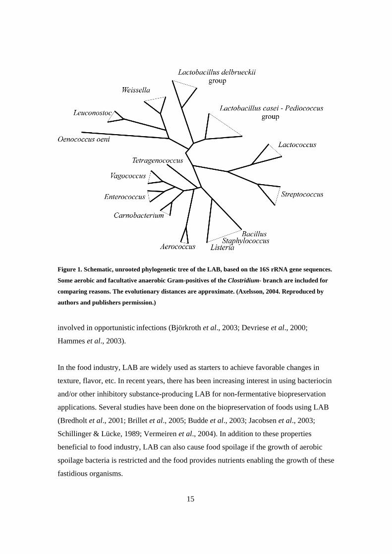
principal LAB associated with foods (Axelsson, 2004). The phylogenetic tree of the LAB
as a group is presented in Figure 1. Bifidobacteria, which also produce lactate and acetate
as the end-products of their carbohydrate metabolism, have a unique pathway of hexose
fermentation, a fructose 6-phosphate shunt that differs from Embden-Meyerhof-Parnas
(EMP), and 6-phosphogluconate metabolic routes of LAB (Schleifer & Ludwig, 1995).
They also have a G+C content of over 50 mol%, and are not related to LAB but to
actinobacteria (Wood & Holzapfel, 1995).
As fastidious organisms, LAB are usually associated with nutritionally rich habitats. They
can be found in the soil, water, manure, sewage, silage, and other plant material. Some
LAB are part of the microbiota on mucous membranes, such as the intestines, mouth and
vagina of both humans and animals and may have a beneficial influence on these
ecosystems. It is therefore understandable that most probiotic products contain LAB.
However, certain LAB, especially the streptococcal species (excluding S. thermophilus),
are pathogenic. In addition to these true pathogens, several other LAB, for example
species in the genera Lactobacillus, Enterococcus, Weissella and Leuconostoc, may be
15
Figure 1. Schematic, unrooted phylogenetic tree of the LAB, based on the 16S rRNA gene sequences.
Some aerobic and facultative anaerobic Gram-positives of the Clostridium- branch are included for
comparing reasons. The evolutionary distances are approximate. (Axelsson, 2004. Reproduced by
authors and publishers permission.)
involved in opportunistic infections (Björkroth et al., 2003; Devriese et al., 2000;
Hammes et al., 2003).
In the food industry, LAB are widely used as starters to achieve favorable changes in
texture, flavor, etc. In recent years, there has been increasing interest in using bacteriocin
and/or other inhibitory substance-producing LAB for non-fermentative biopreservation
applications. Several studies have been done on the biopreservation of foods using LAB
(Bredholt et al., 2001; Brillet et al., 2005; Budde et al., 2003; Jacobsen et al., 2003;
Schillinger & Lücke, 1989; Vermeiren et al., 2004). In addition to these properties
beneficial to food industry, LAB can also cause food spoilage if the growth of aerobic
spoilage bacteria is restricted and the food provides nutrients enabling the growth of these
fastidious organisms.

Tab
le 1
. Phe
noty
pic
char
acte
rs f
or d
iffe
rent
iatin
g th
e ge
nus
Leu
cono
stoc
fro
m th
e ot
her
gene
ra o
f th
e L
AB
(B
jörk
roth
and
Hol
zapf
el, 2
003;
mod
ifie
d).
L
euco
nost
oc
Wei
ssel
la
Hom
ofer
men
tativ
e
ovoi
d sh
aped
LA
B
Gen
us L
acto
baci
llus
P
edio
cocc
us
Car
noba
cter
ium
Ent
eroc
ocus
,
Lac
toco
ccus
,
Stre
ptoc
occu
s
hom
ofer
men
tativ
e he
tero
ferm
enta
tive
Mor
phol
ogy
Ovo
id t
o sh
ort r
ods
(pai
rs a
nd c
hain
s)
Ovo
id o
r ro
ds
(pai
rs a
nd c
hain
s)
Ovo
id
(pai
rs a
nd c
hain
s)
Rod
s R
ods
Rou
nd c
occi
(tet
rads
and
pai
rs)
Rod
s
CO
2 fr
om
gluc
ose
+
+
- +
-
- -
(+)
Hyd
roly
sis
of
argi
nine
-
+ o
r -
+ o
r -
+ o
r -
+ o
r -
+ o
r -
+
Dex
tran
fro
m
sucr
ose
-/+
-/
+
+ o
r -
-/+
-/
+
- -
Lac
tic a
cid
isom
er f
rom
gluc
ose
D(-
) D
(-)
or D
L
L(+
) D
(-),
DL
or
L(+
) D
L
DL
or
L(+
) L
(+)
Pept
idog
lyca
n L
ysA
la/S
era
Lys
Ala
/Ser
a L
ys-A
sp
mai
nly
Lys
-Asp
and
m-A
2pm
Lys
-Asp
and
othe
rsb
Lys
-Asp
m
-A2p
m
Sym
bols
: +, p
ositi
ve r
eact
ion;
-, n
egat
ive
reac
tion;
-/+
, mos
tly n
egat
ive.
A
bbre
viat
ions
: Lys
, lys
ine;
Ala
, ala
nine
; Ser
, ser
ine,
Asp
, asp
arta
te,;
m-A
2pm
, 2,6
-dia
mon
opim
elic
aci
d (2
,6-d
iam
inoh
epta
nedi
oic
acid
).
a No
Lys
-Asp
-typ
es, b
ut d
iffe
rent
iatio
ns o
f L
ys-A
la/S
er ty
pes,
with
the
exce
ptio
n of
W. k
andl
eri.
b Rel
ated
to th
ose
of L
euco
nost
oc a
nd W
eiss
ella
.

17
2.1.1 LAB in meat and non-fermented meat products
The storage of meat at low temperatures will limit spoilage to psycrotrophic organisms,
including many LAB. Under aerobic conditions, LAB grow slowly and are overgrown by
other bacteria, usually pseudomonads (Borch et al., 1996; Huis in't Veld, 1996). Thus,
LAB rarely cause spoilage of aerobically stored fresh meat (Borch et al., 1996; Huis in't
Veld, 1996). However, LAB have shown to be the specific spoilage organisms in vacuum
or MA packaged meat (Björkroth & Korkeala, 1997; Björkroth et al., 2000; Korkeala &
Björkroth, 1997; Samelis & Georgiadou, 2000; Samelis et al., 2000a, 2000b). By vacuum
packaging the microbiome is gradually selected towards LAB due to the increasing CO2
concentration, whereas in MA packaging, the added CO2 results in immediate selection
towards the CO2 tolerant LAB (Borch et al., 1996; Egan, 1983).
Most of the additives used in the meat industry have an impact on shifting the bacterial
population towards LAB. The pink color of cooked products is achieved by the use of
nitrite. Unlike several other bacteria, LAB are not sensitive to nitrite and thus benefit
from the decreased competition (Egan, 1983). The use of acidic marinades has been
considered to inhibit bacterial growth. However, it has been shown that marinated MAP
broiler products have higher LAB counts than the corresponding non-marinated products
(Björkroth, 2005). Marinades usually contain a high amount of carbohydrates utilized by
LAB. The meat buffers the acidity of the marinades, apparently resulting in no hurdle to
LAB growth (Björkroth, 2005). Curing salt (sodium chloride) and other possible
humectants restrict the growth of salt-sensitive organisms, and thus also shift the
microbiome towards salt-tolerant LAB (Egan, 1983).
The typical LAB genera present in meat and non-fermented meat products are
Carnobacterium, Enterococcus, Lactobacillus, Leuconostoc and Weissella (Björkroth et
al., 2005; Borch et al., 1996). Modified by the production method and additives, the
intrinsic factors of meat and meat products greatly influence the LAB composition of the
product.

18
In raw, marinated poultry meat products, Leuconostoc gasicomitatum was the dominating
spoilage organism (Björkroth et al., 2000; Susiluoto et al., 2003), whereas
Carnobacterium divergens and Carnobacterium maltaromaticum dominated in non-
marinated broiler products (Vihavainen et al., 2006). Carnobacteria were also
predominant in vacuum-packaged pork meat (Holley et al., 2004) and in vacuum
packaged beef, C. divergens and Leuconostoc mesenteroides were identified as the
predominant organisms after 4 weeks storage (Jones, 2004).
In cooked products, LAB other than heat resistant (Borch et al., 1988; Niven et al., 1954)
weissellas are mostly regarded as recontaminants (Barakat et al., 2000; Björkroth &
Korkeala, 1997; Samelis et al., 1998a). The composition of the microbiome depends on
the product composition and type, together with the packaking atmosphere (Samelis et
al., 2000a; Samelis & Georgiadou, 2000). The whole ham was spoiled by leuconostoc-
like bacteria, whereas the sliced version of the same product was spoiled due to
recontamination with L. sakei and L. mesenteroides subsp. mesenteroides (Samelis et al.,
1998a). The composition of the microbiome depends also greatly on whether the product
is smoked or not. Smoking seems to shift the microbiome from leuconostocs towards less
sensitive Lactobacillus species. In cooked pork meat, leuconostocs predominated the
non-smoked ham but smoked pork loin and bacon were spoiled by L. sakei/ L. curvatus
(Samelis et al., 2000a). L. carnosum has been reported as a specific spoilage organism in
a cooked, non-smoked, sliced ham product (Björkroth & Korkeala, 1997; Björkroth et al.,
1998); it also predominated with L. mesenteroides in low heat-processed meat products
made of pork, turkey and beef (Yang & Ray, 1994). In oven-cooked, non-smoked turkey
breast fillets Leuconostoc mesenteroides subsp. mesenteroides predominated (Samelis et
al., 2000a, 2000b); in contrast, Lactobacillus sakei subsp. carnosus was predominant in
smoked products (Samelis et al., 2000b). Leuconostoc carnosum and L. sakei dominated
in a Finnish non-smoked product made of turkey breast fillets (Paloranta, 2005).
Departing from this trend, Barakat et al. (2000) identified Lactococcus raffinolactis, C.
divergens and C. maltaromaticum as the dominating microbes in cooked chicken legs.

19
According to culture-dependent studies, the spoilage microbiome of emulsion sausages
seems to be more diverse than the microbiome of whole meat products. There are often
concurrently several different species with quite equal dominance. L. sakei and other
lactobacilli dominated with a large proportionof leuconostocs in vacuum-packaged ring
sausage made of mixed red meat (Korkeala & Mäkela, 1989). L. sakei dominated also in
vacuum or 100% CO2 stored Greek-style smoked tavern sausage consisting mainly of
pork meat (Samelis & Georgiadou, 2000), whereas in air-stored tavern sausages
leuconostocs formed the largest group (Samelis & Georgiadou, 2000). Leuconostocs
(Leuconostoc mesenteroides subsp. mesenteroides and L. gelidum) were also found to
dominate in Vienna sausages (Dykes et al., 1994). In mortadella and pariza sausages,
Leuconostoc citreum and Weissella viridescens comprised a marked proportion of the
microbiome along with L. sakei/L. curvatus and Leuconostoc mesenteroides subsp.
mesenteroides (Samelis et al., 2000a).
2.2 THE CURRENT BACTERIAL SPECIES CONCEPT
Species is a basic element of bacterial taxonomy. Roselló-Mora and Amann (2001)
described it as “a monophyletic and genomically coherent cluster of individual organisms
that show a high degree of overall similarity in many independent characteristics, and is
diagnosable by a discriminative phenotypic property”. This description conforms well to
the widely accepted “polyphasic approach” of species delineation. In polyphasic
taxonomy, as many different techniques (phenotypic, genotypic and phylogenetic) as is
reasonably possible are combined (Vandamme et al., 1996). This should, at best, result in
delineation that is stable, provides a diagnostic scheme for species differentiation, and
reflects phylogenetic relationships.
The need for uniform principles in bacterial nomenclature, and also in taxonomy, was
already noticed in the early 1900s. The Commission on Nomenclature and Taxonomy
was established to fulfill these needs. With time, this Commission has evolved into the
20
current International Committee on Systematics of Prokaryotes (ICSP), an international
committee subordinating to the International Union of Microbiological Societies (IUMS).
The ICSP consists of an executive board, the Judicial Commission, and a number of
subcommittees. The ICSP is responsible for matters relating to prokaryote nomenclature
and taxonomy. The subcommittees deal with matters relating to the nomenclature and
taxonomy of specific groups of prokaryotes; one of their main tasks being bringing about
a minimal standard for species description within a specific group. Unfortunately, LAB
still lack this standard, but the subcommittee of Bifidobacterium, Lactobacillus and
related organisms has decided to act on this (Prof. J. Björkroth, personal communication).
In addition to these permanent commissions, other commissions are formed if needed.
For the definition of bacterial species, the Ad Hoc Committee on the Reconciliation of
Approaches to Bacterial Systematics and the Ad Hoc Committee for the Re-Evaluation of
the Species Definition in Bacteriology were established in the late 1980s and early 2000s
respectively.
However, despite the substantial work done by these ad hoc committees, there is still no
universal concept of polyphasic species definition. At present, the only definition
acknowledged by the Ad Hoc Committee for the Re-Evaluation of the Species Definition
in Bacteriology is that a species is a group of strains sharing 70% or greater DNA-DNA
reassociation values and 5°C or less ∆Tm (difference in the DNA-DNA hybrid melting
points) (Stackebrandt et al., 2002).
In addition to the official definition, also the 16S rRNA gene sequence similarity has
been suggested to be a criterion in species delineation, with a cutoff of 3% divergence.
However, for several reasons, the 16S rRNA gene sequence should not be used as the
only method in species delineation (Stackebrandt & Goebel, 1994).

21
2.3 THE POLYPHASIC APPROACH IN LAB TAXONOMY
Polyphasic taxonomy utilizes both phenetic (phenotypic and genotypic) and phylogenetic
information. In practice, phenetic data is processed by numerical taxonomy based on the
concept of overall similarity (resemblance), whereas phylogenetic analysis is based on
the concept of homology (having a common origin) and parsimony.
Numerical taxonomy, also referred to as Adansonian taxonomy, was introduced by
Sneath and Sokal at the turn of the 1960s and 1970s (Austin & Priest, 1986). It was
created to eliminate the bias originating from human subjectivity in assessing the value of
different characters. In numerical taxonomy, all the characters are basically treated as
equal. The taxonomic level of sampling selected for use in a study is an OTU (operational
taxonomic unit). Depending on the study, it refers to species, populations, individuals,
etc. in question. In pair-wise comparisons of organisms, measuring the similarity (or
sometimes, dissimilarity) values is done between two OTUs. Several formulae or
coefficients, for example, Jaccard, Dice and Pearson, have been developed for this
purpose. The similarity values derived from these calculations are used to cluster the
OTUs hierarchically, usually by the single (nearest neighbor) - or average (unweighted
pair group method with arithmetic averages, UPGMA) -linkage clustering methods. This
clustering can be presented in reader-friendly form as a dendrogram. Instead of the
hierarchical method, OTUs can also be arranged in two- or three-dimensional ordination
diagrams. Most tests used in bacterial taxonomy generate data that can be processed
according to numerical taxonomy. For example, most phenotypical tests have to be
treated as phenetic characters. They cannot be treated as phylogenetic because the
similarities can result from different genes having the same kind of action, from similar
genes formed as a consequence of convergent evolution or from genuine homologous
genes.
In phylogenetic analysis, the aim is to determine the evolutionary branching pattern. The
basic concept in phylogenetic analysis is parsimony, which means that evolution is
assumed to have reached the current situation by the shortest possible route. The data

22
most suitable for phylogenetic analysis is sequences of DNA, RNA or proteins. Due to its
conservativeness, gene-encoding 16S rRNA is probably the most used. The use is further
enhanced by the fact that the 16S rRNA encoding gene database is nowadays the most
comprehensive.
2.3.1 The golden triad: DNA-DNA reassociation, G+C% and 16S rRNA
encoding gene sequence
DNA-DNA reassociation is the official base for species delineation within all bacteria. In
some bacteria, the limit of 70% can be problematic due to its stringency when
phenotypically identical strains differing only by their reassociation values are detected
(Vandamme et al., 1996). In the year 2005, the most used methods were the optical
method of De Ley et al. (1970), based on renaturation rates, and the microdilution
method of Ezaki et al. (1989).
Determination of G+C% was the first DNA-based method in bacterial taxonomy, and is
considered as one of the basic methods of bacterial taxonomy. Among the prokaryotes,
G+C content varies between 24% and 76% (Vandamme et al., 1996). LAB normally have
G+C contents below 50%, although even this is not a strict value since some
Lactobacillus species have values up to 55% (Axelsson, 2004). Generally speaking, the
variation of G+C content is not more than 5% within a species, and 10% within a genus
(Schleifer & Stackebrandt, 1983), but within the genus Lactobacillus also the latter limit
is exceeded (Axelsson, 2004). The similarities in DNA content can be used only in
excluding strains from a species, not in including them. The G+C content can be
determined either from ∆Tm in the thermal denaturation method (Marmur & Doty, 1962;
Xu et al., 2000) or directly from degraded nucleosides by the HPLC method (Mesbah et
al., 1989).
According to the report by the Ad Hoc Committee for the Re-Evaluation of the Species
Definition in Bacteriology, all species descriptions should include an almost complete

23
16S rRNA encoding gene sequence. As a conserved housekeeping gene, it can be used as
a phylogenetic marker in determining the relationship between even distantly related
bacteria. It can also be used in defining species; it has been observed that organisms with
DNA-DNA similarities above 70% usually share more than a 97% sequence similarity in
their genes encoding16S rRNA (Stackebrandt & Goebel, 1994). However, as the G+C%,
the 16S similarity can usually be used only negatively. Among LAB, different species
with identical or nearly identical 16S sequences exist (Björkroth et al., 2002; Cachat &
Priest, 2005; Kim et al., 2003; Leisner et al., 2002; Švec et al., 2001; Vancanneyt et al.,
2001; Yoon et al., 2000) and thus similarity values can reliably be used only as an
excluding criteria.
Most sequencing approaches are based on Sanger’s chain termination method, which
relies on dideoxynucleotides (ddNTPs) lacking the 3’ OH group, and thus causing the
termination of enzymatic DNA synthesis from the single stranded template sequenced.
As a relatively small molecule, the 16S rRNA encoding gene can be sequenced directly
from PCR amplicons created with universal 16S primers without laborious cloning.
In the phylogenetic analysis of 16S (and other gene or protein) sequences, the first step is
always to align the sequences studied. One of the most used alignment methods is Global
alignment, which assumes that the two sequences are basically similar over their entire
length. This alignment attempts to match them to each other from end to end, even
though parts of the alignment are not very convincing, and the insertion of gaps is
needed. To be able to compare several OTUs, multiple alignment (alignment of several
sequences in the same time) is needed instead of pair-wise alignment. This multiple
alignment can be done in several ways. In the progressive alignment method, pair-wise
alignments are located in a matrix from which an initial tree is created by means of
algorithm (usually UPGMA or neighbor-joining) to guide the order of progressive
alignments. The actual phylogenetic tree is constructed from the matrix of this multiple
alignment. The reliability of the tree can be tested by the bootstrap re-sampling
procedure.

24
2.3.2 Sequencing of other housekeeping genes
The Ad Hoc Committee for the Re-Evaluation of the Species Definition in Bacteriology
(Stackebrandt et al., 2002) regarded the sequencing of housekeeping genes (genes
encoding metabolic functions) or other genes as a promising method for phylogenetic
analyses. The recommendation was that the data should be obtained, as an extension of
the multi locus sequence typing (MLST) approach, from the determination of at least five
genes located in diverse chromosomal loci and widely distributed among taxa. However,
even there are MLST schemes for at least streptococcal and enterococcal species
identification (Enright & Spratt, 1998; Enright et al., 2001; Homan et al., 2002; Jones et
al., 2003; King et al., 2002), the combined analysis of several genes sequenced is rarely
used in LAB taxonomy. The sequencing of only one or few genes is far more widely
used. For Enterococcus, Naser et al. (2005) have published an application of MLST
based on RNA polymerase α-subunit (rpoA) and phenylalanyl-tRNA synthase (pheS)
genes. Genes recA, cpn60, tuf and slp have been used with Lactobacillus (Bringel et al.,
2005; Cachat & Priest, 2005; Dellaglio et al., 2004) and gyrA, gyrB, sodA and parC with
Streptococcus (Kawamura et al., 2005).
Single nucleotide polymorphism (SNP) is a variation of single nucleotide in specific
locations of DNA. SNP can be regarded as a simplified and more cost-effective version
of MLST, where small variable fragments of genes are sequenced instead of the whole
gene. Unfortunately, also this method is scarcely used within LAB; there are only few
reports of its use with streptococci (Beres et al., 2004; Rodriguez et al., 2004; Sumby et
al., 2005).
2.3.3 16S and 23S rRNA gene RFLP (Ribotyping)
It was already reported in the original study developing the 16S and 23S rRNA gene
restriction fragment length polymorphism (RFLP), or ribotyping, method that ribosomal
genes are well suited targets in bacterial taxonomy, and that ribotyping is a useful tool for

25
taxonomy studies (Grimont & Grimont, 1986). RFLP is a molecular marker based on the
differential hybridization of a specific DNA sequence (probe) to DNA fragments in a
sample of restriction enzyme digested DNAs; the marker is specific to a single
probe/restriction enzyme combination. In ribotyping, the total genomic DNA is digested
with a restriction enzyme into smaller fragments that are separated by gel electrophoresis.
The fragments are transferred from the gel onto a membrane, and hybridized using a
labeled universal probe targeting the specific conserved domains of ribosomal 16S and
23S rRNA encoding genes. After hybridization, the label in the probe is visualized to
show the fragments where the probe has hybridized as bands. These banding patterns,
called ribotypes, represent the OTUs in numerical similarity analysis within a reference
strain database, and can be used for identification of unknown isolates. They can also be
used for the typing of isolates for epidemiological purposes, which has probably been the
most common use of this method. In the past few years, ribotyping has been used also in
taxonomic studies of Lactobacillus, Streptococcus, Leuconostoc and Weissella species or
subspecies (Björkroth et al., 2000, 2002; Fernández et al., 2004; Kostinek et al., 2005;
Schlegel et al., 2000; Suzuki et al., 2004). Ribotyping should be differentiated from the
PCR-based fragment length polymorphism of 16S-23S spacer regions, which its authors
have misleadingly called PCR ribotyping (Kostman et al., 1992).
2.3.4 Other DNA profiling methods
Other DNA profiling methods recently used in LAB taxonomy include PCR-based
random amplified polymorphic DNA (RAPD) (Dellaglio et al., 2005; Valcheva et al.,
2005), repetitive extragenic palindromic PCR (repPCR) (Gevers et al., 2001; Kostinek et
al., 2005; Švec et al., 2005a, 2005b), amplified fragment length polymorphism AFLP
(Dellaglio et al., 2005; Valcheva et al., 2005; Vancanneyt et al., 2005b), and its
fluorescent modification fAFLP (Vancanneyt et al., 2005a) based on both the use of
restriction endonucleases and PCR. The Ad Hoc Committee for the Re-Evaluation of the
Species Definition in Bacteriology (Stackebrandt et al., 2002) considered all these

26
methods as being of great promise. It was emphasized that all methods used should be
quantitative and the results amenable to appropriate statistical analysis.
2.3.5 Sodium dodecyl sulfate-polyacrylamide gel electrophoresis (SDS-
PAGE) of whole-cell protein extracts
Numerical analysis of protein patterns created by highly standardized SDS-PAGE reveals
a valuable method for investigations of bacterial, also LAB, relationships at a genus or
species level (Dicks, 1995; Dicks & van Vuuren, 1987; Pot et al., 1994; Schleifer &
Stackebrandt, 1983; Vandamme et al., 1996). It can also be used to identify certain LAB
species and subspecies (Björkroth & Holzapfel, 2003; Hammes & Hertel, 2003).
Recently, whole-cell protein analysis has been the most widely used for studying
Lactobacillus, Enterococcus and Streptococcus species (Lawson et al., 2004, 2005;
Mukai et al., 2003; Roos et al., 2005; Teixeira et al., 2001; Vancanneyt et al., 2004,
2005b).
2.3.6 Cell wall composition
Contrary to the remarkably uniform cell wall of Gram-negative bacteria, the cell wall of
Gram positives contains several layers of peptidoglycans (usually more than 30% of the
total cell wall) that vary greatly in composition and structural arrangement (Schleifer &
Kandler, 1972; Schleifer & Stackebrandt, 1983). Besides peptidoglycans, the cell wall of
Gram-positives consists of polysaccharides, teichoic, teichuronic and/or lipoteichoic
(membrane teichoic acids) acids (Schleifer & Stackebrandt, 1983).
On the whole, the peptidoglycan type is a fairly stable character (Schleifer & Kandler,
1972). While the glycan moiety of peptidoglycans is highly consistent, the differences in
the amino acid sequence of the peptide stems of the peptidoglycans and the mode of
cross-linkage between the stems can be used in species differentiation. Determination of
the cell wall composition can be done by either the enzymatic or chemical method

27
(Schleifer & Kandler, 1972). Analysis of the peptidoglycan interbridges is especially
usable in the differentiation of the genus Weissella from the other LAB (Björkroth &
Holzapfel, 2003), and even the A3α (L-lys–L-ser–L-Ala2) type of Weissella minor is also
found in Lactobacillus rossiae (Corsetti et al., 2005). The absence or presence of meso-
or LL- diaminopimelic acid (mDAP, LL -DAP) can be used instead of laborious
peptidoglycan determination to rapidly screen a large number of lactobacillus strains
(Hammes & Hertel, 2003; Kandler & Weiss, 1986).
Cell wall polysaccharides of LAB other than streptococci are poorly studied (Trüper &
Schleifer, 1999). In streptococci they form the basis for serological Lancefield grouping
with the exception of serogroups D and N (Schleifer & Kandler, 1972).
Teichuronic acid consists of glycosidically linked sugar and uronic acid residues.
Teichoic acids are water-soluble polymers containing sugar, D-alanine residues, and
either glycerol or ribitol phosphates. Teichoic acids can be divided into two classes; cell
wall teichoic acids and membrane or lipoteichoic acids. Cell wall teichoic acids are found
only in a limited number of gram-positive bacteria (Schleifer & Stackebrandt, 1983).
Lancefield serogroups D (Enterococcus) and N (Lactococcus) are based on teichoic acids
(Schleifer & Kandler, 1972). The presence or absence of teichoic acids can be used to
differentiate for example lactobacillus species (Kandler & Weiss, 1986).
2.3.7 Cellular fatty acids
The fatty acids in most Gram-positive bacteria are located in the cytoplasmic membrane,
which consists of roughly 50% lipid. These membrane lipids are a diverse group of
molecules that can be used as markers for the classification and identification of
microorganisms. Cells for the fatty acid analysis have to be grown in standardized
conditions because the fatty acid composition is known to depend on several factors
including growth media and growth phase. Analysis is usually done by gas
chromatography. In LAB, the cellular fatty acid profiles have been used for the

28
characterization and identification of Leuconostoc, Weissella and Oenococcus oeni
(Björkroth & Holzapfel, 2003; Samelis et al., 1998b). For carnobacteria, a group- or even
species-specific pattern of the composition of fatty acids can be recognized (Hammes &
Hertel, 2003).
2.3.8 Classical phenotypic characters: physiological and biochemical
characters and morphology
The classical, traditional phenotypical tests constitute the base for the formal description
of bacterial species; the phenotypic consistency of species is required for a useful
classification system. Orla-Jensen used morphology, mode of glucose fermentation,
growth in certain temperatures, and carbon source utilization as a basis for his early
description of LAB (Axelsson, 2004). However, the selection of phenotypic properties is
not standardized, and it depends on the genera studied. Within LAB, it is usually not
possible to differentiate species with these tests alone (Axelsson, 2004), and their
usability in species identification is thus often restricted to the genus level differentiation.
For example, set of certain classical phenotypic characters, supplemented by
peptidoglycan analysis, can be used to differentiate members of the genus Leuconostoc
from other genera of the LAB, as seen in Table 1.
The early bacterial taxonomy is based mostly on morphology, which consists of cellular
and colonial morphology. Cellular morphology includes not only the shape of the cell,
but also endospores, flagellas, inclusion bodies, etc., whereas colonial morphology
consists of color, dimensions, and form of the bacterial colony. LAB are divided into
genera consisting of both bacilliforms and cocci (Weissella), only bacilliforms
(Lactobacillus and Carnobacterium), or only cocci (all the other genera).
The mode of glucose fermentation under standard, non-limiting conditions is used
especially in the differentiation of LAB genera, and also Lactobacillus species. Based on
the hexose (and partly, pentose) fermentation type, genus Lactobacillus can be divided

29
into three groups; obligately homofermentative (ferments hexoses via Embden Meyerhof
Parnas (EMP) pathway, does not ferment pentoses), obligately heterofermentative
(ferments hexoses via phosphoglugluconate pathway, may also ferment pentoses via this
pathway), and facultatively heterofermentative (ferments hexoses via EMP and pentoses,
when needed, via phosphogluconate pathway) species (Hammes et al., 1992).
Other tests widely used within LAB include growth factor requirements, arginine
hydrolysis, acetoin formation (Voges-Proscauer), salt and/or bile tolerance, possible
haemolyse (primarily within streptococci), and the presence of specific enzymes (for
example β-galactosidase, β-glucuronidase).
Even the physiological, nutritional and biochemical tests are often laborious, time-
consuming, and somewhat hard to standardize, these traditional tests still have great
importance in taxonomy. To overcome these problems, several commercial identification
systems based on the nutritional and biochemical characteristics have been developed, for
example, the API system (bioMérieux, France), MicroLog (Biolog Inc., USA) and
Diatabs (Rosco, Denmark). Identification keys for most of these tests are not accurate
with LAB species, but the tests themselves are valuable as a convenient way to carry out
large-scale phenotypical studies.
2.4 HISTORY OF THE LAB GENERA RELEVANT IN MEAT
ECOSYSTEMS
Even the concept of LAB as a group of organisms was not introduced until the early
1900s, two still currently valid LAB genera, Leuconostoc and Streptococcus, were
already described at the end of the 19th century. Within the first few years of the 20th
century, several other LAB genera were described. In 1901, Beijerinck revised the
definition of LAB by excluding the coliforms (Stiles & Holzapfel, 1997). In 1919, Orla-
Jensen published his noted monograph on LAB classification (Axelsson, 2004), and, in

30
1937, Sherman published his review of streptococci, including the proposition for
systematic classification scheme (Sherman, 1937). Subsequently, new species were
described, while the already existing species and genera were re-named and restored. In
total, very little happened within the food-associated LAB between Orla-Jensen’s
monograph and the effective adoption of DNA-based methods into taxonomy in the
1980s; after which changes that have been called even “dramatic” resulted in LAB
taxonomy.
2.4.1 LAB with coccoid morhology
Leuconostoc, described by van Tieghem in 1878, was the first of the prevailing LAB
genera (Euzéby, 1997). The first species was Leuconostoc mesenteroides [Tsenkovskii
1878] van Tieghem 1878 (Euzéby, 1997). It is the only species still existing from the
original composition of the genus, Leuconotoc mesenteroides subsp. mesenteroides as its
current name.
In 1995, Leuconostoc oeni (corrig.), described by Garvie in 1967 (Euzéby, 1997), was
separated from the genus Leuconostoc. Dicks et al. (1995) combined the data from earlier
studies and proposed reclassification of this species in a novel genus Oenococcus, in
which it still is the only species.
The genus Streptococcus and the first species in it, Streptococcus pyogenes, were
described in 1884 by Rosenbach (Euzéby, 1997). With increasing numbers of the species,
it became necessary to make classifications within the genus. In 1937, Sherman (1937)
suggested a division based mostly on currently developed Lancefield serotyping and
some other phenotypical characters. Originally there were four groups, Pyogenic,
Viridans, Lactic and Enterococcus. Later, the first two groups were divided further, but
retained within the genus, whereas the two latter were classified as novel genera. The
genus Streptococcus is the origin of at least Enterococcus, Lactococcus, and Vagococcus.

31
The first species in enterococci, Enterococcus faecalis, was descibed in 1886 as
"Micrococcus ovalis" by Escherich. Later, the same species was re-described separately
as "Micrococcus zymogenes" [MacCallum and Hastings 1899], "Streptococcus
liquefaciens" [Sternberg 1892], "Streptococcus glycerinaceus" [Orla-Jensen 1919], and
also as "Enterocoque" and "Enterococcus proteiformis" [Thiercelin 1902, Thiercelin and
Jouhaud 1903] (Euzéby, 1997). In 1906, the name was established as “Streptococcus
faecalis” after Andrewes and Horder. However, the term “enterococci” remained, and
was widely used for a group of streptococcal species originating from the intestines of
man and other warm blooded animals, even though the status of this group as an
individual genus was questioned (Sherman, 1937). In 1970, Kalina made a major
proposal that S. faecalis and Streptococcus faecium [Orla-Jensen, 1919] should be
transferred to the genus Enterococcus. However, this was not formally accepted until
1984 when Schleifer and Kilpper-Bälz (1984) made the same proposal, basing it on
DNA-DNA hybridization studies.
Like enterococci, lactococci, at that time called “lactic” or “lactic-acid” streptococci,
were known to form a group phenotypically distinct from Streptococcus long before the
genus Lactococcus was described (Sherman, 1937). In 1985, Schleifer et al. (1985)
confirmed this division, and described the novel genus Lactococcus. The first species was
Lactococcus lactis, originally described by Lister in 1873 as “Bacterium lactis”
(Sherman, 1937).
Collins et al. (1989) separated the motile group N streptococci from lactococci and
streptococci in 1989. The classification of these strains in the novel genus Vagococcus
was based on the earlier phenotypical and DNA-23S rRNA hybridization studies of
Schleifer et al. (1985), and the 16S rRNA sequencing studies (Collins et al., 1989). The
first species in this genus was Vagococcus fluvialis.
The genus Pediococcus is ascribed to Claussen, who described the species Pediococcus
damnosus in 1903 (Euzéby, 1997). However, the name Pediococcus was already used in
1884 by Balcke, in his description of Pediococcus cerevisiae for a group of beer-spoilage

32
bacteria (Simpson & Taguchi, 1995). Unfortunately, the strains were never isolated as a
pure culture, and thus the species is not validly published. Probably due to the lack of a
corresponding type strain, the name P. cerevisiae was later used for strains originating
from both spoiled beer and plants. Confusion with the name occurred when it was shown
that the strains in these two groups were distinct. At long last, the Juridical Commission
of the International Committee on Systematic Bacteriology proposed in 1976 (Opinion
52) that the name P. cerevisiae should be rejected, while the genus name Pediococcus
should be conserved, P. damnosus [Claussen 1903] as the type species (Simpson &
Taguchi, 1995).
2.4.2 LAB with bacilliform morphology
Genus Lactobacillus was described by Beijerinck in 1901. The species he included in the
new genus were Lactobacillus delbrueckii (currently L. delbrueckii subsp. delbrueckii),
formerly "Bacillus Delbrücki" [Leichmann 1896], and Lactobacillus fermentum, formerly
"Bacillus δ" [von Freudenreich 1895] (Euzéby, 1997). However, the first lactobacillus
species described is Lactobacillus casei, described by von Freudenreich in 1890 as
"Bacillus α", (Euzéby, 1997), even though it was not named Lactobacillus until 1971
(Hansen & Lessel, 1971). Even after that, the existence of this species has not been
uncomplicated; for example, the species status of the type strain of L. casei is presently
awaiting the statement of the Judicial Commission in ICSP (Klein, 2005). Also the
species L. acidophilus, L. brevis, L. buchneri, L. delbrueckii subsp. Lactis and L.
helveticus were described before the genus, but were not included in it until at least
twenty years after the genus description (Euzéby, 1997). The genus Lactobacillus is
unquestionably the most numerous LAB genera, with 124 species (in October 2005).
However, including all these species under one genus does not represent true
phylogenetic relationships, and further revision of this genus is needed (Axelsson, 2004).
In 1987, Collins et al. described the genus Carnobacterium in which Lactobacillus
divergens [Holzapfel and Gerber 1984] and Lactobacillus piscicola [Hiu et al. 1984]

33
(synonym Lactobacillus carnis [Shaw and Harding 1986]) were transferred as C.
divergens and C. piscicola, together with some strains classified as novel species
Carnobacterium gallinarum and Carnobacterium mobile (Euzéby, 1997; Collins et al.,
1987). Later, one more Lactobacillus species, Lactobacillus maltaromicus [Miller et al.
1974] was reclassified as Carnobacterium maltaromaticum and C. piscicola was also
found to belong to this species (Mora et al., 2003).
2.4.3 LAB with both cocci and bacilliforms: Weissella
The first species in the genus Weissella, W. viridescens was discovered already in 1949,
when Niven et al. reported the greening of sausage surfaces caused by an unidentified
species (Niven et al., 1949). However, the species was not formally described until 1957,
when it was named as L. viridescens, even it was regarded as an atypical Lactobacillus
from the beginning (Niven & Evans, 1957). Later studies showed that some Lactobacillus
species, including L. viridescens, are phylogenetically closer to Leuconostoc
paramesenteroides than to the genus Lactobacillus (Martinez-Murcia & Collins, 1990,
1991; Martinez-Murcia et al., 1993). Later, this so-called L. paramesenteroides group
was separated from leuconostocs and lactobacilli and reclassified under a new genus,
Weissella (Collins et al., 1993).

34
3 AIMS OF THE STUDY
The aim of the present thesis was to clarify the taxonomic status of certain meat-
associated LAB using polyphasic taxonomy. The specific aims were to:
1. clarify the taxonomy of L. curvatus and L. sakei,
2. identify the unknown coccoid isolates detected in modified-atmosphere packaged
(MAP), marinated broiler legs, and canine tonsils,
3. identify unknown bacilliform LAB detected in spoiled and unspoiled MAP broiler
products,
4. identify unknown coccoid LAB isolated from marinated or non marinated, MAP
broiler leg products, and air samples from a large-scale broiler meat processing
plant,
5. identify unknown heterofermentative bacilliform LAB detected in vacuum or MA
packaged “Morcilla de Burgos”, a Spanish cooked meat product,
6. assess the function of the polyphasic approach in the taxonomy of meat-associated
LAB.

35
4 MATERIALS AND METHODS
4.1 BACTERIAL STRAINS AND CULTURING (I-V)
Primary identification of all of the isolates was done using the LAB identification
database at the Department of Food and Environmental Hygiene, University of Helsinki.
The database utilizes numerical analysis of 16 and 23S rRNA gene HindIII RFLP
patterns (ribopatterns), and comprises over 3000 isolates and about 170 type and
reference strains representing all LAB species relevant in food spoilage (Björkroth &
Korkeala, 1996, 1997; Björkroth et al., 2000, 2002; Lyhs et al., 1999). The selection of
the type strains used in studies I-V was made by the similarity of HindIII-ribopatterns of
the strains studied and the type strains.
In study I, the type strains used were L. curvatus subsp. melibiosus CCUG 34545T and its
duplicate CCUG 41580T, L. curvatus subsp. curvatus DSM 20019T, L. sakei subsp. sakei
DSM 20017T and L. sakei subsp. carnosus CCUG 31331T. Seven reference strains, used
also in the studies in which the subspecies division of L. curvatus and L. sakei were
described (Torriani et al., 1996), were included in the numerical analyses of ribotype and
protein patterns. These were L. sakei strains LMG 7941 (DSM 20198), LMG 17301,
LMG 17304, LMG 17305 and LMG 17306 (CCUG 8045, CCUG 30939, CCUG 32077
and CCUG 32584, respectively); and L. curvatus strains LMG 17299 (CCUG 31333) and
LMG 17303 (CCUG 31332). In addition to the culture collection strains, six strains
originating from modified-atmosphere-packaged (MAP) poultry-meat products
(Susiluoto et al., 2003) were included on the basis of their HindIII ribopatterns.
In study II, seven unknown enterococcal isolates were studied. Five of these were
detected in fresh, MAP, marinated broiler legs (Björkroth et al., 2005). Two originated
from canine tonsils. Type strains used were E. pallens LMG 21842T, E. gilvus LMG

36
21841T, E. raffinosus LMG 12888T, E. malodoratus LMG 10747T, E. avium LMG
10744T, and E. pseudoavium LMG 11426T. E. raffinosus LMG 12172 was also included.
For study III, 26 of 67 bacilliform isolates with similar HindIII -ribopatterns detected in
MAP broiler meat products were selected. In the preliminary numerical analyses, these
67 isolates were divided into six different HindIII ribopattern groups (I, IIa, IIb, III, IV
and V). For study III, between 4 and 6 isolates from each ribogroup were selected
randomly, unless the group contained only two isolates (I and III). The type strains
included were L. reuteri DSM 20016T, L. thermotolerans DSM 14792T, L. durianis LMG
19193T, L. vaccinostercus DSM 20634T, L. suebicus DSM 5007T, and L. fermentum
CCUG 30138T.
In study IV, 13 isolates with similar HindIII -ribopatterns detected in the earlier studies
were characterized. Nine of these were isolated in fresh (two days after packaging),
marinated MAP broiler leg products (Björkroth et al., 2005), two in non-marinated MAP
broiler leg products at the end of their shelf lives, and two in air samples from a broiler
meat processing plant (Vihavainen et al., 2006). The other strains used in study IV were
Streptococcus parauberis type strain LMG 12174T and its duplicate LMG 14376T and
reference strain LMG 12173R and its duplicate LMG 14377R. In addition, strains
RM212.1 and RA149.1, isolated from diseased turbots (Romalde et al., 1999) were
kindly provided by Dr. Jesús L. Romalde (Departemento de Microbiología y
Parasitología, Facultat de Biología, and Instituto de Acuicultura, Universidad de
University of Santiago de Compostela, Spain).
In study V, three isolates from a group of unidentified heterofermentative lactic acid
bacterium (LAB) isolates detected in vacuum or modified atmosphere (MA) packaged
“Morcilla de Burgos”, a Spanish cooked meat product, were identified. Three W.
viridescens isolates, also detected in the same product, were also included. The type
strains used were W. viridescens ATCC 12706T, W. confusa LMG 9497T, W. hellenica
LMG 15125T, W. soli DSM 14420T, W. minor LMG 9847T, W. koreensis KCTC 3621T,

37
W. kandleri LMG 14471T, W. halotolerans ATCC 35410T, W. paramesenteroides DSM
20288T, and W. cibaria 17699T.
In all the studies (I-V), the strains were cultured at 25°C either overnight in MRS broth
(Difco, BD Diagnostic Systems, Sparks, MD) or for five days on MRS agar plates
(Oxoid, Hampshire, United Kingdom), unless otherwise stated in the description of
prevailing method. The plates were incubated under anaerobic conditions (Anaerogen,
Oxoid, 9-13% CO2 according to the manufacturer). All isolates were maintained in MRS
broth (Difco) at -70°C.
4.2 RIBOTYPING (I-V)
DNA for all DNA-based analyses (ribotyping, 16S rRNA gene sequencing, determination
of the G+C content, and DNA-DNA reassociation) was isolated by using the guanidium
thiocyanate method of Pitcher et al. (1989) as modified by Björkroth and Korkeala
(1996) by the combined lysozyme and mutanolysin (Sigma) treatment of bacterial cells.
HindIII and EcoRI enzymes were used for the digestion of DNA as specified by the
manufacturer (New England Biolabs, Beverly, MA). Restriction enzyme analysis was
performed as described previously (Björkroth & Korkeala, 1996) and southern blotting
was made using a vacuum device (Vacugene, Pharmacia). The cDNA probe for
ribotyping was labeled by reverse transcription (AMV-RT, Promega and Dig Labelling
Kit, Roche Molecular Biochemicals, Mannheim, Germany) as previously described
(Blumberg et al., 1991). Membranes were hybridized at 58ºC overnight and the detection
of the digoxigenin label was performed as recommended by Roche Molecular
Biochemicals.
Scanned (Hewlett Packard Scan Jet 4c/T, Palo Alto, CA) ribopatterns were analyzed
using the BioNumerics 3.0 (I-II) or 3.5 (III-V) software package. The similarity between

38
all pairs was expressed by the Dice coefficient correlation and UPGMA clustering was
used for the construction of the dendrograms. Based on the use of internal controls,
position tolerance of 1.5% was allowed for the bands.
4.3 MORPHOLOGY AND PHENOTYPICAL TESTS (I-IV)
All isolates were Gram stained (I-IV). For size and precise cell morphology
determination, cells were suspended in physiological NaCl, negatively stained with 1%
phosphotungstic acid and examined using JEOL JEM 100 transmission electron
microscope (II, III).
Growth at different temperatures or in the presence of NaCl was tested in MRS broth
(Difco) incubated until growth was observed or otherwise for at least 21 days. The
temperatures used were 4°C (I-IV), 10°C (IV), 15°C (III), 37°C (I-IV), 40°C (IV) and
45°C (I, II); NaCl percentages were 2% (II-IV), 4% (II-IV), 6.5% (II-IV), and 10% (I-II)
w/v.
Isolates were tested for their carbohydrate fermentation profiles by API 50 CHL
(bioMérieux) (I-IV), and for other biochemical activities by API STREP identification
systems (bioMérieux) (II, IV) according to the manufacturer's instructions.
In study I, the production of acetoin from glucose was tested as described by Reuter
(1970). The production of ammonia from arginine was determined by the method of
Briggs (1953) in studies I and II, in the broth containing 0.5% arginine, 0.5% peptone,
0.3% yeast extract, 0.1% glucose, and 0.016% bromcresol purple.
In study II, isolates were tested for catalase and Lancefield antigen D (Streptococcal
grouping kit, Oxoid); formation of typical colonies for enterococci was tested on bile-

39
esculin (Gibco) and Slanetz-Bartley (Oxoid) agars. Haemolyses were tested on blood agar
in studies II and IV.
In study III, the configuration of lactic acid was determined enzymatically by using the
UV method according to Hohorst (1970) and Stetter (1973). The gas formation of glucose
was tested according to the method of Gibson and Abdel-Malek (1945).
Each of these tests was carried out at least twice.
4.4 SDS-PAGE OF WHOLE-CELL PROTEIN EXTRACTS
For the extraction of whole-cell proteins, strains were grown for 24 hours on MRS agar at
24°C in a microaerobic atmosphere (in O2/CO2/N2 at approx. 5:10:85). The preparation of
cellular protein extracts and PAGE were performed as described by Pot et al. (1994). The
densitometric analysis, normalization and interpolation of the scanned (LKB 2202
UltroScan laser densitometer) protein profiles and the numerical analysis were performed
using the GelCompar 4.2 software package (Applied Maths). Similarity between all pairs
of traces was expressed using Pearson product moment correlation coefficient.
4.5 SEQUENCING OF 16S rRNA ENCODING GENE (I-V)
The nearly complete (over 1400 bases) 16S rRNA encoding gene was amplified by PCR
with a universal primer pair F8-27 (5’-AGAGTTTGATCCTGGCTGAG-3’) and R1541-
1522 (5’- AAGGAGGTGATCCAGCCGCA-3’). Sequencing of the purified (QIAquick
PCR Purification Kit, Qiagen, Venlo, Netherlands) PCR product was performed bi-
directionally by Sanger’s dideoxynucleotide chain termination method using primers
F19-38 (5’-CTGGCTCAGGAYGAACGCTG-3’), F926 (5’-

40
AACTCAAAGGAATTGACGG-3’), R519 (5’- GTATTACCGCGGCTGCTG-3’), and
R1541-1522.
Samples were run in a Global IR2 sequencing device with e-Seq 1.1 (I-II) or 2.0 (III-V)
software (LiCor, Lincoln, NE) according to the manufacturer’s instructions, and the
consensus sequences were created with AlignIR software (LiCor).
These consensus sequences and the sequences of representative strains belonging to the
same phylogenetic group (retrieved from the GenBank, http://www.ncbi.nlm.nih.gov,
using BLASTN 2.2.6, (Altschul et al., 1997) were aligned and a phylogenetic tree was
constructed using the neighbor-joining method and BioNumerics 3.0 (I-II) or 3.5 (III-V)
software package (Applied Maths, Sint-Martens-Latem, Belgium).
4.6 DETERMINATION OF THE G+C CONTENT AND DNA-DNA
REASSOCIATION LEVELS (I-V)
For the determination of the G+C content (II-V), DNA was enzymically degraded into
nucleosides as described by Mesbah et al. (1989). The nucleotide mixture was then
separated by HPLC (SymmetryShield C8, Waters, Massachusetts, USA) at 37°C, 0.02 M
NH4H2PO4 with 1.5% acetonitrile as the solvent. Non-methylated lambda phage DNA
(Sigma) was used as the calibration reference.
In study I, the G+C content was estimated using LightCycler (Roche Molecular
Diagnostics), and Formula A as described by Xu et al. (2000).
The reassociation levels were determined either spectrophotometrically (Gilford
Response spectrophotometer, Giba Corning Diagnostics) from the renaturation rates
according to De Ley et al. (1970) (I) or with photobiotin-labeled probes in microplate
plates as described by Ezaki et al. (1989) using HTS7000 Bio Assay Reader (Perkin-
Elmer) (II-VI).

41
5 RESULTS
5.1 LACTOBACILLUS CURVATUS SUBSP. MELIBIOSUS (I)
The taxonomic position of L curvatus subsp. melibiosus CCUG 34545T was re-evaluated
in a polyphasic study including 16S rRNA gene sequencing, DNA-DNA reassociation,
DNA G+C content determination, numerical analysis of ribotypes and whole-cell protein
patterns and the examination of some fundamental phenotypic properties. The results
obtained indicated that the strain CCUG 34545T and its duplicate, CCUG 41850T, are
Lactobacillus sakei subsp. carnosus strains, and that L. curvatus subsp. melibiosus is a
later synonym of L. sakei subsp. carnosus.
5.2 ENTEROCOCCUS HERMANNIENSIS (II) AND
LACTOBACILLUS OLIGOFERMENTANS (III) SP. NOV.
The results of the polyphasic study, including numerical analysis of ribotypes and whole-
cell protein patterns, 16S rRNA gene sequencing, DNA-DNA reassociation, DNA G+C
content determination, and determination of some phenotypic properties, indicated that
the unknown coccoid isolates represent a novel species in the genus Enterococcus, more
accurately in its E. avium- species group. The isolates showed phenotypic properties
typical for the genus Enterococcus, with the exception of not possessing the Lancefield D
antigen. For this novel species, the name Enterococcus hermanniensis was proposed,
with strain LMG 12317T (=CCUG 48100) as the type strain.
A similar polyphasic taxonomy approach as in the case of E. hermanniensis, with the
exception of omitting whole-cell protein patterns, showed that the bacilliform LAB
isolates represent a new Lactobacillus species within the L. reuteri group. According to
the API 50CHL test, arabinose and xylose were the only carbohydrates fermented
strongly and, unexpectedly for a meat-associated LAB, glucose was utilized very weakly.

42
Thus, the name Lactobacillus oligofermentans sp. nov. was proposed, with strain LMG
22743T (DSM 15707T) as a type strain.
5.3 STREPTOCOCCUS PARAUBERIS (IV) AND WEISSELLA
VIRIDESCENS (V)
In studies IV and V, a polyphasic approach (including numerical analysis of ribotypes,
16S rRNA gene sequencing, DNA-DNA reassociation, DNA G+C content determination,
and determination of some phenotypic properties) similar to studies II and III was used.
In study IV, the 16S rRNA genes of the three isolates sequenced possessed the highest
similarities (over 99%) with the sequence of S. parauberis type strain. However, in the
numerical analysis of HindIII ribopatterns, the type strain did not cluster together with
these isolates. Reassociation values between S. parauberis type or reference strain and
the strains studied varied from 82% to 97%, confirming that these strains belong to S.
parauberis. Unexpectedly, most of the broiler meat-originating strains studied for their
phenotypical properties did not utilize lactose, and the same strains fermented also
galactose very weakly, properties considered atypical for S. parauberis. This was the first
report of lactose negative S. parauberis strains, and also the first report associating S.
parauberis with broiler slaughter and meat products.
In study V, the 16S rRNA genes of the three isolates sequenced possessed the highest
similarities with the corresponding sequences of Weissella species; in the phylogenetic
analysis Weissella viridescens was their closest phylogenetic neighbor. However, in the
numerical analysis of either HindIII or EcoRI ribopatterns, the W. viridescens type strain
did not cluster with these isolates. Nevertheless, the high (from 75% to 83%) DNA-DNA
reassociation values between the strains studied and the W. viridescens type strain
confirmed that these strains belong to the latter species. W. viridescens was found to be a
more divergent species than previously reported.

43
6 DISCUSSION
6.1 LACTOBACILLUS CURVATUS SUBSP. MELIBIOSUS (I)
The G+C% of all L. sakei and L. curvatus strains studied were in agreement with the
previous studies (Wood & Holzapfel, 1995) as were all the phenotypical reactions of type
strains (Berthier & Erlich, 1999; Klein et al., 1996). Results from both of these analyses
were also in harmony with the other analyses performed in the present study.
In this study, the DNA-DNA reassociations and 16S rRNA encoding gene sequence
analysis clearly showed that L. curvatus subsp. melibiosus CCUG 34545T strain and its
duplicate CCUG 41580T belonged to the species L. sakei. The reassociation values
between CCUG 34545T and L. sakei subsp. sakei DSM 20017T and L. sakei subsp.
carnosus CCUG 31331T were 82% and 87%, respectively, unambiguously showing that
these strains belong to the same species. In contrast, the value between 34545T and L.
curvatus subsp. curvatus DSM 20019T was as low as 30%. For some reason, these values
are not in agreement with earlier studies of Klein et al. (1996). However, the L. sakei
group including L. curvatus subsp. melibiosus was clearly separated from L. curvatus
subsp. curvatus type and reference strains also in 16S rRNA encoding gene sequence
analysis in which the were located in two main branches, possessing 100% bootstrap
value. All the reactions of the strain 34545T and its duplicate CCUG 41580T were similar.
This species-level conclusion was further supported by the numerical analyses of protein
and ribopatterns; in both analyses L. curvatus subsp. melibiosus strains clustered together
with L. sakei strains. When the variability among the protein patterns of L. sakei subsp.
carnosus reference strains is considered, the L. curvatus subsp. melibiosus type strain
could not be distinguished from L. sakei subsp. carnosus. This high similarity between L.
curvatus subsp. melibiosus type strain and the L. sakei patterns was not reported by Klein
et al. (1996) although the five L. sakei subsp. carnosus reference strains and the L.

44
curvatus subsp. melibiosus type strain were included also in that study. Thus, as the
DNA-DNA reassociation results, the results from protein pattern analysis only partially
agreed with the results from the previous studies (Klein et al., 1996).
Within the subspecies level, one salient difference was noted in the dendrograms derived
from the whole cell protein profiles and ribotyping profiles. In the numerical analysis of
the whole cell protein patterns, three distinct clusters were delineated. Cluster I
comprised the all the L. curvatus subsp. curvatus reference strains, Cluster II comprised
both the L. sakei subsp. sakei reference strains, and Cluster III comprised all the L. sakei
subsp. carnosus reference strains together with both subcultures of the L. curvatus subsp.
melibiosus type strain. Thus, according to the present study and the study of Klein et al.
(1996), protein fingerprints clearly divided L. sakei into the two subspecies, sakei and
carnosus (including L. curvatus subsp. melibiosus strains of the present study). In the
analyses of HindIII and EcoRI ribotypes all L. sakei strains clustered together, and thus
these analyses cannot be used for the subspecies-level identification of L. sakei.
Considering all previously published data by Klein et al. (1996) and Torriani et al.
(1996), only the DNA-DNA hybridization values show clear discrepancy with our
conclusion. Our study demonstrates that L. curvatus subsp. melibiosus is a later synonym
of L. sakei subsp. carnosus and, as a consequence, the subspecies division within L.
curvatus should be abandoned.
6.2 ENTEROCOCCUS HERMANNIENSIS (II) AND
LACTOBACILLUS OLIGOFERMENTANS (III) SP. NOV.
The polyphasic approach was utilized in the studies of unknown coccoid isolates
originating from fresh, MAP, marinated, broiler legs and canine tonsils and
heterofermentative, bacilliform strains originating mainly from spoiled, marinated, MAP
broiler meat products. The results from all the methods were in good accordance and

45
indicated that these isolates represent two novel species in the genera of Enterococcus
and Lactobacillus, and more specifically, in the E. avium and L. reuteri phylogenetic
groups. Names Enterococcus hermanniensis sp. nov. and Lactobacillus oligofermentans
sp. nov were proposed for these species.
Both E. hermanniensis and L. oligofermentans possessed some phenotypical characters
first regarded as unexpected. Even E. hermanniensis isolates mainly showed classical
reactions for the enterococci as listed by Devriese et al. (1993) and Devriese & Pot
(1995), they all were negative for Lancefield group D antigen. Within the phylogenetic
context this is, however, not surprising because several species in the E. avium group lack
the D antigen.
The unexpected characters of L. oligofermentans are somewhat more confusing.
However, even their existence can be difficult to understand when the habitat of this
species is considered alone, they fit in well with the phylogeny of this species. Glucose
has always been considered as one of the main substrates of meat-associated LAB, but all
the L. oligofermentans strains utilized glucose very weakly. According to the API 50
CHL test, arabinose and xylose, devoid from the meat ecosystem, were the only
carbohydrates strongly fermented. For species L. oligofermentans, L. durianis,
Lactobacillus vaccinostercus, and Lactobacillus suebicus, all located in the same
phylogenetic branch based on the 16S rRNA gene sequences, a common feature is that
they prefer pentose carbohydrates to hexoses, they all ferment xylose well and glucose
poorly. For these species other than L. oligofermentans it thus is not unexpected they
have been detected in plant material (fermented durian fruits, apple and pear mashes and
dried cow dung, respectively) containing pentoses more than in sources of animal origin.
It is astonishing that only one species possessing similar metabolic characters to the
others in the same phylogenetic cluster has been detected in such a different habitat.

46
6.3 STREPTOCOCCUS PARAUBERIS (IV) AND WEISSELLA
VIRIDESCENS (V)
The results from the polyphasic studies of unidentified coccoid LAB strains (detected in
the MAP broiler meat products and air of a broiler meat processing plant) and of
unidentified heterofermentative strains (detected in morcilla) showed that these two
groups belong to the already existing species Streptococcus parauberis and Weissella
viridescens, respectively. The identification of either of these species was not
uncomplicated. Both species possessed more diverse phenotypical and genotypical
characteristic than previously reported.
The differentiation of S. parauberis from Streptococcus uberis has been, and can still be,
difficult and thus it has been mainly regarded as a bovine mastitis pathogen similar to S.
uberis. When the species was described and the type strains selected (Williams & Collins,
1990), it was not detected in any other habitat than the bovine. Lately, it has been shown
to be far more uncommon in mastitis than previously thought (Alber et al., 2004; Bentley
et al., 1993) but it has been detected in other habitats like fish (Doménech et al., 1996;
Romalde et al., 1999) and now also in broiler products and air of a broiler meat
processing plant. This diversity of habitats was reflected also to the variability in
characteristics of the strains; variability which had not been reported in the original
species description. All strains studied, except the S. parauberis type strain LMG 12174T
originating from a mastitis milk sample, were found to be psychrotrophic. They all grew
at 10ºC and some even at 4ºC. Another unexpected character was seen in the utilization
of lactose, when only the strains of bovine origin, LMG 12174T and LMG 12173R,
fermented it clearly. The strains from other origins fermented lactose only weakly or not
at all. The non-lactose-fermenting strains were also poor in fermenting galactose. The
difference between the type strain and the other strains was also seen in the numerical
analysis of HindIII ribotypes in which the type strain and its duplicate were distinguished
from the cluster including all the other strains, also the reference strains. The meaning of
the existence of type strains is that they are selected to represent the typical member of
the species. If a description of a new species is based on only few strains, and even from

47
only one habitat, the knowledge of the variability within the species is restricted. Thus,
the selected type strain can later turn out to be anything else but a typical representative
of the species, as is seemingly happening with S. parauberis. The worst thing is that the
unrepresentative type strain is misleading and can hamper the species identification.
Even though the DNA-DNA reassociation results and the phylogenetic analysis based on
16S rRNA gene sequences confirmed that the unidentified strains detected in morcilla
belonged to W. viridescens, these strains represented distinct pheno- and genotypic
subgroups in it. The strains identified as W. viridescens in the present study represented
four dissimilar phenotypes, and, according to those results, should have been regarded
either as W. viridescens, Leuconostoc fructosum, Lactobacillus sanfransiscencis or
Lactobacillus sp. Also the numerical analyses of both HindIII and EcoRI ribopatterns
resulted in clustering that represented the diversity within the species. However, even
there is a wide variability within the species detected basically by all the methods used,
the type strains of the species so far represents the majority of strains, unlike the type
strain of S. parauberis does.
6.4 POLYPHASIC APPROACH
In the polyphasic approach, results from individual methods were basically congruent in
all the studies even though some minor difficulties rose.
With L. sakei and L. curvatus, the current classification is now in good agreement with
the polyphasic approach at the species level; all the methods gave similar results in
respect of species delineation. However, the subspecies level division of L. sakei was
seen only in the analysis of the protein profiles, and no other method reproduced it.
Regrettably, the same kind of situation is seen with many other species with subspecies.
There are, so far, even less official rules for the subspecies division than there are for
species division; there is literally none. Thus the selection of characters on which the

48
division has been based has been somewhat arbitrary. The question is, whether there is
any use for subspecies created without polyphasic background or at least some character
with definable industrial or other implication. At least with L. sakei, the subspecies
division seems to be rarely used.
With all the species studied, the HindIII ribotyping resulted in species-specific clustering.
However, within L. oligofermentans, S. parauberis, and W. viridescens, strains clustered
at least between two distinct HindIII riboclusters, sometimes even intervened by clusters
of other species, but the clusters themselves always included strains from only one
species at the time. The HindIII riboclusters corresponded also well with the other results,
for example, the DNA-DNA reassociation data. However, when utilizing the HindIII
ribotypes for LAB identification, the dualistic (or even multifidus) clustering should be
taken into account. For the LAB species studied, EcoRI ribotyping seems to be less
valuable due to the small number of fragments created. This results in limited pattern
variability, and thus the clusters obtained in the numerical analysis were not species-
specific. Regardless of whether the clustering was species-specific or not, the clustering
was not phylogenetic in either HindIII or EcoRI analyses. A similar observation has been
made in respect of the genera of Leuconostoc (Björkroth et al., 2000), and Weissella
(Björkroth et al., 2002).
The most intriguing ribopatterns were seen with W. viridescens. The overall HindIII
pattern similarity within the species was only 66.8%. Among LAB in our HindIII
ribopattern-based identification database (Björkroth & Korkeala, 1996; Björkroth et al.,
1998, 2000, 2002; Lyhs et al., 1999) pattern similarities clearly below 70% have usually
indicated that the LAB strains do not belong to the same species. The numerical analysis
of EcoRI ribotypes did not clarify the situation at all, because even though all the W.
viridescens strains clustered together with a higher similarity level of 74.2% this cluster
was not species specific. Instead, it also included strains of other species; that is the type
strains of W. confusa and W. paramesenteroides. The same high variability in the HindIII
patterns and the non-species-specific clustering of the EcoRI patterns has earlier been
associated with other weissellas and leuconostocs (Björkroth et al., 2000, 2002).

49
Sequencing of the 16S rRNA encoding genes seems to be a very suitable method for
genus, and usually also for the species level delineation. However, it severely lacks
species-level differentiation capability within some LAB, especially enterococci. E.
hermanniensis is a good example of this: The three E. hermanniensis isolates sequenced
for their 16s rRNA encoding genes showed sequence similarities of 98.3% to 99.0% for
all type strains of the Enterococcus pallens, Enterococcus pseudoavium, and
Enterococcus avium species. These are clearly above the limit of 97% sometimes
suggested to delineate different species. In the distance matrix tree based on 16S rRNA
gene sequences, these isolates were located in the E. avium group with E. pallens as their
closest phylogenetic neighbor. However, low DNA–DNA hybridization levels (12–30 %)
were obtained between E. hermanniensis strains and the E. pallens and Enterococcus
raffinosus type strains, confirming that E. hermanniensis is different from these species.
As the high 16S rRNA gene sequence similarities are also detected within other
enterococci, especially within the E. avium group (Vancanneyt et al., 2001, Švec et al.,
2005b), and thus other phylogenetical markers, for example, other housekeeping genes,
should be used alongside the 16S rRNA encoding gene. A new MLST schema for
enterococci has been developed (Naser et al., 2005); the pheS and rpoA gene sequences
position E. hermanniensis together with the species clustering into the E. avium group by
16S rRNA genes. However, there is more sequence variation in these than in the 16S
rRNA genes and thus the lineages are clearly more species specific.
With respect to L. oligofermentans, there were no problems in the species delineation by
16S rRNA genei. The 16S rRNA gene sequence similarities between the strains of L.
oligofermentans and the type strain of its closest phylogenetic neighbor, Lactobacillus
durianis from the Lactobacillus reuteri phylogenetic group, were clearly below the limit
of 97%. The DNA-DNA reassociation experiments confirmed that L. oligofermentans
strains and the L. durianis type strain LMG 19193T belong to two different species, as
could already be expected because of the low 16S rRNA similarity values.

50
Altogether, the polyphasic approach with adequate number of tests and isolates is needed
when describing new LAB species; otherwise the descriptions will not be stable. The
golden triad of DNA-DNA reassociation, G+C% and 16S rRNA gene sequencing is an
appropriate base for this approach. Phenotypical tests have always to be included in a
formal description of bacterial species. Even the identification of LAB species is
laborious and sometimes even impossible using phenotypic means; these tests usually
function well at the genus level identification and are still important means for
identification in food and clinical laboratories. Databases constructed on the numerical
analysis of macromolecules can be good tools for species-level identification, but only if
the data included is constantly evaluated by polyphasic means.

51
7 CONCLUSIONS
1. L. curvatus subsp. melibiosus strain CCUG 34545T and its duplicate CCUG 41850T
were found to belong to the species L. sakei, and were reclassified as L. sakei subsp.
carnosus. Thus, L. curvatus subsp. melibiosus is a later synonym of L. sakei subsp.
carnosus.
2. The formerly unidentified coccoid strains detected in modified-atmosphere packaged
(MAP), marinated broiler legs, and canine tonsils represent a novel species,
Enterococcus hermanniensis sp. nov.
3. The formerly unidentified bacilliform LAB detected in spoiled and unspoiled MAP
broiler products represents a novel Lactobacillus species, L. oligofermentans. The
name “oligofermentans” is based on the Greek adjective “oligos” meaning few or
little and the Latin word “fermentans” for fermenting. The name was selected because
of the unusually inactive fermentation pattern of this species.
4. The formerly unknown coccoid LAB from marinated or non-marinated, MAP broiler
leg products and air samples from a large-scale broiler meat processing plant were
identified as Streptococcus parauberis. This was the first time S. parauberis was
detected in poultry. The species was also found to possess characters not reported
previously, most of the strains studies were psycrotrophic, and some did not utilize
lactose. The non-lactose utilizing strains also fermented galactose weakly.
5. The formerly unknown heterofermentative LAB detected in vacuum or MA packaged
“Morcilla de Burgos”, a Spanish cooked meat product, were identified as Weissella
viridescens. This species was found to be far more diverse in both pheno- and
genotypically than previously reported.
6. Within enterococci, the 16S rRNA gene sequencing is not very usable for species-
level differentiation. However, at the genus level it works well. Other phylogenetical
markers should be utilized when studying enterococci.
7. The polyphasic approach including at least the golden triad of DNA-DNA
reassociation, G+C% and 16S rRNA gene sequencing was generally found to be well-
suited for LAB taxonomy.

52
8. Databases constructed on the numerical analysis of macromolecules are good tools
for species-level identification but the data included has to be constantly evaluated by
polyphasic means.

53
8 REFERENCES
Alber, J., El-Sayed, A., Lämmler, C., Hassan, A. A. & Zschöck, M. (2004). Polymerase chain reaction mediated identification of Streptococcus uberis and Streptococcus parauberis using species-specific sequences of the genes encoding Superoxide dismutase A and Chaperonin 60. J Vet Med 51, 180-184.
Altschul, S. F., Madden, T. L., Schaffer, A. A., Zhang, J., Zhang, Z., Miller, W. & Lipman, D. J. (1997). Gapped BLAST and PSI-BLAST: a new generation of protein database search programs. Nucleic Acids Res 25, 3389-3402.
Austin, B. & Priest, F. (1986). Modern bacterial taxonomy, 1st ed. Worcester: Van Nostrand Reinhold Co. Ltd.
Axelsson, L. (2004). Lactic acid bacteria: Classification and Physiology. In Lactic acid bacteria. Microbiological and functional aspects. pp. 1-66. Edited by S. Salminen, A. von Wright & A. Ouwehand. New York: Marcel Dekker, Inc.
Barakat, R. K., Griffiths, M. W. & Harris, L. J. (2000). Isolation and characterization of Carnobacterium, Lactococcus, and Enterococcus spp. from cooked, modified atmosphere packaged, refrigerated, poultry meat. Int J Food Microbiol 62, 83-94.
Bentley, R. W., Leigh, J. A. & Collins, M. D. (1993). Development and use of species-specific oligonucleotide probes for differentation of Streptococcus uberis and Streptococcus parauberis. J Clin Microbiol 31, 57-60.
Beres, S. B., Sylva, G. L., Sturdevant, D. E., Granville, C. N., Liu, M., Ricklefs, S. M, Whitney, A. R., Parkins, L. D., Hoe N. P., Adams, G. J., Low, D. E., DeLeo, F. R., McGeer, A. & Musser, J. M. (2004). Genome-wide molecular dissection of serotype M3 group A Streptococcus strains causing two epidemics of invasive infections. Proc Natl Acad Sci USA 101, 11833-11838.
Berthier, F. & Erlich, S. (1999). Genetic diversity within Lactobacillus sakei and Lactobacillus curvatus and design of PCR primers for its detection using randomly amplified polymorphic DNA. Int J Syst Bacteriol 49, 997-1007.
Björkroth, J. (2005). Microbiological ecology of marinated meat products. Meat Sci 70, 477-480.
Björkroth, J. & Holzapfel, W. (2003). Genera Leuconostoc, Oenococcus and Weissella. In The Prokaryotes: An Evolving Electronic Resource for the Microbiological Community. Edited by M. Dworkin. New York, http://link.springer-ny.com/link/service/books/10125/: Springer-Verlag. Epub March 28, 2003.
Björkroth, J. & Korkeala, H. (1996). rRNA gene restriction patterns as a characterization tool for Lactobacillus sake strains producing ropy slime. Int J Food Microbiol 30, 293-302.
Björkroth, K. J., Geisen, R., Schillinger, U., Weiss, N., De Vos, P., Holzapfel, W. H., Korkeala, H. J. & Vandamme, P. (2000). Characterization of Leuconostoc gasicomitatum sp. nov., associated with spoiled raw tomato-marinated broiler meat strips packaged under modified-atmosphere conditions. Appl Environ Microbiol 66, 3764-3772.

54
Björkroth, K. J. & Korkeala, H. J. (1997). Use of rRNA gene restriction patterns to evaluate lactic acid bacterium contamination of vacuum-packaged sliced cooked whole-meat product in a meat processing plant. Appl Environ Microbiol 63, 448-453.
Björkroth, K. J., Ristiniemi, M., Vandamme, P. & Korkeala, H. (2005). Enterococcus species dominating in fresh modified-atmosphere-packaged, marinated broiler legs are overgrown by Carnobacterium and Lactobacillus species during storage at 6 °C. Int J Food Microbiol 97, 267-276.
Björkroth, K. J., Schillinger, U., Geisen, R., Weiss, N., Hoste, B., Holzapfel, W. H., Korkeala, H. J. & Vandamme, P. (2002). Taxonomic study of Weissella confusa and description of Weissella cibaria sp. nov., detected in food and clinical samples. Int J Syst Evol Microbiol 52, 141-148.
Björkroth, K. J., Vandamme, P. & Korkeala, H. J. (1998). Identification and characterization of Leuconostoc carnosum, associated with production and spoilage of vacuum-packaged, sliced, cooked ham. Appl Environ Microbiol 64, 3313-3319.
Blumberg, H. M., Kiehlbauch, J. A. & Wachsmuth, I. K. (1991). Molecular epidemiology of Yersinia enterocolitica O:3 infections: Use of chromosomal DNA restriction fragment length polymorphisms of rRNA genes. J Clin Microbiol 29, 2368-2374.
Borch, E., Kant-Muermans, M. L. & Blixt, Y. (1996). Bacterial spoilage of meat and cured meat products. Int J Food Microbiol 33, 103-120.
Borch, E., Nerbrink, E. & Svensson, P. (1988). Identification of major contamination sources during processing of emulsion sausage. Int J Food Microbiol 7, 317-330.
Bredholt, S., Nesbakken, T. & Holck, A. (2001). Industrial application of an antilisterial strain of Lactobacillus sakei as a protective culture and its effect on the sensory acceptability of cooked, sliced, vacuum-packaged meats. Int J Food Microbiol 66:191-196.
Briggs, M. (1953). The classification of lactobacilli by means of physiological tests. J Appl Bacteriol 54, 45-56.
Brillet, A., Pilet, M. F., Prevost, H., Cardinal, M. & Leroi, F. (2005). Effect of inoculation of Carnobacterium divergens V41, a biopreservative strain against Listeria monocytogenes risk, on the microbiological, chemical and sensory quality of cold-smoked salmon. Int J Food Microbiol 104, 309-324.
Bringel, F., Castioni, A., Olukoya, D. K., Felis, G. E., Torriani, S. & Dellaglio, F. (2005). Lactobacillus plantarum subsp. argentoratensis subsp. nov., isolated from vegetable matrices. Int J Syst Evol Microbiol 55, 1629-1634.
Budde, B. B., Hornbaek, T., Jacobsen, T., Barkholt, V. & Koch, A. G. (2003). Leuconostoc carnosum 4010 has the potential for use as a protective culture for vacuum-packed meats: culture isolation, bacteriocin identification, and meat application experiments. Int J Food Microbiol 83, 171-184.
Cachat, E. & Priest, F. G. (2005). Lactobacillus suntoryeus sp. nov., isolated from malt whisky distilleries Int J Syst Evol Microbiol 55, 31-34.
Collins, M. D., Ash, C., Farrow, J. A. E., Wallbanks, S. & Williams, A. M. (1989). 16S ribosomal ribonucleic acid sequence analyses of lactococci and related taxa. Description of Vagococcus fluvialis gen. nov., sp. nov. J Appl Bacteriol 67, 453-460.

55
Collins, M. D., Farrow, J. A. E., Phillips, B. A., Ferusu, S. & Jones, D. (1987). Classification of Lactobacillus divergens, Lactobacillus piscicola, and some catalase-negative, asporogenous, rod-shaped bacteria from poultry in a new genus, Carnobacterium. Int J Syst Bacteriol 37, 310-316.
Collins, M. D., Samelis, J., Metaxopoulos, J. & Wallbanks, S. (1993). Taxonomic studies on some leuconostoc-like organisms from fermented sausages: description of a new genus Weissella for the Leuconostoc paramesenteroides group of species. J Appl Bacteriol 75, 595-603.
Corsetti, A., Settani, L., van Sinderen, D., Felis, G. E., Dellaglio, F. & Gobbetti, M. (2005). Lactobacillus rossii sp. nov., isolated from wheat sourdough. Int J Syst Evol Microbiol 55, 35-40.
De Ley, J., Cattoir, H. & Reynaerts, A. (1970). Quantitative measurement of DNA hybridization from renaturation rates. Eur J Biochem 12, 133-142.
Dellaglio, F., Felis, G. E., Castioni, A., Torriani, S. & Germond, J. (2005). Lactobacillus delbrueckii subsp. indicus subsp. nov., isolated from Indian dairy products. Int J Syst Evol Microbiol 55, 401-404.
Dellaglio, F., Torriani, S. & Felis, G. E. (2004). Reclassification of Lactobacillus cellobiosus Rogosa et al. 1953 as a later synonym of Lactobacillus fermentum Beijerinck 1901 Int J Syst Evol Microbiol 54, 809-812.
Devriese, L.A., Baele, M. & Butaye, P. (2000). The genus Enterococcus. In The Prokaryotes: An Evolving Electronic Resource for the Microbiological Community. Edited by M. Dworkin. New York, http://link.springer-ny.com/link/service/books/10125/: Springer-Verlag. Epub July 25, 2000.
Devriese, L. A., Pot, B. & Collins, M. D. (1993). Phenotypic identification of the genus Enterococcus and differentiation of phylogenetically distinct enterococcal species and species groups. J Appl 75, 399-408.
Devriese, L. A. & Pot, B. (1995). The genus Enterococcus. In The Genera of Lactic Acid Bacteria, pp. 327-367. Edited by B. J. B. Wood & W. H. Holzapfel. London: Blackie Academic & Professional.
Dicks, L. M. (1995). Relatedness of Leuconostoc species of the Leuconostoc sensu stricto line of descent, Leuconotoc oenos and Weissella paramesenteroides revealed by numerical analysis of total soluble cell protein pattern. System Appl Microbiol 18: 99-102.
Dicks, L. M., Dellaglio, F. & Collins, M. D. (1995). Proposal to reclassify Leuconostoc oenos as Oenococcus oeni [corrig.] gen. nov., comb. nov. Int J Syst Bacteriol 45, 395-397.
Dicks, L. M. & van Vuuren, H. J. J. (1987). Relatedness of heterofermentative Lactobacillus species revealed by numerical analysis of total soluble cell protein patterns. Int J Syst Bacteriol 37: 437-440.
Doménech, A., Fernández-Garayzábal, J. F., Pascual, C., Garcia, J. A., Cutuli, M. T., Moreno, M. A., Collins, M. D. & Dominiguez, L. (1996). Streptococcosis in cultured turbot, Scophthalmus maximus (L.), associated with Streptococcus parauberis. J Fish Dis 19, 33-38.
Dykes, G. A., Britz, T. J. & von Holy, A. (1994). Numerical taxonomy and identification of lactic acid bacteria from spoiled, vacuum-packaged vienna sausages. J Appl Bacteriol 76, 246-252.
Egan, A. F. (1983). Lactic acid bacteria of meat and meat products. Antonie Van Leeuwenhoek 49, 327-336.
Enright, M. C. & Spratt, B. G. (1998). A multilocus sequence typing scheme for Streptococcus pneumoniae: identification of clones associated with serious invasive disease. Microbiology 144, 3049-3060.

56
Enright, M. C., Spratt, B. G., Kalia, A., Cross, J. H. & Bessen, D. E. (2001). Multilocus sequence typing of Streptococcus pyogenes and the relationships between emm type and clone. Infect Immun 69, 2416-2427.
Euzéby, J. P. (1997). List of Bacterial Names with Standing in Nomenclature: a Folder Available on the Internet. (List of Prokaryotic Names with Standing in Nomenclature. Last full update September 14, 2005. URL: http://www.bacterio.net). Int J Syst Bacteriol 47, 590-592.
Ezaki, T., Hashimoto, Y. & Yabuuchi, E. (1989). Fluorometric deoxyribonucleic acid-deoxyribonucleic acid hybridization in microdilution wells as an alternative to membrane filter hybridization in which radioisotopes are used to determined genetic relatedness among bacterial strains. Int J Syst Bacteriol 39, 224-229.
Fernández, E., Blume, V., Garrido, P., Collins, M. D., Mateos, A., Domínguez, L. & Fernández-Garayzábal, J. F. (2004). Streptococcus equi subsp. ruminatorum subsp. nov., isolated from mastitis in small ruminants. Int J Syst Evol Microbiol 54, 2291-2296.
Gevers, D., Huys, G. & Swings, J. (2001). Applicability of rep-PCR fingerprinting for identification of Lactobacillus species. FEMS Microbiol Lett 205, 31-36.
Gibson, T. & Abdel-Malek, Y. (1945). The formation of carbon dioxide by lactic acid bacteria and Bacillus licheniformis and cultural method of detecting the process. J Dairy Res 14, 35-44.
Grimont, F. & Grimont, P. A. D. (1986). Ribosomal ribonucleic acid gene restriction patterns as potential taxonomic tools. Ann Inst Pasteur Microbiol 137B, 165-175.
Hammes, W. P. & Hertel, C. (2003). The Genera Lactobacillus and Carnobacterium. In The Prokaryotes: An Evolving Electronic Resource for the Microbiological Community. Edited by M. Dworkin. New York. http://link.springer-ny.com/link/service/books/10125/: Springer-Verlag. Epub December 15th, 2003.
Hammes, W. P., Weiss, N. & Holzapfel, W. (1992). The Genera Lactobacillus and Carnobacterium. In The Prokaryotes. A handbook on the Biology of Bacteria: Ecophysiology, isolation, identification, applications. pp. 1535-1594. Edited by A. Balows, H. G. Trüper, M. Dworkin, W. Harder & K. Schleifer. New York: Springer-Verlag.
Hansen, P. A. & Lessel, E. F. (1971). Lactobacillus casei (Orla Jensen) comb. nov. Int J Syst Bacteriol 21, 69-71.
Hohorst, H. J. (1970). L-(+)-Lactat. Bestimmung mit Lactat-Dehydrogenase und NAD. In Methoden der enzymatischen Analyse, pp. 1425-1429. Edited by H. U. Bergmeyer. Weinheim: Verlag Chemie.
Holley, R. A., Peirson, M. D., Lam, J. & Tan, K. B. (2004). Microbial profiles of commercial, vacuum-packaged, fresh pork of normal or short storage life. Int J Food Microbiol 97, 53-62.
Homan, W. L., Tribe, D., Poznanski, S., Li, M., Hogg, G., Spalburg, E., van Embden, J. D. A. & Willems, R. J. L. (2002). Multilocus Sequence Typing Scheme for Enterococcus faecium. J Clin Microbiol 40, 1963-1971.
Huis in't Veld, J. H. J. (1996). Microbial and biochemical spoilage of foods: an overview. Int J Food Microbiol 33, 1-18.
Jacobsen, T., Budde, B. B. & Koch, A. G. (2003). Application of Leuconostoc carnosum for biopreservation of cooked meat products. J Appl Microbiol 95, 242-249.

57
Jones, N., Bohnsack, J. F., Takahashi, S., Oliver, K. A., Chan, M. S., Kunst, F., Glaser, P., Rusniok, C., Crook, D. W., Harding, R. M., Bisharat, N. & Spratt, B. G. other authors (2003). Multilocus Sequence Typing System for Group B Streptococcus. J Clin Microbiol 41, 2530-2536.
Jones, R. J. (2004). Observations on the succession dynamics of lactic acid bacteria populations in chill-stored vacuum-packaged beef. Int J Food Microbiol 90, 273-282.
Kandler, O. & Weiss, N. (1986). Genus Lactobacillus Beijerinck 1901. In Bergey's manual of systematic bacteriology pp. 1209-1234. Edited by P. H. A. Sneath, N. S. Mair, M. E. Sharpe & J. G. Holt. Baltimore: Williams & Wilkins.
Kawamura, Y., Itoh, Y., Mishima, N., Ohkusu, K., Kasai, H. & Ezaki, T. (2005). High genetic similarity of Streptococcus agalactiae and Streptococcus difficilis: S. difficilis Eldar et al. 1995 is a later synonym of S. agalactiae Lehmann and Neumann 1896 (Approved Lists 1980) Int J Syst Evol Microbiol 55, 961-965.
Kim, B., Lee, J., Jang, J., Kim, J. & Han, H. (2003). Leuconostoc inhae sp. nov., a lactic acid bacterium isolated from kimchi. Int J Syst Evol Microbiol 53, 1123-1126.
King, S. J., Leigh, J. A., Heath, P. J., Luque, I., Tarradas, C., Dowson, C. G. & Whatmore, A. M. (2002). Development of a multilocus sequence typing scheme for the pig pathogen Streptococcus suis: identification of virulent clones and potential capsular serotype exchange. J Clin Microbiol 40, 3671-3680.
Klein, G. (2005). International Committee on Systematics of Prokaryotes; Subcommittee on the taxonomy of Bifidobacterium, Lactobacillus and related organisms: Minutes of the meetings, 14 and 15 June 2002, Verona, Italy. Int J Syst Evol Microbiol 55, 1739-1741.
Klein, G., Dicks, L. M. T., Pack, A., Hack, B., Zimmermann, K., Dellaglio, F. & Reuter, G. (1996). Emended Descriptions of Lactobacillus sake (Katagiri, Kitahara, and Fukami) and Lactobacillus curvatus (Abo-Elnaga and Kandler): Numerical classification revealed by protein fingerprinting and identification based on and DNA-DNA hybridizations. Int J Syst Bacteriol 46, 367-376.
Korkeala, H. J. & Björkroth, K. J. (1997). Microbiological spoilage and contamination of vacuum-packaged cooked sausages. A review. J Food Protect 60, 724-731.
Korkeala, H. & Mäkela, P. (1989). Characterization of lactic acid bacteria isolated from vacuum-packed cooked ring sausages. Int J Food Microbiol 9, 33-43.
Kostinek, M., Pukall, R., Rooney, A. P., Schillinger, U., Hertel, C., Holzapfel, W. H. & Franz, C. M. A. P. (2005). Lactobacillus arizonensis is a later heterotypic synonym of Lactobacillus plantarum. Int J Syst Evol Microbiol 55, 2485-2489.
Kostman, J. R., Edlin, T. D., LiPuma, J. J. & Stull, T. L. (1992). Molecular epidemiology of Pseudomonas cepacia determined by polymerase chain reaction ribotyping. J Clin Microbiol 30, 2084-2087.
Lawson, P. A., Foster, G., Falsen, E. & Collins, M. D. (2005). Streptococcus marimammalium sp. nov., isolated from seals. Int J Syst Evol Microbiol 55, 271-274.
Lawson, P. A., Foster, G., Falsen, E., Davison, N. & Collins, M. D. (2004). Streptococcus halichoeri sp. nov., isolated from grey seals (Halichoerus grypus). Int J Syst Evol Microbiol 54, 1753-1756.
Leisner, J. J., Vancanneyt, M., Lefebvre, K., Vandemeulebroecke, K., Hoste, B., Vilalta, N. E., Rusul, G. & Swings, J. (2002). Lactobacillus durianis sp. nov., isolated from an acid-fermented condiment (tempoyak) in Malaysia. Int J Syst Evol Microbiol 52, 927-931.
58
Lyhs, U., Björkroth, J. & Korkeala, H. (1999). Characterisation of lactic acid bacteria from spoiled, vacuum-packaged, cold-smoked rainbow trout using ribotyping. Int J Food Microbiol 52, 77-84.
Marmur, J. & Doty, P. (1962). Determination of the base composition of deoxyribonucleic acid from its thermal denaturation temperature. J Mol Biol 5, 109-118.
Martinez-Murcia, A. J., Harland, N. M. & Collins, M. D. (1993). Phylogenetic analysis of some leuconostocs and related organisms as determined from large-subunit rRNA gene sequences: assessment of congruence of small- and large-subunit rRNA derived trees. J Appl Bacteriol 74, 532-541.
Martinez-Murcia, A. J. & Collins, M. D. (1991). A phylogenetic analysis of an atypical leuconostoc: description of Leuconostoc fallax sp. nov. FEMS Microbiol Lett 66, 55-59.
Martinez-Murcia, A. J. & Collins, M. D. (1990). A phylogenetic analysis of the genus Leuconostoc based on reverse transcriptase sequencing of 16 S rRNA. FEMS Microbiol Lett 58, 73-83.
Mesbah, M., Premachandran, U. & Whitman, W. B. (1989). Precise measurement of the G + C content of deoxyribonucleic acid by high-performance liquid chromatography. Int J Syst Bacteriol 39, 159-167.
Mora, D., Scarpellini, M., Franzetti, L., Colombo, S. & Galli, A. (2003). Reclassification of Lactobacillus maltaromicus (Miller et al. 1974) DSM 20342T and DSM 20344 and Carnobacterium piscicola (Collins et al. 1987) DSM 20730T and DSM 20722 as Carnobacterium maltaromaticum comb. nov. Int J Syst Evol Microbiol 53, 675-678.
Mukai, T., Arihara, K., Ikeda, A., Nomura, K., Suzuki, F. & Ohori, H. (2003). Lactobacillus kitasatonis sp. nov., from chicken intestine. Int J Syst Evol Microbiol 53, 2055-2059.
Naser, S., Thompson, F., Hoste, B., Gevers, D., Dawyndt, P., Vancanneyt, M. & Swings, J. (2005). Application of multilocus sequence analysis (MLSA) for rapid identification of Enterococcus species based on rpoA and pheS genes. Microbiol 151, 2141-2150.
Niven, C. F. J., Castellani, A. G. & Allanson, V. (1949). A study of the lactic acid bacteria that cause surface discolorations of sausages. J Bacteriol 58, 633-641.
Niven, C. F. J. & Evans, J. B. (1957). Lactobacillus viridescens nov. spec., a heterofermentative species that produces a green discoloration of cured meat pigments. J Bacteriol 73, 758-759.
Niven, C. F. J., Buettner, L. G. & Evans, J. B. (1954). Thermal tolerance studies on the heterofermentative lactobacilli that cause greening of cured meat products. Appl Microbiol 2, 26-29.
Paloranta, A. (2005). Tyhjiöpakattuja kalkkunaleikkeitä pilaavat maitohappobakteerit ja niiden tunnistaminen numeerisen 16S ja 23S rRNA geenin RFLP- analyysin avulla. Masters thesis. University of Helsinki, Finland.
Pitcher, D. G., Saunders, N. A. & Owen, R. J. (1989). Rapid extraction of bacterial genomic DNA with guanidium thiocyanate. Lett Appl Microbiol 8, 151-156.
Pot, B., Vandamme, P. & Kersters, K. (1994). Analysis of electrophoretic whole-organism protein fingerprints. In Modern Microbiological Methods: Chemical Methods in Prokaryotic Systematics. pp. 493-521. Edited by M. Goodfellow & A. G. O’Donnell. Chichester, England: John Wiley and Sons Ltd.
Reuter, G. (1970). Lactobazillen und eng verwandte Mikroorganismen in Fleish und Fleisherzeungnissen. 2. Mitteilung: Die Characterisierung der isolierten Lactobazillenstemme. (Lactobacilli and closely related

59
micro-organisms in meat and meat products. 2. Characterization of isolated lactobacilli strains.). Fleischwirtschaft 50, 954-962.
Rodriguez, C. A., Atkinson, R. & Bitar, W., Whitney, C. G., Edwards, K. M., Mitchell, L., Li, J., Sublett, J., Li, C. S., Liu, T., Chesney, P. J. & Tuomanen, E. I. (2004). Tolerance to Vancomycin in Pneumococci: Detection with a Molecular Marker and Assessment of Clinical Impact. J Inf Dis 190, 1481-1487.
Romalde, J. L., Magariños, B., Villar, C., Barja, J. L. & Toranzo, A. E. (1999). Gentetic analysis of turbot pathogenic Streptococcus parauberis strains by ribotyping and random amplified polymorphic DNA. FEMS Microbiol Lett 459, 297-304.
Roos, S., Engstrand, L. & Jonsson , H. (2005). Lactobacillus gastricus sp. nov., Lactobacillus antri sp. nov., Lactobacillus kalixensis sp. nov. and Lactobacillus ultunensis sp. nov., isolated from human stomach mucosa Int J Syst Evol Microbiol82 55, 77-82.
Rosselló-Mora, R. & Amann, R. (2001). The species concept for prokaryotes. FEMS Microbiol Rev 25, 39-67.
Samelis, J. & Georgiadou, K. G. (2000). The microbial association of Greek taverna sausage stored at 4 and 10 degrees C in air, vacuum or 100% carbon dioxide, and its spoilage potential. J Appl Microbiol 88, 58-68.
Samelis, J., Kakouri, A., Georgiadou, K. G. & Metaxopoulos, J. (1998a). Evaluation of the extent and type of bacterial contamination at different stages of processing of cooked ham. J Appl Microbiol 84, 649-660.
Samelis, J., Kakouri, A. & Rementzis, J. (2000a). Selective effect of the product type and the packaging conditions on the species of lactic acid bacteria dominating the spoilage microbial association of cooked meats at 4 °C. Food Microbiol 17, 329-340.
Samelis, J., Kakouri, A. & Rementzis, J. (2000b). The spoilage microflora of cured, cooked turkey breasts prepared commercially with or without smoking. Int J Food Microbiol 56, 133-143.
Samelis, J., Rementzis, J., Tsakalidou, E. & Metaxopoulos, J. (1998b). Usefulness of rapid GC analysis of cellular fatty acids for distinguishing Weissella viridescens, Weissella paramesenteroides, Weissella hellenica and some non-identifiable, arginine-negative Weissella strains of meat origin. Syst Appl Microbiol 21, 260-265.
Schillinger, U. & Lücke, F-K. (1989). Antibacterial activity of Lactobacillus sake isolated from meat. Appl Environ Microbiol 55: 1901-1906.
Schlegel, L., Grimont, F., Collins, M. D., Régnault, B., Grimont, P. A. D. & Bouvet, A. (2000). Streptococcus infantarius sp. nov., Streptococcus infantarius subsp. infantarius subsp. nov. and Streptococcus infantarius subsp. coli subsp. nov., isolated from humans and food. Int J Syst Evol Microbiol 50, 1425-1434.
Schleifer, K. H. & Kandler, O. (1972). Petidoglycan types of bacterial cell walls and their taxonomic impications. Bacteriol Rev 36, 407-477.
Schleifer, K. H. & Kilpper-Bälz, R. (1984). Transfer of Streptococcus faecalis and Streptococcus faecium to the genus Enterococcus nom. rev. as Enterococcus faecalis comb. nov. and Enterococcus faecium comb. nov. Int J Syst Bacteriol 34, 31-34.

60
Schleifer, K. H., Kraus, J., Dvorak, C., Kilpper-Bälz, R., Collins, M. D. & Fischer, W. (1985). Transfer of Streptococcus lactis and related streptococci to the genus Lactococcus gen. nov. Syst Appl Microbiol 6, 183-195.
Schleifer, K. H. & Ludwig, W. (1995). Phylogenetic relationships of lactic acid bacteria. In The genera of lactic acid bacteria, pp. 7-18. Edited by B. J. B. Wood & W. H. Holzapfel. London: Blackie Academic&Professional.
Schleifer, K. H. & Stackebrandt, E. (1983). Molecular systematics of prokaryotes. Ann Rev Microbiol 37, 143-187.
Sherman, J. M. (1937). The streptococci. Bacteriol Rev 1, 3-97.
Simpson, W. J. & Taguchi, H. (1995). The genus Pediococcus, with notes on the genera Tetragenococcus and Aerococcus. In The genera of lactic acid bacteria, pp. 125-172. Edited by B. J. B. Wood & W. H. Holzapfel. London, UK: Blackie Academic&Professional.
Stackebrandt, E., Frederiksen, W., Garrity, G. M., Grimont, P. A., Kampfer, P., Maiden, M. C., Nesme, X., Rossello-Mora, R., Swings, J., Truper, H. G., Vauterin, L., Ward, A. C. & Whitman, W. B. (2002). Report of the Ad Hoc Committee for the Re-Evaluation of the species definition in bacteriology. Int J Syst Evol Microbiol 52, 1043-1047.
Stackebrandt, E. & Goebel, B. M. (1994). Taxonomic note: A place for DNA-DNA reassociation and 16S rRNA sequence analysis in the present species definition in bacteriology. Int J Syst Bacteriol 44, 846-849.
Stetter, K. O. (1973). Physiologisch-biochemische Untersuchungen zur Bildung von Milchsäureisomerengemischen bei Lactobacillen. Thesis. Technische Universität, München, Germany.
Stiles, M. E. & Holzapfel, W. H. (1997). Lactic acid bacteria of foods and their current taxonomy. Int J Food Microbiol 36, 1-29.
Sumby, P., Porcella, S. F., Madrigal, A. G., Barbian, K. D., Virtaneva, K., Ricklefs, S. M., Sturdevant, D. E., Graham, M. R., Vuopio-Varkila, J., Hoe, N. P. & Musser, J. M. (2005). Evolutionary Origin and Emergence of a Highly Successful Clone of Serotype M1 Group A Streptococcus Involved Multiple Horizontal Gene Transfer Events. J Inf Dis 192, 771-782.
Susiluoto, T., Korkeala, H. & Björkroth, K. J. (2003). Leuconostoc gasicomitatum is the dominating lactic acid bacterium in retail modified-atmosphere-packaged marinated broiler meat strips on sell-by-day. Int J Food Microbiol 80, 89-97.
Suzuki, K., Funahashi, W., Koyanagi, M. & Yamashita, H. (2004). Lactobacillus paracollinoides sp. nov., isolated from brewery environments. Int J Syst Evol Microbiol 54, 115-117.
Švec, P., Devriese, L. A., Sedlacek, I., Baele, M., Vancanneyt, M., Haesebrouck, F., Swings, J. & Doskar, J. (2001). Enterococcus haemoperoxidus sp. nov. and Enterococcus moraviensis sp. nov., isolated from water. Int J Syst Evol Microbiol 51, 1567-1574.
Švec, P., Vancanneyt, M., Devriese, L. A., Naser, S. M., Snauwaert, C., Lefebvre, K., Hoste, B. & Swings, J. (2005a). Enterococcus aquimarinus sp. nov., isolated from sea water. Int J Syst Evol Microbiol 55, 2183-2187.
Švec, P., Vancanneyt, M., Koort, J., Naser, S., Hoste, B., Vihavainen, E., Vandamme, P., Swings, J. & Björkroth, J. (2005b). Enterococcus devriesei sp. nov., associated with animal sources. Int J System Evol Microbiol 55, 2479-2484.

61
Teixeira, L. M., Carvalho, M. G. S., Espinola, M. M. B., Steigerwalt, A. G., Douglas, M. P., Brenner, D. J. & Facklam, R. R. (2001). Enterococcus porcinus sp. nov. and Enterococcus ratti sp. nov., associated with enteric disorders in animals. Int J Syst Evol Microbiol 51, 1737-1743.
Torriani, S., Van Reenen, G. A., Klein, G., Reuter, G., Dellaglio, F. & Dicks, L. M. (1996). Lactobacillus curvatus subsp. curvatus subsp. nov. and Lactobacillus curvatus subsp. melibiosus subsp. nov. and Lactobacillus sake subsp. sake subsp. nov. and Lactobacillus sake subsp. carnosus subsp. nov., new subspecies of Lactobacillus curvatus Abo-Elnaga and Kandler 1965 and Lactobacillus sake Katagiri, Kitahara, and Fukami 1934 (Klein et al. 1996, emended descriptions), respectively. Int J Syst Bacteriol 46, 1158-1163.
Trüper, H. G. & Schleifer, K. (1999). Prokaryote Characterization and Identification. In The Prokaryotes: An Evolving Electronic Resource for the Microbiological Community. Edited by M. Dworkin. New York, http://link.springer-ny.com/link/service/books/10125/: Springer-Verlag. Epub May 21, 1999.
Valcheva, R., Korakli, M., Onno, B., Prévost, H., Ivanova, I., Ehrmann, M. A., Dousset, X., Gänzle, M. G. & Vogel, R. F. (2005). Lactobacillus hammesii sp. nov., isolated from French sourdough Int J Syst Evol Microbiol 55, 763-767.
Vancanneyt, M., Devriese, L. A., De Graef, E. M., Baele, M., Lefebvre, K., Snauwaert, C., Vandamme, P., Swings, J. & Haesebrouck, F. (2004). Streptococcus minor sp. nov., from faecal samples and tonsils of domestic animals. Int J Syst Evol Microbiol 54, 449-452.
Vancanneyt, M., Engelbeen, K., De Wachter, M., Vandemeulebroecke, K., Cleenwerck, I. & Swings, J. (2005a). Reclassification of Lactobacillus ferintoshensis as a later heterotypic synonym of Lactobacillus parabuchneri. Int J Syst Evol Microbiol In Press.
Vancanneyt, M., Neysens, P., De Wachter, M., Engelbeen, K., Snauwaert, C., Cleenwerck, I., Van der Meulen, R., Hoste, B., Tsakalidou, E., De Vuyst, L. & Swings, J. (2005b). Lactobacillus acidifarinae sp. nov. and Lactobacillus zymae sp. nov., from wheat sourdoughs. Int J Syst Evol Microbiol 55, 615-620.
Vancanneyt, M., Snauwaert, C., Cleenwerck, I., Baele, M., Descheemaeker, P., Goossens, H., Pot, B., Vandamme, P., Swings, J., Haesebrouck, F. & Devriese, L. A. (2001). Enterococcus villorum sp. nov., an enteroadherent bacterium associated with diarrhoea in piglets. Int J Syst Evol Microbiol 51, 393-400.
Vandamme, P., Pot, B., Gillis, M., de Vos, P., Kersters, K. & Swings, J. (1996). Polyphasic taxonomy, a consensus approach to bacterial systematics. Microbiol Rev 60, 407-438.
Vermeiren, L., Devlieghere, F. & Debevere, J. (2004). Evaluation of meat born lactic acid bacteria as protective cultures for the biopreservation of cooked meat products. Int J Food Microbiol 96, 149-164.
Vihavainen, E., Lundström, H-S., Susiluoto, T., Koort, J. M. K., Paulin, L., Auvinen, P. & Björkroth, J. (2006). Identification of developing spoilage lactic acid bacteria in modified atmosphere packaged broiler meat products and their association with broiler and meat prosessing environment. Submitted manuscript.
Williams, A. M. & Collins, M. D. (1990). Molecular taxonomic studies on Streptococcus uberis types I and II. Description of Streptococcus parauberis sp. nov. J Appl Bacteriol 68, 485-490.
Wood, B. J. B. & Holzapfel, W. H. (eds) (1995). The genera of lactic acid bacteria. London: Blackie Academic & Professional.

62
Xu, H., Kawamura, Y., Li, N., Zhao, L., Li, T., Li, Z., Shu, S. & Ezaki, T. (2000). A Rapid method for determining the G+C content of bacterial chromosomes by monitoring fluorescence intensity during DNA denaturation in capillary tube. Int J Syst Evol Microbiol 50, 1463-1469.
Yang, R. & Ray, B. (1994). Prevalence and biological control of bacteriocin-producing psychrotrophic leuconostocs associated with spoilage of vacuum-packaged processed meats. J Food Prot 57, 209-217.
Yoon, J. H., Kang, S. S., Mheen, T. I., Ahn, J. S., Lee, H. J., Kim, T. K., Park, C. S., Kho Y. H., Kang, K. H. & Park, Y. H. (2000). Lactobacillus kimchii sp. nov., a new species from kimchi. Int J Syst Evol Microbiol 50, 1789-1795.