polymorphisms and biologic effects of acidic mammalian
TRANSCRIPT

Yale UniversityEliScholar – A Digital Platform for Scholarly Publishing at Yale
Yale Medicine Thesis Digital Library School of Medicine
2009
Polymorphisms and Biologic Effects of AcidicMammalian Chitinase in AsthmaHeather WachtelYale University
Follow this and additional works at: http://elischolar.library.yale.edu/ymtdl
Part of the Medicine and Health Sciences Commons
This Open Access Thesis is brought to you for free and open access by the School of Medicine at EliScholar – A Digital Platform for ScholarlyPublishing at Yale. It has been accepted for inclusion in Yale Medicine Thesis Digital Library by an authorized administrator of EliScholar – A DigitalPlatform for Scholarly Publishing at Yale. For more information, please contact [email protected].
Recommended CitationWachtel, Heather, "Polymorphisms and Biologic Effects of Acidic Mammalian Chitinase in Asthma" (2009). Yale Medicine ThesisDigital Library. 124.http://elischolar.library.yale.edu/ymtdl/124

Polymorphisms and Biologic Effects of Acidic Mammalian Chitinase in Asthma
A Thesis Submitted to the
Yale University School of Medicine
in Partial Fulfillment of the Requirements for the
Degree of Doctor of Medicine
By
Heather Wachtel
2009

POLYMORPHISMS AND BIOLOGIC EFFECTS OF ACIDIC MAMMALIAN
CHITINASE IN ASTHMA
Heather Wachtel, Chuan Hua He, and Jack A. Elias. Section of Pulmonary and Critical
Care Medicine, Department of Internal Medicine, Yale University, School of Medicine,
New Haven, CT.
Abstract
In this study, we hypothesize that human acidic mammalian chitinase (AMCase)
binds and is regulated by the epidermal growth factor receptor (EGFR), and that AMCase
interacts with Galectin-3 (Gal-3) to mediate anti-apoptotic functions. We further
hypothesize that asthma-associated polymorphisms of AMCase alter chitinase activity
and modulate anti-apoptotic effects. We investigated the interactions between AMCase,
Gal-3 and EGFR by establishing binding and co-expression in vitro; apoptotic effects
were evaluated via Annexin V/Propidium Iodide staining. Molecular cloning was
performed to generate single nucleotide polymorphisms (SNPs) of AMCase associated
with asthma. Our data showed that co-expression of AMCase and EGFR induces
chitinase activity; we found that AMCase and Gal-3 bind each other in vitro, and that
they co-localize in the cytoplasm of cells. Co-transfection of AMCase and Gal-3
demonstrates greater anti-apoptotic effect than Gal-3 alone, while recombinant Gal-3
induces apoptosis, which is not blocked by incubation with recombinant AMCase. From
these data, we conclude that AMCase is regulated by EGFR, and that AMCase and Gal-3
physically interact, however contrary to our hypothesis, the anti-apoptotic effects of

AMCase are unlikely to be mediated by Gal-3. Further exploration of this pathway using
SNP constructs generated in this study will shed light on the mechanism of AMCase in
asthma.

Acknowledgements
My gratitude and thanks to Dr. Jack Elias for his encouragement and support throughout
my tenure in medical school; to Chuan Hua He for his endless patience, generosity and
untiring scientific aid; to Chun Guen Lee for his help with project planning and involving
me in the work of the laboratory. Many thanks as well to all the members of the Elias lab
who made my time there both enjoyable and a wonderful learning experience.
To my husband Greg, thank you for your years of support and love and midnight help
with cloning strategies; being able to share my experiments and results with you made
them even more exciting. To my parents, Guy and Helen, thank you for always
encouraging me in every pursuit, great or small, and speaking wisdom to me when I
became discouraged; I could never have done this without you. To my sister April, thank
you for always being a breath of fresh air, and for bringing creativity and fun with you
wherever you go.
This project was supported in part by funding from the Yale School of Medicine Office
of Student Research. This work was also supported in part by National Institutes of
Health, NHLB Grants R01-HL-081639 and P01-HL-056389 (to J.E.).
Table of Contents
Introduction………………………………………………………………………………. 1
Hypothesis………………………………………………………………………………. 12
Methods………………………………………………………………………………..... 14
Results………………………………………………………………………………....... 18
Discussion………………………………………………………………………………. 32
Future directions ……………………………………………………………………...... 35
Appendix ……………………………………………………………………………….. 39
References……………………………………………………………………………..... 40

1
Introduction
Molecular pathogenesis of asthma
Asthma is a chronic inflammatory airway disease which affects more than twenty
million Americans and many millions more worldwide. The increasing prevalence of
asthma in developed countries has been correlated with decreases in childhood infections.
Asthma is characterized by reversible airway inflammation, mucus plugging, and
airway hyper-responsiveness leading to airflow obstruction. Allergic asthma, which
constitutes most cases, is believed to develop as an allergic-type reaction to
environmental allergens [1]. Exposure to allergen triggers eosinophil and T cell
infiltration into airways, leading to a state of reversible airway inflammation.
Current theories of allergic and asthmatic disease center on the ‘hygiene
hypothesis’ [2]. This hypothesis proposes that childhood exposure to infection is
protective against the hyper-excitable inflammatory responses characteristic of allergic
and asthmatic disease. The inflammatory response in asthma is believed to be caused by
the dysregulation of a subset of T lymphocytes designated as T helper-2 (Th2) cells [3].
Th2 and T helper-1 (Th1) cells are both mutually antagonistic and self-perpetuating: pro-
Th2 cytokines simultaneously induce Th2 expansion and suppress Th1 proliferation, and
vice versa.
In the early post-natal period, T lymphocyte populations are skewed toward a Th2
predominance, possibly due to selective fetal down-regulation of Th1 cytokines such as
IFN-γ, which are toxic to the placenta [4]. High level allergen exposure – to bacteria,
viruses, and parasites – during post-natal life leads to redistribution of T cell populations

2
via anergy and deletion, creating the Th1-dominant adult state. This process of T cell
restructuring is known as immune deviation and is believed to fail in atopic patients,
leading to allergic and asthmatic disease later in life.
Th2-mediated responses are classically considered to be immunoprotective
against parasitic infections. In the absence of previous parasitic exposure, innocuous
environmental antigens can elicit an inappropriate Th2 response, leading to asthmatic or
allergic disease. Many lines of evidence strongly link Th2 cell predominance to allergic
asthma in humans. Th2-stimulatory cytokines are elevated in the airways of asthmatics
[5], and high levels of Th2 transcription factors such as STAT6 and GATA3 have been
detected on airway biopsy [6, 7]. Th2 cells have been shown to be functionally active in
inflammatory asthma, producing pro-inflammatory cytokines including interleukin (IL) -
4 and IL-13. The Th2-mediated release of IL-4 and IL-13 has been demonstrated to
induce the tissue responses seen in asthma, including recruitment of eosinophils, mucus
metaplasia, airway fibrosis and airway hyperresponsiveness [8, 9]. The exact factors
which determine immune deviation remain to be fully delineated, however.
The complex interplay between genetics and environmental exposures continues
to be the subject of extensive investigation. Chitin is an environmental allergen
commonly found in the coats of parasitic nematodes, as well as in fungal cell walls and
the exoskeletons and digestive tracts of insects [10]. Chitin is the most common
polysaccharide on Earth, after cellulose. Chitin appears to play a role in immune
responses, however conflicting studies exist as to the exact function. Protective chitin
coats help insulate parasites from host defense mechanisms. As with many pathogen
surface markers, chitin has been shown to play a role as a recognition element for hosts,

3
and to elicit defense responses to pathogens in many species of plants [11]. Interestingly,
in a murine model of airway allergy, orally administered chitin was found to down-
regulate Th2 mediated IgE production and eosinophilia, inducing a shift toward Th1
dominance [12].
Chitinases, a family of enzymes that cleave chitin, have been identified in many
organisms [13]. In plants, chitinases are believed to play a role in resistance to infection
by chitin-containing fungi; in fungi, chitinases act as a defense mechanism against
parasites [14]. Until recently it was believed that mammals, which lack chitin, also
lacked chitinases. Two mammalian chitinases have since been identified and
characterized: chitotriosidase [15] and more recently, acidic mammalian chitinase [13].
Chitotriosidase
Chitotriosidase was the first functional mammalian chitinase to be isolated [16].
Exclusively produced by phagocytes, chitotriosidase is enzymatically active in vitro
against both native chitin and synthetic fluorogenic substrate. The 50 kDa active enzyme
secreted by macrophages consists of an N-terminal catalytic domain linked via a hinge
region to the C-terminal chitin-binding domain [15].
Chitotriosidase was first identified in the plasma of patients with symptomatic
Gaucher’s disease [17]. Gaucher’s disease is an autosomal recessive lysosomal storage
disease characterized by low levels of glucocerebrosidase, which results in accumulation
of glucosylceramide inside macrophages. The accumulation of glycolipid results in
hepatosplenomegaly, bone lesions and neurological abnormalities. The clinical phenotype
of Gaucher’s disease is highly variable and difficult to predict.

4
In addition to accumulation of glycolipids, Gaucher’s patients may develop
abnormalities in plasma levels of various hydrolases [15]. Patients with symptomatic
Gaucher’s disease were also observed to have levels of chitotriosidase 100 to 5000-fold
(mean, 600-fold) greater than the median level in healthy controls [17]. Levels of
chitotriosidase frequently correlate with degree of macrophage activation and hence
disease severity; levels decrease with enzyme replacement therapy [18]. As a result,
chitotriosidase is currently the most promising biomarker for monitoring of enzyme
replacement therapy of Gaucher’s disease [19].
Although the exact function of chitotriosidase in mammals has yet to be
determined, the available data suggest that chitotriosidase plays a role in innate
immunity, similar to the function of plant chitinases in host defenses. GM-CSF has been
demonstrated both to increase the synthesis of chitotriosidase in macrophages and to
promote its release from polymorphonuclear leukocytes [20]. In vitro recombinant
human chitotriosidase exhibits anti-fungal activity, and in murine models of systemic
aspergillosis and candidiasis, chitotriosidase significantly increases the survival time of
neutropenic mice. A common genetic variant consisting of a 24 base pair duplication in
the chitotriosidase gene which obviates enzyme activity, has been shown to be associated
with susceptibility to Wucheria bancrofti filarial infections in India [21], and with
bacteremia in children with acute myeloid leukemia [22]. Taken together, these
observations point toward a role for chitotriosidase in immune defenses against chitin-
containing organisms [20].

5
Chitotriosidase has also been studied in the context of asthma. Seibold et al
found that chitotriosidase was upregulated in the lungs of smokers, but not in the lungs of
asthmatics [23]. The low level of chitinase activity in the lungs of subjects with asthma
was consistent with a protective role for chitinases in the setting of allergic airway
disease. Another study found that genetic variants in chitotriosidase have no correlation
with asthma. The 24 base pair duplication in chitotriosidase which eliminates enzyme
activity, as well as polymorphisms at both the five-prime and the three-prime ends of the
gene leading to amino acid exchanges, were found to be evenly distributed between
asthmatics and healthy controls [24]. This difference may be due to distinct patterns of
tissue expression [25] and functionality of AMCase and chitotriosidase. Given that
approximately 6% of the Caucasian and Ashkenazi populations are homozygous for the
24 base pair duplication in chitotriosidase and are therefore deficient in this enzyme [26],
it has been proposed that chitotriosidase no longer plays an essential role in host
immunity. Instead this function is entirely carried out by AMCase [13].
Acidic mammalian chitinase
Acidic mammalian chitinase (AMCase) was the second functional mammalian
chitinase to be identified and cloned [13]. AMCase is located on chromosome 1q13 in
humans, and is highly conserved between humans and mice. However, unlike murine
AMCase which has a major optimum pH at 2 and a secondary optimum pH at 3-6, human
AMCase has a single optimum pH of ~4-5 [27]. Chitotriosidase in contrast has a broad
optimum pH and is inactivated at acidic pH [13]. Both AMCase and chitotriosidase are
members of family 18 of glycosyl hydrolases [27]. In contrast to chitotriosidase which is

6
expressed in macrophages in humans, AMCase is predominantly expressed in the human
lung and gastrointestinal tract. Similar to chitotriosidase, full-length AMCase is secreted
as a 50 kDa protein consisting of an N-terminal catalytic domain linked via a hinge
region to a C-terminal chitin-binding domain [13]. Unlike AMCase, a small proportion
of active chitotriosidase enzyme may be proteolytically cleaved in macrophages to
produce a 39 kDa form [28]. This C-terminally truncated chitotriosidase retains
hydrolytic activity and accumulates in macrophage lysozymes in humans.
In addition to the full-length enzyme, cloned human AMCase exists in two splice
variants (see Figure 1) which have been previously described in the literature as TSA
1902-L and TSA 1902-S [29]. These two isoforms arise from sites of alternative
translation initiation. They begin at the third and fourth in-frame start codons (see
Figure 7), and will be referred to in this study as the ‘long-form’ and ‘short-form’,
respectively.
Figure 1: Isoforms of human AMCase.

7
The short-form is 315 amino acids long, lacks the signal peptide and almost half
of the catalytic domain, and has no chitinase activity. Since it is not biochemically
active, the short form was not investigated in this study. The long-form is 368 amino
acids long and lacks the signal peptide and the first part of the catalytic domain, but
retains the conserved DXXDXDXE catalytic motif that characterizes chitinases although
it is chitinolytically inactive. The long-form also contains a highly conserved histidine
residue (His207 as numbered from the first start codon) in the active site of the enzyme
that has been shown to be responsible for the uniquely acidic optimum pH of AMCase
[30].
AMCase and asthma
A growing body of evidence links AMCase with the development of airway
inflammation in asthma. When it was initially identified, AMCase was observed to be
expressed in the lungs of both humans and rodents [13]. Using a mouse model of allergic
airway inflammation, a proteomics approach demonstrated strikingly elevated levels of
AMCase and the chitinase family proteins Ym1 and Ym2 in the airways of experimental
murine subjects as compared to controls [31]. DNA microarray analysis identified
AMCase as upregulated in two murine models of airway inflammation – transgenic IL-13
mice which express IL-13 in the airways, and IL-13/Epi mice, which express IL-13 in the
airways, and STAT6 only in nonciliated airway epithelial cells [32].
AMCase has been shown to be integrally involved in Th2-mediated inflammation.
In a transgenic mouse model of asthma, our lab has recently shown that AMCase is
selectively induced during Th2 inflammatory responses and acts downstream of IL-13

8
and STAT6 [10]. In this model, inhibition of AMCase activity leads to a markedly
diminished Th2 response with decreased eosinophil recruitment, tissue inflammation, and
airway hyper-responsiveness. These data implicate AMCase as an important mediator of
asthmatic inflammation [33].
Although the role of AMCase in inflammation is still incompletely understood,
recent studies have identified some of its functional interacting partners. In a yeast two-
hybrid screen, our lab identified epidermal growth factor receptor (EGFR) as a putative
binding partner of AMCase. EGFR regulates many functions of epithelial cells,
including cell migration, proliferation, differentiation, and survival [34]. EGFR is
upregulated or mutated in many human cancers: small molecule EGFR inhibitors such as
erlotinib and gefitinib are used as therapy in solid tumors [35].
EGFR has also been implicated in Th2-mediated inflammation in the airways. In
a rodent model of asthma, EGFR has been shown to be upregulated in the airways. Pre-
treatment with an inhibitor of EGFR tyrosine kinase attenuated the development of
inflammation [36]. Bronchial biopsies demonstrated high levels of EGFR mRNA in
asthmatics as compared to healthy controls [37]. EGFR has also been shown to act
downstream of IL-13 in rodent models of airway inflammation; blocking EGFR signaling
prevents IL-13 mediated mucus hypersecretion [38]. Follow-up studies by our lab have
shown that AMCase binds EGFR in vitro. In addition, AMCase secretion was increased
following co-transfection of AMCase and EGFR, but was found to be decreased
following inhibition of EGFR, its upstream transactivator ADAM17, or its downstream
mediator Ras [39].

9
Taken together, this evidence has led our group to theorize that airway
inflammation in asthma is mediated by Th2 activation. A central theme behind these
studies is that AMCase participates in Th2 inflammation by an EGFR-dependant
inhibition of apoptosis mechanism. Secretion of IL-13 by activated Th2 cells stimulates
the EGFR signaling cascade via ADAM17. Acting through its downstream mediator
Ras, EGFR stimulates AMCase secretion, which leads to increased inflammation via an
as yet unknown mechanism, possibly involving inhibition of apoptosis via Galectin-3.
These effects may be modulated by polymorphisms in the AMCase gene.
Polymorphisms in AMCase in human asthma
In addition to evidence from murine models, expression of AMCase mRNA has
been shown to be markedly increased in the lungs of human asthmatics [10]. This
association has been explored in population genetic studies. Haplotypes composed of
seven single nucleotide polymorphisms (SNPs) of AMCase identified in a German
population show a strong correlation (p<10-10) with asthma [40].
Polymorphisms in AMCase have also been associated with responses to albuterol
treatment in asthmatic patients [41] (see Appendix). Polymorphisms correlated with
asthma vary with population and genetic background: one recent study identifying
polymorphisms in a North Indian population demonstrated minimal overlap with asthma-
correlated polymorphisms identified in the German population [40, 42]. Three of the
new SNPs identified in the Indian population were found to be correlated with atopic
asthma and elevated total serum IgE [42]. One of these SNPs, located in the promoter

10
region, altered the transcriptional activity of the AMCase promoter; another of the SNPs
abrogated binding of the transcription factor Oct-1.
Of the seven SNPs identified in the German population, four are of particular
interest to this study (see Table 1). The first, leading to the amino acid variant K17R in
the catalytic domain within exon 5, is strongly associated with asthma both in adult
(p=0.0031) and pediatric populations (p=0.0172) as compared to controls [40]. Two
other polymorphisms in the catalytic domain (Rs2275253 and Rs2275254) were strongly
represented in the haplotypes associated with asthma, and may play a role in altered
catalytic activity. The fourth polymorphism (Rs2256721) is located within exon 11 in the
chitin binding domain (CBD) of AMCase, and as such may modulate chitin binding
activity. The Rs2256721 polymorphism has also been strongly correlated with asthma in
patients (Elias lab, unpublished data). A fifth SNP (Rs3818822) located in exon 4 was
also associated with asthma, particularly in pediatric populations. However, because it
does not alter the amino acid sequence of wildtype AMCase this fifth SNP was not
included in this study.
Table 1: AMCase SNPs associated with asthma [40]
SNP Exon BP# WT Base SNP Base Amino Acid
exchange
-- 5 477 A G K17R
Rs2275253 9 1118 A G I231V
Rs2275254 10 1164 T C F269S
Rs2256721 11 1398 T G V324G

11
None of these five non-synonymous SNPs have been fully functionally
characterized with regard to their effect on chitinase activity or chitin binding. Two of
these SNPs (K17R and Rs2256721) have been associated with asthma independent of
haplotype; two others (Rs2275253 and Rs2275254) are prominently represented in
multiple haplotypes associated with asthma. Although it remains to be established what
impact an isolated SNP may have on functional activity, this strong association data
coupled with plausible biochemical mechanisms seemed to support the investigation of
these SNPs both individually and as part of haplotype constructs.
Summary
These observations show that AMCase is upregulated in both humans and mice
with airway inflammation is involved Th2 cell-mediated inflammation, acts downstream
of IL-13 and EGFR, and that genetic polymorphisms associated with clinical asthma are
present in human populations. Taken together, these data suggest that AMCase plays a
role in the pathobiology of asthma, and that polymorphisms in human populations have
significant implications for understanding mechanisms of asthmatic disease and
treatment. This study will seek to investigate the functional role of AMCase by
developing the molecular tools to further explore the impact of non-synonymous single
nucleotide polymorphisms of AMCase on chitinase activity and apoptotic effects.

12
Hypothesis
We hypothesize that human AMCase participates in Th2 inflammation by an
EGFR-dependant inhibition of apoptosis mechanism. We hypothesize that AMCase
interacts with Galectin-3 to mediate downstream anti-apoptotic functions of AMCase,
and that asthma-associated polymorphisms of human AMCase alter chitinase activity and
modulate downstream anti-apoptotic functions of AMCase, increasing Th2-mediated
inflammation in human asthma.
Specific Aims
In this study, we seek to characterize the effects of human AMCase on apoptosis,
and to develop the biochemical tools to explore the effect of polymorphisms of AMCase
associated with asthma. We approach this problem in two stages:
1. Investigate the interactions between wildtype AMCase and Galectin-3 and their
effect on apoptosis.
In a yeast two-hybrid screen our lab has determined that Galectin-3 binds to
human AMCase. Galectin-3 is a known regulator of apoptosis that is upregulated during
cellular proliferation [43]. Galectin-3 has been shown to have anti-apoptotic effects when
over-expressed in cell lines, conferring resistance to anti-Fas antibody and staurosporine
[44]. Paradoxically, exogenous Galectin-3 has been shown to induce apoptosis in Jurkat
cells, a human T cell line [45]. We believe that the inflammatory actions of AMCase
may be mediated via protein-protein interactions with Galectin-3, inhibiting apoptosis
and thereby increasing Th2 cell-mediated inflammation and the severity of asthmatic

13
disease. In this study, we will seek to establish the effects of interactions between
AMCase and Galectin-3 on apoptosis. We will over-express full-length AMCase and
Galectin-3 in Jurkat T cells and induce apoptosis via staurosporine. We will also study
the effects of exogenous AMCase and Galectin-3 by stimulating un-transfected Jurkat T
cells with recombinant Galectin-3 to induce apoptosis; we will then incubate with
recombinant AMCase to determine whether AMCase blocks Galectin-3 mediated
apoptosis, as detected by Annexin V/PI staining.
2. Develop mammalian expression constructs of AMCase SNPs associated with
asthma.
Polymorphisms of AMCase demonstrating a strong association with human
asthma have been identified by population genetics in multiple studies [40, 42]. Given
that the exact function of AMCase has yet to be fully elucidated, investigation of genetic
polymorphisms with known clinical implications has significant potential to aid in
understandings of the biochemical role of AMCase. In this study, we propose to generate
constructs expressing SNPs of AMCase that have been associated with asthma.
Specifically, we will seek to introduce SNPs identified in the literature [40] and by our
lab (Elias lab, unpublished data) using site-specific mutagenesis. We plan to generate
these constructs both for long-form (Open Biosystems clone) and full-length
(MedImmune clone) for comparison. The SNPs to be investigated in this study are listed
in Table 1.

14
Methods
Cloning. Long-form AMCase was purchased from Open Biosystems (Huntsville, AL),
clone ID #5185486. Full-length AMCase was obtained as a gift from MedImmune
(Gaithersburg, MD). Site-directed mutagenesis was conducted with Quik Change Site-
Directed Mutagenesis Kit (Stratagene, La Jolla, CA) according to the manufacturer’s
instructions. Primers were designed using Stratagene’s Quik Change Mutagenesis
Program. Clones were sequenced to confirm accuracy, and amplified in host bacteria
DH5α. DNA was prepared using QIAprep Spin Miniprep Kit (Qiagen, Germantown,
MD) or using PowerPrep HP Plasmid Maxiprep System (Marligen Biosciences,
Ijamsville, MD) according to the manufacturer’s instructions.
A549 cell transfection. Cultured A549 cells were grown in 6-well culture plates (BD
Biosciences, San Jose, CA) to 80% confluence. Transfection of 2 µg of plasmid DNA per
well was performed using Lipofectamine (Invitrogen, Carlsbad, CA) according to the
manufacturer's instructions. At designated time points the cells were harvested to assay
for chitinase activity or FACS analysis.
Jurkat T cell transfection. Jurkat T cells were cultured in IMDM (Invitrogen) with 10%
FBS at a maximum of 1x10⁶ cells per mL. Cells were harvested by centrifugation,
washed once in IMDM without serum, and resuspended at a final concentration of
2.5x10⁶ cells/mL in a final volume of 100 µl. Maxiprep DNA was added at a
concentration of 100 ug/mL, and samples were electroporated at 250 volts/960 µF.

15
Samples were transferred to 10 mL culture flasks (BD Biosciences, San Jose, CA)
containing IMDM with 10% FBS, and incubated at 37˚C. At designated time points the
cells were harvested for FACS analysis.
Chitinase bioactivity assay. A549 cells were cultured in DMEM (Invitrogen) with 10%
FBS to 80% confluence. Cells were washed twice in serum-free media, transfected as
above, and cultured in serum-free Opti-MEM. Supernatants from transfected cells were
harvested at designated time points. Aliquots (50 µl) of each sample were mixed with 30
µl of buffer (0.1M citrate, 0.2M phosphate, pH 5.2) and 20 µl of 0.5 mg/ml 4-
methylumbelliferyl-D-N,N’-diacetylchitobioside (Sigma, St. Louis, MO) at a final
concentration of 0.17 mM. The samples were incubated at 37˚C for 15 minutes, and the
reactions were stopped by adding 1 mL of stop solution (0.3 M glycine/NaOH buffer, pH
10.6). The fluorescence intensity of released 4-methylumbelliferone was measured using
a fluorometer (excitation 350 nm, emission 450 nm). Chitinase extracted from Serratia
marcescens (Sigma) was used as a positive control.
T cell apoptosis assay. Electroporated Jurkat T cells (1x10⁶/ml) were harvested by
centrifugation, resuspended in RPMI 1640 (Invitrogen) to a final concentration of
2.5x10⁴ cells/mL, and incubated with 0.5 µM of staurosporine in RPMI containing 10%
FBS. Samples were incubated for 6 hours at 37˚C. Apoptosis was detected by FACS
staining for Annexin V/PI, as described below.

16
Recombinant protein stimulation assay. Untransfected Jurkat T cells were harvested by
centrifugation, resuspended in RPMI 1640 (Invitrogen) to a final concentration of
2.5x10⁴ cells/mL, and incubated with either 10 µg/mL recombinant human Galectin-3
(R&D Systems Inc, Minneapolis, MN.), 10 µg/mL recombinant human AMCase, or PBS
negative control in RPMI containing 10% FBS. Samples were pre-incubated for 30
minutes on ice then incubated for 6 hours at 37˚C. Apoptosis was detected by FACS
staining for Annexin V/PI, as described below.
Annexin V/PI staining. Samples were stained with Annexin V/Propidium Iodide kit
(BD Pharmingen, San Jose, CA) according to the manufacturer’s instructions. Briefly,
cells were harvested by centrifugation at indicated time points, washed twice in cold
PBS, and resuspended in 250 µl of 1X binding buffer. Samples were incubated with 5 µl
each of Annexin V and Propidium Iodide for 15 minutes in the dark. Staining was
stopped by the addition of 400 µl of 1X binding buffer, and the cells were analyzed by
FACS.
Immunoprecipitation. Cell monolayers were washed twice with ice-cold PBS containing
1 mM sodium orthovanadate and 1 mM sodium fluoride, and lysed with lysis buffer (15
mM HEPES [pH 7.9], 10% glycerol, 0.5% NP-40, 250 mM NaCl, 0.1 mM EDTA, 1 mM
sodium orthovanadate, 10 mM sodium fluoride, 10 mM DTT, and 1 tablet of Complete
Mini Protease Inhibitor Cocktail (Roche Diagnostics, Pleasanton, CA) per 10 ml lysis
buffer). The lysates were clarified by centrifugation at 10,000 g for 15 minutes, and
supernatant protein concentrations determined. Lysates were incubated 1 hour at 4°C
17
with appropriately diluted antibody-bead conjugates (TBD) to tagged vector, resolved by
centrifugation at 10,000 g for 1 minute and stored at -20°C prior to Western blot analysis.
Western blotting. Samples were mixed with an equal volume of 2x SDS-PAGE sample
buffer (100 mM Tris-Cl [pH 6.8], 200 mM DTT, 4% SDS, 0.2% bromophenol blue, 20%
glycerol) and heated in a boiling water bath, and equal amounts loaded onto 10% or 12%
SDS-polyacrylamide gels (Bio-Rad, Hercules, CA) and transferred to Immun-Blot PVDF
membranes (Bio-Rad). After transfer, the membranes were blocked for 1 hour in 2%
nonfat dried milk in TBST, rinsed, incubated with the appropriate primary antibodies for
1.5 hours at room temperature or overnight at 4°C, washed, incubated with secondary
antibody for 1.5 hours at room temperature, and washed in Tris-buffered saline (pH 7.4)
containing 0.1% Tween-20. Immunoreactive proteins were visualized using the 20x
LumiGLO reagent and 20x Peroxide according to the manufacturer’s instructions (Cell
Signaling Technology Inc., Beverly, MA). The membranes were exposed to BioMax MR
film (Eastman Kodak Company, Rochester, NY).

18
Results
Characterization of the AMCase and Galectin-3 interaction
In a yeast two-hybrid screen, our lab identified Galectin-3 as a direct binding
partner of AMCase. Binding was confirmed in vitro by immunoprecipitation in A549
cell lysates cotransfected with Galectin-3 and AMCase constructs, as shown in Figure 2
(data courtesy of Chun Hua He). A549 cells were transfected with myc-tagged Galectin-
3 (Gal3), full-length AMCase, Galectin-3 and AMCase, or empty vector. 24 hours post-
transfection, samples were harvested and immunoprecipitated with anti-AMCase or anti-
myc antibody, then analyzed by Western blot using anti-AMCase or anti-myc antibody
for detection. Co-transfected samples contained both AMCase and myc-tagged Galectin-
3, while single transfected and empty vector samples failed to contain both proteins.
Figure 2: AMCase binds Galectin-3 in vitro.

19
Our lab has also demonstrated that AMCase and Galectin-3 co-localize in the cytoplasm
of A549 cells. A549 cells were co-transfected with AMCase and Galectin-3. 24 hours
post-transfection, cells were stained with anti-AMCase and anti-Galectin-3 antibodies,
and incubated with secondary antibody tagged with GFP (AMCase) or RFP (Galectin-3).
As show in Figure 3 (data courtesy of Chun Hua He), AMCase and Galectin-3 were
observed to co-localize in the cytoplasm, as indicated by arrows in the merged panel.
Figure 3: AMCase and Galectin-3 co-localize in A549 cells.
The effect of AMCase on Galectin-3 in staurosporine induced apoptosis of Jurkat T
cells
Having demonstrated that AMCase and Galectin-3 physically interact and co-
localize in vitro, the effect of AMCase on Galectin-3 modulation of apoptosis was then
investigated in the Jurkat T cell apoptosis assay. Staurosporine, a broad-spectrum protein
kinase inhibitor induces apoptosis through the mitochondrial apoptosis pathway [46], was

20
incubated with Jurkat T cells which had been electroporated with AMCase, Galectin-3,
both or empty vector; DNA content was equalized with empty vector. Cells were
incubated with staurosporine for 6 hours, and stained with Annexin V/PI. Apoptosis was
assessed by FACS. The results of the pilot experiment are demonstrated in Figure 4.
Figure 4: The effect of AMCase and Galectin-3 on staurosporine induced apoptosis in
Jurkat T cells.
0
10
20
30
40
50
60
70
80
Gal-3 AMCase Gal-3+AMCase Vector
%
Necrotic cellsLive cellsApoptotic cells
Galectin-3 was observed to be mildly protective against staurosporine-induced apoptosis,
consistent with the known anti-apoptotic effect of over-expressed Galectin-3 [44].
AMCase alone showed a greater degree of apoptosis than empty vector alone, however
AMCase co-transfected with Galectin-3 demonstrated less apoptosis than either Galectin-
3 alone or the empty vector control. These data support our hypothesis that Galectin-3
and AMCase interact and Galectin-3 mediates anti-apoptotic effects of AMCase.
21
However, the high level of cell death, indicated by the number of necrotic cells (greater
than 50% in all samples) is a known effect of electroporation we concluded that a
different experimental approach was required to accurately characterize the effect of
AMCase on apoptosis.
As an alternative means to evaluate apoptosis, we next studied the induction of
apoptosis by recombinant Galectin-3. Un-transfected Jurkat T cells were incubated with
recombinant Galectin-3, recombinant AMCase, both, or negative control (PBS) for 6
hours as described in Methods. Samples were stained with Annexin V/PI to detect
apoptosis, and analyzed by FACS. All assays were performed in triplicate.
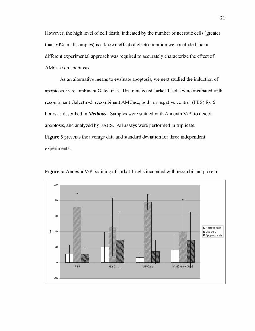
Figure 5 presents the average data and standard deviation for three independent
experiments.
Figure 5: Annexin V/PI staining of Jurkat T cells incubated with recombinant protein.
-20
0
20
40
60
80
100
PBS Gal-3 hAMCase hAMCase + Gal-3
%
Necrotic cellsLive cellsApoptotic cells

22
As seen in Figure 5, recombinant Galectin-3 induced apoptosis in Jurkat T cells,
consistent with previous studies [45]. Incubation with recombinant AMCase produced
levels of apoptosis similar to the negative control. However, co-incubation with
exogenous AMCase and Galectin-3 did not block Galectin-3 mediated apoptosis in this
system.
AMCase chitinase activity and EGFR
AMCase has been shown to demonstrate functional chitinase activity in vitro [13].
Previous studies in our lab demonstrated elevation of chitinase activity with co-
expression of AMCase and EGFR. In order to confirm this finding and to validate the
assay method, A549 cells were transfected with either full-length AMCase (2 µg), EGFR
(2 µg, 4 µg or 6 µg) plus empty vector to equalize amounts of DNA, or co-transfected
with full-length AMCase (2 µg) and EGFR (2 µg, 4 µg or 6 µg). Empty pcDNA3 vector
was used as a negative control. All transfections were done in triplicate. Chitinase
activity was measured at 24 and 56 hours. Figure 6 presents average data and standard
deviation from three independent experiments.
As shown in Figure 6, co-transfection of AMCase and EGFR caused a marked
increase in chitinase activity when compared to AMCase alone (p=0.03 at 24 hour time
point by Student’s T test); this effect did not show a dose-dependent response to the
amount of EGFR transfected.

23
Figure 6: Chitinase activity in transfected A549 cells at 24 and 56 hours.
0
10
20
30
40
50
60
70
80
90
pcDNA 3 AMC full EGFR 1x EGFR 2x EGFR 3x AMC full+EGFR (1:1)
AMC full+EGFR (1:2)
AMC full+EGFR (1:3)
Act
ivity
at 5
6 hr
s hr
s
24 hrs56 hrs
Cloning of polymorphisms of AMCase.
Human AMCase exists endogenously as both full-length and C-terminally
truncated forms. Numerous genetic variants of AMCase including multiple SNPs have
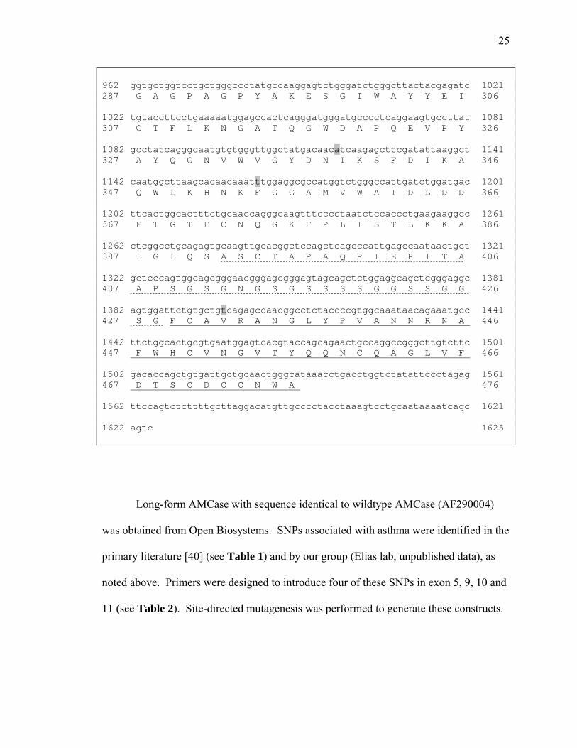
been identified in the literature. The SNPs of interest in this study are shown in Figure 7.
Human AMCase consists of a catalytic domain (amino acids 1-391) which includes the
N-terminal signal peptide (amino acids 1-21, double underline), hinge domain (amino
acids 392-428, dotted underline) and chitin binding domain (amino acids 429-476, single
underline). Start codons for full-length (amino acid 1), long-form (amino acid 109) and
short-form AMCase (amino acid 162) are indicated by bold text. SNPs of interest are

24
highlighted in gray (Exon 5, base pair 477, A to G; Exon 9, base pair 1118, A to G; Exon
10, base pair 1164, T to C; Exon 11, base pair 1398, T to G).
Figure 7: Sequence of human AMCase cDNA (AF290004). 1 gctttccagtctggtggtgaatcctccatagtctgaagcctttgtgataaccacagaatca 61 62 gaacatataaaaagctctgcgggactggtgctgactgcaaccatgacaaagcttattctc 121 1 M T K L I L 6 122 ctcacaggtcttgtccttatactgaatttgcagctcggctctgcctaccagctgacatgc 181 7 L T G L V L I L N L Q L G S A Y Q L T C 26 182 tacttcaccaactgggcccagtaccggccaggcctggggcgcttcatgcctgacaacatc 241 27 Y F T N W A Q Y R P G L G R F M P D N I 46 242 gacccctgcctctgtacccacctgatctacgcctttgctgggaggcagaacaacgagatc 301 47 D P C L C T H L I Y A F A G R Q N N E I 66 302 accaccatcgaatggaacgatgtgactctctaccaagctttcaatggcctgaaaaataag 361 67 T T I E W N D V T L Y Q A F N G L K N K 86 362 aacagccagctgaaaactctcctggccattggaggctggaacttcgggactgcccctttc 421 87 N S Q L K T L L A I G G W N F G T A P F 106 422 actgccatggtttctactcctgagaaccgccagactttcatcacctcagtcatcaaattc 481 107 T A M V S T P E N R Q T F I T S V I K F 126 482 ctgcgccagtatgagtttgacgggctggactttgactgggagtaccctggctctcgtggg 541 127 L R Q Y E F D G L D F D W E Y P G S R G 146 542 agccctcctcaggacaagcatctcttcactgtcctggtgcaggaaatgcgtgaagctttt 601 147 S P P Q D K H L F T V L V Q E M R E A F 166 602 gagcaggaggccaagcagatcaacaagcccaggctgatggtcactgctgcagtagctgct 661 167 E Q E A K Q I N K P R L M V T A A V A A 186 662 ggcatctccaatatccagtctggctatgagatcccccaactgtcacagtacctggactac 721 187 G I S N I Q S G Y E I P Q L S Q Y L D Y 206 722 atccatgtcatgacctacgacctccatggctcctgggagggctacactggagagaacagc 781 207 I H V M T Y D L H G S W E G Y T G E N S 226 782 cccctctacaaatacccgactgacaccggcagcaacgcctacctcaatgtggattatgtc 841 227 P L Y K Y P T D T G S N A Y L N V D Y V 246 842 atgaactactggaaggacaatggagcaccagctgagaagctcatcgttggattccctacc 901 247 M N Y W K D N G A P A E K L I V G F P T 266 902 tatggacacaacttcatcctgagcaacccctccaacactggaattggtgcccccacctct 961 267 Y G H N F I L S N P S N T G I G A P T S 286
25
962 ggtgctggtcctgctgggccctatgccaaggagtctgggatctgggcttactacgagatc 1021 287 G A G P A G P Y A K E S G I W A Y Y E I 306 1022 tgtaccttcctgaaaaatggagccactcagggatgggatgcccctcaggaagtgccttat 1081 307 C T F L K N G A T Q G W D A P Q E V P Y 326 1082 gcctatcagggcaatgtgtgggttggctatgacaacatcaagagcttcgatattaaggct 1141 327 A Y Q G N V W V G Y D N I K S F D I K A 346 1142 caatggcttaagcacaacaaatttggaggcgccatggtctgggccattgatctggatgac 1201 347 Q W L K H N K F G G A M V W A I D L D D 366 1202 ttcactggcactttctgcaaccagggcaagtttcccctaatctccaccctgaagaaggcc 1261 367 F T G T F C N Q G K F P L I S T L K K A 386 1262 ctcggcctgcagagtgcaagttgcacggctccagctcagcccattgagccaataactgct 1321 387 L G L Q S A S C T A P A Q P I E P I T A 406 1322 gctcccagtggcagcgggaacgggagcgggagtagcagctctggaggcagctcgggaggc 1381 407 A P S G S G N G S G S S S S G G S S G G 426 1382 agtggattctgtgctgtcagagccaacggcctctaccccgtggcaaataacagaaatgcc 1441 427 S G F C A V R A N G L Y P V A N N R N A 446 1442 ttctggcactgcgtgaatggagtcacgtaccagcagaactgccaggccgggcttgtcttc 1501 447 F W H C V N G V T Y Q Q N C Q A G L V F 466 1502 gacaccagctgtgattgctgcaactgggcataaacctgacctggtctatattccctagag 1561 467 D T S C D C C N W A 476 1562 ttccagtctcttttgcttaggacatgttgcccctacctaaagtcctgcaataaaatcagc 1621 1622 agtc 1625
Long-form AMCase with sequence identical to wildtype AMCase (AF290004)
was obtained from Open Biosystems. SNPs associated with asthma were identified in the
primary literature [40] (see Table 1) and by our group (Elias lab, unpublished data), as
noted above. Primers were designed to introduce four of these SNPs in exon 5, 9, 10 and
11 (see Table 2). Site-directed mutagenesis was performed to generate these constructs.

26
Table 2: Primers used in site-directed mutagenesis of long-form AMCase (Open
Biosystems).
SNP Exon BP# WT
Base
SNP
Base
Primers
-- 5 477 A G Forward:
5’-catcacctcagtcatcagattcctgcgccagtatg-3’
Reverse:
5’-catactggcgcaggaatctgatgactgaggtgatg-3’
Rs2275253 9 1118 A G Forward:
5'-gtgggttggctatgacaacgtcaagagcttcgatattaa-3'
Reverse:
5'-ttaatatcgaagctcttgacgttgtcatagccaacccac-3'
Rs2275254 10 1164 T C Forward:
5'-tcaatggcttaagcacaacaaatctggaggcgccat-3'
Reverse:
5'-atggcgcctccagatttgttgtgcttaagccattga-3'
Rs2256721 11 1398 T G Forward:
5'-tggattctgtgctggcagagccaacggcc-3'
Reverse:
5'-ggccgttggctctgccagcacagaatcca-3'

27
Constructs were sequenced after each round of mutagenesis to identify clones containing
the desired sequence of base pairs. Constructs developed included the single-nucleotide
polymorphisms identified as significantly correlated with asthma [40] as well as various
combinations of SNPs (Table 3).
Table 3: Long-form AMCase constructs containing combinations of SNPs correlated
with asthma.
Construct Exon 5 SNP Exon 9 SNP Exon 10 SNP Exon 11 SNP
Wildtype
1 +
2 +
3 +
4 +
5 + +
6 + +
7 + +
Full-length AMCase acquired from MedImmune was sequenced and found to contain
nine SNPs (Table 4). Three of these SNPs (numbered 6, 7 and 9 in Table 4) were
previously identified as associated with asthma [40]. Two of these SNPs (numbered 5
and 8 in Table 4) consisted of silent mutations in the catalytic domain, though neither of
these two synonymous mutations have been identified in the current literature as
clinically associated with asthma. The remaining four SNPs (numbered 1, 2, 3, and 4 in

28
Table 4) were located in the signal peptide domain of AMCase, and were of
undetermined clinical significance.
Table 4: Single-nucleotide polymorphisms identified in full-length AMCase clone
(MedImmune) by sequencing.
SNP Domain Exon BP# WT
Base
SNP
Base
WT
AA
New
AA
1 -- SP -- A G AAC
(N)
GAC
(D)
2 -- SP -- G A GAC
(D)
AAC
(N)
3 -- SP -- G T AGG
(R)
ATG
(M)
4 -- SP -- C T AAC
(N)
AAT
(N)
5 -- Catalytic 9 1099 G A GTG
(V)
GTA
(V)
6 Rs2275253 Catalytic 9* 1118 A G ATC
(I)
GTC
(V)
7 Rs2275254 Catalytic 10* 1164 T C TTT
(F)
TCT
(S)
8 -- Catalytic 10 1268 C T GGC
(G)
GGT
(G)

29
9 Rs2256721 CBD 11* 1398 T G GTC
(V)
GGC
(G)
SNPs in the signal peptide domain of full-length AMCase were corrected in a
stepwise manner to generate wildtype sequence using point mutagenesis as described in
the Methods section above. Subsequently, constructs containing the desired clinically-
relevant SNPs (designated by an asterix in Table 4) were generated by sequential
correction via point mutagenesis. Primers used for mutagenesis are listed in Table 5;
SNPs associated with asthma are designated with an asterix.
Table 5: Primers used in site-directed mutagenesis of full-length AMCase
(MedImmune).
SNP Exon BP# WT
Base
SNP
Base
Primers
1 -- -- A G
2 -- -- G A
Forward:
5’-cttcatgcctgacaacatcgacccctgcctctg-3’
Reverse:
5’-cagaggcaggggtcgatgttgtcaggcatgaag-3’
3 -- -- G T Forward:
5’-cgcctttgctgggaggcagaacaacgag-3’
Reverse:
5’-ctcgttgttctgcctcccagcaaaggcg-3’
4 -- -- C T Forward:
5’-ccaccatcgaatggaacgatgtgactctctacc-3’
Reverse:

30
5’-ggtagagagtcacatcgttccattcgatggtgg-3’
5 5* 477 A G Forward:
5’-catcacctcagtcatcagattcctgcgccagtatg-3’
Reverse:
5’-catactggcgcaggaatctgatgactgaggtgatg-3’
6 -- 9 1099 G A Forward:
5'-ccttatgcctatcagggcaatgtgtgggttggctat-3'
Reverse:
5'-atagccaacccacacattgccctgataggcataagg-3'
7 Rs2275253 9* 1118 A G Forward:
5’-tatgggttggctatgacaacatcaagagcttcgatattaag-3’
Reverse:
5’-cttaatatcgaagctcttgatgttgtcatagccaacccata-3’
8 Rs2275254 10* 1164 T C Forward:
5'-tcaatggcttaagcacaacaaatttggaggcgccat-3'
Reverse:
5'-atggcgcctccaaatttgttgtgcttaagccattga-3'
9 -- 10 1268 C T Forward:
5'-gaaggccctcggcctgcagagtgcaag-3'
Reverse:
5'-cttgcactctgcaggccgagggccttc-3'
10 Rs2256721 11* 1398 T G Forward:
5'-tggattctgtgctgtcagagccaacggcc-3'
Reverse:
5'-ggccgttggctctgacagcacagaatcca-3'
Constructs were sequenced after each round of mutagenesis to identify clones containing
the desired sequence of base pairs. The two silent mutations in the catalytic domain were

31
not altered due to low potential for changes in enzyme activity or expression; all
constructs generated contained these two point mutations, making the backgrounds of all
constructs comparable.

32
Discussion
AMCase has been implicated in asthma and other immune-mediated allergic
disease based upon studies from animal models of asthma that demonstrate it is involved
in the development of Th2 inflammatory responses, as well as population-based
association studies that have demonstrated that genetic variation in AMCase is associated
with asthma. The effect of genetic mutations in AMCase on its biologic function remains
completely unknown.
We hypothesized that human AMCase participates in Th2 inflammation by an
EGFR-dependant inhibition of apoptosis mechanism. We theorized that AMCase
interacts with Galectin-3 to mediate downstream anti-apoptotic functions of AMCase,
and that asthma-associated polymorphisms of human AMCase alter chitinase activity and
modulate downstream anti-apoptotic functions of AMCase, increasing Th2-mediated
inflammation in human asthma.
In these studies, we have begun to elucidate the biologic effects of AMCase. We
have demonstrated that AMCase binds to and co-localizes with Galectin-3 in vitro. In an
over-expression system, Galectin-3 demonstrated known anti-apoptotic effects [44],
while co-transfection of AMCase and Galectin-3 was more protective against apoptosis
than either Galectin-3 or empty vector alone. In an exogenous protein assay,
recombinant Galectin-3 induced apoptosis consistent with published data [45], while
recombinant AMCase stimulation resulted in similar levels of apoptosis as observed in
negative controls. These data seem to suggest that while AMCase and Galectin-3 bind
and interact, and co-expression appears to increase anti-apoptotic effects, contrary to our
hypothesis the anti-apoptotic effects of AMCase may be independent of a direct

33
interaction with Galectin-3. It is also possible that the over-expression assay is a better
model of an endogenous system, while the experimental system in which recombinant
protein is added exogenously does not sufficiently resemble in vivo conditions.
Alternatively, the interaction may be a complex one, mediated by different factors
intracellularly as compared with extracellularly.
In this investigation, we also demonstrated that co-transfection of AMCase and
EGFR augments chitinase activity in transfected A549 cells. This data is consistent with
our hypothesis that AMCase and EGFR interact, and with published data from our lab
establishing EGFR as an upstream regulator of AMCase [39]. This raises the intriguing
possibility that the downstream functions of EGFR may be mediated by AMCase.
Lastly, in order to further explore the function of AMCase and particularly the
role that genetic variants of AMCase play in human asthma, in this study we have
developed expression constructs of SNPs associated with clinical asthma. These
constructs have great potential as tools with which to study molecular functionality of
AMCase, and are further discussed in Future directions, below.
Although it is clear that AMCase plays a significant role in Th2-mediated
inflammation in the lung, its exact function is yet to be determined, and contrasting data
exists. In a murine model, chitin has been shown to induce eosinophil and basophil
recruitment to the lung [47], an effect which seemed to be mediated by leukotriene B4
produced by macrophages. BLT1, the high-affinity receptor for leukotriene B4, has an
essential role in CD8+ T-cell recruitment to the lung in allergic asthma and airway hyper-
responsiveness [48, 49]. Eosinophil recruitment to the lung was blocked both by pre-
treatment with recombinant AMCase, and by depletion of macrophages, contrary to

34
previous data which seemed to implicate AMCase as an inducer of Th2-mediated
inflammation [10]. Taken together, these data suggest a role for chitin as a molecular-
recognition element implicated in the innate immune response and recruitment of
eosinophils and basophils to the lung. In this context, AMCase seems to act in an anti-
inflammatory manner to decrease eosinophil influx, in contrast to findings by our lab that
AMCase has a pro-inflammatory function mediated by IL-13 [10]. The causes of this
apparent paradox have yet to be delineated, however, some STAT pathways have been
shown to have both pro- and anti-inflammatory actions [50].

35
Future directions
In this study, we have developed the biochemical tools to investigate genetic
polymorphisms in AMCase that have been clinically associated with asthma. Our initial
data shows that the anti-apoptotic effects of AMCase are not mediated by a simple
interaction with Galectin-3. Rather, it is likely that AMCase acts through a complex
activation cascade which includes Galectin-3 and other as yet unidentified mediators. In
order to further explore the biologic function of AMCase, we have developed constructs
expressing SNPs of AMCase associated with asthma in human populations. Our goal is
to use these constructs in future experiments to delineate the effects of SNPs and
haplotypes of SNPs on chitinase activity and apoptosis.
Chitinase activity is an important characteristic of AMCase. Functional chitinases
are enzymatically active against chitin. Their chitinase activity can be measured by using
a fluorogenic substrate such as 4-methylumbelliferyl-D-N,N’-diacetylchitobioside (4-
MU). Both chitotriosidase and AMCase have been demonstrated to have functional
chitinase activity against 4-MU [13, 16]. Our lab has replicated chitinase activity in
wildtype AMCase.
Using the AMCase constructs generated in this study, future experiments will
seek to quantify chitinase activity in constructs containing polymorphisms associated
with asthma as compared to wildtype AMCase. AMCase constructs will be over-
expressed in A549 cells, and chitinase activity against 4-MU measured. Cells will be
harvested and lysates probed by Western blot to monitor protein expression levels.
In order to further explore the impact of polymorphisms in AMCase, future
experiments would investigate whether polymorphic AMCase constructs demonstrate

36
altered levels of apoptosis as compared to wildtype. Full-length AMCase constructs
generated in this study are currently in a bacterial expression vector containing RFP
(pDSRed, Clontech), which fluoresces in the same range of wavelengths (excitation 557
nm, emission 579 nm) as the tagged Propidium Iodide (BD Pharmingen; emission
spectrum 562-588 nm) which is currently available, precluding the use of Annexin V/PI
staining as an assay for apoptosis. To facilitate future experiments using PI, the existing
panel of AMCase constructs can be cloned into an alternative expression vector such as
pcDNA3 in order to eliminate confounding signal between the RFP and PI. These
constructs can then be over-expressed by transfection in A549 cells, and apoptosis
induced by incubation with Fas ligand as compared to negative control. Transfected
samples can then be evaluated for apoptosis by Annexin V/PI staining.
In order to further investigate the role that polymorphisms in AMCase play in
asthma, future experiments should be aimed at generating and biochemically testing
haplotypes of AMCase which have been identified as clinically significant in asthma. In
their study of German asthmatics, Bierbaum et al identified sixteen haplotypes with a
frequency greater than 1% in either the asthmatic or the control population [40]. The five
haplotypes with the greatest difference in frequency between asthmatics and healthy
controls are shown in Table 6 below.
Table 6: Haplotyes of AMCase identified in a German population [40]
Haplotype*
Polymorphism Position Amino Acid
exchange
1 2 3 4 5

37
Rs12033184 Promoter None wt wt M M M
Rs3818822 Exon 4 None M M wt M M
Exon 5 K17R wt wt wt Wt M
Rs2275253 Exon 9 I231V wt M M Wt M
Rs2275254 Exon 10 F246S M wt M M Wt
Exon 10 F269S wt wt wt Wt Wt
Rs2256721 Exon 11 V324G M wt wt M Wt
*wt= wildtype allele, M=mutant allele
Of these five haplotypes, two (Haplotypes 2 and 4) are positively associated with asthma,
occurring with greater frequency in asthmatic populations as compared with healthy
controls. This difference is particularly marked for pediatric asthmatics as compared
with healthy children, as shown in Table 7. The remaining three haplotypes are
negatively associated with asthma, occurring with greater frequency in control subjects.
Table 7: AMCase haplotype frequency in asthmatics compared with controls [40]
Haplotype Asthma Control Subjects Frequency Difference
Adults Children Adults Children
1 0.105921 0.109709 0.166181 -0.00379 -0.06026
2* 0.232742 0.194970 0.182000 0.037772 0.050742
3 0.073852 0.085775 0.102604 -0.011920 -0.02875
4* 0.146976 0.102581 0.076349 0.044395 0.070627
5 0.099457 0.139256 0.142457 -0.039800 -0.043000
*Haplotype with greater frequency in asthmatics as compared with healthy controls
38
Our goal is to generate the haplotypes of AMCase that appear with greater
frequency in the asthmatic population, and to investigate these haplotypes in vitro for
biochemical and functional activity, including chitinase activity and evaluation of
apoptosis.
Summary
Chitin is a ubiquitous environmental allergen known to have a role as a
recognition element for some host responses. Plant and fungal chitinases serve as
defense mechanisms against parasitic infection. AMCase and chitotriosidase, two
recently discovered mammalian chitinases, are likewise hypothesized to function in
innate immunity. AMCase has been implicated in Th2 cell-mediated inflammation,
which is classically considered to be the branch of the immune system involved in both
parasitic infection and the development of atopic and asthmatic disease. Several lines of
evidence have linked AMCase to clinical asthma, and SNPs of AMCase have been
associated with development of asthmatic disease in human populations. In this study,
we have shown that AMCase is stimulated by co-transfection EGFR, that it inhibits
apoptosis, and that it binds and co-localizes with Galectin-3 in vitro. However, contrary
to our hypothesis, the anti-apoptotic effects of AMCase do not seem to be mediated by a
direct interaction with Galectin-3. We have also developed the molecular tools to explore
the effect of genetic polymorphisms have on the function of AMCase. Future
experiments will be focused on delineating the biochemical activity of polymorphic
AMCase, to help shed light on this important mediator of inflammatory asthma.

39
Appendix: SNPs in AMCase in African American populations [41]
Table 1. Non-synonymous AMCase SNPs Frequency of Noted Allele Variants Alleles AA
Change Position Allele
MX PR AA SNP1+236 A/G Asn/Asp Exon 4 G 0.09 0.135 0.26 SNP2+242 G/A Asp/Asn Exon 4 A 0.09 0.135 0.26 SNP3+285 G/T Arg/Met Exon 4 T 0.09 0.135 0.26 SNP4+407 G/A Gly/Arg Exon 5 A 0.035 0.1 0.15 SNP5+477 G/A Arg/Lys Exon 6 G 0.06 0.1 0.05 SNP6+1118 A/G Ile/Val Exon 10 G 0.36 0.55 0.79 SNP7+1164 C/T Ser/Phe Exon 11 C 0.30 0.39 0.39 SNP8+1398 T/G Val/Gly Exon 12 G 0.35 0.54 0.78 Table 2. Results of African American Case-Control Analysis for Asthma and Atopic Asthma Disease Status
Frequency (%) Odds Ratio (95% CI)
χ2 p Value SNP# Allele
Cases (n=352)
Atopic Cases
(n=202)
Controls(n=352)
Asthma Atopic Asthma
Asthma Atopic Asthma
SNP3 T 17.0 16.2 25.9 .59/(.41,.85) .55/(.35,.87) 0.004 0.009 SNP4 A 16.1 17.8 14.5 1.05 1.27 0.83 0.30 SNP5 G 4.9 4.6 4.5 1.07 0.99 0.85 0.98 SNP6 A 22.5 20.6 20.7 0.90 0.99 0.56 0.95 SNP7 C 32.6 30.4 39.2 0.75 .67/(.47,.98) 0.08 0.04 SNP8 T 23.4 21.5 21.7 1.10 0.93 0.59 0.95 Table 3. Allelic and Genotypic Association test for AMCase SNPs with Bronchodilator Response (Delta-FEV1) in African Americans
Allelic Regression Genotypic Regression SNP Cases
(n=352) Atopic Cases
(n=202) Cases
(n=176) Atopic Cases
(n=101) p value p value p value p value
SNP3 0.887 0.718 0.863 0.902 SNP4 0.852 0.385 0.879 0.417 SNP5 0.052 0.503 0.051 0.491 SNP6 0.018 0.012 0.025 0.013 SNP7 0.403 0.342 0.316 0.343 SNP8 0.009 0.014 0.017 0.016

40
References
1. Afshar, R., B.D. Medoff, and A.D. Luster, Allergic asthma: a tale of many T cells. Clin Exp Allergy, 2008. 38(12): p. 1847-57.
2. Liu, A.H. and S.J. Szefler, Advances in childhood asthma: hygiene hypothesis, natural history, and management. J Allergy Clin Immunol, 2003. 111(3 Suppl): p. S785-92.
3. Larche, M., D.S. Robinson, and A.B. Kay, The role of T lymphocytes in the pathogenesis of asthma. J Allergy Clin Immunol, 2003. 111(3): p. 450-63; quiz 464.
4. Holt, P.G., et al., The role of allergy in the development of asthma. Nature, 1999. 402(6760 Suppl): p. B12-7.
5. Brightling, C.E., et al., TH2 cytokine expression in bronchoalveolar lavage fluid T lymphocytes and bronchial submucosa is a feature of asthma and eosinophilic bronchitis. J Allergy Clin Immunol, 2002. 110(6): p. 899-905.
6. Taha, R., et al., T helper type 2 cytokine receptors and associated transcription factors GATA-3, c-MAF, and signal transducer and activator of transcription factor-6 in induced sputum of atopic asthmatic patients. Chest, 2003. 123(6): p. 2074-82.
7. Nakamura, Y., et al., Gene expression of the GATA-3 transcription factor is increased in atopic asthma. J Allergy Clin Immunol, 1999. 103(2 Pt 1): p. 215-22.
8. Elias, J.A., et al., Transgenic modeling of interleukin-13 in the lung. Chest, 2003. 123(3 Suppl): p. 339S-45S.
9. Kuperman, D.A., et al., Direct effects of interleukin-13 on epithelial cells cause airway hyperreactivity and mucus overproduction in asthma. Nat Med, 2002. 8(8): p. 885-9.
10. Zhu, Z., et al., Acidic mammalian chitinase in asthmatic Th2 inflammation and IL-13 pathway activation. Science, 2004. 304(5677): p. 1678-82.
11. Kaku, H., et al., Plant cells recognize chitin fragments for defense signaling through a plasma membrane receptor. Proc Natl Acad Sci U S A, 2006. 103(29): p. 11086-91.
12. Shibata, Y., et al., Oral administration of chitin down-regulates serum IgE levels and lung eosinophilia in the allergic mouse. J Immunol, 2000. 164(3): p. 1314-21.
13. Boot, R.G., et al., Identification of a novel acidic mammalian chitinase distinct from chitotriosidase. J Biol Chem, 2001. 276(9): p. 6770-8.
14. Duo-Chuan, L., Review of fungal chitinases. Mycopathologia, 2006. 161(6): p. 345-60.
15. Renkema, G.H., et al., Purification and characterization of human chitotriosidase, a novel member of the chitinase family of proteins. J Biol Chem, 1995. 270(5): p. 2198-202.
16. Boot, R.G., et al., Cloning of a cDNA encoding chitotriosidase, a human chitinase produced by macrophages. J Biol Chem, 1995. 270(44): p. 26252-6.
17. Hollak, C.E., et al., Marked elevation of plasma chitotriosidase activity. A novel hallmark of Gaucher disease. J Clin Invest, 1994. 93(3): p. 1288-92.

41
18. Guggenbuhl, P., B. Grosbois, and G. Chales, Gaucher disease. Joint Bone Spine, 2008. 75(2): p. 116-24.
19. Aerts, J.M., et al., Biomarkers for lysosomal storage disorders: identification and application as exemplified by chitotriosidase in Gaucher disease. Acta Paediatr Suppl, 2008. 97(457): p. 7-14.
20. van Eijk, M., et al., Characterization of human phagocyte-derived chitotriosidase, a component of innate immunity. Int Immunol, 2005. 17(11): p. 1505-12.
21. Choi, E.H., et al., Genetic polymorphisms in molecules of innate immunity and susceptibility to infection with Wuchereria bancrofti in South India. Genes Immun, 2001. 2(5): p. 248-53.
22. Lehrnbecher, T., et al., Common genetic variants in the interleukin-6 and chitotriosidase genes are associated with the risk for serious infection in children undergoing therapy for acute myeloid leukemia. Leukemia, 2005. 19(10): p. 1745-50.
23. Seibold, M.A., et al., Chitotriosidase is the primary active chitinase in the human lung and is modulated by genotype and smoking habit. J Allergy Clin Immunol, 2008. 122(5): p. 944-950 e3.
24. Bierbaum, S., A. Superti-Furga, and A. Heinzmann, Genetic polymorphisms of chitotriosidase in Caucasian children with bronchial asthma. Int J Immunogenet, 2006. 33(3): p. 201-4.
25. Boot, R.G., et al., Marked differences in tissue-specific expression of chitinases in mouse and man. J Histochem Cytochem, 2005. 53(10): p. 1283-92.
26. Boot, R.G., et al., The human chitotriosidase gene. Nature of inherited enzyme deficiency. J Biol Chem, 1998. 273(40): p. 25680-5.
27. Chou, Y.T., et al., Kinetic characterization of recombinant human acidic mammalian chitinase. Biochemistry, 2006. 45(14): p. 4444-54.
28. Renkema, G.H., et al., Chitotriosidase, a chitinase, and the 39-kDa human cartilage glycoprotein, a chitin-binding lectin, are homologues of family 18 glycosyl hydrolases secreted by human macrophages. Eur J Biochem, 1998. 251(1-2): p. 504-9.
29. Saito, A., et al., Isolation and mapping of a human lung-specific gene, TSA1902, encoding a novel chitinase family member. Gene, 1999. 239(2): p. 325-31.
30. Bussink, A.P., et al., A single histidine residue modulates enzymatic activity in acidic mammalian chitinase. FEBS Lett, 2008. 582(6): p. 931-5.
31. Zhao, J., et al., Increased lungkine and chitinase levels in allergic airway inflammation: a proteomics approach. Proteomics, 2005. 5(11): p. 2799-807.
32. Kuperman, D.A., et al., Dissecting asthma using focused transgenic modeling and functional genomics. J Allergy Clin Immunol, 2005. 116(2): p. 305-11.
33. Donnelly, L.E. and P.J. Barnes, Acidic mammalian chitinase--a potential target for asthma therapy. Trends Pharmacol Sci, 2004. 25(10): p. 509-11.
34. Puddicombe, S.M., et al., Involvement of the epidermal growth factor receptor in epithelial repair in asthma. Faseb J, 2000. 14(10): p. 1362-74.
35. Mendelsohn, J. and J. Baselga, The EGF receptor family as targets for cancer therapy. Oncogene, 2000. 19(56): p. 6550-65.
36. Takeyama, K., et al., Epidermal growth factor system regulates mucin production in airways. Proc Natl Acad Sci U S A, 1999. 96(6): p. 3081-6.

42
37. Takeyama, K., J.V. Fahy, and J.A. Nadel, Relationship of epidermal growth factor receptors to goblet cell production in human bronchi. Am J Respir Crit Care Med, 2001. 163(2): p. 511-6.
38. Shim, J.J., et al., IL-13 induces mucin production by stimulating epidermal growth factor receptors and by activating neutrophils. Am J Physiol Lung Cell Mol Physiol, 2001. 280(1): p. L134-40.
39. Hartl, D., et al., Acidic Mammalian Chitinase Is Secreted via an ADAM17/Epidermal Growth Factor Receptor-dependent Pathway and Stimulates Chemokine Production by Pulmonary Epithelial Cells. J Biol Chem, 2008. 283(48): p. 33472-82.
40. Bierbaum, S., et al., Polymorphisms and haplotypes of acid mammalian chitinase are associated with bronchial asthma. Am J Respir Crit Care Med, 2005. 172(12): p. 1505-9.
41. Seibold MA, C.S., Tsai HJ, Beckham KB, Nazario S, Casai J, Torres A, Rodriguez-Santana JR, Gomez J, Salas J, Lilly CM, Matallana H, Chapela R, Rodriguez-Cintron W, Burchard EG, Acidic Mammalian Chitinase (AMCase) Coding Variation Associated with Asthma and Bronchodilator Response in Ethnically Diverse Populations.
42. Chatterjee, R., et al., Genetic association of acidic mammalian chitinase with atopic asthma and serum total IgE levels. J Allergy Clin Immunol, 2008. 122(1): p. 202-8, 208 e1-7.
43. Yang, R.Y. and F.T. Liu, Galectins in cell growth and apoptosis. Cell Mol Life Sci, 2003. 60(2): p. 267-76.
44. Yang, R.Y., D.K. Hsu, and F.T. Liu, Expression of galectin-3 modulates T-cell growth and apoptosis. Proc Natl Acad Sci U S A, 1996. 93(13): p. 6737-42.
45. Fukumori, T., et al., CD29 and CD7 mediate galectin-3-induced type II T-cell apoptosis. Cancer Res, 2003. 63(23): p. 8302-11.
46. Zhang, X.D., S.K. Gillespie, and P. Hersey, Staurosporine induces apoptosis of melanoma by both caspase-dependent and -independent apoptotic pathways. Mol Cancer Ther, 2004. 3(2): p. 187-97.
47. Reese, T.A., et al., Chitin induces accumulation in tissue of innate immune cells associated with allergy. Nature, 2007. 447(7140): p. 92-6.
48. Tager, A.M., et al., Leukotriene B4 receptor BLT1 mediates early effector T cell recruitment. Nat Immunol, 2003. 4(10): p. 982-90.
49. Taube, C., et al., The leukotriene B4 receptor (BLT1) is required for effector CD8+ T cell-mediated, mast cell-dependent airway hyperresponsiveness. J Immunol, 2006. 176(5): p. 3157-64.
50. O'Shea, J.J. and P.J. Murray, Cytokine signaling modules in inflammatory responses. Immunity, 2008. 28(4): p. 477-87.