polymorfi - fysikaalinenfarmasia.fi · spectris and the spectris logo are trade marks of ......
TRANSCRIPT

1988
FYSI
KAALISEN FARMASIAN YHDISTYS
SOCIETY OF PHYSICAL PHARM
ACY**
POLYMORFI2010
Current developments in biomaterials and drug formulat ions research
FUNCTIONAL MATERIALS FOR HEALTHCARE
The XXI Symposium of the Finnish Society of Physical Pharmacy
January 28th 2010Old Mill – Turku, Finland

9:00 Registration and coffee
9:45 Opening of the Symposium
Chairman of the Society, Teemu Heikkilä
10:00Bioactive glass scaffolds for bone regeneration and new methods for quantifying their hierarchical pore structure
Julian Jones, Imperial College London, UK
11:00 Different biodegradable silica structures in drug delivery
Mika Jokinen, DelSiTech Ltd., Finland
11:30 Engineering bio-based materials at the nanoscale
Markus Linder, VTT Technical Research Centre of Finland
12:00 Lunch and poster session
14:00 Nanoparticles as delivery systems for bioactives
Thomas Rades, University of Otago, New Zealand
15:00 Coffee break
15:30 Lipid nanocapsules in drug delivery
Samuli Hirsjärvi, University of Angers, France
16:00Acrylic pH-responsive microparticles for targeted gastrointestinal delivery
Abdul Basit, London School of Pharmacy, UK
16:45 Closing words of the Symposium
18:30 Symposium Dinner
The XXI Symposium of the Finnish Society of Physical Pharmacy
FUNCTIONAL MATERIALS FOR HEALTHCARECurrent developments in biomaterials and drug formulations research
Old Mill – Turku, 28.1.2010
SYMPOSIUM PROGRAM:

2
ISSN: 1236-40021458-5820 (PDF)

3
Fysikaalisen farmasian yhdistys kiittää yhteistyökumppaneitaan:
The Finnish Society of Physical Pharmacy gratefully acknowledges the support of the following sponsors:
The Analytical X-ray Company
Advantages:
Optimal performance in the size range from 1 - 100 nm
Analysis results often within minutes
Pre-aligned system that does not require calibration
No expert knowledge required
Automated datacollection and analysis
Particle and pore size analysis with EasySAXS
NANOMATERIALS ANALYSIS
For more information, please contact:
PANalytical B.V., Branch FinlandNikkarinkuja 5FIN-02650 ESPOOT +358 9 2212 580F +358 9 2212 585
R [Å]
Vo
lum
e D
istr
ibu
tio
n D
v(R
)
0
0.5
1.0
1.5
0 50 100 150 200
100
102
104
106
-1 0 1 2 3 4 5 62Theta [deg.]
Int.
[ar
b. u
nit
s]
samplebackgroundbackground-subtracted
Typical measurement: sample, background, background data corrected
Typical analysis result
0 50 100 150
Automated datacollection and analysis
For more information, please contact:
PANalytical B.V., Branch Finland
EasySAXS, the new small-angle X-ray scattering (SAXS) solution on the proven PANalytical X’Pert PRO MPD (multi-purpose X-ray diffractometer) platform.
PN7241.indd 1 07-12-2009 09:55:30
detailed specifications at www.malvern.com
© 2009 MRK500-07
Malvern Instruments Limited Grovewood Road • Malvern • Worcestershire • UK • WR14 1XZ Telephone: +44 (0)1684 892456 • Facsimile: +44 (0)1684 892789
Malvern Instruments WorldwideSales and service centres in over 50 countries. For details visit www.malvern.com/contact
Advanced technology made simple
dist
ribut
or d
etai
ls
Malvern Instruments is part of Spectris plc, the Precision Instrumentation and Controls Company.Spectris and the Spectris logo are Trade Marks of Spectris plc.
All information supplied within is correct at time of publication.
Malvern Instruments pursues a policy of continual improvement due to technical development. We therefore reserve the right to deviate from information, descriptions, and specifications in this publication without notice. Malvern Instruments shall not be liable for errors contained herein or for incidental or consequential damages in connection with the furnishing, performance or use of this material.Malvern and the ‘hills logo’, Bohlin, Gemini, Insitec, ISys, Kinexus, Mastersizer, Morphologi, , Rosand, SyNIRgi, Viscotek and Zetasizer, are International Trade Marks owned by Malvern Instruments Ltd.
Malvernsolutions
r

4
PÄÄTOIMITTAJAN PALSTA ....................................................................................................6
GREETINGS FROM THE CHAIR .............................................................................................7
PUHUJABIOGRAFIAT................................................................................................................9
Julian R. Jones......................................................................................................................................................... 10
Mika Jokinen........................................................................................................................................................... 11
Markus Linder......................................................................................................................................................... 12
Thomas Rades.......................................................................................................................................................... 13
Samuli Hirsjärvi ...................................................................................................................................................... 14
Abdul Basit .............................................................................................................................................................. 15
ESITYSABSTRAKTIT ...............................................................................................................17
Bioactive Scaffolds for Bone Regeneration & New Methods for Quantifying Their Hierarchical Pore Structure 18
Different Biodegradable Silica Structures in Drug Delivery .................................................................................. 19
Engineering Bio-Based Material at the Nanoscale.................................................................................................. 21
Nanoparticles as Delivery System for Bioactives .................................................................................................... 22
Lipid Nanocapsules in Drug Delivery ..................................................................................................................... 25
Acrylic pH-responsive Microparticles for Targeted Gastrointestinal Delivery...................................................... 26
ESITYSKALVOT........................................................................................................................29
Nanoparticles as Delivery System for Bioactives .................................................................................................... 30
Lipid Nanocapsules in Drug Delivery ..................................................................................................................... 42
POLYMORFI 2010
FYSIKAALISEN FARMASIAN YHDISTYKSEN JÄSENLEHTISISÄLLYS

5
POSTERIABSTRAKTIT............................................................................................................45
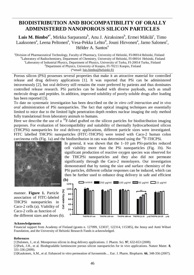
Biodistribution and Biocompatibility of Orally Administered Nanoporous Silicon Particles................................46
Color Recognition Using a Led-Based Multispectral Imaging System...................................................................47
Stability of High Indomethacin Payload Ordered Mesoporous Silica Mcm-41 and Sba-15................................... 48
Properties and Composition of Bioactive Glasses – Recent Research Activities ....................................................49
Effect of Freeze-Drying Conditions on Transfection Efficiency of Cationic Polymer DNA-Complexes ................50
Cellular Automata Model for Swelling-Controlled Drug Release ..........................................................................51
In vitro & In Vivo Characterization of Photo-Crosslinked Poly(ester anhydrides) for Controlled Drug Delivery 52
Fabrication and Characterization of Drug Particles Produced by Electrospraying into Reduced Pressure .........53
Cancer Cell Targeting and Intracellular Delivery of Hydrophobic Agents Using Mesoporous Hybrid Silica asCarrier Systems .......................................................................................................................................................54
Micro-Electroencapsulation of Porous Silicon Nanoparticles for Controlled Oral Drug Delivery Applications... 55
Tablet Formulations of Mesoporous Silicon............................................................................................................56
Cellular Responses to Porous Silicon Micro- and Nanoparticles ............................................................................57
Investigation of the Powder Flow Behaviour of Binary Mixtures of Paracetamol & Microcrystalline Cellulose..58
Optimization of Production Process of PLA Nanoparticles by Electrospraying Technique ..................................59
Mesoporous Silicon Microparticles for Sustained Peptide Delivery: Cardiovascular Effects of Melanotan II inConcious Rats ..........................................................................................................................................................60
VÄITÖSKIRJOJEN TIIVISTELMÄT ......................................................................................61
Physical Modification of Drug Release Controlling Structures – Hydrophobic Matrices and Fast DissolvingParticles....................................................................................................................................................................62
Particle Size Determination During Fluid Bed Granulation – Tools for Enhanced Process Understanding.........63
Prolonged Release Starch Acetate Matrix Tablets – Relationships between Formulation Properties and In VitroDissolution Behavior................................................................................................................................................64
The Caco-2 Cell Line in Studies of Drug Metabolism and Efflux...........................................................................66
PRO GRADUT 2008 ...................................................................................................................67
Helsingin Yliopisto................................................................................................................................................... 68
Kuopion Yliopisto .................................................................................................................................................... 68
Turun Yliopisto, Teollisuusfysiikan Laboratorio....................................................................................................69
Painallus villaisella ......................................................................................................................70
???? PÄHKINÄ ???? ..................................................................................................................73
OSALLISTUJAT.........................................................................................................................74
Pähkinän ratkaisu .......................................................................................................................76
Päätoimittaja: Henrik Ehlers, Helsingin Yliopisto; [email protected]
Julkaisija: Fysikaalisen farmasian yhdistys ry ; www.fysikaalinenfarmasia.fi

6
PÄÄTOIMITTAJAN PALSTA
Hyvät lukijat,
Fysikaalisen farmasian yhdistys on tänä vuonna aloittanut selvittämään omia juuriaan. Vaikkayhdistys on vielä varsin nuori, suuren vaihtuvuuden takia hallituksen piirissä ei ole säilynytmuistoja ja tarinoita yhdistyksen alkutaipaleelta. Tähän olemme onneksi saaneet korjauksen tänävuonna! Olemme haalineet kirjastojen ja jäsenkunnan kätköistä huomattavan määrän yhdistyksenjulkaisemista kirjallisista tuotoksista. Tämä myös näkyy tämän numeron sisällössä; lehden sivuilleon eksynyt aivopähkinä vuodelta 1992 ja pääkirjoitus vuodelta 1993. Kaikesta huolimatta, joitakinjulkaisuja vielä puuttuu kokoelmastamme; käykää vastikään uudistetuilla verkkosivuillammeosoitteessa www.fysikaalinenfarmasia.fi ja tarkistakaa löytyykö kenties Sinulta kaipaamiammeaarteita!
Polymorfissa I/1993 pyydettiin jäseniä liittymään kiinnostusryhmiin. Kiinnostusryhmä oliyksinkertaisesti ryhmä yhdistyksen jäseniä, joilla on samat kiinnostuksen kohteet. Perustelunaryhmien perustamiselle käytettiin seuraavaa: ”Yhdistyksemme jäsenkunnan osaaminen kattaalaajasti fysikaalisen farmasian aihepiirialuetta. Jäsenkuntamme on lukumäärältään melkoinen jakaikkien on mahdotonta henkilökohtaisesti tuntea toisiaan ja meillä on varsin vähän tietoa siitä,mitä kukin työssään tekee. On luultavaa, että meille jokaisella vastaan tulevien fysikaalisenfarmasian ongelmien kanssa painii joku toinenkin, mahdollisesti lukuisia henkilöitäjäsenkunnastamme. Ongelmien jakaminen, niistä keskusteleminen, tietyn tutkimusalueen tiedonkartuttaminen pienessä aktiivisessa joukossa olisi varmaan hedelmällistä ja oletettavasti välilläjopa varsin palkitsevaa ja hauskaakin.”
Yllä oleva lainaus on paikkansapitävä tänäkin päivänä, 17 vuotta myöhemmin. Kiinnostusryhmäteivät tosin enää ole osa yhdistyksen aktiivista toimintaa, mutta ajatus, joka johti kiinnostusryhmienmuodostumiseen, on edelleenkin ajankohtainen ja yksi tärkeä syy koko yhdistyksen olemassaololle.
Tämän vuoden symposium pidetään Turussa ja ajatuksena olisi tuoda biomateriaalitiede jafysikaalinen farmasia saman pöydän ääreen, jotta ymmärtäisimme että meillä on yhteisiämielenkiinnon kohteita. Toteuttakaa siis Polymorfin I/1993 ja I/2010 sanomaa ja keskustelkaatyöstänne, mielenkiinnon kohteistanne, symposiumesityksistä tai vaikka säästä, ajokelistä tai mistätahansa. Tärkeintä on että opitte uusia asioita, verkostoidutte ja nautitte poikkitieteellisestäilmapiiristä; sen takia Fysikaalisen farmasian yhdistys on olemassa!
Helsingissä 21.1.2009
Henrik EhlersPäätoimittaja

7
GREETINGS FROM THE CHAIR
Turku, Finland January 15th, 2010
To summarize my past five years in the steering committee of the Society: great experiences andfun memories. For me the most valuable offering of this association and its board has been thechance to meet and get to know so many friendly and knowledgeable people from diverse fields ofpharmaceutical and biomedical sciences.
Actually, one of the main reasons that the Finnish Society of Physical Pharmacy was grounded over21 years ago was the idea to bring together people from different fields of science (pharmacists,physicists, physical chemists and so on) so that their expertise and knowledge could be combinedand used to advance the common goal of pharmaceuticals research. The aim of the presentsymposium follows the theme of “bringing down the invisible barriers” between disciplines. Thegoal was to broaden the typical audience base by extending the invitation to “new pastures”, whichperhaps previously were not familiar with the Society and its activities. The board then faced therather difficult task of building a program that would appeal to both the new and old audiences.Based on the registration data, it seems that we succeeded in our endeavour quite nicely.
The member base of the Society has been a steady 100-130 person already since the early 1990’s(naturally during this time old members have resigned, while new members have joined). It almostseems that the Society reached its growth limit in Finland a long time ago. Have we really reachedeveryone who might be interested in our activities? Hopefully some of the first time visitors in thepresent symposium will stay with the Society as members and perhaps even take heart to join theboard in the future. Extending and refreshing the steering committee to include members from neworganizations would be important for the growth of the Society. In fact, during this term we’vealready had a new “external member” in the board from Åbo Akademi. I can fully encourage anymotivated member, new or old, to join the steering committee based on my personal experience.
However, does the Society really need to grow? Should it strive towards internationalisation or stayas an intimate forum in Finland for members that know each other well? Both types of associationshave their benefits and drawbacks. In the middle of the 90’s the clear aim of the Society was tomake more international contacts, for example a special international issue of Polymorfi 3/1997 waspublished (this issue is available online). During the last five years the board has contemplated theissue of going international almost every year. I think that the future steering committees shouldconcentrate on expanding this symposium to an “official” international meeting. This means that thesymposium audience needs to have at least 20% of visitors from outside Finland. Since the qualityof the programs of these meetings already meets the international level, the Society would mainlyneed to reform the way the symposium is advertised by publishing high quality “Firstannouncements”, “Invitations” etc and extend their circulation abroad. The coveted internationalcontacts would then surely follow organically!
As the space is running out, the time has come for me to sign out, stand down and let the next boardtake its turn at the steering wheel. I’m now leaving the board after five years of service, but I willalways be part of the Society as a member. See you! Nähdään!
Teemu Heikkilä (chair)The Finnish Society of Physical Pharmacy / Fysikaalisen farmasian yhdistys ry.

4

9
PUHUJABIOGRAFIAT

10
JULIAN R. JONES
Dr. Julian R. Jones
Department of Materials
Imperial College London, SW7 2AZ, UK
Tel: +44 (0)20 7594 6749
Fax: +44 (0)20 7594 6757
Email: julian.r.jones imperial.ac.uk
2004- Royal Academy of Engineering/ EPSRC Research Fellow (Department of Materials,Imperial college, London)
2002 – 2004 Lloyds Tercentenary Foundation Fellow (Department of Materials, Imperial college,London)
1999-2002 Ph.D. (Department of Materials, Imperial College London)
1995-1999 MEng (2.1 Hons) Metallurgy and the Science of Materials (University of Oxford)
Biography
Dr. Julian Jones is a Royal Academy of Engineering and EPSRC Research Fellow. He was awardedthe fellowship in 2004. Prior to this he held a two year Lloyds Tercentenary Foundation Fellowship,having completed his PhD here in the department in 2002. His research interests are in biomaterialsfor regenerative medicine. His work on process development of foamed gel-derived bioactive glass(the first 3D porous scaffold made from bioactive glass) has produced scaffolds suitable for tissueengineering applications with hierarchical structures similar to that of trabecular bone.
His research group consists of 6 PhD students and a PDRA. The group's research interests involvethe development of porous scaffolds for tissue engineering, novel 3D characterisation techniques ofporous materials, the development of novel nanocomposite materials, the processing of glasses,bioactive materials, protein adsoption to nanotextured materials, cell responses to biomaterials andnon-invasive cell-material interaction analysis techniques. He has published extensively on thesetopics and is also the co-editor of a text book on biomaterials and tissue engineering (Biomaterials,artificial organs and tissue engineering, L. Hench, J. Jones, 2005).
In 2007 he was awarded a prestigious Philip Leverhulme Prize for excellence in engineering and in2004 he was awarded the Silver Medal by the Institute of Materials, Mining and Minerals (IOM3 )for outstanding achievement in materials science by a younger researcher and the promotion of thesubject on the international scale.

11
MIKA JOKINEN
Mika Jokinen, PhD (Tech.)
Research Director
DelsiTech Ltd.
and
Principal Lecturer
Faculty of Life Sciences and Business
Turku University of Applied Sciences
Finland
Biography
At present Mika Jokinen holds two positions, he is (part-time) Research Director of DelSiTech Ltdand Principal Lecturer in chemical and biochemical engineering (with teaching and researchresponsibility on biomaterials and tissue engineering among other fields of biotechnology) at theTurku University of Applied Sciences, Faculty of Life Sciences and Business.
Prior to the present posts he was (full-time) Research Director at DelSiTech Ltd 2006-2008, SeniorScientist at Turku Biomaterials Centre, University of Turku 1999-2005, where he coordinated largeresearch projects between academia and industry and instructed several PhD theses, part-timeResearch Instructor at DelSiTech Ltd and its mother company Bioxid Ltd 2001-2005, ResearchScientist at Åbo Akademi University (research on bioceramics), Department of Physical Chemistry1994-1999, Research Assistant and hourly-based teacher at Åbo Akademi University, Laboratory ofIndustrial Chemistry 1992-1994.
He obtained MSc in Chemical Engineering 1993, Licenciate of Technology 1998 and Doctor ofScience in Technology 1999 from Åbo Akademi University, Faculty of Chemical Engineering. Hewas nominated as Adjunct Professor in Medical Biomaterials at Åbo Akademi University in 2003.His current research interests are bioceramics and bioceramic-polymer composites for delivery ofdrugs and other biologically active agents and for tissue engineering.

12
MARKUS LINDER
Markus Linder, PhD (Tech.)
Professor
VTT Technical Research Center of Finland
P.O. Box 1000, FI-02044 VTT
Tel. +358 20 722 111
Fax +358 20 722 7001
Biography
Markus Linder obtained his PhD in technology at the Helsinki University of Technology. He hasworked for VTT Technical Research Center of Finland as a scientist, team-leader and projectmanager in projects concerning nano-biomaterials, protein structure and functions, surfacechemistry and enzymatic hydrolysis of cellulose. As of October 2009 he has been a researchprofessor in functional materials. His research interests are the development and application of awide range of material science –based solutions, with emphasis on nano- and functional materialsand biomimetics.

13
THOMAS RADES
Thomas Rades, PhD
Professor
New Zealand's National School of Pharmacy
University of Otago
Adams Building
18 Frederick Street
PO Box 913
Dunedin
New Zealand
Tel.: +64 (0) 3 479 5410
Fax: +64 (0) 3 479 7034
Biography
Professor Thomas Rades is the Chair in Pharmaceutical Sciences at the National School ofPharmacy, University of Otago, New Zealand.
In 1994 he received a PhD from the University of Braunschweig, Germany for his work onthermotropic and lyotropic liquid crystalline drugs. After working as a Research Scientist in thePreclinical Development and Formulation at F. Hoffmann-La Roche in Basel, Switzerland, hebecame a Senior Lecturer in Pharmaceutical Sciences at Otago in 1999 and since 2003 holds theChair in Pharmaceutical Sciences.
Professor Rades has developed an international reputation for his research in drug delivery andphysical characterisation of drugs. Prof Rades has published more than 185 papers in internationalpeer review journals as well as several book chapters and patents.
Prof Rades also holds a visiting professorship at the Department of Medicine at the University ofAdelaide, Australia and The School of Life and Health at Aston University, Birmingham, UK.
Professor Rades has successfully supervised more than 30 PhD Students. For his undergraduate andpostgraduate teaching he was awarded the University of Otago Teaching Excellence Award and theNew Zealand Tertiary Teaching Excellence Award for Sustained Excellence in 2005.
His research interests include Nanoparticles as delivery systems for drugs and vaccines, and thesolid state of drugs and dosage forms. Research in both areas aims to improve drug therapy throughappropriate formulation and characterisation of medicines and to increase our understanding of thephysico-chemical properties of drugs and medicines. It combines physical, chemical, and biologicalsciences and technology to optimally formulate drugs and vaccines for human and veterinary uses.

14
SAMULI HIRSJÄRVI
Samuli Hirsjärvi, PhD
Post doctoral researcher
Inserm U646
University of Angers
France
Biography
Samuli Hirsjärvi obtained his MSc (Pharm.) (2003) and PhD (Pharm.) (2008) degrees inpharmaceutical technology from the Faculty of Pharmacy, University of Helsinki. In his PhD thesiswork, he focused on the preparation and characterization of poly(lactic acid) nanoparticles forpharmaceutical use. He is currently working as a post doctoral researcher in the Inserm U646research institute in Angers, France. The joint project of four French research institutes, funded bythe French National Research Agency, aims at developing targeted lipid nanocarriers for cancertreatment and elaborating advanced methods in studying of their biodistribution by fluorescenceimaging.

15
ABDUL BASIT
Dr. Abdul Basit
The School of Pharmacy
University of London
29/39 Brunswick square
London WC1N 1AX
United Kingdom
Telephone: +44 20 7753 5865
E-mail: [email protected]
Biography
Dr Abdul Basit holds the position of Senior Lecturer in Pharmaceutics at the School of Pharmacy,University of London. He is also a Visiting Professor in the Faculty of Chemical andPharmaceutical Sciences at the University of Chile. He further holds an Honorary Lectureship inGastroenterology at the Wingate Institute of Neurogastroenterology, Queen Mary College,University of London. Dr Basit read Pharmacy at the University of Bath and graduated in 1993 withfirst class honors. Following a short period with Pfizer in the UK, he undertook post-graduatestudies in Pharmaceutics at the School of Pharmacy, University of London and was awarded a PhDin 1999. Dr Basit's research sits at the interface between pharmaceutical science andgastroenterology and is focused on oral delivery. Dr. Basit leads a large and multi-disciplinaryresearch group of 15 PhD students and post-doctoral fellows. He has published extensively and hasa number of papers, patents, book chapters and abstracts to his name. Dr Basit sits on the scientificadvisory board of several pharmaceutical and healthcare companies and is on the editorial board ofscientific journals. He is a frequent speaker at international conferences and is a consultant to thepharmaceutical industry. In recognition of his research achievements Dr. Basit was the recipient ofthe 2004 Young Investigator Award in Pharmaceutics and Pharmaceutical Technology from theAmerican Association of Pharmaceutical Scientists (AAPS). He is the first scientist based outside ofNorth America to receive this award.

16
The Analytical X-ray Company
Advantages:
Optimal performance in the size range from 1 - 100 nm
Analysis results often within minutes
Pre-aligned system that does not require calibration
No expert knowledge required
Automated datacollection and analysis
Particle and pore size analysis with EasySAXS
NANOMATERIALS ANALYSIS
For more information, please contact:
PANalytical B.V., Branch FinlandNikkarinkuja 5FIN-02650 ESPOOT +358 9 2212 580F +358 9 2212 585
R [Å]
Vo
lum
e D
istr
ibu
tio
n D
v(R
)
0
0.5
1.0
1.5
0 50 100 150 200
100
102
104
106
-1 0 1 2 3 4 5 62Theta [deg.]
Int.
[ar
b. u
nit
s]
samplebackgroundbackground-subtracted
Typical measurement: sample, background, background data corrected
Typical analysis result
0 50 100 150
Automated datacollection and analysis
For more information, please contact:
PANalytical B.V., Branch Finland
EasySAXS, the new small-angle X-ray scattering (SAXS) solution on the proven PANalytical X’Pert PRO MPD (multi-purpose X-ray diffractometer) platform.
PN7241.indd 1 07-12-2009 09:55:30
17
ESITYSABSTRAKTIT

18
BIOACTIVE SCAFFOLDS FOR BONE REGENERATION ANDNEW METHODS FOR QUANTIFYING THEIR HIERARCHICAL
PORE STRUCTURE
Julian R. Jones
Department of MaterialsImperial College London
SW7 2AZUK
The presentation will cover my research interests in bioactive glass scaffolds, bioactive glassnanoparticles and their cellular response. Bioactive glass scaffolds for bone regenerationapplications have been developed with a hierarchical pore structure of nanopores andinterconnected macropores. Particular emphasis will be placed on new techniques for characterisingthis complex pore structure. Melt-derived bioactive glasses have been used in a particulate form asbone fillers for over twenty years. Bioactive glasses bond to bone, are resorbable in the body andtheir dissolution products have been found to stimulate osteogenic cells at the genetic level. Theyare therefore an ideal material to stimulate bone regeneration. However a scaffold is required thatcan act as a temporary template for three dimensional bone growth. The criteria for an ideal scaffoldwill be discussed. Bioactive glass scaffolds that fulfil many of the criteria can be synthesised by theusing the sol-gel foaming process. It is more challenging to produce melt-derived scaffolds becausethe traditional compositions all crystallise during sintering. However we have developed newcompositions that remain amorphous during sintering, therefore the gel-casting foaming process canbe applied to these compositions. The scaffolds have compressive strengths similar to porous boneand commercially available porous hydroxyapatite, but they are brittle under tensile loads. An idealscaffold for all bone regeneration sites in the body must have improved toughness. High toughness,while maintaining bioactivity and controlled resorption, cannot be achieved by conventionalcomposites. Instead, close mimics of the bone nanostructure must be created by developing novelnanocomposite scaffolds. For all tissue scaffolds, it is imperative to be able to quantify the poresizes and more importantly the size of interconnects between the large pores. X-raymicrotomography (µCT) has become a popular tool for obtaining 3D images of tissue scaffolds,however images are only qualitative. We have developed image analysis techniques for quantifyingopen pore networks in 3D. These techniques are suitable for many other types of tissue scaffold. Fora large bone defect to be regenerated successfully blood vessels must grow into the scaffold. Theonly way this will be possible is to use tissue engineering approaches.

19
DIFFERENT BIODEGRADABLE SILICA STRUCTURES INDRUG DELIVERY
Mika Jokinena,b, Harry Jalonena, Ari-Pekka Forsbacka, Mika Koskinena
a DelSiTech Ltd, Turku, Finlandb Faculty of Life Sciences and Business, Turku University of Applied Sciences, Turku, Finland
[email protected], [email protected]
Biodegradable silica can be prepared into various structures and forms, e.g., injectable gels, fibers,coatings, ceramic “compacts” of varying water content and particles of different size by using thesol-gel method (Fig. 1). The sol-gel derived silica has typically a porous structure and it containsvarying amount of silanol groups that both affect the biodegradation rate (dissolution of silica inbody fluids). The structure originates from the dual nature of polymerisation in the sol-gel method.The inorganic, dual polymerisation includes both “molecular polymerisation” (condensation ofsilanols) and aggregation of nanoparticles, i.e., there are 2 types of “monomers” that affect theresulting structure. The chemical reactions and aggregation occur simultaneously, which makes thesol-gel method challenging. On the other hand, it has also provided structural variation that makesthe material suitable for different types of active ingredients and administration routes.
The sol-gel method is as such an old technique and very much studied, but not from the viewpointof adjustable biodegradation. Biodegradation rate is one of the most important parameters in thedevelopment of silica-based drug delivery device. The great challenge has been to combine the pre-cursor ratios, process parameters (e.g., for aging & drying) and different preparation methods withthe large-scale adjustment of the biodegradation rate, incorporation of active ingredients (AI) andconditions (pH, temperature etc.) that are suitable for different types of AI, but on the other handalso for the synthesis.
We have found that by suitable combination of the above-mentioned factors, it is possible to varythe biodegradation rate on a large scale (almost) independently on the pore structure. It means that itis possible to combine different “chemical structures” (degree of condensation, number of silanolgroups) with different pore structures so that we can find (for controlled release) suitable silicastructures for both large AI (e.g., viral vectors with diameter of 40-300 nm) and small-moleculedrugs with 10-100 fold smaller size. For example, it possible to prepare a dense silica implant withlow porosity & specific surface area that dissolves either very fast in some days or slowly in severalmonths.
Figure 1. Different forms of sol-gel derived silica.

20
Another important finding was that, in spite of dramatic changes in the synthesis (e.g. increasing thepH from 2 to 6 due to addition of viral vectors or proteins), it is possible to “freeze” a desired che-mical structure of silica by introducing a fast and often forced gelation (e.g. spray-drying or freeze-drying). The fast gelation preserves the chemical structure achieved prior to the pH adjustment.
The varying silica structures have opened possibilities to develop delivery device for different typesof active ingredients. Mainly matrix-dissolution dependent release of drugs is possible for bothsmall molecules and larger biologicals. Another important aspect has been the possibility to preparesilica implants, particles or gels that contain large amounts of water. It has been of importance whenencapsulating different biopharmaceuticals, such as proteins or viral vectors. The larger wateramount in implants has a positive influence on the preservation of biological activity. Due tocolloidal nature (i.e., due to the fact that structure is based on aggregated nanoscale particles) of thesilica gels, it is possible to prepare implants that are easy to handle during implantation althoughthey contain more than 90% water.
Figure 2. One-pot synthesis of injectable silica.
One of the recent forms of silica that we developed is injectable silica that is made in a simple one-pot processing (Fig. 2). The material could be described to be a sol, where the solid phase consistsof “gel particles”. The gel particles (size varies depending on the formulation & dispersing from 30nm to 10 µm) include the encapsulated drugs. The idea is to first prepare a gel that encapsulates allcomponents of the sol and added drugs into one semi-solid gel body. The formed gel body is thenalmost immediately redispersed by stirring and breaking the gel in extra water and it turns into aflowable and injectable form. The structure of the gel is (partly) reversible (redispersible) only for ashort while. The silica content of the final injectable formulation is very low, 0.5-2.0% and hence, itcan be administered by injection with very thin needles, e.g., 31G needle. It is also possible toinduce a regelation (“solidification”) of the injectable silica, which can be useful after the injectioninto tissue, because the regelled, implant-like structure is more effective in the encapsulation and ithas also an influence on the biodegraration rate. The injectable silica has been found to beespecially suitable matrix for biopharmaceuticals.
In conclusion, both the process and the structural properties have been adapted to the large-scalebiodegradation adjustment. This makes the sol-gel derived silica a potential drug delivery device formany types of active ingredients, from small-molecule drugs to sensitive biopharmaceuticals

21
ENGINEERING BIO-BASED MATERIAL AT THE NANOSCALE
Markus Linder
VTT Technical Research Centre of Finland
Typically we produce complex nanomaterials by assembly of different components. Thesecomponents van vary widely in structure and function, being for example nanotubes, smallorganic molecules, polymers, or inorganic particles. For the functionality of thenanomaterials it is important to have a well understood control of the interactions andstructures of the components. In this respect biomolecules offer some advantageousproperties in comparison to more traditional polymers. Proteins can for example beengineered with molecular precision to have specified properties. Here we show howprotein engineering of surfactant proteins called hydrophobins and nanocellulose and itsinteractions with proteins can be used in designing nanomaterials.

22
NANOPARTICLES AS DELIVERY SYSTEM FOR BIOACTIVESThomas Rades
New Zealand National School of PharmacyUniversity of Otago
Dunedin, New Zealand
With current gene and protein technology it is now possible to identify specific regions of somewhole organisms or cells which are likely to be recognized by the immune system, and to reproducethem synthetically as subunit vaccines. These so called epitopes are very safe because they are non-living but they also tend to be only poorly immune stimulating. To improve the immunogenicity ofa poorly immunogenic antigen, our approach is to use nanoparticles as delivery systems.Nanoparticulate delivery systems are thought to enhance the immune response by more closelymimicking a virus or microorganism due to the possibility of multimeric antigen presentation andtheir large size compared to subunit antigens.
Our group has developed and characterised the following colloidal delivery systems:• functionalised liposomes (mannosylated or including adjuvants such as Quil A) [1-5],• immune stimulating complexes (ISCOMs) [6-13],• cationic ISCOMs (termed Pluscoms) [14-16],• cubosomes [17-20],• ISCOM implants and in situ gelling chitosan solutions containing chitosan nanoparticles
[21-26].
In this presentation we will give an overview about the various nanoparticulate delivery systems ourgroup has developed for the delivery of subunit vaccines. We will describe new results in this field,both on physico-chemical characterisation and immunological activity of these systems.
References
Functionalised liposomes[1] Copland MJ, Baird MA, Rades T, McKenzie JL, Becker B, Reck F, Tyler P, Davies NM.Liposomal Delivery of Antigen to Human Dendritic Cells. Vaccine 21: 883 - 890 (2003)
[2] Copland MJ, Rades T, Davies NM, Baird M, Lipid based particulate formulations for thedelivery of antigen. Immunology and Cell Biology 83: 97-105 (2005)
[3] White K, Rades T, Fernaux RH, Tyler PC, Hook S, Mannosylated liposomes as antigen deliveryvehicles for targeting to dendritic cells. Journal of Pharmacy and Pharmacology 58: 729 - 737(2006)
[4] White K, Rades T, Fernaux R, Kearns P, Toth I, Hook S, Immunogenicity of liposomescontaining lipid core peptides and the adjuvant Quil A. Pharmaceutical Research 23: 1473 – 1481(2006)
[5] Saupe A, McBurney WT, Rades T, Hook SJ, Immunostimulatory colloidal delivery systems forcancer vaccines. Expert Opinion on Drug Delivery 3: 345 - 354 (2006)
23
ISCOMs[6] Demana PH, Vosgerau U, Davies NM, Rades T, Pseudo ternary phase diagrams of aqueoussystems of Quil A, cholesterol and phospholipid for immune stimulating complexes (ISCOMs).International Journal of Pharmaceutics 270: 229-239 (2004)
[7] Demana PH, Davies NM, Berger B, Vosgerau U, Rades T, A comparison of pseudo ternarydiagrams of aqueous mixtures of Quil A, cholesterol and phospholipid prepared by lipid filmhydration and dialysis. Journal of Pharmacy and Pharmacology 56: 573-580 (2004)
[8] Demana PH, Davies NM, Berger B, Rades T, Incorporation of ovalbumin into iscoms andrelated colloidal particles prepared by the lipid film hydration method. International Journal ofPharmaceutics 278: 263-274 (2004)
[9] Demana P, Fehske C, White K, Rades T, Hook S, Effect of incorporation of the adjuvant Quil Aon structure and immune stimulatory capacity of liposomes. Immunology and Cell Biology 82: 547-554 (2004)
[10] Lendemans DG, Myschik J, Hook S, Rades T, Immuno-stimulating complexes prepared byethanol injection. Journal of Pharmacy and Pharmacology 57: 729-733 (2005)
[11] Lendemans DG, Egert AM, Myschik J, Hook S, Rades T, On the dilution behaviour ofimmuno-stimulation complexes. Die Pharmazie 61: 689 - 695 (2006)
[12] Myschik J, Lendemans DG, McBurney WT, Demana P, Hook S, Rades T, On the preparation,microscopic investigation and application of ISCOMs. Micron 37: 724 - 734 (2006)
[13] Demana PH, Nigel M. Davies NM, Sarah Hook S, Rades T, Analysis of Quil A-phospholipidmixtures using drift spectroscopy. International Journal of Pharmaceutics 342: 49 – 61 (2007)
Cationic ISCOMs[14] Lendemans DG, Myschik J, Hook S, Rades T, Cationic Cage-like Complexes Formed by DC-cholesterol, Quil-A and Phospholipid. Journal of Pharmaceutical Sciences 94: 1794-1807(2005)
[15] Lendemans DG, Egert AM, Hook S, Rades T, Cage-like complexes formed by DOTAP, Quil-A and cholesterol. International Journal of Pharmaceutics 332: 192 – 195 (2007)
[16] McBurney WT, Lendemans DG, Myschik J, Hennessy T, Rades T, Hook S, In vivo activity ofcationic immune stimulating complexes (PLUSCOMs). Vaccine 26: 4549 – 4556 (2008)
Cubosomes [17] Rizwan SB, Dong Y, Boyd BJ, Rades T, Hook S, Characterisation of bicontinuous cubicliquid crystalline systems of phytantriol and water using cryo field emission scanning electronmicroscopy (cryo FESEM). Micron 38: 478 – 485 (2007)
[18] Boyd B, Rizwan S, Dong Y-D, Hook S, Rades T, Self-assembled geometric liquid-crystallinenanoparticles imaged in three dimensions - hexosomes are not necessarily flat hexagonal prisms.Langmuir 23: 12461-12464 (2007)
[19] Boyd BJ, Dong Y, Rades T, Non-lamellar liquid crystalline nanostructured particles –advances in materials and structure determination. Journal of Liposome Research 19: 12 – 28(2009)

24
[20] Rizwan SB, Hanley T, Boyd BJ, Rades T, Hook S, Liquid crystallinesystems of phytantriol and glyceryl monooleate containing a hydrophilic protein: characterisation,swelling and release kinetics. Journal of Pharmaceutical Sciences (2009), IN PRESS
Implants and gels[21] Demana PH, Davies NM, Hook S, Rades T, Quil A-lipid powder formulations releasingISCOMs and related colloidal structures upon hydration. Journal of Controlled Release 103: 45-59(2005)
[22] Myschik J, Eberhardt F, Rades T, Hook S, Immunostimulatory biodegradable implantscontaining the adjuvant Quil-A – Part I: Physicochemical characterisation. Journal of DrugTargeting 16: 213 – 223 (2008)
[23] Myschik J, McBurney WT, Hennessy T, Phipps-Green A, Rades T, Hook S,Immunostimulatory biodegradable implants containing the adjuvant Quil-A – Part II: In vivoevaluation. Journal of Drug Targeting 16: 224 – 232 (2008)
[24] Myschik J, McBurney WT, Rades T, Hook S, Immunostimulatory lipid implants containingQuil-A and DC-cholesterol. International Journal of Pharmaceutics 363: 91 – 98 (2008)
[25] Myschik J, Hennessy T, McBurney WT, Phipps-Green A, Rades T, Hook S, Immunogenicityof lipid sustained release implants containing imiquimod, -Galactosylceramide, or Quil-A. DiePharmazie 63: 686 – 692 (2008)
[26] Gordon S, Saupe A, McBurney W, Rades T, Hook S, Comparison of chitosan nanoparticlesand chitosan hydrogels for vaccine delivery. Journal of Pharmacy and Pharmacology 60: 1591 –1600 (2008)

25
LIPID NANOCAPSULES IN DRUG DELIVERYSamuli Hirsjärvi
Inserm U646, University of Angers, France
Lipid nanocapsules (LNCs) are colloidal lipoprotein-like biomimetic carriers with tuneable sizebetween 20 and 100 nm [1]. Their structure can be characterized as a hybrid between polymericnanocapsules and liposomes (an oily core: triglycerides, with a shell consisting of a mixture oflecithin and a pegylated surfactant, stearate of PEG) (Fig. 1). As compared to liposomes which aremanufactured through processes involving organic solvents and are leaky and unstable in biologicalfluids, LNCs are prepared by a solvent-free, low-energy procedure and they possess good stability(physical stability of a disperstion up to 18 months). Their fabrication is based on the phase-inversion temperature phenomenon of emulsion leading to formation of LNCs with narrow sizedistribution [2]. LNCs can encapsulate lipophilic or even hydrophilic active substances into theircore [3].
Figure 1. Structure of LNCs and schematic presentation of their fabrication process.
Thus far, various drug delivery strategies with LNCs have been studied and they are presented e.g.in a comprehensive review [4]. LNCs demonstrate P-glycoprotein inhibiting properties especiallybecause of Solutol® on the surface. It has been shown that with the help of this inhibition,cytotoxicity of paclitaxel (administered in LNCs) in vitro and in vivo on glioma cells has increased.After oral administration of paclitaxel-loaded LNCs, mean plasmatic concentration of the drug was3 times higher compared to the conventional formulation. Also due to their PEGylated surface,LNCs remain in the blood circulation long enough for passive targeting purposes. However, coatingof LNCs with an even longer PEG chain increased significantly docetaxel accumulation in thetumour in vivo compared to a conventional formulation with the same drug. Applying activetargeting strategies, several functional ligands such as monoclonal antibodies, have been grafted onthe LNC surface. With these modifications, binding of LNCs on target cells or accumulation in thebrain has increased significantly.
Ongoing studies with LNCs include e.g. active targeting to cancer cells, RNA/DNA delivery,characterisation and optimisation of the intracellular fate of these carriers, and encapsulationtechniques of hydrophilic substances.
References:
[1] Heurtault et al. 2001, Patent WO0164328.[2] Heurtault et al. 2002, Pharm. Res. 19, 875.[3] Saulnier et al. 2008, Patent WO2009001019.[4] Huynh et al. 2009, Int. J. Pharm. 379, 201.

26
ACRYLIC PH-RESPONSIVE MICROPARTICLES FORTARGETED GASTROINTESTINAL DELIVERY
Abdul BasitThe Schoolof PharmacyUniversity of London
Enteric polymers are commonly applied to conventional solid dosage forms to modify drug release,exploiting the aboral increase in gastrointestinal pH (Evans et al., 1988) to manipulate thedissolution of pH-sensitive polymeric coatings. The small intestine can be targeted with polymershaving a dissolution threshold in the region of 5.0–6.0 while the distal gut requires polymers whichdissolve around pH7.0–7.5. Dissolution in the small intestine is generally used for systemicabsorption, whilst protecting the drug from the conditions in the stomach, or protecting the stomachfrom the effects of the drug. Colon specific targeting is used for the topical treatment of localdisorders e.g. inflammatory bowel disease. However, due to the inherent inter- and intra-individualvariability in the gastrointestinal physiology of man (McConnell et al., 2008a), the targetingefficacy of conventional pH-responsive systems is variable and often poor. For example, entericformulations for targeting the small intestine (coated with acrylic-, cellulose or polyvinyl basedpolymers) are often observed to disintegrate 1.5–2 h post-gastric emptying, rather than immediatelyafter gastric emptying (Hardy et al., 1987; Cole et al., 2002) resulting in delayed release or reducedbioavailability. The variability in time, site and extent of drug release and absorption is attributed tolimited free fluid (Schiller et al., 2005), and highly variable transit times (Fadda et al., 2009).Colon-targeted systems are even more complicated. These systems are reliant, not only on thehighly variable pH at the ileocaecal junction (Fallingborg et al., 1989; Ibekwe et al., 2008), but theirresidence time at this site, feeding status of the subject (Ibekwe et al., 2008) and the limited fluid inthe colon (Schiller et al., 2005). This variability is reflected in the fact that single-unit entericdosage forms for colonic targeting are sometimes voided intact (Ibekwe et al., 2006, 2008; Sinha etal., 2003; Safdi, 2005; Schroeder et al., 1987).
One approach to overcome the limitations of single-unit modified release dosage forms is sizereduction. Multi-unit systems, such as pellets, granules or beads have been proposed, but evenpellets of 0.5–1 mm diameter do not show reliable and fast gastric emptying (Clarke et al., 1995)and enteric coated pellets have shown the same failure to release drug in the colon as single-unitdosage forms (McConnell et al., 2008b). It is possible that further size reduction to microparticlesless than 100 µm may overcome the limitations of larger pellets. Microparticles can be dosed in theform of liquid suspension, which may improve gastric emptying, and the increased surfacearea:volume ratio of the microparticles would enable rapid drug release at the desired location in thegastrointestinal tract. Thus, there has been great interest in the manufacture of entericmicroparticles, but the major issues to date has been the use of toxic solvents, retention of highlevels of solvents in the products, the use of overly complicated methodology, poor microparticlesmorphology, relatively large size, changes upon storage, non-uniformity of size and poor control ofdrug release.
The aim of the presentation is to describe a simple, safe, and universal method for the fabrication ofpH-responsive microparticles for site-specific release in the gastrointestinal tract. Specifically, thepresentation will focus on the preparation and subsequent in vitro and in vivo characterisation ofuniform and spherical microparticles (less than 100 µm in size) of Eudragit L (polymethacrylicacid, methyl methacrylate 1:1; dissolution threshold pH 6.0) for proximal small intestinal targeting,and Eudragit S (polymethacrylic acid, methyl methacrylate 1:2; dissolution threshold pH 7.0) forileo-colonic targeting.

27
References:
Clarke, G.M., Newton, J.M., Short, M.B., 1995. Comparative gastrointestinal transit of pelletsystems of varying density. Int. J. Pharm. 114, 1–11.
Cole, E.T., Scott, R.A., Connor, A.L., Wilding, I.R., Petereit, H.U., Schminke, C., Beckert, T.,Cade, D., 2002. Enteric coated HPMC capsules designed to achieve intestinal targeting. Int. J.Pharm. 231, 83–95.
Evans, D.F., Pye, G., Bramley, R., Clark, A.G., Dyson, T.J., Hardcastle, J.D., 1988. Measurementof gastrointestinal pH profiles in normal ambulant human subjects. Gut 29, 1035–1041.
Fadda, H.M., McConnell, E.L., Short, M.D., Basit, A.W., 2009. Meal-induced accelerationof tablet transit through the human small intestine. Pharm. Res. 26, 356–360.
Fallingborg, J., Christensen, L.A., Ingeman-Nielsen, M., Jacobsen, B.A., Abildgaard, K.,Rasmussen, H.H., 1989. pH-profile and regional transit times of the normal gut measured by aradiotelemetry device. Aliment Pharmacol. Ther. 3, 605–613.
Hardy, J.G., Evans, D.F., Zaki, I., Clark, A.G., Tonnesen, H.H., Gamst, O.N., 1987. Evaluation ofan enteric coated Naproxen Tablet using Gamma-Scintigraphy and pH Monitoring. Int. J. Pharm.37, 245–250.
Ibekwe, V.C., Fadda, H., McConnell, E.L., Khela, M.K., Evans, D.F., Basit, A.W., 2008. Interplaybetween intestinal pH, transit time and feed status on the in vivo performance of pH responsiveileo-colonic release systems. Pharm. Res. 25, 1828–1835.
Ibekwe, V.C., Liu, F., Fadda, H.M., Khela, M.K., Evans, D.F., Parsons, G.E., Basit, A.W., 2006.An investigation into the in vivo performance variability of pH responsive polymers for ileo-colonicdrug delivery using gamma scintigraphy in humans. J. Pharm. Sci. 95, 2760–2766.
McConnell, E.L., Fadda, H.M., Basit, A.W., 2008a. Gut instincts: explorations in intestinalphysiology and drug delivery. Int. J. Pharm. 364, 213–226.
McConnell, E.L., Short, M.D., Basit, A.W., 2008b. An in vivo comparison of intestinal pH andbacteria as physiological trigger mechanisms for colonic targeting in man. J. Control. Release 130,154–160.
Safdi, A.V., 2005. Determination of mesalazine in whole or partial mesalamine delayed-releasetablets recovered fromfecal samples of healthy volunteers. Am. J. Gastroenterol. S159.
Schiller, C., Frohlich, C.P., Geissman, T., Siegmund, W., Monnikes, H., Hosten, N., Weitschies,W., 2005. Intestinal fluid volumes and transit of dosage forms as assessed by magnetic resonanceimaging. Aliment Pharmacol. Ther. 22, 971– 979.
Schroeder, K.W., Tremaine,W.J., Ilstrup, D.M., 1987. Coated oral 5-aminosalicylic acid therapy formildly to moderately active ulcerative colitis. A randomized study. N. Engl. J. Med. 317, 1625–1629.Sinha, A., Ball, B.J., Connor, A.L., Nightingale, J., Wilding, I.R., 2003. Intestinal performance oftwo mesalamine formulations in patients with active ulcerative colitis as assessed by gammascintigraphy. Pract. Gastroenterol. 27, 56–69.

28
The Analytical X-ray Company
MINIPAL 4 PHARMA - ENERGY DISPERSIVE XRF
Heavy metals and inorganicsin pharmaceuticals
• Elemental analysis: Na - U• GUMs identifi cation• Contaminant identifi cation
MiniPal 4 Pharma advantages
MiniPal 4 Pharma is a compact, benchtop instrument that delivers the benefi ts that can only be derived from the state-of-the-art energy dispersiveX-ray fl uorescence (EDXRF) spectroscopy.
• Direct analysis of inorganics and impurities
• Robust analytical data on pharmaceutical ingredients and manufacturing residues
• Identifi es and controls raw and general use materials (GUMs)
• Non-destructive, multi-element analysis that is both qualitative and quantitative
• Proven and powerful software, FDA 21 CFR Part 11 ready
• Statistical Process Control• Automatic Program Selection
choosing the best calibration when measuring unknowns
• Equipped with a 12-position sample changer
• Global customer support from an experienced team
Heavy metals and inorganicsin pharmaceuticals
• Elemental analysis: Na - U• GUMs identifi cation• Contaminant identifi cation
The benefi ts of XRF• Fast and accurate elemental analysis• Sample preparation made easy - Direct analysis of liquids/powders/solids - No chemical reagents, less waste• Unattended automated analysis• Suitable for non-experts• Unmatched repeatability with minimal
recalibration• Unknown contaminant identifi cation
For more information, please contact:
PANalytical B.V., Branch FinlandNikkarinkuja 5FIN-02650 ESPOOT +358 9 2212 580F +358 9 2212 585
PN6742.indd 1 07-12-2009 16:36:43
29
ESITYSKALVOT

30 1
Nanoparticles as deliverysystems for bioactives
Thomas RadesSchool of Pharmacy, University of Otago, Dunedin, New Zealand
Vaccines
• O’Hagan & Rappuoli Adv Drug Delivery Rev2006; 58: 29-51
Challenges
• Old challenges - M. tuberculosis,HIV, malaria
• Emerging problems - MDRbacteria & viruses, pandemicinfluenza, Ebola, non-communicable diseases -cancer/autoimmunity
Changing standards
• Regulatory issues- Live attenuated vaccines and can no
longer be made- Killed whole cell vaccines are also
difficult- Safety is more important than efficacy
- New vaccines are sub-unit vaccines
Subunit vaccines – What are thechallenges?Subunit antigens
+ Highly purified+ Good safety profile- Only poorly immunogenic- Incorporation into particulate
delivery systems- Addition of an adjuvant required- Multiple dosing, booster
injections
NANOPARTICLES AS DELIVERY SYSTEM FOR BIOACTIVES
Thomas RadesNew Zealand National School of Pharmacy
University of OtagoDunedin, New Zealand

31
2
Particulate vaccinedelivery systems
• Liposomes• Immune stimulating complexes
(ISCOMs)• Cationic ISCOMS (Pluscoms)• Cubosomes
DC treated with LPS
DC (“immature”)
DC treated withparticulateformulation
Liposomes
Versatile delivery system
Safe
Inherently not strongly immunestimulatory
OOH
HOHO
O
HO
OOH
HOHO
O
HO
OOH
HO
O (CH2)8O
NH
O PO
O-O
OO
O
(CH2)14CH3
O
(CH2)14CH3
Trimannose conjugateddipalmitoylphosphatidylethanolamine (man3DPPE)
Mannosylated phospholipid Liposome Characterisation
• Antigen Entrapment - FluorescenceSpectroscopy
• Zeta Potential - Zetasizer
• Size - Photon CorrelationSpectroscopy
• Lamellarity - 31P NMR
• Mannosylation - Lectin Agglutination

32
3
Liposome Characteristics
Neutral Negative Mannosylated
Entrapment(FITC-OVA mg/ml)
1.4 ± 0.3 1.3 ± 0.2 1.3 ± 0.2
Zeta potential(mV)
neutral -16.4 ± 2.6 -14.7 ± 4.4
Lamellarity ~ 2 bilayer - ~ 1-2 bilayers
Size (nm) 459 ± 81 302 ± 15 259± 20
Presence of accessible mannoseresidues on mannosylated liposomes
confirmed by agglutination with Con. A
0
500
1000
1500
2000
2500
0 50 100 150 200Time (min)
Siz
e (n
m) neutral
mannosylated
Con. A addition
Uptake of antigen fromvarious formulations
4oC
37oC
0
100
200
300
400
Mea
n Fl
uore
scen
ce In
tens
ity
Neutralliposomes
Negativeliposomes
Mannosylatedliposomes
Antigensolution
Expression ofActivation Markers
0
200
400
600
800
1000
MHCII CD80 CD86 CD83
NeutralLiposomesNegativeLiposomesMannosylatedLiposomesAntigenSolution
MFI
(trea
ted)
as
% o
f MFI
(con
trol)
T Cell Proliferation Assay
0
2000
4000
6000
8000
Antigen Solution Antigen FreeLiposomes
NeutralLiposomes
MannosylatedLiposomes
coun
ts/m
in
Copland M., et al. Immunology and Cell Biology 83: 97-105 (2005).White K., et al. Journal of Pharmacy and Pharmacology 58: 729 - 737 (2006).
Tumour challenge model
measure tumour growth
Inject 1 x 10⁶ cells s.c.
EG7.OVA tumour cells
30 days2 weeks
Immunise s.c. with formulation

34
5
Formationof
ISCOMs • Limited in ability to incorporate antigens
Immune StimulatingComplexes (ISCOMs)
0
5
10
15
20
25
30
Liposomes- fitc-OvaISCOMS-fitc-OvaISCOMS-p-fitc-Ova
% in
corp
orat
ion
DC-cholesterol
Cationic Immune StimulatingComplexes (Pluscoms)
Cationic Immune StimulatingComplexes (Pluscoms)
Cationic Immune StimulatingComplexes (Pluscoms)
0
10
20
30
40
50
60
70
80
90
0 0.05 0.1
protein:lipid ratio
PLUSCOMs-OVA
ISCOMs-OVA
ISCOMs-PE-OVA
Lendemans D. et al. Journal of Pharmaceutical Sciences 94: 1794-1807(2005).Lendemans D. et al. International Journal of Pharmaceutics 332: 192 – 195 (2007).
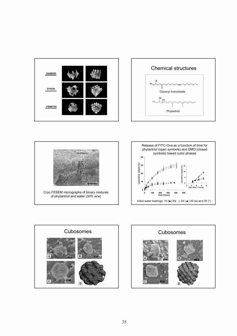
CubosomesBicontinuous Cubic Phase
• Bicontinuous cubic phase– Lipid bilayers– Bilayers are arranged in
periodic 3D cubic latticestructures
• Periodic Minimal Surfaces• Three common bicontinuous
cubic phases– Diamond (D)– Gyroid (G)– Primitive (P)
Nakano, MNakano, M et alet al. 2001. 2001
35
6
GYROID
PRIMITIVE
DIAMOND
Chemical structures
Phytantriol
Glyceryl monooleate
1 µm
Cryo FESEM micrographs of binary mixturesof phytantriol and water (30% w/w)
Release of FITC-Ova as a function of time forphytantriol (open symbols) and GMO (closed
symbols) based cubic phases
0
10
20
30
40
0 100 200 300 400 500time (hours)
cum
ulat
ive
rele
ase
(%)
0
5
10
15
0 2 4 6 8sqrt. time ( hours)
cum
ulat
ive
rele
ase
(%)
Initial water loadings: 10 ( ) 20( ), 25 ( ) 30 ( ) and 35 (*)
A
DC
B
Cubosomes
A
DC
B
Cubosomes

36
7
“…cubosomes were significantly moreefficient at generating antigen-specificcellular responses and equally aseffective in generating humoralresponses when compared toliposomes.”
Shakila Rizwan, PhD thesis, Otago, 2009
Implant vaccine deliverysystems
• Lipids implants• Chitosan gels
Sustained Release Delivery
Formulation strategy to produce safe +effective sub-unit vaccines
Prolonged antigen presentation: increased likelihood of immune response
generation reduced need for multiple immunisations costs compliance
S. Lofthouse. Advanced Drug Delivery Reviews, 54; 863 (2002)
Lipid implants• Adjuvant Quil-A
– Th1-type immuneresponse
• Cholesterol• Phosphatidylcholine
• Immunostimulatory• Biodegradable• Sustained release• Particle release
Demana P, et al. Journal of Controlled Release 103: 45-59 (2005).
Questions to ask…• Does the implant release particles upon
hydration with buffer?• Do different ratios of
adjuvant:cholesterol:phospholipid lead todifferent colloidal structures?
• Can the release of model antigen besustained?
• How much of the antigen is incorporatedinto the particles?
Lipid implants

37
8
0 10 20 30 40 500
20
40
60
80
100
5 % added CHOL 15% added CHOL 40% added CHOL 5% added CHOL 15% added CHOL 40% added CHOLcu
mul
ativ
e re
leas
e [%
]
time [h]
Formulation B
Formulation A
FormulationA
FormulationB
Release
Formulation A Formulation B0
5
10
15
20
25
30
35
40
45
entr
apm
ent [
%]
Entrapment of OVA, p-OVAand PE-OVA
Activation of CD8+ T cells
0.0
5.0
10.0
15.0
20.0
25.0
30.0
QA/OVAimmediate release
formulation
QA/OVA implant OVA implant alum + OVA PBS + OVA
gate
d ce
lls [%
]
*
*
*
*
p 0.001 for lymph nodes and p 0.002 for spleens
Production of Interferon-
0
2
4
6
8
10
12
OVA media
titre
[ng/
ml]
QA/OVA injectable
QA/OVA implant
OVA implant
alum + OVA
PBS + OVA
Conclusion
Immune response achieved by animplant releasing the antigen in asustained manner is comparableto two immunisations given byinjection.
Myschik J., et al. Journal of Drug Targeting (2008)
Chitosan
Biocompatible, biodegradable, cationic Addition of polyol salts temperature-
controlled, gelling solutions Viscous solutions at room temperature and
neutral pH; gel upon heating to bodytemperature
Injectable, in situ gel-forming systems
OO
OH
CH2OH
OH
NHO
CH3
O
NHCOCH3OH
CH2OH
OHO
O
OH
CH2OH
OH
NH2
O
NHCOCH3OH
CH2OH
OHNaOH
A. Chenite, et. al. Biomaterials, 21; 2155 (2000)

38
9
Formulation Chitosan (2.4%) dissolved in 0.1 M HCl;
stirred on ice for 24 h Glycerol 2-phosphate disodium hydrate
(5.7%) added drop-wise to chitosan solution(to promote thermosensitive gel formation)
Solution of OVA protein dispersed inthermosensitive chitosan solution
In vivo ExperimentationT Cells from OT-1 and OT-2
mice
Adoptive Transfer
1. OVA (20ug) + gel2. OVA (10ug) + PBS3. OVA (10ug) + Alum
Immunise s.c. with:
Boost groups2 and 3
1 day
14 days
14 daysPulse all micewith 10ug OVA
2 daysSacrifice mice
– Flow cytometry- IgG ELISA
T Cell Activation – CD8
0.00
2.00
4.00
6.00
8.00
10.00
12.00
14.00
OVA Gel OVA + PBS OVA + Alum
Tran
sgen
ic C
D8 C
ells
(%)
**
0.00
2.00
4.00
6.00
8.00
10.00
12.00
14.00
OVA Gel OVA + PBS OVA + Alum
Tran
sgen
ic C
D8 C
ells
(%)
**
Results are mean ± S.D. ** denotes p 0.01. n = 3 mice per group.
T Cell Activation – CD4*
0.00
0.50
1.00
1.50
2.00
2.50
3.00
OVA Gel OVA + PBS OVA + Alum
Tran
sgen
ic C
D4
Cel
ls (%
)
***
0.00
0.50
1.00
1.50
2.00
2.50
3.00
OVA Gel OVA + PBS OVA + Alum
Tran
sgen
ic C
D4
Cel
ls (%
)
**
Results are mean ± S.D. * denotes p 0.05, while ** denotes p0.01. n = 3 mice per group.
OVA-specific AntibodyProduction
Diamonds represent titres of individual mice; bars represent meanantibody titres. * denotes p 0.05. Data was pooled from threeindependent experiments (n = 3 per experiment).
0.01
0.1
1
10
100
1000
OVA Gel OVA + PBS OVA + Alum
IgG
Ant
ibod
y Ti
tre
**
0.01
0.1
1
10
100
1000
OVA Gel OVA + PBS OVA + Alum
IgG
Ant
ibod
y Ti
tre
**
Conclusions OVA in chitosan gel system showed:
- activation of CD8+ T cells- greater CD4+ T cell activation than OVA
in alum- similar antibody production to OVA in
alumA promising result:
- alum is known to be an effective inducerof CD4 responses and antibodyproduction
- response is due to soluble antigen

39
10
Combining particles and gels?
Combine the demonstrated advantages ofa sustained release chitosan gel vaccinewith those of a particulate system(liposomes) and adjuvant (QA) In vitro formulation and characterisation of
particulate, sustained release system In vivo examination of immunogenicity
Liposome Production- Phosphatidylcholine (PC) 63 wt%- Stearylamine (ST) 7 wt%- Cholesterol (CHOL) 30 wt%
PC, ST, CHOLdissolved in
CHCl3
CHCl3evaporated
Thin lipid film
PBSbuffer pH
7.4
Sonication
Add FITC-OVA + QA,freeze-thaw
Extrusion
Empty Liposomes
FITC-OVA + QALiposomes
Liposome Characterisation
23.5 ± 2.214 ± 3.50.61 ± 0.19650 ± 135
Entrapment(%)
ZetaPotential
(mV)
PDIZ-average(nm)
Formulation of Liposomes inChitosan Hydrogel
Acidic chitosan (2.4%) solution+
Glycerol 2-phosphate disodium (5.7%) topromote thermosensitive gel formation
+Liposomes containing fluorescently-labelled OVA (FITC-OVA) and QA
Heated to 37°C to induce gelation
Liposome in Gel Characterisation–Fluorescence Microscopy
Liposome in Gel Characterisation-Freeze Fracture TEM

40
11
0
10
20
30
40
50
60
70
80
0 1 2 3 4 5 6 7 8
Time (days)
Cum
ulat
ive
Rel
ease
(%)
In Vitro Release Study
• Chitosan gel (just soluble FITC-OVA, black circles)• Liposomes incorporated into chitosan gel (opendiamonds)
In Vivo Experimentation
T Cells from OT-1 and OT-2 mice
Adoptive Transfer
1. OVA (20 µg) + QA inChitosan gel
2. OVA (20 µg) + QA inLiposomes inChitosan gel
3. OVA (20 µg) + QA inLiposomes in PBS
4. OVA (10 µg) + PBS5. OVA (10 µg) + Alum
Immunise s.c.with:
Boost groups4 and 5
1 day
14 days
14 daysPulse all mice
with 10 µg OVA2 days
Sacrifice mice
– Flow cytometry, OVA-specific IgG ELISA
In Vivo T Cell Activation –CD8+ T Cells
0
2
4
6
8
10
12
14
16
OVA + QAin Gel
OVA + QALips in Gel
OVA + QALips in PBS
OVA + PBS OVA +Alum
Tran
sgen
ic C
D8
T C
ells
(%)
**
Results are mean + S.D. * denotes p 0.05. n = 4 mice per group.
In Vivo T Cell Activation –CD4+ T Cells
0
0.5
1
1.5
2
2.5
3
3.5
OVA + QAin Gel
OVA + QALips in Gel
OVA + QALips in PBS
OVA + PBS OVA +Alum
Tran
sgen
ic C
D4
T C
ells
(%)
*
Results are mean + S.D. * denotes p 0.05. n = 4 mice per group.
In Vivo OVA-specific IgGProduction
0.1
1
10
100
OVA + QAin Gel
OVA + QALips in Gel
OVA + QALips in PBS
OVA + PBS OVA +Alum
IgG
Ant
ibod
y Ti
tre
****
** denotes p 0.01, relative to all non-gel groups. n = 4 mice per group.
Conclusions Incorporation of soluble/liposomal antigen
into chitosan gel sustained release A single administration of chitosan gel-based
formulations resulted in:- Increased CD8 and CD4 T cell activation incomparison to non-gel formulations- Significantly greater OVA-specific IgGproduction in comparison to prime + boostOVA in Alum
BUT No observed difference in immunogenicity of
particulate vs. soluble antigen and adjuvant inchitosan gel

41
12
Acknowledgements
Liposomes• Dr Melissa Copland• Dr Karen White
ISCOMs, PLUSCOMs• Dr Patrick Demana• Dr Dirk Lendemans• Dr Warren McBurney
Cubosomes• Dr Ben Boyd• Ms Shakila Rizwan
Funding - Cancer Societyof NZ, FRST, UORG,OMRF, NZPERF
Implants/gelling systems• Dr Anne Saupe• Dr Julia Myschik• Ms Sarah Gordon
Immunological work• Dr Sarah Hook

42 1
Lipid nanocapsulesin drug delivery
Samuli HirsjärviInserm U646
University of AngersFrance
Lipid nanocapsules (LNC)
SCIAM, University of Angers
PEG-hydoxystearate (Solutol®)
Triglycerides (Labrafac®)Lecithin (Lipoid®)
•Lipophilic drugs in the oily core
•Hydrophilic drugs encapsulated as amicroemulsion in the oily core
Properties of lipid nanocapsulesSimple preparation,GRAS excipients
Adjustable size20-200 nm
Easy scale up possible
Dispersion stability > 1 year
Stealth properties
Cytostaticaction in vitroand in vivo toglioma cells
Capacity toinhibit P-gp
(Huynh et al., 2009)
Fabrication process
o/w emulsion(low T)
phase inversion zone w/o emulsion(high T)
dilution and/orcooling
lipidnanocapsules
LNCs 50nm
05
1015
2025
30
40 60 80 100
Temperature (°C)
Con
duct
ivity
(mS
.cm-1
)
LNCs 50nm
Low-energy methodbased on phaseinversion induced bytemperature change
(Heurtault et al., 2002)
Formulation of LNC
Feasibility domain
0 10 20 30 40 50 60 70 80 90 1000
10
20
30
40
50
60
70
80
90
100
OIL
PEG OHstearate
WATER+ NaCl
0
10
20
30
40
50
60
70
80
90
100
P
PP
P
PP
P
PP
PPP
ParticlesPNo structure
Micelles
20-100 nm nanocapsules
(Heurtault et al. 2003)
Temperaturecycles
Dilution withcold water
Pilot-scale fabrication
Batch sizes up to50x (50 g of LNC)
TemperatureConductivity
LIPID NANOCAPSULES IN DRUG DELIVERY
Samuli HirsjärviInserm U646,
University of AngersFrance

43
2
Release profile from LNCs
0 24 48 72 960
20
40
60
80
100
LNC PLGA-NP
drug
rele
ase
[%]
time [h]
Same loading (amiodarone), different kinetics:polymeric nanoparticles (PLGA)
⇒ matrixlipid nanocapsules
⇒ reservoir + membrane
GI stability of LNC and transportacross intestinal epithelium
Lipid rafts
P-gp
Clathrin CaveolaeVesicle
LNCs LNCs LNCs LNCs
3.5-fold transport of paclitaxel across Caco-2 cells compared toTaxol® (Roger et al., J. Control. Release (2009) 140, 174)
Gastric fluid: size remained stable; 12%paclitaxel released
Intestinal fluid: size stable; paclitaxel released:6.5% in fasted state, 30% fed state
(Roger et al., Int. J. Pharm. (2009) 379, 260)
Activation of the complement system
Strong complementadsorption
Strong hemolysis
Sensitised erythrocytes
Normal human serum(proteins of complement)
Stealth nanoparticles
Non-stealth nanoparticles
1
21
2
Weak hemolysis
CH50 test:
Activation of the complement systemComparison of LNC and LNE
0
20
40
60
80
100
120
140
0 500 1000 1500 2000 2500
Particle surface area (cm2 / ml serum)
CH50
uni
t con
sum
ptio
n (%
)
LNC 20 nm LNE 20 nm LNC 50 nm LNE 50 nmLNC 100 nm LNE 100 nm PMMA
•Lipid nanocapsules (LNC) and lipidnanoemulsions (LNE) activate little thecomplement system good stealthproperties
•Complement activation size dependent
Surface modification by post-insertion
• LNC = ”soft particles”LNC
+(DSPE-PEG2000-COOH)
Incubation atelevatedtemperature
Fast dilution/cooling to 4°C
Post-inserted LNC
Micelles
Avanti Polar Lipids, Inc.
Surface modification by post-insertion
-50.02650Starting lipidnanocapsules
+310.06656”Lipochitosan”inserted
-20.04559”Lipodextran”inserted
-420.06257DSPE-PEG2000-COOH inserted
Zeta-potential (mV)Polydispersity indexLNC size (nm)
Lipodextran post-insertion
0
10
20
30
40
50
60
70
80
0 10 20 40 60 100
Lipodextran concentration (mg/ml)
LNC
size
(nm
)
-20-18-16-14-12-10
-8-6-4-20
LNC
zet
a-po
tent
ial (
mV)
LNC 57,5 mg/ml size LNC 57,5 mg/ml zeta
Lipochitosan post-insertion
2025
30
354045
5055
6065
70
0 10 20 40
Lipochitosan conce ntration (mg/ml)
Size
(nm
)
-5
0
5
10
15
20
25
30
35
Zeta
pot
entia
l (m
V)
LNC 57,5 mg/ml size LNC 57,5 mg/ml zeta

44
3
Attachment of a targeting ligand•RGD peptide binds to v 3 integrin on tumour cellsurface
•RAFT (Regioselectively AddressableFunctionalized Template): multimeric presentationof ligands
(Boturyn et al., JACS (2004) 126, 5730)
KK
K
KK
AG
G P
P
c(RG
DfK)
LNC RAFT-RGD+
50LNC
61LNC RAFT-RGD
Size (nm)
Biodistribution of targeted LNCsBrain accumulation
%in
ject
eddo
se /
g t
issu
e
Time (h)
* p< 0,05 (Mann-Whitney)
LNC functionalized
OX26- LNC
Fab’- LNC
*
*
**
*
0,00%
0,02%
0,04%
0,06%
0,08%
0,5 1 12 24
Significant enhancement of brain accumulation of OX26-LNC in rats
Beduneau et al. J. Control. Release (2008)
Encapsulation of fluorescent dyes
• Commercialhydrophobicindocyanines– DiD, DiO, DiR, DiI, ICG
• FRET pairs
PEG-hydoxystearate (Solutol®)
Triglycerides (Labrafac®)Lecithin (Lipoid®)
Fluorescent dye
0,00
0,10
0,20
0,30
0,40
0,50
0,60
0,70
DiR DiD DiO ICGFluorescent dye
Fluo
resc
ence
qua
ntum
yie
ld
LNELNC
In vivo fate of LNC1h30 3h 5h 24h
M200ms 2121-40311
M200ms 2121-25136
Muscle Kidney Adrenal Bladder
Stomach Uterus-Ovaries Liver
TumorIntestine Spleen Pancreas Fat
Heart Lungs Brain Skin
Lymphnode
M200ms 2828-37682
LNC DiD 50 nm
•Still in blood circulation 24 h after injection
In vivo fate of LNCLNC DiD 50nm, 24h after injection
hear
t
lung
brai
n
skin
mus
cle
kidn
ey
adre
nal
blad
der
inte
stin
e
sple
en
panc
reas fa
t
stom
ach
ovar
y
uter
us
liver
tum
or
lym
ph n
ode
Fluo
resc
ence
inte
nsity
(a.u
.)
0
10000
20000
30000
40000
50000
SolutolLipodextranLipochistosan
”Normal” LNCs(Solutol) or coated withdextran or chitosan
Project partners and funding• Inserm U646, Angers
– Samuli HIRSJÄRVI, Emmanuel GARCION CatherinePASSIRANI, Olivier THOMAS, Jean-Pierre BENOIT
• DTBS CEA-LETI, Grenoble– Julien GRAVIER, Isabelle TEXIER
• Laboratoire Colloïdes etMatériaux Divisés, ESPCI,Paris
– Yan QIAO, Audrey ROYERE, Jérôme BIBETTE
• Inserm U823, Grenoble– Sandrine DUFORT, Jean-Luc COLL
• Personal funding– Academy of Finland, Alfred
Kordelin Foundation,L'Association Franco-Finlandaisepour la Recherche Scientifique etTechnique

45
POSTERIABSTRAKTIT
46
THCPSi 97 nm THCPSi 135 nm THCPSi 188 nm THCPSi (1-10 µ m)THCPSi (10-25 µm)
250 µg/ml 50 µg/ml 15 µg/ml100 µg/ml
** * *
control
0
20
40
60
80
100
120
Cel
l via
bilit
y (%
)
BIODISTRIBUTION AND BIOCOMPATIBILITY OF ORALLYADMINISTERED NANOPOROUS SILICON PARTICLES
Luis M. Bimboa*, Mirkka Sarparantab, Anu J. Airaksinenb, Ermei Mäkiläc, TimoLaaksonena, Leena Peltonena, Vesa-Pekka Lehtod, Jouni Hirvonena, Jarno Salonenc,
Hélder A. Santosa
aDivision of Pharmaceutical Technology, Faculty of Pharmacy, University of Helsinki, FI-00014 Helsinki, FinlandbLaboratory of Radiochemistry, Department of Chemistry, University of Helsinki, FI-00014 Helsinki, Finland
cLaboratory of Industrial Physics, Department of Physics, University of Turku, FI-20014 Turku, FinlanddDepartment of Physics, University of Kuopio, FI-70211 Kuopio, Finland
(*email: [email protected])
Porous silicon (PSi) possesses several properties that make it an attractive material for controlledrelease and drug delivery applications [1]. It was reported that PSi can be administeredintravenously [2], but oral delivery still remains the route preferred by patients and thus dominatescontrolled release research. PSi particles can be loaded with diverse payloads, such as smallmolecule drugs and peptides. In addition, improved solubility of poorly soluble drugs after loadinghas been reported [3].To date no systematic investigation has been described on the in vitro cell interaction and in vivooral administration of PSi nanoparticles. The fact that optical imaging techniques are essentiallylimited to mice due to the limited light penetration depth renders nuclear imaging the only methodfully translational from laboratory animals to humans.Here we describe the use of a 18F-label grafted on the silicon particles for biodistribution imagingpurposes. For evaluation of biocompatibility and suitability of thermally hydrocarbonized silicon(THCPSi) nanoparticles for oral delivery applications, different particle sizes were investigated.FITC labelled THCPSi nanoparticles (FITC-THCPSi) were tested with Caco-2 human coloncarcinoma cells (Fig. 1a) and the biodistribution in rats was determined using the 18F-THCPSi.
In general, it was shown that the 1–10 µm PSi-particles reducedcell viability more than the PSi nanoparticles (Fig. 1b). Nosignificant production of reactive oxygen species was observed forthe THCPSi nanoparticles and they also did not permeatesignificantly through the Caco-2 monolayers. Our investigationdemonstrated that by tuning the size and surface chemistry of thePSi particles, different cellular responses can be induced, which canthen be further used to enhance drug delivery in safe and efficient
manner. Figure 1. Particleassociation of FITC-labeledTHCPSi nanoparticles inCaco-2 cells (a). Viability ofCaco-2 cells as function ofthe different sizes and doses (b).
AcknowledgementsFinancial support from Academy of Finland (grants n. 127099, 123037, 122314, 115385), the Jenny and Antti WihuriFoundation, and the University of Helsinki Research Funds is acknowledged.
References[1]Salonen, J., et al. Mesoporous silicon in drug delivery applications. J. Pharm. Sci. 97, 632-653 (2008).[2]Park, J.H., et al. Biodegradable luminescent porous silicon nanoparticles for in vivo applications. Nature Mater. 8,331-336 (2009).[3]Kaukonen, A.M., et al. Enhanced in vitro permeation of furosemide… Eur. J. Pharm. Biopharm. 66, 348-356 (2007).
(a)
(b)

47
COLOR RECOGNITION USING A LED-BASEDMULTISPECTRAL IMAGING SYSTEM
Tuomas Ervastia, Ervin Nippolainenb, Laure Fauchb, Victor Teplovb, JarkkoKetolainena, Jaakko Aaltonena
a School of Pharmacy, Faculty of Health Sciences, University of Eastern Finland,P.O Box 1627, FIN-70211 Kuopio, Finland
b Department of Physics and Mathematics, Faculty of Science, University of Eastern Finland,P.O Box 1627, FIN-70211 Kuopio, Finland
The color of a product is a very important measurable parameter. In pharmaceutics it can tell forexample about consistency of and concentration of different components in a tablet. In some casesalso counterfeit products can be detected on the grounds of color. Accurate color recognition ispossible by means of measuring the reflectance spectrum of the object under investigation. Withtraditional approaches a huge amount of data needs to be stored and analysing a large data set is atime-consuming task.
With a technique based on a novel vector subspace model the time needed for accurate colorclassification is much shorter than with traditional methods: The spectral data are compressedalready at the stage of measurements, so the spectrum can be measured with smaller amount of datareadings and reflectance spectrum can be reconstructed by using simple linear combination. Heresuch a technique is implemented with a computer-controlled set of light-emitting diodes (LED) anda photoreceiver operating in integration regime.
In this study, the recently developed approach of multispectral imaging system was applied inmeasuring the color of pharmaceutical tablets; A tablet set with different concentrations of lactosemonohydrate (100-92%) and riboflavin sodium phosphate (0-8%) was measured and a comparisonbetween the reflectance spectra of tablets surfaces and concentration of riboflavin was made.
The current study found that change of riboflavin concentration can be detected using led-basedmultispectral imaging system. Linear correlation between concentration and reflectance spectracould be found and even as small as 1% difference could be noticed. The results are encouragingand tell about good potential of this novel method in the pharmaceutical field.
Image 1. Schematic representation of the multispectral imaging system.

48
STABILITY OF HIGH INDOMETHACIN PAYLOAD ORDEREDMESOPOROUS SILICA MCM-41 AND SBA-15
Teemu Heikkiläa, f, Tarja Limnellb, Hélder A. Santosb, Sanna Ojanenc, NarendraKumard, Dmitry Yu. Murzind, Leena Peltonenb, Timo Laaksonenb, Marjatta Louhi-
Kultanenc, Jouni Hirvonenb, Jarno Salonena, Vesa-Pekka Lehtoe
a Laboratory of Industrial Physics, Department of Physics and Astronomy, FI-20014 University of Turku, Finlandb Division of Pharmaceutical Technology, Faculty of Pharmacy, FI-00014 University of Helsinki, Finland
c Department of Chemical Technology, Lappeenranta University of Technology, FI-53851 Lappeenranta, Finlandd Laboratory of Industrial Chemistry, Process Chemistry Centre, FI-20500 Åbo Akademi University, Finland
e Department of Physics, P.O. Box 1627, FI-70211 University of Kuopio, Finlandf Graduate School of Materials Research (GSMR), Turku, Finland
Poor solubility and/or permeability of a drug candidate in the intestinal lumen typically lead tounacceptable oral bioavailability. Thus, new solubility enhancing strategies are in demand. Orderedmesoporous silica materials MCM-41 and SBA-15 have shown promise as broad-spectrum drugsolubility enhancing delivery platform, due to their high drug load capacity and inherit rapid drugrelease property [1-3]. As a proof-of-concept, an oral capsule formulation based on the orderedmesoporous silica material SBA-15 has been shown to improve the bioavailability of poorly solubleantifungal drug itraconazole in vivo with rabbits and dogs [4].
Amorphization of indomethacin (IMC, a potent NSAID) improves its dissolution rate by a factor of4-5, however it recrystallizes in days if stored in ambient conditions. Obviously, a delivery systemthat would stabilize the amorphous form but still enable fast release kinetics would be highlydesirable. Herein we prepared high amorphous IMC content (26.3-40.5 %weightIMC/weightIMC+silica)formulations with the ordered mesoporous silica MCM-41 or SBA-15 as the stabilizing carrier. Westudied the physical and chemical stability of the samples, as well as their IMC release performanceafter aging at elevated temperature and humidity conditions (30 °C/60% RH) for three months.Physical and chemical properties of the samples were characterized with laser diffraction, SEM,XRD (Fig. 1, left), DSC, TGA, ATR-FTIR (Fig. 1, middle), Raman, N2-ad/desorption and HPLC.The drug release performance of the formulations was evaluated in vitro at buffer pH 1.2 (Fig. 1,right). The physicochemical stability of the IMC/SBA-15 samples during aging was foundsatisfactory. Some chemical discrepancies were detected in the HPLC assay of IMC in MCM-41samples, which need to be certified in future studies. The present results encourage furtherdevelopment of SBA-15 mesoporous silica as oral drug delivery platform.
Figure 1. XRD (left), ATR-FTIR (middle) and drug release (right) results for high indomethacinpayload SBA-15 before and after storage at 30 °C/ 60% RH/3 months.
References[1] Heikkilä T., et al. Int. J. Pharm. 331: 133-138 , 2007[2] Heikkilä T., et al. Drug Deliv. 14: 337-347 , 2007[3] Heikkilä T., et al. Eur J. Pharm. Biopharm. in press, 2010[4] Mellaerts R., et al., Eur J. Pharm. Biopharm. 69: 223-230, 2008

49
PROPERTIES AND COMPOSITION OF BIOACTIVE GLASSES –RECENT RESEARCH ACTIVITIES
Leena Hupa, Susanne Fagerlund, Linda Fröberg, Di Zhang, Mikko HupaProcess Chemistry Centre, Åbo Akademi University, Biskopsgatan 8, 20500, Turku Finland
Bioactive glass research at Åbo Akademi University started in the early 1980´s. The research hasbeen focused on understanding how physical and chemical properties as well as bioactivity dependon glass composition. Over the years several series of glasses within a large composition range from45 to 65 wt% of SiO2 have been studied. Selected glasses have been studied in vivo as cooperationwith research groups at the Faculty of Medicine at Turku University. The focus of the research hasbeen on developing tools for finding novel compositions of bioactive glasses with uniquecombinations of properties. The manufacturing properties have been expressed with models forviscosity and crystallization characteristics of the glasses. Changes in simulated body fluid and onglasses after immersion have been used to develop models of the in vitro bioactivity of the glassesas function of the oxide composition.
The bioactive glass compositions used in clinical applications show high bioactivity, i.e. the glassesstart to react and bond to living tissues within a few days after implantation. So far, the clinicalapplications of glasses deal with non-load bearing applications only. However, during the past yearsbioactive glasses have been studied intensively as components of composites together withpolymers. Using thin fibers or thin-walled highly porous structures increases the surface area of theglasses. Accordingly, the glasses are likely to react rapidly in vitro and in vivo and thus might losetheir mechanical strength more rapidly than new bone forms. For these applications the glassesshould have a slower bioactivity and higher chemical durability. The need of tailoring glasscompositions with desired in vivo reactivity has encouraged us to further develop our understandingof glasses for medical applications. The results will be applied for finding compositions of bioactiveglasses with a desired product form and a desired in vitro bioactivity ranging from very bioactive toalmost inert compositions. Examples of ÅA property optimized nanoporous sheets, porous scaffoldsand thin fibers are given in Fig. 1.
Figure 1. SEM images of various product of bioactive glasses manufactured from ÅA propertyoptimized bioactive glasses: a) nanoporous thin sheet1; b) porous sintererd scaffolds2 c) cross-section of continuous bioactive glass fibres at two weeks in SBF showing reaction layer3
References[1] Zhang, D. et al, unpublished results, ÅA.[2] Mantsos, T. et al, Biomed. Mater. 4 (2009).[3] Fagerlund, S. et al, Proc. 8th Pacific Rim Conf. on Glass and Ceramics Technology, Vancouver 2009, accepted.

50
EFFECT OF FREEZE-DRYING CONDITIONS ONTRANSFECTION EFFICIENCY OF CATIONIC POLYMER DNA-
COMPLEXESAri Kauppinena, Mika Reinisaloa, Riikka Pellinena, Ossi Korhonena,
Kristiina Järvinena and Jarkko Ketolainena
a School of Pharmacy, Faculty of Health Sciences, University of Eastern Finland,P.O. Box 1627, 70211 Kuopio, Finland
Freeze-drying is a widely used for drying pharmaceuticals, especially protein drugs, to enhancestability and prolong shelf life. A well designed freeze-drying cycle forms an elegant solid cakewhich can inhibit degradation reactions like protein unfolding. Designing of optimal freeze-driedproduct requires knowledge of the critical formulation properties, process and storage conditions.Reverse transfection method can be applied for many cell lines and it allows long period storage ofcomplexes before use.
In this study we determined the effect of freeze-drying conditions, formulation and long-termstorage on reverse transfection efficiency of cationic polymer DNA complexes. Polyethylenimine(PEI25) was used as a DNA carrier and was complexed with pCluc4 reporter plasmid [1].Complexes were formulated in different sucrose dilutions containing also buffer compounds.Critical formulation temperatures were determined prior to freeze-drying. Glass transitiontemperature of the maximally freeze-concentrated solute (Tg’) of formulations was measured with aMettler Toledo DSC 822e differential scanning calorimeter [2]. Collapse temperature (Tc) offormulations was measured with a Zeiss AxioImager A1 freeze-drying microscope equipped with aLinkam FDCS 196 cryo-stage [3]. Freeze-drying cycles were designed based on these properties.Freezing rate and primary drying conditions were altered between different cycles. Freeze-dryingwas performed with VirTis AdVantage Plus benchtop freeze-dryer. Freeze-dried complexes werestored at -20 °C. CV-1 cells were transfected with complexes 2, 4 and 6 months after storage.Transfection efficiency was measured with luciferase reporter assay using a Wallac Victor2 1420Multilabel counter.
Figure 1 presents transfection efficiency of freeze-dried DNA carrier complexes. The first results ofthis study indicate that different process parameters have no significant effect on transfectionefficiency. On the contrary, sucrose appears to decrease transfection efficiency. However,transfection efficiencies were found to increase with storage time. The present findings areconsistent with previous studies [1].
Figure 1. Transfection efficiencies of complexes freeze dryed with fast (1,3 °C/min) freezing rateand intermediate (Ts = -15 °C, pc = 70 mTorr) primary drying conditions.
AcknowledgementsThe authors thank Dr. Henning Gieseler and his freeze-drying research group from University of Erlangen forassistance and advice.
References[1] Reinisalo M, Urtti A, Honkakoski P. J Control Rel 110: 437-443, 2006[2] Beirowski J, Gieseler H. Eur Pharma Rev 6: 63-70, 2008[3] Meister E, Gieseler H. J Pharm Sci, online first, 2008

51
CELLULAR AUTOMATA MODEL FOR SWELLING-CONTROLLED DRUG RELEASE
T. Laaksonena, H. Laaksonenb, J. Hirvonena and L. Murtomäkic,d
a Division of Pharmaceutical Technology, P.O. Box 56 (Viikinkaari 5 E), 00014 University of Helsinkib Brain Research Unit, Low Temperature Laboratory, P.O. Box 5100, 02015 Helsinki University of Technology
cCentre for Drug Research, P.O. Box 56 (Viikinkaari 5 E), 00014 University of Helsinkid Laboratory of Physical Chemistry and Electrochemistry, P.O. Box 6100, 02015 Helsinki University of Technology
E-mail address of the presenting author: [email protected]
A cellular automata approach for modeling swelling-controlled drug release is presented. In themodel, a drug release device is divided into a 2D grid space. Each cell in the grid containsinformation about the material, drug, polymer or solvent in that domain. Cells are allowed to changetheir state according to statistical rules designed to mimic physical phenomena. Diffusion andswelling are modeled by a random walk of mobile cells, and kinetics of chemical or physicalprocesses by probabilities of conversion from one state to another. The model is applied to aswelling controlled drug release device, where polymer swells when in contact with water and drughas to permeate through this hydrogel layer in order to be released. The effect of simulationparameters on the drug release profiles and the locations of erosion and diffusion fronts areconsidered. The model was able to produce realistic simulations and is proposed as a new tool forthe design of controlled release devices [1-2].
The model divides an assumed spherical drug release device or tablet into a 2D array of cells. Thestate of each cell represents the physical contents of that part of the tablet. The states can be eitherwater, polymer, drug or a combination of the three. Each cell represents a domain of the releasedevice, not single molecules or even groups of molecules. During the simulations, cells are allowedto interact with the 4 adjacent cells and change their state according to a simple set of rules. Adissolved drug filled domain is always assumed to be at the saturation concentration andconcentration is represented by the density of these cells. Each cell has a predefined set ofprobabilities for various events such as moving to a neighboring cell or being converted intoanother type of a cell. The probability for its movement is set to 1 and all other probabilities arerelative to this. Swelling and permeation have lower probabilities.
Figure. 1. Schematic illustration of the model in action. All cells follow the rules given above.Arrows indicate the flow of either drug or polymer.
References
[1] Laaksonen, T., Laaksonen, H., Hirvonen, J., Murtomäki, L., Biomaterials 30: 1978-1987, 2009.[2] Laaksonen, H., Hirvonen, J., Laaksonen, T., Int. J. Pharm., 380: 25-32, 2009.
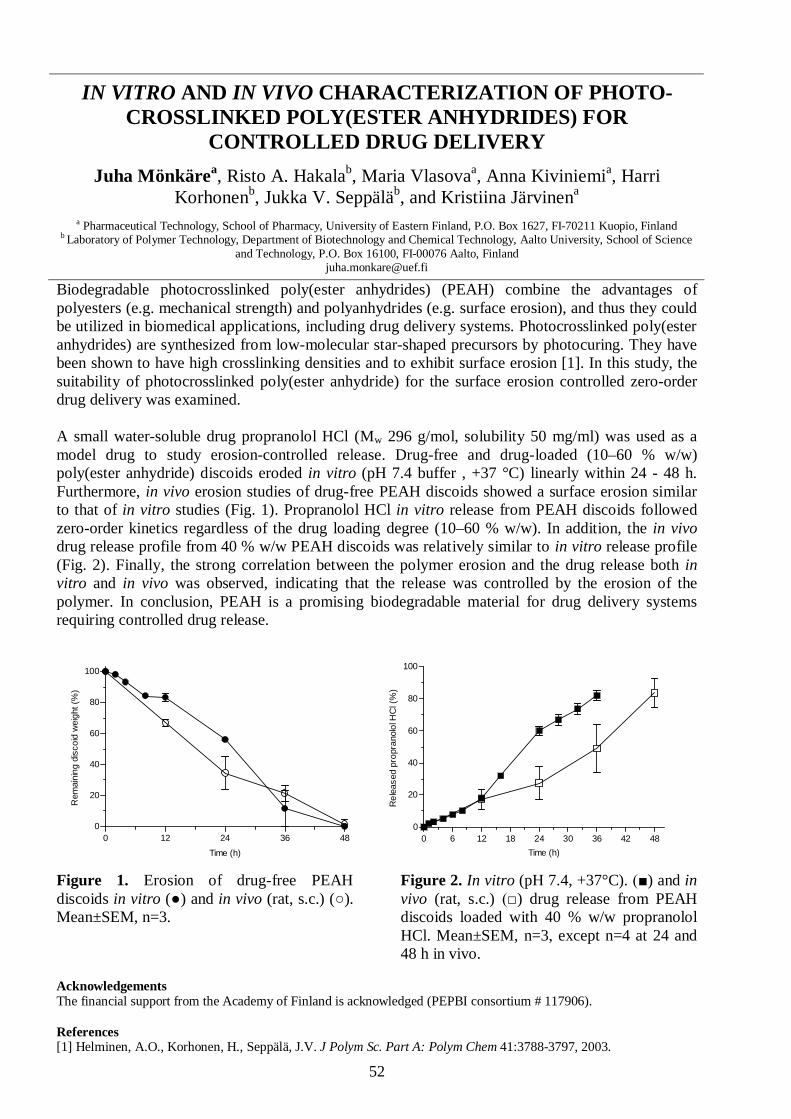
52
IN VITRO AND IN VIVO CHARACTERIZATION OF PHOTO-CROSSLINKED POLY(ESTER ANHYDRIDES) FOR
CONTROLLED DRUG DELIVERYJuha Mönkärea, Risto A. Hakalab, Maria Vlasovaa, Anna Kiviniemia, Harri
Korhonenb, Jukka V. Seppäläb, and Kristiina Järvinena
a Pharmaceutical Technology, School of Pharmacy, University of Eastern Finland, P.O. Box 1627, FI-70211 Kuopio, Finlandb Laboratory of Polymer Technology, Department of Biotechnology and Chemical Technology, Aalto University, School of Science
and Technology, P.O. Box 16100, FI-00076 Aalto, [email protected]
Biodegradable photocrosslinked poly(ester anhydrides) (PEAH) combine the advantages ofpolyesters (e.g. mechanical strength) and polyanhydrides (e.g. surface erosion), and thus they couldbe utilized in biomedical applications, including drug delivery systems. Photocrosslinked poly(esteranhydrides) are synthesized from low-molecular star-shaped precursors by photocuring. They havebeen shown to have high crosslinking densities and to exhibit surface erosion [1]. In this study, thesuitability of photocrosslinked poly(ester anhydride) for the surface erosion controlled zero-orderdrug delivery was examined.
A small water-soluble drug propranolol HCl (Mw 296 g/mol, solubility 50 mg/ml) was used as amodel drug to study erosion-controlled release. Drug-free and drug-loaded (10–60 % w/w)poly(ester anhydride) discoids eroded in vitro (pH 7.4 buffer , +37 °C) linearly within 24 - 48 h.Furthermore, in vivo erosion studies of drug-free PEAH discoids showed a surface erosion similarto that of in vitro studies (Fig. 1). Propranolol HCl in vitro release from PEAH discoids followedzero-order kinetics regardless of the drug loading degree (10–60 % w/w). In addition, the in vivodrug release profile from 40 % w/w PEAH discoids was relatively similar to in vitro release profile(Fig. 2). Finally, the strong correlation between the polymer erosion and the drug release both invitro and in vivo was observed, indicating that the release was controlled by the erosion of thepolymer. In conclusion, PEAH is a promising biodegradable material for drug delivery systemsrequiring controlled drug release.
0 12 24 36 480
20
40
60
80
100
Time (h)
Rem
aini
ng d
isco
id w
eigh
t (%
)
0 6 12 18 24 30 36 42 480
20
40
60
80
100
Time (h)
Rel
ease
d pr
opra
nolo
l HC
l (%
)
Figure 1. Erosion of drug-free PEAHdiscoids in vitro ( ) and in vivo (rat, s.c.) ( ).Mean±SEM, n=3.
Figure 2. In vitro (pH 7.4, +37°C). ) and invivo (rat, s.c.) ) drug release from PEAHdiscoids loaded with 40 % w/w propranololHCl. Mean±SEM, n=3, except n=4 at 24 and48 h in vivo.
AcknowledgementsThe financial support from the Academy of Finland is acknowledged (PEPBI consortium # 117906).
References[1] Helminen, A.O., Korhonen, H., Seppälä, J.V. J Polym Sc. Part A: Polym Chem 41:3788-3797, 2003.
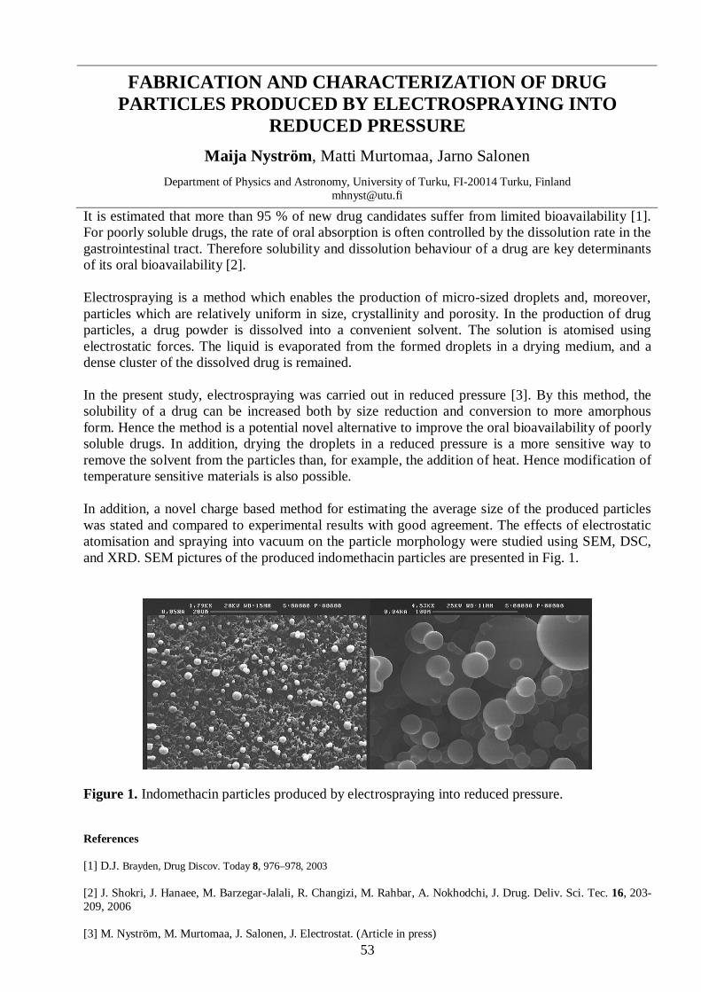
53
FABRICATION AND CHARACTERIZATION OF DRUGPARTICLES PRODUCED BY ELECTROSPRAYING INTO
REDUCED PRESSUREMaija Nyström, Matti Murtomaa, Jarno Salonen
Department of Physics and Astronomy, University of Turku, FI-20014 Turku, [email protected]
It is estimated that more than 95 % of new drug candidates suffer from limited bioavailability [1].For poorly soluble drugs, the rate of oral absorption is often controlled by the dissolution rate in thegastrointestinal tract. Therefore solubility and dissolution behaviour of a drug are key determinantsof its oral bioavailability [2].
Electrospraying is a method which enables the production of micro-sized droplets and, moreover,particles which are relatively uniform in size, crystallinity and porosity. In the production of drugparticles, a drug powder is dissolved into a convenient solvent. The solution is atomised usingelectrostatic forces. The liquid is evaporated from the formed droplets in a drying medium, and adense cluster of the dissolved drug is remained.
In the present study, electrospraying was carried out in reduced pressure [3]. By this method, thesolubility of a drug can be increased both by size reduction and conversion to more amorphousform. Hence the method is a potential novel alternative to improve the oral bioavailability of poorlysoluble drugs. In addition, drying the droplets in a reduced pressure is a more sensitive way toremove the solvent from the particles than, for example, the addition of heat. Hence modification oftemperature sensitive materials is also possible.
In addition, a novel charge based method for estimating the average size of the produced particleswas stated and compared to experimental results with good agreement. The effects of electrostaticatomisation and spraying into vacuum on the particle morphology were studied using SEM, DSC,and XRD. SEM pictures of the produced indomethacin particles are presented in Fig. 1.
Figure 1. Indomethacin particles produced by electrospraying into reduced pressure.
References
[1] D.J. Brayden, Drug Discov. Today 8, 976–978, 2003
[2] J. Shokri, J. Hanaee, M. Barzegar-Jalali, R. Changizi, M. Rahbar, A. Nokhodchi, J. Drug. Deliv. Sci. Tec. 16, 203-209, 2006
[3] M. Nyström, M. Murtomaa, J. Salonen, J. Electrostat. (Article in press)

54
CANCER CELL TARGETING AND INTRACELLULARDELIVERY OF HYDROPHOBIC AGENTS USING
MESOPOROUS HYBRID SILICA AS CARRIER SYSTEMSJessica M. Rosenholm 1,#, Emilia Peuhu 2,3,#, Laurel Tabe Bate-Eya 2,3,
John E. Eriksson 2,3, Cecilia Sahlgren 2,3,*, Mika Lindén 1,*
1Center for Functional Materials, Department of Physical Chemistry, Åbo AkademiUniversity, Porthansgatan 3-5, FI-20500, Turku, Finland;
2Department of Biology, Åbo Akademi University, Artillerigatan 6A, FI-20520 Turku, Finland;3Turku Centre for Biotechnology, University of Turku and Åbo Akademi University, P.P. Box 123, FI-20521, Turku,
Finland#Equal contribution, *Equal contribution, to whom correspondence should be addressed: [email protected],
Mesoporous hybrid silica nanoparticles functionalized by surface hyperbranchingpolymerization of poly(ethylene imine), PEI, were further modified by introducing both fluorescentand targeting moieties, with the aim of specifically targeting cancer cells. Owing to the highabundance of folate receptors in many cancer cells as compared to normal cells, folic acid was usedas the targeting ligand. The internalization of the particles and intracellular delivery of model drugcargo in cell lines expressing different levels of folate receptors was studied.
The presented results show that the total number of particles internalized by the cancercells expressing folate receptors is about an order of magnitude higher than the total number ofparticles internalized by normal cells expressing low levels of the receptor; a difference highenough to be of significant biological importance. Targeted nanoparticle-mediated intracellulardelivery is demonstrated using two hydrophobic fluorophores as model drug cargo. The presentedhybrid carrier system exhibits both cancer cell-targeting ability and capacity to retain a hydrophobicagent with subsequent specific release into the endosomal compartment. Furthermore, theincorporated agent is shown to be able to escape from the endosomes into the cytoplasm, makingthe particles promising candidates as carriers for targeted drug delivery for cancer treatment.
Our results also demonstrate the use of the anti-folate drug, methotrexate (MTX), bothas a nanoparticle targeting ligand and an apoptosis-inducing agent for selective cancer cellelimination. MTX-functionalized nanoparticles induced apoptosis preferably in tumor originatingHeLa cells compared to normal epithelial 293 cells whereas both cell types were equally sensitiveto free MTX. These results present a potential concept for cancer therapy with selective MTX-induced apoptosis in folate receptor expressing tumors.
Figure 1. FITC-conjugated mesoporous hybrid silica nanoparticlesin the cytoplasm of a cancer cell (confocal microscopy image, 40xobjective).
References
[1] Rosenholm JM, Meinander A, Peuhu E, Niemi R, Eriksson JE, Sahlgren Cand Lindén M (2009): Targeting of porous hybrid silica nanoparticles to cancercells. ACS Nano 3:197–206.
[2] Rosenholm JM, Peuhu E, Eriksson JE, Sahlgren C and Lindén M(2009):Targeted Intracellular Delivery of Hydrophobic Agents usingMesoporous hybrid Silica Nanoparticles as Carrier Systems. Nano Letters9:3308–3311.

55
MICRO-ELECTROENCAPSULATION OF POROUS SILICONNANOPARTICLES FOR CONTROLLED ORAL DRUG
DELIVERY APPLICATIONSJorma Roinea,b, Matti Murtomaaa, Jarno Salonena
aLaboratory of Industrial Physics, Department of Physics, University of Turku, FI-20014 Turku, FinlandbGraduate School of Materials Research, Turku, Finland
Where possible, the oral route is often preferred for drug administration due to comfort of usage anda well controlled drug release rate as opposed to, for example, intravenous administration.However, many potential drug molecules possess poor pharmacokinetic properties, such as poorsolubility, dissolution of the drug in the intestinal lumen, poor permeation in the gastrointestinal(GI) tract, or high intestinal or hepatic first pass metabolism. Hence the bioavailability of the drugwill be inadequate when administered orally, resulting in poor efficiency [1-4].
During the past decade, the strengths of porous silicon (PSi) as a potential drug carrier mediumhave been shown. Drugs can be loaded to the pores of PSi nanoparticles in room temperaturesolvents, enabling the usage temperature sensitive drugs such as peptides and hormones. The poreproperties are easily adjustable. Nanoparticles are able to penetrate the blood-brain barrier, makingdelivery of drugs to brain tumors possible. But in general, nanoparticles have a strong tendency toform agglomerates, making them very difficult to handle or dose [1,5-7].
This presentation discusses the potentials of micro-electroencapsulation of drug loaded PSinanoparticles in order to enhance the oral drug delivery process. Experimental settings andpreliminary results are presented. The PSi particles are captured inside a polymer shell by the meansof electrohydrodynamic spraying. A solution containing the shell material and a dispersion(solution) containing the PSi particles are electrosprayed from two separate capillary nozzles, keptat opposite potentials. The sprayed droplets of opposite charges collide, forming (when immiscible)a capsule with the droplet of higher surface tension as the core. With proper choice of solvents andmaterials, the core of the polymer capsule shell will remain a liquid dispersion of PSi particles or aform a solid polymer matrix with embedded PSi particles. [8,9].
The presented work is aimed at eventually helping to overcome fore discussed problems in PSi drugdelivery applications. Our goal is to produce and study a variety of nanostructured micro-scalecomposite drug particles by micro-electroencapsulation. Such composite particles would have thebenefits of the properties of encapsulated PSi nanoparticles in targeted drug delivery, the shieldingproperties of the selected capsule material and good workability due to the size scale. With propermaterial choices, the structure would enable oral administration, safe delivery and controlled releaseof loaded PSi particles in a selected part of the GI tract, unaffected by the digestive metabolism.
References[1] J. Salonen, A. M. Kaukonen, J. Hirvonen, V.-P. Lehto, J. Pharm. Sc. 97, 632 (2008).[2] M. Koulu, J. Tuomisto (editors), Farmakologia ja Toksikologia, 6th edition, s. 81-84, Medicina Oy, Kuopio, 2001.[3] S. S. Davis, L. Illum, Int. J. Pharm. 176, 1 (1998).[4] D. J. Brayden, Drug Disc Today 8, 976 (2003).[5] E. J. Anglin, L. Cheng, W. R. Freeman, M. J. Sailor, Adv. Drug. Del. Rev. 60, 1266 (2008).[6] P. Horcajada, A. Ramila, J. Perez-Pariente, M. Vallet-Regi, Micropor. Mesopor. Mater. 68, 105 (2004).[7] M. Vallet-Regi, Chem. Eur. J. 12, 5934 (2006).[8] A. Jaworek, A.T. Sobczyk, J. Electrost. 66, 197 (2008).[9] A. Jaworek, Powder Tech. 176, 18 (2007).

56
TABLET FORMULATIONS OF MESOPOROUS SILICONT Rotkoa, E Mäkiläb, H Santosa, T Laaksonena, J Salonenb, JHirvonena, L Peltonena
a Division of Pharmaceutical Technology, Faculty of Pharmacy, FI-00014, University of Helsinki, Finlandb Laboratory of Industrial Physics, University of Turku, FI-20014 Turku, Finland
Mesoporous silicon (PSi) is a promising material to enhance the bioavailability of poorly solubledrugs in oral drug delivery. Drug molecules entrapped in the nanosized pores of PSi are retained indisordered form causing increase in the solubility and the dissolution rate of the drug, which hasbeen shown in earlier studies (1).
Indomethacin loaded thermally oxidized PSi-particles (loading degree 16 – 22 w/w %) wereformulated as tablet formulations with excipients (lactose 200 M, cellulose, polyvinylpyrrolidone,croscarmellose sodium, magnesiumstearate and Aerosil 200). Compression of tablets was carriedout in 5 mm mold with eccentric tablet machine (Korsch EK-0, Erweka-Apparatenbau GmbH,Germany). The particle concentrations of tablets were 20, 25, 30 and 35 w/w %. Tablets werecharacterised by measuring crushing strength, thickness and disintegration time (according toPh.Eur., Sotax DT3, Sotax AG, Switzerland). The release of indomethacin from the tablets wasanalysed according to USP paddle method (Erweka DT-D6, Instrulab, Germany) in pH 5.5 buffer atat temperature 37 ± 0.5 °C. Samples from medium were analysed at 318 nm with UV-spectrophotometer (Pharmacia LKB Ultrospec III, Sweden) after filtration with 0.45 µm filter(Sartorius Biotech GmbH, Germany). Comparative dissolution studies with pure indomethacin werecarried out from gelatine capsules.
The results of tablet characterisation studies show that indomethacin-loaded TOPSi-particles can becompressed into tablets with acceptable mechanical properties. Disintegration times forindomethacin loaded TOPSi tablets were under 1 min 40 sec and within the limits given in Ph.Eur.The dissolution of indomethacin from tablet formulations at different drug concentrations wasenhanced and maintained in spite of the tabletting process.
0
50
100
150
200
250
300
0 5 10 15Time (min)
Rel
ease
d am
ount
(%)
25 w - %30 w - %35 w - %Pure indomethacin
Figure 1. The release of indomethacin from TOPSi-particles containing tablet formulations atdifferent drug concentrations compared to the indomethacin release from gelatine capsules.
AcknowledgementsBoehringer Ingelheim Pharma GmbH is acknowledged for financial support of this study.
References [1] Salonen J, Laitinen L, Kaukonen AM, Tuura J, Björkqvist M, Heikkilä T, Vähä-Heikkilä K, Hirvonen J, Lehto V-P.J Control. Release 108: 362 – 374, 200

57
CELLULAR RESPONSES TO POROUS SILICON MICRO- ANDNANOPARTICLES
Hélder A. Santosa,*, Luis M. Bimboa, Jarno Salonenb, Ermei Mäkiläb, JoakimRiikonenc, Teemu Heikkiläb, Timo Laaksonena, Leena Peltonena, Vesa-Pekka Lehtoc,
Jouni Hirvonena
a Division of Pharmaceutical Technology, Faculty of Pharmacy, University of Helsinki, FI-00014 Helsinki, Finlandb Laboratory of Industrial Physics, Department of Physics, University of Turku, FI-20014 Turku, Finland
c Department of Physics, University of Kuopio, FI-70211 Kuopio, Finland*[email protected]
Inorganic nanomaterials have been actively investigated recently, especially in areas like imagingand drug delivery applications [1–4]. These materials are thought to improve the efficiency ofcancer therapy and to provide efficient and user-friendly administration of other activepharmaceutical ingredients. For example,many potential molecules cannot bedelivered in oral form due to their poordissolution/solubility and/or pharmaco-kinetic properties in the intestinal lumen,and poor permeation properties in the GItract. Porous silicon (PSi) materials haveseveral advantages over the existingmaterials for drug delivery, overcomingmost of the abovementioned problems indrug delivery of poorly soluble drugmolecules [2–4]. Beside the beneficialfunctionalities of the nanomaterials, whichstem from their reduced size, also safetyissues need to be assessed at the cellularlevel before they can be implemented inhuman therapeutics [1]. In the presentstudy the in vitro interactions of micro- andnanosized PSi materials with humanintestinal carcinoma-derived cells (Caco-2)and RAW 264.7 mouse macrophage cellswere evaluated (Figure 1). In general, itwas shown that the 1–25 µm PSi-particles reduced cell viability more than the PSi nanoparticles.No significant production of reactive oxygen species was observed for the PSi nanoparticles. TheTHCPSi nanoparticles did not permeate significantly through the Caco-2 monolayers. These NPsalso interacted strongly with the RAW264.7 macrophages, but the significant cell uptake wasprevented due to the surface properties of the particles. Our investigation demonstrated that bytuning the size and surface chemistry of the PSi particles, different cellular responses can beinduced, which can then be further used to enhance drug delivery in safe and efficient manner.
AcknowledgementsThe financial support from the Academy of Finland (grants no. 122314 and 127099) and the University of HelsinkiResearch Funds are acknowledged.References[1] Santos HA, Riikonen J, Salonen J, Heikkilä T, Mälkiä E, Laaksonen T, Peltonen L, Lehto V-P, Hirvonen J. ActaBiomater. 2010 (in press).[2] Salonen J, Kaukonen AM, Hirvonen J, Lehto V-P. J. Pharm. Sci. 97: 632–653, 2008.[3] Kaukonen AM, Laitinen L, Salonen J, Tuura J, Heikkilä T, Limnell T, Hirvonen J, Lehto V-P. Eur. J. Pharm.Biopharm. 66: 348–356, 2007.[4] Salonen J, Laitinen L, Kaukonen AM, Tuura J, Bjorkqvist M, Heikkilä T, Vaha-Heikkila K, Hirvonen J, Lehto V-P.J. Control. Release 108: 362–374, 2005.
Caco-2 monolayers in the presence of fluorescentlylabeled THCPSi nanoparticles (NPs) of 170 nm, c =100 g/ml (a) and c = 15 g/ml.
Figure 1. (a) UNA wascovalently attached tothe THCPSi NPs, afterwhich FTIC was boundto the activated THCPSisurface. (b) Confocalmicroscopy z-stackspictures of differentiated
(b)

58
INVESTIGATION OF THE POWDER FLOW BEHAVIOUR OFBINARY MIXTURES OF PARACETAMOL AND
MICROCRYSTALLINE CELLULOSESoppela Ia, Hatara Ja, Airaksinen Sb, Räikkönen Ha, Yliruusi Ja, Sandler Nb
a Division of Pharmaceutical Technology, Faculty of Pharmacy, P.O. Box 56, 00014 University of Helsinki, FinlandbPharmaceutical Sciences Laboratory, Åbo Akademi University, Turku, Finland
The flowability of a powder is an important characteristic influencing several drug manufacturingsteps. Flowability is affected by the physical properties of the powder, such as particle size andshape, and the processing environment [1]. Partly due to the vast amount of factors influencing theflow properties, there is a lack of direct and accurate methods for measuring flowability of powders.Indirect methods, such as the Hausner ratio, have been used for evaluating flowability but it iscommonly acknowledged that they are not accurate. One reason for many direct methods failing toaccurately measure the flow rate of poorly flowing materials is the arching tendency of cohesivepowders [2]. In this study a novel technology for measuring flowability was studied to increase theunderstanding of the flow behaviour of binary mixtures of paracetamol and different grades ofmicrocrystalline cellulose. Also the effect of lubricant on the flow properties of the differentmixtures was investigated.
Binary mixtures of paracetamol and three microcrystalline celluloses (Avicel PH101, 102 and 200)were prepared in controlled conditions (24±1°C, 50±2% RH). The raw materials were conditionedfor three days and sieved with a 1.68 mm sieve prior to mixing. Samples containing Avicel PH101,PH102 or PH200 and 0 to 25% paracetamol were mixed in 100 ml glass containers using Turbula®mixer. The mixing time was ten minutes, after which 0.5 % magnesium stearate was added insidethe powder bed and mixed for two more minutes.
The flowability of the samples was studied using FlowPro technology (SAY Group, Helsinki,Finland) in controlled conditions (24±1°C, 50±2% RH). The FlowPro instrument is composed of amotor, funnel, orifice and analytical scale connected to a computer. The motor moves the funnelvertically and the upward motion breaks the arch formation enabling the powder to flow freelythrough the orifice until the arch structure forms again. This cycle is repeated until the powder flowis complete. Thus, the device measures the tendency of arching of powders and arch strength. Basedon the experimental data, the flow rate of the powder and the shape of the mass function can bedetermined. Also scanning electron microscope images of each sample were taken.
As expected, the flowability of all the binary mixtures of MCC and paracetamol decreased when theamount of paracetamol increased. However, the flow rate depended also on the MCC grade: thesamples containing Avicel PH200 had the best flow properties while the mixtures of PH101 andparacetamol were the poorest flowing. Yet, magnesium stearate was able to increase the flowabilityof all samples. These findings suggest that the FlowPro instrument is able to measure theflowability of powders reliably and accurately.
AcknowledgementsThe Research Foundation of University of Helsinki is acknowledged for financial support of this study.
References[1] Staniforth JN, Powder flow. In: Aulton ME, editor. Pharmaceutics – the science of dosage form design.
London, Great Britain, Churchill Livingstone, pp. 168–179, 2007
[2] Walker DM, An approximate theory for pressures and arching in hoppers. Chem Eng Sci. 21(11): 975-997,1966

59
OPTIMIZATION OF PRODUCTION PROCESS OF PLANANOPARTICLES BY ELECTROSPRAYING TECHNIQUE
H. Valo1, L. Peltonen1, S. Vehviläinen1, T. Laaksonen1, R. Kostiainen2, J. Hirvonen1
1Division of Pharmaceutical Technology, Faculty of Pharmacy, P.O Box 56, FIN-00014, Finland2Division of Pharmaceutical Chemistry, Faculty of Pharmacy, P.O Box 56, FIN-00014, Finland
The aim in this study was to optimize processing parameters for electrospray system developed forpreparation of beclomethasone dipropionate and salbutamol sulfate loaded biodegradable low Mwpoly(l-lactic acid) nanoparticles. Formulation properties, like flow rate, applied voltage and amountof the polymer and electrolyte were altered for producing spherical drug loaded nano-sized particleswith a controllable particle size. Particle size and morphology were studied by photon correlationspectroscopy (PCS), scanning electron microscopy (SEM) and transmission electron microscopy(TEM). Drug loading and drug encapsulation efficiency were determined by UV-spectroscopy andphysical characterizations of the particles were performed with XRPD and DSC.
The size distributions of the particles were relatively narrow with optimized set-ups of electrospray,and the diameters of the nanoparticles could be controlled from ca. 150 nm to 800 nm. Theproperties of the drug substances remained physically unaltered during the process. With theelectrospray method presented, the entrapment efficiencies of optimized salbutamol sulfate andbeclomethasone dipropionate nanoparticles were 54% and 56%, respectively. According to thisstudy [1], electrospray method is a promising technique for drug delivery purposes: particleproperties are easily controlled by process variables and the process works as well for both thehydrophilic and hydrophobic materials.
Figure 1. The electrospray apparatus used for production of nanoparticles
AcknowledgementsFinancial support from University of Helsinki (HENAKOTO project) is acknowledged.
References[1] Valo, H. Peltonen L, Vehviläinen, S. Karjalainen, M. Kostiainen, R., Laaksonen, T. Hirvonen J. Small 5:1791-1798,2008

60
MESOPOROUS SILICON MICROPARTICLES FOR SUSTAINEDPEPTIDE DELIVERY: CARDIOVASCULAR EFFECTS OF
MELANOTAN II IN CONCIOUS RATSMaria Vlasovaa, Joakim Riikonenb, Vesa-Pekka Lehtob, Jarno Salonenc,
Karl-Heinz Herzigd, Kristiina Järvinena
aDepartment of Pharmaceutics, University of Eastern Finland, FI-70211, Kuopio, FinlandbDepartment of Physics, University of Eastern Finland , FI-70211, Kuopio, Finland
cDepartment of Physics, University of Turku, FI-20014 Turku, FinlanddInstitute of Biomedicine, University of Oulu, FI-90014 Oulu, Finland
Clinical usages of the peptides as drugs are hindered by their short duration of actions in vivo.Different types of mesoporous materials as microparticles have been used to improve permeabilityof large molecule drugs and to control drug release.
In the present study, hydrophobic thermally hydrocarbonized mesoporous silicon (THCPSi)microparticles (38–53 m) were loaded with a model peptide, melanotan II (MTII), to determine ifthis technique could prolong duration of the peptide physiological action.
Melanotan II is a potent, unselective peptide antagonist of melanocortin receptors (MCRs), which,among other physiological effects, clearly affects cardiovascular system [1]. Radiotelemetry system(DSI, St Paul, MN) was used for the monitoring of cardiovascular parameters of conscious rats. Theeffect of MTII on heart rate in rats was investigated after subcutaneous administration (3mg/kg).
Using MTII as a model peptide, a high peptide loading degree was obtained in microparticles (15 %w/w). The effect of MTII loaded THCPSi on the heart rate was prolonged, as compared withsubcutaneous MTII administration as a solution (3 mg/kg in rats, s.c.).
The present findings indicate that THCPSi microparticles are suitable for sustained peptidedelivery.
Figure 1. The increase in the heart rate is delayed after THCPSi+MTII treatment as compared with MTII treatment inrats (MTII dose 3mg/kg, s.c.). Treatments: Vehicle = 5 mg/ml CMC in 0.9 % NaCl, THCPSi = unloaded THCPSimicroparticles suspended in vehicle, THCPSi+MTII= MTII loaded THCPSi microparticles suspended in vehicle;MTII= MTII dissolved in vehicle. Data are presented as mean±SEM, (n=6), # p<0.05, ## p<0.01 THCPSi+MTII vs.vehicle; * p<0.05, ** p<0.01 MTII vs. vehicle.
AcknowledgementsThis study was supported by the FinNano program of the Academy of Finland–PEPBI consortium, Pohjois-SavonKulttuurirahasto (MV).
References: [1] Kuo JJ, Silva AA, Hall JE. Hypertension 41: 768-74, 2003.

61
VÄITÖSKIRJOJEN TIIVISTELMÄT

62
PHYSICAL MODIFICATION OF DRUG RELEASECONTROLLING STRUCTURES – HYDROPHOBIC MATRICES
AND FAST DISSOLVING PARTICLESRiikka Laitinen
Kuopio University Publications A. Pharmaceutical Sciences 117, 2009. 138p.
An oral drug delivery system (e.g. tablet or capsule) is required for administration, which must en-sure delivery to the site of absorption in the gastrointestinal (GI) tract and release control of the ac-tive drug substance in a safe, effective and reliable way. However, many drug compounds are eitherineffectively or incompletely absorbed after oral administration. Fortunately, biopharmaceuticalperformance of drug compounds suffering from such limitations can be effectively improved bymodified-release formulation technologies.
The objective of this study was to evaluate different modified-release technologies both for control-ling the drug release properties of a hydrophobic matrix system and for improving the dissolutionproperties of a poorly soluble drug, in order to allow its intraoral delivery, i.e. to formulate an orallyfast disintegrating tablet (FDT). Matrix systems, which allow retarding the drug dissolution fromthe dosage form, are the most commonly used controlled drug delivery dosage forms due to theirrobustness and low production costs. This can beneficial in the case that the required dosing fre-quency is too high to enable once or twice a day administration due to the excessively short phar-macokinetic half-life of the drug. On the other hand, from a FDT the drug releases in the oral cavityand it can be absorbed through the oral mucosa and delivered directly into the systemic circulation,avoiding first pass metabolism, by ensuring that the drug is rapidly released and dissolved in theoral cavity. Another advantage of the intraoral route is the very fast onset of drug action.
First, the ability of hydrophobic starch acetate (SA) and ethyl cellulose (EC) matrices for control-ling the release of water soluble model drugs was studied. In the study, the release properties ofhighly water soluble saccharides were found to be similar with SA and commercially available EC.It was shown that simply by altering tablet porosity and the relative amount of the excipient in thetablet, the release of saccharides could be controlled over a wide time scale. Subsequently, a simpledry powder agglomeration preparation process for drug/SA mixtures was developed. It was obser-ved that changing the organization of the powder mixture by this process, the release rate of watersoluble model drugs from SA matrix and tablet properties could be modified. The extent of thechange in the mixture structure was found to be dependent on the size and the surface roughness ofthe drug particles.
Finally, an extremely fast dissolution rate of a poorly water soluble drug in a small volume of liquid(pH 6.8) was obtained by utilizing a solid dispersion (SD) approach. The amorphous SD with thebest dissolution and stability characteristics was formulated as a FDT. The formulation preparedwith direct compression, underwent fast disintegration and displayed a fast and immediate onset ofthe release of the drug and also possessed sufficient tensile strength.
In conclusion, simple formulation and processing modifications, which do not require anyexpensive and complicated equipment or process stages, or new chemical entities, displayed a greatpotential in controlled modification of release and dissolution of physicochemically diverse drugs.These simple methods may be helpful in solving the future challenges of developing innovativeformulations and dosage forms, e.g. enhancing the drug solubility and dissolution rate of new, morehydrophobic lead molecules that otherwise would have limited biavailabilities. The results can alsobe useful for developing dosage forms for elderly patients and children, two patient groups whosuffer problems in swallowing conventional dosage forms.

63
PARTICLE SIZE DETERMINATION DURING FLUID BEDGRANULATION – TOOLS FOR ENHANCED PROCESS
UNDERSTANDINGTero Närvänen
Dissertationes bioscientiarum molecularium Universitatis Helsingiensis in Viikki, 21/2009, 57 pp.
Fluid bed granulation (FBG) is a widely used process in pharmaceutical industry to improve thepowder properties for tableting. During the granulation, primary particles are attached to each otherand granules are formed. Since the physical characteristics (e.g. size) of the granules have asignificant influence on the tableting process and hence on the end product quality, processunderstanding and control of the FBG process are of great importance. Process understanding canbe created by exploiting the design of experiment studies in well instrumented FBG environment. Inaddition to the traditional process measurements and off-line analytics, modern process analyticaltechnology (PAT) tools enable more relevant real-time process data acquisition during the FBG.
The aim of this thesis was to study different particle size measurement techniques and PAT toolsduring the FBG in order to get a better insight into the granulation process and to evaluatepossibilities for real-time particle size monitoring and control. Laser diffraction, spatial filteringtechnique (SFT), sieve analysis and new image analysis method (SAY-3D) were used as particlesize determination techniques. In addition to the off-line measurement, SFT was also applied in-lineand at-line, whereas SAY-3D was applied on-line. Modelling of the final particle size and theprediction of the particle size growth during the FBG was also tested using partial least squares(PLS).
SFT studies revealed different process phenomena that could also be explained by the processmeasurement data. E.g., fine particles entrapment into the filter bags, blocking of the distributorplate and segregation in FBG were observed. The developed on-line cuvette enabled SAY-3Dimage acquisition and visual monitoring throughout the granulations and it performed well even invery wet conditions. Predictive PLS models for the final particle size could be constructed. Basedon this information, pulsing of the granulation liquid feed was presented as a controlling tool tocompensate for the excessive moisture content during the FBG. A new concept of utilising theprocess measurement data to predict particle size during FBG was also successfully developed. Itwas concluded that the new methods and PAT tools introduced and studied will enable enhancedprocess understanding and control of FBG process.

64
PROLONGED RELEASE STARCH ACETATE MATRIXTABLETS – RELATIONSHIPS BETWEEN FORMULATIONPROPERTIES AND IN VITRO DISSOLUTION BEHAVIOR
Jari PajanderKuopio University Publications A. Pharmaceutical Sciences 121. 2009. 105 p.
A drug compound is most commonly introduced into the systemic plasma circulation by means of asolid oral dosage form due to convenience, robustness and ease of product handling. The utilisationof preparations that release their contents slowly in the gastrointestinal tract, i.e. prolonged releasepreparations, can reduce several undesired effects, such as unnecessarily frequent administration,unwanted side-effects or local irritation. However, the development of a well-designed prolongeddrug release preparation is a challenging task.
The objective of the study was to find suitable methods to control the structure and subsequent drugrelease properties of hydrophobic starch acetate (ds 2.7) matrix tablets, and to relate the structuralproperties with the drug release behaviour. In addition, the functionality of an in vitro drug releasetest which is routinely used in order to ensure the consistency and safety of the preparation wasevaluated.
The studies indicate that the structure of the matrix tablet can be controlled by altering the particlesize fraction of matrix forming hydrophobic excipient or making the tablet more porous. Whenstarch acetate (ds 2.7) powder with an adequately small particle size fraction is used, it can form apercolating network within the tablet. The consistence of a networking matrix in the tablet has agreat significance. A networking matrix of the hydrophilic drug alone leads to immediate tabletdisintegration and rapid drug release. Co-existing percolating networks of drug and excipient resultin surface erosion and highly variable drug release. When the hydrophobic excipient is percolating,tablets maintain their shape and only crack during dissolution tests. Furthermore, when the tabletporosity of the studied SA particle size-hydrophilic drug (caffeine) combinations is increased over20 %, the drug release determining feature changes from a relaxational component into a diffusionalcomponent.
In tablets where the networking matrix is composed of the hydrophobic excipient, the penetratingliquid weakens the internal bonds and initially this causes tablet expansion which is thentransformed into cracking. The cracking increases the drug release rate, since the formation of acrack shortens the length of the diffusion path, increases the effective surface area and lowers thedegree of tortuosity.
The structure of the tablet and parameters affecting it are crucial considering the drug releasemechanism and rate. However, the properties of the drug compound, such as the water solubilityand solubility rate, also contribute to the drug release mechanism and rate. However, in practice, thesituations of the formation of the matrix and drug release can be extremely complicated andknowledge of maximum water solubility and dissolution rate do not describe this processadequately. The results indicate that although compound exhibits adequate maximum watersolubility and solubility rate, other properties, such as the magnitude and location of hydrophilicand hydrophobic areas, can cause significant interactions with other excipients which might not bebeneficial to drug release. These chemical molecular properties cannot be removed by means oftraditional pharmaceutical processes and, thus the properties and nature of the drug compound inquestion need to be comprehensively characterized in order to fully understand and control the drugrelease process.

65
Finally, the results showed that USP paddle method produces relevant data describing the drugrelease of prolonged hydrophobic tablets if the preparation consists of an extremely water solublecompound with homogenous distribution within the matrix tablet. However, in the case of a lesswater soluble compound whose particle size distribution is wide and the consequent drugdistribution is less homogenous, the in vitro test may not produce results which are meaningful. Theobtained results showed that less water soluble compound clearly concentrated at the bottom edgeof the tablet in contact with the dissolution vessel, although the poor hydrodynamic properties of theUSP paddle method were considered to play a important part in this observation. Thus, the in vitrodissolution test should be chosen extremely carefully for prolonged release preparations or theexisting test should be modified when the drug compound is not highly water soluble and thepreparation is a hydrophobic polymer based matrix tablet.

66
THE CACO-2 CELL LINE IN STUDIES OF DRUG METABOLISMAND EFFLUX
Sanna SiissaloDissertationes bioscientiarum molecularium Universitatis Helsingiensis in Viikki, 11/2009, 60 pp.
Preclinical prediction of intestinal drug absorption is a continuous challenge in drug development.The absorption of a drug from the gastrointestinal tract is a complicated process, involving passivemembrane permeability parameters as well as many active transport and metabolism components.Therefore, well characterised and reliable in vitro methods for studying drug absorption areconstantly devised and under refinement. In this work, the Caco-2 cell line, a widely used model forintestinal drug absorption, was assessed as a platform to study the interplay of phase II metabolismand MRP (multidrug resistance associated protein) -mediated efflux. Expression and function ofseveral metabolic enzymes and efflux proteins have been observed in the intestine and many drugconjugates produced by UDP-glucunorosyltransferases (UGTs) and sulfotransferases (SULTs) aresubstrates for the apical MRP2 and/or the basolateral MRPs. The kinetics of these interactions iscomplex, but the human origin and intestinal-like differentiation under appropriate cultureconditions appoint Caco-2 cells as a potential platform for these studies.
The Caco-2 cell line studied was thoroughly characterised with regards to different efflux proteinsand UGT enzymes. The expression and functionality of MDR1 (multidrug resistance protein 1, P-glycoprotein) and several MRP proteins as well as UGT enzymes were observed in the studiedcells, while the expression of SULTs and GSTs (glutathione-S-transferases) have been previouslyreported by other groups working with Caco-2. In fully differentiated Caco-2 monolayers theexpression of most MRPs and UGTs was significantly higher compared to less differentiated cellsgrown for shorter periods or in flasks, an important observation with direct implications for thesensitivity and specificity of higher throughput cell-based screening assays. Other factors such asthe passage number of the cells and the use of inducers also affected the mRNA expression levels.
Based on the observed efflux and phase II metabolism activities, a Caco-2 based screening methodwas developed for compounds interacting with MRP2, either directly or via their phase IImetabolites. The kinetics of these interactions were investigated more closely in permeabilityexperiments, where conjugation of model compounds and the efflux of their metabolites(indomethacin glucuronide, paracetamol sulfate and naphthol glucuronide) were detected. Substrateor product inhibition of the UGT enzyme(s) was evident at higher 1- naphthol concentrations,whereas the complementary role of basolateral efflux proteins was observed at the highestindomethacin concentration as the apical efflux was saturated. Pharmacokinetic modelling could beutilized as a tool for further interpretation of the results. The combined results of these studies go along way in improving our understanding of the Caco-2 cell line and its suitability as a modelsystem for drug absorption and metabolism in the intestine.

67
PRO GRADUT 2008

68
HELSINGIN YLIOPISTO
Anna Halenius Atomivoimamikroskopian käyttö farmaseuttisessa materiaali-tutkimuksessa.Farmasian teknologia
Hilkka Ylinärä Puristustutkimukset ja puristuksen aikainen Ramanspektroskopia. Farmasianteknologia
Anne Kumpu- EU GMP:n ja standardin SFS-EN 15013585:2003 erot laatujärjestelmänHuhtala vaatimusten suhteen sekä lääkevalmisteen muuttaminen CE-merkityksi
terveydenhuollon laitteeksi ja tarvikkeeksi, Farmasian teknologia
Maria Fernandez Ihmisen hPEPT1-kuljetinproteiini substraattien seulontamenetelmän validointi.Biofarmasia
Elina Järvinen Aktiivisen kuljetuksen tutkiminen in vivo. Sarveiskalvon aktiivisen japassiivisen visen läpäisyn tietokonemallintaminen. Biofarmasia
Heini Kari An investigation of combined pH- and bacterially-triggered oral colon targeteddrug delivery system. Farmasian teknologia
Emilia Galli Encapsulated, genetically engineered cells in the treatment of Parkinson’sdisease. Biofarmasia
KUOPION YLIOPISTO
Kolju Outi Sekoittumattoman vesi- ja limakerroksen vaikutus imeytymiseen in vivo ja invitro. Biofarmasia
Toljamo Kirsi Bentsodiatsepiinien käyttö lapsipotilailla: Syklodekstriinikompleksoinninvaikutus midatsolaamin sublinguaaliseen imeytymiseen. Farmaseuttinenkemia
Järvinen Maiju Dissoluutiokokeiden merkitys teollisessa kiinteiden peroraalistenlääkemuotojen tutkimuksessa, tuotekehityksessä ja valmistuksessa.Farmasian teknologia
Kaasinen Raisa Amorfisen materiaalin stabiloiminen. Farmasian teknologia
Paavola Leila Terahertsispektroskopian farmaseuttisia sovelluksia - Lääkeaineenvapautumisen mallintaminen tärkkelysasetaattitableteista Stella-ohjelmalla.Farmasian teknologia

69
TURUN YLIOPISTO, TEOLLISUUSFYSIIKAN LABORATORIO
Martti Kaasalainen Valonsirontatekniikat huokoisen piin nanopartikkelien karakterisoinnissa
Pekka Jukantupa Kuivajauheen siirtämisestä etenevän sähköstaattisen aallon avulla
Rosita Päivärinne Niukkaliukoisten lääkeaineiden adsorptiotehokkuuden parantamisestahuokoiseen piihin
Maija Nyström Sähköstaattisella atomisaatiolla tuotettujen partikkelien valmistus jakarakterisointi

70
PAINALLUS VILLAISELLA
Anne JuppoTeollisuusfarmasian professori, Helsingin yliopisto
Alla oleva artikkeli julkaistiin pääkirjoituksena Polymorfissa II/1993. Sen on kirjoittanut
Polymorfin silloinen päätoimittaja Anne Juppo 26.10.1993.
Ollessani Marseillessa IUPACin Characterization of Porous Solids –kongressissa tapasin
sakasalaisen arvokkaan vanhan herran, joka oli perehtynyt adsorption historiaan. Häneltä sain tietää,
että ensimmäinen adsorptiokoe on rekisteröity Raamattuun. Uteilaisuuttani otin selvää lähemmin
asiasta. Raamatussa koe on esitetty seuraavasti:
Gideon sanoi Jumalalle: ”Osoita minulle, että todella teet minusta Israelin pelastajan, kuten olet
luvannut. Minä tuon vastakerittyä villaa puimatantereelle. Jos huomisaamuna villassa on kastetta,
mutta maan pinta on kuiva, minä, tiedän että sinä teet minusta Israelin pelastajan, kuten olet
luvannut”.
Kun Gideon varhain seuraavana aamuna puristi villoja, hän sai puserretuksi kastetta kokonaisen
maljallisen. Silloin Gideon sanoi Jumallalle: ”Ethän vihastu minuun, jos vielä tämän kerran puhun
sinulle. Anna minun tehdä vielä yksi koe. Tällä kertaa pysykööt villat kuivina, vaikka maa niiden
ympärillä on kasteesta märkä”.
Ja Jumala teki sinä yönä niin. Villat olivat kuivia, vaikka maa oli kauttaaltaan kasteesta märkä (1).
Tämän ”kokeen” ilmiöt on myös selitetty tieteellisesti (2). Villalla on erittäin huono lämmön
johtokyky, joten sen lämpötila voi olla kastepisteen alapuolella, vaikka ympäröivän maan lämpötila
on vielä kastepisteen yläpuolella. Villakuitujen ominaispinta-ala huomioon ottaen 10-kiloinen
villamäärä voi kerätä itseensä 7 kiloa vettä. Koska villakuidut ovat hydrofobisia, vesi tiivistyy ja
kerääntyy villamassan sisäosiin, jolloin se on suojassa haihtumiselta. Villat säilyvät kosteina,
vaikka ympäröivässä maassa oleva kosteus haihtuu aamuauringossa. Ensimmäinen testi on siis
helposti selitettävissä.
Toinen koe on myös selitettävissä. Jossain olosuhteissa maaperän kosteus diffundoituu ylöspäin
ilmakehään maapinnan kautta. Eli on mahdollista että toisessa kokeessa maasta nouseva vesihöyry

71
kondensoitui jäähtyneille kasveille ja kiville, muttei pystynyt nousemaan maasta villamassan läpi.
Normaali kaste syntyy ilman kosteudesta, kun on tietyt olosuhteet: kirkas taivas, lähellä maanpintaa
olevissa ilmakerroksissa on suuri kosteuspitoisuus ja tuuli on kohtalainen (1-3 m/s). Mikä tahansa
muutos, esimerkiksi tuulen laantuminen, näissä olesuhteissa voi aiheuttaa sen että normaalia
kastetta ei synny. Kuitenkin kasvien lehdillä voi olla vesipisaroita, jotka kondesoituvat maasta
nousevasta vesihöyrystä, eli ns. väärä kaste syntyy. Villat pysyvät lähes kuivina, koska vesihöyry ei
pysty nousemaan maanpinnasta. Vaikka jonkin verran ”väärä kastetta” adsorboituisi villan pintaan,
sen määrä on kymmenesosa ensimmäisessä kokeessa adsorboituneesta oieasta kasteesta ja sekin
imeytyy vilamassan sisäosiin, ja pinta tuntuu kuivalta.
Eli ensimmäisellä kerralla Gideon odotti aamulla niin pitkään ,että kaste oli maasta ehtinyt kuivua
jolloin villat olivat oikeasta kasteesta litimärkiä. Seuraavalla kerralla jokin sääolosuhdeiden muutos
aiheutti sen, että oikeaa kastetta ei syntynyt ja Gideon tuli toetamaan villojen kuivuuden, kun
aurinko ei ollut vielä ehtinyt kuivata maata.
Vesihöyryn adsorptiokokeilla on siis pitkä ja ylväs historia.
Kirjallisuutta
(1) Raamattu, Tuomarien kirja, 6.luku, jakeet 36-40.(2) Giles, C.H., Gideon’s fleece test. The earliest recorded vapor phase adsorption experiment?.
J.Chem.Education 39 (1962) 584-585.

72
Moving science forward
Delivering the tools to shape your solution.
© 2
009
Ther
mo
Fish
er S
cien
tific
Inc.
All
righ
ts re
serv
ed.
Thermo Scientific products andservices meet all of your applicationneeds, from blood banking to food safetyto proteomics and stem cell research.
Rely on us for the broadest selection of quality laboratory equipment
and supplies. We serve more than 350,000 companies worldwide
in the pharmacuetical, biotechnology, environmental and healthcare
industries, as well as leading universities, diagnostic laboratories
and research institutes.
Our proven brands deliver excellence and innovation in:
• Centrifugation
• Biological safety cabinets
• Liquid handling
• Microplate instrumentation
• Water purification
• Incubation
To learn more, visit www.thermo.com
• Cold storage
• Chromatography columns and supplies
• Plastic labware
• Laboratory furnitureand fume hoods
THERMO FISHER SCIENTIFIC OYRatastie 201620 Vantaawww.thermofisher.com
tiedustelut: [email protected] tilaukset: puh. 09-3291 [email protected]
LabSolutions_A4_1109:Layout 1 11/13/09 3:46 PM Page 1

73
???? PÄHKINÄ ????
Tämän vuoden aivopähkinä on Polymorfin edeltäjässä jäsentiedotteessa 2/1992 julkaistun
yhdistyksen kunniajäsenen Ensio Laineen tekemän aivopähkinän näköis-uusinta. Kaikessa
yksinkertaisuudessaan aivopähkinä on seuraavanlainen:
Kysymyksen triviaalisuutta arvuuteltiin vuoden 1993 Symposiumissa Turussa, ja todettiin että
kysymys yksinkertaisuudestaan huolimatta ei ole aivan triviaali. Tällä kertaa ei kysellä, vaan saatte
päätellä pähkinän triviaalisuus-asteen itse. Ratkaisu esitetään Polymorfissa I/1993 sivulla 8 ja
Polymorfissa I/2010 sivulla 76.
74
Jaakko Aaltonen University of Eastern Finland
Hanna Arstila Åbo Akademi University
Abdul Basit London School of Pharmacy, United Kingdom
Lotta Bergman Åbo Akademi University
Luis Bimbo University of Helsinki
Kirsi Collan Thermo Fisher Scientific
Henrik Ehlers University of Helsinki
Tuomas Ervasti University of Eastern Finland
Susanne Fagerlund Åbo Akademi University
Linda Fröberg Åbo Akademi University
Outi Harkki VTT Technical Research Centre of Finland
Teemu Heikkilä University of Turku
Petteri Heljo University of Helsinki
Samuli Hirsjärvi University of Angers, France
Arto Huhtala Thermo Fisher Scientific
Leena Hupa Åbo Akademi University
Mika Jokinen DelSiTech Ltd.
Julian Jones Imperial College London, United Kingdom
Harri Jukarainen Bayer Schering Pharma
Martti Kaasalainen University of Turku
Kirsi Katila Hormos Medical
Ari Kauppinen University of Eastern Finland
Raija Koivuniemi Orion Pharma
Ruzica Kolakovic University of Helsinki
Timo Laaksonen University of Helsinki
Johanna Laru Orion Pharma
Vesa-Pekka Lehto University of Eastern Finland
Laura Leimu Orion Pharma
Tarja Limnell Orion Pharma
Markus Linder VTT Technical Research Centre of Finland
Kimmo Lähteenkorva ConMed Linvatec Biomaterials Ltd.
Fysikaalisen farmasian XXI symposiumOSALLISTUJAT

75
Carita Martin Turku Science Park Ltd.
Jonathan Massera Åbo Akademi University
Matti Murtomaa University of Turku
Ermei Mäkilä University of Turku
Juha Mönkäre University of Eastern Finland
Jouko Nieminen PANalytical B.V.
Maija Nyström University of Turku
Marja Partanen Orion Pharma
Liu Peng University of Helsinki
Emilia Peuhu Åbo Akademi University
Juhani Posti Bayer Schering Pharma
Thomas Rades University of Otago, New Zealand
Joakim Riikonen University of Eastern Finland
Jorma Roine University of Turku
Kirsi Rosenqvist University of Helsinki
Tanja Rotko University of Helsinki
Marko Saalasti Orion Pharma
Kirsti Saarnivaara Orion Pharma
Jarno Salonen University of Turku
Niklas Sandler Åbo Akademi University
Hélder A. Santos University of Helsinki
Anna Shevchenko Orion Pharma
Ira Soppela University of Helsinki
Mikko Tenho University of Turku
Hanna Valo University of Helsinki
Bert van Veen Orion Pharma
Marja-Riitta Viljainen Turku Science Park Ltd.
Maria Vlasova University of Eastern Finland
Marju Väkiparta Orion Pharma
Di Zhang Åbo Akademi University

76
PÄHKINÄN RATKAISU
Pähkinän ratkaisussa lähtökohtana on kaasujen yleinen tilanyhtälö nRTpV = . Kaavassa n on
moolien lukumäärä, eli tarkasteltava kaasumassa jaettuna moolimassalla. Näin ollen kaava voidaan
esittää muodossa RTMmpV = . Kaasun tiheydelle saadaan
RTpM
Vm
==ρ . Kostean ilman
kokonaispaine pkok muodostuu kuivan ilman osapaineesta pi ja vesihöyryn osapaneesta ph. Mi ja Mv
ovat ilman ja veden moolimassat.
Ilman tiheys lasketaan seuraaavasti:
vh
ii M
RTpM
RTp
+=ρ
vh
ihkok M
RTpM
RTpp
+−
=ρ
−−=
kok
h
i
vii
kok
pp
MMM
MRTp
1ρ
−=
kok
hi
kok
pp
MRTp
379,01ρ .
Näin ollen, saman ilmanpaineen pkok vallitessa ilman tiheys on sitä pienempi mitä suurempi on ph,
eli mitä kosteampaa ilma on. Kiitos Ensio Laineelle ja Polymorfin toimituskunnalle vuosina 1992
ja 1993!


The Analytical X-ray Company
X’PERT PRO
New applications for nanomaterials
10
5
0
0 10 20
GobsGcalc
30 40 50 60
-5
G
r
10 20 30 40 50 60 70 80 90 100 110 120 130
2Theta [deg.]
0
2500
10000
22500
40000
62500
Inte
nsi
ty [
cou
nts
]
Experimental dataEmpty container
Typical PDF result. Sample: SiC.Data collected using Ag radiation (λ = 0.5594 Å)
Typical measurement of sample and background
New application for the X’Pert PRO MPD• Long- and short-range ordering• For amorphous and nanocrystalline
materials
Standard X-ray sources and detectors• Mo and Ag tubes compatible
with X’Pert PRO
Available for every X’Pert PRO MPD• Data quality allows for pair
distribution function (PDF) analysis
• Additional components for featureless instrument background
Preparation for synchrotron beam time• Direct access at the home lab
PANalytical, your partner in modern materials analysis!
New applications for nanomaterials
New application for the X’Pert PRO MPD• Long- and short-range ordering• For amorphous and nanocrystalline
materials
Standard X-ray sources and detectors• Mo and Ag tubes compatible
with X’Pert PRO
Available for every X’Pert PRO MPD• Data quality allows for pair
distribution function (PDF) analysis
• Additional components for featureless instrument background
Preparation for synchrotron beam time• Direct access at the home lab
PANalytical, your partner in modern materials analysis!
For more information, please contact:
PANalytical B.V., Branch FinlandNikkarinkuja 5FIN-02650 ESPOOT +358 9 2212 580F +358 9 2212 585
PN7242.indd 1 07-12-2009 09:56:05