plasma cell disorders e tak1 is a pivotal therapeutic
TRANSCRIPT

haematologica | 2021; 106(5) 1401
Received: August 7, 2019.
Accepted: April 2, 2020.
Pre-published: April 9, 2020.
©2021 Ferrata Storti Foundation
Material published in Haematologica is covered by copyright.All rights are reserved to the Ferrata Storti Foundation. Use ofpublished material is allowed under the following terms andconditions: https://creativecommons.org/licenses/by-nc/4.0/legalcode. Copies of published material are allowed for personal or inter-nal use. Sharing published material for non-commercial pur-poses is subject to the following conditions: https://creativecommons.org/licenses/by-nc/4.0/legalcode,sect. 3. Reproducing and sharing published material for com-mercial purposes is not allowed without permission in writingfrom the publisher.
Correspondence: JUMPEI [email protected]
MASAHIRO [email protected]
Haematologica 2021Volume 106(5):1401-1413
ARTICLEPlasma Cell Disorders
https://doi.org/10.3324/haematol.2019.234476
Ferrata Storti Foundation
Along with tumor progression, the bone marrow microenviron-ment is skewed in multiple myeloma (MM), which underlies theunique pathophysiology of MM and confers aggressiveness and
drug resistance in MM cells. TGF-b-activated kinase-1 (TAK1) mediatesa wide range of intracellular signaling pathways. We demonstrate herethat TAK1 is constitutively overexpressed and phosphorylated in MMcells, and that TAK1 inhibition suppresses the activation of NF-κB,p38MAPK, ERK and STAT3 in order to decrease the expression of criti-cal mediators for MM growth and survival, including PIM2, MYC, Mcl-1, IRF4, and Sp1, along with a substantial reduction in the angiogenicfactor VEGF in MM cells. Intriguingly, TAK1 phosphorylation was alsoinduced along with upregulation of vascular cell adhesion molecule-1(VCAM-1) in bone marrow stromal cells (BMSC) in cocultures withMM cells, which facilitated MM cell-BMSC adhesion while inducingIL-6 production and receptor activator of nuclear factor κ-B ligand(RANKL) expression by BMSC. TAK1 inhibition effectively impairedMM cell adhesion to BMSC to disrupt the support of MM cell growthand survival by BMSC. Furthermore, TAK1 inhibition suppressed osteo-clastogenesis enhanced by RANKL in cocultures of bone marrow cellswith MM cells, and restored osteoblastic differentiation suppressed byMM cells or inhibitory factors for osteoblastogenesis overproduced inMM. Finally, treatment with the TAK1 inhibitor LLZ1640-2 markedlysuppressed MM tumor growth and prevented bone destruction and lossin mouse MM models. Therefore, TAK1 inhibition may be a promisingtherapeutic option targeting not only MM cells but also the skewedbone marrow microenvironment in MM.
TAK1 is a pivotal therapeutic target for tumorprogression and bone destruction in myelomaJumpei Teramachi,1,2 Hirofumi Tenshin,2,3 Masahiro Hiasa,2,3 Asuka Oda,2Ariunzaya Bat-Erdene,2,4 Takeshi Harada,2 Shingen Nakamura,2
Mohannad Ashtar,2,3 So Shimizu,2,3 Masami Iwasa,2 Kimiko Sogabe,2
Masahiro Oura,2 Shiro Fujii,2 Kumiko Kagawa,2 Hirokazu Miki,5 Itsuro Endo,6Tatsuji Haneji,1 Toshio Matsumoto7 and Masahiro Abe2
1Department of Histology and Oral Histology, Tokushima University Graduate School,Tokushima, Japan; 2Department of Hematology, Endocrinology and Metabolism,Tokushima University Graduate School, Tokushima, Japan; 3Department of Orthodonticsand Dentofacial Orthopedics, Tokushima University Graduate School, Tokushima, Japan;4Department of Immunology and Laboratory Medicines, School of Biomedicine,Mongolian National University of Medical Sciences, Ulaanbaatar, Mongolia; 5Division ofTransfusion Medicine and Cell Therapy, Tokushima University Hospital, Tokushima,Japan; 6Department of Chronomedicine, Institute of Biomedical Sciences, TokushimaUniversity Graduate School, Tokushima, Japan and 7Fujii Memorial Institute of MedicalSciences, Tokushima University, Tokushima, Japan
ABSTRACT
Introduction
Multiple myeloma (MM) has a unique propensity to almost exclusively developin the bone marrow and generate devastating bone destruction. MM cells enhanceosteoclast (OC) formation and activity, and suppress osteoblastic differentiationfrom bone marrow stromal cells (BMSC), leading to extensive bone destructionwith rapid development of osteolytic lesions.1,2 Angiogenesis is also enhancedthrough these cellular interactions.3,4 The types of cells surrounding MM cells cre-ate a cellular microenvironment suitable for MM cell growth and survival to con-fer drug resistance, which can be termed the “MM niche”.

In order to improve therapeutic efficacy, we need todisrupt the MM niche that confers drug resistance.Therefore, we looked for novel molecules upregulated inthe MM niche to be targeted, and found that proviralintegrations of Moloney virus 2 kinase (PIM2) is constitu-tively overexpressed as an anti-apoptotic mediator inMM cells.5 PIM2 expression has been demonstrated to behigher in hematologic malignancies than solid cancers ortheir normal tissue counterparts, and highest in MM.6,7Importantly, PIM2 can be further upregulated in MM cellsin cocultures with BMSC as well as OC.5Although multiple soluble inhibitors for osteoblastoge-
nesis have been reported to be overproduced in MM,including IL-3,8 IL-7,9 TNF-α,10 TGF-b,11 and activin A,12PIM2 was found to be upregulated in BMSC by theseinhibitory factors acting as a common intracellular medi-ator to suppress their osteoblastogenesis.13 Moreover, wesubsequently reported that PIM2 is induced in osteoclas-tic lineage cells by receptor activator of nuclear factor κ-Bligand (RANKL) to act as a critical mediator of RANKL-induced osteoclastogenesis in MM.14 Therefore, PIM2appears to play a versatile role in tumor progression andbone destruction and bone loss in MM, and is regarded asan important therapeutic target.TGF-b-activated kinase 1 (TAK1) is a member of the
mitogen-activated protein kinase kinase (MAP3K) family,also known as MAP3K7.15,16 It was originally identified asa key kinase in transducing TGF-b signaling down to p38mitogen-activated protein kinase (MAPK) and c-Jun andN-terminal kinase (JNK).15 Subsequently, TAK1 has beendemonstrated to be associated with the activation of awide range of intracellular signaling pathways importantfor various cellular functions, including the activation ofnuclear factor-κB (NF-κB) and extracellular signal-regulat-ed kinase (ERK).15 Therefore, TAK1 appears to be a gatekeeper to facilitate the multiple important intracellularsignaling pathways. Therapeutic efficacy of TAK1 inhibi-tion has been preclinically demonstrated in differenttypes of cancers, including mantle cell lymphoma,17breast cancer,18 and colon cancer.19We demonstrate here that TAK1 is constitutively over-
expressed and phosphorylated in MM cells and thatTAK1 acts as an upstream regulator responsible for mul-tiple signaling pathways critical for MM growth and sur-vival.1 TAK1 phosphorylation is also induced in BMSC incocultures with MM cells, which facilitates MM celladhesion to BMSC between very late antigen-4 (VLA-4)and vascular cell adhesion molecule-1 (VCAM-1), therebyinducing IL-6 production and RANKL expression byBMSC. Importantly, TAK1 inhibition was able to effec-tively induce MM cell death, and alleviate bone destruc-tion though suppression of osteoclastogenesis andrestoration of osteoblastogenesis. Therefore, TAK1appears to be a pivotal therapeutic target in MM to dis-rupt the key signal transduction pathways responsible fortumor progression and bone destruction.
Methods
Ethics All procedures involving human samples from healthy donors
and patients were performed with written informed consent inaccordance with the Declaration of Helsinki and a protocolapproved by the Institutional Review Board for human protec-
tion at the University of Tokushima (Permission number: 240).All animal experiments were conducted under the regulationand permission of the Animal Care and Use Committee ofTokushima University, Tokushima, Japan (toku-dobutsu 13094).
ReagentsReagents used in this manuscript are described in the Online
Supplementary Appendix.
Cells and cell cultureDetails of the cells and cell culture procedures are available in
the Online Supplementary Appendix.
Western blottingCells were collected and lysed in RIPA lysis buffer (Santa
Cruz, Dallas, TX, USA). For cytosolic and nuclear preparation,cells were lysed in NE-PER extraction reagent (Thermo FisherScientific, Waltham, MA) in accordance with the manufacturer'sprotocol. Western blot analysis was done with equal proteinamounts of cell lysate, as described previously.13
Cell viabilityCell viability was determined using the Cell Counting Kit-8
assay (Dojindo, Kumamoto, Japan) in accordance with the man-ufacturer’s instructions. The absorbance of each well was meas-ured at 450-655 nm using an iMark microplate reader (Bio-RadLaboratories, Hercules, CA, USA). In order to assess apoptoticcells, cells were stained with an annexinV-FITC and propidiumiodide labeling kit (MEBCYTO Apoptosis Kit; MBL, Nagano,Japan) in accordance with the manufacturer’s instruction, andanalyzed by flow cytometry.
Small interfering RNA transfectionSmall interfering RNA (siRNA) transduction was performed as
described previously.5,20 Human and mouse TAK1 siRNA werepurchased from Santa Cruz. Human TAK1 siRNA was transfect-ed into MM cells using a Human Nucleofector Kit (Lonza, Basel,Switzerland). Mouse TAK1 siRNA was transfected into mouseBMSC or RAW264.7 cells using siRNA Transfection Reagent(Santa Cruz) in accordance with the manufacturer’s protocol.
Real-time reverse transcription polymerase chain reactionRNA isolation and quantitative real-time reverse transcription
polymerase chain reaction (RT-PCR) were performed asdescribed previously.20 The following primer sequences wereused: human RANKL F: TCGTTGGATCACAGCACATCA andR: TATGGGAACCAGATGGGATGTC, human IL-6 F: TCT-GAGGCTCATTCTGCCCTCGAGC and R: AACTGGACC-GAAGGCGCTTGTGGA, human GAPDH, used as an endoge-nous control to normalize each sample, F: TGTCTTCACCAC-CATGGAGAAGG and R: GTGGATGCAGGGATGAT-GTTCTG
Adhesion assaysAdhesion assays were performed as described previously.21
Human BMSC (2×10⁴ cells/well) were expanded in 96-well cul-ture plates. MM cells were labeled with BCECF-AM (Dojindo)for 2 hours at 37°C and 5% CO2 as described previously.
22
BMSC were washed, and the fluorescence-labeled MM cellswere added onto the BMSC and incubated for 4 hours.Nonadherent BCECF-AM-labeled cells were removed by gentlypipetting four times. Adherent cells were quantitated in a fluo-rescence multi-well plate reader (Infinite® 200 PRO, TECAN,Männedorf. Switzerland).
J. Teramachi et al.
1402 haematologica | 2021; 106(5)
Multiple myeloma animal model and histological analysesMultiple myeloma (MM) mouse models were prepared by
intra-tibial inoculation of mouse luciferase-transfected 5TGM1MM cells (a gift from Dr. Gregory R. Mundy [Vanderbilt Centerfor Bone Biology, Vanderbilt University, Nashville, TN, USA]) intoICR nu/nu mice (CLEA Japan) at 4–6 weeks old as described pre-viously.13,14 The assessment of tumor growth and bone volume,and bone histomorphometric and immunohistochemical analysesare described in the Online Supplementary Appendix.
Statistical analysisStatistical analysis was performed using Student's t-test or one-
way analysis of variance (ANOVA). P<0.05 was considered as asignificant difference. All statistics were performed using theStatistical Package for Social Sciences (SPSS 13.0 for Windows;Chicago, IL, USA).
Results
TAK1 is activated to mediate growth and survival ofmultiple myeloma cellsWe first examined the expression and activation status
of TAK1 in MM cells. TAK1 is highly overexpressed andphosphorylated in MM cell lines and primary MM cellsfrom patients, whereas normal peripheral blood mononu-clear cells (PMBC) only weakly expressed TAK1 (Figure1A). IRF4 and MYC have been regarded as master regula-tors for MM cell survival and function, which MM cellsare addicted to, and their reduction is a major mechanismfor the anti-MM activity of immunomodulatory drugslenalidomide and pomalidomide.23,24 Notably, the TAK1inhibitor LLZ dose-dependently reduced MYC and IRF4 inparallel with the reduction in PIM2 expression and thephosphorylation of the PIM2 substrate 4E-BP1 as well asthe anti-apoptotic factor Mcl-1 in MM cells (Figure 1B,left). We and others reported that transcription factor Sp1is constitutively overexpressed in MM cells, which canserve as an important therapeutic target for MM.25-27 TAK1inhibition also substantially reduced Sp1 (Figure 1B, left).The effects of TAK1 inhibition were confirmed with TAK1knockdown using siRNA (Figure 1B, right). TAK1 inhibition with LLZ dose-dependently induced
cell death in all MM cell lines tested (Figure. 1C, left), andTAK1 knockdown also reduced the viability of MM cells(Figure 1C, right). The induction of apoptosis was con-firmed using annexinV and propidium iodide dual staining(Figure 1D) with activation of caspase-8, caspase-9, andcaspase-3 (Figure 1E) in MM cells, indicating activation ofthe extrinsic as well as the intrinsic caspase-mediatedapoptotic pathways. LLZ at the concentrations up to 10 mM did not apparently impair the viability of normalPBMC (Online Supplementary Figure S1). Therefore, TAK1appears to be a good therapeutic target to effectivelyinduce cell death in MM cells through suppression ofPIM2 plus other pro-survival mediators.
TAK1 inhibition is able to abolish IL-6 and TNF-α-induced signaling in multiple myeloma cellsIL-6 and TNF-α are predominant paracrine factors over-
produced in the bone marrow microenvironment in MM,and these elicit the signaling pathways responsible forMM cell growth and survival.28 As we demonstrated pre-viously,5 PIM2 was substantially upregulated in MM cell
lines in cocultures with BMSC as well as in the presenceof IL-6 or TNF-α (Figure 2A). However, treatment withLLZ was able to abolish PIM2 upregulation in MM cells byBMSC as well as IL-6 or TNF-α. We further examined theeffects of TAK1 inhibition on the signaling pathways inMM cells activated by IL-6 and TNF-α. After starvingwithout serum, phosphorylation of TAK1 was reduced inRPMI8226 cells, but IL-6 (Figure 2B) and TNF-α (Figure2C) promptly induced the phosphorylation of TAK1 andactivation of their corresponding downstream signalingmolecules in serum-depleted media. Treatment with LLZabolished IL-6-induced phosphorylation of STAT3 (Figure2B), and TNF-α-induced phosphorylation and degradationof IκBα and phosphorylation of p38MAPK and ERK(Figure 2C) in the MM cells. Analyses of nuclear extractsfrom MM cells treated with TNF-α showed the nuclearaccumulation of p65; however, treatment with LLZreduced the p65 content in the nuclear extracts in the pres-ence of TNF-α (Figure 2D). Consistent with the notionthat PIM2 is transcriptionally upregulated in MM cells bythe NF-κB signaling pathway,5 the PIM inhibitor SMI-16ashowed only marginal effects on the nuclear accumulationof p65 induced by TNF-α (Figure 2D). Of note, the TAK1inhibitor LLZ as well reduced MM cell viability in cocul-tures with BMSC (Figure 2E). TAK1 knockdown in MMcells mostly reduced their viability even in cocultures withBMSC (Figure 2F). Also, TAK1 knockdown in BMSC par-tially but significantly reduced their supportive activity forMM cell growth (Figure 2G). Therefore, TAK1 activationin both MM cells and BMSC is suggested to play animportant role in MM cell growth and survival in cocul-tures with BMSC. Furthermore, although cytotoxic effectsof doxorubicin on MM cells were blunted in cocultureswith BMSC, TAK1 inhibition with LLZ was able toresume MM cell death by doxorubicin even in the pres-ence of BMSC (Online Supplementary Figure S2). Theseresults suggested that TAK1 inhibition impairs MM cellgrowth and survival supported by the bone marrowmicroenvironment.
TAK1 inhibition impairs multiple myeloma cell adhesion to bone marrow stromal cellsMM cell adhesion to BMSC through the interaction
between VLA-4 and its corresponding ligand, VCAM-1, isamong the predominant mechanisms for cell adhesion-mediated drug resistance (CAM-DR) in MM,22,29 whileenhancing the production of IL-630 and RANKL, an criticalosteoclastogenic factor in MM.31 In addition to TAK1 acti-vation in MM cells, TAK1 was found to be phosphorylat-ed along with PIM2 upregulation in BMSC, when cocul-tured with MM cells (Figure 3A). Furthermore, VCAM-1expression was substantially upregulated in BMSC aftercoculturing with MM cells, which was abolished by TAK1inhibition with LLZ (Figure 3B). TNF-α is known as apotent inducer of VCAM-1 in BMSC through activation ofthe NF-κB signaling pathway.32,33 Treatment with LLZ aswell as TAK1 knockdown by siRNA abrogated the upreg-ulation of VCAM-1 expression in BMSC by TNF-α(Figures 3B and 3C). Treatment with TNF-α promptlyphosphorylated TAK1 and degraded IκBα, and inducedthe phosphorylation of p38MAPK and ERK in BMSC(Figure 3D). However, TAK1 inhibition with LLZ as wellas TAK1 knockdown abolished the degradation of IκBαand reduced the phosphorylation of p38MAPK and ERK inthe presence of TNF-α, indicating efficacious suppression
TAK1 inhibition in myeloma
haematologica | 2021; 106(5) 1403

J. Teramachi et al.
1404 haematologica | 2021; 106(5)
Figure 1. TAK1 is activated to mediate growth and survival of multiple myeloma cells. (A) TAK1 expression and phosphorylation in multiple myeloma (MM) cell lines.TAK1 expression and phosphorylation were examined by western blotting analysis in various MM cell lines and primary MM cells as well as normal peripheral bloodmononuclear cells (PBMC). Glyceraldehyde 3-phosphate dehydrogenase (GADPH) was used as a loading control. (B) The indicated MM cell lines were cultured in theabsence or presence of LLZ at 1 or 5 mM for 2 days (left). The cells were transduced with scrambled (siCTL) or human TAK1 small interfering RNA (siRNA) (siTAK1)and cultured for 13 hours (right). Cell lysates were then collected, and protein levels of the indicated molecules were analyzed by western blotting analysis. b-actinwas used as a loading control. (C) MM cell lines as indicated were incubated in triplicate in the presence of the indicated concentrations of TAK1 inhibitor LLZ1640-2 (LLZ) for 48 hours (left). The indicated MM cell lines transduced with scrambled (siCTL) or human TAK1 siRNA (siTAK1) were cultured in triplicate for 24 hours(right). Cell viability was measured using a WST-8 assay. Data are expressed as means ± standard deviation. (D) The indicated MM cell lines and CD138-positivecells isolated from MM patients (Pt #1 and Pt #2) were cultured in the presence or absence of LLZ at 3 mM for 24 hours. The induction of apoptosis was analyzedusing annexinV and propidium iodide (PI) dual staining. (E) The indicated MM cell lines were cultured alone or in the presence of LLZ at 1 or 5 mM for 2 days, andcell lysates were then collected. Caspase-mediated apoptotic pathways were analyzed using western blotting analysis. b-actin was used as a loading control.
A
B
C
D E

TAK1 inhibition in myeloma
haematologica | 2021; 106(5) 1405
Figure 2. TAK1 inhibition abolishes IL-6- and TNF-α-induced signaling in multiple myeloma cells. (A) Human bone marrow stromal cells (BMSC) were expanded in24-well culture plates. The indicated multiple myeloma (MM) cell lines were cultured alone or cocultured with BMSC (upper), or cultured in the presence or absenceof IL-6 (lower left) or TNF-α (lower right) for 24 hours (h), and cell lysates were then collected. PIM2 expression was analyzed using western blotting. b-actin was blot-ted as a loading control. (B, C) RPMI8226 cells were cultured in α-MEM with 1% FBS for 12 h for serum starvation. The starved cells were then cultured in α-MEMwith 1% fetal bovine serum (FBS) with or without LLZ1640-2 (LLZ) at 3 mM. Three hours later, IL-6 (b) or TNF-α (c) at 10 ng/mL was added. After the indicated timeperiods, cell lysates were collected. The expression of phosphorylated TAK1 (p-TAK1), TAK1, phosphorylated STAT3 (p-STAT3), STAT3, phosphorylated IκBα (p-IκBα),IκBα, phosphorylated p38MAPK (p-p38), p38MAPK (p38), phosphorylated ERK (p-ERK), and ERK were detected using western blotting. b-actin was used as a loadingcontrol. (D) RPMI8226 cells after starvation were cultured in α-MEM with 1% FBS with or without LLZ (5 mM) or the PIM inhibitor SMI-16a (50 mM). Three hours later,TNF-α at 10 ng/mL was added as indicated. After incubating for 15 minutes, whole lysates and nuclear fractions were extracted. The protein levels of p65 were ana-lyzed using western blotting. Glyceraldehyde 3-phosphate dehydrogenase (GADPH) and p84 were used as protein loading controls for cytoplasmic and nuclear pro-teins, respectively. (E) The indicated MM cell lines were cultured alone or cocultured with human BMSC expanded in 24-well culture plates for 24 hours in the pres-ence or absence of LLZ at 5 mM. MM cells were then collected, and their viability was analyzed using a WST8 assay. (F) The indicated MM cell lines were transducedwith scrambled (siCTL) or TAK1 siRNA (siTAK1), cultured alone or cocultured with human BMSC expanded in 24-well culture plates for 24 hours. MM cells were thencollected, and their viability was analyzed using a WST8 assay. (G) Indicated MM cells were cocultured for 24 hours with or without human BMSC which were trans-duced with scrambled (siCTL BMSC) or TAK1 siRNA (siTAK1 BMSC). MM cells were then collected, and their viability was analyzed using a WST8 assay.
A
B
E
F
C D
G
of TNF-α signaling in BMSC by TAK1 inhibition. VLA-4,α4 b1 integrin, is constitutively overexpressed in MMcells. We found that the TAK1 inhibition with LLZ wasable to reduce the expression of b1 integrin in MM cells(Online Supplementary Figure S3), indicating the contribu-tion of TAK1 to b1 integrin expression in MM cells.Consistently, treatment with LLZ suppressed MM celladhesion to BMSC (Figures 3E; Online Supplementary FigureS4), although LLZ did not impair the viability of BMSC(Online Supplementary Figure S5).BMSC are regarded as a major source of IL-6, a growth
and survival factor for MM cells, and the critical osteoclas-togenic factor RANKL. Consistent with the previousobservations,30,31 cocultures with MM cells potently aug-mented IL-6 (Figures 3F) and RANKL (Figures 3G) mRNAexpression in BMSC. However, treatment with LLZ sup-pressed the upregulation of these factors in BMSC incocultures with MM cells. These data demonstrate thatTAK1 inhibition is able to efficaciously suppress MM celladhesion to BMSC and thereby abolish the upregulationof IL-6 and RANKL in BMSC, which may alleviate MMtumor progression in the bone marrow and bone destruc-tion.
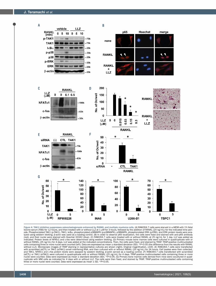
TAK1 inhibition suppresses osteoclastogenesisenhanced by RANKL and multiple myeloma cellsConsistent with previous observations,20,34 RANKL
induced the phosphorylation of TAK1 in parallel with thedegradation of IκBα and phosphorylation of p38MAPKand ERK (Figure 4A), and nuclear localization of the NF-κBsubunit p65 (Figure 4B) in RAW264.7 preosteoclastic cells.However, treatment with LLZ abolished all of theseRANKL-mediated changes, indicating critical involvementof TAK1 in RANKL-induced activation of the NF-κB andMAPK pathways. RANKL induced the expression ofNFATc1 and c-fos, critical transcription factors for osteo-clastogenesis (Figure 4C), and the formation of TRAP-pos-itive multinucleated cells, namely OC, in RAW264.7 cells(Figures 4D); however, treatment with LLZ dose-depen-dently suppressed the RANKL-induced expression ofNFATc1 and c-fos, and OC formation. TAK1 knockdownby siRNA also abolished the induction of NFATc1 and c-fos expression (Figure 4E) and osteoclastogenesis (Figure4F) by RANKL. Furthermore, MM cells potently inducedTRAP-positive multinucleated OC formation from bonemarrow cells; however, treatment with LLZ suppressedOC formation (Figure 4G). These results demonstrate thatTAK1 inhibition is able to suppress osteoclastogenesisenhanced by MM cells.
TAK1 inhibition restores osteoblastogenesis suppressed by multiple myeloma cells as well as majorinhibitors for osteoblastogenesis in multiple myelomaIn contrast to the enhanced osteoclastogenesis,
osteoblastogenesis or bone formation is suppressed inMM. Conditioned media (CM) from MM cell lines as wellas inhibitory factors for osteoblastogenesis overproducedin MM, including IL-3, IL-7, TNF-α, TGF-b, and activinA,8-12 induced the phosphorylation of TAK1 (Figure 5A)and suppressed mineralized nodule formation (Figure 5B)in MC3T3-E1 preosteoblastic cells. However, treatmentwith the TAK1 inhibitor LLZ restored mineralized noduleformation (Figure 5B). Osterix is an essential transcriptionfactor for osteoblastogenesis, known as a downstream tar-get of BMP-2. The upregulation of Osterix by BMP-2 was
reduced in MC3T3-E1 cells in the presence of CM fromMM cell lines or TNF-α; however, treatment with LLZrestored the upregulation of Osterix by BMP-2 (Figure5C). TGF-b inhibits the terminal stage of OB differentiation
or bone mineralization, whereas BMP-2 is a stimulator forosteoblastogenesis.11,35-38 We and others demonstrated thatTGF-b plays a significant role in bone destruction in MM,and that the inhibition of the TGF-b signaling restoredbone formation in MM animal models.11,39-41 Treatmentwith TGF-b induced the phosphorylation of Smad2 andSmad3 in MC3T3-E1 cells (Figure 5D). However, TAK1inhibition with LLZ as well as TAK1 knockdown bysiRNA abolished the phosphorylation of these factors.TGF-b has been shown to counteract the BMP-2 signalingto suppress the terminal differentiation of OB in partthrough the upregulation of Smad6, an inhibitory regula-tor for BMP-2 signaling.42 Treatment with LLZ for 24hours dose-dependently reduced Smad6 protein levels inMC3T3-E1 cells (Figure 5E, upper). Moreover, LLZ inhib-ited TGF-b-induced upregulation of Smad6 in MC3T3-E1cells (Figure 5E, lower). In contrast, treatment with LLZ aswell as TAK1 knockdown by siRNA enhanced the phos-phorylation of Smad1/5 in MC3T3-E1 cells by BMP-2(Figure 5F). These results collectively suggest that TAK1inhibition may resume osteoblastogenesis suppressed inMM.
TAK1 inhibition suppresses vascular endothelial growthfactor secretion by multiple myeloma cellsAngiogenesis also plays an important role in the patho-
genesis and progression of MM. Vascular endothelialgrowth factor (VEGF) appears to be the most criticalangiogenic factor in MM.43,44 VEGF has been demonstratedto be overproduced downstream of the signaling mediatorERK in MM cells.45 As expected, TAK1 inhibition withLLZ as well as TAK1 knockdown with siRNA substantial-ly reduced VEGF production by MM cells (OnlineSupplementary Figure S6). These results suggested thatTAK1 inhibition can impair angiogenesis in MM to retardMM progression.
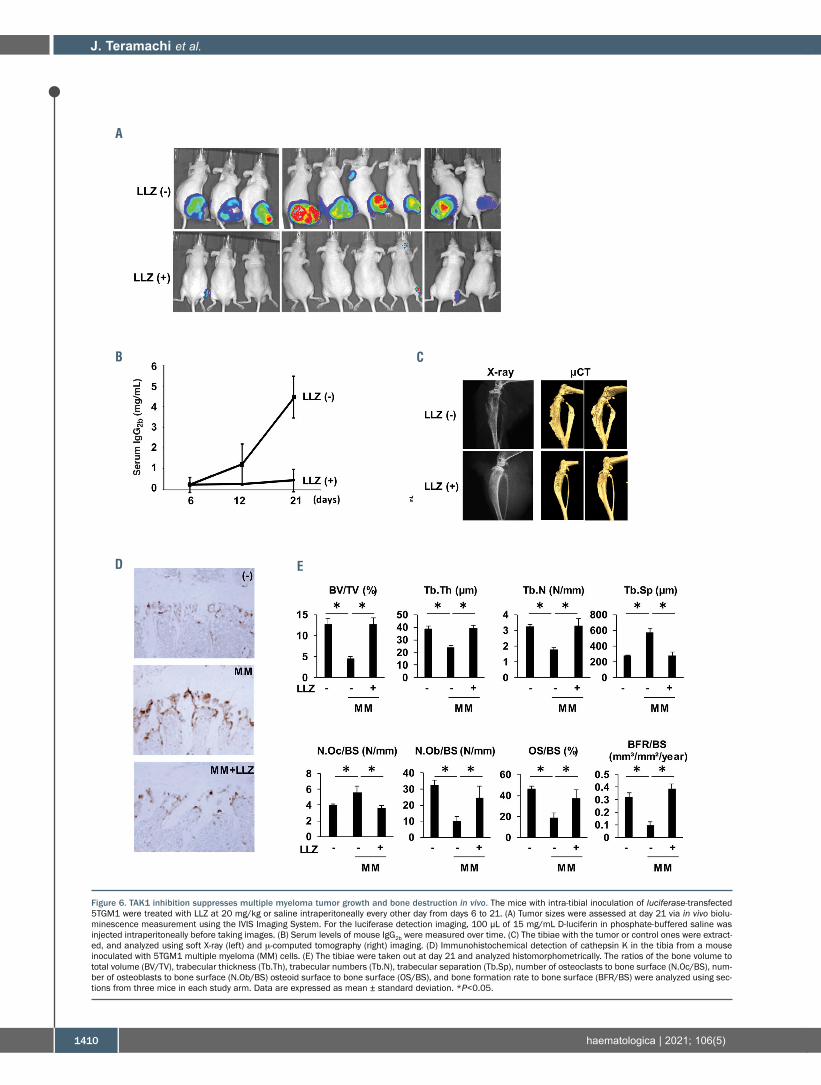
TAK1 inhibition suppresses multiple myeloma tumorprogression and prevents bone destruction in vivoWe next examined the in vivo effects of the TAK1
inhibitor LLZ using MM mouse models by intratibial inoc-ulation of mouse 5TGM1 MM cells. Mice were treatedwith LLZ every other day for 2 weeks from day 6, the dayon which 5TGM1 MM cell-derived IgG2b levels started toincrease in mouse sera. Vehicle-treated mice showed atday 21 large tumor masses around the tibiae where MMcells were inoculated (Figure 6A), and a progressiveincrease in serum IgG2b levels over time (Figure 6B). Bonedestruction in the tibiae was observed at day 21 in plainX-ray as well as m-computed tomography (m-CT) images(Figure 6C). Treatment with LLZ substantially suppressedtumor sizes (Figure 6A) with almost no increase in serumIgG2b (Figure 6B), and prevented bone destruction of thetibiae (Figure 6C). Cathepsin K-expressing OC increasedin number on the surface of bone in 5TGM1-inoculatedtibiae; however, treatment with LLZ reduced the OCnumbers (Figure 6D). These results demonstrate thatTAK1 inhibition is able to suppress MM tumor growthwhile preventing bone destruction in vivo.In order to further clarify the effects of TAK1 inhibition
J. Teramachi et al.
1406 haematologica | 2021; 106(5)

TAK1 inhibition in myeloma
haematologica | 2021; 106(5) 1407
Figure 3. TAK1 inhibition suppresses VCAM-1 expression in bone marrow stromal cells and their adhesion to multiple myeloma cells. (A) Human bone marrow stro-mal cells (BMSC) were expanded in 6-well culture plates. The BMSC were cocultured with the indicated multiple myeloma (MM) cell lines for 24 hours. After washingout MM cells, cell lysates were collected from the BMSC. The indicated protein levels were examined using western blotting. (B) Human BMSC were cultured aloneor cocultured with the indicated MM cell lines, or cultured with TNF-α at 10 ng/mL in the presence or absence of LLZ (5 mM) for 2 days. Cell lysates were collectedfrom BMSC, and VCAM-1 expression was analyzed using western blotting. (C) BMSC were transduced with scrambled (siCTL) or TAK1 small interfering RNA (siRNA)(siTAK1), and then cultured for 2 days with or without TNF-α at 10 ng/mL. Cell lysates were collected, and VCAM-1 expression was analyzed using western blotting.(D) BMSC cells were starved in α-MEM with 1% fetal bovine serum (FBS) for 12 hours. The cells were then cultured in α-MEM with 1% FBS with or without LLZ at 5mM for 3 hours (left), or transduced with scrambled (siCTL) or TAK1 siRNA (siTAK1) (right). TNF-α at 10 ng/mL was added and cells lysates were harvested after theindicated time periods. The indicated protein levels were analyzed using western blotting. (E) Human BMSC were treated with LLZ (5 mM) for 1 day, and the indicatedMM cells were then added in quadruplicate at 105 cells/well, and incubated for 4 hours. By gentle pipetting, non-adherent MM cells were removed, and adherentMM cells were quantitated in a fluorescence multi-well plate reader. Data represent the means ± standard deviation (SD) (n=4). *P<0.05, by ANOVA. (F, G) HumanBMSC prepared in 6-well culture plates were cultured in triplicate alone or cocultured with MM cells as indicated in the presence or absence of LLZ (5 mM) for 1 day.After washing out MM cells, total RNA was isolated from the BMSC. IL-6 (f) and RANKL (g) mRNA expression in the BMSC was determined using quantitative reversetranscription polymerase chain reaction. Data represent the means ± SD (n=3). *P<0.05, by ANOVA.
A
C
E
F
B
D
G
J. Teramachi et al.
1408 haematologica | 2021; 106(5)
Figure 4. TAK1 inhibition suppresses osteoclastogenesis enhanced by RANKL and multiple myeloma cells. (A) RAW264.7 cells were starved in α-MEM with 1% fetalbovine serum (FBS) for 12 hours, and then treated with or without LLZ at 1 mM for 3 hours, followed by the addition of RANKL (20 ng/mL) for the indicated time peri-ods. Phosphorylated TAK1 (p-TAK1), TAK1, IκBα, phosphorylated p38MAPK (p-p38MAPK), p38MAPK, phosphorylated ERK (p-ERK), and ERK protein levels were ana-lyzed using western blotting. b-actin was used as a loading control. (B) In order to observe p65 localization, the cells were fixed and stained with anti-p65 antibody(red), and their nuclei were stained with Hoechst 33342 (blue). (C) RAW264.7 cells were treated with or without RANKL at 25 ng/mL for 1 day. LLZ was added asindicated. Protein levels of NFATc1 and c-fos were determined using western blotting. (D) Primary mouse bone marrow cells were cultured in quadruplicate with orwithout RANKL (25 ng/mL) for 4 days. LLZ was added at the indicated concentrations. Then, the cells were fixed, and stained by TRAP. TRAP-positive multinucleatedcells containing three or more nuclei were counted (left). Data are expressed as mean ± standard deviation (SD). *P<0.05 (the difference from the results with RANKLwithout LLZ). Microscopic images of TRAP staining in representative cultures are shown (right). Original magnification, x100. (E) RAW264.7 cells were transfectedwith scrambled (siCTL) or TAK1 (siTAK1) small interfering RNA, and then cultured with or without RANKL (25 ng/mL) for 24 hours. Cell lysates were then collected,and TAK1, NFATc1 and c-fos protein levels were assayed by western blotting. b-actin served as a loading control. (F) RAW264.7 cells transfected with scrambled(siCTL) or TAK1 (siTAK1) were cultured in quadruplicate with or without RANKL (25 ng/mL) for 4 days. TRAP-positive multinucleated cells containing three or morenuclei were counted. Data were expressed as mean ± standard devaition (SD). *P<0.05. (G) Primary bone marrow cells derived from mice were cocultured in quad-ruplicate with MM cells as indicated for 4 days with or without LLZ. The cells were then fixed, and stained by TRAP. TRAP-positive multinucleated cells containingthree or more nuclei were counted. Data were expressed as mean ± SD. *P<0.05.
A
C
E
G
B
D
F

TAK1 inhibition in myeloma
haematologica | 2021; 106(5) 1409
Figure 5. TAK1 inhibition restores osteoblastogenesis suppressed by multiple myeloma cells as well as major inhibitors for osteoblastogenesis in multiple myelo-ma. (A) Conditioned media from the indicated multiple myeloma (MM) cell lines (MM CM) at 25%, or cytokines including TNF-α (1 ng/ml), TGF-b (10 ng/ml), IL-3 (10ng/ml), IL-7 (10 ng/mL), or activinA (10 ng/mL) were added onto cultures with the MC3T3-E1 cells. After culturing for 24 hours, cell lysates were collected, and phos-phorylated TAK1 (p-TAK1) and TAK1 levels were analyzed using western blotting. β-actin served as a loading control. (B) MC3T3-E1 cells were cultured in the presenceor absence of LLZ (0.3 mM) in osteogenic media with BMP-2 (25 ng/mL) in 24-well culture plates. MM cells CM from the indicated cell lines and primary MM patientwere added at 25%. TNF-α (1 ng/mL), TGF-b (10 ng/mL), IL-3 (10 ng/mL), IL-7 (10 ng/mL), or activinA (10 ng/mL) were added to the indicated wells. After culturingfor 14 days, the cells were fixed and mineralized nodule formation was visualized using Alizarin red staining. (C) MC3T3-E1 cells were cultured for 4 days with MMCM (25%) or TNF-α (1 ng/mL) in the presence or absence of LLZ (0.3 mM) in osteogenic media with BMP-2 (25 ng/mL). Then, cell lysates were collected and Osterix(OSX) expression was assayed using western blotting. (D) MC3T3-E1 cells were starved in α-MEM with 1% FBS for 12 hours, and then treated with or without LLZ at0.3 μM for 3 hours, or transfected with scrambled (siCTL) or TAK1 (siTAK1) small interfering RNA. TGF-b was added at 10 ng/mL, and cell lysates were harvestedafter the indicated time periods. The expression of indicated proteins were analyzed using western blotting. (E) (Upper) MC3T3-E1 cells were treated with LLZ at theindicated concentrations for 12 hours. (Lower) MC3T3-E1 cells were treated with LLZ for 30 minutes prior to adding TGF-b (10 ng/mL), then cultured for 12 hours.The expression of Smad6 protein was analyzed using western blotting. (F) MC3T3-E1 cells were starved with or without LLZ at 0.3 mM for 12 hours, or transfectedwith scrambled (siCTL) or TAK1 (siTAK1) siRNA. BMP-2 was added at 25 ng/mL, and cell lysates were harvested after the indicated time periods. The expression ofindicated proteins were analyzed using western blotting.
A
B
C
D
F
E
J. Teramachi et al.
1410 haematologica | 2021; 106(5)
Figure 6. TAK1 inhibition suppresses multiple myeloma tumor growth and bone destruction in vivo. The mice with intra-tibial inoculation of luciferase-transfected5TGM1 were treated with LLZ at 20 mg/kg or saline intraperitoneally every other day from days 6 to 21. (A) Tumor sizes were assessed at day 21 via in vivo biolu-minescence measurement using the IVIS Imaging System. For the luciferase detection imaging, 100 μL of 15 mg/mL D-luciferin in phosphate-buffered saline wasinjected intraperitoneally before taking images. (B) Serum levels of mouse IgG2b were measured over time. (C) The tibiae with the tumor or control ones were extract-ed, and analyzed using soft X-ray (left) and m-computed tomography (right) imaging. (D) Immunohistochemical detection of cathepsin K in the tibia from a mouseinoculated with 5TGM1 multiple myeloma (MM) cells. (E) The tibiae were taken out at day 21 and analyzed histomorphometrically. The ratios of the bone volume tototal volume (BV/TV), trabecular thickness (Tb.Th), trabecular numbers (Tb.N), trabecular separation (Tb.Sp), number of osteoclasts to bone surface (N.Oc/BS), num-ber of osteoblasts to bone surface (N.Ob/BS) osteoid surface to bone surface (OS/BS), and bone formation rate to bone surface (BFR/BS) were analyzed using sec-tions from three mice in each study arm. Data are expressed as mean ± standard deviation. *P<0.05.
A
B
D
C
E

on bone metabolism in MM, we histomorphometricallyanalyzed bone lesions in the mouse models. In vehicle-treated mice, bone volume over total volume (BV/TV), tra-becular thickness (Tb.Th), trabecular numbers (Tb.N),number of osteoblast surface over bone surface(N.Ob/BS), osteoid surface over bone surface (OS/BS), andbone formation rate over bone surface (BFR/BS) weredecreased, whereas trabecular separation (Tb.Sp) and thenumber of OC surface over bone surface (N.Oc/BS) wereincreased (Figure 6E). However, treatment with LLZimproved these changes in the 5TGM1-inoculated tibiae.These results demonstrate that the TAK1 inhibition notonly suppresses osteoclastic bone destruction but alsorestores osteoid and bone formation in MM bone lesions.In order to further clarify the direct roles of TAK1 inhibi-tion on pathological bone loss without tumor cells, weinvestigated the effects of LLZ on bone loss in ovariec-tomized (OVX) mice. In vehicle-treated OVX mice, boneloss was revealed in m−CT; and BV/TV, Tb.Th and Tb.Nwere decreased, whereas Tb.Sp was increased in bonemorphometric analysis (Online Supplementary Figure S7).However, treatment with LLZ was able to prevent OVX-induced pathological changes, suggesting a protectiveaction of TAK1 inhibition on non-malignant bone loss.
Discussion
Although MM cells perturb bone metabolism with bonedestruction, crosstalk between MM cells and the microen-vironment in bone lesions leads to a progressive viciouscycle of tumor growth and bone destruction. The presentstudy demonstrated that TAK1 plays a critical role in thevicious cycle, and suggested that TAK1 is a pivotal thera-peutic target to disrupt the key signal transduction path-ways responsible for tumor progression and bone destruc-tion in MM. TAK1 activation appears to govern the expres-sion of PIM2 in MM cells and osteoclastic as well asosteoblastic lineage cells; the TAK1-PIM2 signaling path-way is critical for MM tumor expansion and bone destruc-tion. In addition to PIM2 upregulation, TAK1 activationinduced Sp1 expression in MM cells. Sp1 is a ubiquitouszinc-finger transcription factor that binds guanine–cyto-sine-rich elements in the promoter region of its target genesand upregulates various important genes for cancer initia-tion and progression.46,47 Sp1 is known to be constitutivelyoverexpressed in many cancers and is associated with poorprognosis.46 Sp1 expression and its DNA binding activityhas been also demonstrated to be upregulated in MMcells.25We and others reported that inhibition of Sp1 expres-sion with Sp1 siRNA markedly induced apoptosis in MMcells, indicating that Sp1 as a novel therapeutic target forMM.25-27 Our results showed that TAK1 activation con-tributes to Sp1 over-expression in MM cells, and that TAK1inhibition reduces Sp1 expression to impair MM cellgrowth and survival. TAK1 inhibition was found to reduceSp1-mediated IRF4 expression in MM cells. As IMiD havebeen reported to downregulate IRF4 expression thoughdegradation of IKZF1/3,23,24 TAK1 inhibition may synergizethe downregulation of IRF4 expression in combinationwith IMiD. TAK1 was also demonstrated to play a criticalrole in facilitating MM cell-BMSC adhesion via VLA-4-VCAM-1 interaction. TAK1 inhibition reduced VCAM-1expression in BMSC upregulated by MM cells or TNF-α,and impaired MM cell adhesion to BMSC. MM cell-BMSC
adhesion induced IL-6 production and RANKL expressionin BMSC in a manner dependent on TAK1 activation.Given that the adhesion of MM cells to BMSC via VLA-4-VCAM-1 interaction confers CAM-DR in MM cells29,48,49and osteoclastogenesis,50 these results suggested the thera-peutic impact of TAK1 inhibition on CAM-DR as well asosteoclastogenesis induced by the MM-bone marrow inter-action.RANKL plays an important role in osteoclastogenesis
enhanced in MM. As reported previously,34 RANKL inducedthe phosphorylation of TAK1 in RAW264.7 preosteoclasticcells in parallel with the degradation of IκBα and phospho-rylation of p38MAPK and ERK (Figure 4A). However, TAK1inhibition abolished these changes in the RANKL-mediatedintracellular signaling and suppressed the formation ofTRAP-positive OC from bone marrow cells upon treatmentwith exogenous RANKL (Figures 4D, F) as well as in cocul-tures with MM cells (Figure 4G). Together with suppressionof the TAK1-dependent induction of RANKL expression inBMSC (Figure 3G), TAK1 inhibition can reduce osteoclasto-genesis enhanced in MM through blockade of RANKL-mediated signaling in osteoclastic lineage cells. As forosteoblastogenesis, major inhibitors for osteoblastogenesisin MM, including IL-3, IL-7, TNF-α, TGF-b, and activinA, aswell as MM cell CM-induced TAK1 phosphorylation whilesuppressing osteoblastogenesis in MC3T3-E1 preosteoblas-tic cells; however, TAK1 inhibition restored their osteoblas-togenesis (Figures 5A,B). Taken together, these resultsunderscored the value of TAK1 inhibition for preventingprogression of bone destruction and restoring bone forma-tion in MM. Finally, we validated the therapeutic effects ofTAK1 inhibition in vivo. Treatment with LLZ markedly sup-pressed MM tumor growth and prevented bone destructionin mouse MM models with intra-tibial injection of 5TGM1MM cells (Figure 6). Although various bone-modifyingagents have been developed, bone loss still remains a seri-ous unmet issue in patients with MM; bone formation ishard to be restored in MM bone lesions by clinically avail-able anti-resorptive agents, namely zoledronic acid anddenosumab. In contrast to these agents, TAK1 inhibitorsappear to be bone anabolic and anti-resorptive agents withtumor-suppressing activity, which may bring considerablebenefits for patients with malignant diseases exhibitingbone loss, such as MM patients. Moreover, because TAK1inhibition is a novel mechanism of action, combinationwith TAK1 inhibition can improve the therapeutic efficacyof currently available anti-cancer agents while preventingcancer-related and cancer treatment-induced bone loss.Therefore, TAK1 inhibition may be a promising therapeuticoption with anti-tumor and bone modifying action, target-ing the interaction between MM cells and their surroundingmicroenvironment. The present results warrant furtherstudy for development of novel TAK1 inhibitors useful forMM treatment. We are currently synthesizing novel com-pounds with better specificity for TAK1 with less toxicity.Further translational research will elucidate the dividendsthey may yield in improved clinical outcomes.
DislosuresMA received research funding from Chuagai Pharmaceutical,
Sanofi K.K., Pfizer Seiyaku K.K., Kyowa Hakko Kirin, MSDKK, Astellas Pharma, Takeda Pharmaceutical, Teijin Pharmaand Ono Pharmaceutical, and honoraria from Daiichi SankyoCompany. The other authors have no conflicts of interest todeclare.
TAK1 inhibition in myeloma
haematologica | 2021; 106(5) 1411
ContributionsJT and MA designed the research and conceived the project;
PCR was performed by JT, HT, AO and SS; flow cytometry byAO, MH and TH; immunoblotting by JT, HT, MH, AO, AB,TH, SN, MA, SS and MI; transfection by JT, HT, MH, AO andTH; and cell cultures by JT, HT, MH, AO, AB, TH, SN, MA,SS, MI, KS, MO, SF, KK and HM, JT, HT, MH, AO, TH,SN, MH, IE, TH, TM, and MA analyzed the data; JT andMA wrote the manuscript.
FundingThis work was supported in part by JSPS KAKENHI Grant
Numbers JP18K08329, JP16K11504, JP17KK0169,JP18H06294; and Aki Horinouchi Research Grant; JapanLeukemia Research Fund; Yasuda Memorial MedicalFoundation; the Ichiro Kanehara Foundation; and the ResearchClusters program of Tokushima University. The funders had norole in study design, data collection and analysis, decision to pub-lish, or preparation of the manuscript.
J. Teramachi et al.
1412 haematologica | 2021; 106(5)
References 1. Raje N, Roodman GD. Advances in the
biology and treatment of bone disease inmultiple myeloma. Clin Cancer Res. 2011;17(6):1278-1286.
2. Silbermann R, Roodman GD. Myelomabone disease: pathophysiology and man-agement. J Bone Oncol. 2013;2(2):59-69.
3. Tanaka Y, Abe M, Hiasa M, et al. Myelomacell-osteoclast interaction enhances angio-genesis together with bone resorption: arole for vascular endothelial cell growthfactor and osteopontin. Clin Cancer Res.2007;13(3):816-823.
4. Cackowski FC, Anderson JL, Patrene KD,et al. Osteoclasts are important for boneangiogenesis. Blood. 2010;115(1):140-149.
5. Asano J, Nakano A, Oda A, et al. The ser-ine/threonine kinase Pim-2 is a novel anti-apoptotic mediator in myeloma cells.Leukemia. 2011;25(7):1182-1188.
6. Lu J, Zavorotinskaya T, Dai Y, et al. Pim2 isrequired for maintaining multiple myelomacell growth through modulating TSC2phosphorylation. Blood. 2013;122(9):1610-1620.
7. Johrer K, Obkircher M, Neureiter D, et al.Antimyeloma activity of the sesquiterpenelactone cnicin: impact on Pim-2 kinase as anovel therapeutic target. J Mol Med (Berl).2012;90(6):681-693.
8. Ehrlich LA, Chung HY, Ghobrial I, et al. IL-3 is a potential inhibitor of osteoblast dif-ferentiation in multiple myeloma. Blood.2005;106(4):1407-1414.
9.Giuliani N, Colla S, Morandi F, et al.Myeloma cells block RUNX2/CBFA1 activ-ity in human bone marrow osteoblast pro-genitors and inhibit osteoblast formationand differentiation. Blood. 2005;106(7):2472-2483.
10.D'Souza S, del Prete D, Jin S, et al. Gfi1expressed in bone marrow stromal cells is anovel osteoblast suppressor in patientswith multiple myeloma bone disease.Blood. 2011;118(26):6871-6880.
11. Takeuchi K, Abe M, Hiasa M, et al. Tgf-Beta inhibition restores terminal osteoblastdifferentiation to suppress myelomagrowth. PloS One. 2010;5(3):e9870.
12. Vallet S, Mukherjee S, Vaghela N, et al.Activin A promotes multiple myeloma-induced osteolysis and is a promising targetfor myeloma bone disease. Proc Natl AcadSci U S A. 2010;107(11):5124-5129.
13.Hiasa M, Teramachi J, Oda A, et al. Pim-2kinase is an important target of treatmentfor tumor progression and bone loss inmyeloma. Leukemia. 2015;29(1):207-217.
14. Teramachi J, Hiasa M, Oda A, et al. Pim-2is a critical target for treatment of osteoclas-togenesis enhanced in myeloma. Br JHaematol. 2018;180(4):581-585.
15.Mihaly SR, Ninomiya-Tsuji J, Morioka S.TAK1 control of cell death. Cell Death
Differ. 2014;21(11):1667-1676. 16. Sakurai H. Targeting of TAK1 in inflamma-
tory disorders and cancer. TrendsPharmacol Sci. 2012;33(10):522-530.
17. Buglio D, Palakurthi S, Byth K, et al.Essential role of TAK1 in regulating mantlecell lymphoma survival. Blood. 2012;120(2):347-355.
18. Safina A, Ren MQ, Vandette E, Bakin AV.TAK1 is required for TGF-beta 1-mediatedregulation of matrix metalloproteinase-9and metastasis. Oncogene. 2008;27(9):1198-1207.
19. Singh A, Sweeney MF, Yu M, et al. TAK1inhibition promotes apoptosis in KRAS-dependent colon cancers. Cell. 2012;148(4):639-650.
20. Tenshin H, Teramachi J, Oda A, et al. TAK1inhibition subverts the osteoclastogenicaction of TRAIL while potentiating itsantimyeloma effects. Blood Adv. 2017;1(24):2124-2137.
21.Wang LH, Yang XY, Zhang X, Farrar WL.Inhibition of adhesive interaction betweenmultiple myeloma and bone marrow stro-mal cells by PPARgamma cross talk withNF-kappaB and C/EBP. Blood. 2007;110(13):4373-4384.
22.Abe M, Hiura K, Ozaki S, Kido S,Matsumoto T. Vicious cycle betweenmyeloma cell binding to bone marrow stro-mal cells via VLA-4-VCAM-1 adhesion andmacrophage inflammatory protein-1alphaand MIP-1beta production. J Bone MinerMetab. 2009;27(1):16-23.
23. Zhu YX, Braggio E, Shi CX, et al.Identification of cereblon-binding proteinsand relationship with response and survivalafter IMiDs in multiple myeloma. Blood.2014;124(4):536-545.
24. Tang S, Ma D, Cheng B, et al. Crucial roleof HO-1/IRF4-dependent apoptosisinduced by panobinostat and lenalidomidein multiple myeloma. Exp Cell Res.2018;363(2):196-207.
25. Fulciniti M, Amin S, Nanjappa P, et al.Significant biological role of sp1 transacti-vation in multiple myeloma. Clin CancerRes. 2011;17(20):6500-6509.
26. Bat-Erdene A, Miki H, Oda A, et al.Synergistic targeting of Sp1, a critical tran-scription factor for myeloma cell growthand survival, by panobinostat and protea-some inhibitors. Oncotarget.2016;7(48):79064-79075.
27. Kikuchi J, Wada T, Shimizu R, et al.Histone deacetylases are critical targets ofbortezomib-induced cytotoxicity in multi-ple myeloma. Blood. 2010;116(3):406-417.
28. Yasui H, Hideshima T, Richardson PG,Anderson KC. Novel therapeutic strategiestargeting growth factor signalling cascadesin multiple myeloma. Br J Haematol. 2006;132(4):385-397.
29.Damiano JS, Cress AE, Hazlehurst LA, ShtilAA, Dalton WS. Cell adhesion mediated
drug resistance (CAM-DR): role of integrinsand resistance to apoptosis in humanmyeloma cell lines. Blood. 1999;93(5):1658-1667.
30.Chauhan D, Uchiyama H, Akbarali Y, et al.Multiple myeloma cell adhesion-inducedinterleukin-6 expression in bone marrowstromal cells involves activation of NF-kappa B. Blood. 1996;87(3):1104-1112.
31.Giuliani N, Colla S, Morandi F, Rizzoli V.The RANK/RANK ligand system isinvolved in interleukin-6 and interleukin-11up-regulation by human myeloma cells inthe bone marrow microenvironment.Haematologica. 2004;89(9):1118-1123.
32.Hiruma Y, Honjo T, Jelinek DF, et al.Increased signaling through p62 in the mar-row microenvironment increases myelomacell growth and osteoclast formation.Blood. 2009;113(20):4894-4902.
33. Teramachi J, Silbermann R, Yang P, et al.Blocking the ZZ domain of sequesto-some1/p62 suppresses myeloma growthand osteoclast formation in vitro andinduces dramatic bone formation in myelo-ma-bearing bones in vivo. Leukemia.2016;30(2):390-398.
34.Mizukami J, Takaesu G, Akatsuka H, et al.Receptor activator of NF-kappaB ligand(RANKL) activates TAK1 mitogen-activat-ed protein kinase kinase kinase through asignaling complex containing RANK,TAB2, and TRAF6. Mol Cell Biol.2002;22(4):992-1000.
35. Spinella-Jaegle S, Roman-Roman S,Faucheu C, et al. Opposite effects of bonemorphogenetic protein-2 and transforminggrowth factor-beta1 on osteoblast differen-tiation. Bone. 2001;29(4):323-330.
36.Maeda S, Hayashi M, Komiya S, ImamuraT, Miyazono K. Endogenous TGF-beta sig-naling suppresses maturation of osteoblas-tic mesenchymal cells. EMBO J.2004;23(3):552-563.
37.Miyazono K. TGF-beta signaling by Smadproteins. Cytokine Growth Factor Rev.2000;11(1-2):15-22.
38.Alliston T, Choy L, Ducy P, Karsenty G,Derynck R. TGF-beta-induced repressionof CBFA1 by Smad3 decreases cbfa1 andosteocalcin expression and inhibitsosteoblast differentiation. EMBO J.2001;20(9):2254-2272.
39.Matsumoto T, Abe M. TGF-beta-relatedmechanisms of bone destruction in multi-ple myeloma. Bone. 2011;48(1):129-134.
40.Nyman JS, Merkel AR, Uppuganti S, et al.Combined treatment with a transforminggrowth factor beta inhibitor (1D11) andbortezomib improves bone architecture ina mouse model of myeloma-induced bonedisease. Bone. 2016;91:81-91.
41. Lu A, Pallero MA, Lei W, et al. Inhibition oftransforming growth factor-beta activationdiminishes tumor progression and osteolyt-ic bone disease in mouse models of multi-

ple myeloma. Am J Pathol. 2016;186(3):678-690.
42. Fujii M, Takeda K, Imamura T, et al. Rolesof bone morphogenetic protein type Ireceptors and Smad proteins in osteoblastand chondroblast differentiation. Mol BiolCell. 1999;10(11):3801-3813.
43. Podar K, Tai YT, Davies FE, et al. Vascularendothelial growth factor triggers signalingcascades mediating multiple myeloma cellgrowth and migration. Blood. 2001;98(2):428-435.
44. Bellamy WT. Expression of vascularendothelial growth factor and its receptorsin multiple myeloma and other hematopoi-etic malignancies. Semin Oncol. 2001;28(6):551-559.
45.Giuliani N, Lunghi P, Morandi F, et al.Downmodulation of ERK protein kinaseactivity inhibits VEGF secretion by humanmyeloma cells and myeloma-inducedangiogenesis. Leukemia. 2004;18(3):628-635.
46. Beishline K, Azizkhan-Clifford J. Sp1 andthe 'hallmarks of cancer'. FEBS J. 2015;28282):224-258.
47. Tornin J, Martinez-Cruzado L, Santos L, etal. Inhibition of SP1 by the mithramycinanalog EC-8042 efficiently targets tumorinitiating cells in sarcoma. Oncotarget.2016;7(21):30935-30950.
48. Landowski TH, Olashaw NE, Agrawal D,Dalton WS. Cell adhesion-mediated drugresistance (CAM-DR) is associated with
activation of NF-kappa B (RelB/p50) inmyeloma cells. Oncogene. 2003;22(16):2417-2421.
49.Hazlehurst LA, Damiano JS, Buyuksal I,Pledger WJ, Dalton WS. Adhesion tofibronectin via beta1 integrins regulatesp27kip1 levels and contributes to cell adhe-sion mediated drug resistance (CAM-DR).Oncogene. 2000;19(38):4319-4327.
50.Michigami T, Shimizu N, Williams PJ, et al.Cell-cell contact between marrow stromalcells and myeloma cells via VCAM-1 andalpha(4)beta(1)-integrin enhances produc-tion of osteoclast-stimulating activity.Blood. 2000;96(5):1953-1960.
TAK1 inhibition in myeloma
haematologica | 2021; 106(5) 1413