physiology of the ovary lesson - university of...
TRANSCRIPT

Physiology of the ovary
Page no.1 Institute of Life Long Learning, University of Delhi
Lesson: Physiology of the ovary
Author: Dr. Anjali Nagendra
College/Department: Hindu College Zoology
Department,
University of Delhi

Physiology of the ovary
Page no.2 Institute of Life Long Learning, University of Delhi
Table of Contents
Chapter: PHYSIOLOGY OF THE OVARY
Introduction
Morphology of the adult ovary
Histology of the ovary
Oogenesis
Oocyte life span
Folliculogenesis
Gonadotropin dependent phase of folliculogenesis
Role of cytokines in primordial follicle recruitment
Gonadotropin dependent phase of folliculogenesis
Two cell two gonadotropin hypothesis
Molecular mechanism of FSH action
Gonadotropins and intrafollicular cytokines
Ovulation
Meiotic cytoplasmic maturation of oocyte
Factors regulating ovulation
Molecular mechanism of ovulation
Luteinization
Luteotropins
Luteolysis
The ovarian cycle and follicular development
Feedback regulation in ovarian cycle
Regulation of gonadotropin secretion
Regulation at the hypothalamic level
Regulation at the anterior pituitary level
Summary
Exercise/ Practice
Glossary
References/ Bibliography/ Further Reading

Physiology of the ovary
Page no.3 Institute of Life Long Learning, University of Delhi
INTRODUCTION
The ovary (From Latin: ovarium, literally "egg" or "nut") is a gamete producing
reproductive organ in females. Ovaries work as both female gonads and endocrine
glands. Though the process of gamete formation in the ovary is initiated in female fetus,
however the ovary starts functioning as an endocrine gland only after puberty.
The process of gamete formation in the ovary is termed as oogenesis, involves
formation of an ovum from primordial germ cells through a number of morphological,
genetic and physiological changes. These changes consist of oocyte maturation,
cytoplasmic maturation and meiotic division. The primordial germ cells once at the
genital ridge, differentiates into oogonia. These cells further divide exponentially to form
huge pile of oogonial germ cells. A few of these oogonia develop into large cells called
primary oocytes and are surrounded by a number of somatic cells; this structure is
termed ovarian follicle. After onset of puberty, a number of primary oocytes are
recruited to undergo maturation and meiotic division, eventually give rise to ovum.
Folliculogenesis is a sub-process involving maturation of ovarian follicle. It
describes the progression of a number of small primordial follicles into large pre-
ovulatory follicles.
During the process of ovulation, the ovum is released and if fertilized, forms a
zygote. The left out follicle in the ovary undergo differentiation to form corpus luteum,
the process is termed as Luteinization. Corpus luteum actively secretes hormone in
anticipation of pregnancy and is maintained as such in the ovary throughout or for
duration of pregnancy depending upon the species. However, in the absence of
pregnancy the corpus luteum degenerates to corpus albicans, the process termed as
Luteolysis.
The cyclic changes in the uterine epithelium collectively termed as menstrual
cycle are the manifestations of endocrine release of steroid hormones synthesized in the
ovary during the process of folliculogenesis. Thus, the process of folliculogenesis is
tightly coordinated with the menstrual cycle. The follicular phase in ovary corresponds to
the proliferative phase and the luteal phase corresponds to secretory phase of menstrual
cycle.
All these process of oogenesis; folliculogenesis, ovulation and luteinization are
tightly regulated by the endocrine secretions from pituitary. The tropic hormones
released from the hypothalamus termed as Gonadotropin Releasing Hormones (GnRH)
regulate the endocrine secretions of pituitary, which in turn releases two hormones,
named as Follicle Stimulating Hormone (FSH) and Luteinizing Hormone (LH), regulating
the secretions of ovary.
The endocrine secretions of ovary mainly consist of steroid hormones, estrogens
and progesterone. These hormones are responsible for maintenance of reproductive
organs and development of secondary sexual characters in the female. Moreover,
these hormones also govern their own synthesis by feedback mechanism at the level of
both pituitary and hypothalamus.
In this chapter sequential events of gamete formation are first described followed
by a description of their regulation.

Physiology of the ovary
Page no.4 Institute of Life Long Learning, University of Delhi
Morphology of the adult ovary
Ovaries are paired glands, in case of humans, they are oval shaped and whitish in colour
and lie on either side of the uterus. Ovaries are located in a shallow depression called
ovarian fossa one on either side in the lateral wall of the pelvic cavity. Ovary descends to
the brim of the superior portion of the pelvic cavity during the third month of fetal
development in humans and remains attached to the lateral pelvic wall. A series of
ligaments holds them in position.
The BROAD LIGAMENT of the uterus attaches to the ovaries by a double layered
fold of peritoneum called the MESOVARIUM.
The OVARIAN LIGAMENT anchors the ovaries to the uterus
The SUSPENSORY LIGAMENT attaches ovaries to the pelvic wall.
Each ovary contains a HILUM, the point of entrance and exit for blood vessels and nerves
along which the mesovarium is attached. (Fig. 1)
Fig1. Location and attachments of an adult ovary
Source http://en.wikipedia.org/wiki/Adnexa_of_uterus#mediaviewer/File:Gray1161.png

Physiology of the ovary
Page no.5 Institute of Life Long Learning, University of Delhi
Histology of the ovary
Each ovary consists of the following parts:
Germinal epithelium:is a layer of simple epithelial (low cuboidal or squamose) that
covers the surface of the ovary.
Tunica albuginea: is a whitish capsule of dense irregular connective tissue located
immediately deep to the germinal epithelium. The germinal epithelium and tunica
albuginea together form ovarian capsuleThe layer deep to the tunica albuginea is termed
ovarian cortex. It consists of ovarian follicles surrounded by a dense layer of connective
tissue that contains collagen and fibroblast like cells called stromal cells. The oocyte
surrounded by a layer of differentiated stromal cells is termed as an ovarian follicle (or
primordial follicle). These primordial follicles undergo a series of changes through
primary, secondary, tertiary and preovulatory follicle to finally release the secondary
oocyte. Following ovulation, the follicle becomes corpus luteum and in absence of
pregnancy it degenerates to form corpus albicans and all these types of follicles are
present in the cortical region of the ovary. (Fig. 2)
The ovarian medulla makes the central region of the ovary deep to the cortex. The
border between cortex and medulla is indistinct, but the medulla contains more loosely
arranged connective tissues,lymphatic vessels, blood vessels and nerves.
Fig. 2. Section of the ovary. 1. Germinal epithelium. 2. Central stroma. 3. Peripheral stroma. 4. Blood vessels. 5. Vesicular follicles in their earliest stage. 6, 7, 8. More advanced follicles. 9. An almost mature follicle. 9. Follicle from which the ovum has escaped. 10. Corpus luteum.
Physiology of the ovary
Page no.6 Institute of Life Long Learning, University of Delhi
Source
http://en.wikipedia.org/wiki/Germinal_epithelium_(female)#mediaviewer/File:Gray1163.
png
Oogenesis
The process of formation of gametes in ovary is termed as oogenesis. This process of
gamete formation begins in ovary even before a female is born.
It occurs in the following sequence (Fig. 3)
During early fetal development, the primordial (primitive) germ cells migrate
from yolk sac epithelium to the ovaries.
These germ cells differentiate into Oogonia within the ovaries.
Oogonia are diploid stem cells which divide mitotically to produce millions of germ
cells most of which degenerate*.
However, a few of them develop into larger cells called Primary oocytesthat enter
prophase of meiosis I but do not complete that until after puberty. During this
arrested stage of development, each primary oocyte is surrounded by a single
layer of flat follicular cells, and the entire structure is termed as Primordial follicle.
At puberty, a few primary oocytes complete meiosis I and produce a secondary
oocyte and first polar body. The secondary oocyte has half the number of
chromosomes (haploid set). The released polar body may or may not divide
again.
The secondary oocyte enters meiosis II, but does not complete it. The oocyte
remains arrested at metaphase II during the process of ovulation.
Only after fertilization, the secondary oocyte resumes meiosis II and form an
ovum by releasing a second polar body.
Eventually, the nuclear membrane of the ovum fuses with that of spermatozoa to
form a zygote with a complete set of chromosomes (2n).
VALUE ADDITION: VIDEO
Heading text: Oogenesis
Body text: Watch the video clip 3D-animated in the link given below and see how
oogenesis and folliculogenesis occurs:
https://www.youtube.com/watch?v=gTSUWHWe70Q

Physiology of the ovary
Page no.7 Institute of Life Long Learning, University of Delhi
VALUE ADDITION : Interesting to know!!!
Heading text: Difference between male and female gametogenesis
SPERMATOGENESIS OOGENESIS
Male gametogenesis -the process of
formation of spermatozoa, including
both spermatocytogenesis and
spermiogenesis.
Female gametogenesis -the
production or development of an
ovum.
No meiotic divisions during fetal
period
Primitive germ cell enter into
meiosis (arrested at diplotene)
during fetal period
It is a continuous process and
completes approximately in 74
days.
It is a discontinuous process and
completed in few days to few years.
Although from puberty to old age
sperm cells are constantly being
generated, the production is subject
to extreme fluctuations regarding
both quantity and quality.
Using up the oocytes generated
before birth.Continual decreaseof
the oocytes, beginning with the
fetal period and complete
exhaustion at menopause.
A spermatogonium forms four
functioning small spermatozoa
(head 4µm)
An oogonia forms only one large
ovum (diameter 120µm).
No polar bodies are formed. Polar bodies are formed.
It is generally completed in the
testes and thus mature sperms are
released from the testes.
Oocyte arrested at metaphase II
are released from the ovary and
oocyte maturation is required which
is completed in reproductive tract.
Mammalian sperms Mammalian ovum
A sperm is made up of four parts: a
head, neck, middle piece and a tail.
Ovum is alecithal and has an
exocentric nucleus.
Small amount of cytoplasm is Large amount of cytoplasm is

Physiology of the ovary
Page no.8 Institute of Life Long Learning, University of Delhi
present. present.
Nucleus condensed with no
nucleoplasm.
Nucleus is bloated with nucleoplasm
and is called germinal vescicle.
Centriole is present Centriole is absent
Sperm is flagellated and motile Ovum is spherical and non-motile.
Surrounded by only plasma
membrane.
Surrounded by a number of egg
envelopes such as zona pellucida.
Fig.3 The process of oogenesis
(source: http://commons.wikimedia.org/wiki/File:Figure_28_02_03.JPG)

Physiology of the ovary
Page no.9 Institute of Life Long Learning, University of Delhi
VALUE ADDITION: DID YOU KNOW?
Heading text: The life span of oocytes
Body text: The number of germ cells that arrive at the genital ridge (future ovaries) is
not more than few hundred; however, they divide rapidly to form 10,000 oogonia by six
weeks of gestation. The number further rises to 600,000 by the eighth week and to 6-7
million by twentieth week of gestation. However, after 20th week (midgestation), the
number of oocytes rapidly decline for two reasons; first is decreasing rate of oogonial
mitosia and second is extensive oogonial atresia. Thus, from midgestation onwards the
progressive decline in germ cell number leaves only 700,000 primordial follicles at birth.
This number decreases further to approximately 300,000 by the onset of puberty. Out of
these only 400-500 ovulate in the entire course of reproductive life span. (Fig.4)
Fig 4. The pool of primordial follicle reaches its maximum around 20 weeks of
gestational period and then decrease in a logarithmic fashion throughout the
life until complete depletion occurs around the age of menopause (Fig. 4).
Reproductive life is initiated when less than 10% (0.5 million) of primordial
follicles are left.
Source:Author
Folliculogenesis
The process by which the primordial follicles grow into a preovulatory follicle is termed
as folliculogenesis. This follicular growth is tightly coordinated with oogenesis. During
folliculogenesis,the sequential changes in the morphology of the follicle are described in
Fig. 5 and Fig.6.

Physiology of the ovary
Page no.10 Institute of Life Long Learning, University of Delhi
In a primordial follicle, the oocyte is surrounded by a single non-dividing layer of
granulosa cells covered by a basal lamina. The primordial follicles are 0.03 to 0.05 mm
in size.
The non dividing cuboidal cells of primordial follicle differentiate into dividing and
secretory columnar cells and now the follicle is termed as primary follicle (diameter 0.1
mm).
With the proliferating granulosa cells, multiple layers are formed surrounding the oocyte.
The stromal cells in connection with the granulosa cells also differentiate into thecal
cells; the follicle is now termed as secondary follicle (diameter 0.2mm). With the
formation of secondary follicles, the granulosa cells also acquire the receptors to bind
gonadotropins.
Various secretions from granulosa cells starts accumulating in the intercellular spaces of
the secondary follicle, the secretions eventually accumulate and form a fluid filled cavity
in between the granulosa cells called as antral cavity (antrum) and the fluid is called as
follicular fluid. The follicle with oocyte surrounded by multiple layers of granulosa cells,
layer of thecal cells and with an antral cavity is termed as tertiary follicle or early tertiary
follicle. On the basis of its size it is divided into five classes.
Class 1 follicles are 0.2 mm in diameter,
Class 2 about 0.4 mm,
Class 3 about 0.9 mm,
Class 4 about 2 mm,
Class 5 about 5 mm. At this stage, the thecal cells also differentiate into theca
interna and fibrous theca externa.
The late tertiary follicle is even bigger in size. The granulosa cells in these follicles are
differentiated into three types based on the distance from the oocyte. The layer of
granulosa cells surrounding the oocyte is termed as corona radiata. The layer far away
from oocyte, close to the basement membrane is termed as mural granulosa or
membrana granulosa. The layer of granulosa cells that connects corona radiata and
mural granulosa is termed as cumulous oophorous. At this stage the follicle is also
termed as Graafian follicle. On the basis of size tertiary follicle is divided into class 6
(10mm), class 7 (16mm) and class 8 (20mm).
The final stage of follicular growth is termed as preovulatory follicle (>20mm). At this
stage, the follicle undergoes ovulation. Until the preovulatory stage, the follicle contains
a primary oocyte that is arrested in prophase of meiosis I. During the late preovulatory
stage, the oocyte continues meiosis and becomes a secondary oocyte, arrested in
metaphase II. There is only one preovulatory follicle in the ovary at a time.
VALUE ADDITION
HEADING TEXT: IMPORTANCE OF THECAL CELLS
Body text: Theca cells function in a diverse range of necessary roles during
folliculogenesis; to synthesize androgens, provide crosstalk with granulosa cells and
oocytes during development, and provide structural support of the growing follicle as it
progresses through the developmental stages to produce a mature and fertilizable
oocyte. Thecal cells are thought to be recruited from surrounding stromal tissue by
factors secreted from an activated primary follicle. The precise origin and identity of
these recruiting factors are currently not clear, but it appears that thecal recruitment

Physiology of the ovary
Page no.11 Institute of Life Long Learning, University of Delhi
and/or differentiation involves not just one signal, but a complex and tightly controlled
combination of multiple factors. It is clear that thecal cells are fundamental for follicular
growth, providing all the androgens required by the developing follicle(s) for conversion
into estrogens by the granulosa cells. Their function is enabled through the
establishment of a vascular system providing communication with the pituitary axis
throughout the reproductive cycle, and delivering essential nutrients to these highly
active cells. During development, the majority of follicles undergo atresia, and the theca
cells are often the final follicular cell type to die. For those follicles that do ovulate, the
theca cells then undergo hormone-dependent differentiation into luteinized thecal cells of
the corpus luteum.
Source: Author
Fig.5 Ovarian follicles. a) Primordial follicles. Primary oocytes are surrounded
by a layer of flattened granulosa cells. b) Primary follicle. The oocyte is
surrounded by a layer of cuboidal granulosa cells. c) Secondary follicle. The
oocyte is surrounded by the early zona pellucida and many layers of granulosa
cells. d) Early antral follicle. The oocyte is surrounded by many layers of
granulosa cells with accumulation of intercellular follicular fluid and a fully
formed layer of zona pellucida. Theca interna and theca externa are clearly
visible e) Graafian follicle. The oocyte is surrounded by layers of cumulous
cells. Follicular fluid has accumulated forming a large antrum.
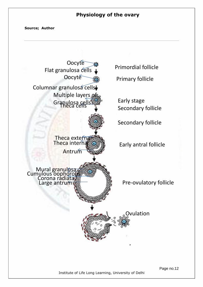
Physiology of the ovary
Page no.12 Institute of Life Long Learning, University of Delhi
Source; Author
Primordial follicle
Primary follicle
Early stage
Secondary follicle
Secondary follicle
Early antral follicle
Pre-ovulatory follicle
Ovulation
Oocyte
Flat granulosa cells
Oocyte
Columnar granulosa cells
Theca cells
Multiple layers of Granulosa cells
Antrum
Theca interna
Theca externa
Mural granulosa Cumulous oophorous
Corona radiata Large antrum
Physiology of the ovary
Page no.13 Institute of Life Long Learning, University of Delhi
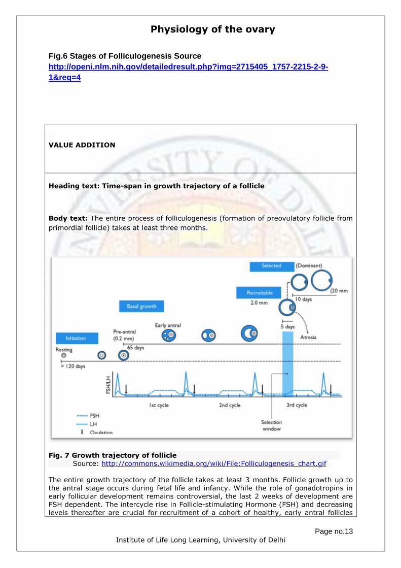
Fig.6 Stages of Folliculogenesis Source
http://openi.nlm.nih.gov/detailedresult.php?img=2715405_1757-2215-2-9-
1&req=4
VALUE ADDITION
Heading text: Time-span in growth trajectory of a follicle
Body text: The entire process of folliculogenesis (formation of preovulatory follicle from
primordial follicle) takes at least three months.
Fig. 7 Growth trajectory of follicle
Source: http://commons.wikimedia.org/wiki/File:Folliculogenesis_chart.gif
The entire growth trajectory of the follicle takes at least 3 months. Follicle growth up to
the antral stage occurs during fetal life and infancy. While the role of gonadotropins in
early follicular development remains controversial, the last 2 weeks of development are
FSH dependent. The intercycle rise in Follicle-stimulating Hormone (FSH) and decreasing
levels thereafter are crucial for recruitment of a cohort of healthy, early antral follicles

Physiology of the ovary
Page no.14 Institute of Life Long Learning, University of Delhi
and subsequent single dominant selection. Following puberty, anovulation may persist
for years and this may presage the development of adult anovulatory infertility. The
menopause is preceded by a period of reduced fertility.
Folliculogenesis can be divided into two phases: one gonadotropin independent phase
and other gonadotropin dependent phase. Follicular growth up to the formation of
secondary follicle is independent of gonadotropin. The granulosa cells in secondary
follicle become responsive to gonadotropins. The formation of antral follicle and their
growth up to preovulatory follicle followed by ovulation are the gonadotropin dependent
stages of folliculogenesis. (Fig.8)
Gonadotropin independent phase of folliculogenesis The pool of primordial follicles is known to be maintained in a dormant state by various
forms of inhibitory signals and molecules which are not regulated by gonadotropins. Loss
of function of any of these inhibitory molecules of follicular activation, leads to premature
and irreversible activation of the primordial follicle pool.
Many years of research have demonstrated that complex bidirectional signaling between
the oocyte and the surrounding somatic cells, involving specific cytokines and growth
factors are required to control of primordial follicle activation. In contrast, the
intracellular signalling pathways activated during follicle development remain largely
uncharacterized and are fundamental to understand the molecular systems responsible
for ensuring timely delivery of functional oocytes for fertilization.
Fig.8 The transition of the follicle from the preantral to early antral stage is the
"penultimate" stage of development in terms of gonadotropin (Gn) dependence
and follicle destiny (growth versus atresia).
Source
http://openi.nlm.nih.gov/detailedresult.php?img=2715405_1757-2215-2-9-1&req=4
GGrraaaaffiiaann
PPoosstt--OOvvuullaattiioonn EEaarrllyy
AAnnttrraall

Physiology of the ovary
Page no.15 Institute of Life Long Learning, University of Delhi
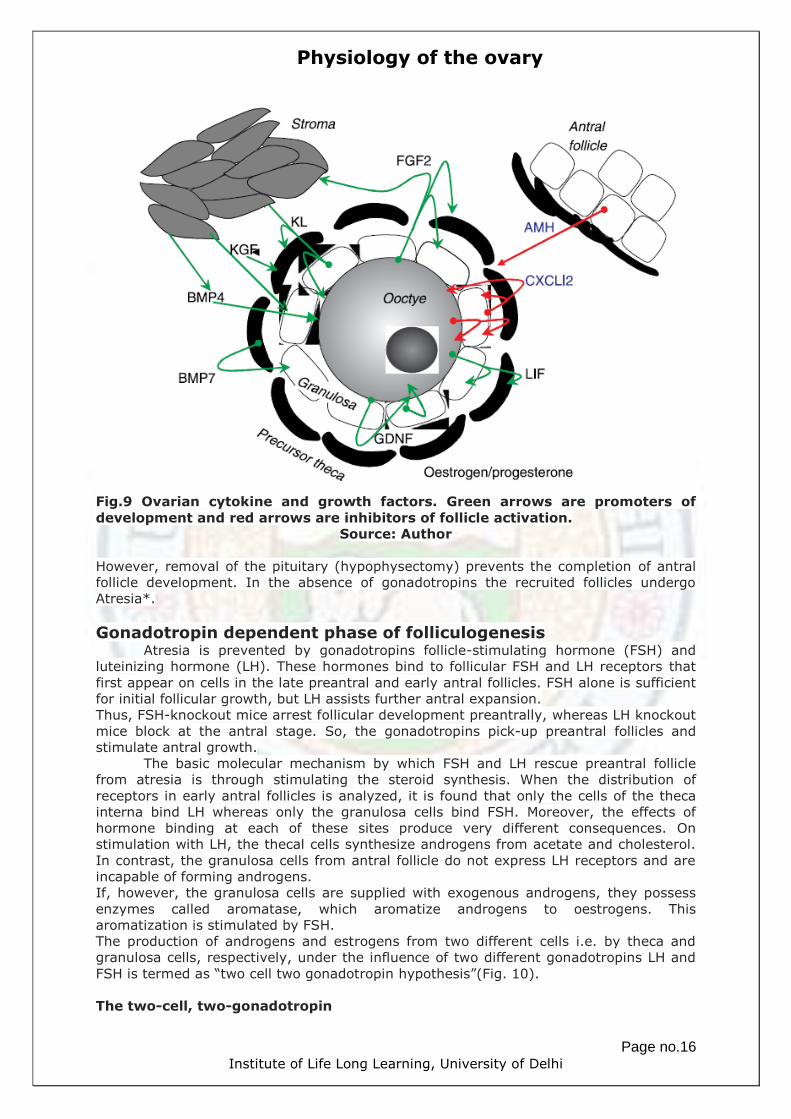
Cytokines involved in primordial to primary follicle transition (Fig.9)
Facilitate primordial follicle recruitment
1. SCF/KL (Stem Cell Factor/Kit Ligand)- Released from granulosa cells and
a) initiates oocyte growth in primordial follicle
b) stimulates thecal cells mitosis
c) maintains growth of preantral and antral granulosa cells
d) boosts androgen release from thecal cells.
2. FGF2/KGF (Fibroblast Growth Factor)- Released from primordial granulosa
cells and growing oocyte
a) promotes recruitment of primordial follicle
b) suppresses granulosa cell apoptosis
c) stimulates theca-interstitial cells
3. BMP7 (Bone Morphogenetic Factor 7)-Released from precursor thecal cells
a) Promotes granulosa cell mitosis
4. BMP4 (Bone Morphogenetic Factor 4)-Released from stromal cell
a) Promotes oocyte maturation
5. KGF (Keratinocyte Growth Factor)-Released from stromal cells
a) Promotes thecal cell formation and granulosa cell proliferation
6. LIF (Leukamia Inhibitory Factor)-Released from primordial follicle Oocyte
a) Stimulate precursor thecal cell and granulosa cell proliferation
GDNF (Glial Cell Derived Neurotropic Factor)-Released from primordial
a) follicle granulosa cells and the oocyte and stimulate oocyte maturation.
Antagonize follicle recruitment
1. MIH/AMH (Mullerian Inhibiting Hormone/ Anti Mullerian Hormone)-
Released from granulosa cells up to early antral stage
a) Depresses recruitment of primordial and preantral follicles
2. CXCL12/SDF1 Chemokine (C-X-C motif) ligand 12/Stromal Cell
Derived Factor 1-Released from granulosa cells and oocyte of primordial
follicle
a) Suppress follicle recruitment by its inhibitory action on oocyte as well as
granulosa cells of primordial follicle.
Physiology of the ovary
Page no.16 Institute of Life Long Learning, University of Delhi
Fig.9 Ovarian cytokine and growth factors. Green arrows are promoters of
development and red arrows are inhibitors of follicle activation.
Source: Author
However, removal of the pituitary (hypophysectomy) prevents the completion of antral
follicle development. In the absence of gonadotropins the recruited follicles undergo
Atresia*.
Gonadotropin dependent phase of folliculogenesis Atresia is prevented by gonadotropins follicle-stimulating hormone (FSH) and
luteinizing hormone (LH). These hormones bind to follicular FSH and LH receptors that
first appear on cells in the late preantral and early antral follicles. FSH alone is sufficient
for initial follicular growth, but LH assists further antral expansion.
Thus, FSH-knockout mice arrest follicular development preantrally, whereas LH knockout
mice block at the antral stage. So, the gonadotropins pick-up preantral follicles and
stimulate antral growth.
The basic molecular mechanism by which FSH and LH rescue preantral follicle
from atresia is through stimulating the steroid synthesis. When the distribution of
receptors in early antral follicles is analyzed, it is found that only the cells of the theca
interna bind LH whereas only the granulosa cells bind FSH. Moreover, the effects of
hormone binding at each of these sites produce very different consequences. On
stimulation with LH, the thecal cells synthesize androgens from acetate and cholesterol.
In contrast, the granulosa cells from antral follicle do not express LH receptors and are
incapable of forming androgens.
If, however, the granulosa cells are supplied with exogenous androgens, they possess
enzymes called aromatase, which aromatize androgens to oestrogens. This
aromatization is stimulated by FSH.
The production of androgens and estrogens from two different cells i.e. by theca and
granulosa cells, respectively, under the influence of two different gonadotropins LH and
FSH is termed as “two cell two gonadotropin hypothesis”(Fig. 10).
The two-cell, two-gonadotropin

Physiology of the ovary
Page no.17 Institute of Life Long Learning, University of Delhi
According to the hypothesis, estrogen synthesis occurs in the following steps:
Step 1. LH stimulates the theca cell, through the adenylyl cyclase pathway, to
increase the synthesis of LDL receptors and the side-chain–cleavage enzyme.
Step 2. Thus stimulated, the theca cell increases the synthesis of androstenedione.
Step 3. The androstenedione synthesized in the theca cells freely diffuses to the
granulosa cells.
Step 4. FSH, also acting through the adenylyl cyclase pathway, stimulates the
granulosa cell to produce aromatase.
Step 5. The aromatase converts androstenedione to estrone. 17β-HSD then converts
the estrone to estradiol. Alternatively, 17β-HSD can first convert the same
androstenedione to testosterone, and then the aromatase can convert this product to
estradiol. By these pathways, theca-derived androgens are converted to estrogens in
the granulosa cell.
Step 6. The estradiol diffuses into the blood vessels.
Fig. 10 Two cell two gonadotropin hypothesis.
Source Author
Thus, in the ovary, androgens produced by developing follicles are derived exclusively
from thecal cells and the oestrogens arise from granulosa cells. However, progesterone
can be produced by both the cell types.
Antral follicles also account for 30–70% of the circulating androgens found in
women, mainly androstenedione and testosterone (the remainder coming from the
adrenal).The antral follicles produce and release increasing amounts of steroids as they
grow (Fig. 10).
In addition, to their release systemically via secretion into the blood, androgens also
have important local intrafollicular roles as following:
1) They serve as substrates for conversion to estrogens.

Physiology of the ovary
Page no.18 Institute of Life Long Learning, University of Delhi
2) Acting with FSH, they stimulate the proliferation of granulosa cells and thereby
follicular growth.
3) They stimulate aromatase activity, thereby promoting estrogen synthesis.
Thus, the rising thecal levels of androgens stimulate a massive increase in estrogen
biosynthesis. Since the estrogens are mitogens (the molecules which induce mitosis)
through autocrine mode of action, they stimulate granulosa cells to proliferate. Thus, a
powerful positive feedback system is operating that culminates in a surge, in this case a
surge of circulating oestrogens towards the end of antral expansion from the most
advanced follicle(s).
Molecular mechanism of FSH action
The granulosa cells secrete insulin like growth factor 1 or 2 (IGF1or 2) depending on the
species. The IGF appears to mediate both
a) the stimulation by LH of androgen output by the thecal cells and
b) its FSH-dependent aromatization to oestrogen by granulosa cells.
FSH regulates the availability of IGF to promote follicular development by following three
ways
Stimulating its production by granulosa cells.
Suppressing the ovarian endogenous release of IGF-binding proteins (IGFBPs).
Promoting the formation of a protease, called pregnancy-associated plasma
protein A (PAPP-A), which cleaves the IGF from its IGFBP, thereby releasing it for
action, by granulosa cells.
Gonadotropins and intrafollicular cytokines
Besides steroids, the production and activity of several cytokines are also
stimulated by the gonadotrophins during the antral phase to mediate and/or modulate
the actions of steroids and gonadotrophin, summarized in table
In particular, the inhibins and activins are of particular importance and well
studied. There are two types of inhibins, A and B produced by the granulosa cells.
However, whereas inhibin B production is stimulated by FSH, inhibin A is stimulated by
both FSH and LH. Thus, the ratio of inhibin A: B rises as the follicle expands to peak at
ovulation. Thus, the ratio acts as a marker for follicular expansion, in addition to the
rising oestrogen output.
Androgens and oestrogens are particularly important for follicular growth and
maturation, but progesterone assumes major significance only as ovulation approaches.
Cytokines that promotes antral follicle growth
1 IGF 1 (Insulin like Growth Factor) (pig, rat), IGF 2 (human, cow, sheep)-
Release from dividing granulosa cells from antal follicle (rat, pig, human) or
thecal cells (cow, sheep)
a) Promotes FSH-induced mitosis
b) differentiation and estradiol output of granulosa cells
c) LH-induced androgen synthesis by thecal cells
2. Inhibin B- Released from granulosa cells in early/mid antral follicle (and decline
thereafter)
a) Enhances FSH stimulation of granulosa cells
3. GDF9 (Growth Differentiation Factor)- Released from preantral and antral
oocytes
a) Co-stimulates with FSH granulosa cell mitosis
b) stimulates estradiol and depresses progesterone output to prevent
premature luteinisation
c) may interact with BMP15
4. Inhibin A-Released from granulosa cells in large antral follicle, preovulatory
follicle an luteinized cells
a) Enhances LH-induced androgen rise in thecal cells a
b) Progesterone rise in luteinized
5. TGF-β- Released from thecal cells in antral follicles
a) Inhibits granulosa/thecal cell proliferation but stimulates GC differentiation

Physiology of the ovary
Page no.19 Institute of Life Long Learning, University of Delhi
b) Also stimulate production of inhibin and estradiol
Cytokines that inhibits follicle growth and/or promotes atresia
1. Activins- Released from granulosa cells in preantral phase, declining as follicle
matures (can block further growth if stay high)
a) Promote FSH receptor expression on early granulosa cells
b) stimulate granulosa cells proliferation
c) attenuate LH-induced rise in androgen output from thecal cells
2. IGFBPs (Insulin like Growth Factor Binding Proteins)-Released from
granulosa cells (sheep, pigs); thecal cells (cow). Depressed by FSH higher in atretic
follicles
a) Bind and attenuate IGFs (which can be released through action of
IGFBP protease-pregnancy-associated protein A (PAPP-A) made by
granulosa cells under FSH control
3. TNFα (Tumour Necrosis Factor) and leptin
a) Promote atresia
b) Antagonize FSH effects to depress follicle growth and steroidogenesis
Cytokines involved in dominant follicle selection
1. BMP15 (Bone Morphogenetic Protein)- Released from preantral and antral
oocytes
a) Depresses PAPP-A output and may be involved in selection of dominant
follicle
VALUE ADDITION
Heading text: Important facts
1. The number of follicles ovulating in any one cycle is characteristic for each
species and ranges from one to several hundred.
2. Thus, dominant follicles are characterized by high ratios of both inhibin:activin
and IGF:IGFBP

Physiology of the ovary
Page no.20 Institute of Life Long Learning, University of Delhi
Ovulation
Under the influence of a brief LH surge the expanding antral follicles enter into
preovulatory phase to ovulate. If it does not, the expanded antral follicle dies. The LH
surge coincides with the appearance of LH receptors on the outer granulosa cells.
The surge of LH affects these advanced follicles in two ways.
1) It causes major changes in both the oocyte and the follicle cells, which results
in the oocyte expulsion from the follicle at ovulation.
2) It changes the whole endocrinology of the follicle, which becomes a corpus
luteum at ovulation.
The oocyte undergoes major preovulatory changes within 3–12 h of the beginning of a
surge of LH (depending on the species).Changes occurring in the oocyte can be listed as:
1) The nuclear membrane surrounding the dictyate chromosomes breaks
down.
2) The arrested meiotic prophase ends, many years after its initiation and
the chromosome progress through the remainder of the first meiotic
division forming a secondary oocyte with half number of chromosomes
and releasing first polar body.
3) The chromosome in the secondary oocyte immediately enter the
second meiotic division and come to lie on the second metaphase
spindle.
4) Then suddenly, meiosis arrests yet again at Metaphase II.
5) The oocyte is ovulated in this arrested metaphase state.
6) This maturation through the arrest and the ovulation is accompanied
by a set of cytoplasmic events which are as follows:
Meiotic Cytoplasmic maturation of oocyte
The intimate contact between the oocyte and the granulosa cells of the cumulus
is broken by withdrawal of the cytoplasmic processes.
The Golgi apparatus of the oocyte synthesizes lysosomal-like granules, which migrate
towards the surface of the oocyte to assume a subcortical position as cortical granules.
Remarkably, centrioles are lost at pachytene, and thereafter only pericentriolar material
(PCM) organizes microtubules (Fig.11). Protein synthetic activity continues at the same
rate, but new and distinctive proteins are synthesized. This activity prepares the oocyte
for fertilization.
Fig.11 Meiotic maturation of oocyte during ovulation.
Source http://www2.mrc-lmb.cam.ac.uk/groups/mschuh/thebasics.html
Physiology of the ovary
Page no.21 Institute of Life Long Learning, University of Delhi
VALUE ADDITION
Heading text: Important facts
If an oocyte is shed from its follicle prematurely, or removed surgically before the
completion of these cytoplasmic maturational events, then its ability to undergo
fertilization is much reduced. For this reason, in clinical in vitro fertilization programs,
human oocytes aspirated from preovulatory follicles are usually cultured for a few hours
before addition of spermatozoa. This procedure improves the chances of a complete
maturation of the oocyte
Factors regulating ovulation
As mentioned above meiotic and cytoplasmic maturation of the oocyte is
stimulated by the surge of LH, yet it is clear that LH cannot, and does not bind to the
oocyte itself. Therefore, its effect must be mediated via the cumulus cells of the follicle,
where it is thought to act by suppressing cAMP levels.
Preovulatory growth of follicle cells is associated with a major endocrine switch. The
preovulatory growth in follicular size is matched by changes in the pattern of steroid
secretion. Within 2 h or so of the beginning of the LH surge, there is a transient rise in
the output of follicular oestrogens and androgens followed by a decline. This rise
coincides with distinctive changes in the thecal layer, which appears transiently
stimulated and hyperemic.
The outer cells of the granulosa layer also show a marked change in their properties.
1) They stop dividing and no longer convert androgen to oestrogen, but instead
synthesize progesterone.
2) LH stimulates this synthesis of progesterone via the newly acquired LH receptors.
3) The cells have lost, or reduced, their capacity to bind oestrogen and FSH, but
gained the capacity to bind progestins.
This acquisition of the capacity to respond to LH by synthesizing progesterone (and then
to be self-stimulated by positive feedback) results in an exponential release of
progesterone from the follicle, which becomes significant in the human several hours
before ovulation, although in most species only just before or immediately after
ovulation. This rising progestin output has three important functional consequences.
1) It depresses growth in the less mature developing follicles.
2) It is essential for ovulation itself. Thus, the progesterone inhibitors such as
mifepristone (RU486) suppress ovulation and females genetically knockout for
progesterone receptors do not ovulate.
3) It promotes the transition to the progestogenic phase of the ovarian cycle.

Physiology of the ovary
Page no.22 Institute of Life Long Learning, University of Delhi
Fig.12 Changes in mural granulosa cells after LH surge.
Source: AUTHOR
Molecular events associated with ovulation
A number of proteolytic enzymes are known to dissolve the protein matrix to release
ovum. Under LH influence, directly and/or indirectly via progesterone and/or
prostaglandins the activity of these enzymes increases especially at a region called
stigma from where oocyte flows out along with the follicular fluid. These enzymes are:
1) Members of a large family of matrix metalloproteinases (MMPs-zinc dependent
endopeptidases) like collagenase, gelatinase and their natural tissue inhibitors
(TIMPs)
2) Members of family of serine proteases such as plasmin and plasminogen activator.
VALUE ADDITION
Heading text: Important facts
In many species, including humans, the ovarian surface is directly exposed to the
peritoneal cavity, but in some (e.g. the sheep, horse and rat) a peritoneal capsule or
bursa encloses the ovary to varying degrees and acts to retain the oocyte cumulus
mass(es) close to the ovary. There they are collected by cilia on the fimbria of the
oviduct, which sweep the cumulus mass into the oviducal ostium. The residual parts of
the follicle within the ovary collapse into the space left by the fluid, the oocyte and the
cumulus cells, and within this cavity a clot forms. Thus, the postovulatory follicle is
composed of a fibrin core, surrounded by several collapsed layers of granulosa cells,
enclosed within a fibrous outer thecal capsule
Source: Author

Physiology of the ovary
Page no.23 Institute of Life Long Learning, University of Delhi
Luteinization
As mentioned in the text earlier, the post ovulatory follicle in the ovary collapse and
transforms into a corpus luteum which produces progestogens. This transformation is
termed as Luteinization, it involves:
1) Within the follicular antrum, the fibrin core undergoes fibrosis over a period of
several days.
2) The membrane propria between the granulosa and thecal layers breaks down
and blood vessels invade.
3) Both granulosa cells and cells from the theca interna contribute to the corpus
luteum, although many thecal cells also disperse to the stromal tissue.
4) The granulosa cells have now ceased dividing and hypertrophy to form large
lutein cells, rich in mitochondria, smooth endoplasmic reticulum, lipid
droplets, Golgi bodies and, in many species, a carotenoid pigment, lutein,
which may give the corpora lutea a yellowish or orange tinge and called
granulosa lutein cells. (Fig.13)
5) The thecal cells transformed to smaller lutein cells, produce progesterone and
androgens, and seem to be richer in LH receptors called thecal lutein cells.
Fig.13 Corpous Luteum from rat ovary; in luteinized follicle the granulosa
cells are luteinized to granulosa lutein cell (relatively bigger in size) and
secrete progesterone. While the thecal cells luteinized to form thecal lutein
cells which also secret progesterone.
Source Author
VALUE ADDITION
Heading text: Important facts
Body text: In most species, the principal progestogen secreted from the large lutein
cells is progesterone, but secretion of significant quantities of 17-hydroxyprogesterone

Physiology of the ovary
Page no.24 Institute of Life Long Learning, University of Delhi
in primates and of 20-hydroxyprogesterone in the rat and hamster also occurs. In a few
species, notably the great apes and humans, and to a lesser extent the pig, the corpus
luteum also secretes oestrogens, particularly oestradiol 17β. Its source also seems to be
the large luteal cells, using as substrate androgens derived from the small luteal cells (a
hangover from granulosa function in preovulatory follicles). In most species (e.g. the
monkey, sheep, cow, rabbit, rat and horse), however, the corpus luteum secretes
onlytrivial amounts of oestrogen.
Source: Author
The corpus luteum also secretes two other hormones. 1) Inhibin A is secreted in large
amounts in higher primates. It acts to promote production of progesterone 2) The
second hormone is oxytocin, which comes from the large lutein cells and is important for
‘Luteolysis’
Luteotrophins: endocrine support of the corpus luteum.
LH surge is required for initial transformation; however, for maintenance its small
amount is required. Also, in some species prolactin and/or progesterone also help in
maintenance of corpus luteum.
Luteolysis: death of the corpus luteum may be active or passive depending on
the species
The life of the corpus luteum in the non-pregnant female varies among species from 2 to
14 days. Luteal regression or luteolysis involves a collapse of the lutein cells, ischemia
and progressive cell death with a consequent fall in the output of progestogens. The
whitish scar tissue remaining, the corpus albicans, is absorbed into the stromal tissue of
the ovary over a period that varies from weeks to months, depending on the species.
Luteolysis can be caused by two ways
1) Withdrawal or inadequacy of the luteotrophic complex (e.g. humans)
2) Active production of a luteolytic factor that brings about normal luteal
regression (e.g. rat).
Thus, in latter case, both luteotrophic support and antiluteolytic support are required to
sustain the corpus luteum. The major luteolytic substance is prostaglandin F2 released
by endometrium.
The ovarian cycle and the follicular development One complete ovarian cycle is the interval between successive ovulations, where each
ovulation is preceded by a period of oestrogen dominance. It could be divided into three
phases as:
Follicular phase
Preovulatory phase
Luteal phase
A continuous recruitment of developing primordial, preantral and early antral follicles
occurs throughout the cycle, a lot of which undergo atresia if not rescued by FSH and LH,
which take them through full antral expansion. This doesn’t affect the blood steroid level.

Physiology of the ovary
Page no.25 Institute of Life Long Learning, University of Delhi
However, one (or two) of these rescued follicles survive to become the dominant antral
follicle, these dominant follicle/s release high levels of oestrogens in the blood-Follicular
phase.
This advanced follicle is then converted to a preovulatory follicle by the transient high
levels of LH that are measured in the blood at this time. The other antral follicles become
atretic- Preovulatory phase
The successful ovulatory follicle forms a corpus luteum, which secretes progesterone and
oestrogen, until luteolysis 14 or so days later- the luteal phase. (At this phase, LH and
FSH levels are comparatively low and are insufficient to maintain antral development,
which is also suppressed by elevated progesterone levels, so follicular atresia occurs.)
After luteolysis, a new cycle then begins as tonic gonadotrophin levels are elevated and
progesterone is low.
Two important features emerge from this description of the human ovarian cycle: first,
we need to understand what controls the fluctuating levels of gonadotrophin output (this
will be discussed in detail in the next chapter); second, the complete antral expansion
phase, ovulation and the complete luteal phase occupy one complete ovarian cycle.
The follicular phase of the cycle
Follicular phase corresponds to the proliferative phase of the uterine cycle (menstruation
cycle). By convention, the menstrual cycle begins with the first day of menstruation.
Initiation of menstruation is governed by the preceding luteolysis, which results in
declining levels of luteal oestrogen, progesterone and inhibin A.
As a consequence, negative feedback inhibition is relaxed, and both FSH and LH levels
rise. These rises permit antral growth to proceed, resulting in first the rising output of
inhibin B followed by androgens and oestrogens.
As a consequence, negative feedback is gradually re-exerted and so FSH levels fall and
LH levels plateau.
The selection of a dominant follicle leads to a further rise in oestrogen (together with
their biosynthetically associated androgens), culminating in an oestrogen (and androgen)
surge and a switch from inhibin B to inhibin A output under the combined stimulation of
LH and FSH.
This output of oestrogen and inhibin A reflects the development of only the most
advanced follicle(s), thus, is a measure of nearness to ovulation.
The preovulatory phase
The surge in oestrogens triggers a rapid rise in LH and FSH levels via its positive
feedback effect, and ovulation follows.
As a result of follicular collapse, androgen and thus oestrogen outputs fall, and
progesterone levels rise.
The LH and FSH levels now fall equally precipitously because, at least in part, they lack a
continuing positive feedback stimulus.
The luteal phase of the cycle
The luteal phase of the cycle corresponds to the secretory phase of the uterine cycle.
This phase is characterized by rising concentrations of plasma progesterone and 17α-
hydroxyprogesterone, which peak around 8 days after the LH surge. The luteinized cells
of the corpus luteum also make large amounts of oestrogen and inhibin A.
The progesterone depresses levels of both gonadotrophins (negative feedback),
moreover inhibin A is also active in suppressing FSH. Growth of antral follicles is
therefore suppressed and so androgens are also at a low level.
At the end of the luteal phase, if conception has not occurred, oestrogens, progesterone
and inhibin A decline at luteolysis, the negative feedback effect of these hormones is
relaxed and LH and FSH levels start to rise, thereby permitting the rescue of preantral
follicles and the initiation of another cycle.

Physiology of the ovary
Page no.26 Institute of Life Long Learning, University of Delhi
Fig. 14 Follicular development and the ovarian cycle
http://en.wikipedia.org/wiki/Menstrual_cycle#mediaviewer/File:Figure_28_02_07.jpg

Physiology of the ovary
Page no.27 Institute of Life Long Learning, University of Delhi
VALUE ADDITION: VIDEO
Heading text: LH surge, Ovulation and Menstrual cycle are co-ordinated events
Body text: Watch the video clip 3D animated and narrated in the link given below and
see how LH surge, ovulation and menstrual cycle are co-ordinated:
https://www.youtube.com/watch?v=WGJsrGmWeKE
Source: YouTube
Feedback by steroid hormones and the inhibins regulates the menstrual cycle
Estradiol regulation of gonadotropin secretion
The oestradiol secreted from ovary has dual function in regulating gonadotrophin
secretion.
1) At low circulating levels, it exerts negative feedback control over FSH and LH.
2) However, at higher levels, maintained for a duration, it exerts positive feedback
resulting in gonadotropin surge.
Progesterone regulation of gonadotropin secretion
Progesterone also has two effects.
1) First, the high plasma concentration of progesterone, such as is seen in the
luteal phase (4–8 ng/ml in humans) enhances the negative feedback effects of
oestradiol, FSH and LH secretion being held down to a very low level.
2) Second, the positive feedback effect of oestradiol is blocked.
The inhibins regulate only FSH secretion
Data indicates a role for inhibin in the negative feedback regulation of FSH secretion.
Fig.15 Annotation of the graph of ovarian cycle.
Source: Author
Increase in FSH stimulates development of the Graafian follicles (1) and estrogen
production from follicles (Positive feedback). Estrogen levels increase repairing and
thickening of endometrium (2). Also, stimulates LH production (Positive feedback). A
peak of LH stimulates ovulation, formation of corpous luteum and thus production of
progesterone (3) Increase in progesterone maintains endometrium in preparation for
fertilization (4). If fertilization does take place progesterone levels remain high and the
endometrium is maintained. If fertilization does not occur, the decreasing levels of
progesterone cause mensuration to take place. Decrease in progesterone stimulates FSH
production (Negative feedback)

Physiology of the ovary
Page no.28 Institute of Life Long Learning, University of Delhi
Regulation of gonadotropin secretion The gonadotrophs of anterior pituitary secrete both the gonadotropins; FSH and LH and
lactotrophs secrete prolactin, which is involved in regulating ovarian functions. The
pituitary lies close to the hypothalamus (a small part of brain) and have both neural and
vascular connections with it. The synthesis and secretion of both FSH and LH depend on
a gonadotrophin-releasing hormone (GnRH) released by hypothalamus. The GnRH is
released as a series of pulses into the portal vessels; it binds to receptors on the
gonadotrophs, and drives gonadotrophin secretion in a pulsatile manner. Therefore,
alterations in the output of LH and FSH could be achieved by:
(1) increasing or decreasing either the amplitude or the frequency of these pulses of
GnRH
(2) modulating the response of the gonadotrophs to the pulses.
Positive and negative feedback are mediated at the levels of both
hypothalamus and pituitary
The ovarian hormones influence gonadotrophin output by exerting its effect on two sites
1) the anterior pituitary: the hormones might regulate FSH and LH secretion by a
direct action on the gonadotrophs. This could be achieved by modulating their
sensitivity to hypothalamic GnRH pulses via changing GnRH receptor
expression/affinity. There are abundant receptors for oestrogens, progestogens
and inhibins in the anterior pituitary, emphasizing the potential importance of this
site.
2) the hypothalamus: the ovarian hormones might change the GnRH output signal
either directly by affecting the GnRH neurons in the hypothalamus, or indirectly
by changing the activity of other neural systems that exert a modulator influence
on GnRH release (both GnRH pulse amplitude and frequency). Large regional
concentrations of receptors for oestradiol and progesterone exist in different
regions of hypothalamus and its surroundings. However, there is no evidence to
support a hypothalamic site of action of inhibin.
Regulation at the level of anterior pituitary
Within the anterior pituitary, oestradiol appears to exert its positive feedback effects by:
1) inducing and maintaining GnRH receptors and
2) sensitizing the self-priming process whereby GnRH induces its own receptors.
There is less information on the ways in which oestradiol might cause a decrease in
gonadotrophin secretion to mediate its negative feedback. Nor is there detailed
information on the way that inhibin exerts its selective depressant effect on FSH
secretion.
Regulation at the level of hypothalamus
The accurate nuclei and preoptic/anterior hypothalamic areas are rich in oestradiol
receptors, and the GnRH content of neurons in this area changes in response to
oestradiol. However, GnRH+ve neurons do not express progesterone or oestradiol-
(ER) receptors, and express ERβ only weakly which suggests that steroid effects on
GnRH secretion must be mediated indirectly via other steroid-sensitive neural systems,
which then converge onto GnRH neuronal cell bodies or terminals. In particular, it is
assumed that different pathways are likely to mediate negative and positive feedback
effects.
Among the hypothalamic neural systems most strongly implicated in the regulation of
GnRH secretion, key players having steroid receptors are the amino acid γ-amino-butyric
acid (GABA), glutamate, opioid peptide β-endorphin, and noradrenaline (norepinephrine)
systems. Among the many other transmitters and peptides that have been suggested to
influence GnRH secretion, the recently studied peptide kisspeptin seems of particular
interest in the context of feedback. However, it remains a puzzle as to how these various
Physiology of the ovary
Page no.29 Institute of Life Long Learning, University of Delhi
neural mechanisms, which can affect GnRH secretion and are sensitive to steroid
hormones, interact under physiological circumstances to regulate the cyclic changes in
GnRH output.
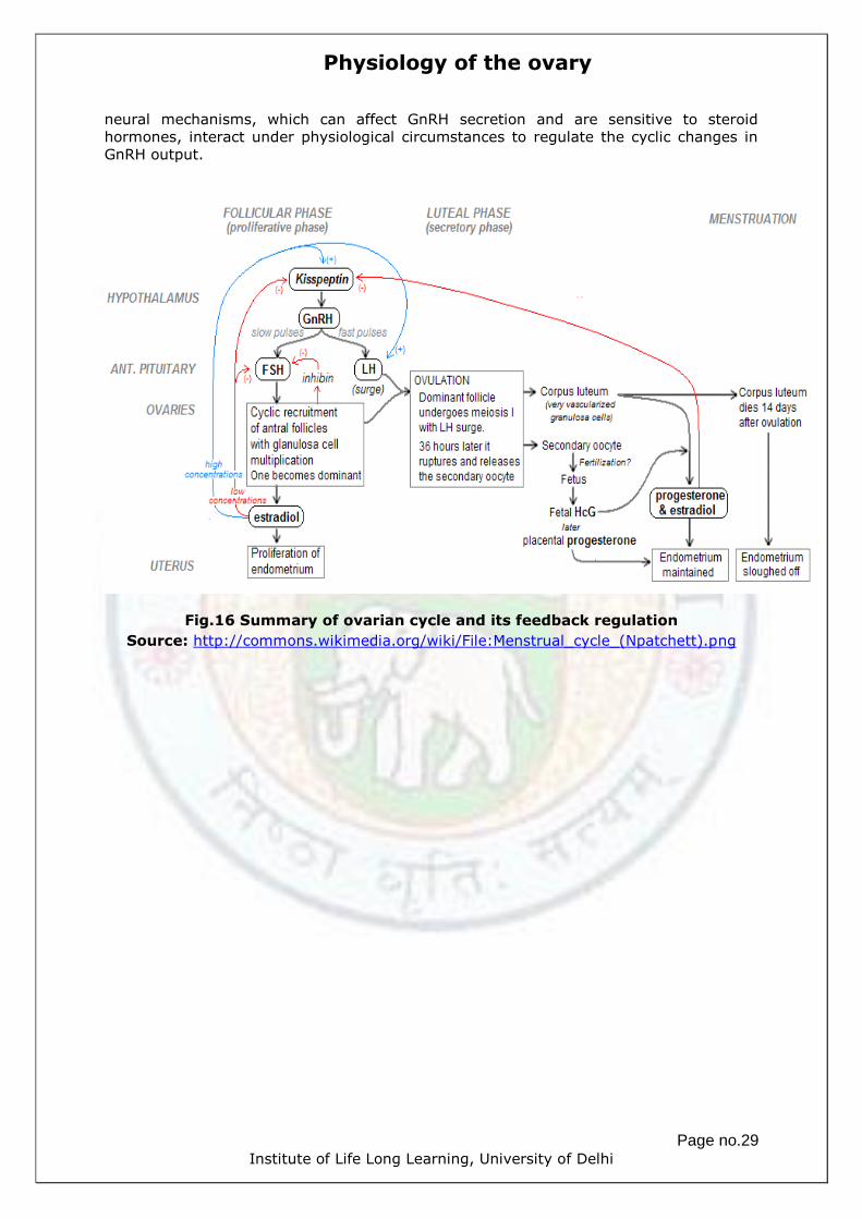
Fig.16 Summary of ovarian cycle and its feedback regulation
Source: http://commons.wikimedia.org/wiki/File:Menstrual_cycle_(Npatchett).png

Physiology of the ovary
Page no.30 Institute of Life Long Learning, University of Delhi
SUMMARY
Oogenesis is the process of gamete (ovum) formation in the ovary.
Oogenesis begins in ovary even before a female is born.
During early fetal development, the primitive germ cells migrate from yolk sac to
the ovaries.
These germ cells differentiate into OOGONIA within the ovaries.
Oogonia are diploid stem cells which divide mitotically to produce millions of germ
cells.
Even before birth most of these germ cells degenerate by a process called
ATRESIA.
A few however develop into larger cells called PRIMARY OOCYTES that enter
prophase of meiosis I but do not complete that until after puberty.
During this arrested stage of development, each primary oocyte is surrounded by
a single layer of flat follicular cells, and the entire structure is termed as
PRIMORDIAL FOLLICLE. These follicles lie in the ovarian stroma (cortical region)
and are surrounded by a thin layer of basal lamina.
The progression of the primordial follicle from a quiescent stage through a series
of changes in the morphology and gene expression into a preovulatory follicle
that undergo ovulation to form corpus luteum is termed as FOLLICULOGENESIS.
The growing follicle is the main source of hormones in the ovary.
As follicle grow in size the primary oocyte also complete meiosis I (reductional
division) to become haploid secondary oocyte, release a polar body and enter into
meiosis II. This process is termed at OOCYTE MATURATION. In preovulatory
follicle, the oocyte is arrested at metaphase II.
OVULATION is the process of releasing secondary oocyte (which is arrested at
metaphase II) from preovulatory follicle into oviduct.
The secondary oocyte only after fusion with spermatozoa (the process termed
FERTILIZATION) undergoes completion of meiosis II, releases a polar body and
forms zygote.
The cells in the left out follicle in the ovary undergo differentiation, the process
termed as LUTEINIZATION, to form corpus luteum.
The corpus luteum actively secrets progesterone hormone (in anticipation of
pregnancy) under the influence of LUTEOTROPIC COMPLEX and it is maintained
throughout or for a duration during pregnancy.

Physiology of the ovary
Page no.31 Institute of Life Long Learning, University of Delhi
However, in the absence of pregnancy the corpus luteum degenerates after a few
days of ovulation to form corpous albicans the process is termed as LUTEOLYSIS.
The process of folliculogenesis, ovulation and luteolysis are repeated again and
again in the ovary throughout the reproductive age of female. This cycle is
termed as OVARIAN CYCLE.
All the processes mentioned here are tightly regulated via HYPOTHALAMUS-
HYPOPHYSEAL-GONADAL axis. Any abnormality in any of the above processes can result
in an ovulation and infertility.

Physiology of the ovary
Page no.32 Institute of Life Long Learning, University of Delhi
GLOSSARY
Atresia: A periodic process of degeneration of immature ovarian follicles and
subsequently re-absorption during the follicular phase of the menstrual cycle. Out of 20
follicles maturing each month, a single follicle undergoes ovulation and rest atresia.
Estrogen: A hormone that is primary for women. It causes the uterine wall to thicken
monthly, helping spur ovulation. There are different forms of estrogen hormones. The
main form is called Estradiol
Follicle: A round sac, found in the ovary, containing an egg and cells that produce
hormones.
Follicle-stimulating hormone (FSH): A hormone, produced by the pituitary gland,
which stimulates the growth of the follicle. The hormone is used in medical treatments,
contained in injectable ovulation drugs to help spur growth of the follicles.
Folliculogenesis: The process by which the primordial follicles grow into a preovulatory
follicle.
Luteinizing hormone (LH): a gonadotropic hormone that is secreted by the anterior
pituitary
Luteotrophins: endocrine support of the corpus luteum.
Luteinization: The postovulatory follicle in the ovary collapse and transforms into a
corpus luteum, which produces progestrogens. This transformation is termed as
Luteinization.
Luteolysis: death of the corpus luteum.
Menstural cycle: hormonally controlled cyclic changes occurring in female reproductive
system resulting in the monthly replacement of the lining of the uterus.
Ooycte: The official medical term for an egg.
Ovary: The ovaries or "egg sacs" are a pair of reproductive organs of female
reproductive system. There are two ovaries, located on located in the pelvis, one on
each side of the uterus. The ovaries have two functions: they produce eggs (ova) and
female hormones.
Ovulation: When an ovary releases an egg into the fallopian tube.
Progesterone: A hormone that helps prepare the lining of the uterus for the
implantation of a fertilized egg. It is present during the second half of the menstrual
cycle.
Tunica albuginea: a whitish capsule of dense irregular connective tissue located
immediately deep to the germinal epithelium.
Physiology of the ovary
Page no.33 Institute of Life Long Learning, University of Delhi
EXERCISE
A. MULTIPLE CHOICE QUESTIONS
1. At birth, a girl has in her ovaries many……….that have started meiosis but stopped
at prophase I.
a) Primary oocytes
b) Secondary oocytes
c) Ova
d) Oogonia
2. A primary oocyte divides to produce a(n)
a) Oogonium
b) Secondary oocyte
c) Polar body
d) Both b)&c)
3. A layer of clear viscous fluid deposited around a primary oocyte is called
a) corona radiata
b) Cumulous oophorous
c) Zona pellucida
d) Primordial follicle
4. In the process of oogenesis, the polar body
a) Is formed before fertilization
b) Is formed after fertilization
c) Normally receives most of the cytoplasm of the cell
d) Both a) & b)
5. While the follicle is developing, a positive feedback loop occurs in
which……….stimulates the follicle, which increases the secretion of………….., which
stimulates the secretion of GnRH.
a) LH, estrogen
b) FSH, estrogen
c) LH, progesterone
d) FSH, progesterone
6. Which of the following ovarian hormones is involved in positive feedback loop
with the hypothalamus and the anterior pituitary?
a) Progesterone
b) Estrogen
c) Testosterone
d) GnRH
7. A positive feedback loop causes a self amplifying cycle wherea physiological
change leads to even greater change in the same direction
a) True
b) False
8. When a primary follicle enlarges and there are a several layers of granulosa cells,
it is called an
a) Primordial follicle
b) Primary follicle
c) Secondary follicle

Physiology of the ovary
Page no.34 Institute of Life Long Learning, University of Delhi
d) Tertiary follicle
9. Which of the following has an antrum
a) Primordial follicle
b) Primary follicle
c) Secondary follicle
d) Tertiary follicle
10. The layer of granulosa cells surrounding the oocyte in a preovulatory follicle is
called
a) Cumulus oophorous
b) Mural granulosa
c) Corona radiata
d) Zona pellucida
11. Progesterone has a negative feedback effect on GnRH and LH
a) True
b) False
12. The period of time when secondary sexual characteristics begin to develop and
the potential for sexual reproduction is reached is called
a) Menarch
b) Puberty
c) Menopause
d) Spermatogenesis
13. The first menses is called _____, and the permanent cessation of menses is
called _____.
a) Menarch, Puberty
b) Menarch, menopause
c) Menopause, Menarch
d) Menopause, puberty
14. After ovulation, progesterone is produced by
a) Corpus albicans
b) Graafian follicle
c) Secondary follicle
d) Corpus luteum
15. State true and false
a) The initiation of preantral phase of follicular development is under the control of
LH
b) The development of antral follicle depends on the presence of FSH and LH
receptors.
c) Ovulation occurs at day 14 of menstrual cycle.
d) The period before ovulation is known as luteal phase.
e) The luteal phase is associated with the large increase in plasma progesterone.
f) The enometrium proliferates under the influence of progesterone.
16. The primary female sex hormone is
a) Testosterone
b) Estrogen
c) Progesterone
d) DHEA
17. At which stage of meiosis does the primary oocyte is arrested at birth
a) Pachytene
b) Diplotene
c) MetaphaseI
d) Anaphase I
18. At which stage of meiosis does the secondary oocyte is arrested at ovulation
a) Metaphase I
b) Metaphase II

Physiology of the ovary
Page no.35 Institute of Life Long Learning, University of Delhi
c) Anaphase II
d) Anaphase I
B. SHORT ANSWER TYPE QUESTIONS
1. Define: a) Ovulation b) Luteolysis c) Atresia d) Primordial follicle e) Antrum
2. Differentiate:
a) Gonadotropin dependent folliculogenesis and gonadotropin independent
folliculogenesis
b) Oogenesis and Folliculogenesis
c) Follicular phase and luteal phase
d) LH and FSH
e) Estrogen and progestrone
C. LONG ANSWER TYPE QUESTIONS
1. Describe the histology of ovary with a neatly labeled diagram.
2. Write Short notes on:
a. Folliculogenesis
b. Regulation of Gonadotropin secretion
c. Ovarian cycle
d. Factors regulating ovulation
3. Describe the role of intrafollicular cytokines.
4. Describe Two-cell two-gonadotropin hypothesis with neatly labeled diagram.
5. Describe the factors regulating ovulation.

Physiology of the ovary
Page no.36 Institute of Life Long Learning, University of Delhi
REFERENCES
General reading
Essential Reproduction - 6th Edn-Martin H. Johnson
Principles of Anatomy and Physiology-12th Edn-Gerard J. Tortora and Bryan
Derrickson-2009
www.expertconsultbook.com
Further reading
Eppig JJ et al. (2002). The mammalian oocyte orchestrates the rate of ovarian
follicular development. Proceedings of the National Academy of Sciences of the
USA 99, 2890–2894.
Fortune JE et al. (2004). Follicular development: the role of the follicular
microenvironment in selection of the dominant follicle. Animal Reproduction
Science 82–83, 109–126.
Erickson GF, Shimasaki S (2001). The physiology of folliculogenesis: the role of
novel growth factors. Fertility and Sterility 76, 943–949.
Wang Y et al. (2005) Gonadotropin control of inhibin secretion and the
relationship to follicle type and number in the hpg mouse. Biology of
Reproduction 73, 610–618.
Weblinks:
http://education.med.nyu.edu/Histology/courseware/modules/fem-repro-
sy/female.reproductive.01.html
http://www.cytochemistry.net/microanatomy/medical_lectures/oviduct_and_uter
us.htm
https://www.glowm.com/
http://embryology.med.unsw.edu.au/
http://www.histologyworld.com/photoalbum/displayimage.php?album=39&pid=3
619
http://www.rci.rutgers.edu/~uzwiak/AnatPhys/PPFall03Lect8_files/image002.jpg
http://www.austincc.edu/rfofi/NursingRvw/PhysText/Reproductive.html
http://www.vetmed.vt.edu/education/curriculum/vm8054/Labs/Lab28/lab28.htm
#