physiologic monitoring: improving safety or increasing...
TRANSCRIPT
Physiologic Monitoring: Improving Safety or Increasing Risk?
Charles G Durbin Jr MD FAARC
IntroductionA Brief History of AnesthesiaAdvances to Enhance and Extend the Senses of CaregiversPersonal Travels in Anesthesia and Patient MonitoringMonitoring With Pulse OximetryEarly Studies of Pulse Oximetry in the Operating RoomEffect of Improved Monitor Function on Caregiver BehaviorConfusion Arises When Monitors Are Used to Make a DiagnosisNew Monitors, New Problems: AlarmsConclusions
This paper will present a focused and personal history of physiologic monitoring, beginning with thediscovery of modern anesthesia and its development from a technical practice to a scientific disci-pline. Emphasis will be on the essence of monitoring in the anesthesia evolution, and this work willattempt to answer the question of how to evaluate the impact of monitoring on patient outcome.Understanding that monitors are passive and that only caregivers using monitors can impactoutcome is at the crux of this approach to analysis. The limited quality data involving monitoringanalysis, including that from pulse oximetry, will be discussed and critiqued. The invention andrapid spread of pulse oximetry will be highlighted and used as an example throughout, but theprinciples developed will apply to other monitors and patient monitoring in general. The problemscreated by monitoring alarms will also be discussed. Key words: physiologic monitoring; pulse oxi-metry; intensive care unit; alarms; false alarms; alarm fatigue; patient outcome; anesthesia. [RespirCare 2016;61(8):1110–1121. © 2016 Daedalus Enterprises]
Introduction
The continued evolution of physiologic monitoring de-vices in recent years has raised the promise of better pa-
tient outcomes, especially in the care of the critically ill.Although the technical aspects of monitoring devices areimproving and changing rapidly, very little informationsuggesting better patient outcomes from their use has beenforthcoming.1 This is especially true of invasive monitors,such as the pulmonary artery catheter, the use of which isassociated with no improvement and, in some studies, worsepatient outcomes.2 Despite this lack of positive data, sev-eral organizations and institutions are pushing to deployelectronic physiologic monitoring to all hospital wards aspart of a misplaced belief that this is a sure route to safetyand better patient care.
In fact, even in ICUs, the frequency and variety of alarmstriggered by multiple monitors cause sensory overload anddesensitization of caregivers, leading to a decrease in safetyand a worse outcome in vulnerable patients.3 The problem
Dr Durbin is affiliated with the Department of Anesthesiology, Univer-sity of Virginia, Charlottesville, Virginia.
Dr Durbin has disclosed a relationship with Halyard Medical Company.
Dr Durbin presented a version of this paper as the 42nd Donald F EganScientific Memorial Lecture at the 61st AARC Congress, held November7-10, 2015, in Tampa, Florida.
Correspondence: Charles G Durbin Jr MD FAARC, Box 800710, Char-lottesville, VA 22908. E-mail: [email protected].
DOI: 10.4187/respcare.04931
1110 RESPIRATORY CARE • AUGUST 2016 VOL 61 NO 8
of alarm management has been widely recognized as acritical patient safety issue. Concern has lead to a universalcall for action to find a solution for alarm issues.4
This paper will present a focused and personal historyof physiologic monitoring, beginning with the discoveryof modern anesthesia and its development from a technicalpractice to a scientific discipline. Emphasis will be on theessence of monitoring, and this work will attempt to an-swer the question: Why is there so little quality evidencedemonstrating a positive impact on patient outcomes fromapplication of current monitors and monitoring standards?Development of new monitors and monitoring standardsare unlikely to lead to demonstrably better patient out-comes; however, they may mitigate some of the currentrisks that have accumulated from dissemination of poorlyintegrated devices and lack of understanding of monitor-ing principles.
A Brief History of Anesthesia
Although sedative and analgesic biologics had been usedfor thousands of years, the discovery and application of theanesthetic effects of diethyl ether, chloroform, and nitrousoxide officially date from the middle of the 19th century(1842–1847). Together with increased understanding ofhuman anatomy and physiology, these agents allowed rapidadvancement in the complexity and success of surgicalprocedures. The use of these newly discovered anestheticsfostered the need for skilled anesthesia providers and en-hanced patient monitoring.
Shortly after the first successful public demonstration ofpainless surgery in the United States, ether became thepredominant agent used due to its simplicity of adminis-tration, preservation of airway-protective reflexes, stimu-lation of respiration, production of muscle relaxation, andpredictable prolonged analgesia after surgery (if the pa-tient survived!). The administration of ether was directedby the surgeon and often delivered by the “etherizer,” anurse, technician, or medical student with little or no train-ing or experience in delivering this potent drug. The onlyequipment needed was a cone or handkerchief placed overthe nose and mouth of the patient and intermittently sat-urated with the volatile liquid ether. Monitoring of patientsreceiving ether for surgery consisted of the surgeon ob-serving the depth and character of spontaneous breathing,color of the skin and blood, and, infrequently, palpation ofa pulse. The surgeon was the decision maker for the choiceand amount of the anesthetic administered. At the time,surgical mortality was high and included both the lethaleffects of the anesthetic and the surgical procedure itself.In addition to the deleterious effects of ether on humanphysiology, ether is highly flammable and can easily catchfire. The first recorded patient death from a fire duringsurgery with ether was reported in 1850.
The anesthetic potential of chloroform was soon iden-tified, and it quickly became the most popular anestheticagent in Europe. Chloroform is more potent than ether andsafer in that it is not flammable. The use of and acclaim forchloroform during childbirth by Queen Victoria undoubt-edly influenced its wide acceptance throughout the BritishIsles. The first death believed to be due to chloroform andnot surgery was recorded in 1848, a year after its firstdemonstration in a human. Unlike ether, chloroform re-quires complex equipment and careful attention to dose forsafe administration. In addition, chloroform is toxic to theliver and frequently causes cardiac arrhythmias.
Acceptance and evaluation of the risks of anesthesiaapart from surgery came much later than discovery anduse of anesthetic agents. It was not until the late 1890s thata suggestion was made that vital signs even be recordedduring surgical procedures. In fact, no systematic writtenrecords were routinely used during anesthesia for nearly100 years after its discovery. Monitoring of a patientduring anesthesia consisted of watching the depth, fre-quency, and characteristics of breathing; assessing skeletalmuscle tone; determining the frequency and characteristicsof the peripheral pulse; and observing the color of thepatient’s skin. These observations were used to determineadequacy of the anesthetic depth for the surgery and, af-terward, to determine whether the patient had survived theexperience. Patient survival was assessed by the presenceof spontaneous breathing and a pulse following surgery,and success of the surgery was declared based on survivalat the end of the procedure. Long-term survival was notused as a marker of surgical success. At the time, therewere no intravenous therapies (including fluids and bloodtransfusions), mechanical or manual ventilation techniques,or resuscitation standards or practices. If the heart stoppedduring surgery, open massage by the surgeon was attemptedbut usually failed to restore the dead patient. These intra-operative deaths were generally blamed on the surgery orthe patient’s disease; if anesthesia contributed, this was notseparately noted.
Medical practice during the 1800s was one of making adiagnosis from simple clinical observations and predictingsurvival or death.5 Treatments were many but were notfounded on scientific evidence and often not helpful. Phy-sicians of the time, however, were careful clinical observ-ers. By feeling the temperature and moisture of the skin,frequency and characteristics of the peripheral pulse, heartrate and characteristics of breathing, color of the skin, odorof breath and urine, and taste (yes taste!) of urine, the bestphysicians would make the correct diagnosis, determine atreatment plan (phlebotomy or patent medicine), and pre-dict the outcome of seriously ill patients (eg, approxi-mately when they would die). Patient monitoring duringsurgery consisted only of the eyes, ears, and hands of theperson administering the anesthetic or performing the sur-
PHYSIOLOGIC MONITORING
RESPIRATORY CARE • AUGUST 2016 VOL 61 NO 8 1111
gery. Identification of changes and trends in patient phys-iology noted by the senses could be used to titrate an-esthetic drug administration and detect surgicalcomplications. The only possible interventions whenthings were obviously deteriorating were to speed upthe surgery (ie, stop the bleeding), stop the anestheticadministration, or both.
New techniques for observing and characterizing nor-mal and abnormal physiology were developed in the yearsfollowing the discovery of anesthesia. Whereas direct de-termination of arterial blood pressure was performed bySteven Hales in 1733,6 noninvasive blood pressure mea-surement using the Riva-Rocci mercury sphygmomanom-eter was described in 1896.7 Blood pressure measurementduring anesthesia was first advocated by Cushing in 1910but was not routinely practiced until the mid-1900s.
Harvey Cushing, a pioneer of modern neurosurgery,while a medical student, was required to administer etherfor a patient of one of his surgical teachers. He had pre-viously seen a patient die during surgery, possibly precip-itated by inelegant administration of the anesthetic. As aresult of the anxiety surrounding this experience, Cushingdeveloped the anesthesia record which initially includeddocumenting the patient’s vital signs, pulse, and respira-tory rate (1894). Later, he recommend adding blood pres-sure measurement as part of the anesthesia monitoring andrecording process (1910).8,9 The rationale for creating andcollecting a record of the patient’s vital signs during sur-gery was his belief that anesthesia did contribute to mor-tality and that improvements in the conduct of anesthesiawould only come from careful observation of vital signsand review of collective previous experiences of deliver-ing anesthesia.
With surgical advances and increased need for anesthe-sia and therapeutic skills, dedicated individuals who de-livered anesthesia on a regular basis led to the establish-ment of the profession of anesthesiology. Over the past100 years, the mantra of professional anesthesia practi-tioners has been “Vigilance,” meaning continuous (one-on-one) attention to the patient receiving anesthesia. Thefirst scientific anesthesia journal was established in 1922in the United States (Anesthesia and Analgesia). Devicesdesigned to extend the sensitivity and expand the range ofhuman senses to detect normal and changing patient con-ditions were developed and applied in operating roomsand, later, in recovery areas and ICUs. The desire forimproved physiologic monitoring using specially designeddevices came from the collective anxiety experienced bythose who administered the anesthesia for surgical care.From these beginnings emerged an understanding of theindependent contribution of anesthesia to surgical patientmortality and a desire to improve the safety of anesthesia.
Advances to Enhance and Extend the Sensesof Caregivers
Early patient monitors evolved in parallel with medicaldiagnostic equipment. These forward steps began with me-chanical devices that enhanced human senses. In the early1800s, the physical examination included percussion and“immediate auscultation” of the chest. Physicians placedtheir ear directly on the patient’s chest to detect and char-acterize internal sounds to make cardiac and pulmonarydiagnoses. Credit for inventing the first stethoscope is givento a French physician, Rene Laennec who first used arolled sheet of thick paper and later a wooden tube, tolisten to the chest in 1816. Over the next several years, thissimple chest auscultation device was improved to includeamplification and bi-aural hearing, which enabled physi-cians to make better observations of normal and abnormalcardiac and lung function that were unapparent to theunaided ear.10 This improved the diagnostic abilities ofphysicians to predict the outcome of diseases and toprescribe treatments. Unfortunately, most therapies ofthat time were unlikely to alter disease progress, and theprimary role of diagnosis was to predict when death wasimminent.
Determination of the approach of death was aided bythe use of these advances in diagnostic devices. Aus-cultation of a heartbeat could be detected for some timeafter palpable pulses were no longer apparent. However,cessation of breathing remained the cardinal sign ofdeath, and apnea was the usual sign that death hadoccurred.11 Auscultation of the chest could detect faintbreathing efforts that may not have been apparent to theunaided eye. Once the prognosis was confirmed as grim,family members stayed with the dying patient (deathwatch) and were the ones who identified the time ofdeath when breathing stopped. Determining the occur-rence of death was not considered the domain of phy-sician practice at this time.
The human electrocardiogram was described byEinthoven in 1895 using a large string galvanometer. Elec-tricity was developed and deployed during the 20th cen-tury, and electronic devices were then invented that wereable to amplify and to display the electrical activity of theheart. These were used to diagnose and treat cardiac ab-normalities. By the middle of the 20th century, oscillo-scopes able to display a single lead cardiac electrical com-plex were common in cardiac care areas and wereoccasionally used during cardiac operations. Concernsabout the flammability of ether and the potential for ex-plosion with cyclopropane (introduced into anesthesia clin-ical practice in 1934) discouraged deployment of electricaldevices in operating rooms.
The first new successful, non-flammable inhalation an-esthetic agent was halothane, a highly halogenated hydro-
PHYSIOLOGIC MONITORING
1112 RESPIRATORY CARE • AUGUST 2016 VOL 61 NO 8
carbon, which was introduced into clinical use in 1956. Atthis time, patient monitoring during anesthesia was left tothe person providing the anesthetic and had changed verylittle since the discovery of ether. Anesthesia was usuallyadministered by a specially trained nurse and consisted ofintermittently feeling the pulse; occasionally obtaining amanual blood pressure measurement; and observing mus-cle tone, skin color, respiratory characteristics, and breath-ing frequency. Endotracheal tubes were available but wereinfrequently used, and positive pressure manually assistedventilation was used with or without intubation. Monitor-ing had changed little over the 100 years since anesthesiawas discovered.
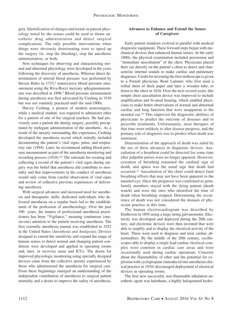
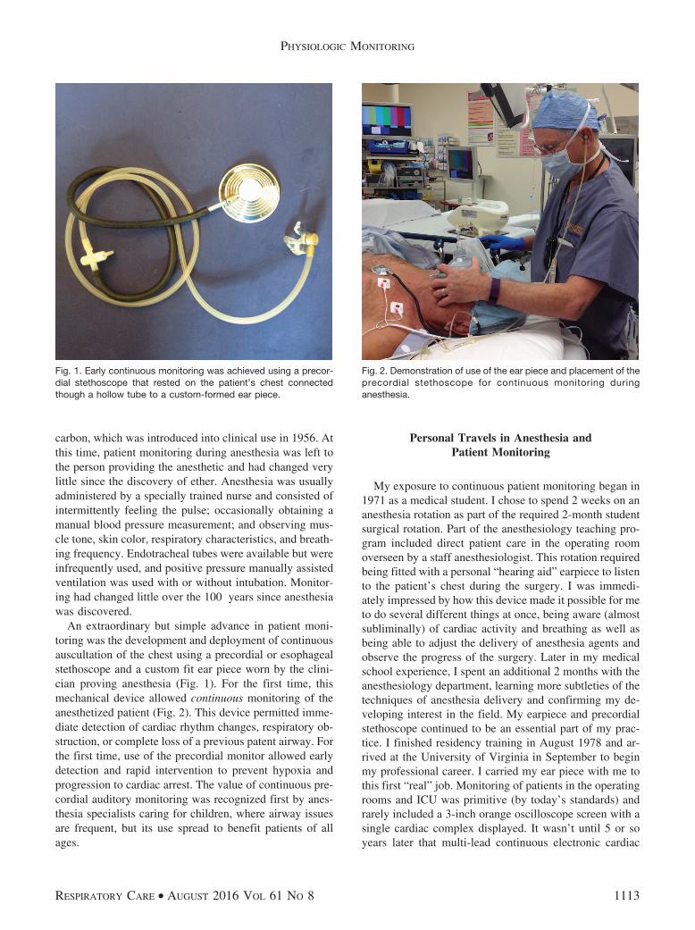
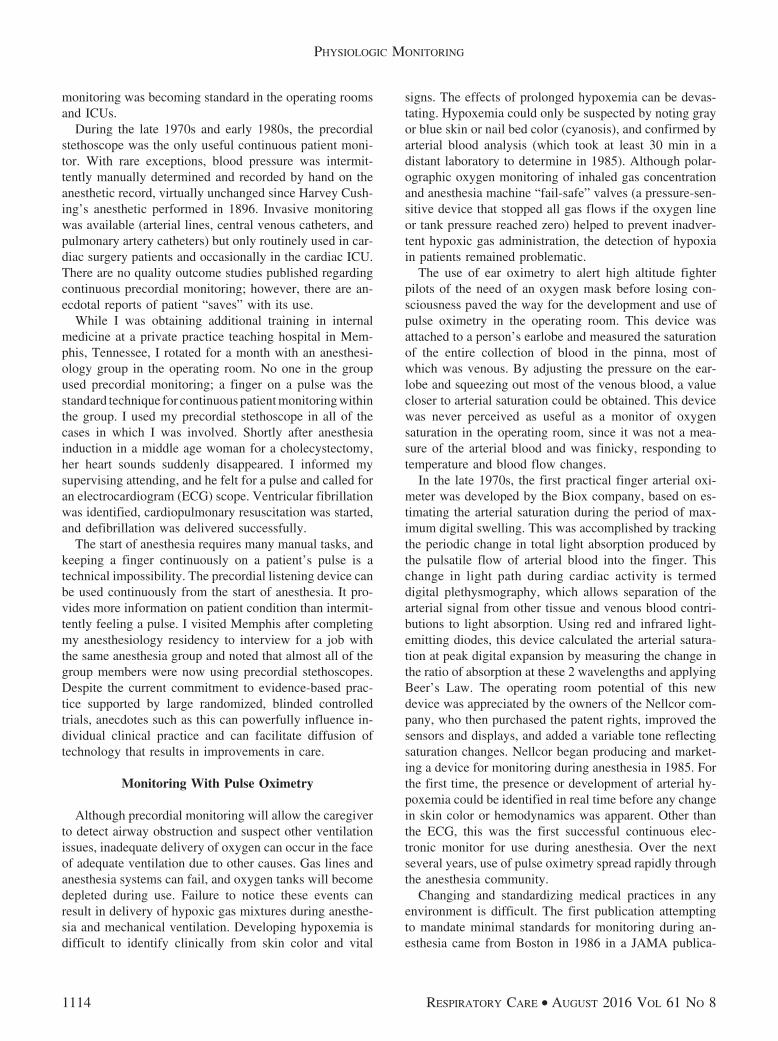
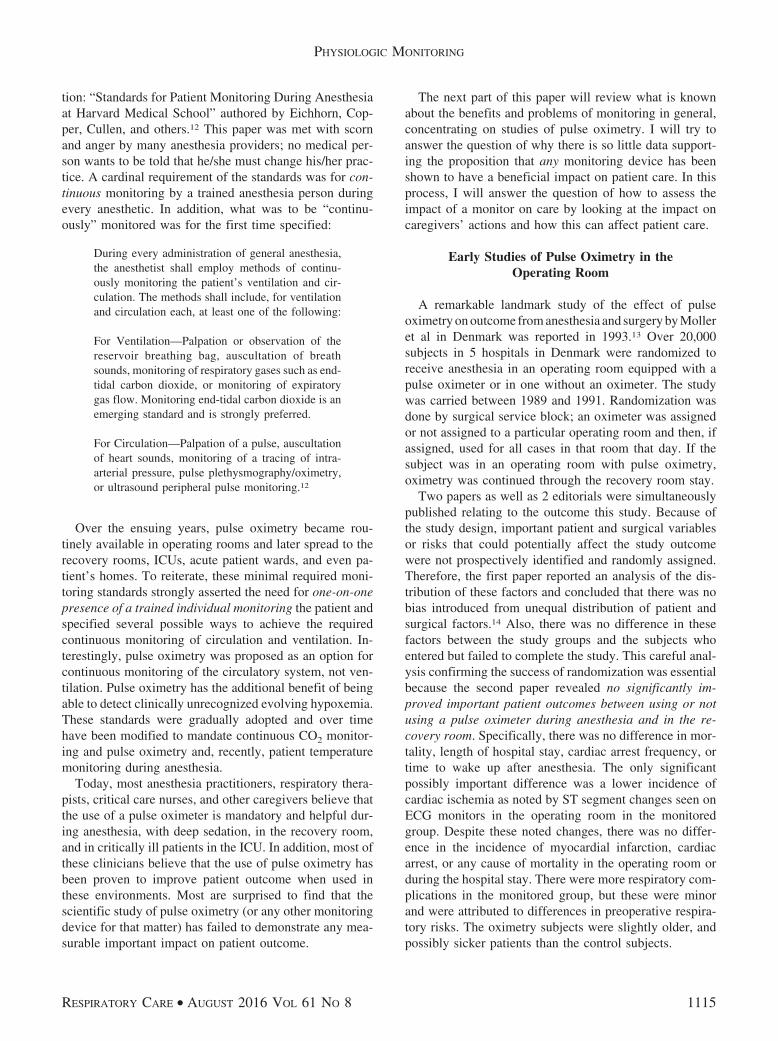
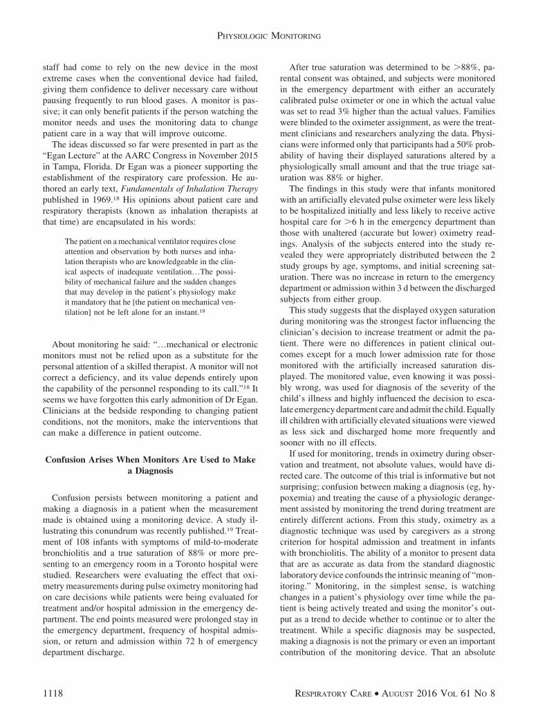
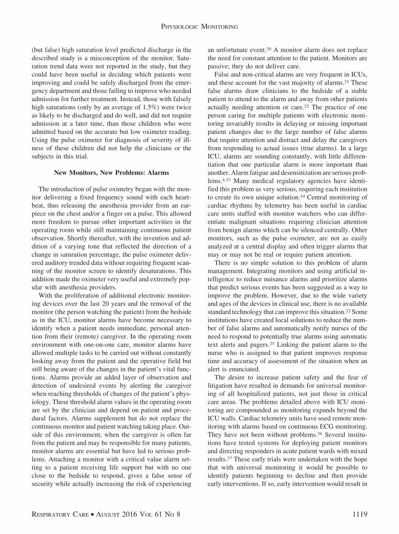
An extraordinary but simple advance in patient moni-toring was the development and deployment of continuousauscultation of the chest using a precordial or esophagealstethoscope and a custom fit ear piece worn by the clini-cian proving anesthesia (Fig. 1). For the first time, thismechanical device allowed continuous monitoring of theanesthetized patient (Fig. 2). This device permitted imme-diate detection of cardiac rhythm changes, respiratory ob-struction, or complete loss of a previous patent airway. Forthe first time, use of the precordial monitor allowed earlydetection and rapid intervention to prevent hypoxia andprogression to cardiac arrest. The value of continuous pre-cordial auditory monitoring was recognized first by anes-thesia specialists caring for children, where airway issuesare frequent, but its use spread to benefit patients of allages.
Personal Travels in Anesthesia andPatient Monitoring
My exposure to continuous patient monitoring began in1971 as a medical student. I chose to spend 2 weeks on ananesthesia rotation as part of the required 2-month studentsurgical rotation. Part of the anesthesiology teaching pro-gram included direct patient care in the operating roomoverseen by a staff anesthesiologist. This rotation requiredbeing fitted with a personal “hearing aid” earpiece to listento the patient’s chest during the surgery. I was immedi-ately impressed by how this device made it possible for meto do several different things at once, being aware (almostsubliminally) of cardiac activity and breathing as well asbeing able to adjust the delivery of anesthesia agents andobserve the progress of the surgery. Later in my medicalschool experience, I spent an additional 2 months with theanesthesiology department, learning more subtleties of thetechniques of anesthesia delivery and confirming my de-veloping interest in the field. My earpiece and precordialstethoscope continued to be an essential part of my prac-tice. I finished residency training in August 1978 and ar-rived at the University of Virginia in September to beginmy professional career. I carried my ear piece with me tothis first “real” job. Monitoring of patients in the operatingrooms and ICU was primitive (by today’s standards) andrarely included a 3-inch orange oscilloscope screen with asingle cardiac complex displayed. It wasn’t until 5 or soyears later that multi-lead continuous electronic cardiac
Fig. 1. Early continuous monitoring was achieved using a precor-dial stethoscope that rested on the patient’s chest connectedthough a hollow tube to a custom-formed ear piece.
Fig. 2. Demonstration of use of the ear piece and placement of theprecordial stethoscope for continuous monitoring duringanesthesia.
PHYSIOLOGIC MONITORING
RESPIRATORY CARE • AUGUST 2016 VOL 61 NO 8 1113
monitoring was becoming standard in the operating roomsand ICUs.
During the late 1970s and early 1980s, the precordialstethoscope was the only useful continuous patient moni-tor. With rare exceptions, blood pressure was intermit-tently manually determined and recorded by hand on theanesthetic record, virtually unchanged since Harvey Cush-ing’s anesthetic performed in 1896. Invasive monitoringwas available (arterial lines, central venous catheters, andpulmonary artery catheters) but only routinely used in car-diac surgery patients and occasionally in the cardiac ICU.There are no quality outcome studies published regardingcontinuous precordial monitoring; however, there are an-ecdotal reports of patient “saves” with its use.
While I was obtaining additional training in internalmedicine at a private practice teaching hospital in Mem-phis, Tennessee, I rotated for a month with an anesthesi-ology group in the operating room. No one in the groupused precordial monitoring; a finger on a pulse was thestandard technique for continuous patient monitoring withinthe group. I used my precordial stethoscope in all of thecases in which I was involved. Shortly after anesthesiainduction in a middle age woman for a cholecystectomy,her heart sounds suddenly disappeared. I informed mysupervising attending, and he felt for a pulse and called foran electrocardiogram (ECG) scope. Ventricular fibrillationwas identified, cardiopulmonary resuscitation was started,and defibrillation was delivered successfully.
The start of anesthesia requires many manual tasks, andkeeping a finger continuously on a patient’s pulse is atechnical impossibility. The precordial listening device canbe used continuously from the start of anesthesia. It pro-vides more information on patient condition than intermit-tently feeling a pulse. I visited Memphis after completingmy anesthesiology residency to interview for a job withthe same anesthesia group and noted that almost all of thegroup members were now using precordial stethoscopes.Despite the current commitment to evidence-based prac-tice supported by large randomized, blinded controlledtrials, anecdotes such as this can powerfully influence in-dividual clinical practice and can facilitate diffusion oftechnology that results in improvements in care.
Monitoring With Pulse Oximetry
Although precordial monitoring will allow the caregiverto detect airway obstruction and suspect other ventilationissues, inadequate delivery of oxygen can occur in the faceof adequate ventilation due to other causes. Gas lines andanesthesia systems can fail, and oxygen tanks will becomedepleted during use. Failure to notice these events canresult in delivery of hypoxic gas mixtures during anesthe-sia and mechanical ventilation. Developing hypoxemia isdifficult to identify clinically from skin color and vital
signs. The effects of prolonged hypoxemia can be devas-tating. Hypoxemia could only be suspected by noting grayor blue skin or nail bed color (cyanosis), and confirmed byarterial blood analysis (which took at least 30 min in adistant laboratory to determine in 1985). Although polar-ographic oxygen monitoring of inhaled gas concentrationand anesthesia machine “fail-safe” valves (a pressure-sen-sitive device that stopped all gas flows if the oxygen lineor tank pressure reached zero) helped to prevent inadver-tent hypoxic gas administration, the detection of hypoxiain patients remained problematic.
The use of ear oximetry to alert high altitude fighterpilots of the need of an oxygen mask before losing con-sciousness paved the way for the development and use ofpulse oximetry in the operating room. This device wasattached to a person’s earlobe and measured the saturationof the entire collection of blood in the pinna, most ofwhich was venous. By adjusting the pressure on the ear-lobe and squeezing out most of the venous blood, a valuecloser to arterial saturation could be obtained. This devicewas never perceived as useful as a monitor of oxygensaturation in the operating room, since it was not a mea-sure of the arterial blood and was finicky, responding totemperature and blood flow changes.
In the late 1970s, the first practical finger arterial oxi-meter was developed by the Biox company, based on es-timating the arterial saturation during the period of max-imum digital swelling. This was accomplished by trackingthe periodic change in total light absorption produced bythe pulsatile flow of arterial blood into the finger. Thischange in light path during cardiac activity is termeddigital plethysmography, which allows separation of thearterial signal from other tissue and venous blood contri-butions to light absorption. Using red and infrared light-emitting diodes, this device calculated the arterial satura-tion at peak digital expansion by measuring the change inthe ratio of absorption at these 2 wavelengths and applyingBeer’s Law. The operating room potential of this newdevice was appreciated by the owners of the Nellcor com-pany, who then purchased the patent rights, improved thesensors and displays, and added a variable tone reflectingsaturation changes. Nellcor began producing and market-ing a device for monitoring during anesthesia in 1985. Forthe first time, the presence or development of arterial hy-poxemia could be identified in real time before any changein skin color or hemodynamics was apparent. Other thanthe ECG, this was the first successful continuous elec-tronic monitor for use during anesthesia. Over the nextseveral years, use of pulse oximetry spread rapidly throughthe anesthesia community.
Changing and standardizing medical practices in anyenvironment is difficult. The first publication attemptingto mandate minimal standards for monitoring during an-esthesia came from Boston in 1986 in a JAMA publica-
PHYSIOLOGIC MONITORING
1114 RESPIRATORY CARE • AUGUST 2016 VOL 61 NO 8
tion: “Standards for Patient Monitoring During Anesthesiaat Harvard Medical School” authored by Eichhorn, Cop-per, Cullen, and others.12 This paper was met with scornand anger by many anesthesia providers; no medical per-son wants to be told that he/she must change his/her prac-tice. A cardinal requirement of the standards was for con-tinuous monitoring by a trained anesthesia person duringevery anesthetic. In addition, what was to be “continu-ously” monitored was for the first time specified:
During every administration of general anesthesia,the anesthetist shall employ methods of continu-ously monitoring the patient’s ventilation and cir-culation. The methods shall include, for ventilationand circulation each, at least one of the following:
For Ventilation—Palpation or observation of thereservoir breathing bag, auscultation of breathsounds, monitoring of respiratory gases such as end-tidal carbon dioxide, or monitoring of expiratorygas flow. Monitoring end-tidal carbon dioxide is anemerging standard and is strongly preferred.
For Circulation—Palpation of a pulse, auscultationof heart sounds, monitoring of a tracing of intra-arterial pressure, pulse plethysmography/oximetry,or ultrasound peripheral pulse monitoring.12
Over the ensuing years, pulse oximetry became rou-tinely available in operating rooms and later spread to therecovery rooms, ICUs, acute patient wards, and even pa-tient’s homes. To reiterate, these minimal required moni-toring standards strongly asserted the need for one-on-onepresence of a trained individual monitoring the patient andspecified several possible ways to achieve the requiredcontinuous monitoring of circulation and ventilation. In-terestingly, pulse oximetry was proposed as an option forcontinuous monitoring of the circulatory system, not ven-tilation. Pulse oximetry has the additional benefit of beingable to detect clinically unrecognized evolving hypoxemia.These standards were gradually adopted and over timehave been modified to mandate continuous CO2 monitor-ing and pulse oximetry and, recently, patient temperaturemonitoring during anesthesia.
Today, most anesthesia practitioners, respiratory thera-pists, critical care nurses, and other caregivers believe thatthe use of a pulse oximeter is mandatory and helpful dur-ing anesthesia, with deep sedation, in the recovery room,and in critically ill patients in the ICU. In addition, most ofthese clinicians believe that the use of pulse oximetry hasbeen proven to improve patient outcome when used inthese environments. Most are surprised to find that thescientific study of pulse oximetry (or any other monitoringdevice for that matter) has failed to demonstrate any mea-surable important impact on patient outcome.
The next part of this paper will review what is knownabout the benefits and problems of monitoring in general,concentrating on studies of pulse oximetry. I will try toanswer the question of why there is so little data support-ing the proposition that any monitoring device has beenshown to have a beneficial impact on patient care. In thisprocess, I will answer the question of how to assess theimpact of a monitor on care by looking at the impact oncaregivers’ actions and how this can affect patient care.
Early Studies of Pulse Oximetry in theOperating Room
A remarkable landmark study of the effect of pulseoximetry on outcome from anesthesia and surgery by Molleret al in Denmark was reported in 1993.13 Over 20,000subjects in 5 hospitals in Denmark were randomized toreceive anesthesia in an operating room equipped with apulse oximeter or in one without an oximeter. The studywas carried between 1989 and 1991. Randomization wasdone by surgical service block; an oximeter was assignedor not assigned to a particular operating room and then, ifassigned, used for all cases in that room that day. If thesubject was in an operating room with pulse oximetry,oximetry was continued through the recovery room stay.
Two papers as well as 2 editorials were simultaneouslypublished relating to the outcome this study. Because ofthe study design, important patient and surgical variablesor risks that could potentially affect the study outcomewere not prospectively identified and randomly assigned.Therefore, the first paper reported an analysis of the dis-tribution of these factors and concluded that there was nobias introduced from unequal distribution of patient andsurgical factors.14 Also, there was no difference in thesefactors between the study groups and the subjects whoentered but failed to complete the study. This careful anal-ysis confirming the success of randomization was essentialbecause the second paper revealed no significantly im-proved important patient outcomes between using or notusing a pulse oximeter during anesthesia and in the re-covery room. Specifically, there was no difference in mor-tality, length of hospital stay, cardiac arrest frequency, ortime to wake up after anesthesia. The only significantpossibly important difference was a lower incidence ofcardiac ischemia as noted by ST segment changes seen onECG monitors in the operating room in the monitoredgroup. Despite these noted changes, there was no differ-ence in the incidence of myocardial infarction, cardiacarrest, or any cause of mortality in the operating room orduring the hospital stay. There were more respiratory com-plications in the monitored group, but these were minorand were attributed to differences in preoperative respira-tory risks. The oximetry subjects were slightly older, andpossibly sicker patients than the control subjects.
PHYSIOLOGIC MONITORING
RESPIRATORY CARE • AUGUST 2016 VOL 61 NO 8 1115
The use of the pulse oximeter resulted in a markeddifference between the treatment and control in the iden-tification and treatment of hypoxemia, which was 19 timesmore frequent in the monitored group (eg, the device wasbetter at diagnosing hypoxemia than clinicians were atdetecting early cyanosis). Subjects in the oximetry groupmore frequently received supplemental oxygen in the re-covery room and were more likely to be discharged to thefloor on oxygen. As a group, the monitored subjects stayedan average of 15 min longer in the recovery room beforedischarge to an acute hospital floor. The postoperativecomplication rate was about 10% in both groups. Changesin clinical care were more frequent in the monitored groupand increased with increasing preoperative patient illness.In the sickest group of subjects with oximetry (AmericanSociety of Anesthesiologists physical status 4), over 20%had care changed in the operating room and over 25% inthe post-anesthesia care unit, mostly receiving increasedinspired oxygen or narcotic reversal agents, identified byoxygen saturation changes.
The most interesting findings from this study came fromthe survey of the anesthesiologists who participated. De-spite knowing the lack of impact pulse oximetry had onserious patient outcomes, 92% of the 104 anesthesiologistsbelieved that using a pulse oximeter during anesthesia andin the recovery room would be beneficial and improvesafety. In addition, 18% stated they experienced a specificevent where pulse oximetry allowed avoidance of a seri-ous event or complication. Ninety-four percent reportedthat they had experienced an event where pulse oximetrywas very helpful at guiding clinical care. Only one personreported that the device had provided false security duringan event leading to (possibly) a worse outcome. Eightypercent said that they felt more secure when using a pulseoximeter, and 54% said its use had changed their clinicalpractice of anesthesia.
Since the publication of this early, large study, severalother trials evaluating the impact of oximetry in the oper-ating room have been carried out. Despite these efforts, nosignificant improvements in important patient outcomeshave been identified with pulse oximetry use in the peri-operative patient population. This has led to a CochraneCollaborative authors’ conclusion in 201415 that is essen-tially unchanged from the initial conclusions in 1993, 2003,and 2009:
The studies confirmed that pulse oximetry can de-tect hypoxemia and related events. However, wehave found no evidence that pulse oximetry affectsthe outcome of anesthesia for patients. The conflict-ing subjective and objective results of the studies,despite an intense methodical collection of data froma relatively large general surgery population, indi-cate that the value of perioperative monitoring with
pulse oximetry is questionable in relation to im-proved reliable outcomes, effectiveness, and effi-ciency. Routine continuous pulse oximetry moni-toring did not reduce either transfer to ICU ormortality, and it is unclear if there is any real ben-efit from the application of this technology for pa-tients…15
At first blush, this evidence-based conclusion, whichhas stood unchallenged by additional data for over 20 years,seems unlikely because of the universal belief that earlydetection and prevention of hypoxemia will improve pa-tient outcome in the operating room and post-anesthesiacare unit. This belief is widely shared by patients andclinicians, and the use of pulse oximetry has become man-datory as a practice standard in anesthesia. Possibly as aby-product of mandatory monitoring with pulse oximetryduring anesthesia, dramatically reduced malpractice insur-ance rates in anesthesiology have occurred. This is despitethe lack of scientific support demonstrating less patientrisk of harm with its use.
The fundamental problem with analysis of monitors isthey do not deliver care; they only deliver data that aresponsible caregiver can use to deliver or change care. Toassociate a patient’s outcome (good or bad) with use of amonitor, the clinician must take an action that requiresusing the data being monitored, an action that would nothave been taken without the monitor being available. Earlydevelopment of oxygen desaturation is indeed difficult toclinically detect and can be caused by mechanical failures,technical faults, patient physiological changes, and otherthings. Identifying early desaturations can alert the care-giver of the need to look more carefully for a developingproblem with the oxygen source, the anesthesia machinefunction, the patient’s breathing, the mechanical ventila-tor, or changed cardiac or vascular function. Severe hy-poxemia is associated with detectable clinical changes (cy-anosis) and may have severe patient consequences, but thisis only likely if hypoxia is not quickly reversed or if car-diac activity is severely compromised (ie, cardiac arrest orprofound hypotension). To summarize, pulse oximetry candeliver notice of impending severe hypoxemia, but thedevice itself is only helpful to the patient by alerting thecaregiver of this possibility. The pulse oximeter does notidentify the cause or deliver any treatment that could ben-efit the patient. If patient outcome is changed by using amonitor, then the actions of the caregivers must havechanged due to having the monitored data, and these ac-tions are the cause for the changed outcome, not the mon-itor.
To appropriately evaluate the impact of a particular mon-itor on patients, one must study the impact of the monitoron the caregiver behavior rather than on the patient out-come only. Few studies of pulse oximetry, or any other
PHYSIOLOGIC MONITORING
1116 RESPIRATORY CARE • AUGUST 2016 VOL 61 NO 8
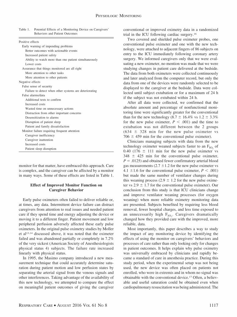
monitor for that matter, have embraced this approach. Careis complex, and the caregiver can be affected by a monitorin many ways. Some of these effects are listed in Table 1.
Effect of Improved Monitor Function onCaregiver Behavior
Early pulse oximeters often failed to deliver reliable or,at times, any data. Intermittent device failure can distractcaregivers from attention to real issues and needed patientcare if they spend time and energy adjusting the device ormoving it to a different finger. Patient movement and lowperipheral perfusion adversely affected these early pulseoximeters. In the original pulse oximetry studies by Molleret al13,14 discussed above, it was noted that the oximeterfailed and was abandoned partially or completely in 7.2%of the very sickest (American Society of Anesthesiologistsphysical status 4) subjects. The failure rate increasedlinearly with physical status.
In 1995, the Masimo company introduced a new mea-surement technique that could accurately determine satu-ration during patient motion and low perfusion states byseparating the arterial signal from the venous signals andother interferences. Taking advantage of the availability ofthis new technology, we attempted to compare the effecton meaningful patient outcomes of giving the caregiver
conventional or improved oximetry data in a randomizedtrial in the ICU following cardiac surgery.16
Two covered and shielded pulse oximeter probes, oneconventional pulse oximeter and one with the new tech-nology, were attached to adjacent fingers of 86 subjects onentry to the ICU immediately following coronary arterysurgery. We informed caregivers only that we were eval-uating a new oximeter, no mention was made that we werestudying changes in patient care delivered at the bedside.The data from both oximeters were collected continuouslyand later analyzed from the computer record, but only thedata from one of the devices were randomly selected to bedisplayed to the caregiver at the bedside. Data were col-lected until subject extubation or for a maximum of 24 hif the subject was not extubated within 24 h.
After all data were collected, we confirmed that theabsolute amount and percentage of nonfunctional moni-toring time were significantly greater for the conventionalthan for the new technology (8.7 � 16.4% vs 1.2 � 3.3%for the new pulse oximeter, P � .001) and the time toextubation was not different between the 2 groups(634 � 328 min for the new pulse oximeter vs706 � 459 min for the conventional pulse oximeter).
Clinicians managing subjects with data from the newtechnology oximeter weaned subjects faster to an FIO2
of0.40 (176 � 111 min for the new pulse oximeter vs348 � 425 min for the conventional pulse oximeter,P � .0125) and obtained fewer confirmatory arterial bloodgas measurements (2.7 �1.2 for the new pulse oximeter vs4.1 �1.6 for the conventional pulse oximeter, P � .001)but made the same number of ventilator changes duringthis weaning process (2.9 � 1.2 for the new pulse oxime-ter vs 2.9 � 1.7 for the conventional pulse oximeter). Ourconclusion from this study is that ICU clinicians changeand improve ventilator weaning processes (for oxygenweaning) when more reliable oximetry monitoring dataare presented. Subjects benefited by requiring less bloodremoval, fewer hospital charges, and less time exposed toan unnecessarily high FIO2
. Caregivers dramaticallychanged how they provided care with the improved, morereliable, data.
Most importantly, this paper describes a way to studythe impact of any monitoring device by identifying theeffects of using the monitor on caregivers’ behaviors andprocesses of care rather than only looking only for changesin patient outcomes. It helps explain why pulse oximetrywas universally embraced by clinicians and rapidly be-came a standard of care in anesthesia practice. During thisstudy period, when the experimental setup was not beingused, the new device was often placed on patients notenrolled, who were in extremis and in whom no signal wasobtainable with the conventional device.17 Often, a believ-able and useful saturation could be obtained even whencardiopulmonary resuscitation was being administered. The
Table 1. Potential Effects of a Monitoring Device on Caregivers’Behaviors and Patient Outcomes
Positive effectsEarly warning of impending problems
Better outcomes with actionable eventsIncreased patient safetyAbility to watch more than one patient simultaneouslyLower costs
Assurance that things monitored are all rightMore attention to other tasksMore attention to other patients
Negative effectsFalse sense of security
Failure to detect when other systems are deterioratingFalse alarms/data
Additional tests to confirmIncreased costsWasted time on unnecessary actionsDistraction from other important concernsDesensitization to alarmsDisruption of patient sleepPatient and family dissatisfaction
Monitor failure requiring frequent attentionCaregiver inefficiencyCaregiver inattentionIncreased costsPatient sleep disruption
PHYSIOLOGIC MONITORING
RESPIRATORY CARE • AUGUST 2016 VOL 61 NO 8 1117
staff had come to rely on the new device in the mostextreme cases when the conventional device had failed,giving them confidence to deliver necessary care withoutpausing frequently to run blood gases. A monitor is pas-sive; it can only benefit patients if the person watching themonitor needs and uses the monitoring data to changepatient care in a way that will improve outcome.
The ideas discussed so far were presented in part as the“Egan Lecture” at the AARC Congress in November 2015in Tampa, Florida. Dr Egan was a pioneer supporting theestablishment of the respiratory care profession. He au-thored an early text, Fundamentals of Inhalation Therapypublished in 1969.18 His opinions about patient care andrespiratory therapists (known as inhalation therapists atthat time) are encapsulated in his words:
The patient on a mechanical ventilator requires closeattention and observation by both nurses and inha-lation therapists who are knowledgeable in the clin-ical aspects of inadequate ventilation…The possi-bility of mechanical failure and the sudden changesthat may develop in the patient’s physiology makeit mandatory that he [the patient on mechanical ven-tilation] not be left alone for an instant.18
About monitoring he said: “…mechanical or electronicmonitors must not be relied upon as a substitute for thepersonal attention of a skilled therapist. A monitor will notcorrect a deficiency, and its value depends entirely uponthe capability of the personnel responding to its call.”18 Itseems we have forgotten this early admonition of Dr Egan.Clinicians at the bedside responding to changing patientconditions, not the monitors, make the interventions thatcan make a difference in patient outcome.
Confusion Arises When Monitors Are Used to Makea Diagnosis
Confusion persists between monitoring a patient andmaking a diagnosis in a patient when the measurementmade is obtained using a monitoring device. A study il-lustrating this conundrum was recently published.19 Treat-ment of 108 infants with symptoms of mild-to-moderatebronchiolitis and a true saturation of 88% or more pre-senting to an emergency room in a Toronto hospital werestudied. Researchers were evaluating the effect that oxi-metry measurements during pulse oximetry monitoring hadon care decisions while patients were being evaluated fortreatment and/or hospital admission in the emergency de-partment. The end points measured were prolonged stay inthe emergency department, frequency of hospital admis-sion, or return and admission within 72 h of emergencydepartment discharge.
After true saturation was determined to be �88%, pa-rental consent was obtained, and subjects were monitoredin the emergency department with either an accuratelycalibrated pulse oximeter or one in which the actual valuewas set to read 3% higher than the actual values. Familieswere blinded to the oximeter assignment, as were the treat-ment clinicians and researchers analyzing the data. Physi-cians were informed only that participants had a 50% prob-ability of having their displayed saturations altered by aphysiologically small amount and that the true triage sat-uration was 88% or higher.
The findings in this study were that infants monitoredwith an artificially elevated pulse oximeter were less likelyto be hospitalized initially and less likely to receive activehospital care for �6 h in the emergency department thanthose with unaltered (accurate but lower) oximetry read-ings. Analysis of the subjects entered into the study re-vealed they were appropriately distributed between the 2study groups by age, symptoms, and initial screening sat-uration. There was no increase in return to the emergencydepartment or admission within 3 d between the dischargedsubjects from either group.
This study suggests that the displayed oxygen saturationduring monitoring was the strongest factor influencing theclinician’s decision to increase treatment or admit the pa-tient. There were no differences in patient clinical out-comes except for a much lower admission rate for thosemonitored with the artificially increased saturation dis-played. The monitored value, even knowing it was possi-bly wrong, was used for diagnosis of the severity of thechild’s illness and highly influenced the decision to esca-late emergency department care and admit the child. Equallyill children with artificially elevated situations were viewedas less sick and discharged home more frequently andsooner with no ill effects.
If used for monitoring, trends in oximetry during obser-vation and treatment, not absolute values, would have di-rected care. The outcome of this trial is informative but notsurprising; confusion between making a diagnosis (eg, hy-poxemia) and treating the cause of a physiologic derange-ment assisted by monitoring the trend during treatment areentirely different actions. From this study, oximetry as adiagnostic technique was used by caregivers as a strongcriterion for hospital admission and treatment in infantswith bronchiolitis. The ability of a monitor to present datathat are as accurate as data from the standard diagnosticlaboratory device confounds the intrinsic meaning of “mon-itoring.” Monitoring, in the simplest sense, is watchingchanges in a patient’s physiology over time while the pa-tient is being actively treated and using the monitor’s out-put as a trend to decide whether to continue or to alter thetreatment. While a specific diagnosis may be suspected,making a diagnosis is not the primary or even an importantcontribution of the monitoring device. That an absolute
PHYSIOLOGIC MONITORING
1118 RESPIRATORY CARE • AUGUST 2016 VOL 61 NO 8
(but false) high saturation level predicted discharge in thedescribed study is a misconception of the monitor. Satu-ration trend data were not reported in the study, but theycould have been useful in deciding which patients wereimproving and could be safely discharged from the emer-gency department and those failing to improve who neededadmission for further treatment. Instead, those with falselyhigh saturations (only by an average of 1.5%) were twiceas likely to be discharged and do well, and did not requireadmission at a later time, than those children who wereadmitted based on the accurate but low oximeter reading.Using the pulse oximeter for diagnosis of severity of ill-ness of these children did not help the clinicians or thesubjects in this trial.
New Monitors, New Problems: Alarms
The introduction of pulse oximetry began with the mon-itor delivering a fixed frequency sound with each heart-beat, thus releasing the anesthesia provider from an ear-piece on the chest and/or a finger on a pulse. This allowedmore freedom to pursue other important activities in theoperating room while still maintaining continuous patientobservation. Shortly thereafter, with the invention and ad-dition of a varying tone that reflected the direction of achange in saturation percentage, the pulse oximeter deliv-ered auditory trended data without requiring frequent scan-ning of the monitor screen to identify desaturations. Thisaddition made the oximeter very useful and extremely pop-ular with anesthesia providers.
With the proliferation of additional electronic monitor-ing devices over the last 20 years and the removal of themonitor (the person watching the patient) from the bedsideas in the ICU, monitor alarms have become necessary toidentify when a patient needs immediate, personal atten-tion from their (remote) caregiver. In the operating roomenvironment with one-on-one care, monitor alarms haveallowed multiple tasks to be carried out without constantlylooking away from the patient and the operative field butstill being aware of the changes in the patient’s vital func-tions. Alarms provide an added layer of observation anddetection of undesired events by alerting the caregiverwhen reaching thresholds of changes of the patient’s phys-iology. These threshold alarm values in the operating roomare set by the clinician and depend on patient and proce-dural factors. Alarms supplement but do not replace thecontinuous monitor and patient watching taking place. Out-side of this environment, when the caregiver is often farfrom the patient and may be responsible for many patients,monitor alarms are essential but have led to serious prob-lems. Attaching a monitor with a critical value alarm set-ting to a patient receiving life support but with no oneclose to the bedside to respond, gives a false sense ofsecurity while actually increasing the risk of experiencing
an unfortunate event.20 A monitor alarm does not replacethe need for constant attention to the patient. Monitors arepassive; they do not deliver care.
False and non-critical alarms are very frequent in ICUs,and these account for the vast majority of alarms.21 Thesefalse alarms draw clinicians to the bedside of a stablepatient to attend to the alarm and away from other patientsactually needing attention or care.22 The practice of oneperson caring for multiple patients with electronic moni-toring invariably results in delaying or missing importantpatient changes due to the large number of false alarmsthat require attention and distract and delay the caregiversfrom responding to actual issues (true alarms). In a largeICU, alarms are sounding constantly, with little differen-tiation that one particular alarm is more important thananother. Alarm fatigue and desensitization are serious prob-lems.4,23 Many medical regulatory agencies have identi-fied this problem as very serious, requiring each institutionto create its own unique solution.24 Central monitoring ofcardiac rhythms by telemetry has been useful in cardiaccare units staffed with monitor watchers who can differ-entiate malignant situations requiring clinician attentionfrom benign alarms which can be silenced centrally. Othermonitors, such as the pulse oximeter, are not as easilyanalyzed at a central display and often trigger alarms thatmay or may not be real or require patient attention.
There is no simple solution to this problem of alarmmanagement. Integrating monitors and using artificial in-telligence to reduce nuisance alarms and prioritize alarmsthat predict serious events has been suggested as a way toimprove the problem. However, due to the wide varietyand ages of the devices in clinical use, there is no availablestandard technology that can improve this situation.22 Someinstitutions have created local solutions to reduce the num-ber of false alarms and automatically notify nurses of theneed to respond to potentially true alarms using automatictext alerts and pagers.25 Linking the patient alarm to thenurse who is assigned to that patient improves responsetime and accuracy of assessment of the situation when analert is enunciated.
The desire to increase patient safety and the fear oflitigation have resulted in demands for universal monitor-ing of all hospitalized patients, not just those in criticalcare areas. The problems detailed above with ICU moni-toring are compounded as monitoring expands beyond theICU walls. Cardiac telemetry units have used remote mon-itoring with alarms based on continuous ECG monitoring.They have not been without problems.26 Several institu-tions have tested systems for deploying patient monitorsand directing responders in acute patient wards with mixedresults.27 These early trials were undertaken with the hopethat with universal monitoring it would be possible toidentify patients beginning to decline and then provideearly interventions. If so, early intervention would result in
PHYSIOLOGIC MONITORING
RESPIRATORY CARE • AUGUST 2016 VOL 61 NO 8 1119
fewer rapid response team calls and ICU admissions andshorter lengths of stay for those admitted to an ICU.
In one study, the researchers implemented a patient sur-veillance system based on pulse oximetry with nursingnotification of violation of alarm limits via wireless pager.28
Data were collected for several months before and severalmonths after implementation of the monitoring and callsystem. Concurrently, matching outcome data were col-lected on 2 other postoperative units not being monitored.Baseline threshold alarm limits were set to reach a balancebetween actionable and false positive alarms (SpO2
�80%and heart rate �50 and �140 beats/min). These limitscould be altered by the nursing staff up to �10% or alteredfurther, for a specific reason, by a physician. Notificationdelay is an important issue in alarm frequency manage-ment.29 Appropriate delay eliminates many transient andmotion artifact-generated false alarms; a 15-s audio alarmdelay at the bedside and an additional 15-s delay for pagerannunciation was chosen for this study (leading to a 30-sdelay before a nurse would be notified by pager of viola-tion of alarm thresholds).
There were half as many deaths observed after imple-mentation (2 deaths) compared with the time before (4deaths) in the studied unit. These included deaths on theward and those occurring after transfer to an ICU. Thisobservation did not reach statistical significance. Rescueevents were dramatically less frequent in the monitoredunit compared with the non-monitored units and decreasedfrom 3.4 (1.89–4.85) to 1.2 (0.53–1.88) per 1,000 patientdischarges after implementation. No significant changeswere seen for rescue events in the comparison units be-tween the 2 time periods. Transfers to the ICU also de-clined significantly, from 5.6 (3.7–7.4) per 1,000 patientdays to 2.9 (1.4–4.3) in the monitored unit, whereas onlysmall, nonsignificant changes were observed in the 2 com-parison units between the 2 study periods. This study sug-gests that there may be benefits of implementing universalmonitoring, but a balance between reaching the alarmthreshold and delaying the nursing call must be carefullydecided to reduce the false negative calls. Also, trainedstaff and reliable communication systems must be estab-lished and meticulously maintained to achieve success.This study only included monitoring with pulse oximetry;adding additional monitors increases manyfold the oppor-tunities for failure. Although this study is mildly encour-aging, the authors state that they experienced failure inseveral previous institutional attempts to deploy universalmonitoring. They focus in this paper on the details of howthey improved the response of those staff who must reactto all alarms if the system is to be fail-safe.
In summary, solutions to detection of patients who be-gin to deteriorate outside of intensive monitoring areaswhere responders are not close to the bedside cannot de-pend on our current use of monitoring devices and the
monitoring practices as employed in ICU settings. There isa misguided belief that universal monitoring of all hospi-talized patients with pulse oximetry or other monitors willprevent undesired outcomes and improve patient safety.30
As emphasized throughout this paper, monitors are pas-sive; they do not provide patient care. Only caregivers canimprove outcomes by making correct interventions whenthey are needed, and only by being at the bedside at theright time. Monitors and alarms can alert responsible care-givers as to when to be at the bedside, but adequate andtrained personnel must be readily available to arrive swiftlyand administer needed care. No current monitoring or alert-ing system is 100% accurate, and massive numbers offalse alarms will still be issued, compounding the need forpatient and monitor attention. Dealing with the distractingfalse calls but remaining vigilant for the true ones is thebiggest problem preventing successful response to alarms.Deployment of monitors is easy; establishing prompt, re-liable, and informed responses is hard. Doing the firstwithout the second is a prescription for failure and in-creased patient harm. Such a failure can inflict a hugeburden of guilt on the caregivers involved and increase thepotential liability to the institution.
Solutions to early detection of patient deterioration needto be more robust than the simple alarms issued from themonitoring devices currently in use. Research needs to bedone integrating multiple patient variables from large mon-itored patient populations to identify markers that reliablypredict the need for care escalation. Human factors, suchas fatigue, need exploration and interventions to improvecaregiver vigilance during long care shifts. Adequatebackup personnel and frequent breaks are helpful in pre-venting exhaustion and desensitization to repetitive alarms.Institutions considering providing universal monitoringwould be wise to proceed deliberately and with adequateattention to the devices selected and the humans respon-sible for the care to be delivered.
Conclusions
Monitors are passive; they do not deliver care. By them-selves they cannot achieve or affect important patient out-comes. The lack of quality outcome data is not a condem-nation of monitoring; the question of how to study monitorsmust be restated. Monitors (ie, the caregivers who arewatching patients) do affect patient outcome. When care isone-on-one, a physiological monitor can help the caregiverto give the best care by demonstrating the immediate needfor and effects of the intervention. When the care is pro-vided to several patients simultaneously, monitors can pro-vide alarms that attract caregiver attention. If most alarmsare not actionable, care will suffer. Finding safe and reli-able solutions to too many false alarms in this setting is
PHYSIOLOGIC MONITORING
1120 RESPIRATORY CARE • AUGUST 2016 VOL 61 NO 8
essential but has not yet been achieved. For now, patientsand caregivers suffer daily from alarm overload.
REFERENCES
1. Ospina-Tascon GA, Cordioli RL, Vincent JL. What type of moni-toring has been shown to improve outcomes in acutely ill patients?Intensive Care Med 2008;34(5):800-820.
2. Hadian M, Pinsky MR. Evidence-based review of the use of thepulmonary artery catheter: impact data and complications. Crit Care2006;10(Suppl 3):S8.
3. Drew BJ, Harris P, Zegre-Hemsey JK, Mammone T, Schindler D,Salas-Boni R, et al. Insights into the problem of alarm fatigue withphysiologic monitor devices: a comprehensive observational studyof consecutive intensive care unit patients. PLoS One 2014;9(10):e110274.
4. Mitka M. Joint commission warns of alarm fatigue: multitude ofalarms from monitoring devices problematic. JAMA 2013;309(22):2315-2316.
5. Powner DJ, Ackerman BM, Grenvik A. Medical diagnosis of deathin adults: historical contributions to current controversies. Lancet1996;348(9036):1219-1223.
6. Hales S. An account of some hydraulic and hydrostatical experi-ments made on the blood and blood-vessels of animals. 1710 [clas-sical article]. J Clin Monit Comput 2000;16(1):45-47.
7. Sakula A. K.T.H. Laennec 1781-1826: his life and work: a bicente-nary appreciation. Thorax 1981;36(2):81-90.
8. Fisher JA, Bromberg IL, Eisen LB. On the design of the anaesthesiarecord forms. Can J Anaesth 1994;41(10):973-983.
9. Wright AJ. Early use of the Cushing-Codman Anesthesia Record.Anesthesiology 1987;66(1):92.
10. MacIntyre JWR. Stethoscopy during anaesthesia. Can J Anaesth 1997;44(5):535-542.
11. Gardiner D, Shemie S, Manara A, Opdam H. International perspec-tive on the diagnosis of death. Br J Anaesth 2012;108(Suppl 1):i14-i28.
12. Eichhorn JH, Cooper JB, Cullen DJ, Maier WR, Philip JH, SeemanRG. Standards for patient monitoring during anesthesia at HarvardMedical School. JAMA 1986;256(8):1017-1020.
13. Moller JT, Pedersen T, Rasmussen LS, Jensen PF, Pedersen BD,Ravlo O, et al. Randomized evaluation of pulse oximetry in 20,802patients: I. design, demography, pulse oximetry failure rate, andoverall complication rate. Anesthesiology 1993;78(3):436-444.
14. Moller JT, Johannessen NW, Espersen K, Ravlo O, Pedersen BD,Jensen PF, et al. Randomized evaluation of pulse oximetry in 20,802patients: II. perioperative events and postoperative complications.Anesthesiology 1993;78(3):445-453.
15. Pedersen T1, Nicholson A, Hovhannisyan K, Møller AM, Smith AF,Lewis SR. Pulse oximetry for perioperative monitoring. CochraneDatabase Syst Rev 2014;(3):CD002013.
16. Durbin CG Jr, Rostow SK. More reliable oximetry reduces the fre-quency of arterial blood gas analyses and hastens oxygen weaningafter cardiac surgery: a prospective, randomized trial of the clinicalimpact of a new technology. Crit Care Med 2002;30(8):1735-1740.
17. Durbin CG Jr, Rostow SK. Advantages of new technology pulseoximetry with adults in extremis. Anesth Analg 2002;94(1 Suppl):S81-S83.
18. Egan DF. Fundamentals of Inhalation Therapy. St Louis: Mosby;1969. 371, 402.
19. Schuh S, Freedman S, Coates A, Allen U, Parkin PC, Stephens D,et al. Effect of oximetry on hospitalization in bronchiolitis: a ran-domized clinical trial. JAMA 2014;312(7):712-718.
20. Schmid F, Goepfert G, Reuter DA. Patient monitoring alarms in theICU and in the operating room. Crit Care 2013;17(2):216.
21. Siebig S, Kuhls S, Imhoff M, Gather U, Scholmerich J, Wrede CE.Intensive care unit alarms: how many do we need? Crit Care Med2010;38(2):451-456.
22. Cvach M. Monitor alarm fatigue: an integrative review. BiomedInstrum Technol 2012;46(4):268-277.
23. Bonafide CP, Lin R, Zander M, Graham CS, Paine CW, Rock W,et al. Association between exposure to nonactionable physiologicmonitor alarms and response time in a children’s hospital. J HospMed 2015;10(6):345-351.
24. Chopra V, McMahon LF. Redesigning hospital alarms for patientsafety: alarmed and potentially dangerous. JAMA 2014;311(12):1199-1200.
25. Paine CW, Goel VV, Ely E, Stave CD, Stemler S, Zander M, BonafideCP. Systematic review of physiologic monitor alarm characteristicsand pragmatic interventions to reduce alarm frequency. J Hosp Med2016;11(2):136-144.
26. Gazarian PK. Nurses’ response to frequency and types of electro-cardiography alarms in a non-critical care setting: a descriptive study.Int J Nurs Stud 2014;51(2):190-197.
27. Voepel-Lewis T, Parker ML, Burke CN, Hemberg J, Perlin L, Kai S,Ramachandran SK. Pulse oximetry desaturation alarms on a generalpostoperative adult unit: a prospective observational study of nurseresponse time. Int J Nurs Stud 2013;50(10):1351-1358.
28. Taenzer AH, Pyke JB, McGrath SP, Blike GT. Impact of pulseoximetry surveillance on rescue events and intensive care unit trans-fers: a before-and-after concurrence study. Anesthesiology 2010;112(2):282-287.
29. Cvach MM, Frank RJ, Doyle P, Stevens ZK. Use of pagers with analarm escalation system to reduce cardiac monitor alarm signals. JNurs Care Qual 2014;29(1):9-18.
30. Stoelting RK. Continuous postoperative electronic monitoring andthe will to require it. Anesth Analg 2015;121(3):579-581.
PHYSIOLOGIC MONITORING
RESPIRATORY CARE • AUGUST 2016 VOL 61 NO 8 1121