physicochemical characterization of iron oxide ... · tarfi aziz1*, shah md. masum1, md. rakibul...
TRANSCRIPT

_____________________________________________________________________________________________________ *Corresponding author: E-mail: [email protected] ;
International Research Journal of Pure & Applied Chemistry
11(1): 1-9, 2016, Article no.IRJPAC.23408 ISSN: 2231-3443, NLM ID: 101647669
SCIENCEDOMAIN international
www.sciencedomain.org
Physicochemical Characterization of Iron Oxide Nanoparticle Coated with Chitosan for Biomedical
Application
Tarfi Aziz1*, Shah Md. Masum1, Md. Rakibul Qadir2, Abdul Gafur2
and Dilruba Huq1
1Department of Applied Chemistry and Chemical Engineering, Faculty of Engineering and Technology, University of Dhaka, Bangladesh.
2Pilot Plant and Process Development Center, Bangladesh Council of Scientific and Industrial
Research, Dhaka, Bangladesh.
Authors’ contributions
This work was carried out in collaboration between all authors. Author TA performed the analysis, wrote the protocol and the first draft of the manuscript. Authors SMM, AG and DH supervised the
research, helped with literature searches and provided the instrumental facilities. Author MRQ helped to perform the XRD analysis. All authors read and approved the final manuscript.
Article Information
DOI: 10.9734/IRJPAC/2016/23408
Editor(s):
(1) Bengi Uslu, Department of Analytical Chemistry, Ankara University, Ankara, Turkey. Reviewers:
(1) Aline Elesbao Do Nascimento, Universidade Catolica De Pernambuco, Brazil.
(2) Heshu Sulaiman Rahman, University Putra Malaysia, Malaysia. Complete Peer review History: http://sciencedomain.org/review-history/12868
Received 29th
November 2015 Accepted 22nd December 2015
Published 1st
January 2016
ABSTRACT Aims: Synthesize and characterize a bio-polymer coated iron oxide nanoparticle, which has the potency to be used as an appropriate biomaterial for biomedical purpose. Magnetic iron oxide nanoparticles (MNPs) with biological coating exhibits many properties that can be exploited in a variety of biomedical applications. Place and Duration of Study: The study was performed at Bangladesh Council of Scientific and Industrial Research (BCSIR), Dhaka, Bangladesh from August 2013 to August 2014. Methodology: This research work describes the in-situ synthesis of chitosan-coated magnetic nanoparticles (CS MNPs) by co precipitation method where trisodium phosphate is added as ionic
Original Research Article

Aziz et al.; IRJPAC, 11(1): 1-9, 2016; Article no.IRJPAC.23408
2
cross-linker. Analytical characterization of synthesized nanoparticles was performed using X-Ray Diffraction (XRD), Fourier Transform Infrared Spectroscopy (FTIR), Thermal gravimetric analysis (TGA) and Scanning Electron Microscope (SEM). Results: The XRD analysis proved that the synthesized iron oxide was magnetite (Fe3O4). The layer of chitosan on the magnetite surface was confirmed by FTIR. SEM image demonstrated a comparable morphology. The average diameter of synthesized nanoparticle was found to be 13.4 nm from XRD crystallite size analysis. TGA result indicated that the chitosan content of CS MNP were 29% by weight. Conclusion: The synthesized MNPs have many potential applications in biomedicine including targeted drug delivery, magnetic resonance imaging (MRI), and magnetic hyperthermia.
Keywords: Chitosan; Iron oxide nanoparticle; biomedical application; physicochemical
characterization. 1. INTRODUCTION Magnetic particles with dimensions ranging from the nanometer to the micrometer scales are being used in an increasing number of medical applications. The most important properties of magnetic particles for clinical diagnostics and medical therapies are nontoxicity, biocompatibility, injectability, and high-level accumulation in the target tissue or specific organ [1]. Magnetic iron oxide nanoparticles are the primary choice. The unique feature of MNPs to be guided by an external magnetic field has been used in magnetic resonance imaging (MRI), tissue repair, hyperthermia, drug delivery, and in cell separation [2,3,4,5]. Two other advantages of iron oxide nanoparticles are their low toxicity on cells and superparamagnetic properties. Superparamagnetic nanoparticles exhibit outstanding magnetic properties because they show no magnetization in the absence of a magnetic field but become strongly magnetized in the presence of magnetic field [6]. With the ability to utilize magnetic attraction and or specific targeting of disease biomarkers, MNPs offer an attractive means of remotely directing therapeutic agents specifically to a disease site while simultaneously reducing dosage and the deleterious side effects associated with non-specific uptake of cytotoxic drugs by healthy tissue [7]. For biomedical applications, iron oxide MNP must be precoated with substances that assure their stability, biodegradability, and non-toxicity in the physiological medium in order to achieve combined properties of high magnetic saturation, biocompatibility, and interactive functions on the surface [8]. Size and surface properties of iron oxide nanoparticles are the two important factors which could dramatically affect the nanoparticle efficiency as well as their stability. MNPs are preferred to be super- paramagnetic and have high magnetization
property so that their movement in the blood can be controlled with external magnetic field and be immobilized close to the targeted tissue [4]. Surfactants are used to coat magnetic nanoparticles which acts as a steric barrier to prevent aggregation caused by magnetic dipole–dipole attractions between particles and avoid opsonization [9]. In addition, polymeric coatings provide a means to tailor the surface properties of MNPs such as surface charge and chemical functionality [10]. The iron oxide core can be coated with polymers during the synthesis process and it must be a long chain. Among the coating materials studied to date, chitosan has drawn considerable attention. Chitosan, prevents the particles reacting with blood proteins and receptors [11]. In this study magnetic iron oxide nanoparticles were synthesized by the co-precipitation method because of its potential for large-scale manufacturing, cost-effectiveness, ease of production, and hydrophilicity of nanocrystals [12]. Hydrophilicity of nanoparticles is a key requirement for biomedical applications [13]. Chitosan, made from naturally occurring chitin, is a biodegradable, biocompatible, linear polysaccharide [14] and has many reactive functional groups that can serve as an anchor for conjugation of therapeutics, targeting ligands, and imaging agents [15]. In comparison with many other polymers, the chitosan backbone contains number of free amine groups, which allow binding of many agents. The chitosan coated IONPs have a remarkable heating effect which has great potential in hyperthermia therapy [16].
For biomedical applications, chitosan-coated magnetic nanoparticles (CS MNPs) are generally synthesized by in situ coating method which is

Aziz et al.; IRJPAC, 11(1): 1-9, 2016; Article no.IRJPAC.23408
3
alkaline coprecipitation of Fe (II) and Fe (III) precursors in aqueous solutions of hydrophilic chitosan polymers. These polymers serve to limit the core growth of iron oxide during the preparation, and to stabilize via steric repulsions when the nanoparticles disperse in aqueous media [17]. The co-precipitation method involves the precipitation of iron salts in the presence of chitosan and trisodium phosphate, which acts as a cross linker. Trisodium phosphate cross links the adsorbed chitosan molecules to each other through the ionic interaction. This work aims to evaluate the magnetic chitosan coated iron oxide nanoparticle for biomedical application. The prepared biocomposite was characterized by several analysis for comparative study.
2. MATERIALS AND METHODS Iron (II) chloride tetrahydrate (FeCl2·4H2O), iron (III) chloride hexahydrate (FeCl3·6H2O), acetic acid (CH3COOH), and ammonium hydroxide (NH4OH) were obtained from Merck, Germany; chitosan (LMW, 84.57% deacetylated) and tri sodium phosphate were purchased from Sigma-Aldrich Chemie GmbH, Germany.
2.1 Synthesis of Iron Oxide Nanoparticle Magnetic iron oxide nanoparticles (MNPs) were prepared by alkaline co-precipitation of ferrous chloride tetrahydrate, FeCl2·4H2O (1.34 g) and ferric chloride hexahydrate FeCl3·6H2O (3.40 g) at 1:2 ratio. The salts were dissolved in 150 mL deionized water within a three necked glass balloon. The glass balloon was placed in a heating mantle and stirred with a magnetic stirrer. It was vigorously stirred at 90ºC in the presence of N2 gas. Ammonium hydroxide (NH4OH) was added to the system drop wise. The process was ended by washing with deionized water until the solution pH was 9.0. The solution was then centrifuged at 5000 rpm for 30 minutes. The precipitates were collected and dried in the incubator at 55ºC. The black precipitates were then turned into brown.
2.2 Synthesis of Chitosan-coated Magnetic Iron Oxide Nanoparticles
Chitosan-coated magnetic iron oxide nanoparticles (CS MNP) were in situ synthesized by the co-precipitation of Fe (II) and Fe (III) salts in the presence of chitosan and trisodium
phosphate molecules. Chitosan was previously prepared with degree of deacetylation 75% by titrimetric method [18,19]. Trisodium phosphate was used for the crosslinking of low molecular weight chitosan polymers. Chitosan (0.15 g) was dissolved in 30 ml of 1% acetic acid and the pH was adjusted to 4.8 by 10M NaOH. Iron salts (1.34 g FeCl2·4H2O and 3.40 g FeCl3·6H2O) were dissolved in 30 ml of 0.5% chitosan solution. The solution was then vigorously stirred at 2000 rpm. 10 ml of 22.5% trisodium phosphate and different amounts of 32% NH4OH (18, 20, 22, 25 mL) were added to the solution to obtain the final NH4OH concentration of 31%, at room temperature. The ammonia solution was added very slowly to produce smaller sized nanoparticles. The resulting solution was stirred for an additional 1 hour. The colloidal chitosan coated magnetic Fe3O4 nanoparticles were extensively washed (3 times) with deionized water and separated by centrifugation and drying.
2.3 Analytical Characterization Characterization of MNPs, chitosan biopolymers and CSMNPs were carried out by X-ray diffraction (XRD), Fourier Transformed Infrared Spectroscopy (FTIR) and Laser Diffraction Particle Size Analyzer. 2.3.1 X-ray diffraction (XRD) X-ray diffractometer (XRD) (D8 ADVANC, BRUKERE AXS GmbH, Germany) was used to investigate the mineral phase of obtained materials. XRD measurements were performed with Cu Kα radiation (λ = 1.54178 Å), with the operation voltage and current at 40 kV and 40 mA, respectively. The 2θ range was from 5°-90° in steps of 0.02°. 2.3.2 Fourier transformed infrared
spectroscopy (FTIR)
The FTIR spectroscopy of the sample was taken in the region between 500-4000 cm-1 (with Perkin Elmer 1650, USA) on a Thermo-Nicolet Avatar 370 model FTIR in order to understand the chemical and structural nature of the Chitosan coated iron oxide nanoparticle. 2.3.3 Laser diffraction particle size analyzer Particle size of bare MNP and CSMNP were determined by using Laser Diffraction particle Size Analyzer (Microtrac Model S3500, USA).

2.3.4 Thermo-gravimetric analysis /differential thermal analysis/derivative thermogravimetric analysis (TG/DTA/DTG)
TGA shows the change in mass with increase of temperature. This studies were carried out by TG/DTA 6300, SII Nano Technology, Japan by heating the sample at 20ºC /min in the temperature range 1001000ºC in nitrogen atmosphere.
2.3.5 Morphological analysis of the CSMNP
The size of the prepared sample (CSMNP) was related to properties such as bioavailability and surface modification for in vivo application. The particles were inspected with Scanning Electron Microscopy (SEM, model: JEOL JSMidentify average diameter, interconnectivity, and any agglomeration.
3. RESULTS AND DISCUSSION
In the synthesis of bare MNPs (FigFe3O4 (Fig. 1a) were formed at the temperature of 90ºC. The characteristic properties of synthesized bare and CSMNPs (Figbeen analyzed by various analytical methods.
Crystal structures of synthesized MNPs were analyzed by XRD. The chemical groups and chemical interactions involved in synthesized MNPs were identified using the FTIR methods. The sizes of magnetic core and morphological properties were observed through SEThe qualitative and quantitative information about the volatile compounds of CSMNP were performed using TGDTA.
3.1 XRD
Fig. 2a shows the XRD pattern of synthesized crystalline iron oxide (Fe3O4) nanoparticle. Feexhibits a strong and sharp peak in the diffarctogram. The diffraction peaks are obtained at (220), (311), (400), (422), (511), and (which are the characteristic peaks of magnetite (Fe3O4). The peaks in the XRD patterns of iron
Fig. 1. Pure iron oxide (a) bare magnetic
Aziz et al.; IRJPAC, 11(1): 1-9, 2016; Article no.IRJPAC.
4
analysis /differential thermal analysis/derivative thermo-
(TG/DTA/DTG)
TGA shows the change in mass with the studies were
carried out by TG/DTA 6300, SII Nano by heating the sample at
/min in the temperature range 100ºC -
of the CSMNP
The size of the prepared sample (CSMNP) was related to properties such as bioavailability and surface modification for in vivo application. The particles were inspected with Scanning Electron Microscopy (SEM, model: JEOL JSM-6490LA) to identify average diameter, interconnectivity, and
SSION
In the synthesis of bare MNPs (Fig. 1b), pure 1a) were formed at the temperature
of 90ºC. The characteristic properties of synthesized bare and CSMNPs (Fig. 1c) have been analyzed by various analytical methods.
Crystal structures of synthesized MNPs were analyzed by XRD. The chemical groups and chemical interactions involved in synthesized
fied using the FTIR methods. The sizes of magnetic core and morphological properties were observed through SEM images. The qualitative and quantitative information about the volatile compounds of CSMNP were
Fig. 2a shows the XRD pattern of synthesized ) nanoparticle. Fe3O4
exhibits a strong and sharp peak in the diffarctogram. The diffraction peaks are obtained
), (311), (400), (422), (511), and (440), which are the characteristic peaks of magnetite
). The peaks in the XRD patterns of iron
oxide are compared with the standards, (JCPDS card file no 77-1545), appeared in both bare and chitosan coated iron oxide nanoparticle. The diffractogram of chitosan are showed in Fig. 2a&c, which revealed the characteristic peak between 9º-20º with comparable degrcrystallinity (Fig. 2a) [20,21]. The XRD spectrum of chitosan is reported in various literature which shows peaks with low intensity values around 2θ=10º and a broad peak at around 2XRD result exposed a broader peak in chitosan coated iron oxide nanoparticle (CSMNP), which indicates the smaller crystallite size of the produced CSMNP than that of the starting iron oxide nanoparticles and this may be devoted to the method of preparation of composite which causes the coating of chitosan on iron oxide nanoparticle. The crystallite size of both iron oxide and produced composite (CSMNP) were calculated from peaks at 2θ = 35.55º corresponding to iron oxide phase using Scherrer Equation
�� =0.94�
��������
In the equation, β is the FWHM of diffracpeak, λ is the wave length of X-ray (0.154 nm), L is the crystallite size, and θ is the Bragg peak position. The crystallite size was found to be 13.4 nm and 11.6 nm for both MNP and CSMNP respectively.
3.2 FTIR The chemical composition of nanoparticles was confirmed by the FTIR. The FTIR results were given with their auto baseline corrections. The presence of Fe3
be identified by the strong stretching absorption band at 581.55 cm
-1, which corresponded to the
Fe-O. The peak located in the 570 cmwas found in bare and chitosan coated nanoparticle spectra (Fig. 3), which confirmed that the product contained magnetite.
Fig. 1. Pure iron oxide (a) bare magnetic nanoparticle (b) and CSMNP (c)
; Article no.IRJPAC.23408
th the standards, (JCPDS 1545), appeared in both bare and
chitosan coated iron oxide nanoparticle. The diffractogram of chitosan are showed in Fig. 2a&c, which revealed the characteristic peak
20º with comparable degree of The XRD spectrum
of chitosan is reported in various literature which shows peaks with low intensity values around
º and a broad peak at around 2θ=15º-35º. XRD result exposed a broader peak in chitosan
article (CSMNP), which indicates the smaller crystallite size of the produced CSMNP than that of the starting iron oxide nanoparticles and this may be devoted to the method of preparation of composite which causes the coating of chitosan on iron oxide
The crystallite size of both iron oxide and produced composite (CSMNP) were calculated
º corresponding to iron oxide phase using Scherrer Equation [22].
In the equation, β is the FWHM of diffraction ray (0.154 nm), L
is the crystallite size, and θ is the Bragg peak position. The crystallite size was found to be 13.4 nm and 11.6 nm for both MNP and CSMNP
The chemical composition of synthesized nanoparticles was confirmed by the FTIR. The FTIR results were given with their auto baseline
3O4 core could be identified by the strong stretching absorption
, which corresponded to the he peak located in the 570 cm-1 region
was found in bare and chitosan coated nanoparticle spectra (Fig. 3), which confirmed that the product contained magnetite.
(c)

Fig. 2. XRD pattern of synthesized crystalline iron oxide (Fe
Fig. 3. FTIR spectra of bare MNP The peaks around 1621.20 cm
-1, assigned to the
-NH2 group bend scissoring, are present in both chitosan and chitosan coated nanoparticle spectra, which proved that the magnetite nanoparticles were successfully coated by chitosan polymer [23]. In the IR spectrum of chitosan (Fig. 3b), the band at 1588.41 cmassigned to –NH2 group bend scissoring, the peak at 1421 cm
-1 to OH bending of primary
Aziz et al.; IRJPAC, 11(1): 1-9, 2016; Article no.IRJPAC.
5
Fig. 2. XRD pattern of synthesized crystalline iron oxide (Fe3O4) nanoparticle (a) chitosan (b)
and CSMNP (c)
Fig. 3. FTIR spectra of bare MNP (a), chitosan (b) and CSMNP (c)
, assigned to the NH2 group bend scissoring, are present in both
chitosan and chitosan coated nanoparticle spectra, which proved that the magnetite
lly coated by . In the IR spectrum of
1588.41 cm-1
was NH2 group bend scissoring, the
to OH bending of primary
alcoholic group, and 1155.38 cm-1
in chitosan. In the spectrum of CS MNP S1 (Fig. 3c), the 1588.41cm-1 peak of scissoring in chitosan, shifted to 1621.20 cmand a new peak at 570.94 cm-1 was appeared. All characteristic peaks of chitosan and iron oxide were present in the spectrum of CSMNPs. Similar results were also observed in [24,
; Article no.IRJPAC.23408
(a) chitosan (b)
1 to C-N stretch
in chitosan. In the spectrum of CS MNP S1 (Fig. peak of –NH2 bend
hitosan, shifted to 1621.20 cm-1
was appeared.
All characteristic peaks of chitosan and iron oxide were present in the spectrum of CSMNPs.
results were also observed in [24,25].
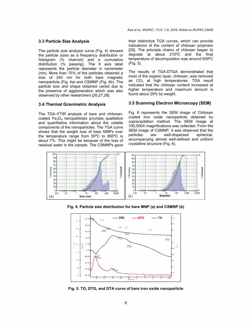
3.3 Particle Size Analysis The particle size analysis curve (Fig. 4) showed the particle sizes as a frequency distribution or histogram (% channel) and a cumulative distribution (% passing). The X axis label represents the particle diameter in nanometer (nm). More than 75% of the particles obtsize of 240 nm for both bare magnetic nanoparticle (Fig. 4a) and CSMNP (Fig. 4b). The particle size and shape obtained varied due to the presence of agglomeration which was alsoobserved by other researchers [26,27,28] 3.4 Thermal Gravimetric Analysis The TGA–FTIR analysis of bare and chitosancoated Fe3O4 nanoparticles provides qualitative and quantitative information about the volatile components of the nanoparticles. The TGA curve shows that the weight loss of bare MNPs over the temperature range from 50ºC to 950ºC is about 7%. This might be because of the loss of residual water in the sample. The CSMNPs gave
Fig. 4. Particle size distribution
Fig. 5. TG, DTG, and DTA curve
Aziz et al.; IRJPAC, 11(1): 1-9, 2016; Article no.IRJPAC.
6
e size analysis curve (Fig. 4) showed the particle sizes as a frequency distribution or histogram (% channel) and a cumulative distribution (% passing). The X axis label represents the particle diameter in nanometer (nm). More than 75% of the particles obtained a size of 240 nm for both bare magnetic nanoparticle (Fig. 4a) and CSMNP (Fig. 4b). The particle size and shape obtained varied due to the presence of agglomeration which was also observed by other researchers [26,27,28].
Analysis
FTIR analysis of bare and chitosan-nanoparticles provides qualitative
and quantitative information about the volatile components of the nanoparticles. The TGA curve shows that the weight loss of bare MNPs over
ºC to 950ºC is about 7%. This might be because of the loss of residual water in the sample. The CSMNPs gave
their distinctive TGA curves, which can provide indications of the content of chitosan polymers [29]. The principle chains of chitosandegrade at about 210ºC and the temperature of decomposition was around 600ºC (Fig. 5).
The results of TGA-DTGA demonstrated that most of the organic layer, chitosan, was removed as CO2 at high temperatures. TGA result indicated that the chitosan content increased at higher temperature and maximum amount is found about 29% by weight.
3.5 Scanning Electron Microscopy (SEM) Fig. 6 represents the SEM image of Chitosan coated iron oxide nanoparticle obtained by coprecipitation method. The SEM 100,000X magnifications was collected. From the SEM image of CSMNP, it was observed that the particles are well-dispersed spheaccompanying almost well-definedcrystalline structure (Fig. 6).
Particle size distribution for bare MNP (a) and CSMNP (b)
Fig. 5. TG, DTG, and DTA curve of bare iron oxide nanoparticle
; Article no.IRJPAC.23408
their distinctive TGA curves, which can provide indications of the content of chitosan polymers
. The principle chains of chitosan began to degrade at about 210ºC and the final temperature of decomposition was around 600ºC
DTGA demonstrated that most of the organic layer, chitosan, was removed
at high temperatures. TGA result itosan content increased at
higher temperature and maximum amount is
Scanning Electron Microscopy (SEM)
Fig. 6 represents the SEM image of Chitosan coated iron oxide nanoparticle obtained by coprecipitation method. The SEM image at 100,000X magnifications was collected. From the SEM image of CSMNP, it was observed that the
dispersed spherical, defined and uniform

Fig. 6. The SEM image There was also a higher agglomerations. The island growth of the tightly packed spherical arrangement was clearly observed. In some regions, the big nanoparticles were surrounded by smaller nanoparticles. The average diameter measured is about 22.4 nm which is nearly in agreement with the particle size obtained from XRD analysis. Simalso found from [30,31].
4. CONCLUSION The synthesis of nanoparticles with small size and uniform size distribution is a subject of intensive research in recent years. In Chitosan polymer was coated on iron oxide core by ionic-cross linking of trisodium phosphate. Size-tunable synthesis of chitosanmagnetic nanoparticles by in situ coprecipitation method at this small size range is very recently been reported. This method is advantageous than the earlier published methods [the process is simple and carried out under mild conditions without using hazardous organic solvents. The nanoparticles obtained by our method are expected to have better biocompatibility than covalently crosschitosan [33,34] as the chitosan was crosswith ionic interactions. The XRD pattern indicated crystalline structure of iron oxide nanoparticle and the coating with chitosan resulted a noisy amorphous peak because chitosan raw material is essentially noncrystalline [19]. The crystallite size obtained from X-Ray powder diffraction is about 13.4 nm which successfully conforms to the biological application. The SEM image of CSMNPs showed that the particles were spherical. The FTIR spectra confirmed the presence of metalbond. Further studies will be done for antimicrobial analysis and cytotoxicity assay to check its bioavailability.
Aziz et al.; IRJPAC, 11(1): 1-9, 2016; Article no.IRJPAC.
7
Fig. 6. The SEM image of bare MNP (a) and CS MNP (b)
There was also a higher tendency of agglomerations. The island growth of the tightly packed spherical arrangement was clearly observed. In some regions, the big nanoparticles were surrounded by smaller nanoparticles. The average diameter measured is about 22.4 nm
in agreement with the particle size obtained from XRD analysis. Similar result is
The synthesis of nanoparticles with small size and uniform size distribution is a subject of intensive research in recent years. In this study Chitosan polymer was coated on iron oxide core
cross linking of trisodium phosphate. tunable synthesis of chitosan-coated
magnetic nanoparticles by in situ coprecipitation method at this small size range is very recently
ted. This method is advantageous the earlier published methods [32] because
the process is simple and carried out under mild conditions without using hazardous organic solvents. The nanoparticles obtained by our method are expected to have better
valently cross-linked as the chitosan was cross-linked
The XRD pattern indicated crystalline structure of iron oxide nanoparticle and the coating with chitosan resulted a noisy amorphous peak because chitosan raw material is essentially non-
. The crystallite size obtained from Ray powder diffraction is about 13.4 nm which
successfully conforms to the biological application. The SEM image of CSMNPs showed
e spherical. The FTIR spectra confirmed the presence of metal-oxygen bond. Further studies will be done for antimicrobial analysis and cytotoxicity assay to
ACKNOWLEDGEMENTS The instrumental supports of Pilot Plant and Process development Centre, Bangladesh Council of Scientific and Industrial Research (BCSIR), Centre for Advanced Research in Sciences (CARS) and Department of Applied Chemistry and Chemical Engineering,of Dhaka are greatly acknowledged.
COMPETING INTERESTS Authors have declared that no competing interests exist.
REFERENCES
1. Ito A, Shinkai M, Honda H, Medical application of functionalized magnetic nanoparticles. J Biosci Bioeng. 2005;100(1):1-11.
2. Mornet S, Vasseur S, Grasset F, P, Goglio G, Demourgues A, Portier J, Pollert E, Duguet E. Magnetic nanoparticle design for medical applications. State Chem. 2006;34:237-247.
3. Gupta AK, Gupta M. Synthesis and surface engineering of iron oxide nanoparticles for biomedical applicBiomaterials. 2005;26:3995-
4. Gupta AK, Wells S. Surfacesuperparamagnetic nanoparticles for drug delivery: Preparation, characterization, and cytotoxicity studies. NanoBioscience, IEEE Transactions on. 2004;3(1):6673.
5. McCarthy JR, Kelly KA, Sun EY, Weissleder R. Targeted delivery of multifunctional magnetic nanoparticles. Future Med; 2007.
6. Corrot C, Robert P, Idee JM, Port M. Recent advances in iron oxide nanocrystal
; Article no.IRJPAC.23408
of Pilot Plant and evelopment Centre, Bangladesh
Council of Scientific and Industrial Research Centre for Advanced Research in
) and Department of Applied Chemistry and Chemical Engineering, University of Dhaka are greatly acknowledged.
Authors have declared that no competing
Ito A, Shinkai M, Honda H, Kobayashi T. Medical application of functionalized magnetic nanoparticles. J Biosci Bioeng.
Mornet S, Vasseur S, Grasset F, Veverka P, Goglio G, Demourgues A, Portier J, Pollert E, Duguet E. Magnetic nanoparticle design for medical applications. Prog Solid
247.
AK, Gupta M. Synthesis and surface engineering of iron oxide
al applications. 4021.
Wells S. Surface-modified superparamagnetic nanoparticles for drug
, characterization, and cytotoxicity studies. NanoBioscience, IEEE
6673.
R, Kelly KA, Sun EY, Targeted delivery of
multifunctional magnetic nanoparticles.
Corrot C, Robert P, Idee JM, Port M. Recent advances in iron oxide nanocrystal

Aziz et al.; IRJPAC, 11(1): 1-9, 2016; Article no.IRJPAC.23408
8
technology for medical imaging. Adv. Drug Deliver. Rev. 2006;58:1471-1504.
7. Alexiou C, Arnold W, Hulin P, Klein RJ, Renz H, Parak FG, Lübbe AS. Magnetic mitoxantrone nanoparticle detection by histology, X-ray and MRI after magnetic tumor targeting. J Magnetism and Magnetic Mat. 2001;225(1):187-193.
8. Tartaj P, del Puerto Morales M, Veintemillas-Verdaguer S, Gonzalez-Carreno T, Serna CJ. The preparation of magnetic nanoparticles for applications in biomedicine. J Physics D: App Physics. 2003;36(13):R182.
9. Denkbaş EB, Kiliçay E, Birlikseven C, Öztürk E. Magnetic chitosan microspheres: Preparation and characterization. Reactive and Functional Polymers. 2002;50(3):225-232.
10. Berry CC, Curtis ASG. Functionalisation of magnetic nanoparticles for applications in biomedicine. J Phy D: App Physics. 2003;36:R198-R206.
11. Weissleder R, Bogdanov A, Neuwelt EA, Papisov M. Long-circulating iron oxides for MR imaging. Adv Drug Delivery Rev. 1995;16:321-334.
12. Qu S, Yang H, Ren D, Kan S, Zou G, Li D, Li M. Magnetite nanoparticles prepared by precipitation from partially reduced ferric chloride aqueous solutions. J Colloid and Interface Sci. 1999;215:190-192.
13. Massart R, Cabuil V. Effect of some parameters on the formation of colloidal magnetite in alkaline-medium-yield and particle-size control. Journal de Chimie Physique et de Physico-Chimie Biologique. 1987;84(7-8):967-973.
14. Chung HJ, Go DH, Bae JW, Jung IK, Lee JW, Park KD. Synthesis and characterization of Pluronic® grafted chitosan copolymer as a novel injectable biomaterial. Current Applied Physics. 2005;5:485-488.
15. Kim DK, Zhang Y, Voit W, Rao KV, Muhammed M. Synthesis and characterization of surfactant-coated superparamagnetic monodispersed iron oxide nanoparticles. J Magnetism and Magnetic Mat. 2001;225:30-36.
16. Qu Y, Hua J, Tian H. Colorimetric and ratiometric red fluorescent chemosensor for fluoride ion based on diketopyrrolopyrrole. Organic letters. 2010;12(15):3320-3323.
17. Mornet S, Vasseur S, Grasset F, Duguet E. Magnetic nanoparticle design for
medical diagnosis and therapy. J Mat Chem. 2004;14(14):2161-2175.
18. Nessa F, Masum SM, Asaduzzaman M, Roy SK, Hossain MM, Jahan MS. A process for the preparation of chitin and chitosan from prawn shell waste. Bangladesh J Scientific and Indust Res. 2010;45(4):323-330.
19. Kabiraj MK, Jahan IA, Masum SM, Islam MM, Hasan SMM, Saha B, Nur HP. Effective Removal of Chromium (VI) Ions from Tannery Effluent using Chitosan-Alumina Composite. Int Res J Pure Appl Chem. 2015;10(3):1-12.
20. Li MC, Liu C, Xin M, Zhao H, Wang M, Feng Z, Sun X. Preparation and characterization of acylated chitosan. Chem Res Chinese U. 2005;21(1):114-116.
21. Lu G, Kong L, Sheng B, Wang G, Gong Y, Zhang X. Degradation of covalently cross-linked carboxymethyl chitosan and its potential application for peripheral nerve regeneration. Eur Polym J. 2007;43:3807-3818.
22. Patterson AL. The Scherrer formula for X-ray particle size determination. Physical rev. 1939;56(10):978.
23. Coates J. Encyclopedia of analytical chemistry. In: Me-yers RA, (ed), Interpretation of infrared spectra, a practical approach. Wiley, Chichester. 2000;10815-10837.
24. Ma L, Gao C, Mao Z, Zhou J, Shen J, Hu X, Han C. Collagen/chitosan porous scaffolds with improved biostability for skin tissue engineering. Biomaterials. 2003; 24(26):4833-4841.
25. Zhang M, Smith A, Gorski W. Carbon nanotube-chitosan system for electrochemical sensing based on dehydrogenase enzymes. Analyt Chem. 2004;76(17):5045-5050.
26. Li G, Jiang Y, Huang K, Ding P, Chen J. Preparation andproperties of magnetic Fe3O4–chitosan nanoparticles. J Alloy Compd. 2008;66:451-456.
27. Dung DTK, Hai TH, Phuc LH, Long BD, Vinh LK, Truc PN. Preparation and characterization of magnetic nanoparticles with chitosan coating, APCTP–ASEAN Workshop on Advanced Materials Science and Nano-technology (AMSN08) IOP Publishing J of Physics, Conference Series. 2009;187:012036.
28. Zhang L, Zhu X, Sun H, Chi G, Xu J, Sun Y. Control synthesis of magnetic Fe3O4–

Aziz et al.; IRJPAC, 11(1): 1-9, 2016; Article no.IRJPAC.23408
9
chitosan nanoparticles under UV irradiation in aqueous system. Curr Appl Phys. 2010;10:828-833.
29. Paama L, Pitkänen I, Halttunen H, Perämäki P. Infrared evolved gas analysis during thermal investigation of lanthanum, europium and samarium carbonates. Thermochimica Acta. 2003;403(2):197-206.
30. Shrifian EA, Salhi MT, Mojtaba NE, Ekramian E. Chitosan-modified superparamgnetic iron oxide nanoparticles: design, fabrication, characterization and antibacterial activity. CHEMIK. 2015;69(1):19-32.
31. Ali A, Muhammad MTM, Sijam K, Siddiqui Y. Effect of chitosan coatings on the
physicochemical characteristics of Eksotika II papaya (Carica papaya L.) fruit during cold storage. Food Chemistry. 2011;124(2):620-626.
32. Tiaboonchai W, Limpeanchob N. Formulation and characterization of amphotericin B-chitosan-dextran sulfate nanoparticles. Int J Pharm. 2007;329:142-149.
33. Agnihotri SA, Mallikarjuna NN, Aminabhavi TM. Recent advances on chitosan-based micro and nanoparticles in drug delivery. J Cont Release. 2004;100(1):5-28.
34. Park JH, Saravanakumar G, Kim K, Kwon IC. Targeted delivery of low molecular drugs using chitosan and its derivatives. Adv Drug Deliv Rev. 2010;62:28-41.
_________________________________________________________________________________ © 2016 Aziz et al.; This is an Open Access article distributed under the terms of the Creative Commons Attribution License (http://creativecommons.org/licenses/by/4.0), which permits unrestricted use, distribution, and reproduction in any medium, provided the original work is properly cited.
Peer-review history: The peer review history for this paper can be accessed here:
http://sciencedomain.org/review-history/12868