1, iman m. al zaka 2 1, 2 · teeba talal abdul aziz1, iman m. al zaka 2 1, 2department of...
TRANSCRIPT

International Journal of Science and Research (IJSR) ISSN: 2319-7064
Impact Factor (2018): 7.426
Volume 8 Issue 1, January 2019
www.ijsr.net Licensed Under Creative Commons Attribution CC BY
Efficacy of XP-endo Finisher, XP-endo Finisher R,
CanalBrush and EndoActivator in the removal of
Intracanal Medicament (An in Vitro Study)
Teeba Talal Abdul Aziz1, Iman M. Al Zaka
2
1, 2Department of Conservative Dentistry, College of Dentistry, Al-Mustansiriya University, Iraq
Abstract: The aim of this study was to evaluate and compare the efficacy of XP-endo Finisher, XP-endo Finisher R, CanalBrush and
EndoActivator in the removal of Intracanal medicament. Sixty freshly extracted upper molar teeth with straight palatal roots were used
in this study; all roots were shortened to a length of 12 mm. Canals were prepared with RECIPROC blue file R40 and irrigated with 10
ml of 2.5% NaOCl. Then the roots were split longitudinally. Two of the three standard grooves were created in the coronal and apical
parts of one of root halves, and the other in the middle part of the second halves then each of the root halves and grooves was irrigated
with 5 ml 17% EDTA for 60 sec. and 5 ml 2.5% NaOCl for 60 sec. The standardized grooves were filled with Ca(OH)2 and the root
halves were reassembled. After 7 days, R40 file at WL was used to get a space for irrigation needle and instruments.After thatspecimens
were randomly divided into 4 experimental groups (n=15/group). Ca(OH)2 was removed with different instruments as follow: Group
(І): CanalBrush, Group (П): EndoActivator, Group (III): XP-endo Finisher, and Group (IV): XP-endo Finisher R. 5 ml 2.5% NaOCl
irrigation was used before and after every cleaning procedure with each instrument.The amount of remaining Ca(OH)2 in the grooves
was scored under a Stereomicroscope at 30X magnification. Statistical evaluation was performed using Kruskal–Wallis and Wilcoxon
Sum rank test. The results showed that cleaning with XP-endo Finisher R (IV) resulted in significantly cleaner canals (P ≤ 0.05) than
CanalBrush (І), EndoActivator (П), and XP-endo Finisher (III) at all canal levels. XP-endo Finisher resulted in cleaner canals than
CanalBrush, EndoActivator at all levels, but with insignificant statistical difference (P>0.05). Apical level of all canals showed greater
amountofCa(OH)2 remnants in compared to middle and coronal levels, whatever instruments were used. Apical groove was
significantly difference from coronal groove (P≤0.05) in all groups.None of the investigated instruments were able to completely
removed all Ca(OH)2 from the three root regions.Keywords: EndoActivator; XP-endo Finisher; XP-endo Finisher R; CanalBrush;
Ca(OH)2 .
1. Introduction
The primary aims of endodontic treatment are the removal
of microorganisms and prevention of reinfection inside the
root canal system. Even after mechanical instrumentation
and irrigation procedures, resistant microorganisms can stay
within the root canal system as a result of its complex
anatomy [1].
Many studies [2; 3; 4] established the efficiency of
intracanal medications in fighting the antimicrobial resistant
microorganisms.In endodontics, calcium hydroxide
(Ca(OH)2)is utilized as an intracanal medication, owing to
its antimicrobial efficacy and biological properties. The
antimicrobial properties of Ca(OH)2medicamentwere owing
to its ability in releasing of hydroxyl ions and providing a
highly alkaline environment with a pH value is about 12.5
[5]. It is well recognized that Ca(OH)2 residual must be
eliminated due to its effect on the sealing and bonding of
endodontic materials. Moreover, the residual of
Ca(OH) 2 may result in an adverse chemical reactions with
root canal sealer, leading to unpredictable prognosis by
reducing its flow and working time[6].
Remaining medicament also has been established to impact
negatively the accuracy of electronic working length (WL)
measurement [7]. Therefore, complete elimination of
Ca(OH) 2 medicament prior to root filling is necessary.
Since irrigation using sodium hypochlorite (NaOCl) only
was not effective in the elimination of the medication [8; 9],
for that reason a combination of chemical and mechanical
removal would be essential.
Root canal instrumentation using a master apical file (MAF)
and copious irrigation is the most commonly described
removal technique [10; 11].
Several instruments and devices have been introduced for
root canal cleaning. Some of these instruments, for instance
CanalBrush [12], laser systems [13], EndoActivator [14]
[15; 7], EndoVac and XP-endo Finisher [16][17] have been
utilized for the elimination of intracanal medicaments. XP-
endo Finisher R recently introduced to the market for
retreatment case. The results regarding the efficacy of these
instruments and devices in removal of intracanal
medicaments are controversial. Therefore, the cleaning
ability of these instruments require further investigation.
2. Materials and Methods
2.1 Methods
2.1.1 Sample Collection and Selection
Palatal roots of sixty freshly extracted maxillary molars
human teeth were selected. The criteria for root selection:
1) Straight root canal.
2) Mature centrally located apical foramen.
3) Patent apical foramen.
4) Root devoid of any resorption (internal and external
resorption), crack or fracture.
Immediately after the extraction, calculus, stains and soft
tissues on the tooth surface were removed manually by the
use of a cumine, disinfected with 3.0 % NaOCl solution for
30 minutes [18], followed by washing with tap water. The
Paper ID: ART20191176 10.21275/ART20191176 919
International Journal of Science and Research (IJSR) ISSN: 2319-7064
Impact Factor (2018): 7.426
Volume 8 Issue 1, January 2019
www.ijsr.net Licensed Under Creative Commons Attribution CC BY
teeth were then stored in container containing thymol
solution.
2.1.2. Sample Preparation The length of the palatal root was standardized to 12mm
from the anatomic apex by using a digital caliper and a
permanent red marker. The tooth was then fixed on a bench
vice, a double-faced diamond disc with straight handpiece,
under water coolant, was utilized for cutting off the palatal
root perpendicular to the long axis of the root in accordance
with the drawn line. Then all root were measured using
digital veneer. The pulpal tissues were extirpated by using
barbed broaches, and the location of the apical foramen and
the patency of the canals were verified by the insertion of a
#15 stainless steel K-file and introduced slowly till it is
visualized at the apical foramen Then the silicon stopper
was set on the file, then the file was withdraw and measured
by using an endodontic ruler, after that the correct WL was
measured by subtracting 1mm from that measurement [19].
The samples were embedded, except for the coronal 3
millimeters, in an Eppendorf vial containing silicon rubber
base impression material (putty consistency) to aid handling
of the samples throughout the following steps. A straight
fissure bur fixed on a low speed straight handpiece was
used to make a hole in the base of the plastic vial to permit
for the air in the tube to release during silicon putty
insertion. According to the manufacturer’s instructions the
putty was mixed with the catalyst gel and inserted in the
plastic vial then the sample was embedded in the silicon
putty. Then the silicon material was left for complete setting
and forming a block. After that the plastic vials were fixed
using a bench vice in order to achieve a standardized
position during the entire procedure.
2.1.3. Root Canal Instrumentation
The root canals were irrigatedwith 10 ml of 2.5% NaOCl
solution during the preparation delivered by a 5.0 ml
disposable syringe witha 30-gauge side-vented needle.
During all irrigation phases, the needle was placedshort 2
mm from the WL.
The root canals were prepared with RECIPROC blue
(40/06) NiTi file usingVDW.SILVER®RECIPROC®
endomotors. According to the manufacturer ,s
instructions,the silicone stopper was set on the Reciproc®
blue instrument at two thirds of WL. After that, the
Reciproc® blue file was advanced in the canal by a slow
pecking motion (in- and out- movements) without
withdrawing the instrument totally out of the canal. The
amplitude of pecking motions must not be surpass 3–4 mm.
The instrument introduced easily in the canal in an apical
direction. After 3 pecks, or if resistance was felt before the
3 pecks were completed, the instrument was withdrawn out
of the canal to clean it. For the purpose of checking the
patency of the canal a #15 hand file was utilized.
Then the canal was copiously irrigated and Reciproc® blue
instrument was then re-used in the same way till it reached
the two thirds of the estimated WL. The canal was irrigated
and a #15 file was used to check patency up to the WL. The
Reciproc® blue instrument was re-used as previously-
mentioned till it reached the WL. Once the WL was
reached, the Reciproc® blue instrument was pulled out of
the canal for the aim of avoiding an unnecessary over-
enlargement [20]. For standardization purposes, one rotary
file was used to prepare three canals and then discarded.
2.1.4. Root Sectioning
After complete root canal instrumentation, the roots were
removed from their vials and a marker was utilized to draw
guiding lines longitudinally on the buccal and palatal sides.
Then the roots were fixed on the bench vice and
longitudinally grooved buccopalatally on the previously-
mentioned lines by using a diamond disc fixed on a low
speed straight handpiece with water cooling, conserving the
inner shelf of dentin surrounding the canal. The grooves
were then cleaned from any remaining dust and debris by a
short blast of air. Then the roots were cleaved by placing a
surgical blade #15 in the groove and striking the blade
gently with a small mallet [21]. The longitudinal section of
each root with ≤180° of the canal circumference was
selected for the study since the sections with >180 of canal
circumference would possibly hinder the total canal
visualization thru photography [22]. If teeth showing that
the canals had been penetrated, the tooth was then
discarded and replaced [23].
2.1.5. Artificial Standard Grooves
The specimens were positioned in silicone on Microscope
slide. A graphite mark was made 2 mm apart from the apex
to indicate the apical end of the apical groove. A second
graphite mark was made 3 mm away from the first mark to
indicate the other end of the apical groove. This maneuver
was achieved by using an Electronic digital caliper with
graded ends that were fixed 3 mm from one another in order
to standardize the groove size and location. A similar
graphite marks were made 8-11 mm from the apex for the
coronal groove in the same half of the apical groove and 5-8
mm from the apex for the middle groove.
Then an ultrasonic tip P1 (0.3mm)was used to create
artificial standard grooves inside the canal in each half
which were 3 mm in length, 0.3 mm in width and 0.5 mm in
depth and were located 2–5 mm from the apex for apical
sections, 8-11 mm from the apex for coronal sections, and
5-8 mm from the apex in the opposite half for middle
sections[24]. Then the dimension of grooves was checked
under a Stereomicroscope at 30X magnification[11; 29,
30].
The root halves and grooves were irrigated with 5 ml 17%
EDTA for 60 sec. and 5 mL 2.5% NaOCl for 60 sec .
Then, the root canal was dried with paper points [25]. The
root halves were dried with gauze.
2.1.6 Calcium Hydroxide Paste Insertion Each groove was filled with Ca(OH)2 by inserting the
needle tip of Ca(OH]2 paste on the groove and adapted by
paper point [26; 17]and the Stereomicroscope was used to
confirm that the groove was fully filled with Ca(OH)2 paste
[25].
After that a ligature wire 0.3 mm in diameter was used for
the fixation of root halves and interlocked using orthodontic
plier and then the root halves was reassembled by using a
A
Paper ID: ART20191176 10.21275/ART20191176 920

International Journal of Science and Research (IJSR) ISSN: 2319-7064
Impact Factor (2018): 7.426
Volume 8 Issue 1, January 2019
www.ijsr.net Licensed Under Creative Commons Attribution CC BY
thin layer of cyanoacrylate glue and was remounted in
Eppendorf vials [19].
Next, the roots were filled with Ca(OH)2 paste starting from
the apical aspect with the needle slowly advancing
coronally until the paste was visualized at the canal orifice
then using lentulo spiralin a slow speed handpiece.
Radiographs were taken to ensure that canals were
completely filled with Ca(OH)2 paste. Excess material was
wiped off with moist cotton.
For all samples in the four groups the root canal was sealed
coronally with a temporary filling material. Then the
samples were stored in an incubator at 37°C with 100%
relative humidity for 1 week.
2.1.7. Sample grouping
After this period, the temporary filling material was
removed. For the removal of Ca(OH)2, an R40 file at WL
and 1 ml 2.5 % NaOCL was utilized to get a space for
irrigation needle and instruments [19]. After removal of
R40 file, all specimens were randomly divided into four
experimental groups (n=15) according to the instrument was
used for removal of Ca(OH)2
2.1.8 Ca(OH)2 Removal
Group (І): CanalBrush
The root canals were irrigated with 5 ml 2.5% NaOCl and
then were brushed with a Canal Brush (medium size). A 2–
3 mm up and down motion was made by the Canal Brush at
600 rpm for 1min; according to manufacturer’s instructions.
The Canal Brush was introduced 1 mm short of the WL.
After that, Canal Brush was moved around in small vertical
movementsfollowed by a final flush of the root canal with 5
ml 2.5% NaOCland was dried with RECIPROC paper
points. For standardization purpose one canal brush was
used for cleaning a single canal, and then was discarded
[25; 24; 19].
Group (П): EndoActivator
After irrigation of the root canals with 5 ml 2.5% NaOCl.
The medium size polymer tip was used to remove the paste.
The tip was fit passively inside the canal, 2mm shorter than
the WL. EndoActivator was activated at 10,000 cpm for
1min with pumping action in short 2-3mm vertical strokes
with the amplitude of 2mm for 1minaccording to the
manufacture's instruction. Then the root canal was
underwent a final flush with 5 ml 2.5% NaOCl and was
dried with RECIPROC paper points. For standardization
purposes the activator tip was used for cleaning a single
canal then discarded [25; 24; 19].
Group (III): XP-endo Finisher
The XP-endo Finisher (25/00)was used according to the
instruction of the manufacture, at 800 rpm for the speed and
1.0 Ncm for the torque.The stopper was set 1 mm short of
the WL with the provided tube, the XP-endo- Finisher was
cooled down with cold spray while it is inside the tube .
After irrigating the canal with 5.0 ml 2.5% NaOCl, the XP-
endo Finisher was removed from the tube and activated
while its tip was inside the canal. The Finisher was
activated for 1 min inside the canal using gentle 7-8mm
lengthwise movements to contact the full length of the
canal. Then the root canal underwent a final rinse (5.0 ml
2.5% NaOCl) and drying of the canal with RECIPROC
paper points. For standardization purposes, each file was
used for cleaning a single canal, and then discarded [25;24].
Group (IV): XP-endo Finisher R
The XP-endo Finisher R (30/00) was used according to the
instruction of the manufacture, at 800 rpm for the speed and
1.0 Ncm for the torque. The stopper was set 1 mm short of
the WL with the provided tube; the XP-endo Finisher R was
cooled down with cold spray while it is inside the tube. So
after irrigating the canal with 5.0 ml 2.5% NaOCl, the XP-
endoFinisher R was removed from the tube and activated
while its tip was inside the canal. The XP-endo Finisher R
was activated for 1 min inside the canal using gentle 7-8mm
lengthwise movements to contact the full length of the canal
, followed by the final rinse (5.0 ml 2.5% NaOCl) and
drying of the canal with RECIPROC paper points.
For standardization purposes, each file was used for
cleaning a single canal, and then discarded[25; 24].
Both XP-Endo Finisher and XP-endo Finisher Rfiles were
used under 35 °C by using heater [27; 28].
After finished all roots were removed and each half was
separated, then all grooves in both half were evaluated
under Stereomicroscope. The photographs were taken using
a Stereomicroscope equipping with a digital camera at 30X
magnification [11; 29; 30; 33].
2.1.9. Stereomicroscope Evaluation
The amount of remaining Ca(OH)2 in the grooves was
assessed under a Stereomicroscope at 30× magnification
and equipped with a digital camera by using an numeric
evaluation scale as described by[31;26;30].
Score 0: the cavity is free of Ca(OH)2 remnants.
Score 1: less than the half of the cavity is filled with
Ca(OH)2 remnants.
Score 2: more than the half of the cavity is filled with
Ca(OH)2 remnants.
Score 3: the cavity is filled with Ca(OH]2 remnants
completely.
The remnants in each groove were traced by using specific
software tool magnetic lasso tool and the total number of
pixels occupied by the remnants were reported using the
histogram function in the software [32; 33].
The 4 experimental groups were analyzed using Kruskal-
Wallis and Wilcoxon Sum rank test at a significance level
of P ≤ 0.05. All statistical Analyses were performed using
IBM SPSS Statistics 21 software.
3. Results
Kruskal–Wallis test and Wilcoxon Sum rank test were used
due to the ordinal nature of the scores [25; 24]
Paper ID: ART20191176 10.21275/ART20191176 921
International Journal of Science and Research (IJSR) ISSN: 2319-7064
Impact Factor (2018): 7.426
Volume 8 Issue 1, January 2019
www.ijsr.net Licensed Under Creative Commons Attribution CC BY
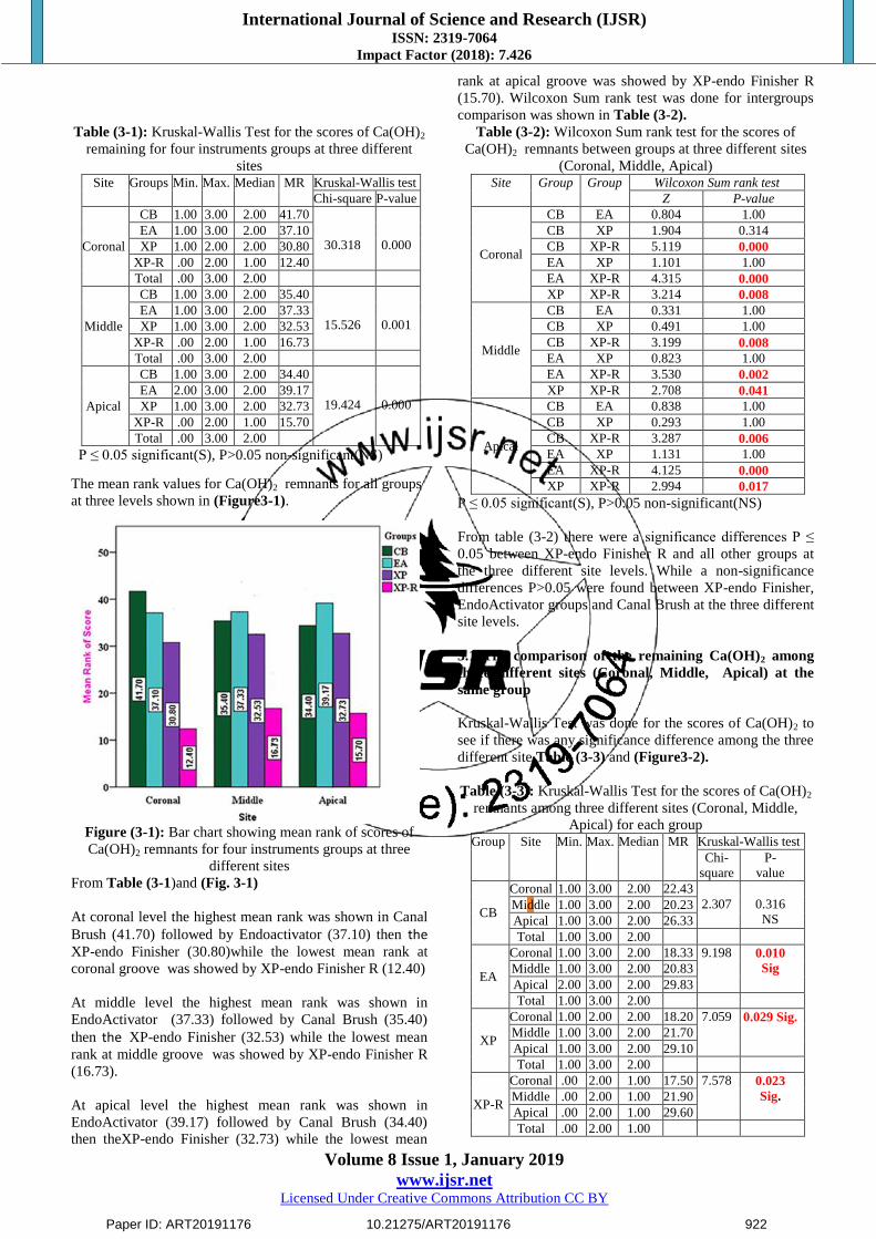
Table (3-1): Kruskal-Wallis Test for the scores of Ca(OH)2
remaining for four instruments groups at three different
sites Site Groups Min. Max. Median MR Kruskal-Wallis test
Chi-square P-value
Coronal
CB 1.00 3.00 2.00 41.70
30.318
0.000 EA 1.00 3.00 2.00 37.10
XP 1.00 2.00 2.00 30.80
XP-R .00 2.00 1.00 12.40
Total .00 3.00 2.00
Middle
CB 1.00 3.00 2.00 35.40
15.526
0.001 EA 1.00 3.00 2.00 37.33
XP 1.00 3.00 2.00 32.53
XP-R .00 2.00 1.00 16.73
Total .00 3.00 2.00
Apical
CB 1.00 3.00 2.00 34.40
19.424
0.000 EA 2.00 3.00 2.00 39.17
XP 1.00 3.00 2.00 32.73
XP-R .00 2.00 1.00 15.70
Total .00 3.00 2.00
P ≤ 0.05 significant(S), P>0.05 non-significant(NS)
The mean rank values for Ca(OH)2 remnants for all groups
at three levels shown in (Figure3-1).
Figure (3-1): Bar chart showing mean rank of scores of
Ca(OH)2 remnants for four instruments groups at three
different sites
From Table (3-1)and (Fig. 3-1)
At coronal level the highest mean rank was shown in Canal
Brush (41.70) followed by Endoactivator (37.10) then the XP-endo Finisher (30.80)while the lowest mean rank at
coronal groove was showed by XP-endo Finisher R (12.40)
At middle level the highest mean rank was shown in
EndoActivator (37.33) followed by Canal Brush (35.40)
then the XP-endo Finisher (32.53) while the lowest mean
rank at middle groove was showed by XP-endo Finisher R
(16.73).
At apical level the highest mean rank was shown in
EndoActivator (39.17) followed by Canal Brush (34.40)
then theXP-endo Finisher (32.73) while the lowest mean
rank at apical groove was showed by XP-endo Finisher R
(15.70). Wilcoxon Sum rank test was done for intergroups
comparison was shown in Table (3-2).
Table (3-2): Wilcoxon Sum rank test for the scores of
Ca(OH)2 remnants between groups at three different sites
(Coronal, Middle, Apical) Site Group Group Wilcoxon Sum rank test
Z P-value
Coronal
CB EA 0.804 1.00
CB XP 1.904 0.314
CB XP-R 5.119 0.000
EA XP 1.101 1.00
EA XP-R 4.315 0.000
XP XP-R 3.214 0.008
Middle
CB EA 0.331 1.00
CB XP 0.491 1.00
CB XP-R 3.199 0.008
EA XP 0.823 1.00
EA XP-R 3.530 0.002
XP XP-R 2.708 0.041
Apical
CB EA 0.838 1.00
CB XP 0.293 1.00
CB XP-R 3.287 0.006
EA XP 1.131 1.00
EA XP-R 4.125 0.000
XP XP-R 2.994 0.017
P ≤ 0.05 significant(S), P>0.05 non-significant(NS)
From table (3-2) there were a significance differences P ≤
0.05 between XP-endo Finisher R and all other groups at
the three different site levels. While a non-significance
differences P>0.05 were found between XP-endo Finisher,
EndoActivator groups and Canal Brush at the three different
site levels.
3.1 The comparison of the remaining Ca(OH)2 among
three different sites (Coronal, Middle, Apical) at the
same group
Kruskal-Wallis Test was done for the scores of Ca(OH)2 to
see if there was any significance difference among the three
different site Table (3-3) and (Figure3-2).
Table (3-3): Kruskal-Wallis Test for the scores of Ca(OH)2
remnants among three different sites (Coronal, Middle,
Apical) for each group Group Site Min. Max. Median MR Kruskal-Wallis test
Chi-
square
P-
value
CB
Coronal 1.00 3.00 2.00 22.43
2.307
0.316
NS Middle 1.00 3.00 2.00 20.23
Apical 1.00 3.00 2.00 26.33
Total 1.00 3.00 2.00
EA
Coronal 1.00 3.00 2.00 18.33 9.198 0.010
Sig Middle 1.00 3.00 2.00 20.83
Apical 2.00 3.00 2.00 29.83
Total 1.00 3.00 2.00
XP
Coronal 1.00 2.00 2.00 18.20 7.059 0.029 Sig. Middle 1.00 3.00 2.00 21.70
Apical 1.00 3.00 2.00 29.10
Total 1.00 3.00 2.00
XP-R
Coronal .00 2.00 1.00 17.50 7.578 0.023
Sig. Middle .00 2.00 1.00 21.90
Apical .00 2.00 1.00 29.60
Total .00 2.00 1.00
Paper ID: ART20191176 10.21275/ART20191176 922

International Journal of Science and Research (IJSR) ISSN: 2319-7064
Impact Factor (2018): 7.426
Volume 8 Issue 1, January 2019
www.ijsr.net Licensed Under Creative Commons Attribution CC BY
P ≤ 0.05 significant(S), P>0.05 non-significant (NS).
Figure (3-2): Bar chart showing mean rank for scores of
Ca(OH]2 remnants among three different sites for each
group
By performing Kruskal-Wallis test Table (3-3):
For all groups (І, П, III, and IV) the highest of mean rank
was found in apical site. While the lowest mean rank was
found in coronal site for all group except for CanalBrush
where the lowest mean rank was found in the middle site. In
CanalBrush group a non-significance differences was found
while for other groups a significance differences were
found.
Table (3-4): Wilcoxon Sum rank test for Ca(OH)2 scores
between three different sites (Coronal, Middle, Apical) for
each group Group Site Site Wilcoxon Sum rank test
Z P-value
EA
Coronal Middle 0.627 1.00 NS
Middle Apical 2.256 0.072 NS
Apical Coronal 2.883 0.012 Sig.
XP
Coronal Middle 0.835 1.00 NS
Middle Apical 1.766 0.232 NS
Apical Coronal 2.602 0.028 Sig.
XP-R
Coronal Middle 0.989 0.968 NS
Middle Apical 1.730 0.251 NS
Apical Coronal 2.719 0.020 Sig.
P ≤ 0.05 significant(S), P>0.05 non-significant (NS)
Wilcoxon Sum rank test for the score of Ca(OH)2 remaining
at three different sites for each group showed that in all
groups except Canal Brush, a significant differences (p≤
0.05) were found between coronal and apical grooves while
no significant difference (p> 0.05) were found between
coronal and middle grooves, and between apical and middle
grooves (Table 3-4) .
4. Discussion
The primary aim of root canal treatment is to reduce or
eliminate bacteria and their by-products from an infected
root canal system [34]. To this end, intracanal medication
has been advocated to disinfect the root canal system and as
a consequence increase therapeutic success [35]. For this
purpose, Ca(OH]2 paste as intracanal medicament during
endodontic therapy due to its antimicrobial efficacy and
biological properties is used, but reportedly, failing to
completely remove Ca(OH)2 from the root canal walls
following therapy influences dentin bond strength [36] and
sealer penetration into the dentinal tubules,as well as
markedly compromises the quality of the seal provided by
the root filling. As a result of the previously mentioned
reasons, removal of the Ca(OH)2 medicament prior to root
filling is mandatory. After removal of Ca(OH)2 paste from
the main canal, remnants can persist in canal extensions or
in irregularities and this was the case in this study.
4.1 Discussion of methodology
Root canal anatomy possess numerous geometrical
probabilities in cross section; for instance: round, oval, long
oval, flattened or irregular [37]. These anatomic
complexities have been represented physical restrictions
that pose a serious challenge for adequate root canal
instrumentation and disinfection [38]. Palatal root of freshly
extracted human maxillary molars teeth was used in this
study, since its canal has a more round cross section when
compared to other teeth. In view of the fact that most of the
enlarging instruments was resulted in circular carving
motion [39]. Therefore, using a root canal with round cross
section would result in similarity between root canal
geometry and the enlarging instrument, which consequently
increased the similarity and standardization among the
samples.
The length of the root was standardized to a uniform length
(12mm) to obtain fixed and reliable reference point and for
enhancement of instrumentation, and elimination of any
variables in the access during the instrumentation and to
facilitate the creation of groove standardized in size and
location.
A preparation size (40) was estimated for representation of
an adequate balance between the apical enlargement, the
preservation of tooth structure, and the inhibition of
technical mishaps. In addition, the preparation eased
insertion of the irrigation and agitation devices to 1–2 mm
short of WL, because the penetration depth of the irrigation
needle has an influence on the mechanical efficiency of
irrigation [40] and subsequently on the elimination of
medicament.
The usage of NaOCl alone as a sole irrigation solution in
this study, was done to concentrate on the effects of
different instruments rather than investigate the influences
of the irrigation solution. In this study, the agent, volume
and the procedure for irrigation have been standardized for
all experimental groups [41].
The benefit of the groove model was standardized the size,
location of the grooves and the volumes of medicament
used [31; 26; 29; 30; 17].Grooves also may be considered
as typical irregularities of a root canal which are most
difficult to clean from the medication. In spite of this, the
complexity of root canal anatomy cannot be completely
simulated by this model [42].
Paper ID: ART20191176 10.21275/ART20191176 923
International Journal of Science and Research (IJSR) ISSN: 2319-7064
Impact Factor (2018): 7.426
Volume 8 Issue 1, January 2019
www.ijsr.net Licensed Under Creative Commons Attribution CC BY
Scoring of cleanliness of the grooves probably would be
easier and more reproducible than scoring of such a large
area as the whole root canal wall [42]. Additionally, this
experimental design did not address medicament that
diffused into the dentinal tubules. The model of this study
was based on studies illustrated by [31; 26;29; 24].
Various techniques have been applied for evaluating
Ca(OH)2 removal from the root canal system [43; 44; 11].
In this study, the samples were longitudinally split, and the
percentage of residual Ca(OH]2 was calculated from images
have been taken under magnification of the whole area
desired at Stereomicroscopy. Three different parts of the
tooth were evaluated: the apical, middle, and coronal
grooves. This method has been described as an effective
method in determining the amount of Ca(OH)2 residue
using Stereomicroscopic analysis that evaluated the
remnants in a more wide view considering the debris,
differently from SEM that enables the researchers to view
both debris and the smear layer [45]. In addition to the
above-mentioned difference SEM have been required
sectioning of tooth which may cause loss of material which
effect the result. In addition to that only small area of the
root canal could be observed since it operated at very high
magnifications, resulting in certain observer bias.
Nainanet al. in 2013 [46] have been used volumetric
analysis with spiral CT to evaluate the removal of three
calcium hydroxide preparations used as intracanal
medicaments. Although this technique permitted, three-
dimensional measurement of Ca(OH)2 existed in canal
system and could be done without sectioning the teeth and
thus preventing any loss of Ca(OH)2 paste. In spite of that,
it was not possible to assess the removal of filling material
segmented into thirds, as long as the CT image analysis
software did not provide a tool for dividing the structure
examined into an equal parts. For that reason, the volume of
initial and residual filling material were analyzed as a
whole.
Moreover, μ-CT has anthor limitation that should be taken
into consideration in evaluating the removal efficacy of
Ca(OH)2 paste in separate from other root canal filling
materials such as gutta-percha or sealer. Generally, the
radioopacity of Ca(OH)2 paste, with the exclusion of
iodoform-based material, is lower than that of gutta-percha
or sealer. For that reason, the removal efficiency of less
radiopaque material could be exaggerated as the residual
material might not be recognized in scanned images [47].
Certain studies calculated the Ca(OH)2 remnants in the root
canal system by means of measuring the surface area of the
Ca(OH)2 residue existing in the canal in terms of mm2[43;
8].On the other hand, the disadvantage of this technique
might be the loss of material throughout sectioning.
In this study, a pixel count of Ca(OH)2 remaining on the
artificially created grooves was calculated and measured in
terms of percentile values of the overall groove surface and
these were then scored from 0 to 3 [32; 33].The elimination
of the subjectivity of some previous studies where
evaluators gave the grooves a score of no, light, moderate or
heavy for the amount of Ca(OH)2 residual was the benefit of
this method.
In this study, the effectiveness of different activation
instruments Canal Brush, EndoActivator, XP-endo Finisher
and XP-endo Finisher R were compared for their efficiency
in removing Ca(OH)2 paste.
All the canals were instrumented by one operator (the
researcher) to minimize the variables during the study, The
researcher was trained to practice the removal techniquesof
Ca(OH)2 paste before starting the actual experimental
research work, which was achieved according to the
manufacturer’s instructions.
4.2 Discussion of results
4.2.1 Comparison of Ca(OH)2 removal between the four
different instruments groups
According to the results of this study, none of the
instruments could completely remove all remnants of
Ca(OH)2 in all three root regions,this finding has also been
described by other studies [25; 24]. XP-endoFinisher R
showed the lowest score than other groups in the removal of
Ca(OH)2 in all 3 sites of the root canal and significantly
difference from XP-endoFinisher, EndoActivator and Canal
Brush.
This result may be related to the features of XP-endo
Finisher Rwhich is a new variation of the XP-endo Finisher
file. The XP-endo Finisher R have differents in its core
diameter larger (ISO 30) and in the angulation of its tip in
comparison with the XP-endoFinisher (ISO 25), making it
stiffer and also more efficient in removing root fillings
materials adhering to the root canal walls [28]. According to
the manufacturer, it has been developed for removal of
medicament and for retreatment cases, depending on the
shape-memory principles of the alloy MaxWire and due to
its flexibility, the XP-endoFinisher R has the ability to
expand and contract with an improved reach of 6 mm in
diameter or 100-fold when compared with a standard
instrument of the same size. Its ability to expand and its
sickle shape permits it to access and clean areas previously
impossible to reach [16].
No studies in literature, were found in term of Ca(OH)2
removal by XP-endoFinisher R in compared with XP-
endoFinisher, EndoActivator and Canal Brush.
Hashemi and Mackevičiūtė stated that the use of XP-endo
Finisher R showed a significant difference in cleaning
ability in every part (coronal, middle and apical) and
increased cleaning efficiency when comparing with other
instrument and found that there was a significant difference
between the two groups in term of the total root canal filling
material removal.
The XP-endo Finisher R have been made from entirely
different technology that permits it to adapt and “scout” in
searching for the remnants of root canal filling material and
in following the morphology more precisely owing to its
mixed phase technology.
Paper ID: ART20191176 10.21275/ART20191176 924
International Journal of Science and Research (IJSR) ISSN: 2319-7064
Impact Factor (2018): 7.426
Volume 8 Issue 1, January 2019
www.ijsr.net Licensed Under Creative Commons Attribution CC BY
Even there was a non-significant difference between XP-
endo Finisher file, EndoActivator and Canal Brush; XP-
endo Finisher file showed less amount of Ca(OH)2 remnants
than both of them in all three grooves. This finding have
been related to the ability of debris and smear layer removal
by XP-endo Finisher file which evaluated through SEM to
its metallurgy which mean that when the file is subjected to
the body temperature (the canal) it would convert it to
austenitic phase in the rotation mode because of its shape-
memory, this feature would permit the file to contact and
clean areas that are otherwise difficult to reach with regular
instruments [48].
It have been noticed that Canal Brush and EndoActivator
groups did not have the same ability to follow the canal
morphology and making a straight path towards the apex
and not “scout” for Ca(OH)2 remnants as XP-endo Finisher
and XP-endo Finisher R.
CanalBrush that is used with a circumferential motion with
a 2–3 mm up and down motion as a file creating space by
scraping the Ca(OH)2 bulk and the packed, scattered
Ca(OH)2 in the irregularities of the root canal, for that
reason it was less effective in removing Ca(OH)2 paste from
the irregularities of the root canal system [7; 19].
While EndoActivator polymer tip have been produced
vigorous intracanal fluid agitation throughout its swirling
movement and cavitation by its pumping action in short 2-
3mm vertical strokes at 10,000 cycles/min, 2mm shorter
than the WL. Once EndoActivator tip is placed in the apical
level of the root canal, there is a potential dampening effect
that occur when there is contact with the apical root canal
walls that might have restrained the displacement amplitude
of the agitation devices and thus decreased agitation energy.
This result is coincided with the result of Göktürket al.,
2018 [49] this study was evaluated the removal efficacy of
Ca(OH)2 by using CanalBrush, XP-endo Finisher, laser-
activated irrigation (LAI), conventional syringe irrigation
(CSI), passive ultrasonic irrigation (PUI) and Vibringe in
the root canal walls and found that none of the protocols
resulted in complete removal of Ca(OH)2 from the canal
walls and there were no significant differences among the
investigated protocols. Also this result in parallel with the
result of Göktürk et al., 2016 [25]; Göktürk et al., 2017 [24]
who found a non-significance difference between XP-endo
Finisher file and Canal Brush in the middle and apical
region.
The result of this study in agreement with the result of
Elnaghy et al., 2017 [48] who found that there was no
significant difference between XP-endo Finisher and
EndoActivator groups.
But this result in disagreement with Keskin et al., 2017 [19]
who found that XP-endo Finisher removed significantly
more Ca(OH)2 than EndoActivator and CanalBrush. The
reasons for this difference between the results may reflect
different variable that the former study removed Ca(OH)2
from standardized internal resorption simulated cavities
with 0.8-mm depth and 1.6-mm diameter which prepared
with burs at the level 5 mm above the anatomic apex in each
halves of samples. These cavities have regular borders in
comparison with the natural resorption, which are irregular
lesions. Moreover, XP -endo Finisher had the ability to
adapt and “scout” in search for root canal filling remnants
and follow the round morphology of the internal resorption
more precisely than Canal Brush and EndoActivator groups
which did not have the same ability to follow the canal
morphology, and making a straight path towards the apex.
Thus, it might be easier to remove Ca(OH)2 from simulated
cavities [19]. While in this study, Ca(OH)2 was removed
from artificial standardized grooves (coronal, middle, apical
third] with 3mm in length,0.3 in width and 0.5 in depth.
Also this result disagreed with the result of Fahad and Luis
who found in their study that EndoActivator was
significantly removed more Ca(OH)2 from root canal walls
than XP-endo Finisher file. The reasons for this
disagreement might be due to difference in methodological
design including samples, evaluation technique and the
concentration of irrigant solution; the authors used single-
rooted teeth and after the combined usage of the removal
instruments and 18% EDTA and 1% NaOCl as irrigant
solution, the teeth were sectioned and Ca(OH)2 remnants
were evaluated by using SEM.
On the other hand Wigler et al., 2016[17] was assessed the
efficacy of XP-endo Finisher file to remove Ca(OH)2 from
artificial grooves and stated that the claims of reaching the
irregularities of the root canal anatomy were not fulfilled
and also reported that XP-endo Finisher file have a possible
shortcoming is that the operator has little influence on the
amount of time that the file would actually contact a certain
irregular area since the working time is the only factor that
have been influenced by the operator.
There was no significance difference between
EndoActivator and Canal Brush in the result of this study.
This result was supported by other studies Topcuoglu et al.,
2015 [7]; Keskin et al., 2017 [19]; Pabel and Hülsmann,
2017 [42].This finding was coincided with Al-Garni et al
studywhich have been reported that the use of
EndoActivator system did not improve the efficiency of
Ca(OH)2 removal in comparison with other group at the
middle and apical thirds.
And in agreement withSingh et alwho stated that the
efficiency of Canal Brush is more than EndoActivator but
the difference was not statistically significant (P > 0.05).
4.2.2 Comparison of Ca(OH)2 removal at the three
different sites (apical, middle, coronal).
The apical third was exhibited higher amounts of Ca(OH)2
residual than the middle and coronal thirds in all
experimental groups, apically is statistically significant
difference from coronally except for Canal Brush . This
finding is in line with the results of Gorduysus et al., 2012
[50]; Elnaghy et al., 2017 [48]; Gokturk et al., 2017 t
Although the middle section showed no significance
difference from the apical and coronal sections, it exhibited
lower amount of Ca(OH)2 residual than apical section. The
aforementioned observations might be correlated with the
accumulation and transmission of Ca(OH)2 residual to the
Paper ID: ART20191176 10.21275/ART20191176 925
International Journal of Science and Research (IJSR) ISSN: 2319-7064
Impact Factor (2018): 7.426
Volume 8 Issue 1, January 2019
www.ijsr.net Licensed Under Creative Commons Attribution CC BY
apical region, that has smaller canal area and smaller
volume of irrigation solution, in company with the anatomic
complexity of apical third; therefore, the action and
circulation of irrigants might be hindered.
In addition to previously mentioned factors, the depth of the
irrigation needles penetration was restricted to 2 mm from
the WL.
Therefore, apical section of canals would not get sufficient
irrigation and subsequently obtain high scores. However, as
compared to apical and middle section of the canals, coronal
section of the canals which have a relatively larger volume
would get a sufficient mechanical flushing. Simultaneously,
the vapor lock which occurs as a consequence of air bubbles
stuck in the apical third throughout irrigation decreases the
effects of the irrigation solutions in the apical region [51].
This result disagree with Pabel and Hülsmann, 2017[42]
who found that the coronal grooves showed more Ca(OH)2
remnants than the apical groove. The conflicts in the
published results might be related to many different factors
like the diameter of apical preparation, volume of irrigant,
length of time devoted to irrigation and the irrigation
protocol that was used.
XP endoFinisher removed less Ca(OH)2 in the apical groove
than middle and coronal regions. This result is in parallel
with the findings reported by Göktürk et al., 2018 [49] and
could be related to change in instrument shape to a spoon
upon rotation in the canal and expansion of the middle part
of the instrument by more than its tip.
Although Canal Brush exhibited higher amounts of residual
Ca(OH)2 in the apical third in compared to the coronal and
middle thirds, the difference was not statistically significant.
This result in line with other studies Göktürk et al., 2016
[25]; Göktürk et al., 2017 [24]. Also it was reported that the
minimum mean of Ca(OH)2 remnants scores in Canal Brush
group were observed at the middle grooves and this finding
in parallel with Uzunoglu et al., in 2015 [52].Since Canal
brush was more effective regarding debris removal in the
narrower parts of the root canal where it was in better
contact with the root canal surface [53]. On the other hand,
Canal brush was reported to be packed the remnant of
Ca(OH)2 mainly in the apical third of the root .The Canal
brush might be act as a file creating space by means of
scraping the Ca(OH)2 bulk, the packed and scattered
Ca(OH)2 in the irregularities [50; 7; 19].
EndoActivator removed more Ca(OH)2 in the coronal and
middle third in compared with the apical third of the root
canal. The reason for the above-mentioned results that once
EndoActivator tip is placed in the apical level, there is a
potential amount of dampening effect that happens when
there is contact with the canal walls since the amplitude at
the antinode might be as large as 2 mm[19] which is larger
than the diameter of a root canal which denotes extensive
wall contact between the tip and the root canal wall. This
prevents free oscillation of the sonic tip, reducing the
efficient streaming of the irrigant and as consequence the
activation of the irrigant.
In parallel with the findings of the present study,Özyürek et
al., 2017 [54] was reported thatCanal Brush showed less
residual in the middle groove but higher residual in the
coronal one when compared with Endoactivator.
5. Conclusions and Suggestions
5.1 Conclusions
According to the proposed methodology and based on the
results of this an in vitro study, the following conclusions
could be drawn:-
1) None of the investigated instruments were able to
completely remove all Ca(OH)2 from three regions.
2) XP-endo Finisher R removed significantly more
Ca(OH)2 paste than other instruments at all canal levels.
3) 3. No difference in the ability of XP-endo Finisher,
Canal Brush and EndoActivator in the removal of
Ca(OH)2.
4) Apical level of all canals showed greater
amountofCa(OH)2 remnants in compared to middle and
coronal levels, regardless of instruments were used.
5.2 Suggestions
Suggestions for further studies:
1) Evaluation of the efficacy of investigated instruments in
removal of other
2) types of intracanal medicaments.
3) Evaluation of the efficacy of investigated instruments for
the removal of Ca(OH)2 with different irrigation
protocols.
4) Evaluation of the efficacy of anthor instruments for the
removal of Intracanal medicaments.
5) Evaluation of the efficacy of the instruments in the
removing of intracanal medicament using other
assessment method.
6) Evaluation of the efficacy of the instruments in the
removal of intracanal medicament in curved root canals.
References
[1] BT. Turk, BH. Sen, T. Ozturk. “In vitro antimicrobial
activity of calcium hydroxide mixed with different
vehicles against Enterococcus faecalis and Candida
albicans”. Oral Surg Oral Med Oral Pathol Oral Radiol
Endod, vol. 108(2), pp. 297–301, 2009.
[2] B. Athanassiadis, PV. Abbott, LJ. Walsh. “The use of
calcium hydroxide, antibiotics and biocides as
antimicrobial medicaments in endodontics”. Aust Dent
J, vol.52, pp. 64-82, 2007.
[3] MC. Valera, KC. Silva, LE. Maekawa, CA. Carvalho,
CY. Koga-Ito, CH. Camargo, RS. Lima.
“Antimicrobial activity of sodium hypochlorite
associated with intracanal medication for Candida
albicans and Enterococcus faecalis inoculated in root
canals”. J Appl Oral Sci.; vol. 17 (6), pp. 555–559,
2009.
Paper ID: ART20191176 10.21275/ART20191176 926

International Journal of Science and Research (IJSR) ISSN: 2319-7064
Impact Factor (2018): 7.426
Volume 8 Issue 1, January 2019
www.ijsr.net Licensed Under Creative Commons Attribution CC BY
[4] MC. Valera, JA. da Rosa, LE. Maekawa, CA.
Carvalho, CY. Koga-Ito, CH. Camargo, RS. Lima.
“Action of propolis and medications against
Escherichia coli and endotoxin in root canals”. Oral
Surg Oral Med Oral Pathol Oral Radiol Endod.;
vol.110 (4), pp. 70–74, 2010.
[5] Jr JE Siqueira and HP Lopes. Mechanisms of
antimicrobial activity of calcium hydroxide.
International Endodontic Journal.1999; 32,361-369.
[6] N. Hosoya, H. Kurayama, F. Iino, T. Arai. “Effects of
calcium hydroxide on physical and sealing properties of
canal sealers”. Int Endod J, vol. 37, pp. 178–84, 2004.
[7] HS. Topcuoglu, S. Du¨zgu¨n, KT. Ceyhan, A. Akti, K.
Pala, B. Kesim. “Efficacy of different irrigant
techniques in the removal of calcium hydroxide from a
simulated internal root resorption cavity”. Int Endod J,
vol. 48(4), pp. 309–316, 2015.
[8] DM. Kenee, JD. Allemang, JD. Johnson, J. Hellstein,
BK. Nichol. “A quantitative assessment of efficacy of
various calcium hydroxide removal techniques”. J
Endod, vol. 32, pp. 563–565, 2006.
[9] RJC. Salgado, C. Moura-Netto, AK. Yamazaki, LN.
Cardoso, AA. De Moura, I. Prokopowitsch.
“Comparison of different irrigants on calcium
hydroxide medication removal: microscopic cleanliness
evaluation”. Oral Surg Oral Med Oral Pathol Oral
Radiol Endod, vol. 107, pp. 580–584, 2009.
[10] T. Lambrianidis, E. Kosti, C. Boutsioukis, M. Mazinis.
“Removal efficacy of various calcium
hydroxide/chlorhexidine medicaments from the root
canal”. Int Endod J, vol. 39, pp. 55–61, 2006.
[11] T. Rödig, S. Vogel, A. Zapf, M. Hülsmann. “Efficacy
of different irrigants in the removal of calcium
hydroxide from root canals”. Int Endod J. 2010;
43(6):519- 527.
[12] Coltène/Whaledent. “Roeko Canal Brush TM Dental
World Catalogue.” Internet:
(https://eecodent.gr/images/ProductsInfo/ROEKO/DW
_Catalogue_0910_UK.pdf). pp. 1-171, 2009/10.
[13] Eymirli, E. Nagas, MO. Uyanik, ZC. Cehreli. “Effect of
laser-activated irrigation with ethylene
diaminetetraacetic acid and phytic acid on the removal
of calcium hydroxide and triple antibiotic paste from
root dentin. Photomed Laser Surg“. vol. 35, pp. 43–48,
2017.
[14] DENTSPLY “Tulsa Dental Specialties, Tulsa,
OK[EndoActivator Brochure].”Internet
https://www.dentsplysirona.com/content/dam/dentsply/
pim/manufacturer/Endodontics/Irrigation__Activation/I
rrigants/EndoActivator/Endo-Activator-5cjx6za-en-
1402).Pp. 1-2, 2018.
[15] A. Wiseman, TC. Cox, A. Paranjpe, NM. Flake, N.
Cohenca, JD. Johnson. “Efficacy of sonic and
ultrasonic activation for removal of calcium hydroxide
from mesial canals of mandibular molars: a
microtomographic study”. J Endod, vol. 37, pp.235–
238, 2011.
[16] FKG Dentaire SA, “XP-endo Finisher & Finisher R -
Brochure La Chaux-de-Fonds:
Switzerland”:Internet(https://www.fkg.ch/sites/default/f
iles/201610_fkg_xp_endo_finisher_brochure_v2_en_w
eb.pdf). pp. 1-20, 2016.
[17] R. Wigler, R. Dvir, A. Weisman, S. Matalon, A. Kfir.
“Efficacy of XP-endo finisher files in the removal of
calcium hydroxide paste from artificial standardized
grooves in the apical third of oval root canals”. Int
Endod J, vol. 50(7), pp. 700-705, 2016.
[18] M. Aminsobhani, A. Ghorbanzadeh, S. Dehghan, AN.
Niasar, MJ. Kharazifard. “A comparison of canal
preparations by Mtwo and RaCe rotary files using full
sequence versus one rotary file techniques; a cone-
beam computed tomography analysis”. Saudi EndodJ,
vol. 2, pp.70-76, 2014.
[19] C. Keskin, E. Sariyilmaz, O. Sariyilmaz. “Efficacy of
XP-Endo Finisher file in removing Calcium Hydroxide
from simulated internal resorption cavity”. J Endod,
vol. 43(1), pp. 126-130, jan 2017.
[20] G. Yared. “Reciproc blue: the new generation of
reciprocation. Giornale Italiano di Endodonzia” vol. 31,
pp. 96-101, 2017.
[21] RA. Sabins, JD. Johnson, JW. Hellstein. “A
comparison of the cleaning efficacy of short – term
sonic and ultrasonic passive irrigation after hand
instrumentation in molar root canals”. J Endod, vol. 29,
pp.674-678, 2003.
[22] HA. Hussein, MR.A. Hameed. “Study to compare the
cleaning efficiency of different irrigation systems for
macro debris removal in instrumented canals (An in
vitro study]”.J Bagh Coll Dentistry. Vol. 27(2), pp. 11-
16, 2015.
[23] GA. Mohanad , AM. Jamal. “Cleaning efficiency of
XP-endo Finisher file in comparison with Sonic and
Ultrasonic irrigation systems: (An in vitro study)”.
IOSR Journal of Dental and Medical Sciences.vol.
16(4), pp. 80-86, 2017.
[24] H. Göktürk, I. Özkoçak, F. Büyükgebiz, O. Demir.
“Effectiveness of various irrigation protocols for the
removal of calcium hydroxide from artificial
standardized grooves”. J Appl Oral Sci, vol. 25(3), pp.
290–298, 2017.
[25] H. Gokturk, I. Ozkocak, F. Buyizukgebiz, O.
Dermir.“An in vitro evaluation of various irrigation
techniques for the removal of double antibiotic paste
from root canal surfaces”.J Appl Oral Sci.vol. 24(6),
pp.568-574, 2016.
[26] LW. Van der Sluis, MK. Wu, PR. Wesselink. “The
evaluation of removal of calcium hydroxide paste from
an artificial standardized groove in the apical root canal
using different irrigation methodologies”. Int Endod J,
vol.40, pp.52-57, 2007.
[27] FR. Alves, MF. Marceliano-Alves, JC. Sousa, SB.
Silveira, JC. Provenzano, Jr. JF. Siqueira. “Removal of
Root Canal Fillings in Curved Canals Using Either
Reciprocating Single- or Rotary Multi-instrument
Systems and a Supplementary Step with the XP-Endo
Finisher”. J Endod, vol. 42(7), pp. 1114-1119, 2016.
[28] LJM. Silva, OF. Pessoa, MBG. Teixeira, CH. Gouveia,
RR. Braga. “Micro-CT evaluation of calcium
hydroxide removal through passive ultrasonic irrigation
associated with or without an additional instrument”.
Int Endod J., vol. 48(8), pp. 717-814, 2014.
[29] T. Rödig, M. Hirschleb, A. Zapf, M. Hülsmann.
“Comparison of ultrasonic irrigation and RinsEndo for
the removal of calcium hydroxide and Ledermix paste
Paper ID: ART20191176 10.21275/ART20191176 927
International Journal of Science and Research (IJSR) ISSN: 2319-7064
Impact Factor (2018): 7.426
Volume 8 Issue 1, January 2019
www.ijsr.net Licensed Under Creative Commons Attribution CC BY
from root canals”. Int Endod J, vol. 44(12), pp. 1155-
1161, 2011.
[30] ID. Capar, E. Ozcan, H. Arslan, H. Ertas, HA.
Aydinbelge. “Effect of different final irrigation
methods on the removal of calcium hydroxide from an
artificial standardized grove in the apical third of root
canals”. J Endod, vol.40, pp. 451–454, 2014.
[31] S. Lee, M. Wu, PR. Wesselink. “The efficacy of
ultrasonic irrigation to remove artificially placed
dentine debris from different-sized simulated plastic
root canals”. Int Endod J, vol. 37, pp. 607–612, 2004.
[32] Ok. Evren, M. Altunsoy, M. Tanriver, ID. Capar.
“Effectiveness of different irrigation protocols on
calcium hydroxide removal from simulated immature
teeth after apexification“Vol 1(1), pp. 1–5, 2015.
[33] S. Eren. Küçükkaya, H. Aksel, P. Parashos. “A novel
model for testing the efficiency of removal of calcium
hydroxide from complex root canal anatomies”. Aust
Endod J. 2017; 43(1):5-10.
[34] K. Takahashi. “Microbiological, pathological,
inflammatory, immunological and molecular biological
aspects of periradicular disease”. Int Endod J, vol. 31,
pp. 311-325. 1998.
[35] M. Tanomaru-Filho, MR. Leonardo, LAB. Silva.
“Effect of irrigation solution and Calcium Hydroxide
root canal dressing on the repair of apical and
periapical tissues of teeth with periapical lesion”. J
Endod, vol. 28, pp. 295-299, 2002.
[36] Erdemir, H. Ari, H. Gungunes, S. Belli. “Effect of
medications for root canal treatment on bonding to root
canal dentin“. J Endod, vol. 30, pp. 113–116, 2004.
[37] YT. jou, B. Karabucak, L. Levin, D. Liu. “Endodontic
working width current concepts and techniques”. Dent
Clin N Am, vol. 48, pp. 323-335, 2004.
[38] Jr. Jf.Siquerira, FRF. Alves, BM. Almeida, JCM. De.
Oliveira, IN. Rocas. “Ability of chemomechanical
preparation with either rotary instruments or Self-
Adjusting File to disinfect oval-shaped root canals”. J
Endod. v. 36, pp.1860-1865, 2010.
[39] FJ. Vertucci. “Root canal morphology and its
relationship to endodontic procedures”. Endod Top,
vol.10, pp. 3-29, 2005.
[40] CM. Sedgley, AC. Nagel, D. Hall, B. Applegate.
“Influence of irrigant needle depth in removing
bioluminescent bacteria inoculated into instrumented
root canals using real-time imaging an in vitro”. Int
Endod J, vol. 38, pp. 97–104, 2005.
[41] H. Li, C. Zhang, Q. Li, C. Wang , Y. Song.
“Comparison of cleaning efficiency and deformation
characteristics of Twisted File and ProTaper rotary
instruments”. Eur J Dent, vol. 8(2), pp. 191–196, 2014.
[42] AK Pabel, M Hülsmann .“Comparison of different
techniques for removal of calcium hydroxide from
straight root canals: an in vitro study”..Odontology, vol.
105(4), pp. 453-459, 2017.
[43] T. Lambrianidis, J. Margelos, P. Beltes. “Removal
efficiency of calcium hydroxide dressing from the root
canal”. J Endod, vol. 25, pp. 85–88, 1999.
[44] S. Nandini, N. Velmurugan, D. Kandaswamy.
“Removal efficiency of calcium hydroxide intracanal
medicament with two calcium chelators: volumetric
analysis using spiral CT, an in vitro study”.J
Endod.Vol. 32, pp. 1097–1101, 2006.
[45] EM. Ribeiro, YT. Silva-Sousa, AE. Souza-Gabriel.
“Debris and smear removal in flattened root canals
after use of different irrigant agitation protocols”..
Microsc Res Tech. vol.75, pp. 781–790, 2012.
[46] MT. Nainan, D. Nirupama, S. Benjamin “Comparison
of the efficacy of ethylene diamine tetraacetic acid and
maleic acid in the removal of three calcium hydroxide
intra-canal dressings: A spiral computerized
tomography volumetric analysis”. J Conserv Dent. vol.
16, pp. 56–60, 2013.
[47] MJ. Lim,HJ. Jang, MK. Yu, KW. Lee, KS.
Min.“Removal efficacy and cytotoxicity of a calcium
hydroxide paste using N-2-methyl-pyrrolidone as a
vehicle”.Restor Dent Endod., vol. 42(4), pp. 290–300,
2017.
[48] AM. Elnaghy, A. Mandorah, SE. Elsaka.
“Effectiveness of XP-endo Finisher, EndoActivator,
and File agitation on debris and smear layer removal in
curved root canals: a comparative study“. Odontology.
vol. 105(2), pp. 178-183, Apr 2017.
[49] H. Göktürk, I. Özkoçak, F. Büyükgebiz, O. Demir.
“Effectiveness of various irrigation protocols in
removing calcium hydroxide from root canals”.
Meandros Med Dent J. 2018; 19:49-56.
[50] M. Gorduysus, Z. Yilmaz, O. Gorduysus, B. Atila, SO.
Karapinar. “Effectiveness of a new canal brushing
technique in removing calcium hydroxide from the root
canal system: a scanning electron microscope study”. J
Conserv Dent, vol. 15, pp. 367–371, 2012.
[51] FR. Tay, LS. Gu, GJ. Schoeffel, C. Wimmer, L. Susin,
K. Zhang,SN. Arun, J. Kim, SW. Looney, DH. Pashley.
“Effect of vapor lock on root canal debridement by
using a side-vented needle for positive-pressure irrigant
delivery”. J Endod, vol. 36(4), pp.745–750, 2010.
[52] E. Uzunoglu, M. Gorduysus, E. Nagas. “Efficacy of
different devices in removing calcium hydroxide from
the root canal”. Cumhuriyet Dent J, vol. 18(4), pp. 318-
326, 2015.
[53] Y Garip, H Sazak, Gunday M, Hatipoglu S.
“Evaluation of smear layer removal after use of a canal
brush: An SEM study”. Oral Surg Oral Med Oral
Pathol Oral Radiol Endod. vol. 110 (2), pp. 62–
66,2010.
[54] T. Özyürek, K. Yılmaz, G. Uslu. “Comparison of the
smear layer removal ability of four different final
activation techniques after retreatment procedures: a
SEM investigation”.Turk Endod J, vol. 2(1), pp.10–16,
2017.
Paper ID: ART20191176 10.21275/ART20191176 928