physical review letters 120, 138101 (2018)
TRANSCRIPT

Mechanistic Insights into Human Brain Impact Dynamics through Modal Analysis
Kaveh Laksari,1 Mehmet Kurt,2,* Hessam Babaee,3 Svein Kleiven,4 and David Camarillo51Department of Bioemedical Engineering, University of Arizona, Tucson, Arizona 95719, USA
2Department of Mechanical Engineering, Stevens Institute of Technology, Hoboken, New Jersey 07030, USA3Department of Mechanical Engineering and Materials Science, University of Pittsburgh, Pittsburgh, Pennsylvania 15261, USA
4Division of Neuronic Engineering, KTH—Royal Institute of Technology, Huddinge 114 28, Sweden5Department of Bioengineering, Stanford University, Stanford, California 94305, USA
(Received 24 October 2016; revised manuscript received 26 October 2017; published 30 March 2018)
Although concussion is one of the greatest health challenges today, our physical understanding of thecause of injury is limited. In this Letter, we simulated football head impacts in a finite element modeland extracted the most dominant modal behavior of the brain’s deformation. We showed that the brain’sdeformation is most sensitive in low frequency regimes close to 30 Hz, and discovered that for mostsubconcussive head impacts, the dynamics of brain deformation is dominated by a single global mode. Inthis Letter, we show the existence of localized modes and multimodal behavior in the brain as ahyperviscoelastic medium. This dynamical phenomenon leads to strain concentration patterns, particularlyin deep brain regions, which is consistent with reported concussion pathology.
DOI: 10.1103/PhysRevLett.120.138101
Traumatic brain injury (TBI) is a major cause of deathand disability in the United States, contributing to about30% of all injury-related deaths [1,2]. Every year, millionsof Americans are diagnosed with TBI [3,4], 80% of whichare categorized as mild [2]. Undiagnosed cases, due toeither lack of clinical expertise or underreporting, mightbe twice as high [5–8]. Given that mild TBI (MTBI), orconcussion, has become a serious health concern in society,the burden of understanding and preventing it has becomeever more indisputable for clinicians and physicists alike.Efforts to model the brain’s physics date back to the
1940s when Holbourn proposed the head as a mechanicalsystem and explored the relation between the input to thissystem (in the form of head motion) to the output (in theform of relative brain displacement) [9,10]. Kornhauserproposed isodisplacement curves in a second-order spring-mass system representing relative brain displacement as ameasure for classifying injury [11]. Others have alsoshowed that, in different loading regimes, injury couldbe more sensitive to peak acceleration or maximum changein velocity or a combination of both [12,13]. Since then,many scientists have investigated the brain’s response insevere scenarios of TBI with skull flexure [14,15], andmore recently in mild scenarios with mostly inertial loadingon the brain [16–18]. In particular, for helmeted sports,much of previous research has focused on brain deforma-tion while assuming a rigid skull. In time, with the advancesin imaging techniques, axonal injury, which requiresexcessive regional stretching of axons [19,20], has becomeone of the leading hypotheses behind the mechanism ofconcussions. Confirming this hypothesis, strain in the brainand specifically strain in the periventricular region of the
brain—with the highest density of axon fibers—have beenshown to correlate best with acute concussion and long-term neurological deficits [21–24]. However, dynamicalbehavior of the brain during rapid head motions withvarious amplitudes, durations, and directions, as well as thereason for higher susceptibility of these deep regions of thebrain to strain are still largely unknown [22,25].As a complex dynamical system with an intricate geom-
etry, nonuniformly compliant boundary conditions andsignificantly inhomogeneous material properties, under-standing the mechanical characteristics of the brain requiresa multifaceted approach that takes into account both thespatial and temporal aspects of this system. A force impulseon the head creates nonlinear traveling shear waves insidethe brain, which propagate at different speeds and attenuateat different rates, and can create localized strain concen-trations at different regions of a linear [26] and nonlinear [27]viscoelastic medium. Gurdjian et al. observed that shearstrains were developed within the brain, which were con-centrated in the vicinity of the brain stem [28]. A similarbehavior was observed in the corpus callosum region, whichinterfaces with stiff ventricular or membranous structures.Relatively rigid structures inside the head, e.g., falx cerebriand tentorium cerebelli, are also highly influential inreflecting or redirecting the shear waves [29,30].In addition to the spatial distribution of these shear waves,
our understanding of their temporal dynamics is ratherlimited. This is an important gap in knowledge especiallysince impact biomechanics of the brain is by-and-large atransient phenomenon, spanning a few to a hundred milli-seconds [22,31]. Previous studies have shown a strongdependence of brain motion and deformation on the
PHYSICAL REVIEW LETTERS 120, 138101 (2018)Editors' Suggestion Featured in Physics
0031-9007=18=120(13)=138101(7) 138101-1 © 2018 American Physical Society

frequency of the input loading [32–36]. Margulies et al.showed that the maximum strain induced in a brain surrogatematerial had a strong dependence on the frequency of theapplied head motion with peak values occurring near 25 Hz[37,38]. Most recently, using a lumped-parameter modelvalidated with tagged-MRI measurements and cadavericimpact experiments, we modeled the governing dynamicsof the brain in the sagittal plane and discovered an amplifiedglobal behavior for relative brainmotion at around20Hz [35].In this Letter, we study the spatiotemporal characteristics
of brain deformation during athletic events. By leveragingmode decomposition techniques, we demonstrate localizeddynamical phenomena during brain tissue deformationand report on its implications on injury assessment andpreventive equipment design.Modal analysis is widely used in structural and fluid
mechanics, as well as biomedical signal analysis to extractmodal behavior of a data sequence in the absence of anunderlying model [39–41]. Here we used dynamic modedecomposition (DMD), which is a multivariate methoddeveloped to reduce the dimensionality of a given time-series data set [42]. DMD gives a compact representation ofthe original data set by computing a set of independentmodes, each oscillating at a fixed frequency. It can give thesystem’s characteristics such as energy and decay rate.In order to understand how DMD works, we consider a
temporal sequence of the brain’s nodal displacement fields,Uðx; y; z; tÞ, where x, y, z are the spatial location of eachnode considered at time t. Assume that we have N equallyspaced snapshots of M nodes, which in this case translatesto 3 ×M degrees of freedom in each snapshot. Using theDMD definition, we decompose the displacement fieldsinto the following form:
Uðx; y; z; tÞ ¼XN
n¼1
an expðλntÞϕnðx; y; zÞ ð1Þ
such that UN1 ¼ fu1;u2;…;uNg. Here, an is the modal
coefficient, λn is the complex modulus, and ϕn is the spatialdistribution of each mode. The main assumption in DMD isthat each snapshot in time is assumed as a linear combi-nation of the previous snapshots such that ujþ1 ¼ Auj,which in turn leads to UN
1 ¼ fu1;Au1;…;AN−1u1g. Thegoal here is to extract the dynamic characteristics, i.e.,eigenvalues and eigenvectors of the dynamical processdescribed by A, which is a large square matrix of size3M × N. As the number of grid nodes increases, so doesthe size of this matrix. In order to avoid forming this largematrix A, a companion matrix S̃ ¼ HTAH is formedinstead through singular value decomposition (SVD),where H is the left singular vectors of U. By using theeigenvectors of S̃, the dynamic modes can be represented as
Ψ ¼ UN−11 T; ð2Þ
where Tj are the left eigenvectors of S̃. In the followingsections, we will utilize frequency [ωj ¼ ReðλnÞ], decayrate [ζj ¼ ImðλnÞ], and amplitude (jΨjj) of these dynamicmodes. λn’s are calculated by using the eigenvalues of S̃.For a more complete description of the DMDmethodology,please refer to Refs. [42,43].Other dimension reduction techniques such as proper
orthogonal decomposition (POD) have been used in struc-tural mechanics, where spatial structures of the modes areextracted based on the system’s statistically steady-stateresponse [44]. A crucial disadvantage of these energy-ranking methods is that, unlike DMD, they might losevaluable phase information since they utilize energy rankingschemes and are not able to give temporal and spatialseparation [42]. We performed a quantitative comparisonbetween DMD and POD and showed that for a highlytransient event, such as a head impact during football games,DMD is better equipped to capture the presence of intrinsicnonlinearities in contrast to POD, which predicts invariantspatial substructures for different loading conditions (seeSupplemental Material [45], Fig. S4).To investigate the brain’s response in real-world head
impact conditions, we used our lab’s previously publishedhead kinematics from athletic events [22]. We havecollected 537 head impact kinematics by instrumenting31 athletes, two of whom sustained concussions: onesuffered loss of consciousness (LOC), and the other self-reported subtle post-concussive symptoms. The recordedrotational and linear acceleration magnitudes and peakfrequencies (representing impulse durations) span a largerange with no apparent clustering patterns (seeSupplemental Material [45], Figs. S1B,C). To capturethe dynamic characteristics of the complex nonlinearbrain-skull system, we randomly selected and simulateda subset of 187 noninjury collisions and the two injurycollisions using an FE model developed at the KTH RoyalInstitute of Technology in Stockholm, Sweden (seeSupplemental Material [45], videos S1 and S2 for theLOC case).Having simulated the local brain tissue deformations
with the above skull kinematics, we applied DMD toextract the dominant spatiotemporal characteristics of thebrain’s nodal displacements, which we can represent as acombination of modes that oscillate and decay at thecorresponding modal frequencies and decay rates. As anexample, we show the brain’s modal behavior for theLOC case in Fig. 1. The first mode oscillates at 28 Hz anddecays with a rate of 18.4 s−1, whereas the third modeoscillates at 42 Hz and decays with a rate of 29.4 s−1. Notethat the displacements in the third mode appear anddisappear approximately twice in the one cycle of the firstmode shown here. The spatial distribution of the first andthird modes are apparent in the snapshots, where thedisplacements initiate in the central region of the brainand propagate outwards in the form of a torus [Figs. 1(a)
PHYSICAL REVIEW LETTERS 120, 138101 (2018)
138101-2
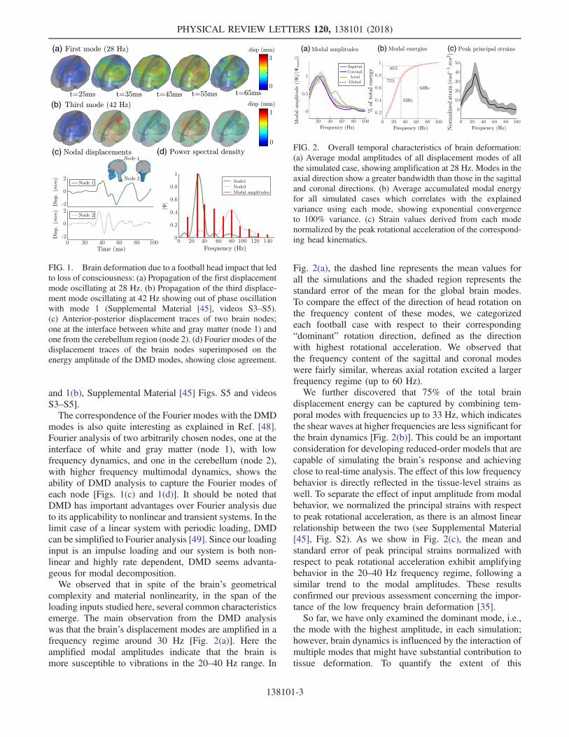
and 1(b), Supplemental Material [45] Figs. S5 and videosS3–S5].The correspondence of the Fourier modes with the DMD
modes is also quite interesting as explained in Ref. [48].Fourier analysis of two arbitrarily chosen nodes, one at theinterface of white and gray matter (node 1), with lowfrequency dynamics, and one in the cerebellum (node 2),with higher frequency multimodal dynamics, shows theability of DMD analysis to capture the Fourier modes ofeach node [Figs. 1(c) and 1(d)]. It should be noted thatDMD has important advantages over Fourier analysis dueto its applicability to nonlinear and transient systems. In thelimit case of a linear system with periodic loading, DMDcan be simplified to Fourier analysis [49]. Since our loadinginput is an impulse loading and our system is both non-linear and highly rate dependent, DMD seems advanta-geous for modal decomposition.We observed that in spite of the brain’s geometrical
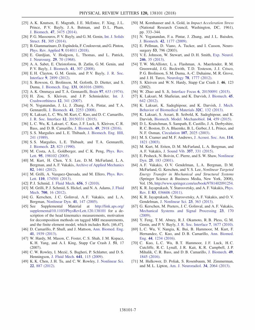
complexity and material nonlinearity, in the span of theloading inputs studied here, several common characteristicsemerge. The main observation from the DMD analysiswas that the brain’s displacement modes are amplified in afrequency regime around 30 Hz [Fig. 2(a)]. Here theamplified modal amplitudes indicate that the brain ismore susceptible to vibrations in the 20–40 Hz range. In
Fig. 2(a), the dashed line represents the mean values forall the simulations and the shaded region represents thestandard error of the mean for the global brain modes.To compare the effect of the direction of head rotation onthe frequency content of these modes, we categorizedeach football case with respect to their corresponding“dominant” rotation direction, defined as the directionwith highest rotational acceleration. We observed thatthe frequency content of the sagittal and coronal modeswere fairly similar, whereas axial rotation excited a largerfrequency regime (up to 60 Hz).We further discovered that 75% of the total brain
displacement energy can be captured by combining tem-poral modes with frequencies up to 33 Hz, which indicatesthe shear waves at higher frequencies are less significant forthe brain dynamics [Fig. 2(b)]. This could be an importantconsideration for developing reduced-order models that arecapable of simulating the brain’s response and achievingclose to real-time analysis. The effect of this low frequencybehavior is directly reflected in the tissue-level strains aswell. To separate the effect of input amplitude from modalbehavior, we normalized the principal strains with respectto peak rotational acceleration, as there is an almost linearrelationship between the two (see Supplemental Material[45], Fig. S2). As we show in Fig. 2(c), the mean andstandard error of peak principal strains normalized withrespect to peak rotational acceleration exhibit amplifyingbehavior in the 20–40 Hz frequency regime, following asimilar trend to the modal amplitudes. These resultsconfirmed our previous assessment concerning the impor-tance of the low frequency brain deformation [35].So far, we have only examined the dominant mode, i.e.,
the mode with the highest amplitude, in each simulation;however, brain dynamics is influenced by the interaction ofmultiple modes that might have substantial contribution totissue deformation. To quantify the extent of this
(a)
(b)
(c) (d)
FIG. 1. Brain deformation due to a football head impact that ledto loss of consciousness: (a) Propagation of the first displacementmode oscillating at 28 Hz. (b) Propagation of the third displace-ment mode oscillating at 42 Hz showing out of phase oscillationwith mode 1 (Supplemental Material [45], videos S3–S5).(c) Anterior-posterior displacement traces of two brain nodes;one at the interface between white and gray matter (node 1) andone from the cerebellum region (node 2). (d) Fourier modes of thedisplacement traces of the brain nodes superimposed on theenergy amplitude of the DMD modes, showing close agreement.
(a) (b) (c)
FIG. 2. Overall temporal characteristics of brain deformation:(a) Average modal amplitudes of all displacement modes of allthe simulated case, showing amplification at 28 Hz. Modes in theaxial direction show a greater bandwidth than those in the sagittaland coronal directions. (b) Average accumulated modal energyfor all simulated cases which correlates with the explainedvariance using each mode, showing exponential convergenceto 100% variance. (c) Strain values derived from each modenormalized by the peak rotational acceleration of the correspond-ing head kinematics.
PHYSICAL REVIEW LETTERS 120, 138101 (2018)
138101-3

contribution, we investigated how the peak principal strainscalculated from the FE model compare against the pre-dictions of the second-order mass-spring system proposedby Kornhauser [50]. The spring-mass system was used toapproximate the contribution of the dominant mode to therelative brain displacement. We used the largest directionalcomponent of rotational acceleration as base excitation. Weused previously published values for the moment of inertia[51], and based on the results shown in Fig. 2(a), weassumed a 28 Hz natural frequency.The rationale here was that if the global mode is the main
contributing factor to brain strain, we would expect therelation between the maximum relative displacement fromthe lumped model to correlate almost linearly with the peakprincipal strains from the FE model. The results are shownin Fig. 3(a), where the dashed line represents the best linearfit (R2 ¼ 0.72) between peak principal strain and maximumdisplacement from the lumped model. As expected, forlow brain strains (< 15%), this relationship maintains itslinearity. As the brain strains become larger, the linearcorrelation decreases and more outliers start to emerge,which we attribute to stronger multimodal behavior in brainregions. Because of the limited number of high strain cases,we superimposed strain and displacement predictions for58 (25 injury cases) previously reported National FootballLeague (NFL) head impact cases (represented by graycrosses) [52]. To formally investigate this effect, we usedthe parameter dynamic range, which we defined as the spanof frequencies that exist within 50% of the dominantmode’s amplitude. The dynamic range increases from zero
to 25 Hz as we go towards higher strain levels. By groupingthe strain values so that they fall within 10 Hz dynamicrange bins, we see an increase in the average error forhigher strains. As shown in Fig. 3(b), the correlationbetween the 1D model and the FE model’s predictionsdecreases with increasing dynamic range, indicating richerdynamical behavior (p < 0.01).The multimodal behavior of the brain presented here
provides interesting insights into possible mechanisticcauses of sports-related concussion. Almost all of theinjury cases we examined fell within the highest dynamicrange when compared with other football cases, signaling astrongly multimodal behavior. It is not surprising to see thatmost of this multimodal behavior falls into the higher strainregions since higher skull kinematics lead to higher tissuedeformations, intensify the effects of nonlinear materialbehavior and geometric nonlinearities, and enrich theresultant dynamics in the frequency domain.The richer temporal dynamics caused by the multimodal
behavior has the potential to create more spatiotemporaldifferences within the brain structures, hence resulting inlocalized dynamical phenomena and exacerbating theregional relative displacements. The TBI community haslong regarded the periventricular area of the brain to be aspecifically vulnerable region during rapid head motions[53] and tissue deformation in this area has been proposedas a potential cause for concussion [22,54]. To explore thisfurther, we examined the spatial variation of the dominantmode frequency within the brain for seven different brainstructures spanning deep and cortical regions of the brain:corpus callosum (CC), gray matter (GM), brain stem (BS),midbrain (MB), white matter (WM), thalamus (TH), andcerebellum (CB) [Fig. 4(a)]. We compared the modalbehavior for each region, between the LOC case and theaverage case, where we took the mean and standarddeviation of all the 187 noninjury cases in both frequencyand normalized strain measures [represented by horizontaland vertical error bars in Fig. 4(b)]. Superimposing theregional frequencies and normalized peak principal strainsonto the global trends [repeated from Fig. 2(c)], we foundthat corpus callosum has a distinct modal frequency around25 Hz and the corresponding contribution of this modecreates the highest strain levels in the brain, when com-pared with other regions. The local mode in CC for LOChas an even higher contribution to the tissue-level strains,resulting in 20% higher normalized strains than the brain’saverage at 25 Hz. Other brain regions have distinct modalfrequencies as well, with the brain stem having the highestmodal frequency around 40 Hz. We did not observe cleardifferences in the variance of local decays rates between thetwo cases (see Supplemental Material [45], Fig. S6). TheLOC case, which has a high dynamic range, leads todistinct local frequencies, specifically in the periventricularregion. In contrast, the average case with a low dynamicrange, expresses a smooth distribution of local frequencies.
(a) (b)
FIG. 3. Dynamic ranges of brain modes indicate multimodalbehavior for higher input amplitudes: (a) Football data fromStanford mouthguard and NFL data from Ref. [52] are comparedagainst the best linear fit calculated by using the global dominantmode [Fig. 4(a)]. Multimodal behavior is observed for higherstrain values, where a single mode fails to predict the peakprincipal strain. The dynamic range of football cases reveals atransition towards a multimodal behavior at higher strain values.(b) The dynamic ranges of individual football simulationssignificantly increase as they are further away from the singlemode prediction.
PHYSICAL REVIEW LETTERS 120, 138101 (2018)
138101-4

The increased localization in CC for the LOC casecompared with the average case is noteworthy, especiallysince this region was identified as a good correlate forconcussion classification in our previous study [22][Figs. 4(b)–4(d)]. These results suggest a potential linkbetween mode localization and tissue damage.A complete investigation of the effect of multimodal
behavior on regional brain dynamics requires a moreextensive study; however, a closer examination of theLOC head impact case against the average modal behaviorsupports our injury hypothesis above: localized dynamicscaused by the multimodal behavior of the brain tends toexcite more dangerous dynamics in the deep brain structures.From a mechanistic point of view, understanding the
initiation of these localized modes requires taking intoaccount the nonlinear material properties and geometry ofthe skull-brain system. Even in the absence of skulldeformation, external forces on the head will translate tothe brain tissue. Given the compliance of the skull-braininterface [55,56] and the viscoelastic and nonlinear behaviorof the brain tissue [57], this energy translation will lead to theinitiation of shear and longitudinal waves inside the brain.Motion and deformation of the brain under external loading,which is governed by damped hyperbolic equations, willhave nonlinear shear and longitudinal components[27,58,59]. The Zabolotskaya equation describes the propa-gation of finite-amplitude, two-dimensional, linearly polar-ized, shear waves in nonlinear solids [60]. Let Uðx; y; tÞdepict the 2D displacement field in a nonlinear media and xbe the direction of shear wave propagation. The shear wavepropagation can then be represented as follows [61]:
ðUt þ βU2UxÞx þ1
2Uyy ¼ 0; ð3Þ
where β is a parameter that characterizes the materialproperties of the solid. The cubic nonlinear term encounteredin the above equation is known to generate localizednonlinear modes in structural systems with geometric non-linearities [62,63], systems with nonlinear absorbers orenergy sinks [64], and granular dimer chains [65,66].Localization and motion confinement are observed whenvibrational energy tends to be confined to one particular areaof a structure or continuous media. They have first beenobserved for periodic linear structures presenting a structuralirregularity [67]. A generic property of nonlinear modelocalization is high localized amplitudes in a particular areaof the media with a distinct oscillation frequency. In thisstudy, through the use of dynamic mode decomposition, wehave found evidence regarding the existence of such modesas well as strain localization in the brain, particularly in thedeep white matter brain regions. To the best of our knowl-edge, there has been no experimental evidence regarding theexistence of such modes in nonlinear viscoelastic media upto date. By combining experimental measurements withcomputational modeling, our study presents the first indirectempirical evidence regarding the existence of localizedmodes with varying eigenvalues in a nonlinear, viscoelastic,and inhomogeneous media.In summary, we used the FE model to simulate 187 head
impacts collected during football game and practice. Thisprovided over 40 000 snapshots of over 5000 nodal braindisplacements. We utilized DMD analysis to extract modalbehavior of the brain from these highly transient events.Our findings show that (i) the brain-skull system isdominated by low frequency dynamics and more than75% of the brain’s displacement can be captured bytemporal modes with frequencies under 33 Hz, (ii) strainin brain tissue is amplified in frequencies close to 30 Hz,(iii) high dynamic range and multimodal behavior might be
(b)(a) (c) (d)
FIG. 4. Dominant frequencies of brain structures: (a) Schematic of the FE model of the head showing various parts of the brain.(b) Strain values derived from each mode normalized by the peak rotational acceleration of the corresponding head kinematics. Here, wesuperimpose regional dominant frequencies for each brain part for the LOC case (red circles) and the average and standard deviations(blue solid dots) for all cases. Brain part abbreviations: corpus callosum (CC), gray matter (GM), brain stem (BS), midbrain (MB), whitematter (WM), thalamus (TH), corpus callosum, cerebellum (CB). (c) Structural distribution of the dominant frequency of brain regionsfor the LOC case, indicating out of phase oscillation due to significant discrepancy between regions especially in the periventricularregion. (d) Structural distribution of dominant frequencies for an average head impact case, which indicates a lower dynamic range.
PHYSICAL REVIEW LETTERS 120, 138101 (2018)
138101-5

associated with higher risks of injury, and (iv) periventric-ular regions of the brain are prone to mode localization,which is the first such evidence for this type of phenome-non in a nonlinear hyperviscoelastic medium.There are several limitations associated with the work
presented here. The FE model used in this study hadpreviously been partially validated with sparse cadaverexperiments and not thoroughly validated against in vivoMRI data [25,68]. We believe that more accurate and regionspecific material properties and more detailed geometries,particularly the cortical gyri, will render the dynamicdifferences more pronounced. The constitutive materialmodels used in the FE simulations incorporate a linearviscosity definition, whichmight explainwhy the local decayrates in LOC and average cases were similar (seeSupplementalMaterial [45], Fig. S6). In addition, the loadingspace used in this study is limited to head impacts in football.Although similar results might be expected in higher loadingregimes, such as car accidents, the loading bias in differentanatomical regions might have a significant effect on theresulting brain dynamics. Also, we had relatively low numberof axial rotation cases from our football data compared tocoronal and sagittal, which might render the frequencyregimes of the axial modes [Fig. 2(a)] less accurate.Finally, there are small errors in the mouthguard measure-ments, previously quantified in anthropomorphic test dummyand in vivo video-tracking experiments [69,70].The spatiotemporal characteristics of the human brain we
identified in this Letter are particularly interesting becausestrain in corpus callosum has previously been shown to bethe best predictor for concussion cases in football [17,22].Similar observations have been made in previous diffusiontensor imaging studies to detect brain abnormalities inpatients with concussions. Fiber tracts in the deep brainstructures such as corpus callosum, fornix, hippocampus,thalamus, and cingulum bundles have been shown to be themost common locations of abnormal fractional anisotropyand mean diffusivity [71]. There needs to be more clinicalobservations to corroborate with the findings of this study.With the advancing imaging technology in acquiring
more spatially and temporally resolved experimental data,and more detailed finite element models, it is conceivablethat ever larger data sets will become available in the future.As such, our proposed methodology is particularly helpfulin extracting most notable characteristics of brain dynam-ics. A natural next step would be to use this technique indeveloping reduced-order models of brain deformation thatcould be used to perform near real-time simulations of headimpacts, providing crucial information to clinicians forinformed return-to-play decisions.
This research was supported by Child Health ResearchInstitute, Lucile Packard Foundation for Children’sHealth, Stanford CTSA (UL1 TR001085) and ThrasherResearch Foundation, who funded this work through theThrasher Early Career Award. This study was also partially
supported by the National Institutes of Health (NIH)(Grant No. 3R21EB01761101S1) National Institute ofBio-medical Imaging and Bioengineering (NIBIB)3R21EB01761101S1. We also thank Dr. AndrewKnutsen and Dr. Philip Bayly for sharing the MRI data.
K. Laksari and M. Kurt contributed equally to this work.
*[email protected][1] M. Faul, L. Xu, M. M. Wald, and V. Coronado, Atlanta, GA:
Centers for Disease Control and Prevention, National Centerfor Injury Prevention and Control (2010), https://stacks.cdc.gov/view/cdc/5571.
[2] C. L. MacDonald, A. M. Johnson, E. C. Nelson, N. J.Werner, R. Fang, S. F. Flaherty, and D. L. Brody,J. Neurotrauma 31, 889 (2014).
[3] J. A. Langlois, W. Rutland-Brown, and M.M. Wald, J. HeadTrauma Rehabilitation 21, 375 (2006).
[4] Centers for Disease Control and Prevention, MMWR MorbMortal Wkly Rep. 60, 1337 (2011).
[5] M. McCrea, T. Hammeke, G. Olsen, P. Leo, and K.Guskiewicz, Clinical J. Sport Medicine 14, 13 (2004).
[6] J. G. Garrick, Clinical journal of sport medicine 15, 385(2005).
[7] I. J. S. Williamson and D. Goodman, British Journal ofSports Medicine 40, 128 (2006).
[8] S. R. Laker, PM&R 3, S354 (2011).[9] A. Holbourn, Lancet 242, 438 (1943).
[10] A. Holbourn, J. Neurosurg. 1, 190 (1944).[11] M. Kornhauser, J. Appl. Mech. 21, 371 (1954).[12] A. Ommaya and A. E. Hirsch, J. Biomech. 4, 13 (1971).[13] S. Margulies and L. Thibault, J. Biomech. 25, 917 (1992).[14] J. Versace, SAE Technical Paper 710881, 1971, https://www
.sae.org/publications/technical-papers/content/710881/.[15] W. C. Moss, M. J. King, and E. G. Blackman, Phys. Rev.
Lett. 103, 108702 (2009).[16] L. Zhang, K. H. Yang, and A. I. King, J. Biomech. Eng. 126,
226 (2004).[17] S. Kleiven, Int. J. Crashworthiness 11, 65 (2006).[18] B. Coats, S. A. Eucker, S. Sullivan, and S. S. Margulies,
Int. J. Dev. Neuroscience: The Official Journal of theInternational Society for Developmental Neuroscience 30,191 (2012).
[19] T. M. Talavage, E. a. Nauman, E. L. Breedlove, U. Yoruk,A. E. Dye, K. E. Morigaki, H. Feuer, and L. J. Leverenz,J. Neurotrauma 31, 327 (2014).
[20] J. J. Bazarian, T. Zhu, J. Zhong, D. Janigro, E. Rozen,A. Roberts, H. Javien, K. Merchant-Borna, B. Abar, andE. G. Blackman, PLoS One 9, e94734 (2014).
[21] S. Kleiven, Stapp Car Crash J. 51, 81 (2007).[22] F. Hernandez, L. C. Wu, M. C. Yip, K. Laksari, A. R.
Hoffman, J. R. Lopez, G. A. Grant, S. Kleiven, and D. B.Camarillo, Ann. Biomed. Eng. 43, 1918 (2015).
[23] S. Sullivan, S. A. Eucker, D. Gabrieli, C. Bradfield, B.Coats, M. R. Maltese, J. Lee, C. Smith, and S. S. Margulies,Biomech. Model. Mechanobiol. 14, 877 (2015).
[24] C. Giordano and S. Kleiven, SAE Technical Paper 2014-22-0002, 2014, https://www.sae.org/publications/technical-papers/content/2014-22-0002/.
PHYSICAL REVIEW LETTERS 120, 138101 (2018)
138101-6
[25] A. K. Knutsen, E. Magrath, J. E. McEntee, F. Xing, J. L.Prince, P. V. Bayly, J. A. Butman, and D. L. Pham,J. Biomech. 47, 3475 (2014).
[26] P. G. Massouros, P. V. Bayly, and G.M. Genin, Int. J. SolidsStruct. 51, 305 (2014).
[27] B.Giammarinaro,D. Espíndola, F. Coulouvrat, andG. Pinton,Phys. Rev. Applied 9, 014011 (2018).
[28] E. Gurdjian, V. Hodgson, L. Thomas, and L. Patrick,J. Neurosurg. 29, 70 (1968).
[29] A. A. Sabet, E. Christoforou, B. Zatlin, G. M. Genin, andP. V. Bayly, J. Biomech. 41, 307 (2008).
[30] E. H. Clayton, G. M. Genin, and P. V. Bayly, J. R. Soc.Interface 9, 2899 (2012).
[31] S. Rowson, G. Brolinson, M. Goforth, D. Dietter, and S.Duma, J. Biomech. Eng. 131, 061016 (2009).
[32] A. K. Ommaya and T. A. Gennarelli, Brain 97, 633 (1974).[33] H. Zou, S. Kleiven, and J. P. Schmiedeler, Int. J.
Crashworthiness 12, 341 (2007).[34] N. Yoganandan, J. Li, J. Zhang, F. A. Pintar, and T. A.
Gennarelli, J. Biomech. 41, 2253 (2008).[35] K. Laksari, L. C. Wu, M. Kurt, C. Kuo, and D. C. Camarillo,
J. R. Soc. Interface 12, 20150331 (2015).[36] L. C. Wu, K. Laksari, C. Kuo, J. F. Luck, S. Kleiven, C. R.
Bass, and D. B. Camarillo, J. Biomech. 49, 2918 (2016).[37] S. S. Margulies and L. E. Thibault, J. Biomech. Eng. 111,
241 (1989).[38] S. S. Margulies, L. E. Thibault, and T. A. Gennarelli,
J. Biomech. 23, 823 (1990).[39] M. Costa, A. L. Goldberger, and C. K. Peng, Phys. Rev.
Lett. 95, 198102 (2005).[40] M. Kurt, H. Chen, Y. S. Lee, D. M. McFarland, L. A.
Bergman, and A. F. Vakakis, Archive of Applied Mechanics82, 1461 (2012).
[41] M. Grilli, A. Vazquez-Quesada, and M. Ellero, Phys. Rev.Lett. 110, 174501 (2013).
[42] P. J. Schmid, J. Fluid Mech. 656, 5 (2010).[43] M. Grilli, P. J. Schmid, S. Hickel, and N. A. Adams, J. Fluid
Mech. 700, 16 (2012).[44] G. Kerschen, J. C. Golinval, A. F. Vakakis, and L. A.
Bergman, Nonlinear Dyn. 41, 147 (2005).[45] See Supplemental Material at http://link.aps.org/
supplemental/10.1103/PhysRevLett.120.138101 for a de-scription of the head kinematics measurements, motivationfor decomposition methods on tagged MRI measurements,and the finite element model, which includes Refs. [46,47].
[46] D. Camarillo, P. Shull, and J. Mattson, Ann. Biomed. Eng.41, 1939 (2013).
[47] W. Hardy, M. Mason, C. Foster, C. S. Shah, J. M. Kopacz,K. H. Yang, and A. I. King, Stapp Car Crash J. 51, 17(2007).
[48] C. W. Rowley, I. Mezić, S. Bagheri, P. Schlatter, and D. S.Henningson, J. Fluid Mech. 641, 115 (2009).
[49] K. K. Chen, J. H. Tu, and C.W. Rowley, J. Nonlinear Sci.22, 887 (2012).
[50] M. Kornhauser and A. Gold, in Impact Acceleration Stress(National Research Council, Washington, DC, 1961),pp. 333–344.
[51] N. Yoganandan, F. a. Pintar, J. Zhang, and J. L. Baisden,J. Biomech. 42, 1177 (2009).
[52] E. Pellman, D. Viano, A. Tucker, and I. Casson, Neuro-surgery 53, 796 (2003).
[53] V. E. Johnson, W. Stewart, and D. H. Smith, Exp. Neurol.246, 35 (2013).
[54] T. W. McAllister, L. a. Flashman, A. Maerlender, R. M.Greenwald, J. G. Beckwith, T. D. Tosteson, J. J. Crisco,P. G. Brolinson, S. M. Duma, A.-C. Duhaime, M. R. Grove,and J. H. Turco, Neurology 78, 1777 (2012).
[55] S. Kleiven and W. N. Hardy, Stapp Car Crash J. 46, 123(2002).
[56] W. Zhao and S. Ji, Interface Focus 6, 20150091 (2015).[57] K. Laksari, M. Shafieian, and K. Darvish, J. Biomech. 45,
642 (2012).[58] K. Laksari, K. Sadeghipour, and K. Darvish, J. Mech.
Behavior of Biomedical Materials 32C, 132 (2013).[59] K. Laksari, S. Assari, B. Seibold, K. Sadeghipour, and K.
Darvish, Biomech. Model. Mechanobiol. 14, 459 (2015).[60] D. L. Kraitchman, S. Sampath, E. Castillo, J. A. Derbyshire,
R. C. Boston, D. A. Bluemke, B. L. Gerber, J. L. Prince, andN. F. Osman, Circulation 107, 2025 (2003).
[61] M. S. Cramer and M. F. Andrews, J. Acoust. Soc. Am. 114,1821 (2003).
[62] M. Kurt, M. Eriten, D. M. McFarland, L. A. Bergman, andA. F. Vakakis, J. Sound Vib. 357, 331 (2015).
[63] E. Pesheck, N. Boivin, C. Pierre, and S. W. Shaw, NonlinearDyn. 25, 183 (2001).
[64] A. F. Vakakis, O. V. Gendelman, L. A. Bergman, D.M.McFarland, G. Kerschen, and Y. S. Lee, Nonlinear TargetedEnergy Transfer in Mechanical and Structural Systems(Springer Science & Business Media, New York, 2008),Vol. 156, http://www.springer.com/us/book/9781402091254.
[65] K. R. Jayaprakash, Y. Starosvetsky, and A. F. Vakakis, Phys.Rev. E 83, 036606 (2011).
[66] K. R. Jayaprakash, Y. Starosvetsky, A. F. Vakakis, and O. V.Gendelman, J. Nonlinear Sci. 23, 363 (2013).
[67] G. Kerschen, M. Peeters, J. C. Golinval, and A. F. Vakakis,Mechanical Systems and Signal Processing 23, 170(2009).
[68] Y. Feng, T. M. Abney, R. J. Okamoto, R. B. Pless, G. M.Genin, and P. V. Bayly, J. R. Soc. Interface 7, 1677 (2010).
[69] L. C. Wu, V. Nangia, K. Bui, B. Hammoor, M. Kurt, F.Hernandez, C. Kuo, and D. B. Camarillo, Ann. Biomed.Eng. 44, 1234 (2016).
[70] C. Kuo, L. C. Wu, B. T. Hammoor, J. F. Luck, H. C.Cutcliffe, R. C. Lynall, J. R. Kait, K. R. Campbell, J. P.Mihalik, C. R. Bass, and D. B. Camarillo, J. Biomech. 49,1845 (2016).
[71] M. Hulkower, D. Poliak, S. Rosenbaum, M. Zimmerman,and M. L. Lipton, Am. J. Neuroradiol. 34, 2064 (2013).
PHYSICAL REVIEW LETTERS 120, 138101 (2018)
138101-7