phylogenetic study on proales daphnicola thompson, … · within rotifera, many morphological,...
TRANSCRIPT
Pr
Ea
b
c
a
ARRA
KMMTPSC
1
l(2eoimabrmipWGi
o2
0d
Zoologischer Anzeiger 251 (2012) 180–196
Contents lists available at SciVerse ScienceDirect
Zoologischer Anzeiger
journa l homepage: www.e lsev ier .de / jcz
hylogenetic study on Proales daphnicola Thompson, 1892 (Proalidae) and itselocation to Epiphanes (Rotifera: Epiphanidae)
.F. Wiltsa,b,∗, D. Brunsa, D. Fontanetoc, W.H. Ahlrichsa
Systematics and Evolutionary Biology, Department of Biology and Environmental Sciences, Carl von Ossietzky University Oldenburg, 26111 Oldenburg, GermanySenckenberg Research Institute, German Centre for Marine Biodiversity Research (DZMB), 26382 Wilhelmshaven, GermanyImperial College London, Division of Biology, Silwood Park Campus, Ascot Berkshire SL5 7PY, United Kingdom
r t i c l e i n f o
rticle history:eceived 24 March 2011eceived in revised form 22 August 2011ccepted 25 August 2011
a b s t r a c t
Within Rotifera, many morphological, taxonomic and phylogenetic questions still remain unanswered.Many families and genera have only poor phylogenetic support and classification is doubtful in a largenumber of species. To address these problems, a comprehensive reinvestigation of various species is nec-essary, especially for families such as Proalidae, in which several species have frequently been movedin and out. Here, we reinvestigated the species Proales daphnicola using light and electron microscopical
eywords:onogonontaorphology
axonomyhylogenyEMOI
techniques as well as morphological and molecular phylogenetic analyses in order to evaluate its phylo-genetic position. Based on our results, we reassign P. daphnicola to Epiphanes and give a redescription ofEpiphanes daphnicola (Thompson, 1892) n. comb. As a consequence of this conclusion, we also reassignProales kostei to Epiphanes.
© 2011 Elsevier GmbH. All rights reserved.
. Introduction
Within the past decade, our understanding of rotifer phy-ogeny has improved by using modern morphological methodse.g. Sørensen, 2002; Segers and Wallace, 2008; Riemann et al.,009), molecular methods (e.g. Herlyn et al., 2003; Yoshinagat al., 2004; Garcia-Varela and Nadler, 2006) and a combinationf both (Sørensen and Giribet, 2006). Admittedly, phylogeneticnvestigations do not exist for many taxa of this group of small
icroinvertebrates. In particular, morphological cladistic analysesre difficult to perform without time-consuming reinvestigations,ecause detailed morphological descriptions are lacking for mostotifers. In an ongoing investigation, we study the inner and outerorphology of different rotifer species mainly belonging to Proal-
dae, in order to clarify the phylogeny and existing taxonomicalroblems of this monogonont taxon as stressed by Sørensen (2005),
ilts et al. (2009a) and Wilts and Ahlrichs (2010). The genus Proalesosse, 1886, currently accommodating 44 species (Segers, 2007),s one of the most problematic genera from a systematic point of
∗ Corresponding author at: Systematics and Evolutionary Biology, Departmentf Biology and Environmental Sciences, Carl von Ossietzky University Oldenburg,6111 Oldenburg, Germany. Tel.: +49 441 798 3369.
E-mail address: [email protected] (E.F. Wilts).
044-5231/$ – see front matter © 2011 Elsevier GmbH. All rights reserved.oi:10.1016/j.jcz.2011.08.005
view, because it represents a taxonomically unsatisfactory assem-blage of diverse species. The genus was subdivided into differentgroups very early (see Remane, 1929–33; Voigt, 1956–57). Koste(1978) subdivided the genus into two main groups: freshwaterspecies resembling the type species of Proales, Proales decipiens(Ehrenberg, 1832), group A, and the predominantly marine speciesresembling Proales reinhardti (Ehrenberg, 1834), group B. However,several species of Proales do not fit into any of the two categories.Two of these species, Proales sigmoidea (Skorikov, 1896) and Proaleswerneckii (Ehrenberg, 1834), have recently been reassigned to Pleu-rotrocha (Notommatidae) and Pourriotia (Notommatidae) by Wiltset al. (2009a) and De Smet (2009), and possibly further reassign-ments will be required when more detailed data are acquired foradditional species. In the present study, we reinvestigated Proalesdaphnicola Thompson, 1892, a limnic species which clearly differsfrom Proales species belonging to the subgroups A and B. In fact,our morphological investigation reveals that P. daphnicola ratherresembles the genus Epiphanes in respect of habitus, corona, andtrophi morphology and also biology. Phylogenetic analyses of mor-phological data support this hypothesis; moreover, also molecularphylogeny is in agreement with a closer relationship of P. daphni-
cola to Epiphanes than to other species of Proales. The species Proaleskostei Nogrady and Smol, 1989 has close resemblance to P. daphni-cola, and it should likewise be closely related to Epiphanes accordingto results of our morphological analyses.
er Anz
2
2
cna8sfdsGalmcascdgatfiA
(ddtws7(fFsalaw(soiatow
2
mao(KS(scth
E.F. Wilts et al. / Zoologisch
. Material and methods
.1. Morphological preparations and species identification
P. daphnicola was found living in large numbers on Daphnia sp.ollected with a plankton sieve (64 �m mesh size) from a pondear Leer, Germany (53◦25′03.50′′N, 7◦52′48.25′′E) in October 2007nd 2008 and from a ditch in Oldenburg, Germany (53◦15′27.41′′N,◦16′60.12′′E) in October 2008 and 2009. Under a stereomicro-cope (LEICA MZ125), the single Daphnia specimens were testedor attached rotifer specimens. The crustaceans were isolated in arop of water on a glass slide, where the rotifers left the crustaceanseparately. Epiphanes senta was found in a ditch in Oldenburg,ermany (53◦15′31.31′′N, 8◦16′87.53′′E) in March 2010. Individu-ls of both rotifer species were studied by differential interferenceight microscopy (LEICA DMLB) as well as by scanning electron
icroscopy (ZEISS DSM 940). Isolated rotifer specimens were nar-otized with bupivacaine and fixed with 4% OsO4 solution and picriccid formaldehyde at 240 mOsm (after Melone, 1998), and pre-erved using a 4% paraformaldehyde solution. Dehydration wasarried out in a graded ethanol series followed by critical pointrying. Then, specimens were mounted on stubs and coated withold and platinum, respectively. Trophi were generally preparedpplying the procedure described by De Smet (1998) but dissolvingissues surrounding the trophi with a mixture of SDS/DTT (modi-ed after Kleinow et al., 1990). Line drawings were created withdobe Illustrator® CS2 and Adobe Photoshop® CS2.
Although the initial description of P. daphnicola by Thompson1892) does not contain information on or drawings of its trophi,ifferent authors (e.g. Harring and Myers, 1924) gave a sufficientescription of the species, also including drawings of general habi-us and trophi. In our determination, we refer to Harring and Myersho described a rounded posterio-dorsal ramus margin for the
pecies (see Plate XVIII, Fig. 5 in Harring and Myers, 1924 and Figs.B and 8B in this study). In his great work on the Proalidae, De Smet1996a) presented modified drawings and SEM pictures of trophieaturing a distinct spine on the posterio-dorsal ramus margin (seeigs. 333, 334 and Plate 13, Fig. 3 in De Smet, 1996a) for the samepecies. Admittedly, this posterior-dorsal spine was not present inny specimen of P. daphnicola collected in Oldenburg and Leer (aocation 60 km from Oldenburg), whereas Proales pejleri (Fig. 1Bnd C in De Smet et al., 1993) shows this character. The speciesas synonymized with P. daphnicola by De Smet (1996a). De Smet
personal communication) noticed a length variation of the dorsalpine depending on the studied population, however, according tour data and the description given by Harring and Myers (1924),t is also conceivable that two different species exist, one withoutspine and one with a spine varying in length, making a revalida-
ion of P. pejleri necessary. In future studies, a detailed comparisonf our specimens with those featuring a dorsal spine on the ramiould help to address this problem.
.2. Morphological character matrix and cladistic analysis
For phylogenetic analyses, 20 species were included in a dataatrix and opposed to 43 morphological characters (see Table 1
nd list of characters in Appendix A) compiled from direct personalbservations and from the following literature sources: Thompson1892), Harring and Myers (1924), Voigt (1956–57), Koste (1978),oste and Terlutter (2001), Kleinow et al. (1990), Nogrady andmol (1989), De Smet (1996a, 1996b), Melone (2001), Sørensen2002), De Smet and Gibson (2008). The character matrix was con-
tructed in Nexus Data Editor (NDE, version 0.5.0, Page, 2001)ontaining 27 characters of binary and 16 characters of mul-istate coding. Six characters are parsimony uninformative, butave been retained in the analysis, since they are illustrativeeiger 251 (2012) 180–196 181
synapomorphies for the ingroup taxa. All characters, except forcharacter no. 35, were treated as unordered and all characterswere equally weighted without any a priori assumptions on char-acter polarization. Therefore, the coding 0 does not necessarilyrepresent a plesiomorphic condition. The taxon sampling for theingroup included P. daphnicola, P. kostei, and several other rotifersof the subtaxon Ploima with malleate or modified malleate trophi(Transversiramida, see Sørensen, 2002). At least one species fromeach transversiramid family (Brachionidae, Euchlanidae, Lepadell-idae, Lecanidae, Mytilinidae, Proalidae, Trichotriidae) was includedin the matrix. For Epiphanidae, several representatives from allgenera (i.e. Epiphanes, Rhinoglena, Mikrocodides and Cyrtonia) wereincluded in order to obtain a better resolution of the family. Dueto our presumption P. daphnicola may be related to them. P. pejleriDe Smet, van Rompu and Beyens 1993 was not included becauseof its incomplete habitus description. Furthermore, species of Pro-ales group A (namely P. fallaciosa and P. tillyensis) and of Proalesgroup B (namely P. reinhardti and P. theodora) were included inthe matrix, in order to test if P. daphnicola and P. kostei are relatedto one or the other Proales group. The notommatid species Pleu-rotrocha petromyzon (Ehrenberg, 1830) was selected as outgroup,because this ploimid taxon is expected to stand apart from otherploimid species with malleate trophi (see Sørensen, 2002). Thisspecies has virgate trophi and adequate data are available (seeWilts et al., 2009a). While compiling the data matrix, we consid-ered further characters to be included, such as the ultrastructureof protonephridia, the detailed construction of the corona or themorphology of the somatic musculature, but such information iscurrently restricted to a very limited number of species.
The maximum parsimony analysis of the matrix was carriedout with PAUP* 4.0 b10 (Swofford, 2002) using the branch-and-bound search method. To calculate the node robustness of theobtained tree, a bootstrap analysis with 1000 replicates was run.Additionally, the Bremer support (Bremer 1988) (decay index) wascalculated in TreeRot 3 (Sorenson and Franzosa, 2007). The pro-gram FigTree 1.3.1 (Rambaut, 2006–2009) was used to visualizethe trees. The character evolution was retraced with MESQUITE2.74 (Maddison and Maddison, 2010) using ACCTRAN (acceleratedtransformation) character optimisation.
2.3. Molecular data and phylogenetic analysis
In order to provide support for the hypothesis of P. daphnicolabeing more similar to Epiphanes than to Proales, we performed aphylogenetic analysis for a taxon set very similar to the one usedfor morphological analysis. We focused on the mitochondrial genecytochrome oxidase c subunit I (COI). It is known that COI is notvery reliable to resolve relationships between families; neverthe-less, using the translated amino acid alignment can improve itsusefulness (Fontaneto and Jondelius, 2011). Moreover, a prelim-inary screening of genetic distances between and within Proalesand other genera of Ploima from loci already available in GenBank(COI, 18S and 28S) did not provide evidence for ribosomal loci beingbetter than COI in solving relationships.
To obtain new COI sequences, single clean individual rotiferswere isolated, put in a 0.5 ml tube and dehydrated. DNA was thenextracted by adding 35 �l of Chelex (Bio-Rad Instagene Matrix) toeach sample processed at 56 ◦C for 20 min and at 100 ◦C for 10 min.A fragment of COI was then amplified using the primers COI-F(AGTTCTAATCATAARGATATYGG) and COI-R (TAAACTTCAGGGT-GACCAAAAAATCA). Cycle conditions were 94 ◦C for 5 min, 40 cyclesof 94 ◦C for 30 s, 50 ◦C for 40 s, 72 ◦C for 40 s, and a final extension
step of 72 ◦C for 7 min. Purification and sequencing were performedby Macrogen Korea. Sequences were checked and aligned by eyeand no gaps had to be inserted. Phylogenetic reconstructions wereperformed (1) on the nucleotide alignment using the GRT + G + I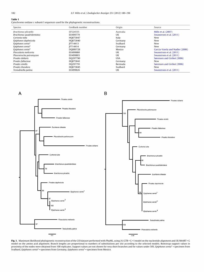
182 E.F. Wilts et al. / Zoologischer Anzeiger 251 (2012) 180–196
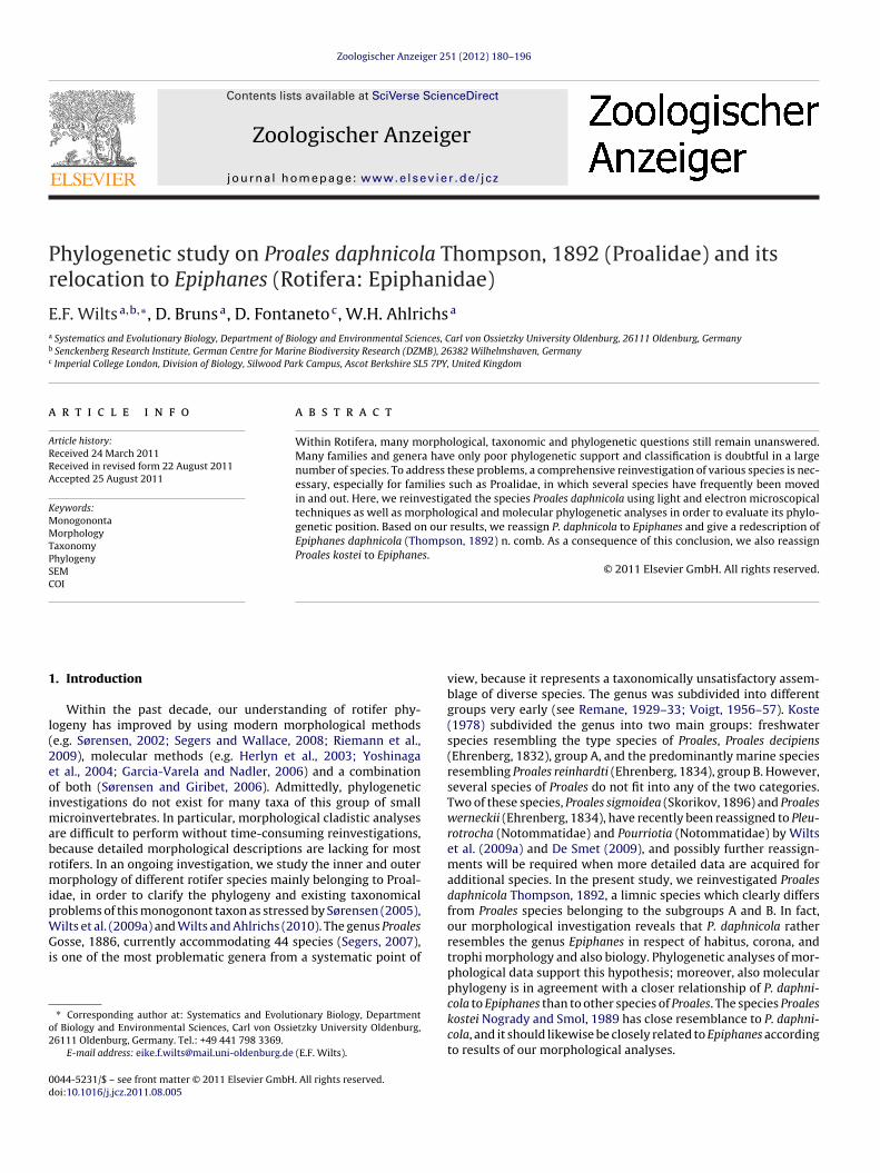
Table 1Cytochrome oxidase c subunit I sequences used for the phylogenetic reconstructions.
Species GenBank number Origin Source
Brachionus plicatilis EF524555 Australia Mills et al. (2007)Brachionus quadridentatus EU499779 UK Swanstrom et al. (2011)Cyrtonia tuba HQ873047 Italy NewEpiphanes daphnicola HQ873040 Germany NewEpiphanes senta1 JF714413 Svalbard NewEpiphanes senta2 JF714414 Germany NewEpiphanes senta3 DQ089728 Mexico Garcia-Varela and Nadler (2006)Floscularia melicerta EU499880 UK Swanstrom et al. (2011)Pleurotrocha petromyzon EU499803 UK Swanstrom et al. (2011)Proales doliaris DQ297790 USA Sørensen and Giribet (2006)Proales fallaciosa HQ873041 Germany NewProales similis DQ297791 Bermuda Sørensen and Giribet (2006)Proales theodora HQ873045 Svalbard NewTestudinella patina EU499826 UK Swanstrom et al. (2011)
Fig. 1. Maximum likelihood phylogenetic reconstruction of the COI dataset performed with PhyML, using (A) GTR + G + I model on the nucleotide alignment and (B) MtART + Gmodel on the amino acid alignment. Branch lengths are proportional to numbers of substitutions per site according to the selected models. Bootstrap support values inproximity of the nodes were obtained from 100 replicates. Support values are not shown for very short branches and for values under 50%. Epiphanes senta1 = specimen fromSvalbard, Epiphanes senta2 = specimen from Germany, Epiphanes senta3 = specimen from Mexico.
E.F. Wilts et al. / Zoologischer Anzeiger 251 (2012) 180–196 183
Fig. 2. Strict consensus tree of 36 equally parsimonious trees at 108 steps, generated by PAUP*. Numbers behind nodes indicate bootstrap support values, numbers followingafter/Bremer support indices. Numbers in black squares refer to selected synapomorphic character states: (1) epidermis stiffened to lorica (character 1): absent; habitusshape (character 2): bulbous saccate, type Epiphanes senta; direction of ramus foramen subbasalis in relative to the fulcrum (character 27): directing inferio-posteriorly, typeEpiphanes senta; ventral manubrial chamber enlarged (character 35): tendency present, type Mikrocodides chlaena; epipharynx (character 38): present, type Mikrocodideschlaena; hypopharynx (character 39): cuticular strands, type P. daphnicola; sexual dimorphism (character 40): males well developed, little smaller than females, type Epiphanessenta. (2) Position of dorsal antenna (character 11): displaced posteriorely, type Epiphanes senta; tips of ramus basal apophyses (character 24): with hairs, type Epiphanessenta; ventral manubrial chamber enlarged (character 35): well developed, type Epiphanes senta. (3) Position of lateral antennae (character 12): middle of the trunk, typeP. daphnicola. (4) Eye position (character 19): ventrally on brain, type Epiphanes senta; manubrium foramen dorsalis (character 36): opened, chamber reduced to flat plane,t ped (c( is (cha3 racter( ent, ty
m(w2aa
trtiblTdirPt2
ype Epiphanes daphnicola. (5) Number of eyes (character 18): no eyes; incus y-shacharacter 23): present, type Epiphanes daphnicola; shape of ramus foramen subbasal2): curved plate with curved teeth, type Epiphanes daphnicola; subuncus shape (chacharacter 38): absent; deposition of eggs on exterior substrate (character 43): pres
odel, which was suggested as the best model by ModelGeneratorKeane et al., 2006), and (2) on the translated amino acid alignmentith the MtART + G model suggested by ProtTest 2.4 (Abascal et al.,
005). Maximum Likelihood reconstructions for both nucleotidend amino acid datasets were performed with PhyML 3.0 (Guindonnd Gascuel, 2003) with 100 bootstrap replicates.
As an additional test, we used the matrix of pairwise genetic dis-ances to support the hypothesis that P. daphnicola is more closelyelated to Epiphanes than to Proales. We obtained pair-wise dis-ances between P. daphnicola and the species in the two generan two ways: (1) by calculating the uncorrected genetic distancesetween sequences in the nucleotide alignment, and (2) by calcu-
ating the patristic distances between terminals in the two trees.hus, we had three datasets to perform the test: the first on rawistances not assuming any evolutionary model, the other two
ncorporating different evolutionary models from phylogenetic
econstructions based on nucleotide and amino acid alignments.air-wise genetic distances and linear models (LM) to performhe tests were computed in R 2.10.0 (R Development Core Team,010).haracter 21): present, type Epiphanes daphnicola; ramus basal apophyses enlargedracter 26): 0 = very large, oval, type Epiphanes daphnicola; shape of uncus (character34): basing on largest uncus tooth, lamellar, type Epiphanes daphnicola; epipharynxpe Proales reinhardti.
3. Results
3.1. Phylogeny
3.1.1. Molecular analysisThe molecular phylogenetic reconstruction using COI sequences
from both the nucleotide (Fig. 1A) and the amino acid alignments(Fig. 1B) support our two hypotheses: (1) that Proales is a poten-tially polyphyletic genus, and (2) that P. daphnicola clusters withEpiphanes and not with Proales (Fig. 1). Unfortunately, bootstrapsupport values for the monophyly of Epiphanes + P. daphnicola arenever very high, ranging between 59 and 69.
Uncorrected genetic distances between P. daphnicola andEpiphanes are 16.3–18.8%, whereas they are 26.1–32.2% betweenP. daphnicola and Proales. These differences are statistically signif-icant (LM: p = 0.002). The uncorrected genetic distances between
P. daphnicola and Epiphanidae (Epiphanes and Cyrtonia) are allbelow 20%, distances to other Ploima, including Proalidae arebetween 20 and 33% and distances to Gnesiotrocha (Testudinellaand Floscularia) are above 30%. Thus, a strong phylogenetic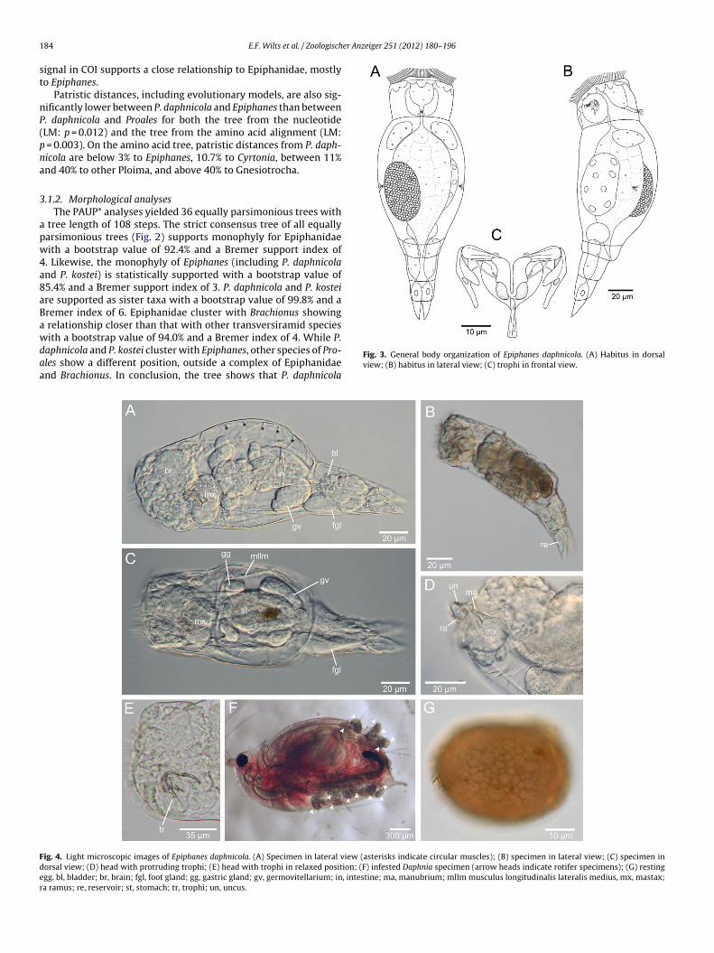
1 er Anzeiger 251 (2012) 180–196
st
nP(pna
3
apw4a8aBawdaa
Fder
84 E.F. Wilts et al. / Zoologisch
ignal in COI supports a close relationship to Epiphanidae, mostlyo Epiphanes.
Patristic distances, including evolutionary models, are also sig-ificantly lower between P. daphnicola and Epiphanes than between. daphnicola and Proales for both the tree from the nucleotideLM: p = 0.012) and the tree from the amino acid alignment (LM:= 0.003). On the amino acid tree, patristic distances from P. daph-icola are below 3% to Epiphanes, 10.7% to Cyrtonia, between 11%nd 40% to other Ploima, and above 40% to Gnesiotrocha.
.1.2. Morphological analysesThe PAUP* analyses yielded 36 equally parsimonious trees with
tree length of 108 steps. The strict consensus tree of all equallyarsimonious trees (Fig. 2) supports monophyly for Epiphanidaeith a bootstrap value of 92.4% and a Bremer support index of
. Likewise, the monophyly of Epiphanes (including P. daphnicoland P. kostei) is statistically supported with a bootstrap value of5.4% and a Bremer support index of 3. P. daphnicola and P. kosteire supported as sister taxa with a bootstrap value of 99.8% and aremer index of 6. Epiphanidae cluster with Brachionus showingrelationship closer than that with other transversiramid species
ith a bootstrap value of 94.0% and a Bremer index of 4. While P.aphnicola and P. kostei cluster with Epiphanes, other species of Pro-les show a different position, outside a complex of Epiphanidaend Brachionus. In conclusion, the tree shows that P. daphnicola
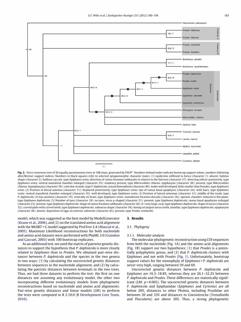
Fig. 3. General body organization of Epiphanes daphnicola. (A) Habitus in dorsalview; (B) habitus in lateral view; (C) trophi in frontal view.
ig. 4. Light microscopic images of Epiphanes daphnicola. (A) Specimen in lateral view (asterisks indicate circular muscles); (B) specimen in lateral view; (C) specimen inorsal view; (D) head with protruding trophi; (E) head with trophi in relaxed position; (F) infested Daphnia specimen (arrow heads indicate rotifer specimens); (G) restinggg. bl, bladder; br, brain; fgl, foot gland; gg, gastric gland; gv, germovitellarium; in, intestine; ma, manubrium; mllm musculus longitudinalis lateralis medius, mx, mastax;a ramus; re, reservoir; st, stomach; tr, trophi; un, uncus.
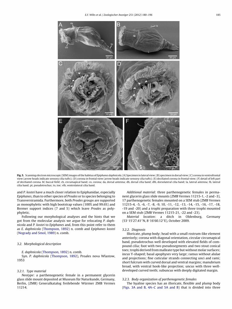
E.F. Wilts et al. / Zoologischer Anzeiger 251 (2012) 180–196 185
F icola.v ads ino antennc
aETaBp
gna(
3
1
3
gB1
ig. 5. Scanning electron microscopic (SEM) images of the habitus of Epiphanes daphniew (arrow heads indicate sensory cilia tufts); (D) corona in frontal view (arrow hef deciliated corona. bf, buccal field; cb, circumapical band; co, corona; da, dorsalilia band; pt, pseudotrochus; to, toe; vlb, ventrolateral cilia band.
nd P. kostei have a much closer relation to Epiphanidae, especiallypiphanes, than to other species of Proales or to species belonging toransversiramida. Furthermore, both Proales groups are supporteds monophyletic with high bootstrap values (100% and 99.6%) andremer support indices (7 and 5) which leave Proales as poly-hyletic.
Following our morphological analyses and the hints that weot from the molecular analysis we argue for relocating P. daph-icola and P. kostei to Epiphanes and, from this point refer to thems E. daphnicola (Thompson, 1892) n. comb and Epiphanes kosteiNogrady and Smol, 1989) n. comb.
.2. Morphological description
E. daphnicola (Thompson, 1892) n. comb.Syn. P. daphnicola (Thompson, 1892), Proales nova Wlastow,
953
.2.1. Type material
Neotype: a parthenogenetic female in a permanent glycerinlass slide mount deposited at Museum für Naturkunde, Germany,erlin, (ZMB) Generalkatalog freilebende Würmer ZMB Vermes1214.
(A) Specimen in lateral view; (B) specimen in dorsal view; (C) corona in ventrofrontaldicate sensory cilia tufts); (E) deciliated corona in frontal view; (F) detail of left parta; db, dorsal cilia band; dlb, dorsolateral cilia band; la, lateral antenna; lb, lateral
Additional material: three parthenogenetic females in perma-nent glycerin glass slide mounts (ZMB Vermes 11215-1, -2 and -3),17 parthenogenetic females mounted on a SEM stub (ZMB Vermes11215-4, -5, -6, -7, -8, -9, 10, -11, -12, -13, -14, -15, -16, -17, -18,-19 and -20) and a trophi preparation with three trophi mountedon a SEM stub (ZMB Vermes 11215-21, -22 and -23).
Material location: a ditch in Oldenburg, Germany(53◦15′27.41′′N, 8◦16′60.12′′E), October 2009.
3.2.2. DiagnosisIlloricate, plump body; head with a small rostrum-like element
anteriorly; corona with diagonal orientation, circular circumapicalband, pseudotrochus well developed with elevated fields of com-pound cilia; foot with two pseudosegments and two stout conicaltoes; trophi derived from malleate type but without molar surfaces;incus Y-shaped; basal apophyses very large; ramus without alulaeand projections; fine cuticular strands connecting unci and rami;short fulcrum with curved dorsal and ventral margins; manubriumbroad, with ventral hook-like projection; uncus with three well-developed curved teeth; subuncus with deeply digitated margin.
3.2.3. Body organization of parthenogenetic femalesThe hyaline species has an illoricate, flexible and plump body
(Figs. 3A and B, 4A–C and 5A and B) that is divided into three
186 E.F. Wilts et al. / Zoologischer Anz
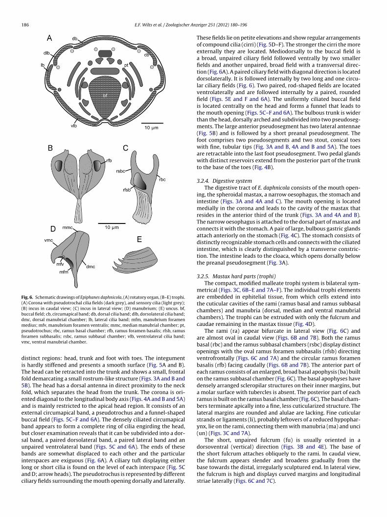
Fig. 6. Schematic drawings of Epiphanes daphnicola, (A) rotatory organ, (B–E) trophi.(A) Corona with pseudotrochal cilia fields (dark grey), and sensory cilia (light grey);(B) incus in caudal view; (C) incus in lateral view; (D) manubrium; (E) uncus. bf,buccal field; cb, circumapical band; db, dorsal cilia band; dlb, dorsolateral cilia band;dmc, dorsal manubrial chamber; lb, lateral cilia band; mfm, manubrium foramenmedius; mfv, manubrium foramen ventralis; mmc, median manubrial chamber; pt,pfv
diTf5feaebbbsubilac
seudotrochus; rbc, ramus basal chamber; rfb, ramus foramen basalis; rfsb, ramusoramen subbasalis; rsbc, ramus subbasal chamber; vlb, ventrolateral cilia band;mc, ventral manubrial chamber.
istinct regions: head, trunk and foot with toes. The integuments hardly stiffened and presents a smooth surface (Fig. 5A and B).he head can be retracted into the trunk and shows a small, frontalold demarcating a small rostrum-like structure (Figs. 3A and B andB). The head has a dorsal antenna in direct proximity to the neckold, which separates the head from the trunk. The corona is ori-nted diagonal to the longitudinal body axis (Figs. 4A and B and 5A)nd is mainly restricted to the apical head region. It consists of anxternal circumapical band, a pseudotrochus and a funnel-shapeduccal field (Figs. 5C–F and 6A). The densely ciliated circumapicaland appears to form a complete ring of cilia engirding the head,ut closer examination reveals that it can be subdivided into a dor-al band, a paired dorsolateral band, a paired lateral band and annpaired ventrolateral band (Figs. 5C and 6A). The ends of theseands are somewhat displaced to each other and the particular
nterspaces are exiguous (Fig. 6A). A ciliary tuft displaying eitherong or short cilia is found on the level of each interspace (Fig. 5Cnd D; arrow heads). The pseudotrochus is represented by differentiliary fields surrounding the mouth opening dorsally and laterally.
eiger 251 (2012) 180–196
These fields lie on petite elevations and show regular arrangementsof compound cilia (cirri) (Fig. 5D–F). The stronger the cirri the moreexternally they are located. Mediodorsally to the buccal field isa broad, unpaired ciliary field followed ventrally by two smallerfields and another unpaired, broad field with a transversal direc-tion (Fig. 6A). A paired ciliary field with diagonal direction is locateddorsolaterally. It is followed internally by two long and one circu-lar ciliary fields (Fig. 6). Two paired, rod-shaped fields are locatedventrolaterally and are followed internally by a paired, roundedfield (Figs. 5E and F and 6A). The uniformly ciliated buccal fieldis located centrally on the head and forms a funnel that leads tothe mouth opening (Figs. 5C–F and 6A). The bulbous trunk is widerthan the head, dorsally arched and subdivided into two pseudoseg-ments. The large anterior pseudosegment has two lateral antennae(Fig. 5B) and is followed by a short preanal pseudosegment. Thefoot comprises two pseudosegments and two stout, conical toeswith fine, tubular tips (Fig. 3A and B, 4A and B and 5A). The toesare retractable into the last foot pseudosegment. Two pedal glandswith distinct reservoirs extend from the posterior part of the trunkto the base of the toes (Fig. 4B).
3.2.4. Digestive systemThe digestive tract of E. daphnicola consists of the mouth open-
ing, the spheroidal mastax, a narrow oesophagus, the stomach andintestine (Figs. 3A and 4A and C). The mouth opening is locatedmedially in the corona and leads to the cavity of the mastax thatresides in the anterior third of the trunk (Figs. 3A and 4A and B).The narrow oesophagus is attached to the dorsal part of mastax andconnects it with the stomach. A pair of large, bulbous gastric glandsattach anteriorly on the stomach (Fig. 4C). The stomach consists ofdistinctly recognizable stomach cells and connects with the ciliatedintestine, which is clearly distinguished by a transverse constric-tion. The intestine leads to the cloaca, which opens dorsally belowthe preanal pseudosegment (Fig. 3A).
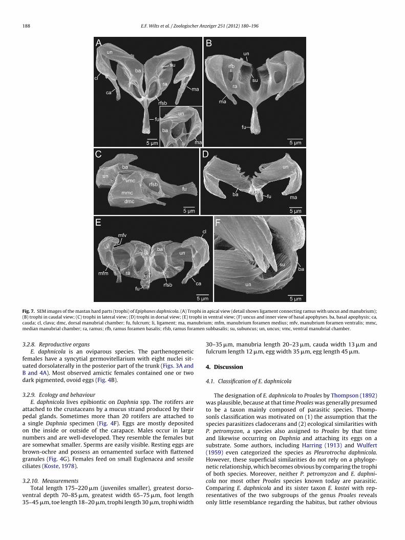
3.2.5. Mastax hard parts (trophi)The compact, modified malleate trophi system is bilateral sym-
metrical (Figs. 3C, 6B–E and 7A–F). The individual trophi elementsare embedded in ephitelial tissue, from which cells extend intothe cuticular cavities of the rami (ramus basal and ramus subbasalchambers) and manubria (dorsal, median and ventral manubrialchambers). The trophi can be extruded with only the fulcrum andcaudae remaining in the mastax tissue (Fig. 4D).
The rami (ra) appear bifurcate in lateral view (Fig. 6C) andare almost oval in caudal view (Figs. 6B and 7B). Both the ramusbasal (rbc) and the ramus subbasal chambers (rsbc) display distinctopenings with the oval ramus foramen subbasalis (rfsb) directingventrofrontally (Figs. 6C and 7A) and the circular ramus foramenbasalis (rfb) facing caudally (Figs. 6B and 7B). The anterior part ofeach ramus consists of an enlarged, broad basal apophysis (ba) builton the ramus subbasal chamber (Fig. 6C). The basal apophyses havedensely arranged scleropilar structures on their inner margins, buta molar surface with tubercles is absent. The posterior part of eachramus is built on the ramus basal chamber (Fig. 6C). The basal cham-bers terminate apically into a fine, less cuticularized structure. Thelateral margins are rounded and alulae are lacking. Fine cuticularstrands or ligaments (li), probably leftovers of a reduced hypophar-ynx, lie on the rami, connecting them with manubria (ma) and unci(un) (Figs. 3C and 7A).
The short, unpaired fulcrum (fu) is usually oriented in adorsoventral (vertical) direction (Figs. 3B and 4E). The base ofthe short fulcrum attaches obliquely to the rami. In caudal view,
the fulcrum appears slender and broadens gradually from thebase towards the distal, irregularly sculptured end. In lateral view,the fulcrum is high and displays curved margins and longitudinalstriae laterally (Figs. 6C and 7C).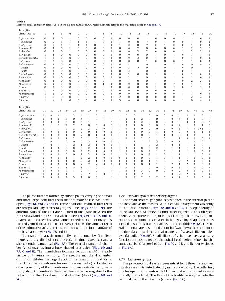
E.F. Wilts et al. / Zoologischer Anzeiger 251 (2012) 180–196 187
Table 2Morphological character matrix used in the cladistic analyses. Character numbers refer to the characters listed in Appendix A.
Taxa (20)Characters (43) 1 2 3 4 5 6 7 8 9 10 11 12 13 14 15 16 17 18 19 20
P. petromyzon 0 5 0 1 0 0 0 0 0 0 0 0 1 0 0 0 1 1 0 0P. fallaciosa 0 0 1 1 1 1 1 0 0 1 0 0 ? 0 1 0 0 1 0 0P. tillyensis 0 0 1 1 1 1 0 0 0 1 0 0 ? 0 1 0 0 1 0 0P. reinhardti 0 4 0 1 0 0 0 0 0 0 0 2 0 0 0 0 1 2 3 1P. theodora 0 4 0 1 0 0 0 0 0 0 0 2 ? 0 0 0 1 2 3 0B. plicatilis 1 2 0 0 0 0 0 0 0 0 0 0 0 1 0 1 0 1 ? 0B. quadridentatus 1 1 0 0 0 0 0 0 0 0 0 3 0 1 0 1 0 1 0 0E. dilatata 1 2 0 0 0 0 0 0 0 0 0 0 1 0 0 0 1 1 0 0P. daphnicola 0 3 0 0 0 0 0 0 0 4 2 1 0 1 0 1 0 0 – 0P. kostei 0 3 0 0 0 0 0 0 0 0 2 ? 0 1 0 1 0 0 – 0E. senta 0 3 0 0 0 0 0 0 0 0 2 1 0 1 0 1 0 1 1 0E. brachionus 0 3 0 0 0 0 0 0 0 0 2 0 0 1 0 1 0 1 0 0E. clavulata 0 6 0 0 0 0 0 0 0 0 2 1 0 1 0 1 0 1 0 0R. frontalis 0 3 0 0 0 0 0 0 1 0 1 0 0 1 0 1 0 2 2 0M. chlaena 0 3 0 0 0 0 0 1 0 0 0 0 0 1 0 ? 0 1 1 0C. tuba 0 3 0 0 0 0 0 0 0 0 0 0 0 1 0 ? 0 1 1 0T. tetractis 1 7 0 0 0 0 0 0 0 0 0 0 0 0 0 0 1 1 1 0M. mucronata 1 8 0 0 0 0 0 0 0 0 0 0 0 0 0 0 1 1 0 0L. patella 1 9 0 0 0 0 0 0 0 2 0 0 0 0 0 0 1 2 4 0L. inermis 1 A 1 0 0 0 0 0 0 3 0 0 0 0 0 0 1 0 – 0
Taxa (20)Characters (43) 21 22 23 24 25 26 27 28 29 30 31 32 33 34 35 36 37 38 39 40 41 42 43
P. petromyzon 0 0 0 – 2 4 1 0 3 1 1 2 0 – 0 0 0 0 4 ? 0 0 1P. fallaciosa 0 0 0 2 0 5 1 0 1 1 1 0 1 2 0 0 0 0 3 0 0 0 1P. tillyensis 0 0 0 2 0 5 1 0 1 1 1 0 1 2 0 0 0 0 3 0 0 0 1P. reinhardti 0 1 1 3 2 5 0 0 0 1 1 0 1 0 0 0 0 0 2 1 0 1 1P. theodora 0 1 1 4 2 6 2 0 0 1 1 0 1 0 0 0 0 0 ? ? 0 0 + 1 1B. plicatilis 0 0 0 1 0 2 0 1 0 0 1 0 1 0 0 1 1 0 0 0 0 1 0B. quadridentatus 0 0 0 1 0 ? 0 1 0 0 1 0 1 0 0 1 1 0 0 0 0 0 0E. dilatata 0 0 0 1 2 5 1 0 0 1 1 0 ? ? 0 1 0 1 ? 0 0 0 0P. daphnicola 1 0 1 0 0 0 2 1 0 0 1 1 1 1 2 2 0 0 1 1 0 0 1P. kostei 1 0 1 0 2 1 2 1 0 0 1 1 1 1 2 2 0 0 1 1 0 0 ?E. senta 0 0 0 0 0 2 2 1 0 0 1 0 1 0 2 2 0 1 1 1 0 0 0E. brachionus 0 0 0 0 0 2 2 1 0 0 1 0 1 0 2 1 0 1 1 1 0 0 0E. clavulata 0 0 0 0 0 3 2 1 0 0 1 0 1 0 2 1 0 1 1 1 1 0 0R. frontalis 0 0 0 1 1 2 2 1 0 0 0 0 1 0 0 1 0 1 1 1 0 0 0M. chlaena 0 0 0 1 0 2 2 1 2 0 0 0 1 0 1 1 0 1 ? ? 0 0 0C. tuba 0 0 0 1 0 2 2 ? 0 0 0 0 1 0 1 1 0 ? 1 ? 0 0 0T. tetractis 0 0 0 ? 3 ? ? 0 1 1 1 0 ? ? 0 0 0 0 ? ? 0 0 0M. mucronata 0 0 0 ? 1 2 1 0 1 1 1 0 1 3 0 0 0 0 2 0 0 0 0L. patella 0 0 0 1 1 ? ? 0 0 1 1 0 1 ? 0 1 0 0 ? ? 0 0 0L. inermis 0 0 0 ? 3 7 0 0 1 1 1 0 ? ? 0 ? 0 0 ? ? 0 0 0
aoaarAlot
msb7v(tdtr7
The paired unci are formed by curved plates, carrying one smallnd three large, bent unci teeth that are more or less well devel-ped (Figs. 6E and 7D and F). Three additional reduced unci teethre recognizable by their straight jugal lines (Figs. 6E and 7F). Thenterior parts of the unci are situated in the space between theamus basal and ramus subbasal chambers (Figs. 6C and 7A and D).large subuncus with several lamellar teeth at its inner margin is
ocated ventral to each uncus. In live specimens, the lamellar teethf the subuncus (su) are in close contact with the inner surface ofhe basal apophyses (Fig. 7B and F).
The manubria attach proximally to the unci by fine liga-ents and are divided into a broad, proximal clava (cl) and a
hort, slender cauda (ca) (Fig. 7A). The ventral manubrial cham-er (vmc) extends into a hook-shaped protrusion (Figs. 6D andA, C and E). The manubrium foramen ventralis (mfv) is clearlyisible and points ventrally. The median manubrial chambermmc) constitutes the largest part of the manubrium and formshe clava. The manubrium foramen medius (mfm) is located in
irect proximity of the manubrium foramen ventralis facing ven-rally also. A manubrium foramen dorsalis is lacking due to theeduction of the dorsal manubrial chamber (dmc) (Figs. 6D andC).3.2.6. Nervous system and sensory organsThe small cerebral ganglion is positioned in the anterior part of
the head above the mastax, with a caudal enlargement attachingto the dorsal antenna (Figs. 3A and B and 4A). Independently ofthe season, eyes were never found either in juvenile or adult spec-imens. A retrocerebral organ is also lacking. The dorsal antennacomposed of numerous cilia encircled by a ring-shaped collar, islocated posteriorly on the head near the neck fold (Fig. 5A). The lat-eral antennae are positioned about halfway down the trunk uponthe dorsolateral surfaces and also consist of several cilia encircledby a flat collar (Fig. 5B). Small ciliary tufts that may have a sensoryfunction are positioned on the apical head region below the cir-cumapical band (arrow heads in Fig. 5C and D and light grey circlesin Fig. 6A).
3.2.7. Excretory systemThe protonephridal system presents at least three distinct ter-
minal organs distributed laterally in the body cavity. The collectingtubules open into a contractile bladder that is positioned ventro-caudally in the trunk. The fluid of the bladder is emptied into theterminal part of the intestine (cloaca) (Fig. 3A).
188 E.F. Wilts et al. / Zoologischer Anzeiger 251 (2012) 180–196
Fig. 7. SEM images of the mastax hard parts (trophi) of Epiphanes daphnicola. (A) Trophi in apical view (detail shows ligament connecting ramus with uncus and manubrium);( phi inc ubrium men
3
fuBd
3
apaonabgc
3
v3
B) trophi in caudal view; (C) trophi in lateral view; (D) trophi in dorsal view; (E) troauda; cl, clava; dmc, dorsal manubrial chamber; fu, fulcrum; li, ligament; ma, manedian manubrial chamber; ra, ramus; rfb, ramus foramen basalis; rfsb, ramus fora
.2.8. Reproductive organsE. daphnicola is an oviparous species. The parthenogenetic
emales have a syncytial germovitellarium with eight nuclei sit-ated dorsolaterally in the posterior part of the trunk (Figs. 3A andand 4A). Most observed amictic females contained one or two
ark pigmented, ovoid eggs (Fig. 4B).
.2.9. Ecology and behaviourE. daphnicola lives epibiontic on Daphnia spp. The rotifers are
ttached to the crustaceans by a mucus strand produced by theiredal glands. Sometimes more than 20 rotifers are attached tosingle Daphnia specimen (Fig. 4F). Eggs are mostly deposited
n the inside or outside of the carapace. Males occur in largeumbers and are well-developed. They resemble the females butre somewhat smaller. Sperms are easily visible. Resting eggs arerown-ochre and possess an ornamented surface with flattenedranules (Fig. 4G). Females feed on small Euglenacea and sessileiliates (Koste, 1978).
.2.10. MeasurementsTotal length 175–220 �m (juveniles smaller), greatest dorso-
entral depth 70–85 �m, greatest width 65–75 �m, foot length5–45 �m, toe length 18–20 �m, trophi length 30 �m, trophi width
ventral view; (F) uncus and inner view of basal apophyses. ba, basal apophysis; ca,m; mfm, manubrium foramen medius; mfv, manubrium foramen ventralis; mmc,
subbasalis; su, subuncus; un, uncus; vmc, ventral manubrial chamber.
30–35 �m, manubria length 20–23 �m, cauda width 13 �m andfulcrum length 12 �m, egg width 35 �m, egg length 45 �m.
4. Discussion
4.1. Classification of E. daphnicola
The designation of E. daphnicola to Proales by Thompson (1892)was plausible, because at that time Proales was generally presumedto be a taxon mainly composed of parasitic species. Thomp-sonı̌s classification was motivated on (1) the assumption that thespecies parasitizes cladocerans and (2) ecological similarities withP. petromyzon, a species also assigned to Proales by that timeand likewise occurring on Daphnia and attaching its eggs on asubstrate. Some authors, including Harring (1913) and Wulfert(1959) even categorized the species as Pleurotrocha daphnicola.However, these superficial similarities do not rely on a phyloge-netic relationship, which becomes obvious by comparing the trophiof both species. Moreover, neither P. petromyzon and E. daphni-
cola nor most other Proales species known today are parasitic.Comparing E. daphnicola and its sister taxon E. kostei with rep-resentatives of the two subgroups of the genus Proales revealsonly little resemblance regarding the habitus, but rather obvious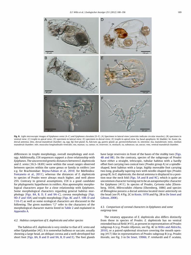
E.F. Wilts et al. / Zoologischer Anzeiger 251 (2012) 180–196 189
Fig. 8. Light microscopic images of Epiphanes senta (A–C) and Epiphanes clavulata (D–F). (A) Specimen in lateral view (asterisks indicate circular muscles); (B) specimen inventral view; (C) trophi in apical view; (D) specimen in lateral view; (E) specimen in dorsal view; (E) trophi in apical view. ba, basal apophysis; bl, bladder; br, brain; da,d m; ggm ; re, re
doEabeFt2flSp91fmA
4
oss
orsal antenna; dmc, dorsal manubrial chamber; eg, egg; fgl, foot gland; fu, fulcruanubrial chamber; mlv, musculus longitudinalis ventralis; mx, mastax; ra, ramus
ifferences in trophi morphology, overall morphology and ecol-gy. Additionally, COI sequences support a close relationship withpiphanes. The uncorrected genetic distances between E. daphnicoland E. senta (16.3–18.8%) were within the usual ranges observedetween species within the same genus or family in rotifers (see.g. for Brachionidae: Reyna-Fabian et al., 2010; for Bdelloidea:ontaneto et al., 2011), whereas the distances of E. daphnicolao species of Proales were always much higher, and well above5%. Contrary to general assumptions, COI is a good candidateor phylogenetic hypotheses in rotifers. Also apomorphic morpho-ogical characters argue for a close relationship with Epiphanes.ome morphological characters regarding general habitus mor-hology (Figs. 8A, B, D, E and 9A–C), corona morphology (Figs.D–F and 10A) and trophi morphology (Figs. 8C and F, 10B–D and1A–F) as well as some ecological characters are discussed in theollowing. The given numbers “()” refer to the characters of the
orphological character matrix listed in Table 2 and explained inppendix A.
.2. Habitus comparison of E. daphnicola and other species
The habitus of E. daphnicola is very similar to that of E. senta andther Epiphanidae (#2). It is somewhat bulbous or saccate, usuallyhowing a large head, an oblique corona and a well-developed buthort foot (Figs. 8A, B and D and 9A, B, D and E). The foot glands
, gastric gland; gv, germovitellarium; in, intestine; ma, manubrium; mmc, medianservoir; st, stomach; su, subuncus; un, uncus; vmc, ventral manubrial chamber.
have large reservoirs in front of the bases of the stubby toes (Figs.4B and 8B). On the contrary, species of the subgroups of Proaleshave either a straight, telescopic, tubular habitus with a hardlyoffset foot carrying two conical toes (Proales group A) or a spindle-shaped, bent habitus with a large, highly moveable foot carryingtwo long, gradually tapering toes with needle-shaped tips (Proalesgroup B). In E. daphnicola, the dorsal antenna is displaced to a posi-tion near the neck fold (Figs. 5A and B and 9C), which is quite anuncommon character turning out to be an autapomorphic characterfor Epiphanes (#11). In species of Proales, Cyrtonia tuba (Ehren-berg, 1834), Mikrocodides chlaena (Ehrenberg, 1886) and speciesof Rhinoglena possess a dorsal antenna located more anteriorly onthe head (see Pl. 4 Fig. 2C in Koste, 1978 and Fig. 2B in De Smet andGibson, 2008).
4.3. Comparison of coronal characters in Epiphanes and someoutgroup taxa
The rotatory apparatus of E. daphnicola also differs distinctlyfrom those in species of Proales. E. daphnicola has no ventralextended buccal field (#15), as present in representatives of Proales
subgroup A (e.g. Proales tillyensis, see Fig. 4C in Wilts and Ahlrichs,2010), or a paired epidermal structure covering the mouth open-ing (#17) like in representatives of Proales subgroup B (e.g. Proaleslitoralis, see Fig. 3 in De Smet, 1996b; P. reinhardti and P. oculata,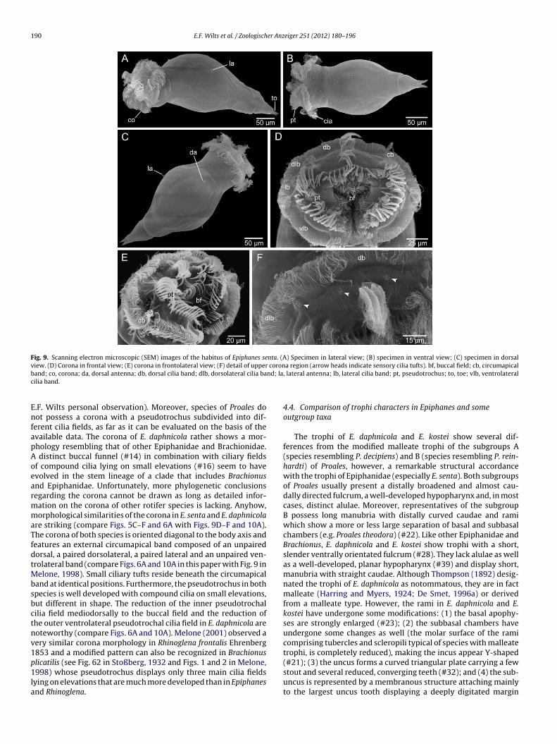
190 E.F. Wilts et al. / Zoologischer Anzeiger 251 (2012) 180–196
Fig. 9. Scanning electron microscopic (SEM) images of the habitus of Epiphanes senta. (A) Specimen in lateral view; (B) specimen in ventral view; (C) specimen in dorsalview. (D) Corona in frontal view; (E) corona in frontolateral view; (F) detail of upper corona region (arrow heads indicate sensory cilia tufts). bf, buccal field; cb, circumapicalb nd; lac
EnfapAoearmmaTfdtMbsbctnv1p1la
and; co, corona; da, dorsal antenna; db, dorsal cilia band; dlb, dorsolateral cilia bailia band.
.F. Wilts personal observation). Moreover, species of Proales doot possess a corona with a pseudotrochus subdivided into dif-
erent cilia fields, as far as it can be evaluated on the basis of thevailable data. The corona of E. daphnicola rather shows a mor-hology resembling that of other Epiphanidae and Brachionidae.distinct buccal funnel (#14) in combination with ciliary fields
f compound cilia lying on small elevations (#16) seem to havevolved in the stem lineage of a clade that includes Brachionusnd Epiphanidae. Unfortunately, more phylogenetic conclusionsegarding the corona cannot be drawn as long as detailed infor-ation on the corona of other rotifer species is lacking. Anyhow,orphological similarities of the corona in E. senta and E. daphnicola
re striking (compare Figs. 5C–F and 6A with Figs. 9D–F and 10A).he corona of both species is oriented diagonal to the body axis andeatures an external circumapical band composed of an unpairedorsal, a paired dorsolateral, a paired lateral and an unpaired ven-rolateral band (compare Figs. 6A and 10A in this paper with Fig. 9 in
elone, 1998). Small ciliary tufts reside beneath the circumapicaland at identical positions. Furthermore, the pseudotrochus in bothpecies is well developed with compound cilia on small elevations,ut different in shape. The reduction of the inner pseudotrochalilia field mediodorsally to the buccal field and the reduction ofhe outer ventrolateral pseudotrochal cilia field in E. daphnicola areoteworthy (compare Figs. 6A and 10A). Melone (2001) observed aery similar corona morphology in Rhinoglena frontalis Ehrenberg853 and a modified pattern can also be recognized in Brachionus
licatilis (see Fig. 62 in Stoßberg, 1932 and Figs. 1 and 2 in Melone,998) whose pseudotrochus displays only three main cilia fieldsying on elevations that are much more developed than in Epiphanesnd Rhinoglena.
, lateral antenna; lb, lateral cilia band; pt, pseudotrochus; to, toe; vlb, ventrolateral
4.4. Comparison of trophi characters in Epiphanes and someoutgroup taxa
The trophi of E. daphnicola and E. kostei show several dif-ferences from the modified malleate trophi of the subgroups A(species resembling P. decipiens) and B (species resembling P. rein-hardti) of Proales, however, a remarkable structural accordancewith the trophi of Epiphanidae (especially E. senta). Both subgroupsof Proales usually present a distally broadened and almost cau-dally directed fulcrum, a well-developed hypopharynx and, in mostcases, distinct alulae. Moreover, representatives of the subgroupB possess long manubria with distally curved caudae and ramiwhich show a more or less large separation of basal and subbasalchambers (e.g. Proales theodora) (#22). Like other Epiphanidae andBrachionus, E. daphnicola and E. kostei show trophi with a short,slender ventrally orientated fulcrum (#28). They lack alulae as wellas a well-developed, planar hypopharynx (#39) and display short,manubria with straight caudae. Although Thompson (1892) desig-nated the trophi of E. daphnicola as notommatous, they are in factmalleate (Harring and Myers, 1924; De Smet, 1996a) or derivedfrom a malleate type. However, the rami in E. daphnicola and E.kostei have undergone some modifications: (1) the basal apophy-ses are strongly enlarged (#23); (2) the subbasal chambers haveundergone some changes as well (the molar surface of the ramicomprising tubercles and scleropili typical of species with malleatetrophi, is completely reduced), making the incus appear Y-shaped
(#21); (3) the uncus forms a curved triangular plate carrying a fewstout and several reduced, converging teeth (#32); and (4) the sub-uncus is represented by a membranous structure attaching mainlyto the largest uncus tooth displaying a deeply digitated margin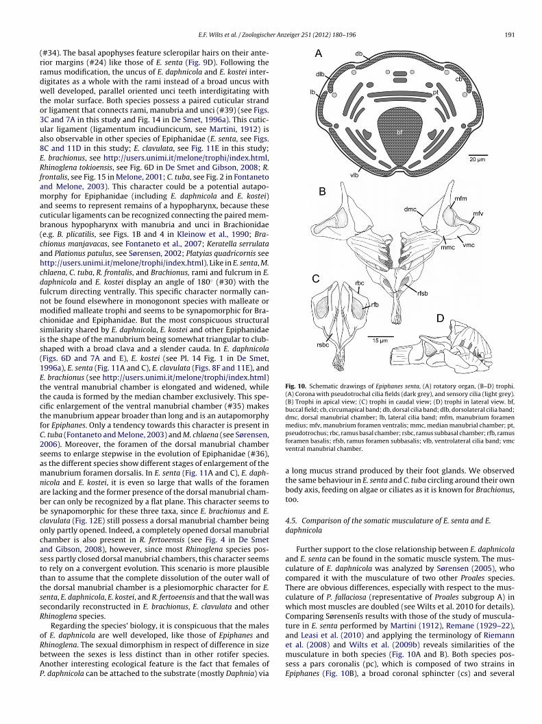
er Anzeiger 251 (2012) 180–196 191
(rrdwto3ua8ERfamacb(cahcdfnmcsis(1EttctfC2samnabbcocastttssR
oRbAP
Fig. 10. Schematic drawings of Epiphanes senta, (A) rotatory organ, (B–D) trophi.(A) Corona with pseudotrochal cilia fields (dark grey), and sensory cilia (light grey).(B) Trophi in apical view; (C) trophi in caudal view; (D) trophi in lateral view. bf,buccal field; cb, circumapical band; db, dorsal cilia band; dlb, dorsolateral cilia band;dmc, dorsal manubrial chamber; lb, lateral cilia band; mfm, manubrium foramenmedius; mfv, manubrium foramen ventralis; mmc, median manubrial chamber; pt,
E.F. Wilts et al. / Zoologisch
#34). The basal apophyses feature scleropilar hairs on their ante-ior margins (#24) like those of E. senta (Fig. 9D). Following theamus modification, the uncus of E. daphnicola and E. kostei inter-igitates as a whole with the rami instead of a broad uncus withell developed, parallel oriented unci teeth interdigitating with
he molar surface. Both species possess a paired cuticular strandr ligament that connects rami, manubria and unci (#39) (see Figs.C and 7A in this study and Fig. 14 in De Smet, 1996a). This cutic-lar ligament (ligamentum incudiuncicum, see Martini, 1912) islso observable in other species of Epiphanidae (E. senta, see Figs.C and 11D in this study; E. clavulata, see Fig. 11E in this study;. brachionus, see http://users.unimi.it/melone/trophi/index.html,hinoglena tokioensis, see Fig. 6D in De Smet and Gibson, 2008; R.
rontalis, see Fig. 15 in Melone, 2001; C. tuba, see Fig. 2 in Fontanetond Melone, 2003). This character could be a potential autapo-orphy for Epiphanidae (including E. daphnicola and E. kostei)
nd seems to represent remains of a hypopharynx, because theseuticular ligaments can be recognized connecting the paired mem-ranous hypopharynx with manubria and unci in Brachionidaee.g. B. plicatilis, see Figs. 1B and 4 in Kleinow et al., 1990; Bra-hionus manjavacas, see Fontaneto et al., 2007; Keratella serrulatand Plationus patulus, see Sørensen, 2002; Platyias quadricornis seettp://users.unimi.it/melone/trophi/index.html). Like in E. senta, M.hlaena, C. tuba, R. frontalis, and Brachionus, rami and fulcrum in E.aphnicola and E. kostei display an angle of 180◦ (#30) with theulcrum directing ventrally. This specific character normally can-ot be found elsewhere in monogonont species with malleate orodified malleate trophi and seems to be synapomorphic for Bra-
hionidae and Epiphanidae. But the most conspicuous structuralimilarity shared by E. daphnicola, E. kostei and other Epiphanidaes the shape of the manubrium being somewhat triangular to club-haped with a broad clava and a slender cauda. In E. daphnicolaFigs. 6D and 7A and E), E. kostei (see Pl. 14 Fig. 1 in De Smet,996a), E. senta (Fig. 11A and C), E. clavulata (Figs. 8F and 11E), and. brachionus (see http://users.unimi.it/melone/trophi/index.html)he ventral manubrial chamber is elongated and widened, whilehe cauda is formed by the median chamber exclusively. This spe-ific enlargement of the ventral manubrial chamber (#35) makeshe manubrium appear broader than long and is an autapomorphyor Epiphanes. Only a tendency towards this character is present in. tuba (Fontaneto and Melone, 2003) and M. chlaena (see Sørensen,006). Moreover, the foramen of the dorsal manubrial chambereems to enlarge stepwise in the evolution of Epiphanidae (#36),s the different species show different stages of enlargement of theanubrium foramen dorsalis. In E. senta (Fig. 11A and C), E. daph-
icola and E. kostei, it is even so large that walls of the foramenre lacking and the former presence of the dorsal manubrial cham-er can only be recognized by a flat plane. This character seems toe synapomorphic for these three taxa, since E. brachionus and E.lavulata (Fig. 12E) still possess a dorsal manubrial chamber beingnly partly opened. Indeed, a completely opened dorsal manubrialhamber is also present in R. fertoeensis (see Fig. 4 in De Smetnd Gibson, 2008), however, since most Rhinoglena species pos-ess partly closed dorsal manubrial chambers, this character seemso rely on a convergent evolution. This scenario is more plausiblehan to assume that the complete dissolution of the outer wall ofhe dorsal manubrial chamber is a plesiomorphic character for E.enta, E. daphnicola, E. kostei, and R. fertoeensis and that the wall wasecondarily reconstructed in E. brachionus, E. clavulata and otherhinoglena species.
Regarding the species’ biology, it is conspicuous that the malesf E. daphnicola are well developed, like those of Epiphanes and
hinoglena. The sexual dimorphism in respect of difference in sizeetween the sexes is less distinct than in other rotifer species.nother interesting ecological feature is the fact that females of. daphnicola can be attached to the substrate (mostly Daphnia) viapseudotrochus; rbc, ramus basal chamber; rsbc, ramus subbasal chamber; rfb, ramusforamen basalis; rfsb, ramus foramen subbasalis; vlb, ventrolateral cilia band; vmcventral manubrial chamber.
a long mucus strand produced by their foot glands. We observedthe same behaviour in E. senta and C. tuba circling around their ownbody axis, feeding on algae or ciliates as it is known for Brachionus,too.
4.5. Comparison of the somatic musculature of E. senta and E.daphnicola
Further support to the close relationship between E. daphnicolaand E. senta can be found in the somatic muscle system. The mus-culature of E. daphnicola was analyzed by Sørensen (2005), whocompared it with the musculature of two other Proales species.There are obvious differences, especially with respect to the mus-culature of P. fallaciosa (representative of Proales subgroup A) inwhich most muscles are doubled (see Wilts et al. 2010 for details).Comparing Sørensenı̌s results with those of the study of muscula-ture in E. senta performed by Martini (1912), Remane (1929–22),and Leasi et al. (2010) and applying the terminology of Riemann
et al. (2008) and Wilts et al. (2009b) reveals similarities of themusculature in both species (Fig. 10A and B). Both species pos-sess a pars coronalis (pc), which is composed of two strains inEpiphanes (Fig. 10B), a broad coronal sphincter (cs) and several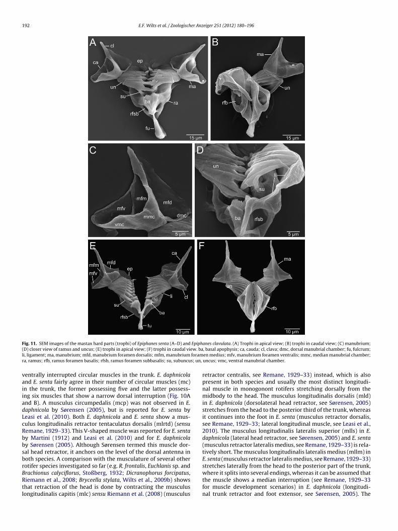
192 E.F. Wilts et al. / Zoologischer Anzeiger 251 (2012) 180–196
Fig. 11. SEM images of the mastax hard parts (trophi) of Epiphanes senta (A–D) and Epiphanes clavulata. (A) Trophi in apical view; (B) trophi in caudal view; (C) manubrium;( ew. bal foramr s; un,
vaiiadLcRbbsbrBRtl
D) closer view of ramus and uncus; (E) trophi in apical view; (F) trophi in caudal vii, ligament; ma, manubrium; mfd, manubrium foramen dorsalis; mfm, manubriuma, ramus; rfb, ramus foramen basalis; rfsb, ramus foramen subbasalis; su, subuncu
entrally interrupted circular muscles in the trunk. E. daphnicoland E. senta fairly agree in their number of circular muscles (mc)n the trunk, the former possessing five and the latter possess-ng six muscles that show a narrow dorsal interruption (Fig. 10And B). A musculus circumpedalis (mcp) was not observed in E.aphnicola by Sørensen (2005), but is reported for E. senta byeasi et al. (2010). Both E. daphnicola and E. senta show a mus-ulus longitudinalis retractor tentaculatus dorsalis (mlrtd) (sensuemane, 1929–33). This V-shaped muscle was reported for E. sentay Martini (1912) and Leasi et al. (2010) and for E. daphnicolay Sørensen (2005). Although Sørensen termed this muscle dor-al head retractor, it anchors on the level of the dorsal antenna inoth species. A comparison with the musculature of several otherotifer species investigated so far (e.g. R. frontalis, Euchlanis sp. and
rachionus calyciflorus, Stoßberg, 1932; Dicranophorus forcipatus,iemann et al., 2008; Bryceella stylata, Wilts et al., 2009b) showshat retraction of the head is done by contracting the musculusongitudinalis capitis (mlc) sensu Riemann et al. (2008) (musculus, basal apophysis; ca, cauda; cl, clava; dmc, dorsal manubrial chamber; fu, fulcrum;en medius; mfv, manubrium foramen ventralis; mmc, median manubrial chamber;uncus; vmc, ventral manubrial chamber.
retractor centralis, see Remane, 1929–33) instead, which is alsopresent in both species and usually the most distinct longitudi-nal muscle in monogonont rotifers stretching dorsally from themidbody to the head. The musculus longitudinalis dorsalis (mld)in E. daphnicola (dorsolateral head retractor, see Sørensen, 2005)stretches from the head to the posterior third of the trunk, whereasit continues into the foot in E. senta (musculus retractor dorsalis,see Remane, 1929–33; lateral longitudinal muscle, see Leasi et al.,2010). The musculus longitudinalis lateralis superior (mlls) in E.daphnicola (lateral head retractor, see Sørensen, 2005) and E. senta(musculus retractor lateralis medius, see Remane, 1929–33) is rela-tively short. The musculus longitudinalis lateralis medius (mllm) inE. senta (musculus retractor lateralis medius, see Remane, 1929–33)stretches laterally from the head to the posterior part of the trunk,
where it splits into several endings, whereas it can be assumed thatthe muscle shows a median interruption (see Remane, 1929–33for muscle development scenarios) in E. daphnicola (longitudi-nal trunk retractor and foot extensor, see Sørensen, 2005). The
E.F. Wilts et al. / Zoologischer Anzeiger 251 (2012) 180–196 193
Fig. 12. Schematic drawing of the somatic musculature of (A) Epiphanes daphnicola (after Sørensen, 2005) and (B) Epiphanes senta (after Martini, 1912). cs, coronal sphincter;lcr, lateral coronal retractor; mc I–VI; musculus circularis I–VI; mcp, musculus circumpedalis; mlc, musculus longitudinalis capitis; mld musculus longitudinalis dorsalis; mllimusculus longitudinalis lateralis inferior; mllm, musculus longitudinalis lateralis medius; mlls, musculus longitudinalis lateralis superior; mlrdt, musculus longitudinalisl pars c
mcltrpt
ateralis retractor dorsalis tentaculatus; mlv, musculus longitudinalis ventralis; pc,
usculus longitudinalis lateralis inferior (mlli) in E. senta (mus-ulus retractor lateralis inferior, see Remane, 1929–33; ventralongitudinal muscle, see Leasi et al., 2010) stretches from the corona
o the posterior part of the trunk. In E. daphnicola (ventral footetractor, see Sørensen, 2005), the muscle is doubled with oneart terminating near the midbody region and the other stranderminating in the head region. Both species feature a musculusoronalis.
longitudinalis ventralis (mlv) (E. senta: musculus retractor ven-tralis, see Remane, 1929–33 and ventral longitudinal muscle,see Leasi et al., 2010; E. daphnicola: ventral trunk retractor, see
Sørensen, 2005) extending from the corona to the base of the foot,presenting medially a short hook-like branch. Additionally, E. daph-nicola shows a lateral coronal retractor (lcr) (see Sørensen, 2005)in the head with diagonal direction. Maybe, this muscle represents
1 er Anz
oda
5
saBpjkopbsoeit
A
ihhabcpafSmE
A
94 E.F. Wilts et al. / Zoologisch
nly a secondarily separated anterior part of the musculus longitu-inalis ventralis, because it is more developed showing anteriorlysimilar, diagonal direction in E. senta.
. Conclusion
Our assumption that P. daphnicola is more closely related topecies of Epiphanidae than to other Proales species was testednd supported via morphological and molecular data analyses.y demonstrating apomorphies, retracing character evolution andresenting further morphological and ecological evidence, we have
ustified a relocation of P. daphnicola and its assumed sister taxon P.ostei to Epiphanes. Unfortunately, there are no further sequencesf other Epiphanidae and Proalidae available at present so that thehylogenetic relationships within Epiphanidae remain to be solvedy future molecular analyses using more sequences of furtherpecies. This study may encourage taxonomists to reinvestigatether rotifer species that have already been described in detail,specially those which do not well fit the taxa they are classified in,n order to discover existing para- and polyphylies and to contributeo their elimination.
cknowledgements
We would like to thank: Giulio Melone for providing us SEMmages of the corona of E. senta (Fig. 9E), Michael Plewka fromttp://www.plingfactory.de/Science/Biohome.html for supplyingabitus micrographs of E. senta (Fig. 8A and B), Willem De Smetnd grant ARCFAC026129-2009-131 for the samplings from Sval-ard. We also thank Olaf R.P. Bininda-Emonds for his help inalculating the bootstrap and Bremer support values for the mor-hological cladistic analyses in an early version of the manuscriptnd Philip Fleischhauer for providing the COI sequence of E. sentarom Germany. Moreover, we especially like to thank Martin V.ørensen for his helpful and valuable editorial comments. Further-ore, we gratefully acknowledge the financial support granted to
ike F. Wilts by Evangelisches Studienwerk e.V. Villigst.
ppendix A. Appendix
Description of characters
(1) Epidermis stiffened to lorica: 0 = absent, 1 = present, type Bra-chionus quadridentatus.
(2) Habitus shape: 0 = tubular, type Proales fallaciosa,1 = rectangular, type B. quadridentatus, 2 = oval,type B. plicatilis, 3 = bulbous saccate, type E. senta(Figs. 9A and B and 10A–C), 4 = spindle-shaped, bent, type P.reinhardti, 5 = inflated elongate, type P. petromyzon, 6 = giant,saccate, type Epiphanes clavulata, 7 = quadrangular, type Tri-chotria tetractis, 8 = elongated, oval, type Mytilina mucronata,9 = flattened, oval, type Lepadella ovalis, A = flattened,elongated-oval, type Lecane inermis.
(3) Neck pseudosegment: 0 = absent, 1 = present, type P. fallaciosa.This character refers to the presence of a short trunk pseu-dosegment following the head (see De Smet, 1996a).
(4) Lumbal pseudosegment: 0 = absent, 1 = present, type P. falla-ciosa.
This character refers to the presence of a posterior trunk
pseudosegment located in front of the preanal pseudoseg-ment.(5) Several dorsal and longitudinal trunk folds: 0 = absent,1 = present, type P. fallaciosa.
eiger 251 (2012) 180–196
Some Proales species display a number of deep, longitudinalinfoldings running across the dorsal and lateral sides of thetrunk (see De Smet, 1996a).
(6) Foot: 0 = distinctly offset from trunk, type P. reinhardti, 1 = footand trunk gradually tapering, type P. fallaciosa.
(7) Knob-like appendage above toes: 0 = absent, 1 = present, typeP. fallaciosa.
(8) Direction of toes: 0 = both toes in same direction, 1 = one toeabducted, type M. chlaena.
(9) Head with proboscis: 0 = absent, 1 = present, type R. frontalis.In Rhinoglena the head features an apical elongation called
proboscis (see Melone, 2001).(10) Head: 0 = anteriorly rounded, without structure, 1 = with
offset, short rostrum, type P. fallaciosa, 2 = with offset,cap-like rostrum, type Lepadella, 3 = with semicircular ros-trum, type L. inermis, 4 = with offset, small element, typeE. daphnicola.
In some proalid rotifers, the dorsal epidermis of the rotatoryorgan frontally runs out into a hyaline projection, which variesin the different taxa in form.
(11) Position of dorsal antenna: 0 = centrally on head, type P. fal-laciosa, 1 = displaced anteriorly, type R. frontalis, 2 = displacedposteriorly, type E. senta (Figs. 6A and B, 9D and 10C).
(12) Position of lateral antenna: 0 = posterior third of trunk, typeP. petromyzon, 1 = middle of the trunk, type E. daphnicola(Figs. 3A and 10A and C), 2 = close to preanal pseudosegment,type P. reinhardti, 3 = displaced on spines, type B. quadridenta-tus.
(13) Openings of retrocerebral organ: 0 = absent, 1 = present, typeP. petromyzon.
The retrocerebral organ is usually associated with a pairedduct that leads near the center of the corona (see Wilts et al.,2009a).
(14) Distinct buccal funnel: 0 = absent, 1 = present, type E. senta(Figs. 6C and 10D).
(15) Corona with caudally elongated buccal field: 0 = absent,1 = present, type P. fallaciosa.
Like most species of Dicranophoridae, Notommatidae andLindiidae, species of Proales group A possess an elongated pos-toral, ciliated field in the ventral head region.
(16) Corona with compound cilia arranged on elevations:0 = absent, 1 = present, type E. senta.
This character refers to the elevated pseudotrochal ciliafields in Epiphanidae that display compound cilia or mem-branellae (Figs. 6C–F and 10D–F).
(17) Corona caudally with labial projections: 0 = absent,1 = present, type P. reinhardti.
This character refers to the presence of epidermal projec-tions that border the corona caudally. It can be found e.g. inspecies of Proales group B, Pleurotrocha (Wilts et al., 2009a),Bryceella (Wilts et al., 2009b) and Lepadellidae (Wilts et al., inpress).
(18) Number of eyes: 0 = no eyes, 1 = one eye present, 2 = two eyespresent.
(19) Eye position: 0 = posterior on brain, type P. petromyzon,1 = ventrally on brain, type E. senta, 2 = displaced on proboscis,type R. frontalis, 3 = apically, type P. reinhardti, 4 = laterally incorona, type L. ovalis.
(20) Rami asymmetry: 0 = absent, 1 = present, type P. reinhardti.(21) Incus Y-shaped: 0 = absent, 1 = present, type E. daphnicola.
This character refers to the unique shape of the incus of E.daphnicola (Fig. 7C) and E. kostei (see De Smet, 1996a).
(22) Ramus chambers separating from each other: 0 = absent,
1 = present, type P. theodora.This character refers to the fact that in species of Proalesgroup B the cuticular chambers building the rami show a sep-

er Anz
(
(
(
(
(
(
(
(
((
((
Herlyn, H., Piskurek, O., Schmitz, J., Ehlers, U., Zischler, H., 2003. The syndermatan
E.F. Wilts et al. / Zoologisch
aration being incomplete in P. reinhardti and complete in P.theodora (see De Smet, 1996a).
23) Ramus basal apophyses enlarged: 0 = absent, 1 = present, typeE. daphnicola.
24) Tips of ramus basal apophyses: 0 = with hairs, type E. senta,1 = with small, regular teeth, type R. frontalis, 2 = with spines,type P. fallaciosa, 3 = with irregular margin, type P. reinhardti,4 = with blunt gradually decreasing teeth, type P. theodora,5 = blunt, without teeth or hairs, type B. plicatilis.
In several taxa the basal apophyses display varying struc-tures distally from regular teeth in Rhinoglena (see De Smetand Gibson, 2008) to fine hairs in Epiphanes (Figs. 8A and Dand 12D and E).
25) Lateral margin of ramus: 0 = rounded, type E. daphnicola,1 = crenated, type R. frontalis, 2 = with alula, type P. petromyzon,3 = with rounded alula, type T. tetractis.
26) Shape of ramus foramen subbasalis: 0 = very large, oval, typeE. daphnicola, 1 = very large, elongated oval, type E. kostei,2 = medium-sized rounded, type E. senta, 3 = large rounded,type E. clavulata, 4 = small rounded, type P. petromyzon,5 = very small, type P. reinhardti, 6 = very large, circular, type P.Theodora, 7 = medium-sized, circular, type L. inermis.
27) Direction of ramus foramen subbasalis in relative to the ful-crum: 0 = directing posteriorly, type P. tillyensis, 1 = directinginferiorly, type P. petromyzon, 2 = directing inferioposteriorly,type E. senta (Fig. 12A, D and E).
This character refers to the orientation of the ramus fora-men subbasalis which varies among the different rotifer taxa.In Lepadellidae e.g. they face like the ramus basal chamberssuperiorly.
28) Fulcrum orientation: 0 = ±caudally, type P. petromyzon,1 = ±ventrally, type E. senta.
This character refers to the position the fulcrum takes up inrest position in the animal. In most monogonont rotifer speciesthe fulcrum lies in the longitudinal axis of the rotifer (hori-zontally). In Brachionus and Epiphanes (Figs. 4B and 5E) thefulcrum is oriented vertically.
29) Fulcrum length: 0 = short, maximal half the length of theramus, type E. senta, 1 = as long as ramus, type P. tillyensis,2 = medium long, maximal two-third of the ramus length, typeM. chlaena, 3 longer than ramus length, type P. petromyzon.
30) Angle of ramus and fulcrum: 0 = ramus and fulcrum planarforming an angle of ±180◦, type E. senta, 1 = ramus and fulcrumangled, type P. petromyzon.
This character refers to the angle that is formed by ramusand fulcrum. In Epiphanidae fulcrum and ramus lie in a moreor less planar level whereas in other monogonont rotifersramus and fulcrum are angled.
31) Number of major uncus teeth: 0 = 7–10 teeth, 1 = 0–6 teeth.32) Uncus shape: 0 = rectangular plate with parallel orientated
teeth, type E. senta, 1 = curved plate with curved teeth, type E.daphnicola (Figs. 7E and 8D), 2 = crescentic, type P. petromyzon.
33) Subuncus: 0 = absent, 1 = present.34) Subuncus shape: 0 = basing on complete uncus, evenly brush-
like, type B. plicatilis, 1 = basing on largest uncus tooth,lamellar, type E. daphnicola, 2 = brush-like elements basing onsingle uncus teeth, type P. fallaciosa, 3 = scleropilar element onramus, type M. mucronata.
This character refers to the development of the subuncus.The subuncus is a structure residing below the uncus that canbe brush-like with sclereopili or laminar with finger- or teeth-like indentions. In E. senta and E. clavulata (Fig. 12A, D and E)and B. plicatilis (Kleinow et al., 1990) it stretches below the
complete uncus, in E. daphnicola and E. kostei it is lamellarand only attaches the principal uncus tooth (E. daphnicola)(Fig. 8A, B and F). In species of Proales-group A it consists ofeiger 251 (2012) 180–196 195
some large brush-like elements that originate mainly from thelargest uncus teeth and see P. tillyensis (see Wilts and Ahlrichs,2010; P. fallaciosa, E.F. Wilts personal observation).
(35) Ventral manubrial chamber enlarged: 0 = absent, 1 = tendencypresent, type M. chlaena, 2 = well developed, type E. senta[character ordered 0-1-2].
(36) Manubrium foramen dorsalis: 0 = small, type P. tillyensis,1 = large, type Epiphanes brachionus, 2 = opened, chamberreduced to a flat plane, type E. daphnicola.
(37) Manubria with twisted shape: 0 = absent, 1 = present, type B.plicatilis. This character refers to the wringled form of thecauda in Brachionus species.
(38) Epipharynx: 0 = absent, 1 = present, type M. chlaena.(39) Hypopharynx: 0 = membranous structures with cuticular
strands, type B. plicatilis, 1 = cuticular strands, type E. daphni-cola, 2 = large element with fine-denticulated platelets, type P.reinhardti, 3 = large element with two-teethed platelets, typeP. tillyensis, 4 = small fork-like structure, type P. petromyzon.
(40) Sexual dimorphism: 0 = dwarf males, type B. plicatilis,1 = males well developed, little smaller than females, type E.senta.
(41) Germovitellarium band-like: 0 = absent, 1 = 1 present, type E.clavulata.
(42) Macro habitat: 0 = fresh water, 1 = marine environment.This character refers to the habitat. It is coded ± for P.
theodora because the species occurs in fresh- and marinewater.
(43) Deposition of eggs on exterior substrate: 0 = absent,1 = present, type P. reinhardti.
References
Abascal, F., Zardoya, R., Posada, D., 2005. ProtTest: selection of best-fit models ofprotein evolution. Bioinformatics 21, 2104–2105.
De Smet, W.H., 1998. Preparation of rotifer trophi for light and scanning electronmicroscopy. Hydrobiologia 387/388, 117–121.
De Smet, W.H., van Rompu, E.A., Beyens, L., 1993. Contribution to the rotifer fauna ofsubarctic Greenland (Kangerlussuaq and Ammassalik area). Hydrobiologia 432,73–89.
De Smet, W.H., 1996a. Rotifera 4: the proalidae (Monogononta). In: Dumont, H.J.,Nogrady, T. (Eds.), Guides to the Identification of the Microinvertebrates of theContinental Waters of the World. SPB Academic Publishing B.V., Amsterdam, pp.1–102.
De Smet, W.H., 1996b. Description of Proales litoralis sp. nov. (Rotifera, Mono-gononta: Proalidae) from the marine littoral. Hydrobiologia 335, 203–208.
De Smet, W.H., Gibson, J.A.E., 2008. Rhinoglena kutikovae n. sp. (Rotifera: Mono-gononta: Epiphanidae) from the Bunger Hills, East Antarctica: a probable relictspecies that survived Quaternary glaciations on the continent. Polar Biol. 31,595–603.
De Smet, W.H., 2009. Pourriotia carcharodonta, a rotifer parasitic on Vaucheria (Xan-thophyceae) causing taxonomic problems. Bull. Soc. Zool. Fr. 134 (3–4), 195–202.
Fontaneto, D., Melone, G., 2003. On some rotifers new for the Italian fauna. Ital. J.Zool. 70, 253–259.
Fontaneto, D., Giordani, I., Melone, G., Serra, M., 2007. Disentangling the morpho-logical stasis in two rotifer species of the Brachionus plicatilis species complex.Hydrobiologia 583, 297–307.
Fontaneto, D., Iakovenko, N., Eyres, I., Kaya, M., Wyman, M., Barraclough, T.G., 2011.Cryptic diversity in the genus Adineta Hudson & Gosse, 1886 (Rotifera: Bdel-loidea: Adinetidae): a DNA taxonomy approach. Hydrobiologia 662, 27–33.
Fontaneto, D., Jondelius, U., 2011. Broad taxonomic sampling of mitochondrialcytochrome c oxidase subunit I does not solve the relationships between Rotiferaand Acanthocephala. Zoologischer Anzeiger 250, 80–85.
Garcia-Varela, M., Nadler, S.A., 2006. Phylogenetic relationships among Syndermatainferred from nuclear and mitochondrial gene sequences. Mol. Phylogenet. Evol.40, 61–72.
Guindon, S., Gascuel, O., 2003. A simple, fast and accurate method to estimate largephylogenies by maximum-likelihood. Syst. Biol. 52, 696–704.
Harring, H.K., 1913. Synopsis of the Rotatoria. Bull. Am. Mus. Nat. Hist. 81, 1–226.Harring, H.K., Myers, F.J., 1924. The rotifer fauna of Wisconsin. II. A revision of the
notommatid rotifers, exclusive of the Dicranophorinae. T. Wisc. Acad. Sci. 21,415–549.
phylogeny and the evolution of acanthocephalan endoparasitism as inferredfrom 18S rDNA sequences. Mol. Phylogenet. Evol. 26, 155–165.
Keane, T.M., Creevey, C.J., Pentony, M.M., Naughton, T.J., Mc Inerney, J.O., 2006.Assessment of methods for amino acid matrix selection and their use on empir-
1 er Anz
K
K
K
L
M
M
MM
M
N
P
R
R
R
R
R
R
Zool., in press.
96 E.F. Wilts et al. / Zoologisch
ical data shows that ad hoc assumptions for choice of matrix are not justified.BMC Evol. Biol. 6, 29.
leinow, W., Klusemann, J., Wratil, H., 1990. A gentle method for the preparation ofhard parts (trophi) of the mastax of rotifers and scanning electron microscopyof the trophi of Brachionus plicatilis (Rotifera). Zoomorphology 109, 329–336.
oste, W., 1978. Rotatoria. In: Die Rädertiere Mitteleuropas. Ein Bestim-mungswerk, begründet von Max Voigt. Ueberordnung Monogononta, 2nd edn.I. Textband/Gebrüder Borntraeger, Berlin, Stuttgart.
oste, W., Terlutter, H., 2001. Die Rotatorienfauna einiger Gewässer desNaturschutzgebietes “Heiliges Meer” im Kreis Steinfurt. Osnabrücker Naturwiss.Mitt. 27, 113–177.
easi, F., Fontaneto, D., Melone, G., 2010. Phylogenetic constraints in the muscu-lar system of rotifer males: investigation on the musculature of males versusfemales of Brachionus manjavacas and Epiphanes senta (Rotifera, Monogononta).J. Zool. 282, 109–119.
addison, W.P., Maddison, D.R., 2010. Mesquite: A Modular System for EvolutionaryAnalysis. Version 2.7.4 http://mesquiteproject.org.
artini, E., 1912. Studien über die Konstanz histologischer Elemente. III. Hydatinasenta. Z. Wiss. Zool. Abt. A 102, 425–645.
elone, G., 1998. The rotifer corona by SEM. Hydrobiologia 387/388, 131–134.elone, G., 2001. Rhinoglena frontalis (Rotifera, Monogononta): a scanning electron
microscopic study. Hydrobiologia 446/447, 291–296.ills, S., Lunt, D.H., Giomez, A., 2007. Global isolation by distance despite strong
regional phylogeography in a small metazoan. BMC Evol. Biol. 7, 225.ogrady, T., Smol, J.P., 1989. Rotifera from five high arctic ponds (Cape Herschel,
Ellesmere Island, NWT). Hydrobiologia 173, 231–242.age, R.D.M., 2001. Nexus Data Editor for Windows (NDE). Version 0.5.0. University
of Glasgow. Available at: http://taxonomy.zoology.gla.ac.uk/rod/rod.html.ambaut, A., 2006–2009. FigTree Tree Figure Drawing Tool. Institute of Evolutionary
Biology, University of Edinburgh.Development Core Team, 2010. R: A Language and Environment for Statis-
tical Computing. R Foundation for Statistical Computing, Vienna, Austria.http://www.R-project.org.
emane, A., 1929–33. Rotatoria. In: Bronn, H.G. (Ed.), Klassen und Ordnungen desTier-Reichs. Akademische Verlagsgesellschaft, Leipzig, Bd. 4, Abt. II/1, pp. 1–577.
iemann, O., Martínez Arbizu, P., Kieneke, A., 2008. Organisation of body muscu-lature in Encentrum mucronatum Wulfert, 1936, Dicranophorus forcipatus (O.F.Müller, 1786) and in the ground pattern of Ploima (Rotifera: Monogononta).Zool. Anz. 247, 133–145.
iemann, O., Kieneke, A., Ahlrichs, W.H., 2009. Phylogeny of Dicranophoridae(Rotifera: Monogononta) – a maximum parsimony analysis based on morpho-
logical characters. J. Zool. Syst. Evol. Res. 47, 61–76.eyna-Fabian, M.E., Laclette, J.P., Cummings, M.P., Garcia-Varela, M., 2010. Vali-dating the systematic position of Plationus Segers, Murugan & Dumont, 1993(Rotifera: Brachionidae) using sequences of the large subunit of the nuclearribosomal DNA and of cytochrome C oxidase. Hydrobiologia 644, 361–370.
eiger 251 (2012) 180–196
Segers, H., 2007. Annotated checklist of the rotifers (Phylum Rotifera), with noteson nomenclature, taxonomy and distribution. Zootaxa 1564, 1–104.
Segers, H., Wallace, R.L., 2008. Phylogeny and classification of the Conochilidae(Rotifera, Monogononta, Flosculariacea). Zool. Scr. 30, 37–48.
Sorenson, M.D., Franzosa, E.A., 2007. TreeRot, Version 3. Boston University, Boston,MA.
Sørensen, M.V., 2002. On the evolution and morphology of the rotiferan trophi, witha cladistic analysis of Rotifera. J. Zool. Syst. Evol. Res. 40, 129–154.
Sørensen, M.V., 2005. Musculature in three species of Proales (Monogononta,Rotifera) stained with phalloidin-labeled fluorescent dye. Zoomorphology 124,47–55.
Sørensen, M.V., 2006. On the rotifer fauna of Disko Island, Greenland, with notes onselected species from a stagnant freshwater lake. Zootaxa 1241, 37–49.
Sørensen, M.V., Giribet, G., 2006. A modern approach to rotiferan phylogeny: com-bining morphological and molecular data. Mol. Phylogenet. Evol. 40, 585–608.
Stoßberg, K., 1932. Zur Morphologie der Rädertiergattung Euchlanis, Brachionus undRhinoglena. Z. Wiss. Zool. 142, 313–424.
Swanstrom, J., Chen, K., Castillo, K., Barraclough, T.G., Fontaneto, D., 2011. Test-ing for evidence of inefficient selection in bdelloid rotifers: do sample size andheterogeneity matter? Hydrobiologia 662, 19–25.
Swofford, D.L., 2002. PAUP* – Phylogenetic Analysis Using Parsimony, Version4.0b10. Sinauer Associates, Sunderland, MA.
Thompson, J.C., 1892. Notes on a parasitic tendency of rotifers of the genus Proales,with an account of a new species. Sci. Gossip. 28, 219–221.
Voigt, M., 1956–57. Rotatoria. Die Rädertiere Mitteleuropas. I. Textband, 508 p. II.Tafelband, 115 Tafeln. Gebrüder Bornträger, Berlin.
Wilts, E.F., Bininda-Emonds, O.R.P., Ahlrichs, W.H., 2009a. Comparison of thepredatory rotifers Pleurotrocha petromyzon (Ehrenberg, 1830) and Pleurotrochasigmoidea Skorikov, 1896 (Rotifera: Monogononta: Notommatidae) based onlight and electron microscopic observations. Zootaxa 2130, 1–20.
Wilts, E.F., Ahlrichs, W.H., Martínez Arbizu, P., 2009b. The somatic musculatureof Bryceella stylata (Milne, 1886) (Rotifera: Proalidae) as revealed by confocallaser scanning microscopy with additional new data on its trophi and overallmorphology. Zool. Anz. 248, 161–175.
Wilts, E.F., Ahlrichs, W.H., 2010. Proales tillyensis sp. n. (Monogononta: Proalidae)a new rotifer species from North-West Germany, with reconstruction of itssomatic musculature. Invert. Zool. 7, 29–46.
Wilts, E.F., Ahlrichs, W.H., Martínez Arbizu, P. The musculature of Squatinella rostrum(Milne, 1886) (Rotifera: Lepadellidae) as revealed by confocal laser scanningmicroscopy with additional new data on its trophi and overall morphology. Acta
Wulfert, K., 1959. Rotatorien des Siebengebirges. Decheniana, Beihefte 7, 59–69.Yoshinaga, T., Minegishi, Y., Rumengan, I.F.M., Kaneko, G., Furukawa, S., Yanagawa,
Y., Tsukamoto, K., Watabe, S., 2004. Molecular phylogeny of the rotifers withtwo Indonesian Brachionus lineages. Coast. Mar. Sci. 29, 45–56.