phosphorus compounds in pharmaceutical drugs and …article.aascit.org/file/pdf/8900878.pdf ·...
TRANSCRIPT

International Journal of Chemical and Biomedical Science
2015; 1(3): 56-69
Published online June 10, 2015 (http://www.aascit.org/journal/ijcbs)
Keywords Phosphorus Compounds,
Bisphosphonates,
Aminophosphonates,
Antioxidants,
Antidiabetics
Received: May 13, 2015
Revised: June 1, 2015
Accepted: June 2, 2015
Phosphorus Compounds in Pharmaceutical Drugs and Their Rising Role as Antioxidants and Antidiabetics: A Review
Azza A. Kamel
Chemical Industries Division, National Research Centre (NRC), Dokki, Cairo, Egypt
Email address [email protected]
Citation Azza A. Kamel. Phosphorus Compounds in Pharmaceutical Drugs and Their Rising Role as
Antioxidants and Antidiabetics: A Review. International Journal of Chemical and Biomedical
Science. Vol. 1, No. 3, 2015, pp. 56-69.
Abstract Phosphorus compounds in general and phosphonates in particular, are a corner stone in
pharmaceutical drugs. Many of these compounds exhibit antifungal, antibacterial,
anticancer and significant analgesic/antiinflammatory properties. Bisphosphonates and
aminophosphonates, taken as representative examples, are important precursors of the
corresponding bisphosphonic acid with, in many cases, remarkable pharmacologically
interesting properties. The benefits of these compounds use in the prevention of bone mass
loss in addition to their antitumor potential when used in the adjuvant setting were
approved in many literatures. This article aims to shed light on phosphorus compounds as
rising stars in pharmaceutical drugs in addition to their increasing role as antioxidants and
antidiabetics. The available body of clinical trials supporting the use of these compounds
in this setting was briefly scrutinized. These results recommended for a careful
consideration of the role that phosphorus compounds can act as a new generation of
antioxidants and antidiabetics. The emphasis is on the recent information added to this
topic. Although the author has attempted the review to be encyclopedic with respect to the
topics, the article is not exhaustive.
1. Introduction
Phosphorus is essential to life and its derivatives are used in a multitude of
technical/industrial applications. Today the main source of phosphorus worldwide is based
on mining of phosphate rocks. This natural resource is limited because natural phosphorus
cycle is very long. Practically, it cannot be considered as being renewable. Nowadays,
there is a need to switch to more phosphorus resources. A phosphorus heterocyclic system
in particular, continues to be one of the most active areas for research. Phosphonates are
one of the three sources of phosphate intake in biological cells; the other two are inorganic
phosphates and organophosphates1.
2. Phosphorus Compounds in Pharmaceutical Drugs
Phosphonates or phosphonic acids are organ phosphorus compounds containing
C-PO(OH)2 or C-PO(OR)2 groups (where R=alkyl, aryl). They are quite common among
different organisms, from prokaryotes to eubacteria as well as, fungi, mollusks, insects,
and others1-4
.
It has been proved in many literatures5-8
that, the introduction of the phosphor moiety into
N- and/or S-heterocycles enhances, usually the biological and pharmacological activities.

57 Azza A. Kamel: Phosphorus Compounds in Pharmaceutical Drugs and Their Rising Role as Antioxidants and Antidiabetics:
A Review
Some phosphonates exhibit antifungal5, antibacterial
6 and
anticancer activity7. Many commercially important
compounds are phosphonates, including: Glyphosate, the
herbicide "Roundup" and Ethephon, a widely used plant
growth regulator. Despite the structural and electronic
differences between the phosphonate- and carboxylic
functionalities (in terms of size, shape, acidity, and geometry),
the phosphonate is regarded as a bioisostere of the carboxylic
group8. The biological role of the natural phosphonates is still
poorly understood.
2.1. Bisphosphonates
2.1.1. History
Bisphosphonates(BPs) were developed in the 19th century
but were first investigated in the 1960s for use in disorders of
bone metabolism. In oncology9-14
, the introduction of BP
therapy since 1969 has radically improved the management
and prevention of skeletal related events (SREs) associated
with malignancy disseminated to the bones, including
pathologic fractures, bone pain, impaired mobility, spinal cord
compression, and hypercalcemia10-14
. In addition to inhibiting
bone resorption, bisphosphonates (BPs) have also been shown
to exhibit antitumor effects such as inhibition of proliferation
and induction of apoptosis in cultured human breast cancer
cells. Recent studies reported that BP- drug treatment
interferes with breast cancer cell adhesion to bone matrix and
inhibit cell migration and invasion9.
2.1.2. Chemistry
Bis- or polyphosphonates have not been found to occur
naturally. There are a great number of reports on the
preparation and biochemical studies on such compounds15-36
.
The bisphosphonates (BPs) are synthetic organic compounds
characterized by a P-C-P backbone structure. They are called
bisphosphonates because they have two phosphonate (PO3)
groups. They are chemically stable analogues of the
endogenous metabolites, inorganic pyrophosphates (PPi) (Fig.
1). The P–C–P structure allows a great number of possible
variations, mostly by changing the two lateral chains on the
carbon. Small changes in the structure of the bisphosphonates
can lead to extensive alterations in their physicochemical,
biological, therapeutic, and toxicological characteristics.
Unlike PPi, BPs are resistant to breakdown by enzymatic
hydrolysis, due to a carbon atom bridging the two
phosphonate groups.
Fig 1. The analogue structures of bisphosphonate & pyrophosphate (From Wikipedia, the free encyclopedia).
2.1.3. Pharmacological Studies
Bisphosphonates (BPs) (also called diphosphonates) are a
class of drugs that prevent the loss of bone mass17
.
Table 1. Potency of various BP-drugs relative to that of etidronate.
BP-Drug Potency relative to etidronate
Etidronate (Didronel) 1
Clodronate (Bonefos, Loron) 10
Tiludronate (Skelid) 10
Pamidronate (APD, Aredia) 100
Neridronate (Nerixia) 100
Olpadronate 500
Alendronate (Fosamax) 500
Ibandronate (Boniva) 1000
Risedronate (Actonel) 2000
Zoledronate (Zometa, Aclasta) 10000
The biological effects of BPs on calcium metabolism were
originally ascribed to their physicochemical effects to impede
the dissolution of hydroxyapatite crystals18
. Moreover, R1
substituent, such as hydroxyl or amino group, enhances
chemisorption to bone mineral, while R2 substituent results in
variations in the antiresorptive potency of several orders of
magnitude. The antiresorptive potency19
(Table 1) observed
with the different R2 groups in different BPs is thought to be
linked to their effects on biochemical activity, for example,
inhibition of the enzyme farnesyl pyrophosphate synthase
(FPPS), and to their ability to bind to hydroxyapatite. Many
bisphosphonates have been investigated in humans with
respect to their effects on bone, and a lot of them are
commercially available today for treatment of bone disease19
.
The uses of bisphosphonates were discussed in many
litreatures17-36
. They include the prevention and treatment of
osteoporosis, osteitisdeformans ("Paget's disease of bone"),
as they inhibit the digestion of bone by encouraging
osteoclasts to undergo apoptosis, or cell death, thereby
slowing bone loss, bone metastasis (with or without
hypercalcaemia), multiple myeloma, primary
hyperparathyroidism, osteogenesisimperfecta, fibrous
dysplasia, and other conditions that feature bone fragility.
One of their non-medical uses was to soften water in
irrigation systems used in orange groves.
The use of bisphosphonates in cancers can also be traced
back to the early 1980s. Several groups showed the impressive
efficacy, particularly of clodronate22,23
and pamidronate24
,in

International Journal of Chemical and Biomedical Science 2015; 1(3): 56-69 58
the treatment of hypercalcaemia of malignancy, associated
with myeloma and bone metastases. However, it took many
more years before the large-scale trials were done that enabled
the registration of these drugs for the prevention of skeletal
related events associated with a variety of cancers25
.
Clezardin et al.28
, in their accompanying review of the
scientific basis of using BPs in cancers, discuss the relative
contribution of direct antitumour effects versus effects
mediated through inhibition of bone resorption. An exciting
possibility is that synergistic antitumour effects may be
achievable in the presence of other chemotherapeutic agents.
In this context, the recent studies carried out by Abdou et.
al.29-36
have been directed toward the construction of bioactive
heterocycle gem-diphosphor esters, especially those
associated with antitumor30,31
, antiinflammatory32-35
, and
antiosteoporosis potencies29, 32-36
.
2.1.4. Mode of Action
The initial rationale for BPs use in humans was their
potential in preventing the dissolution of hydroxylapatite, the
principal bone mineral, thus arresting bone loss. Their actual
mechanism of action was only demonstrated with the initial
launch of Fosamax (Alendronate) by Merck & Co.
Bisphosphonates' mechanisms of action all stem from their
structures' similarity to pyrophosphate (see Fig. 1), thereby
inhibiting activation of enzymes that utilize pyrophosphate.
Bisphosphonate-based drugs' specificity comes from the two
phosphonate groups (and possibly a hydroxyl at R1) that work
together to coordinate calcium ions. Bisphosphonate
molecules preferentially "stick" to calcium and bind to it. The
largest store of calcium in the human body is in bones, so they
accumulate to a high concentration only in bones17
.
In Figure 2, the stages of uptake and release of
bisphosphonates from bone are illustrated20
as follows: Oral or
intravenously administered bisphosphonates bind to bone
mineral. a) Liberated during bone resorption, bisphosphonates
are taken up by osteoclasts. b) After engulfing
bisphosphonates, osteoclasts undergo changes, including loss
of ruffled border, and become inactive, preventing further
resorption. c) Eventually, osteoclasts detach from bone surface
and can persist in bone marrow as large, multinucleated
inactive cells, while the resorption lacuna is filled with new
bone. d) After treatment discontinuation, new bone
remodeling cycles can release embedded bisphosphonates.
Uptake of these compounds by osteoclasts results in decreased
resorption. e) Bisphosphonates might also be released from
bone by desorption, at a rate dependent on their binding
affinity.
Fig 2. Uptake and release of bisphosphonates from bone (Papapoulos, 2013)20.
There are two classes of bisphosphonates that work
differently in killing osteoclast cells: (i) Non-N-containing
bisphosphonates including: Etidronate (Didronel), Clodronate
(Bonefos, Loron), Tiludronate (Skelid). They are metabolised
in the cell to compounds that replace the terminal
pyrophosphate moiety of ATP, forming a nonfunctional
molecule that competes with adenosine triphosphate (ATP) in
the cellular energy metabolism. The osteoclast initiates
apoptosis and dies, leading to an overall decrease in the
breakdown of bone. (ii) N-containing bisphosphonates
including: Pamidronate (APD, Aredia), Neridronate (Nerixia),
Alendronate (Fosamax), Ibandronate (Boniva), Risedronate
(Actonel), Zoledronate (Zometa, Aclasta). They act on bone
metabolism by binding and blocking the enzyme
farnesylpyrophosphate synthase (FPPS) in the HMG-CoA
reductase pathway (also known as the mevalonate pathway,

59 Azza A. Kamel: Phosphorus Compounds in Pharmaceutical Drugs and Their Rising Role as Antioxidants and Antidiabetics:
A Review
Fig. 3)27
. They interfere with FPPS enzyme and
geranylgeranyl pyrophosphate synthase (GGPPS), two key
enzymes in the mevalonate pathway. As a consequence, the
disruption of the mevalonate pathway by N-BPs results in the
accumulation of isopentenyl pyrophosphate (IPP), which is
then converted to a cytotoxic ATP analogue called ApppI.
Fig 3. N-BPs interfere with FPPS and GGPPS in the mevalonate pathway
(From Wikipedia, the free encyclopedia).
2.2. Aminophosphonates: History, Chemistry
& Pharmacological Applications
The naturally-occurring phosphonate
2-aminoethylphosphonic acid was first identified in 1959 in
plants and many animals, where it is localized in membranes.
Aminophosphonates have been the focus of attention in recent
years because of their structural analogy to the corresponding
α-aminoacids as well as heterocyclic phosphonates37,38
and ω-
aminophosphonates39
, which have found a wide range of
applications in agricultural and medicinal chemistry40-43
.
Several approaches44
have been developed for the synthesis
of α-aminophosphonates (Scheme 1). Two main pathways are:
(i) Kabachnik–Fields, multi components one pot reaction, in
which a carbonyl, an amine and a di- or tri-alkyl phosphite
react in a single-pot; (ii) Pudovik reaction, in which the key
step is the nucleophilic addition of amine to a carbonyl
compound followed by addition of a dialkyl (or diaryl)
phosphite to the resulting imine. In some reports, these
reactions were carried out in straight-forward one-pot
procedure without any catalysts45,46
while, in most cases, it
was performed using catalysts47-50
.
Scheme 1. Synthesis of aminophosphonates (Prasad, 2003).
As part of their research program on the synthesis of α- and
β-aminophosphonates, Abdou, et. al.51-56
, have reported a
modified multi-components reaction in a one-pot synthesis, to
give substituted N-heteocyclicaminomethylene
diphosphonates, in high rates52
. In most cases, the synthesized
phosphorus compounds (mono- or diphosphonates and/or
phosphonic acids) showed significant
analgesic/antiinflammatory properties. Particularly
remarkable is their anticancer activity51-57
.
Aminophosphonic acid derivatives can serve as haptens in
catalytic enzyme antibody generation and as transition-state
analogues for, e.g. peptide coupling reactions and peptide
hydrolysis8, making them important targets in the
development of new enzyme inhibitors58-60
. In sequel, the
versatile research directed toward the synthesis of α- and
β-aminophosphonic acids has resulted in a new class of drugs
and other bioactive compounds with a great variety of
commercial applications, ranging from medicine to
agriculture7,61,62
.
3. Phosphorus Compounds as
Antioxidants
Oxidative stress” as a concept in redox biology and

International Journal of Chemical and Biomedical Science 2015; 1(3): 56-69 60
medicine has been formulated in 1985; at the beginning of
2015, approx. 138,000 PubMed entries show for this term.
Oxidation is a free-radical chain process. Therefore, the most
useful stabilizing agents will be those which combine with
free radicals to give stable species incapable of further
reaction. These stabilizing agents are called antioxidants.
Antioxidants slow down the process of degradation so that the
energetic action of the environment can lead to higher
sustainability. They interact with free-radicals, making
possible their reaction with oxygen63
.
3.1. Groups of Antioxidants
Antioxidants can be grouped into synthesis antioxidants
and natural antioxidants. The difference between the two
categories is that most synthesis antioxidants generate
substances that develop cancer or other diseases. Antioxidants
can be also classified, depending on their function or nature,
into two large, basic groups: (i) Chain terminating or primary
antioxidants; (ii) Hydroperoxide decomposers or secondary
antioxidants, frequently called synegists63
.
3.1.1. Primary Antioxidants
They are strically hindered phenols or aromatic amines,
capable of undergoing fat reaction with peroxy radicals and so
are often called radical scavengers. This could be represented
in Scheme 2.
Scheme 2. Action of primary antioxidants.
The estrogens with phenolic structure possessed substantial
activities with respect to the inhibition of lipid peroxidation
(LPO). Concentrations of estradiol and estriol required to
achieve 50% inhibition of membrane phospholipid
peroxidation were about 4- to 6-times that of α-tocopherol,
respectively64
.
3.1.2. Secondary Antioxidants
They are usually sulfur compounds or trimesters of
phosphorous acid (phosphates). They have the ability to react
with hydroperoxides to yield nonradical products, following
heterocyclic mechanism. The reaction of phosphites to
phosphates is an example (Eq. 1).
Eq. 1
3.2. How to Prevent Oxidation
There is no doubt that successful prevention is the key to
controlling morbidity and mortality from chronic diseases
affecting humankind. The possibility has arisen within the
last three decades; those major diseases that directly affect
humankind worldwide may be preventable by the simple
improving of the dietary intake of the nutrient substances that
have become called “antioxidant nutrients”. The major role in antioxidant defense is fulfilled by
antioxidant enzymes, not by small-molecule antioxidant
compounds. Investigations of oxidative responses in different
in vivo models suggest that, in complex organisms such as
mammals, organs and tissues contain distinct antioxidant
systems, and this may form the basis for differential
susceptibility to environmental toxic agents. Thus,
understanding the pathways leading to the induction of
antioxidant responses will enable development of strategies
to protect against oxidative damage65
.
The organism has several biological defense mechanisms
against intracellular oxidative stress; including the enzymatic
antioxidants such as superoxide dismutase, catalase,
glutathione peroxidase and the non-enzymaticantioxidants
such as glutathione, vitamins A, B, C and E, riboflavin66
.The
redox signaling area of research is rapidly expanding, and
future work will examine new pathways and clarify their
importance in cellular pathophysiology.
Methods of prevention include: (i) Methods to avoid
occurrence of disease and most population-based health
promotion efforts are of this type; (ii) Methods to diagnose
and treat extant disease in early stages before it causes
significant morbidity; (iii) Methods to reduce negative
impact of extant disease by restoring function and reducing
disease-related complications; (iv) Methods to mitigate or
61 Azza A. Kamel: Phosphorus Compounds in Pharmaceutical Drugs and Their Rising Role as Antioxidants and Antidiabetics:
A Review
avoid results of excessive interventions in the health system.
3.3. Mode of Action
Free radicals and other reactive oxygen species (ROS) are
constantly formed in the human body. Free-radical
mechanisms have been implicated in the pathology of several
human diseases, including cancer, atherosclerosis, malaria,
rheumatoid arthritis, and neurodegenerative diseases. For
example, the superoxide radical (O2·−
) and hydrogen peroxide
(H2O2) are known to be generated in the brain and nervous
system in vivo, and several areas of the human brain are rich in
iron, which appears to be easily mobilizable in a form that can
stimulate free-radical reactions.
Antioxidant defenses to remove O2·−
and H2O2 exist.
Superoxide dismutases (SOD) remove O2·−
by greatly
accelerating its conversion to H2O2. Catalyses in peroxisomes
convert H2O2 into water and O2 and help to dispose of H2O2
generated by the action of the oxidase enzymes that are
located in these organelles. Other important H2O2-removing
enzymes in human cells are the glutathione peroxidases.
When produced in excess, ROS can cause tissue injury.
However, tissue injury can itself cause ROS generation (e.g.,
by causing activation of phagocytes or releasing transition
metal ions from damaged cells), which may (or may not,
depending on the situation) contribute to a worsening of the
injury.
Imbalance in cell redox status resulting from excessive
production of ROS and/or insufficient antioxidant capacity
promotes both endothelial dysfunction and insulin resistance;
therefore, restoring physiological redox balance is an
attractive treatment approach67
.
Assessment of oxidative damage to biomolecules by means
of emerging technologies based on products of oxidative
damage to DNA (e.g., 8-hydroxydeoxyguanosine), lipids (e.g.,
isoprostanes), and proteins (altered amino acids) would not
only advance our understanding of the underlying
mechanisms but also facilitate supplementation and
intervention studies designed and conducted to test
antioxidant efficacy in human health and disease68
.
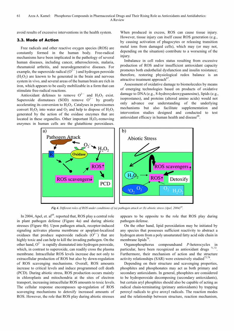
Fig 4. Different roles of ROS under conditions of (a) pathogen attack or (b) abiotic stress (Apel, 2004)69.
In 2004, Apel, et. al69
. reported that, ROS play a central role
in plant pathogen defense (Figure 4a) and during abiotic
stresses (Figure 4b). Upon pathogen attack, receptor-induced
signaling activates plasma membrane or apoplast-localized
oxidases that produce superoxide radicals (O2·−
) that are
highly toxic and can help to kill the invading pathogen. On the
other hand, O2·−
is rapidly dismutated into hydrogen peroxide,
which, in contrast to superoxide, can readily cross the plasma
membrane. Intracellular ROS levels increase due not only to
extracellular production of ROS but also by down-regulation
of ROS scavenging mechanisms. Overall, ROS amounts
increase to critical levels and induce programmed cell death
(PCD). During abiotic stress, ROS production occurs mainly
in chloroplasts and mitochondria at the sites of electron
transport, increasing intracellular ROS amounts to toxic levels.
The cellular response encompasses up-regulation of ROS
scavenging mechanisms to detoxify increased amounts of
ROS. However, the role that ROS play during abiotic stresses
appears to be opposite to the role that ROS play during
pathogen defense.
On the other hand, lipid peroxidation may be initiated by
any species that possesses sufficient reactivity to abstract a
hydrogen atom from a poly unsaturated fatty acid side chain in
membrane lipids70
.
Organophosphorus compoundsand P-heterocycles in
particular, have been recognized as antioxidant drugs 71,72
.
Furthermore, their mechanism of action and the structure
activity relationships (SAR) were extensively studied73,74
.
Depending on their structure and scavenging properties,
phosphites and phosphonates may act as both primary and
secondary antioxidants. In general, phosphites are considered
to be hydroperoxide decomposing (secondary antioxidants),
but certain aryl phosphites should also be capable of acting as
radical chain-terminating (primary antioxidants) by trapping
peroxyl radicals to give aroxyl radicals. The reaction modes
and the relationship between structure, reaction mechanism,
International Journal of Chemical and Biomedical Science 2015; 1(3): 56-69 62
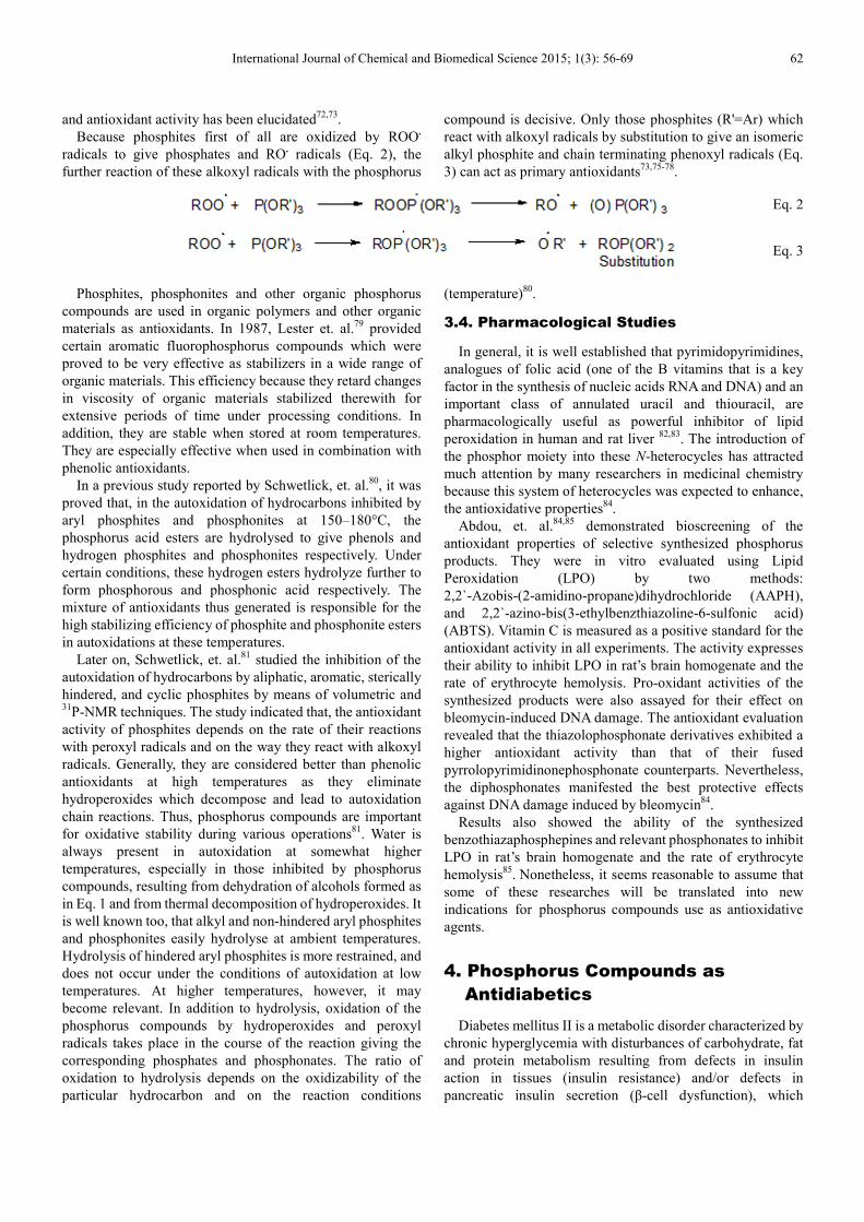
and antioxidant activity has been elucidated72,73
.
Because phosphites first of all are oxidized by ROO.
radicals to give phosphates and RO. radicals (Eq. 2), the
further reaction of these alkoxyl radicals with the phosphorus
compound is decisive. Only those phosphites (R'=Ar) which
react with alkoxyl radicals by substitution to give an isomeric
alkyl phosphite and chain terminating phenoxyl radicals (Eq.
3) can act as primary antioxidants73,75-78
.
Eq. 2
Eq. 3
Phosphites, phosphonites and other organic phosphorus
compounds are used in organic polymers and other organic
materials as antioxidants. In 1987, Lester et. al.79
provided
certain aromatic fluorophosphorus compounds which were
proved to be very effective as stabilizers in a wide range of
organic materials. This efficiency because they retard changes
in viscosity of organic materials stabilized therewith for
extensive periods of time under processing conditions. In
addition, they are stable when stored at room temperatures.
They are especially effective when used in combination with
phenolic antioxidants.
In a previous study reported by Schwetlick, et. al.80
, it was
proved that, in the autoxidation of hydrocarbons inhibited by
aryl phosphites and phosphonites at 150–180°C, the
phosphorus acid esters are hydrolysed to give phenols and
hydrogen phosphites and phosphonites respectively. Under
certain conditions, these hydrogen esters hydrolyze further to
form phosphorous and phosphonic acid respectively. The
mixture of antioxidants thus generated is responsible for the
high stabilizing efficiency of phosphite and phosphonite esters
in autoxidations at these temperatures.
Later on, Schwetlick, et. al.81
studied the inhibition of the
autoxidation of hydrocarbons by aliphatic, aromatic, sterically
hindered, and cyclic phosphites by means of volumetric and 31
P-NMR techniques. The study indicated that, the antioxidant
activity of phosphites depends on the rate of their reactions
with peroxyl radicals and on the way they react with alkoxyl
radicals. Generally, they are considered better than phenolic
antioxidants at high temperatures as they eliminate
hydroperoxides which decompose and lead to autoxidation
chain reactions. Thus, phosphorus compounds are important
for oxidative stability during various operations81
. Water is
always present in autoxidation at somewhat higher
temperatures, especially in those inhibited by phosphorus
compounds, resulting from dehydration of alcohols formed as
in Eq. 1 and from thermal decomposition of hydroperoxides. It
is well known too, that alkyl and non-hindered aryl phosphites
and phosphonites easily hydrolyse at ambient temperatures.
Hydrolysis of hindered aryl phosphites is more restrained, and
does not occur under the conditions of autoxidation at low
temperatures. At higher temperatures, however, it may
become relevant. In addition to hydrolysis, oxidation of the
phosphorus compounds by hydroperoxides and peroxyl
radicals takes place in the course of the reaction giving the
corresponding phosphates and phosphonates. The ratio of
oxidation to hydrolysis depends on the oxidizability of the
particular hydrocarbon and on the reaction conditions
(temperature)80
.
3.4. Pharmacological Studies
In general, it is well established that pyrimidopyrimidines,
analogues of folic acid (one of the B vitamins that is a key
factor in the synthesis of nucleic acids RNA and DNA) and an
important class of annulated uracil and thiouracil, are
pharmacologically useful as powerful inhibitor of lipid
peroxidation in human and rat liver 82,83
. The introduction of
the phosphor moiety into these N-heterocycles has attracted
much attention by many researchers in medicinal chemistry
because this system of heterocycles was expected to enhance,
the antioxidative properties84
.
Abdou, et. al.84,85
demonstrated bioscreening of the
antioxidant properties of selective synthesized phosphorus
products. They were in vitro evaluated using Lipid
Peroxidation (LPO) by two methods:
2,2`-Azobis-(2-amidino-propane)dihydrochloride (AAPH),
and 2,2`-azino-bis(3-ethylbenzthiazoline-6-sulfonic acid)
(ABTS). Vitamin C is measured as a positive standard for the
antioxidant activity in all experiments. The activity expresses
their ability to inhibit LPO in rat’s brain homogenate and the
rate of erythrocyte hemolysis. Pro-oxidant activities of the
synthesized products were also assayed for their effect on
bleomycin-induced DNA damage. The antioxidant evaluation
revealed that the thiazolophosphonate derivatives exhibited a
higher antioxidant activity than that of their fused
pyrrolopyrimidinonephosphonate counterparts. Nevertheless,
the diphosphonates manifested the best protective effects
against DNA damage induced by bleomycin84
.
Results also showed the ability of the synthesized
benzothiazaphosphepines and relevant phosphonates to inhibit
LPO in rat’s brain homogenate and the rate of erythrocyte
hemolysis85
. Nonetheless, it seems reasonable to assume that
some of these researches will be translated into new
indications for phosphorus compounds use as antioxidative
agents.
4. Phosphorus Compounds as
Antidiabetics
Diabetes mellitus II is a metabolic disorder characterized by
chronic hyperglycemia with disturbances of carbohydrate, fat
and protein metabolism resulting from defects in insulin
action in tissues (insulin resistance) and/or defects in
pancreatic insulin secretion (β-cell dysfunction), which

63 Azza A. Kamel: Phosphorus Compounds in Pharmaceutical Drugs and Their Rising Role as Antioxidants and Antidiabetics:
A Review
eventually includes loss of pancreatic insulin-secreting
cells86,87
.
In 2001, Zimmet, et.al.88
expected that, the number of
people with diabetes would double to ∼300 million within 20
years. The associated complications of diabetes, such as
cardiovascular disease, peripheral vascular disease, stroke,
diabetic neuropathy, diabetic nephropathy, and diabetic
retinopathy (eventually blindness) result in increasing
disability, reduced life expectancy, and enormous health costs.
4.1. Pharmacological Studies
In patients with type 2 diabetes, the pancreatic and hepatic
glucokinase activity, as well as the pancreatic glucokinase
mRNA, is reduced by at least 50%. However, the exact
glucokinase status of individual patients is not known and is
not used as an indicator of the individual stage of the disease89
.
Glucokinase mutations may be related to maturity onset of
diabetes of the permanent neonatal diabetes mellitus90,91
.
Altogether, a broad range of “glucokinase diseases” exist, with
close to 250 known missense and nonsense mutations, as well
as insertions, deletions, and splice variants92
.
The established drugs [sulfonylureas, glinides,
glucagon-like peptide 1 (GLP-1) receptor agonists, metformin,
thiazolidinediones, and α-glucosidase inhibitors] generally
target insulin resistance or β-cell dysfunction by increasing
insulin secretion or tissue sensitivity to insulin93
.
Drugs addressing other aspects of the disease including
promising emerging biological/molecular targets are still
under investigation. Many compounds are derived from
physiological compounds (hormones) aiming at improving
their kinetics and selectivity, and others are chemical
compounds that were obtained by screening for a newly
identified target in the physiological or pathophysiological
machinery. Many unsolved problems exist including: a)
Reduced β-cell sensitivity to glucose (“sensor defect”); b)
Loss of β-cell number/function (no halting of diabetes
progression); c) Loss of oscillations of insulin secretion; d)
Loss of first-phase insulin response to a glucose challenge; e)
Elevated pro-insulin/insulin ratio; f) Abnormally high
secretion of amylin; g) Increased glucagon secretion
(gluconeogenesis, glucose production).
In addition, many marketed drugs have major drawbacks
that hamper therapy, and modifications in dosing and/or new
compounds should be developed to overcome these issues94
.
Among others, the following problems continue to plague
current therapy: a) Hypoglycemias (especially when initiating
therapy; severe hypoglycemias are known to lead to
myocardial infarction and to the development of dementias); b)
Many of these pathophysiological parameters are linked to
more than one secondary complication, such as increased
vascular permeability, alterations in blood flow, and
stimulation of neovascularization95
; c) Weight gain (a leading
factor driving the epidemic of diabetes); d) Increase in insulin
resistance; e) β-Cell destruction.
4.2. Recent Trends in Therapy
Hyperglycemia induces various diabetic complications via
different mechanisms, which are the basis for therapies. Since
many defects overlap between various complications, they are
first solely listed and afterward the remedies are described.
Current therapies for type 2 diabetes mellitus have mainly
centered on elevating plasma insulin levels (direct insulin
administration or oral agents that promote insulin secretion),
improving insulin sensitivity of tissues, and eventually
reducing the rate of carbohydrate absorption from the
gastrointestinal tract95
.
This review article spots two of the most attractive recent
trends in therapies for type 2 diabetes96-109
. The first trend is a
new perspective, even a paradigm change, which has recently
been brought forward by a new class called incretin
enhancers96-100
.
Fig 5. Summary of effects of GLP-1 (Verspohl, 2009)97.

International Journal of Chemical and Biomedical Science 2015; 1(3): 56-69 64
Incretins are defined as being responsible for a higher
insulin response to oral intake of glucose compared with an
equal intravenous glucose load (i.e., reaching equivalent
plasma glucose levels)96
. GLP-1 receptor function and
subsequent signaling has been demonstrated in recent
litreatures97-100
(Fig. 5). Albiglutide improves glycemic
control across a variety of doses and dosing schedules,
Albiglutide mimics the full range of GLP-1 actions, second
messengers, and mechanisms99
.
The clinical properties of the GLP-1 receptor agonist
albiglutide (phase III; structural details in Fig. 6) have been
reviewed99,100
. On March 2014, GLP-1 receptor agonist
albiglutide (GLP-1RAs) has been established as an important
total treatment strategy for patients with type 2 diabetes who
do not tolerate metformin99
. It is approved as add-on therapy
in combination with other blood-glucose lowering drugs
including insulin when these, together with diet and exercise,
do not provide adequate glycaemic control. Albiglutide is
injected under the skin once a week with a single use pen
injector100
. Its long plasma half-life of 5 days (improved
pharmacokinetics) enables once-weekly dosing, as a result of
covalent binding of albumin. The tandem repeat structure (Fig.
6) improves the potency observed when only one GLP-1
moiety was covalently linked to albumin (note: a bulky carrier
molecule)98
.
Fig 6. Theoretical structure of albiglutide (Rosenstock, 2009)98.
The second trend is the development of glucokinase
activators has a long history101,102
. Pancreatic β cells and the
liver play key roles in blood glucose homeostasis103
. In both
organs, glucose is transported into the cell by the low-affinity
glucose transporter GLUT2. Rate-limiting phosphorylation of
glucose by glucokinase is the first step initiating glycogen
synthesis in the liver104
and insulin release in β cells102
.
Glucokinase - also known as hexokinase IV, hexokinase D
(ATP: d-glucose 6-phosphotransferase; EC 2.7.1.2), or
d-glucose- phosphorylates other hexoses, such as d-fructose,
d-mannose, or 2-deoxy-d-glucose by ATP according to the
following equation (Eq. 4):
Eq. 4
Glucokinase has a higher Km (6–10 mM) for glucose than
the other hexokinases, which are saturated at this
concentration. Therefore, only glucokinase activity correlates
with physiological rises of blood glucose concentrations from
fasting (5 mM) to postprandial (10–15 mM) levels. This is
why glucokinase is often referred to being a “glucose sensor”
in β cell105
and the “glucostat” concept was developed106
. As a
sensor, it determines the rate and threshold concentration of
glucose (∼5 mM) required to initiate the signaling cascade
leading to insulin release107
. In Fig. 7, various roles of
glucokinase are summarized, including those for β cells and
liver.

65 Azza A. Kamel: Phosphorus Compounds in Pharmaceutical Drugs and Their Rising Role as Antioxidants and Antidiabetics:
A Review
Fig 7. Role of glucokinase in various tissues (Verspohl, 2012)106.
Sarabu, et. al.108,109
described a pharmacophore model of
the heterogeneous chemical group of most known classes of
glucokinase activators. The key structural features common to
both single atom-centered (carbon or nitrogen) and aromatic
ring-centered glucokinase activators, including three
attachments, two of which are hydrophobic groups (with at
least one consisting of an aromatic ring structure) and the
other contributes a hydrogen bond donor-acceptor pair. The
establishment of a crystal structure of recombinant human
glucokinase was mainly put forward by the availability of
glucokinase activators110
. For the allosteric activator site, as
many as nine contact amino acids, depending on the chemistry
of the drug, have been identified, encompassing Val62, Arg63,
Glu210, Ile211, Tyr214, Tyr215, Met235, Val452, and Val455.
The link between enzyme kinetic and structure-activity
relationship (SAR) has not yet been sufficiently
investigated101,110,111
.
Increased superoxide anion production induced by
hyperglycemia leads to decreased activity of
glycerinaldehyde-3-phosphate dehydrogenase and to
consequential increased activity of alternative pathways,
including the polyol, hexosamine, diacylglycerol, PKC, and
AGE pathways. Glucose pathways during hyperglycemia
resulting mainly in cell dysfunctions. Excessive glucose
metabolism generates NADH and overload of the electron
transport chain, causing oxidative stress. Finally, many
pathways are activated, leading to inflammation and neuronal
dysfunction112-114
(Fig. 8).
Fig 8. Pathophysiological factors (Conway, 2009)113.

International Journal of Chemical and Biomedical Science 2015; 1(3): 56-69 66
Some methods to mitigate or avoid diabetic complications
include: a) Inhibition of increased glucose flux through the
polyol pathway (aldose reductase inhibition); b) Inhibition of
increased formation of advanced glycation end-products
(AGE); c) Inhibition of protein kinase C (PKC) isoforms; d)
Inhibition of increased hexosamine biosynthesis pathway; e)
Inhibition of reactive oxygen species (ROS) and superoxide
formation; f) Inhibition of the transforming growth factor-β
(TGF-β) secretion; g) Activation of transketolase, and
inhibition of poly(ADP-ribose) polymerase (PARP).
Normal physiological functions such as gastric emptying
(slowing) or renal glucose re-absorption (blocking to increase
glucose loss) could also be potential targets for future
therapies. One possibility is the comprehensive gene
expression analyses of critical tissues for understanding the
molecular signature of type 2 diabetes. Serial analysis of gene
expression techniques has made it possible to compare tag
levels among independent libraries and to identify previously
unrecognized genes with novel functions that may be
important in the development of diseases. Such serial analysis
of gene expression-based approaches may lead to the
identification of novel therapeutic targets for the treatment of
type 2 diabetes and its complications.
In some areas, great progress is observed (e.g., incretin area) 96-100
; in others, no great progress is obvious (e.g., glucokinase
activators) 101-109
, and other areas are not recommended for
further research.
4.3. Uses of Organophosphorus Compounds
as Antidiabetics
Several drugs such as metformin, biguanides/ metformin
(Glucovance), sulfonylureas/ metformin (glibenclamide), and
others are presently available to reduce hyperglycemia in
diabetes mellitus. Despite their wide spread use, none of the
presently available agents is ideal; each has its shortcoming
and side effects115
. Thus, the continuous search for novel
antidiabetic agents that are more effective and safe is a target
of research by many investigators.
Heterocyclic phosphor esters are known to serve as both
hyperglycemic and hypoglycemic agents in different
concentrations115-118
, e.g., diisopropylphosphorofluoridate has
the activity to reduce the glucose level (hypoglycemia)116
. In
addition, studies on the effect of organophosphorus
compounds (OPC) on carbohydrate metabolism showed an
increase in blood glucose in various constituents of brain rats
after treatment with malathion117
(Structure 1). Nevertheless,
glycogen levels were decreased in rat liver when treated with
dichlorovos118
(Structure 2).
Structure 1. Malathion ( Al-Ghanim, 2012)117.
Structure 2. Dichlorovos (Lakshmanan, 2013)118.
Recentely, Abdou, et. al.84,85
have evaluated the antidiabetic
activity of selective synthesized substituted bicyclic 6,6- and
6,5- membered phosphonates, the results were presented
while % potency of the tested phosphonatesvs blood glucose
levels of diabetic rats was displayed, and glibenclamide was
used as a reference standard. The tested phosphonates have
shown hypoglycemia effectthat can decrease the blood
glucose levels in diabetic rats. The screening for the
antidiabetic effect of the tested products was carried out in
ethanolic solution on streptozotocin-induced diabetic rat in
duration dependent fashion. Streptozotocin injection induced
diabetes mellitus, which may be due to destruction of β-cells
of Islets of Langerhans as proposed by others119
. After 7 days
and 14 days supplementation of ethanol solutions of the tested
compounds resulted in significant diminution of fasting blood
glucose level in respect to diabetic rat, but no significant
alteration of fasting blood glucose level to the control, which
further strengthens the antidiabetogenic action of these
compounds. Fasting blood glucose level of all animals before
treatment was within the normal range then it was
significantly elevated after 24 h of streptozotocin injection
with respect to the control level. The screening results showed
that the tested phosphonates exhibit a potent to moderate
effects on diabetes mellitus II, suggesting new generation of
antidiabetogenic drugs84,85
. At the time being, there has been a
return to laboratory studies that are helping to solve how these
OPC can work at a cellular level. As a result, their full
therapeutic potential is gradually being realized. The journey,
as usual started with chemistry, which led to laboratory studies
related to the mechanism of action of such OPC as antidiabetic
agents.
5. Conclusion and Prospective
In summary, the discovery and development of phosphorus
compounds as a major class of drugs for treatment of many
diseases has been a fascinating saga that is not yet completed.
The introduction of bisphosphonates in oncology has
dramatically changed the management of patients with
metastatic bone disease. Particularly remarkable is their
anticancer activity. In contrast, many studies proved that
aminophosphonates exhibited promising antimicrobial,
antioxidant, and anticancer activity.
Many phosphorus compounds are proved to be important
agents for oxidative stability during various operations.
Phosphites and phosphonates may act as both primary and
secondary antioxidants. Results showed the ability of many
phosphonates to inhibit LPO and the rate of erythrocyte

67 Azza A. Kamel: Phosphorus Compounds in Pharmaceutical Drugs and Their Rising Role as Antioxidants and Antidiabetics:
A Review
hemolysis in rat’s brain homogenate. Consequently, the
potential benefits of phosphorus compounds as antioxidative
agents are needed to be considered.
On the other hand, and regarding to type 2 diabetes mellitus,
none of the presently available drugs to reduce hyperglycemia
is ideal. Thus, the continuous search for novel, more effective
and safe antidiabetic agents -that may involve OPC- is a target
of future research. Despite the synthesis of hundreds of
compounds, no clear-cut structure-effect relationship has been
unraveled up to now.
References
[1] Mader, M. M., Bartlett P. A., Chem. Rev., 1997, 97, 1281-1301.
[2] Holla, B. S., Ashok, M., Phosphorus, Sulfur, Silicon, and Relat. Elem. 2007,182, 981-991.
[3] Bul, E. O. J., Naidu, M. S. R., Phosphorus, Sulfur, Silicon, and Relat. Elem., 2000,162, 231-243.
[4] Gilard, V., Martino, R., Malet-Martino, M., Niemeyer, U., Pohl, J., J. Med. Chem., 1999, 42, 2542-2560.
[5] Maier, L., Diel, P. J., Phosphorous Sulphur and Silicon, 1991, 57, 57-64.
[6] Leon, A., Liu, L., Yang, Y., Hudock, M. P., Hall, P., Yin, F., Studer, D., Puan, K. J., Morita, C. T., Oldfield, E., J. Med. Chem., 2006, 49, 7331-7341.
[7] Kafarski, P., Lejczak, B., Curr. Med. Chem. Anticancer Agents, 2001, 1, 301-312.
[8] Gouverneur, V., Lalloz, M. N., Tetrahedron Lett., 1996, 37, 6331-6334.
[9] Yoneda, T., Sasaki, A., Dustan, C., William, P. J., Bauss, F., De Clerck, Y. A., Mundy, G. R., J. Clin. Invest., 1997, 99, 2509-2517.
[10] Ross, J. R., Saunders, Y., Edmonds, P. M., BMJ, 2003, 327, 469-474.
[11] Tim, V. W., Manon, T. H., Eric, F., Jan, B. V., The Oncologist, 2009, 14, 181-191.
[12] Sanders, J. M., Ghosh, S., Chan, J. M., Meints, G., Wang, H., Raker, A., J. Med Chem., 2004, 47, 375-384.
[13] Thompson, K., Rogers, M. J., J. Bone Miner Res., 2004, 19, 278-288.
[14] Graham, R., Russell, G., Bone, 2011, 49, 2-19.
[15] Waldmann, H., Bialy, L., Angew. Chem., 2005, 44, 3814-3819.
[16] Gautier, A., Garipova, G., Salcedo, C., Balieu, S., Piettre, S. R., Angew. Chem. Int. Ed., 2004, 43, 5963-5967.
[17] Drake, M. T., Cremers, S. C., Mol. Interv., 2010, 10,141-152.
[18] Martin, T. J., Grill, V., Australian Prescriber, 2000, 23, 130-132.
[19] Fernandes, C., Leite, R., Rodrigo, S., Lancas, F. M., Quimica Nova, 2005, 28, 274-280, Chem. Abstr. 2005, 142, 366626.
[20] Papapoulos, S. E., Nature Reviews Rheumatology, 2013, 9, 263-264.
[21] Boikos, S. A., Hammers, H. J., Journal of Clinical Oncology (JCO), 2012, 30, e299.
[22] Saunders, Y., Palliat Med., 2004, 18, 418-431.
[23] Hung, S. H., Tsai, W. Y., Tsao, P. N., Chou, H. C., Hsieh, W. S., Journal of the Formosan Medical Association, 2003, 102, 801-804.
[24] Graham R., Russell G., Bone, 2011, 49, 2-19.
[25] McClung M. R., J. Clin. Densitom., 2010, 13, 132.
[26] Reid I. R., Brown J. P., Burckhardt P., Horowitz Z., Richardson P., Trechsel U. N., Engl. J. Med., 2002, 346, 653-661
[27] van-Beek, E., Löwik, C., van der Pluijm, G., Papapoulos, S., J. Bone Miner. Res., 1999, 14, 722–729.
[28] Clézardin, P., Ebetino, F. H., Fournier, P. G., Cancer Res., 2005, 65, 4971-4974.
[29] Abdou, W. M., Ganoub, N. A., Fahmy, A. F. M., Shaddy, A. A., Monatsh. Chem. 2006, 136, 105-116.
[30] Abdou, W. M., Khidre, R. E., Kamel, A. A., Arch. Pharm. Chem. Life Sci., 2012, 345, 123-136.
[31] Abdou, W. M., Khidre, M. D., Sediek, A. A., The design and synthesis of sulfur and nitrogen containing bisphosphonic acids and their role in oncology in: The chemistry and biologically activity of synthetic and natural compounds, modern aspects of chemistry of heterocycles,Russian Academy of Natural Science, Kartsev, V. G. (ed.).2010, pp. 209-212.
[32] Abdou, W. M., Shaddy, A. A., J. Med. Chem. Res., 2010, 19, 39-40.
[33] Abdou, W. M., Khidre, R. E., Shaddy, A. A., J. Heterocyclic Chem., 2013, 50, 33-41.
[34] Abdou, W. M., Kamel, A. A., Shaddy, A. A., Eur. J. Med. Chem., 2010, 45, 5217-5224.
[35] Abdou, W. M., Shaddy, A. A., Arkivoc, 2009, 14,143-182.
[36] Abdou, W. M., Ganoub, N. A., EL-Khoshnieh, Y. O., Synlett, 2003, 785-790.
[37] Prasad, G. S., Rao, G. N., Journal of Modern Medicinal Chemistry, 2013, 1, 49-60.
[38] Moonen, K., Laureyn, I., Stevens, C. V., Chem Rev, 2004, 104, 6177-6185
[39] Laureyn, I., Stevens, C. V., Soroka, M., Malyse, P., Arkivoc, 2003, 6, 102-115.
[40] Kafarski, P., Lejczak, B., Current Medicinal Chemistry - Anti-Cancer Agents, 2001, 1, 301-312.
[41] Abdel-Monem, W. R., Eur. J. Chem., 2010, 1, 168-172.
[42] Schug, K. A., Lindner, W., Chem. Rev. 2005, 105, 67-114.
[43] Wrobleski, S. T., Lin, S., Hynes, J. Jr., Wu, H., Pitt, S., Bioorg. Med. Chem. Lett., 2008, 18, 2739-2744.
[44] Prasad, G. S., Rao, G. N., Journal of Modern Medicinal Chemistry, 2013, 1, 49-60.

International Journal of Chemical and Biomedical Science 2015; 1(3): 56-69 68
[45] Chandrasekhar, S., Narsihmulu, Ch., Shameen, S. S., Saritha, B., Jayaprakash, S., Synlett, 2003, 505-506.
[46] Takahashi, H., Yoshioka, M., Imai, N., Onimura, K., Kobayashi, S., Synthesis, 1994, 8, 763-764.
[47] Heydari, A., Karimian, A., Ipaktschi, J., Tetrahedron Lett, 1998, 39, 6729-6732.
[48] Azizi, N., Saidi, M. R. Eur. J. Org. Chem., 2003, 46, 30-33.
[49] Lee S., Park J. H., Kang J, Lee J. K., J., Chem.Soc. Chem.Commun., 2001, 1698-1699.
[50] Akiyama, T., Sanada, M., Fuchibe, K., Synlett, 2003, 1463-1464.
[51] Abdou, W. M., Barghash, R. F., Bekhiet, M. S., RSC Org. Advances, 2013, 1528-1540.
[52] Shaddy, A. A., Kamel, A. A., Abdou, W. M., Synth. Commun., 2013, 43, 236-252.
[53] Abdou, W. M., Barghash, R. F., Sediek, A. A., Eur. J. Med. Chem., 2012, 57, 362-372.
[54] Abdou, W. M., Barghash, R. F., Bekheit, M. S., Arch. Pharm. Chem. Life Sci. 2012, 345, 884-895.
[55] Kamel, A. A., Geronikaki, A., Abdou, W. M., Eur. J. Med. Chem. 2012, 51, 239-249
[56] Abdou, W. M., Kamel, A. A., Khidre, R. E., Geronikaki, A., Ekonomopoulou, M. T., Chem. Biol. & Drug Des., 2012, 79, 719-730.
[57] Abdou, W. M., Barghash, R. F., Khidre, R. E., Monatsh. Chem., 2013, 144, 1233-1242.
[58] Hirschmann, R., Smidt, A. B., Taylor, C. M., Benkovic, P. A., Taylor, S. D., Yager, K. M., Sprengeler, P. A., Benkovic, S. J., Science, 1994, 265, 234-237.
[59] Meyer, F., Laaziri, A., Papini, A. M., Uziel, J., Juge, S., Tetrahedron, 2004, 60, 3593-3597.
[60] Smith, W. W., Bartlett, P. A., J. Am. Chem. Soc., 1998, 120, 4622-4628.
[61] For examples and applications of different naturally occurring β-aminophophonates/ phosphonic acids see, e.g.: Aminophosphonic and aminophosphinic acids, chemistry and biological activity, ed. V. P. Kukhar, H. R. Hudson, John Wiley and Sons, NY, 2000.
[62] He, X. P., Xie, J., Tang, Y., Li, J., Chen, G. R., Curr. Med. Chem., 2012, 19, 2399-2405.
[63] Butnariu, M., Grozea, I., J. Bioequiv. Availab., 2012, 4, xvii-xix.
[64] Sugioka, K., Shimosegawa, Y., Nakano, M., FEBS Letters, 1987, 210, 37-90.
[65] Pacheco, J., Gonsebatt, M., Mutation Research/Genetic Toxicology and Environmental Mutagenesis, 2009, 674, 137–147.
[66] Evans, P., Halliwell, B., British Journal of Nutrition, 2001, 85, 67S-74S.
[67] Potenza, N., Papa, U., Russo, A., Cell Biol. Int., 2009, 33, 734-738.
[68] Aruoma, O. I., Journal of the American Oil Chemists' Society, 1998, 75, 199-212.
[69] Apel, K., Hir, H., Annual Review of Plant Biology, 2004, 55, 373-399.
[70] Catalá, A., The International Journal of Biochemistry & Cell Biology, 2006, 38, 1482–1495.
[71] Nurulain, S. M., Szegi, P., Tekes, K., Naqvi S. N., 2013, 64, 169-177.
[72] Schwetlick, K., Pionteck, J., Habicher, T. W. D., Eur. Polymer J. 1987, 23, 383-388.
[73] Földes, E., Maloschik, E.,Kriston, I., Staniek, P., Pukánszky, B., Polymer Degradation and Stability, 2006, 91,479–487.
[74] Schwetlick, K., In mechanism of polymer of degradation and stabillisation, ed. Elsevier Applied Science, London and New York, 1990.
[75] Hall, G. H., Neal, M. A., Jenkins, S. D., Siddiqui, J. A., US6827897 B2, 2004, US 09/818,334.
[76] Vulic, I.,Vitarelli,G.,Zenner, J. M., Polymer Degradation and Stability, 2002,78, 27–34.
[77] Schwetlick, K., Habicher, W. D., Die Angewandte Makromolekulare Chemie, 1995, 232, 239–246.
[78] Habiche, W. D., Bauer, I., Pospíšil, J., Macromolecular Symposia, 2005, 225, 147–164.
[79] Lester, P. J., Burton, US 4912155 A, 1987.
[80] Schwetlick, K., König, T., Rüger, C.,Pionteck, J., Habicher, W. D., Polymer Degradation and Stability, 1986, 15, 97–108.
[81] Schwetlick, K., Pionteck, J., Winkler, A., Hfihner, U., Kroschwitz, H., Habicher, W. D., Polymer Degradation and Stability, 1991, 31, 219-228.
[82] Ahmed, O. M., Hussein, A. M., Ahmed, R. R., Med.Chem., 2012, 2, 20-28.
[83] De la Cruz, J. P., Carrasco, T., Ortega, G., Sanchez, C. F., Lipids, 1992, 27, 192-194.
[84] Kamel, A. A., Khidre, M. D., Abdou, W. M., Heterocyclic Chemistry, accepted for publications, 2014, DOI 1002/jhet.2260, Published online 00 Month 2014 in Wiley Online Library (wileyonlinelibrary.com).
[85] Abdou, W. M., Ganoub, N. A., Barghash, R. F., Synthetic Communications, 2014, 44, 2669-2678.
[86] Rosenbloom, A. L., Joe, J. R., Young, R. S., Winter, W. E., Diabetes Care, 1999, 22, 345-354.
[87] Kamaeswara, B. R., Giri, R., Kesavulu, M. M., Apparao, C. H, J. Ethnopharmacol., 2001, 74, 69-74.
[88] Zimmet, P., Alberti, K. G., Shaw, J., Nature, 2001, 414, 782-787.
[89] Caro, J. F., Triester, S., Patel, V. K., Tapscott, E. B., Frazier, N. L., Dohm, G. L., Hormone and Metabolic Research, 1995, 27, 19-22.
[90] Stride, A., Shields, B., Gill-Carey, O., Chakera, A. J., Colclough, K., Ellard, S., Hattersley, A. T., Diabetologia, 2014, 57, 54-56.

69 Azza A. Kamel: Phosphorus Compounds in Pharmaceutical Drugs and Their Rising Role as Antioxidants and Antidiabetics:
A Review
[91] Gloyn, A. L., Hum. Mutat., 2003, 22, 353-362.
[92] Gloyn, A. L., Front diabetes, in Glucokinase and Glycemic Disease: From Basics to Novel Therapeutics, Matschinsky, F. M., Magnuson, M. A. (eds.), 16, pp 92-109, Karger, Basel, 2004.
[93] Abdul-Ghani, M. A., DeFronzo, R. A., Endocr. Pract., 2008, 14, 782-790.
[94] Agius, L., Biochem., 2008,414, 1-18.
[95] Takagi C, Bursell S. E., Lin Y. W., Takagi H., Duh E., Jiang Z., Clermont A. C., King G. L., Invest. Ophthalmol. Vis. Sci., 1996, 37, 2504-2518.
[96] Deacon, C. F., Carr, R. D., Holst, J. J., Frontiers in Bioscience, 2008, 13, 1780-1794.
[97] Verspohl E. J., Pharmacol. Ther., 2009, 124, 113-138.
[98] Rosenstock, J., Reusch, J., Bush, M., Yang, F., Stewart, M., Diabetes Care, 2009, 32, 1880- 1886.
[99] Trujillo, J. M., Nuffer, W., Ann Pharmacother., 2014, 48, 1494-1501.
[100] Yabe, D. , Kuwata, H., Usui, R. , Kurose, T. , and Seino, Y., Current Medical Research & Opinion. Informa Healthcare, Posted online on May 20, 2015 (doi:1185/030079201045471).
[101] Priyadarsini, R. L., Namratha, J. R., Reddy, D. R., International Journal of Pharmacy and Pharmaceutical Sciences, 2012, 4, 81-87.
[102] Pal, M., Curr. Med. Chem., 2009, 16, 3858-3874.
[103] Bae, J., Kim, T., Kim M., Park, J., Ahn, Y., Sensors, 2010, 10, 5031-5053.
[104] Matschinsky, F. M., Nat. Rev. Drug Discov., 2009, 8, 399-416.
[105] Matschinsky, F. M., Diabetes, 2002, 51, S394-S404.
[106] Verspohl, E. J., Pharmacological Reviews, 2012, 64, 188-237.
[107] Grimsby, J., Matschinsky, F. M., Grippo, J. F., Discovery and
actions of glucokinase activators, in Glucokinase and Glycemic Disease: From Basics to Novel Therapeutics, Matschinksy, F. M., Magnuson, M. A. (eds), 16, pp 360-378, Karger, Basel, 2004.
[108] Sarabu, R., Berthel, S. J., Kester, R. F., Tilley, J. W., Expert Opin. Ther. Pat, 2008, 18, 759-768.
[109] Grimsby, J., Berthel, S. J., Sarabu, R., Curr. Top. Med. Chem., 2008, 8, 1524-1532.
[110] Dunten, P., Swain, A., Kammlot, U., Crowther, R., Lukacs, C. M., Levin, W., Reik, L., Grimsby, J., Corbett, W. L., Magnuson, M. A., Matschinsky, F. M., Grippo, J. F., Crystal structure of human liver glucokinase bound to a small molecule allosteric activator. Insights into the activating mutations, in Glucokinase and Glycemic Disease: From Basics to Novel Therapeutics Front Diabetes, Matschinsky, F. M., Magnuson, M. A. (eds.), 16, pp 145-154, Karger, Basel, 2004.
[111] Kamata, K., Mitsuya, M., Nishimura, T., Eiki, J., Nagata, Y., Structure, 2004, 12, 429-438.
[112] Gnudi, L., Gruden, G., Viberti, G., Pathogenesis of diabetic nephropathy, in Textbook of Diabetes, Pickup, J. C., Williams, G. (eds.), pp 1-21, Blackwell Science, Oxford, 2003.
[113] Conway, B. R., Maxwell, A. P., Nephron., 2009, 112, 213-221.
[114] Obrosova, I. G., Neurotherapeutics, 2009, 6, 638-647.
[115] Shu, Y., Sheardown, S. A., Brown, C., Owen, R. P., Zhang, S., Castro, R. A., Ianculescu, A. G., Yue, L., Lo, J. C., Burchard, E. G., Brett, C. M., Giacomini, K. M. J., Clin. Invest. 2007, 117, 1422-1431.
[116] Chatterjee, A. K., Kaveeshwar, U., Defence Science Journal, 1991, 41, 143-147.
[117] Al-Ghanim, K. A., Scientific Research and Essays, 2012, 7, 1674-1680.
[118] Lakshmanan, S., Rajendran, A., Sivasubramaniyan, C., International Journal of Research in Biological Sciences, 2013, 3, 34-38.
[119] Kavalali, G. H., Tuncel, S., Goksel, H. H., Hatemi, J., Ethnopharmacol., 2002, 84, 241-245.