phospholipase a2 structure/function, mechanism, and signaling
TRANSCRIPT
Phospholipase A2 structure/function, mechanism,and signaling1
John E. Burke and Edward A. Dennis2
Departments of Chemistry and Biochemistry, and Department of Pharmacology, School of Medicine,University of California, San Diego, La Jolla, CA 92093-0601
Abstract Tremendous advances in understanding thestructure and function of the superfamily of phospholipaseA2 (PLA2) enzymes has occurred in the twenty-first century.The superfamily includes 15 groups comprising four maintypes including the secreted sPLA2, cytosolic cPLA2, calcium-independent iPLA2, and platelet activating factor (PAF) ace-tyl hydrolase/oxidized lipid lipoprotein associated (Lp)PLA2.We review herein our current understanding of the structureand interaction with substrate phospholipids, which residesin membranes for a representative of each of these maintypes of PLA2. We will also briefly review the developmentof inhibitors of these enzymes and their roles in lipid sig-naling.—Burke, J. E., and E. A. Dennis. Phospholipase A2structure/function, mechanism, and signaling. J. Lipid Res.2009. 50: S237–S242.
Supplementary key words lipid signaling • phospholipids • arachi-donic acid
The last 25 years has witnessed a virtual explosion in ourknowledge about the superfamily of phospholipase A2
(PLA2) enzymes. PLA2 hydrolyzes the fatty acid from thesn-2 position of membrane phospholipids. In vivo, thesn-2 position of phospholipids frequently contains poly-unsaturated fatty acids, and when released, these can bemetabolized to form various eicosanoids and related bio-active lipid mediators (1). The remaining lysophospholipidcan also have important roles in biological processes (2).
From the end of the nineteenth and beginning of thetwentieth century (3), PLA2 was known to be a major com-ponent of snake venoms, and it was later recognized thatPLA2 from old world snakes (group I) differed in theirdisulfide bond pattern from new world snakes (group II).Later it was discovered that the major mammalian digestiveenzyme, pancreatic PLA2, was more similar to that from theold world snakes such as the Indian cobra (group IA), andhence the pancreatic enzyme was named group IB. With
the isolation, sequencing, and cloning of the PLA2 fromhuman synovial fluid in 1988 (group IIA) (4, 5), which hada disulfide bond pattern more similar to the new worldrattlesnakes (group II), the more complicated PLA2 frombee venom (group III) (6), and in 1991 the human cytosoliccalcium-dependent PLA2 from macrophages (group IVA)(7, 8), the need for a more elaborate “group numberingsystem” became obvious (9). As the discovery of additionalPLA2s continued such as the macrophage secreted group VPLA2 (10, 11) and the calcium-independent PLA2 (group VI)(12), this system was expanded with 14 distinct groups andmany subgroups appearing by 2000 (13). The latest review(14) lists 15 distinct groups of PLA2. They cluster in fourmain categories or types: secreted sPLA2s, cytosolic cPLA2s,calcium-independent iPLA2s, and platelet activating factor(PAF) acetyl hydrolase/oxidized lipid lipoprotein associ-ated (Lp)PLA2s. Each of these types has been implicatedin diverse kinds of lipid metabolism and disease progres-sion so there has been a tremendous interest in the phar-maceutical and biotechnology industry in developingselective and potent inhibitors of each of these types.
SECRETED PLA2
The secreted PLA2s were the first type of PLA2 enzymesdiscovered. They are found in sources as diverse as venomsfrom various snakes, scorpions, etc.; components of pan-creatic juices; arthritic synovial fluid; and in many differentmammalian tissues (13). They are characterized by a lowmolecular weight (13–15 kDa), histidine in the catalyticsite, Ca21 bound in the active site, as well as six conserveddisulfide bonds with one or two variable disulfide bonds.These enzymes all catalyze the hydrolysis through thesame mechanism of abstraction of a proton from a watermolecule followed by a nucleophilic attack on the sn-2bond. The water molecule is activated by the presence of
This work was supported by National Institutes of Health Grant GM20501 (E.A.D).
Manuscript received 7 October 2008 and in revised form 13 November 2008.
Published, JLR Papers in Press, November 14, 2008.DOI 10.1194/jlr.R800033-JLR200
1Guest editor for this article was Martha K. Cathcart, Lerner Re-search Institute, the Cleveland Clinic.
2 To whom correspondence should be addressed.e-mail: [email protected]
Copyright © 2009 by the American Society for Biochemistry and Molecular Biology, Inc.
This article is available online at http://www.jlr.org Journal of Lipid Research April Supplement, 2009 S237
a histidine/aspartic acid dyad in a Ca21 dependent man-ner (15, 16). Most of the secreted PLA2 enzymes share theproperty of exhibiting an increase in activity termed inter-facial activation when substrate is presented as a large lipidaggregate, rather than in monomeric form. More detailedreviews of interfacial kinetics can be found elsewhere(17, 18).
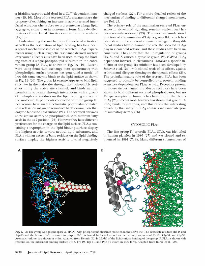
Understanding the mechanism of interfacial activationas well as the orientation of lipid binding has long beena goal of mechanistic studies of the secreted PLA2s. Experi-ments using nuclear magnetic resonance derived nuclearoverhauser effect results have been used to map the bind-ing sites of a single phospholipid substrate in the cobravenom group IA PLA2 as shown in Fig. 1A (19). Recentwork using deuterium exchange mass spectrometry withphospholipid surface present has generated a model ofhow this same enzyme binds to the lipid surface as shownin Fig. 1B (20). The group IA enzyme appears to bind lipidsubstrate in the active site through the hydrophobic resi-dues lining the active site channel, and binds neutralmembrane substrate through interactions with a groupof hydrophobic residues on the lipid binding surface ofthe molecule. Experiments conducted with the group IIIbee venom have used electrostatic potential-modulatedspin relaxation magnetic resonance to determine how thatenzyme binds the lipid surface (21). The secreted enzymesshow similar activity to phospholipids with different fattyacids in the sn-2 position (22). However they have differentpreferences for the charge on the lipid surface. PLA2s con-taining a tryptophan in the lipid binding surface displaythe highest activity toward neutral lipid substrates, andPLA2s with an excess of basic residues on the lipid bindingsurface display the highest activity toward negatively
charged surfaces (22). For a more detailed review of themechanism of binding to differently charged membranes,see Ref. 23.
The primary role of the mammalian secreted PLA2 en-zymes in eicosanoid signaling remains unclear and hasbeen recently reviewed (23). The most well-understoodfunction of a mammalian sPLA2 is group IIA, which hasbeen shown to be a potent antimicrobial agent. Many dif-ferent studies have examined the role the secreted PLA2splay in eicosanoid release, and these studies have been in-conclusive. They show that the up-regulation of groupsIIA, V, and X caused a cytosolic group IVA (GIVA) PLA2
dependent increase in eicosanoids. However a specific in-hibitor of the group IIA inhibitor has been developed bySchevitz et al. (24), with clinical trials of its efficacy againstarthritis and allergens showing no therapeutic effects (23).The proinflammatory role of the secreted PLA2 has beensuggested to possibly be controlled by a protein bindingevent not dependent on PLA2 activity. Receptors presentin mouse tissues named the M-type receptors have beenshown to bind different secreted phospholipases, but noM-type receptor in humans has been found that bindsPLA2 (25). Recent work however has shown that group IIAPLA2 binds to integrins, and this raises the interestingpossibility that integrin-PLA2 contacts may mediate pro-inflammatory activity (26).
CYTOSOLIC PLA2
The first group IV cytosolic PLA2, GIVA, was identifiedin human platelets in 1986 (27) and was cloned and se-quenced in 1991 (7, 8). Many different submembers of
Fig. 1. A: The group IA phospholipase A2 (PLA2) with phospholipid substrate modeled in the active site. The active site residues His-48 andAsp-93 and the bound Ca21 is shown in purple. Ca21 is bound by Asp-49 as well as the carbonyl oxygens of Tyr-28, Gly-30, and Gly-32.Aromatic residues are shown in white. Adapted from Dennis (9). B: Model of the lipid surface binding of the group IA PLA2 is shown withresidues on the interfacial binding surface Tyr-3, Trp-19, Trp 61, and Phe 64 shown in stick form. Adapted from Burke et al. (20).
S238 Journal of Lipid Research April Supplement, 2009
the group IV family have been discovered since then andtheir properties are reviewed (28). The most well-studiedcytosolic enzyme is the GIVA PLA2. It is characterized by anactive site serine and aspartic acid dyad, requirement forCa21 for activity, and it is the only PLA2 with a preferencefor arachidonic acid in the sn-2 position of phospholipids(7, 28). GIVA PLA2 also possesses lysophospholipase activity,as well as transacylase activity (29). Arachidonic acid is theprecursor for the generation of eicosanoids, and this en-zyme has been proposed to play a major role in inflamma-tory diseases. This was proven through the use of knockoutmouse models, where the absence of the GIVA PLA2 genesignificantly reduced the effects of many inflammatory dis-eases (30–32). GIVA PLA2 is now generally considered to bea central enzyme mediating generation of eicosanoids andhence many inflammatory processes.
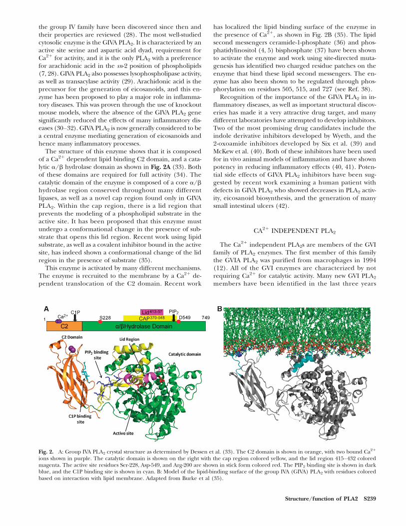
The structure of this enzyme shows that it is composedof a Ca21 dependent lipid binding C2 domain, and a cata-lytic a/b hydrolase domain as shown in Fig. 2A (33). Bothof these domains are required for full activity (34). Thecatalytic domain of the enzyme is composed of a core a/bhydrolase region conserved throughout many differentlipases, as well as a novel cap region found only in GIVAPLA2. Within the cap region, there is a lid region thatprevents the modeling of a phospholipid substrate in theactive site. It has been proposed that this enzyme mustundergo a conformational change in the presence of sub-strate that opens this lid region. Recent work using lipidsubstrate, as well as a covalent inhibitor bound in the activesite, has indeed shown a conformational change of the lidregion in the presence of substrate (35).
This enzyme is activated by many different mechanisms.The enzyme is recruited to the membrane by a Ca21 de-pendent translocation of the C2 domain. Recent work
has localized the lipid binding surface of the enzyme inthe presence of Ca21, as shown in Fig. 2B (35). The lipidsecond messengers ceramide-1-phosphate (36) and phos-phatidylinositol (4, 5) bisphosphate (37) have been shownto activate the enzyme and work using site-directed muta-genesis has identified two charged residue patches on theenzyme that bind these lipid second messengers. The en-zyme has also been shown to be regulated through phos-phorylation on residues 505, 515, and 727 (see Ref. 38).
Recognition of the importance of the GIVA PLA2 in in-flammatory diseases, as well as important structural discov-eries has made it a very attractive drug target, and manydifferent laboratories have attempted to develop inhibitors.Two of the most promising drug candidates include theindole derivative inhibitors developed by Wyeth, and the2-oxoamide inhibitors developed by Six et al. (39) andMcKew et al. (40). Both of these inhibitors have been usedfor in vivo animal models of inflammation and have shownpotency in reducing inflammatory effects (40, 41). Poten-tial side effects of GIVA PLA2 inhibitors have been sug-gested by recent work examining a human patient withdefects in GIVA PLA2 who showed decreases in PLA2 activ-ity, eicosanoid biosynthesis, and the generation of manysmall intestinal ulcers (42).
CA21 INDEPENDENT PLA2
The Ca21 independent PLA2s are members of the GVIfamily of PLA2 enzymes. The first member of this familythe GVIA PLA2 was purified from macrophages in 1994(12). All of the GVI enzymes are characterized by notrequiring Ca21 for catalytic activity. Many new GVI PLA2
members have been identified in the last three years
Fig. 2. A: Group IVA PLA2 crystal structure as determined by Dessen et al. (33). The C2 domain is shown in orange, with two bound Ca21
ions shown in purple. The catalytic domain is shown on the right with the cap region colored yellow, and the lid region 415–432 coloredmagenta. The active site residues Ser-228, Asp-549, and Arg-200 are shown in stick form colored red. The PIP2 binding site is shown in darkblue, and the C1P binding site is shown in cyan. B: Model of the lipid-binding surface of the group IVA (GIVA) PLA2 with residues coloredbased on interaction with lipid membrane. Adapted from Burke et al (35).
Structure/function of PLA2 S239
(as reviewed in Ref. 14). The best characterized of theGVI PLA2 enzymes is the GVIA PLA2 (43). It is found incells expressed in multiple different splice variants (44).The active splice forms of the enzyme GIVA-1, and GIVA-2are composed of 7-8 ankyrin repeats, a linker region and acatalytic domain. This enzyme, similar to GIV PLA2, uses aserine in the active site to catalyze the cleavage of the sn-2ester bond; however it does not show specificity for an ara-chidonic acid in the sn-2 position. The GVIA PLA2 alsoposseses a lysophospholipase activity, as well as transacylaseactivity (44). The activity of the GVIA PLA2 has been sug-gested to be regulated throughmany different mechanisms,including ATP binding, caspase cleavage, calmodulin, andpossible ankyrin repeat mediated protein aggregation (38).
The role of the GVIA PLA2 in different signaling path-ways has been shown to be very complex. Initial reports ofthe functions of the GVIA PLA2 were determined using theinhibitor bromoenollactone (44). Recent work has shownthat this inhibitor is not specific for GVIA PLA2 and actu-ally functions through activation of the inhibitor by GVIAPLA2 followed by nonspecific covalent modification ofcysteine residues in all proximally located enzymes (45).Therefore it has been hard to evaluate early experimentsusing this inhibitor to determine the function of the GVIAPLA2. Experiments using the inhibitor bromoenollactoneare reviewed elsewhere (46). Two major factors have al-lowed the determination of GVI PLA2ʼs cellular functions.First the recent generation of GVIA PLA2 deficient micehas shown the importance of this enzyme in bone forma-tion, apoptosis, insulin secretion, and sperm development(47–50). Second the recent development of specific fluoro-ketone inhibitors of GVIA PLA2 (51) have shown in mousemodels that the GVIA PLA2 in combination with the GIVAPLA2 play an important role in Wallerian degenerationand axon regeneration in nerve injury (52). Recent workusing antisense oligodeoxyribonucleotide toward GVIAPLA2 with monocytes has shown decreases in monocyte re-cruitment and directionality (53).
PAF ACETYL HYDROLASE/OXIDIZED LIPID LPPLA2
The PAF acetyl hydrolase/oxidized lipid LpPLA2 is amember of the GVII family of PLA2 enzymes. This enzymewas named for its ability to cleave the acetyl group fromthe sn-2 position of PAF, as well as its association with lipo-proteins. This name is misleading because this enzyme cancleave oxidized lipids in the sn-2 position up to 9 carbonslong, not just PAF (13). This enzyme has also been shownto access substrate in the aqueous phase unlike all otherPLA2s studied (54). Its active site is composed of a serine,histidine, and aspartic acid hydrolase triad (55), which isunlike all other PLA2s, which have dyads. Recent workhas identified c-terminal regions of the enzyme that are re-quired for binding to HDL and LDL (56). This enzymewas cloned from human plasma in 1995 and was shownto have anti-inflammatory activity in vivo (57). These origi-nal studies led to the hypothesis that this enzyme mightfunction in a protective role by stopping the proinflamma-
tory roles of PAF; however several clinical studies of GVIIAPLA2 levels in patients have now established this enzyme asa definitive marker of coronary heart disease (58, 59).
With the classification of this enzyme as a positive riskfactor in coronary heart disease, it has become a very at-tractive drug target. A specific inhibitor of this enzymewas developed in 2003 by GlaxoSmithKline (60), and re-cent clinical trials with this inhibitor have shown a de-crease in the complex atherosclerotic lesions that lead tounstable lesions, as well as other cardiovascular diseasemarkers (61–63).
CONCLUSION
Experiments with members of the PLA2 superfamily ofenzymes have been carried out for over 100 years. Earlykinetic and structural work established PLA2 as an impor-tant model of lipid enzymology. With the discovery of mul-tiple different family members of PLA2 and their structuralcharacterization, as well as the discovery of their cellularfunctions, the PLA2 family has become a major drug targetfor many different diseases. The future of this field is veryexciting as new knockout mouse models, along with spe-cific inhibitors of these enzymes, lead to further elucida-tion of PLA2sʼ roles in cellular processes, along with newpotential therapeutics.
REFERENCES
1. Funk, C. D. 2001. Prostaglandins and leukotrienes: advances ineicosanoid biology. Science. 294: 1871–1875.
2. Rivera, R., and J. Chun. 2008. Biological effects of lysophospho-lipids. Rev. Physiol. Biochem. Pharmacol. 160: 25–46.
3. Stephens, W. W., J. L. Walker, and W. Myers. 1898. The action ofcobra poison on the blood: a contribution to the study of passiveimmunity. J. Pathol. Bacteriol. 5: 279–301.
4. Seilhamer, J. J., W. Pruzanski, P. Vadas, S. Plant, J. A. Miller, J. Kloss,and L. K. Johnson. 1989. Cloning and recombinant expression ofphospholipase A2 present in rheumatoid arthritic synovial fluid.J. Biol. Chem. 264: 5335–5338.
5. Kramer, R. M., C. Hession, B. Johansen, G. Hayes, P. McGray, E. P.Chow, R. Tizard, and R. B. Pepinsky. 1989. Structure and propertiesof a human non-pancreatic phospholipase A2. J. Biol. Chem. 264:5768–5775.
6. Kuchler, K., M. Gmachl, M. J. Sippl, and G. Kreil. 1989. Analysis ofthe Cdna for phospholipase-A2 from honeybee venom glands - thededuced amino-acid sequence reveals homology to the correspond-ing vertebrate enzymes. Eur. J. Biochem. 184: 249–254.
7. Clark, J. D., L. L. Lin, R. W. Kriz, C. S. Ramesha, L. A. Sultzman, A. Y.Lin, N. Milona, and J. L. Knopf. 1991. A novel arachidonic acid-selective cytosolic PLA2 contains a Ca(21)-dependent translocationdomain with homology to PKC and GAP. Cell. 65: 1043–1051.
8. Kramer, R. M., E. F. Roberts, J. Manetta, and J. E. Putnam. 1991.The Ca2(1)-sensitive cytosolic phospholipase A2 is a 100-kDa pro-tein in human monoblast U937 cells. J. Biol. Chem. 266: 5268–5272.
9. Dennis, E. A. 1994. Diversity of group types, regulation, and func-tion of phospholipase A2. J. Biol. Chem. 269: 13057–13060.
10. Chen, J., S. J. Engle, J. J. Seilhamer, and J. A. Tischfield. 1994. Clon-ing and recombinant expression of a novel human low-molecular-weight Ca21-dependent phospholipase-a(2). J. Biol. Chem. 269:2365–2368.
11. Balboa, M. A., J. Balsinde, M. V. Winstead, J. A. Tischfield, and E. A.Dennis. 1996. Novel group V phospholipase A2 involved in arachi-donic acid mobilization in murine P388D1 macrophages. J. Biol.Chem. 271: 32381–32384.
S240 Journal of Lipid Research April Supplement, 2009
12. Ackermann, E. J., E. S. Kempner, and E. A. Dennis. 1994. Ca(21)-independent cytosolic phospholipase A2 from macrophage-likeP388D1 cells. Isolation and characterization. J. Biol. Chem. 269:9227–9233.
13. Six, D. A., and E. A. Dennis. 2000. The expanding superfamily ofphospholipase A(2) enzymes: classification and characterization.Biochim. Biophys. Acta. 1488: 1–19.
14. Schaloske, R. H., and E. A. Dennis. 2006. The phospholipase A2superfamily and its group numbering system. Biochim. Biophys. Acta.1761: 1246–1259.
15. Scott, D. L., S. P. White, Z. Otwinowski, W. Yuan, M. H. Gelb, andP. B. Sigler. 1990. Interfacial catalysis: the mechanism of phospho-lipase A2. Science. 250: 1541–1546.
16. Yu, L., and E. A. Dennis. 1991. Critical role of a hydrogen bond inthe interaction of phospholipase A2 with transition-state and sub-strate analogues. Proc. Natl. Acad. Sci. USA. 88: 9325–9329.
17. Gelb, M. H., M. K. Jain, A. M. Hanel, and O. G. Berg. 1995. Inter-facial enzymology of glycerolipid hydrolases: lessons from secretedphospholipases A2. Annu. Rev. Biochem. 64: 653–688.
18. Carman, G. M., R. A. Deems, and E. A. Dennis. 1995. Lipid signalingenzymes and surface dilution kinetics. J. Biol. Chem. 270: 18711–18714.
19. Plesniak, L. A., L. Yu, and E. A. Dennis. 1995. Conformation of mi-cellar phospholipid bound to the active site of phospholipase A2.Biochemistry. 34: 4943–4951.
20. Burke, J. E., M. J. Karbarz, R. A. Deems, S. Li, V. L. Woods, Jr., andE. A. Dennis. 2008. Interaction of group IA phospholipase A2 withmetal ions and phospholipid vesicles probed with deuterium ex-change mass spectrometry. Biochemistry. 47: 6451–6459.
21. Lin, Y., R. Nielsen, D. Murray, W. L. Hubbell, C. Mailer, B. H.Robinson, and M. H. Gelb. 1998. Docking phospholipase A2 onmembranes using electrostatic potential-modulated spin relaxationmagnetic resonance. Science. 279: 1925–1929.
22. Singer, A. G., F. Ghomashchi, C. Le Calvez, J. Bollinger, S. Bezzine,M. Rouault, M. Sadilek, E. Nguyen, M. Lazdunski, G. Lambeau,et al. 2002. Interfacial kinetic and binding properties of the com-plete set of human and mouse groups I, II, V, X, and XII secretedphospholipases A2. J. Biol. Chem. 277: 48535–48549.
23. Lambeau, G., and M. H. Gelb. 2008. Biochemistry and physiologyof mammalian secreted phospholipases A2. Annu. Rev. Biochem. 77:495–520.
24. Schevitz, R. W., N. J. Bach, D. G. Carlson, N. Y. Chirgadze, D. K.Clawson, R. D. Dillard, S. E. Draheim, L. W. Hartley, N. D. Jones,E. D. Mihelich, et al. 1995. Structure-based design of the first potentand selective inhibitor of human non-pancreatic secretory phos-pholipase A2. Nat. Struct. Biol. 2: 458–465.
25. Lambeau, G., and M. Lazdunski. 1999. Receptors for a growingfamily of secreted phospholipases A2. Trends Pharmacol. Sci. 20:162–170.
26. Saegusa, J., N. Akakura, C. Y. Wu, C. Hoogland, Z. Ma, K. S. Lam,F. T. Liu, Y. K. Takada, and Y. Takada. 2008. Pro-inflammatory secre-tory phospholipase A2 type IIA binds to integrins {alpha}v{beta}3and {alpha}4{beta}1 and induces proliferation of monocytic cellsin an integrin-dependent manner. J. Biol. Chem. 283: 26107–26115.
27. Kramer, R. M., G. C. Checani, A. Deykin, C. R. Pritzker, and D.Deykin. 1986. Solubilization and properties of Ca21-dependenthuman platelet phospholipase A2. Biochim. Biophys. Acta. 878:394–403.
28. Ghosh, M., D. E. Tucker, S. A. Burchett, and C. C. Leslie. 2006.Properties of the group IV phospholipase A2 family. Prog. LipidRes. 45: 487–510.
29. Reynolds, L., L. Hughes, A. I. Louis, R. A. Kramer, and E. A. Dennis.1993. Metal ion and salt effects on the phospholipase A2, lysophos-pholipase, and transacylase activities of human cytosolic phospho-lipase A2. Biochim. Biophys. Acta. 1167: 272–280.
30. Bonventre, J. V., Z. Huang, M. R. Taheri, E. OʼLeary, E. Li, M. A.Moskowitz, and A. Sapirstein. 1997. Reduced fertility and post-ischaemic brain injury in mice deficient in cytosolic phospholipaseA2. Nature. 390: 622–625.
31. Uozumi, N., K. Kume, T. Nagase, N. Nakatani, S. Ishii, F. Tashiro, Y.Komagata, K. Maki, K. Ikuta, Y. Ouchi, et al. 1997. Role of cytosolicphospholipase A2 in allergic response and parturition. Nature. 390:618–622.
32. Uozumi, N., and T. Shimizu. 2002. Roles for cytosolic phospholipaseA2alpha as revealed by gene-targeted mice. Prostaglandins OtherLipid Mediat. 68–69: 59–69.
33. Dessen, A., J. Tang, H. Schmidt, M. Stahl, J. D. Clark, J. Seehra, andW. S. Somers. 1999. Crystal structure of human cytosolic phospho-
lipase A2 reveals a novel topology and catalytic mechanism. Cell. 97:349–360.
34. Hsu, Y. H., J. E. Burke, D. L. Stephens, R. A. Deems, S. Li, K. M.Asmus, V. L. Woods, Jr., and E. A. Dennis. 2008. Calcium bindingrigidifies the C2 domain and the intra-domain interaction of GIVAphospholipase A2 as revealed by hydrogen/deuterium exchangemass spectrometry. J. Biol. Chem. 283: 9820–9827.
35. Burke, J. E., Y. H. Hsu, R. A. Deems, S. Li, V. L. Woods, Jr., and E. A.Dennis. 2008. A phospholipid substrate molecule residing in themembrane surface mediates opening of the lid region in groupIVA cytosolic phospholipase A2. J. Biol Chem 283: 31227–31236.
36. Stahelin, R. V., P. Subramanian, M. Vora, W. Cho, and C. E. Chalfant.2007. Ceramide-1-phosphate binds group IVA cytosolic phos-pholipase a2 via a novel site in the C2 domain. J. Biol. Chem. 282:20467–20474.
37. Six, D. A., and E. A. Dennis. 2003. Essential Ca(21)-independentrole of the group IVA cytosolic phospholipase A(2) C2 domainfor interfacial activity. J. Biol. Chem. 278: 23842–23850.
38. Burke, J. E., and E. A. Dennis. 2008. Phospholipase A2 biochemistry.Cardiovasc. Drugs Ther. In press.
39. Six, D. A., E. Barbayianni, V. Loukas, V. Constantinou-Kokotou,D. Hadjipavlou-Litina, D. Stephens, A. C. Wong, V. Magrioti, P.Moutevelis-Minakakis, S. F. Baker, et al. 2007. Structure-activityrelationship of 2-oxoamide inhibition of group IVA cytosolic phos-pholipase A(2) and group v secreted phospholipase A(2). J. Med.Chem. 50: 4222–4235.
40. McKew, J. C., K. L. Lee, M. W. Shen, P. Thakker, M. A. Foley, M. L.Behnke, B. Hu, F. W. Sum, S. Tam, Y. Hu, et al. 2008. Indole cyto-solic phospholipase A2 alpha inhibitors: discovery and in vitro andin vivo characterization of 4-{3-[5-chloro-2-(2-{[(3,4-dichlorobenzyl)sulfonyl]amino}ethyl)-1-(dipheny lmethyl)-1H-indol-3-yl]propyl}benzoic acid, efipladib. J. Med. Chem. 51: 3388–3413.
41. Yaksh, T. L., G. Kokotos, C. I. Svensson, D. Stephens, C. G. Kokotos,B. Fitzsimmons, D. Hadjipavlou-Litina, X. Y. Hua, and E. A. Dennis.2006. Systemic and intrathecal effects of a novel series of phospho-lipase A(2) inhibitors on hyperalgesia and spinal prostaglandin E-2release. J. Pharmacol. Exp. Ther. 316: 466–475.
42. Adler, D. H., J. D. Cogan, J. A. Phillips 3rd, N. Schnetz-Boutaud,G. L. Milne, T. Iverson, J. A. Stein, D. A. Brenner, J. D. Morrow,O. Boutaud, et al. 2008. Inherited human cPLA(2alpha) deficiencyis associated with impaired eicosanoid biosynthesis, small intestinalulceration, and platelet dysfunction. J. Clin. Invest. 118: 2121–2131.
43. Balboa, M. A., J. Balsinde, S. S. Jones, and E. A. Dennis. 1997. Iden-tity between the Ca21-independent phospholipase A2 enzymesfrom P388D1 macrophages and Chinese hamster ovary cells. J. Biol.Chem. 272: 8576–8580.
44. Winstead, M. V., J. Balsinde, and E. A. Dennis. 2000. Calcium-independent phospholipase A(2): structure and function. Biochim.Biophys. Acta. 1488: 28–39.
45. Song, H., S. Ramanadham, S. Bao, F. F. Hsu, and J. Turk. 2006.A bromoenol lactone suicide substrate inactivates group VIA phos-pholipase A2 by generating a diffusible bromomethyl keto acid thatalkylates cysteine thiols. Biochemistry. 45: 1061–1073.
46. Balsinde, J., and M. A. Balboa. 2005. Cellular regulation and pro-posed biological functions of group VIA calcium-independent phos-pholipase A(2) in activated cells. Cell. Signal. 17: 1052–1062.
47. Bao, S., D. J. Miller, Z. Ma, M. Wohltmann, G. Eng, S. Ramanadham,K. Moley, and J. Turk. 2004. Male mice that do not express groupVIA phospholipase A2 produce spermatozoa with impaired motilityand have greatly reduced fertility. J. Biol. Chem. 279: 38194–38200.
48. Bao, S., H. Song, M. Wohltmann, S. Ramanadham, W. Jin, A.Bohrer, and J. Turk. 2006. Insulin secretory responses and phos-pholipid composition of pancreatic islets from mice that do not ex-press Group VIA phospholipase A2 and effects of metabolic stresson glucose homeostasis. J. Biol. Chem. 281: 20958–20973.
49. Bao, S., Y. Li, X. Lei, M. Wohltmann, W. Jin, A. Bohrer, C. F.Semenkovich, S. Ramanadham, I. Tabas, and J. Turk. 2007. Attenu-ated free cholesterol loading-induced apoptosis but preservedphospholipid composition of peritoneal macrophages from micethat do not express group VIA phospholipase A2. J. Biol. Chem.282: 27100–27114.
50. Ramanadham, S., K. E. Yarasheski, M. J. Silva, M. Wohltmann, D. V.Novack, B. Christiansen, X. Tu, S. Zhang, X. Lei, and J. Turk. 2008. Age-related changes in bone morphology are accelerated in group VIAphospholipase A2 (iPLA2beta)-null mice. Am. J. Pathol. 172: 868–881.
51. Baskakis, C., V. Magrioti, N. Cotton, D. Stephens, V. Constantinou-Kokotou, E. A. Dennis, and G. Kokotos. 2008. Synthesis of poly-
Structure/function of PLA2 S241
fluoro ketones for selective inhibition of human phospholipase A2enzymes. J. Med. Chem. 51(24): 8027–8037.
52. Lopez-Vales, R., X. Navarro, T. Shimizu, C. Baskakis, G. Kokotos, V.Constantinou-Kokotou, D. Stephens, E. A. Dennis, and S. David.2008. Intracellular phospholipase A2 group IVA and group VIA playimportant roles in Wallerian degeneration and axon regenerationafter peripheral nerve injury. Brain.
53. Mishra, R. S., K. A. Carnevale, and M. K. Cathcart. 2008. iPLA2beta:front and center in human monocyte chemotaxis to MCP-1. J. Exp.Med. 205: 347–359.
54. Gelb, M. H., J. H. Min, and M. K. Jain. 2000. Do membrane-boundenzymes access their substrates from the membrane or aqueousphase: interfacial versus non-interfacial enzymes. Biochim. Biophys.Acta. 1488: 20–27.
55. Tjoelker, L. W., C. Eberhardt, J. Unger, H. L. Trong, G. A. Zimmerman,T. M. McIntyre, D. M. Stafforini, S. M. Prescott, and P. W. Gray. 1995.Plasma platelet-activating factor acetylhydrolase is a secreted phos-pholipase A2 with a catalytic triad. J. Biol. Chem. 270: 25481–25487.
56. Gardner, A. A., E. C. Reichert, M. K. Topham, and D. M. Stafforini.2008. Identification of a domain that mediates association ofplatelet-activating factor acetylhydrolase with high density lipopro-tein. J. Biol. Chem. 283: 17099–17106.
57. Tjoelker, L. W., C. Wilder, C. Eberhardt, D. M. Stafforini, G.Dietsch, B. Schimpf, S. Hooper, H. Le Trong, L. S. Cousens,G. A. Zimmerman, et al. 1995. Anti-inflammatory properties of aplatelet-activating factor acetylhydrolase. Nature. 374: 549–553.
58. Lavi, S., J. Herrmann, R. Lavi, J. P. McConnell, L. O. Lerman, and A.
Lerman. 2008. Role of lipoprotein-associated phospholipase A2 inatherosclerosis. Curr. Atheroscler. Rep. 10: 230–235.
59. Lerman, A., and J. P. McConnell. 2008. Lipoprotein-associatedphospholipase A2: a risk marker or a risk factor? Am. J. Cardiol.101: 11F–22F.
60. Blackie, J. A., J. C. Bloomer, M. J. Brown, H. Y. Cheng, B.Hammond, D. M. Hickey, R. J. Ife, C. A. Leach, V. A. Lewis, C. H.Macphee, et al. 2003. The identification of clinical candidateSB-480848: a potent inhibitor of lipoprotein-associated phospho-lipase A2. Bioorg. Med. Chem. Lett. 13: 1067–1070.
61. Serruys, P. W., H. M. Garcia-Garcia, P. Buszman, P. Erne, S. Verheye,M. Aschermann, H. Duckers, O. Bleie, D. Dudek, H. E. Botker, et al.2008. Effects of the direct lipoprotein-associated phospholipaseA(2) inhibitor darapladib on human coronary atherosclerotic plaque.Circulation. 118: 1172–1182.
62. Mohler 3rd, E. R., C. M. Ballantyne, M. H. Davidson, M. Hanefeld,L. M. Ruilope, J. L. Johnson, and A. Zalewski. 2008. The effect ofdarapladib on plasma lipoprotein-associated phospholipase A2 ac-tivity and cardiovascular biomarkers in patients with stable coronaryheart disease or coronary heart disease risk equivalent: the resultsof a multicenter, randomized, double-blind, placebo-controlledstudy. J. Am. Coll. Cardiol. 51: 1632–1641.
63. Wilensky, R. L., Y. Shi, E. R. Mohler 3rd, D. Hamamdzic, M. E.Burgert, J. Li, A. Postle, R. S. Fenning, J. G. Bollinger, B. E. Hoffman,et al. 2008. Inhibition of lipoprotein-associated phospholipase A(2)reduces complex coronary atherosclerotic plaque development.Nat. Med. In press.
S242 Journal of Lipid Research April Supplement, 2009