phlebotomus papatasi exposure cross-protects mice … · phlebotomus papatasi exposure...
TRANSCRIPT
Pmh
TD
a
ARR1AA
KSPPMC
1
gm(fltoOd
zig
mv
h0
Acta Tropica 144 (2015) 9–18
Contents lists available at ScienceDirect
Acta Tropica
jo ur nal home p age: www.elsev ier .com/ locate /ac ta t ropica
hlebotomus papatasi exposure cross-protects mice against Leishmaniaajor co-inoculated with Phlebotomus duboscqi salivary gland
omogenate
ereza Lestinova ∗, Michaela Vlkova, Jan Votypka, Petr Volf, Iva Rohousova ∗
epartment of Parasitology, Faculty of Science, Charles University in Prague, Vinicna 7, 128 44 Prague 2, Czech Republic
r t i c l e i n f o
rticle history:eceived 19 September 2014eceived in revised form9 November 2014ccepted 7 January 2015vailable online 15 January 2015
eywords:and fly salivahlebotomus papatasihlebotomus duboscqiacrophage
ross-protection
a b s t r a c t
Leishmania parasites are inoculated into host skin together with sand fly saliva and multiple exposuresto uninfected sand fly bites protect mice against Leishmania infection. However, sand fly vectors differin composition of the saliva and therefore the protection elicited by their salivary proteins was shownto be species-specific. On the other hand, the optimal vaccine based on sand fly salivary proteins shouldbe based on conserved salivary proteins conferring cross-reactivity. In the present study we thereforefocused on cross-protective properties of saliva from Phlebotomus papatasi and Phlebotomus duboscqi,the two natural vectors of Leishmania major. Two groups of mice exposed to bites of P. papatasi and twocontrol, non-immunized groups were infected with L. major promastigotes along with either P. papatasior P. duboscqi salivary gland homogenate. All mice were followed for the development of Leishmanialesions, parasite burdens, specific antibodies, and for production of NO, urea, or cytokines by peritonealmacrophages. Protection against Leishmania infection was observed not only in exposed mice challengedwith homologous saliva but also in the group challenged with P. duboscqi saliva. Comparing both exposed
groups, no significant differences were observed in parasite load, macrophage activity, or in the levels ofanti-L. major and anti-P. papatasi/P. duboscqi antibodies. This is the first study showing cross-protectioncaused by salivary antigens of two Phlebotomus species. The cross-protective effect suggests that theanti-Leishmania vaccine based on P. papatasi salivary proteins might be applicable also in areas where L.major is transmitted by P. duboscqi.ublis
© 2015 The Authors. P. Introduction
The causative agents of leishmaniasis, protozoans from theenus Leishmania (Kinetoplastida: Trypanosomatidae), are trans-itted to the vertebrate hosts by the bites of female sand flies
Diptera: Phlebotominae). During the bloodfeeding process, sandy saliva is obligatorily inoculated into the feeding lesion and ifhe females are infected, it plays a crucial role in the establishment
f Leishmania infection in vertebrate host (reviewed in Gomes andliveira (2012)). In naive mice, it aggravates the development of theisease by suppression of the immune system (Titus and Ribeiro,Abbreviations: DUB, Phlebotomus duboscqi; EXP, exposed; L., Leishmania; Lu., Lut-omyia; NB, nitrocellulose membrane; nEXP, non-exposed; P., Phlebotomus; p.i., postnfection; PAP, Phlebotomus papatasi; PM�, peritoneal macrophages; SGH, salivaryland homogenate; SI, stimulation index.∗ Corresponding authors. Tel.: +420 221 951 814; fax: +420 224 919 704.
E-mail addresses: [email protected] (T. Lestinova),[email protected] (M. Vlkova), [email protected] (J. Votypka),
[email protected] (P. Volf), [email protected] (I. Rohousova).
ttp://dx.doi.org/10.1016/j.actatropica.2015.01.005001-706X/© 2015 The Authors. Published by Elsevier B.V. This is an open access article un
hed by Elsevier B.V. This is an open access article under the CC BY-NC-NDlicense (http://creativecommons.org/licenses/by-nc-nd/4.0/).
1988), conversely, in mice repeatedly exposed to sand fly saliva, itelicits strong Th1-derived immune milieu protective against leish-maniasis (Belkaid et al., 1998; Kamhawi et al., 2000; Oliveira et al.,2008; Rohousová et al., 2011; Thiakaki et al., 2005; Xu et al., 2011).
Repeated exposures to sand fly bites induce saliva-specificimmune response, both humoral and cell-mediated. While anti-saliva antibody response correlates with the intensity of exposure(Hostomska et al., 2008; Vlkova et al., 2011; Vlkova et al., 2012) andcan be used as a reliable epidemiological tool (e.g. Clements et al.,2010; Gidwani et al., 2011), specific cellular immunity, particu-larly the delayed type hypersensitive reaction (DTH), is responsiblefor protection against leishmaniasis (Gomes et al., 2008; Kamhawiet al., 2000; Valenzuela et al., 2001). In the murine model, thisimmune response is characterized by powerful recruitment of lym-phocytes and macrophages (M�) to the site of bite and it correlateswith elevated production of IFN-� and IL-12 (Kamhawi et al., 2000;
Valenzuela et al., 2001).Macrophages are the key cells responsible for healing or forprogress of Leishmania infection (reviewed in Horta et al. (2012)).Their biological properties and function depend on the type of
der the CC BY-NC-ND license (http://creativecommons.org/licenses/by-nc-nd/4.0/).

1 ta Trop
ammapfisL
t(ico2V
cmvVnRpOpv2
bvUlgpAp
2
2
sfNsamEbowAit
2
2uSflN
One week after the last exposure, mice of all four groups wereintradermally challenged in the right ear with 104 stationary phasepromastigotes of L. major in the presence of either (1) the equiv-alent of 0.5 salivary gland of P. papatasi (groups: EXP + P.pap and
EXP+P.pap
EXP+P.dub
nEXP+P.pap
nEXP+P.dub
exposu re
infecti on
3T2T1T
measurement of les ion size
Fig. 1. Timeline of the experiment. Mice were divided into four groups. Two groupsof mice were exposed to Phlebotomus papatasi bites (black squares, EXP), two groupsremained unexposed (white squares, nEXP). One week after the second exposure,all mice were intradermally challenged with 104 L. major promastigotes in the pres-ence of either (1) the equivalent of 0.5 salivary gland of P. papatasi (+P.pap) or (2) the
0 T. Lestinova et al. / Ac
ctivation which determines the processing of L-arginine, a com-on substrate for two different enzymatic pathways. In the Th2ilieu (alternative way of M� activation), the metabolism of L-
rginine is diverted towards the production of L-ornithine, therecursor of polyamines, which are utilized by Leishmania parasites
or their intracellular growth (Kropf et al., 2005). On the contrary,n the Th1 environment, macrophages are activated by IFN-� (clas-ical way of M� activation) and L-arginine is transformed into-citrulline and nitric oxide (NO); the latter, together with otheroxic intermediates, successfully eliminates Leishmania parasitesMurray and Cartelli, 1983). Therefore individual salivary proteinsnducing strong Th1 DTH reaction were suggested as promisingandidates for anti-Leishmania vaccine and thus are the subjectf intensive scientific research (da Silva et al., 2011; Gomes et al.,008; Morris et al., 2001; Oliveira et al., 2006; Tavares et al., 2011;alenzuela et al., 2001; Xu et al., 2011).
However, several facts complicate the utilization of salivaryompounds in the control of Leishmania infection; such vaccinesight have limited use due to different saliva composition between
arious sand fly species (Ribeiro et al., 2010; Rohousová et al., 2012;olf and Rohousová, 2001; Volf et al., 2000). Indeed, the immu-ity elicited by sand fly salivary proteins (Drahota et al., 2009;ohousova et al., 2005; Volf and Rohousová, 2001) as well as therotection (Thiakaki et al., 2005) was shown to be species-specific.n the other hand, the vaccine could be cross-protective betweenhylogenetically related vector species with more conserved sali-ary proteins (Ribeiro et al., 2010; Rohousová et al., 2012; Volf et al.,000) and as such could be applicable in more endemic foci.
In this study, we focused on Phlebotomus papatasi and Phle-otomus duboscqi, two sand fly species which serve as the naturalectors of Leishmania major (Killick-Kendrick, 1999; Ready, 2013).sing a BALB/c model, P. papatasi pre-exposed mice were chal-
enged with L. major along with P. papatasi or P. duboscqi salivaryland homogenate and examined for the lesion size development,arasite load, macrophage activity as well as for antibody response.s far as we are aware, this is the first study describing the cross-rotection between Phlebotomus sand fly species.
. Methods
.1. Ethical statement
All animals used in this study were maintained and handledtrictly in accordance with institutional guidelines and legislationor the care and use of animals for research purpose Czech Acto. 359/2012 coll on Protection Animals against Cruelty in present
tatutes at large that complies with all relevant European Unionnd international guidelines for experimental animals. The experi-ents were approved by the Committee on the Ethics of Animal
xperiments of the Charles University in Prague (Permit Num-er: 24,773/2008-10001) and were performed under the Certificatef Competency (Registration Number: CZU 934/05) in accordanceith the Examination Order approved by Central Commission fornimal Welfare of the Czech Republic. All efforts were made to min-
mize the number and the suffering of experimental animals duringhe study.
.2. Sand flies and salivary gland dissection
Colonies of P. papatasi (originating from Turkey, colonized in005) and P. duboscqi (originating from Senegal, 1994) were reared
nder standard conditions as described in Volf and Volfova (2011).alivary glands were dissected from 3 to 5 day-old female sandies, placed into Tris-buffered saline (TBS) (20 mM Tris, 150 mMaCl, pH 7.8) and stored at −20 ◦C until needed. Before use, salivaryica 144 (2015) 9–18
glands were disrupted by three cycles of freezing-thawing to pre-pare salivary gland homogenate (SGH). In SGH of both species theconcentration of salivary proteins was measured using QubitTM Flu-orometer (Invitrogen). The protein concentrations were as follows:0.296 �g/gland in P. papatasi and 0.347 �g/gland in P. duboscqi.
2.3. Leishmania parasites
L. major (strain MHOM/IL/67/LRC-L137 Jericho II) promastig-otes were maintained at 23 ◦C in RPMI 1640 medium with HEPES(Sigma) supplemented with 10% heat-inactivated fetal bovineserum (Gibco), 0.1% amikin (Bristol-Myers Squibb), 1% BME vita-mins (Sigma), and 0.5% sterile urine. Before use, parasites werewashed in 0.9% saline solution by centrifugation for 8 min at2900 × g, resuspended in saline and the concentration of parasiteswas determined using Burker chamber.
2.4. Mice exposure and infection
Twenty-eight BALB/c mice in total (8-week-old females) fromAnlab (Czech Republic) were used within three independent exper-iments. This mouse strain is highly susceptible to Leishmaniainfection and widely used to study the protective effect againstleishmaniasis (e.g. Belkaid et al., 1998; Kamhawi et al., 2000;Rohousová et al., 2011; Thiakaki et al., 2005). Mice were divided intofour groups of seven mice each. Two groups of mice were intraperi-toneally anesthetized by a combination of ketamine (150 mg/kg)and xylazine (15 mg/kg) and were exposed individually to 35females of P. papatasi, twice in 1 week interval (EXP groups) (Fig. 1).The snout part and eyes were covered with damp cotton woolto avoid drying and sand fly feeding on these parts. During eachexposure, an average of 29 females fed on each mouse (standarderror = ±0.7). The other two groups remained without any expo-sure to sand flies (nEXP groups). Blood samples were collected fromthe tail vein of all mice before exposure and 3 days after the lastexposure. The obtained sera were kept at −20 ◦C until needed.
equivalent of 0.5 salivary gland of P. duboscqi (+P.dub). The development of Leishma-nia lesion size was measured weekly for 9 consecutive weeks (gray squares). Bloodsamples were collected from the tail vein of all mice before exposure (T1), 3 daysafter the last exposure (T2) and 9 weeks post infection (T3). Each square represents1 week.

ta Trop
nddbescpm
2
vBDRaGpDTieLna
2
1sgmwcarwiri�mta
2
oGwi52amw1
2
s(a
T. Lestinova et al. / Ac
EXP + P.pap) or (2) the equivalent of 0.5 salivary gland of P.uboscqi (groups: EXP + P.dub and nEXP + P.dub). The infectiousose by needle inoculation was chosen to approximate the num-er of parasites naturally transmitted by sand fly bites (e.g. Kimblint al., 2008; Maia et al., 2011; Warburg and Schlein, 1986). Lesionize was measured weekly for 9 consecutive weeks using a digitalaliper. Mice were sacrificed 9 weeks after infection and sam-led for blood, both ears, draining lymph nodes, and peritonealacrophages (PM�).
.5. Detection and quantification of Leishmania parasites in mice
Parasite numbers were determined by quantitative PCR as pre-iously described (Myskova et al., 2008) with some modifications.riefly, total DNA was isolated from homogenized samples usingNA tissue isolation kit (High Pure PCR Template Preparation Kit;oche) according to the manufacturer’s instruction. Leishmania par-sites were quantified using SYBR Green detection method (iQSYBRreen Supermix, Bio-Rad). Kinetoplast DNA was targeted usingrimers described by Mary et al. (2004). One microliter of elutedNA was used for reaction which was performed in duplicate.hermal cycling scheme was 3 min at 95 ◦C followed by 45 repet-
tive cycles: 10 s at 95 ◦C, 10 s at 56 ◦C, and 10 s at 72 ◦C (Myskovat al., 2008). Calibration was performed in the range of 101–106
eishmania promastigotes blended with homogenized liver fromon-infected mouse. The liver without Leishmania parasites serveds a negative control.
.6. Macrophage activation studies
PM� were obtained by peritoneal lavage of BALB/c mice using0 ml of RPMI 1640 medium with 10% heat-inactivated fetal calferum, 2 mM L-glutamine, 50 �M 2-mercaptoethanol, 50 �g/mlentamicin, 100 U/ml penicilin (in the text referred to as completeedium). After centrifugation (8 min, 4 ◦C, 260 × g), the cell pelletas resuspended in complete medium, mixed with Trypan Blue and
ounted by the CountessTM Automated Cell Counter (Invitrogen)ccording to the manufacturer’s guidelines and following crite-ia: cell size = 8–20, roundness = 80, sensitivity = 5. Murine PM�ere cultured in 96-well plates at a concentration of 2 × 10⁵ liv-
ng cells/ml at 37 ◦C, 5% CO2. After 2 h, non-adherent cells wereemoved by washing with warm complete medium. Cells were thenncubated alone in complete RPMI 1640, with a combination of IFN-
(25 U/ml, AbD SEROTEC) and LPS (0.5 �g/ml, Sigma), or with L.ajor promastigotes (2 × 10⁵ cells per well). After 72 h of incuba-
ion, the supernatant and cell lysate were used for nitrite/cytokinend urea analysis, respectively.
.6.1. Nitrite analysis to measure NO productionThe accumulation of NO2
− produced by cultured macrophagesver a 72 h period was determined in a microplate assay usingriess reagent. A total of 100 �l of culture supernatant was mixedith 50 �l of 60 mM sulfanilamide in 2.5% phosphoric acid and
ncubated at room temperature in the dark for 5 min. Thereafter,0 �l of 12 mM N-1-naphthylethylenediamine dihydrochloride in.5% phosphoric acid was added and incubated in the dark fordditional 5 min. The absorbance was read at 550 nm using theicroplate reader (Tecan Infinite M200). The NO2
− concentrationas determined using sodium nitrite as a standard in the range of
2.5–100 �M.
.6.2. Urea analysis to measure arginase activity
Arginase activity was analyzed in macrophages lysate by mea-uring the conversion of L-arginine to urea as previously describedKropf et al., 2007) with slight modifications. Cells were lysed with
solution of Tris–HCl in combination with protease inhibitors
ica 144 (2015) 9–18 11
(Complete Mini Roche, one tablet per 10 ml of solution), Triton Xand MnCl2. The enzyme was then activated by heating and argi-nine hydrolysis was carried out by incubation of the activatedenzyme with arginine (Sigma–Aldrich) at 37 ◦C with 5% CO2 for120 min. The reaction was stopped with 400 �l of solution contain-ing H2SO4, H3PO4 and distilled water. Color reaction was developedin the presence of 20 �l 550 mM �-isonitrosopropiophenone (dis-solved in 100% ethanol) after incubation at 100 ◦C for 45 min. Theabsorbance was read at 540 nm. Urea concentration was deter-mined using urea as a standard in the range of 0.004–0.6 mg/ml.
2.6.3. Detection of cytokine productionThe production of IL-10 and TNF-� was determined by sandwich
enzyme-linked immunosorbent assays (ELISA) (Fig. 1). Microtiterplate wells were coated with primary antibody (Purified Anti-mouse IL-10 or TNF-�; eBioscience) in concentration of 2 �g/ml inphosphate-buffered saline (PBS) (150 mM NaCl, 3 mM KCl, 8 mMNa2HPO4 × 12H2O, 1 mMKH2PO4, pH 7.2) at 4 ◦C overnight. Toblock free binding sites, washed wells were incubated with 6%low fat dry milk diluted in PBS with 0.05% Tween 20 (PBS–Tween)for 2 h. Plates with 100 �l of undiluted macrophage supernatantper well were incubated at 4 ◦C overnight. Secondary antibodiesconjugated with biotin (Biotin Conjugated Anti-mouse IL-10 orTNF-�; eBioscience) were diluted in PBS–Tween to a concentra-tion of 2 �g/ml and incubated for 1 h at room temperature. Tovisualize the antigen–antibody complex avidine-peroxidase wasapplied at concentration of 500 �g/ml for 30 min at room tempera-ture. Orthophenylendiamine and H2O2 in phosphate–citrate buffer(0.11 M Na2HPO4 × 12H2O, 0.5 M citric acid; pH 5.5) were used asa substrate solution. Absorbance was measured at 492 nm usinga microplate reader (Tecan Infinite M200). Data are stated in theform of stimulation index (SI); each cytokine absorbance value wasdivided by the relevant negative control.
2.7. Detection of anti-P. papatasi and anti-P. duboscqi salivaantibodies
Anti-P. papatasi and anti-P. duboscqi IgG antibodies were mea-sured in sera of BALB/c mice obtained at three intervals: beforeexposure to P. papatasi bites (T1), 3 days after the last exposureto P. papatasi (T2) and 9 weeks post infection (T3). Microtiter platewells were coated with P. papatasi or P. duboscqi SGH (equivalent of1/5 salivary gland per well) in 20 mM carbonate–bicarbonate buffer(20 mM Na2CO3–NaHCO3, pH 9.0–9.5) at 4 ◦C overnight. To blockfree binding sites, washed wells were incubated with 6% low fat drymilk diluted in PBS–Tween. Mice sera were diluted 1:200 in 2% lowfat dry milk and incubated for 90 min at 37 ◦C. Secondary antibodies(goat anti-mouse IgG conjugated with peroxidase, Serotec) werediluted 1:1000 in PBS–Tween and incubated for 45 min at 37 ◦C.Reaction was developed and measured as described above. Simi-lar protocol was used to determine anti-saliva IgG subclasses withthe following modifications: sera were incubated overnight at 4 ◦C;secondary antibodies (goat anti-mouse IgG1 and IgG2a conjugatedwith peroxidase, Serotec) were diluted 1:9000 for IgG1 and 1:200for IgG2a in PBS–Tween and incubated for 45 min and 2 h, respec-tively. Sera of two non-exposed and non-infected BALB/c mice wereused as a negative control.
2.8. Detection of anti-L. major antibodies
Anti-L. major IgG antibodies were measured in sera of BALB/cmice obtained 9 weeks post infection. Stationary promastigotes of L.
major were used as an antigen. Parasites were washed two-times insaline solution, centrifuged for 8 min at 2900 × g, counted in Burkerchamber and frozen until used. Microtiter plate wells were coatedwith crude L. major promastigotes at a concentration of 107 cells/ml
1 ta Trop
abwawisawmdba
2
grcwyafwT1a(i(da
2
dauarSTSGg
3
3
gbpb
scdtml
r
2 T. Lestinova et al. / Ac
t 37 ◦C for 2 h. To block free binding sites, washed wells were incu-ated with 6% low fat dry milk diluted in PBS–Tween. Mice seraere diluted 1:400 in 2% low fat dry milk and incubated for 60 min
t 37 ◦C. Secondary antibodies (goat anti-mouse IgG conjugatedith peroxidase, Serotec) were diluted 1:1000 in PBS–Tween and
ncubated for 45 min at 37 ◦C. Reaction was developed and mea-ured as described above. Similar protocol was used to determinenti-L. major IgG subclasses with the following modifications: seraere incubated overnight at 4 ◦C; secondary antibodies (goat anti-ouse IgG1 and IgG2a conjugated with peroxidase, Serotec) were
iluted 1:9000 for IgG1 and 1:200 for IgG2a in PBS–Tween and incu-ated for 45 min and 2 h, respectively. Sera of the two non-exposednd non-infected BALB/c mice were used as a negative control.
.9. Western blot analysis
P. papatasi and P. duboscqi SGH was separated on 12% SDS–PAGEel under non-reducing conditions using the Mini-Protean III appa-atus (BioRad). For the salivary profile, separated proteins of botholonies (10 and 8.5 salivary glands for P. papatasi a P. duboscqi perell, respectively) were stained by silver. For the Western blot anal-
sis, salivary proteins (10 gland pairs per well) were blotted onto nitrocellulose membrane (NB) by Semi-Phor equipment (Hoe-er Scientific Instruments) and NB was cut into strips. The strips
ere then blocked with 5% low fat dry milk in TBS with 0.05%ween 20 (TBS-Tw) and subsequently incubated with sera diluted:100 for 1 h. Sera from BALB/c mice immunized 10 times by P. pap-tasi or P. duboscqi bites or never exposed to sand flies were usednone of those mice were infected by L. major). Then the strips werencubated for 1 h with peroxidase-conjugated goat anti-mouse IgGSerotec) diluted 1:1000 in TBS-Tw. The chromogenic reaction waseveloped using a substrate solution containing diaminobenzidinend H2O2.
.10. Statistical analysis
The data were analyzed using NCSS 6.0.21 software. Lesion sizeevelopment was analyzed by general linear models (GLM) ANOVAnd Scheffe’s Multiple Comparison Test after data normalizationsing ln(x + 1) transformation formula. The differences betweennd within groups (parasite load, macrophage activity and antibodyesponse) were determined by the non-parametric Wilcoxon Rankum Test for Differences in Medians and Wilcoxon Signed-Rankest for Difference in Medians, respectively. The non-parametricpearman Rank Correlation Matrix was used to test correlations.raphPad Prism 5.00 software was used for creating the correlationraphs.
. Results
.1. Development of L. major infection
The development of L. major infection was monitored in fourroups of mice; mice were followed for 9 weeks after the infectiony measuring the ear lesion size, by quantification of Leishmaniaarasites in the ear dermis and in the draining lymph nodes, andy detection of IgG antibodies against L. major antigens (Fig. 2).
Importantly, since week 5, both immunized groups revealedignificantly smaller Leishmania lesions when compared to theontrols (p < 0.05) (Fig. 2A,B). Since the same week, no significantifference was detected between control groups. On the other hand,he differences were found between the immunized groups, for
ost of the time points with EXP + P. pap group having smalleresion sizes than EXP + P. dub.
Nine weeks after infection, the inoculated ears and the cor-esponding draining lymph nodes of all mice were sampled and
ica 144 (2015) 9–18
Leishmania burden was quantified using qPCR. In EXP + P.pap group,a significant reduction in the amount of the Leishmania parasites(p < 0.05) was detected, when compared to nEXP + P.pap group;fivefold in the inoculated ears and threefold in the draining lymphnodes (Fig. 2C,D). Similar results were achieved comparing theEXP + P.dub and nEXP + P.dub groups (p < 0.05); the parasite burdenin the draining lymph nodes was 11-fold reduced in the immunizedgroup and the same trend was observed also in the inoculated ears,although the difference was not significant (p = 0.064) (Fig. 2C,D).Moreover, the cumulative parasite load (the sum of parasites inthe inoculated ear and in the draining lymph node) was alsosignificantly reduced (p < 0.05) in both immunized groups, whencompared to the either control group (data not shown). No signif-icant difference in the amount of Leishmania parasites was foundbetween the immunized groups, or the controls. Importantly, posi-tive correlation was detected between the ear lesion size measuredin the ninth week after infection and parasite burden in the inoc-ulated ear (� = 0.781; p < 0.05) (Fig. 2F) as well as between thenumber of Leishmania parasites in the inoculated ear and in thedraining lymph node (� = 0.381; p < 0.05) (Table S1).
Supplementary material related to this article found, inthe online version, at http://dx.doi.org/10.1016/j.actatropica.2015.01.005.
The IgG antibodies against L. major were measured in the seraof mice in the ninth week after the infection as a marker of dis-ease severity. No significant difference was found between the twoimmunized groups (EXP + P.pap and EXP + P.dub). However, signif-icantly lower levels of specific antibodies (p < 0.05) were detectedin the EXP + P.pap group, when compared to the nEXP + P.papgroup (Fig. 2E). Although insignificantly, similar trend was observedbetween the group infected together with heterologous antigen(EXP + P.dub) and its control (nEXP + P.dub). Importantly, positivecorrelation was found between the levels of anti-L. major IgG andthe Leishmania lesion size in the ears (� = 0.583; p < 0.05) (Fig. 2G)as well as between the levels of specific antibodies and the numberof parasites in the inoculated ears (� = 0.619; p < 0.05) (Table S1).
Furthermore, we also determined levels of specific IgG2a andIgG1 antibodies. Anti-L. major IgG1 antibodies were the dominantIgG subclass, whereas IgG2a remained near the background lev-els of the negative control. No significant difference was detectedbetween the groups for either IgG subclasses or the IgG1:IgG2a ratio(Fig. S1).
Supplementary material related to this article found, inthe online version, at http://dx.doi.org/10.1016/j.actatropica.2015.01.005.
3.2. Macrophage activities
The activity of macrophages obtained by the peritoneal lavagewas determined by measuring the production of nitric oxide (NO)(marker of classically activated macrophages), urea (marker ofalternatively activated macrophages), and cytokine production inthe ninth week after infection (Figs. 3 and 4).
No significant difference in the production of NO was detectedbetween the groups (Fig. 3A). Differences in urea productionwere observed between the two groups challenged togetherwith P. duboscqi saliva; EXP + P.dub produced approximatelythreefold more urea (p < 0.05) than nEXP + P.dub, both whenthe macrophages were stimulated with LPS and IFN-� and inmacrophages without any stimulation (Fig. 3B). A similar trend inurea production was observed in the exposed and non-exposedgroups (EXP + P.pap compared to nEXP + P.pap and EXP + P.dub
compared to nEXP + P.dub) incubated with Leishmania parasites.When compared to nEXP + P.pap group, macrophages fromthe EXP + P.pap group produced significantly higher amountof TNF-� and lower amounts of IL-10 after stimulation with

T. Lestinova et al. / Acta Tropica 144 (2015) 9–18 13
Fig. 2. Development of Leishmania infection. Kinetics of L. major infection was studied in four groups of BALB/c mice. Two groups of mice immunized by P. papatasi bites(EXP, open marks) and two control non-immunized groups (nEXP, full marks) were infected with L. major promastigotes along with either P. papatasi SGH (EXP + P.pap,nEXP + P.pap; circles) or P. duboscqi SGH (EXP + P. dub, nEXP + P.dub; triangles). The development of Leishmania lesion size was measured weekly for 9 consecutive weeks(A). The ninth week after the infection, photos of the representative inoculated ears were taken (B), parasite burdens in the inoculated ears (C) as well as in correspondingdraining lymph nodes (D) were determined using qPCR, and anti-L. major antibodies in the sera were measured using ELISA (E). Data are summarized from three independentexperiments. Positive correlation was achieved between the lesion size and the amount of parasites in the inoculated ear (F) and between the lesion size and the levels ofa +, #, † nt dif( d imma < 0.05t
Llpnbpmng
nti-L. major IgG (G). Graph A: symbols are used as follows: indicate significaEXP + P.dub versus nEXP + P. dub), P.pap groups (EXP + P.pap versus nEXP + P.pap), anre shown for the week 5 onwards. Graphs C–E: * indicates significant difference (phe means.
eishmania parasites (p < 0.05) (Fig. 4). In the groups chal-enged together with P. duboscqi salivary gland homogenate, theroduction of TNF-� and IL-10 was comparable (Fig. 4). Sig-ificant differences (p < 0.05) in tested cytokines were recordedetween the non-exposed groups after incubation with Leishmania
romastigotes; the nEXP + P.dub group produced significantlyore TNF-� and significantly less IL-10 compared to groupEXP + P.pap. No differences were found between the exposedroups (EXP + P.pap compared to EXP + P.dub) (Fig. 4).
ference (p < 0.05) in the lesion size in the individual weeks between P.dub groupsunized groups (EXP + P.pap versus EXP + P.dub), respectively. Significant differences) between indicated groups. In all graphs, vertical bars represent standard errors of
3.3. Anti-saliva antibody response
As a marker of exposure, the anti-P. papatasi and anti-P.duboscqi saliva IgG were measured in sera of mice of all fourgroups.
Two exposures to P. papatasi females did not result in signif-icantly increased levels of anti-P. papatasi IgG or anti-P. duboscqiIgG at T2 (Fig. 5A,B). However, 9 weeks later, at T3, there wasa significant increase of anti-P. papatasi and anti-P. duboscqi

14 T. Lestinova et al. / Acta Trop
Fig. 3. Effect of sand fly saliva on the macrophages activity. BALB/c mice wereexposed to P. papatasi bites (EXP) or left non-exposed (nEXP). Mice were theninfected with L. major promastigotes along with either P. papatasi (EXP + P.pap,nEXP + P.pap) or P. duboscqi (EXP + P.dub, nEXP + P.dub) SGH. The activity ofmacrophages obtained by the peritoneal lavage in the ninth week after infectionwas determined by production of nitric oxide using Griess reaction (A) and by pro-duction of urea using Arginase assay (B). Obtained macrophages were incubatedalone (neg) or stimulated ex vivo by combination of IFN-� and LPS, or with Leishma-ng
aE
npg
a significant difference particularly within the 10–50 kDa range
FmlsDV
ia major promastigotes (Leish). * Indicates significant difference (p < 0.05) betweenroups. Vertical bars represent standard errors of the means.
ntibodies (p < 0.05) in both exposed groups (EXP + P.pap andXP + P.dub) (Fig. 5A,B).
At T3, EXP + P.pap as well as EXP + P.dub mice produced sig-
ificantly higher levels of anti-P. papatasi IgG antibodies (p < 0.05,< 0.01, respectively) compared to the relevant non-exposedroups. The levels of specific antibodies in the two exposed groups
ig. 4. Cytokine production of the peritoneal macrophages. BALB/c mice were exposed toajor promastigotes along with either P. papatasi (EXP + P.pap, nEXP + P.pap) or P. dubosc
avage in the ninth week after infection and stimulated by combination of IFN-� and LPS
timulation were used as the negative control (Neg). The production of TNF-� (A) and Iata are stated in the stimulation index form; the cytokine absorbance was divided by theertical bars represent standard errors of the means.
ica 144 (2015) 9–18
were comparable throughout whole study. Comparison of thesera of the non-exposed groups from the last sampling point (T3)revealed that nEXP + P.pap presented significantly higher levels ofanti-P. papatasi IgG (p < 0.05) than nEXP + P.dub group (Fig. 5A).
When comparing the production of anti-P. duboscqi antibodiesbetween the non-exposed groups in the ninth week after infec-tion, the nEXP + P.dub sera possessed significantly higher levels ofspecific IgG (p < 0.05) than the nEXP + P.pap group (Fig. 5B). No sig-nificant difference in the levels of specific antibodies was detectedbetween the exposed groups, although a trend towards higher pro-duction in EXP + P.dub group was observed at T3.
A negative correlation was found between the levels of anti-P.papatasi IgG at T3 and the number of L. major parasites in the drain-ing lymph node (� = -0.548; p < 0.01) as well as between the levelsof anti-P. papatasi IgG and the size of ear lesion (� = -0.406; p < 0.05)(Table S1).
Furthermore, we also determined specific IgG2a and IgG1 anti-bodies. Since we did not detect any significant difference in theproduction of total anti-saliva IgG between pre-immune sera (T1)and sera after the second exposure (T2), the IgG1 and IgG2a anti-bodies were measured only in T3 samples. Anti-P. papatasi IgG1antibodies were the dominant IgG subclass in exposed groups,whereas IgG2a remained near the background level of the negativecontrol (Fig. 5C). EXP + P.pap as well as EXP + P.dub mice producedsignificantly higher anti P. papatasi IgG1 antibodies (p < 0.01) com-pared to the appropriate non-exposed groups (Fig. 5C). The levelsof IgG1 and IgG2a in the two exposed as well as the non-exposedgroups were comparable (Fig. 5C).
No significant difference in the production of anti-P. duboscqiIgG1 and IgG2a antibodies was detected between the groups, albeitthe trend of higher IgG1 production in exposed groups was alsoobserved (Fig. 5D).
3.4. Cross-reactivity of P. papatasi and P. duboscqi salivaryantigens
The protein profile of P. papatasi and P. duboscqi salivary glandswas studied using SDS–PAGE. In P. papatasi and P. duboscqi saliva,11 and 13 prominent protein bands, respectively, were visualizedusing silver staining. Comparison of both salivary profiles revealed
(Fig. 6A).To test the cross-reactivity of anti-P. papatasi and anti-P.
duboscqi antibodies, Western blot analysis was performed using
P. papatasi bites (EXP) or left non-exposed (nEXP). Mice were then infected with L.qi (EXP + P.dub, nEXP + P.dub) SGH. Macrophages were obtained by the peritoneal
(IFN-� + LPS) or Leishmania major promastigotes (Leish). Macrophages without anyL-10 (B) was determined from the supernatant collected after 72 h of incubation.
negative control value.* indicates significant difference (p < 0.05) between groups.

T. Lestinova et al. / Acta Tropica 144 (2015) 9–18 15
Fig. 5. Anti-sand fly saliva antibody response. BALB/c mice were exposed to P. papatasi bites (EXP) or left non-exposed (nEXP). Mice were then infected with L. majorpromastigotes along with either P. papatasi (EXP + P.pap, nEXP + P.pap) or P. duboscqi (EXP + P.dub, nEXP + P.dub) SGH. IgG against saliva of P. papatasi (A) and P. duboscqi(B) were measured in sera of mice at three time points: before sand fly exposure (T1), after the last sand fly exposure (T2), and in the ninth week post infection (T3). IgG1and IgG2a against saliva of P. papatasi (C) and P. duboscqi (D) were measured at T3. Symbols are used as follows: # indicates significant difference (p < 0.05) in production ofs differs
sbtb1bvcttmbwd
4
ePaetset
pecific antibodies within the group at different time points; * indicates significanttandard errors of the means.
era of mice repeatedly bitten by either P. papatasi or P. duboscqi. Inoth sand fly species, strong reaction was observed in the samplesested against homologous antigen (Fig. 6B). The sera of mice bitteny P. papatasi recognized 8–11 P. papatasi salivary proteins within0–50 kDa range. Similar reactivity was observed in sera of miceitten by P. duboscqi; they also reacted with 8–11 P. duboscqi sali-ary antigens of the same molecular weights (Fig. 6B). Substantialross-reactivity was observed among both sand fly species whenested by reaction with heterologous antigen; however the reac-ions were less intense compared to homologous antigen. Sera of
ice bitten by P. duboscqi recognized 6 out of 11 P. papatasi proteinands (of approximately 12, 22, 23, 28, 29, and 32 kDa) (Fig. 6B),hile sera of mice bitten by P. papatasi recognized only three P.
uboscqi antigens, of about 15, 23, and 38 kDa (Fig. 6B).
. Discussion
This study demonstrates for the first time the cross-protectiveffect between saliva of two closely related Phlebotomus species:. papatasi and P. duboscqi, both the natural vectors of L. major. Inccordance with previous studies (Belkaid et al., 1998; Kamhawit al., 2000), mice immunized by P. papatasi saliva were pro-
ected against L. major infection co-inoculated with P. papatasialivary gland homogenate. It was reflected in significantly smallerar lesion size and lower number of Leishmania parasites inhe inoculated ear as well as in the draining lymph node. Theence (p < 0.05) between groups within the same time point. Vertical bars represent
course of infection in mice exposed to P. papatasi sand flies butinfected together with P. duboscqi SGH was similar. Herein, thecross-protective effect was demonstrated by significantly smallerear lesion size which corresponded to lower number of Leishmaniaparasites in the draining lymph node with trend to lower numberof parasites also in the inoculated ear. Importantly, when com-pared to P. papatasi-challenged group, there was no difference inparasite load in the inoculated ear and draining lymph node oranti-Leishmania IgG level.
It has been shown that the prior exposure of mice to P. papatasisaliva attracts several immune cells to the bite site and pro-motes the Th1-derived immune milieu capable of effective defenseagainst L. major co-inoculated with sand fly saliva or transmittedby sand fly bite (Belkaid et al., 1998; Kamhawi et al., 2000). On theother hand, the Th2 type inducing saliva, for example of Lu. inter-media, failed to protect against challenge comprised of L. brasiliensisand homologous saliva (de Moura et al., 2007). Therefore, in addi-tion to the course of infection, we also tested several aspects ofcellular and humoral immunity in the infected mice.
The peritoneal macrophages were employed to study the sys-temic immune response. Although the protective effect elicitedby the P. papatasi bite was observed in both exposed groups, the
cytokine profile of peritoneal macrophages (PM�) was affectedonly in the group challenged in the presence of homologous anti-gen. In this group, the production of the proinflammatory cytokineTNF-�, was enhanced, whereas the production of IL-10, the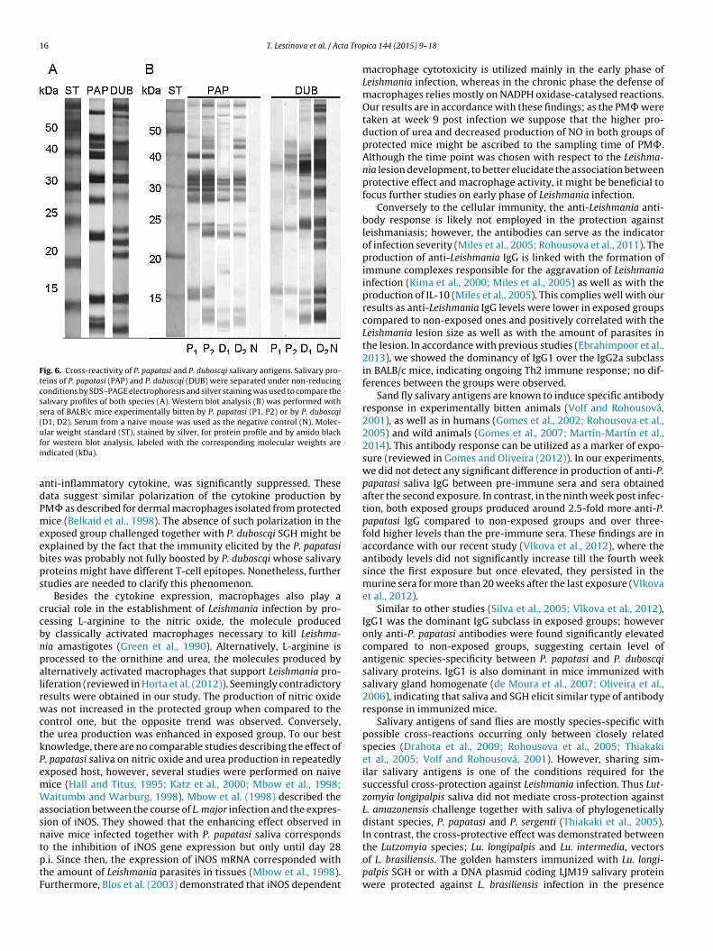
16 T. Lestinova et al. / Acta Trop
Fig. 6. Cross-reactivity of P. papatasi and P. duboscqi salivary antigens. Salivary pro-teins of P. papatasi (PAP) and P. duboscqi (DUB) were separated under non-reducingconditions by SDS–PAGE electrophoresis and silver staining was used to compare thesalivary profiles of both species (A). Western blot analysis (B) was performed withsera of BALB/c mice experimentally bitten by P. papatasi (P1, P2) or by P. duboscqi(D1, D2). Serum from a naive mouse was used as the negative control (N). Molec-ufi
adPmeebps
ccbnpalrwctkPemWasntptF
lar weight standard (ST), stained by silver, for protein profile and by amido blackor western blot analysis, labeled with the corresponding molecular weights arendicated (kDa).
nti-inflammatory cytokine, was significantly suppressed. Theseata suggest similar polarization of the cytokine production byM� as described for dermal macrophages isolated from protectedice (Belkaid et al., 1998). The absence of such polarization in the
xposed group challenged together with P. duboscqi SGH might bexplained by the fact that the immunity elicited by the P. papatasiites was probably not fully boosted by P. duboscqi whose salivaryroteins might have different T-cell epitopes. Nonetheless, furthertudies are needed to clarify this phenomenon.
Besides the cytokine expression, macrophages also play arucial role in the establishment of Leishmania infection by pro-essing L-arginine to the nitric oxide, the molecule producedy classically activated macrophages necessary to kill Leishma-ia amastigotes (Green et al., 1990). Alternatively, L-arginine isrocessed to the ornithine and urea, the molecules produced bylternatively activated macrophages that support Leishmania pro-iferation (reviewed in Horta et al. (2012)). Seemingly contradictoryesults were obtained in our study. The production of nitric oxideas not increased in the protected group when compared to the
ontrol one, but the opposite trend was observed. Conversely,he urea production was enhanced in exposed group. To our bestnowledge, there are no comparable studies describing the effect of. papatasi saliva on nitric oxide and urea production in repeatedlyxposed host, however, several studies were performed on naiveice (Hall and Titus, 1995; Katz et al., 2000; Mbow et al., 1998;aitumbi and Warburg, 1998). Mbow et al. (1998) described the
ssociation between the course of L. major infection and the expres-ion of iNOS. They showed that the enhancing effect observed inaive mice infected together with P. papatasi saliva corresponds
o the inhibition of iNOS gene expression but only until day 28.i. Since then, the expression of iNOS mRNA corresponded withhe amount of Leishmania parasites in tissues (Mbow et al., 1998).urthermore, Blos et al. (2003) demonstrated that iNOS dependentica 144 (2015) 9–18
macrophage cytotoxicity is utilized mainly in the early phase ofLeishmania infection, whereas in the chronic phase the defense ofmacrophages relies mostly on NADPH oxidase-catalysed reactions.Our results are in accordance with these findings; as the PM� weretaken at week 9 post infection we suppose that the higher pro-duction of urea and decreased production of NO in both groups ofprotected mice might be ascribed to the sampling time of PM�.Although the time point was chosen with respect to the Leishma-nia lesion development, to better elucidate the association betweenprotective effect and macrophage activity, it might be beneficial tofocus further studies on early phase of Leishmania infection.
Conversely to the cellular immunity, the anti-Leishmania anti-body response is likely not employed in the protection againstleishmaniasis; however, the antibodies can serve as the indicatorof infection severity (Miles et al., 2005; Rohousova et al., 2011). Theproduction of anti-Leishmania IgG is linked with the formation ofimmune complexes responsible for the aggravation of Leishmaniainfection (Kima et al., 2000; Miles et al., 2005) as well as with theproduction of IL-10 (Miles et al., 2005). This complies well with ourresults as anti-Leishmania IgG levels were lower in exposed groupscompared to non-exposed ones and positively correlated with theLeishmania lesion size as well as with the amount of parasites inthe lesion. In accordance with previous studies (Ebrahimpoor et al.,2013), we showed the dominancy of IgG1 over the IgG2a subclassin BALB/c mice, indicating ongoing Th2 immune response; no dif-ferences between the groups were observed.
Sand fly salivary antigens are known to induce specific antibodyresponse in experimentally bitten animals (Volf and Rohousová,2001), as well as in humans (Gomes et al., 2002; Rohousova et al.,2005) and wild animals (Gomes et al., 2007; Martín-Martín et al.,2014). This antibody response can be utilized as a marker of expo-sure (reviewed in Gomes and Oliveira (2012)). In our experiments,we did not detect any significant difference in production of anti-P.papatasi saliva IgG between pre-immune sera and sera obtainedafter the second exposure. In contrast, in the ninth week post infec-tion, both exposed groups produced around 2.5-fold more anti-P.papatasi IgG compared to non-exposed groups and over three-fold higher levels than the pre-immune sera. These findings are inaccordance with our recent study (Vlkova et al., 2012), where theantibody levels did not significantly increase till the fourth weeksince the first exposure but once elevated, they persisted in themurine sera for more than 20 weeks after the last exposure (Vlkovaet al., 2012).
Similar to other studies (Silva et al., 2005; Vlkova et al., 2012),IgG1 was the dominant IgG subclass in exposed groups; howeveronly anti-P. papatasi antibodies were found significantly elevatedcompared to non-exposed groups, suggesting certain level ofantigenic species-specificity between P. papatasi and P. duboscqisalivary proteins. IgG1 is also dominant in mice immunized withsalivary gland homogenate (de Moura et al., 2007; Oliveira et al.,2006), indicating that saliva and SGH elicit similar type of antibodyresponse in immunized mice.
Salivary antigens of sand flies are mostly species-specific withpossible cross-reactions occurring only between closely relatedspecies (Drahota et al., 2009; Rohousova et al., 2005; Thiakakiet al., 2005; Volf and Rohousová, 2001). However, sharing sim-ilar salivary antigens is one of the conditions required for thesuccessful cross-protection against Leishmania infection. Thus Lut-zomyia longipalpis saliva did not mediate cross-protection againstL. amazonensis challenge together with saliva of phylogeneticallydistant species, P. papatasi and P. sergenti (Thiakaki et al., 2005).In contrast, the cross-protective effect was demonstrated between
the Lutzomyia species; Lu. longipalpis and Lu. intermedia, vectorsof L. brasiliensis. The golden hamsters immunized with Lu. longi-palpis SGH or with a DNA plasmid coding LJM19 salivary proteinwere protected against L. brasiliensis infection in the presence
ta Trop
oi2tpideph
ioabimsiisw
A
KpEtpewTdr
R
B
B
C
d
d
D
E
G
T. Lestinova et al. / Ac
f Lu. intermedia saliva with reduced number of parasites in thenoculated ear and in the draining lymph node (Tavares et al.,011). In accordance with the aforementioned rules for effec-ive cross-protectivity, we observed cross-reactivity between P.apatasi and P. duboscqi salivary antigens using sera of hyper-
mmunized mice. This cross-reactivity was observed despite theifferences in the saliva composition between both species (Katot al., 2006; Volf et al., 2000), but was likely efficient enough torovide the protective effect to the mice infected together witheterologous antigen.
In conclusion, this is the first study showing the cross-protectionn P. papatasi-exposed mice challenged with L. major in the presencef P. duboscqi saliva. The cross-protective effect suggests that thenti-Leishmania vaccine based on P. papatasi salivary proteins coulde applicable also in sub-Saharan endemic areas where L. major
s transmitted by P. duboscqi. Moreover, similar cross-protectionight be possible also between other closely related sand fly
pecies such as Larroussius species responsible for transmission of L.nfantum in Mediterranean basin. We would like to further analyzemmune mechanisms of this cross-protective phenomenon using aand fly bite challenge model that mimic more closely the naturalay of Leishmania transmission.
cknowledgements
We would like to thank Helena Kulikova, Lenka Zitkova andristina Simkova for great technical and administrative sup-ort. This study was partially funded by EU grant FP7-261504DENext and is catalogued by the EDENext Steering Commit-ee as EDENext203 (http://www.edenext.eu). The contents of thisublication are the sole responsibility of the authors and do not nec-ssarily reflect the views of the European Commission. This studyas conducted under the frame of the EurNegVec COST Action
D1303 and COST-CZ LD14076. The funders had no role in studyesign, data collection and analysis, decision to publish, or prepa-ation of the manuscript.
eferences
elkaid, Y., Kamhawi, S., Modi, G., Valenzuela, J., Noben-Trauth, N., Rowton, E.,Ribeiro, J., Sacks, D.L., 1998. Development of a natural model of cutaneousleishmaniasis: powerful effects of vector saliva and saliva preexposure on thelong-term outcome of Leishmania major infection in the mouse ear dermis. J.Exp. Med. 188, 1941–1953.
los, M., Schleicher, U., Soares Rocha, F.J., Meissner, U., Röllinghoff, M., Bogdan, C.,2003. Organ-specific and stage-dependent control of Leishmania major infec-tion by inducible nitric oxide synthase and phagocyte NADPH oxidase. Eur. J.Immunol. 33, 1224–1234.
lements, M.F., Gidwani, K., Kumar, R., Hostomska, J., Dinesh, D.S., Kumar, V., Das, P.,Müller, I., Hamilton, G., Volfova, V., Boelaert, M., Das, M., Rijal, S., Picado, A., Volf,P., Sundar, S., Davies, C.R., Rogers, M.E., 2010. Measurement of recent exposureto Phlebotomus argentipes, the vector of Indian visceral leishmaniasis, by usinghuman antibody responses to sand fly saliva. Am. J. Trop. Med. Hyg. 82, 801–807.
a Silva, R.A., Tavares, N.M., Costa, D., Pitombo, M., Barbosa, L., Fukutani, K., Miranda,J.C., de Oliveira, C.I., Valenzuela, J.G., Barral, A., Soto, M., Barral-Netto, M.,Brodskyn, C., 2011. DNA vaccination with KMP11 and Lutzomyia longipalpis sali-vary protein protects hamsters against visceral leishmaniasis. Acta Trop. 120,185–190.
e Moura, T.R., Oliveira, F., Novais, F.O., Miranda, J.C., Clarêncio, J., Follador, I., Car-valho, E.M., Valenzuela, J.G., Barral-Netto, M., Barral, A., Brodskyn, C., de Oliveira,C.I., 2007. Enhanced Leishmania braziliensis infection following pre-exposure tosandfly saliva. PLoS Negl. Trop. Dis. 1, e84.
rahota, J., Lipoldová, M., Volf, P., Rohousová, I., 2009. Specificity of anti-salivaimmune response in mice repeatedly bitten by Phlebotomus sergenti. ParasiteImmunol. 31, 766–770.
brahimpoor, S., Pakzad, S.R., Ajdary, S., 2013. IgG1 and IgG2a profile of serum anti-bodies to Leishmania major amastigote in BALB/c and C57BL/6Mice. Iran J. AllergyAsthma Immunol. 12, 361–367.
idwani, K., Picado, A., Rijal, S., Singh, S.P., Roy, L., Volfova, V., Andersen, E.W., Uranw,S., Ostyn, B., Sudarshan, M., Chakravarty, J., Volf, P., Sundar, S., Boelaert, M.,Rogers, M.E., 2011. Serological markers of sand fly exposure to evaluate insectici-dal nets against visceral leishmaniasis in India and Nepal: a cluster-randomizedtrial. PLoS Negl. Trop. Dis. 5, e1296.
ica 144 (2015) 9–18 17
Gomes, R., Oliveira, F., 2012. The immune response to sand fly salivary proteins andits influence on leishmania immunity. Front. Immunol. 3, 110.
Gomes, R., Teixeira, C., Teixeira, M.J., Oliveira, F., Menezes, M.J., Silva, C., de Oliveira,C.I., Miranda, J.C., Elnaiem, D.E., Kamhawi, S., Valenzuela, J.G., Brodskyn, C.I.,2008. Immunity to a salivary protein of a sand fly vector protects against thefatal outcome of visceral leishmaniasis in a hamster model. Proc. Natl. Acad. Sci.U. S. A. 105, 7845–7850.
Gomes, R.B., Brodskyn, C., de Oliveira, C.I., Costa, J., Miranda, J.C., Caldas, A., Valen-zuela, J.G., Barral-Netto, M., Barral, A., 2002. Seroconversion against Lutzomyialongipalpis saliva concurrent with the development of anti-Leishmania chagasidelayed-type hypersensitivity. J. Infect. Dis. 186, 1530–1534.
Gomes, R.B., Mendonca, I.L., Silva, V.C., Ruas, J., Silva, M.B., Cruz, M.S., Barral, A., Costa,C.H., 2007. Antibodies against Lutzomyia longipalpis saliva in the fox Cerdocyonthous and the sylvatic cycle of Leishmania chagasi. Trans. R. Soc. Trop. Med. Hyg.101, 127–133.
Green, S.J., Meltzer, M.S., Hibbs, J.B., Nacy, C.A., 1990. Activated macrophages destroyintracellular Leishmania major amastigotes by an L-arginine-dependent killingmechanism. J. Immunol. 144, 278–283.
Hall, L.R., Titus, R.G., 1995. Sand fly vector saliva selectively modulates macrophagefunctions that inhibit killing of Leishmania major and nitric oxide production. J.Immunol. 155, 3501–3506.
Horta, M.F., Mendes, B.P., Roma, E.H., Noronha, F.S., Macêdo, J.P., Oliveira, L.S., Duarte,M.M., Vieira, L.Q., 2012. Reactive oxygen species and nitric oxide in cutaneousleishmaniasis. J. Parasitol. Res. 2012, 203818.
Hostomska, J., Rohousova, I., Volfova, V., Stanneck, D., Mencke, N., Volf, P., 2008.Kinetics of canine antibody response to saliva of the sand fly Lutzomyia longi-palpis. Vector Borne Zoonotic Dis. 8, 443–450.
Kamhawi, S., Belkaid, Y., Modi, G., Rowton, E., Sacks, D., 2000. Protection againstcutaneous leishmaniasis resulting from bites of uninfected sand flies. Science290, 1351–1354.
Kato, H., Anderson, J.M., Kamhawi, S., Oliveira, F., Lawyer, P.G., Pham, V.M., San-gare, C.S., Samake, S., Sissoko, I., Garfield, M., Sigutova, L., Volf, P., Doumbia,S., Valenzuela, J.G., 2006. High degree of conservancy among secreted salivarygland proteins from two geographically distant Phlebotomus duboscqi sandfliespopulations (Mali and Kenya). BMC Genomics 7, 226.
Katz, O., Waitumbi, J.N., Zer, R., Warburg, A., 2000. Adenosine, AMP, and proteinphosphatase activity in sandfly saliva. Am. J. Trop. Med. Hyg. 62, 145–150.
Killick-Kendrick, R., 1999. The biology and control of phlebotomine sand flies. Clin.Dermatol. 17, 279–289.
Kima, P.E., Constant, S.L., Hannum, L., Colmenares, M., Lee, K.S., Haberman, A.M.,Shlomchik, M.J., McMahon-Pratt, D., 2000. Internalization of Leishmania mexi-cana complex amastigotes via the Fc receptor is required to sustain infection inmurine cutaneous leishmaniasis. J. Exp. Med. 191, 1063–1067.
Kimblin, N., Peters, N., Debrabant, A., Secundino, N., Egen, J., Lawyer, P., Fay, M.P.,Kamhawi, S., Sacks, D., 2008. Quantification of the infectious dose of Leishmaniamajor transmitted to the skin by single sand flies. Proc. Natl. Acad. Sci. U.S.A. 105,10125–10130.
Kropf, P., Baud, D., Marshall, S.E., Munder, M., Mosley, A., Fuentes, J.M., Bangham,C.R., Taylor, G.P., Herath, S., Choi, B.S., Soler, G., Teoh, T., Modolell, M., Müller, I.,2007. Arginase activity mediates reversible T cell hyporesponsiveness in humanpregnancy. Eur. J. Immunol. 37, 935–945.
Kropf, P., Fuentes, J.M., Fahnrich, E., Arpa, L., Herath, S., Weber, V., Soler, G., Celada, A.,Modolell, M., Muller, I., 2005. Arginase and polyamine synthesis are key factorsin the regulation of experimental leishmaniasis in vivo. FASEB J. 19, 1000–1002.
Maia, C., Seblova, V., Sadlova, J., Votypka, J., Volf, P., 2011. Experimental transmis-sion of Leishmania infantum by two major vectors: a comparison between aviscerotropic and a dermotropic strain. PLoS Negl. Trop. Dis. 5, e1181.
Martín-Martín, I., Molina, R., Rohousová, I., Drahota, J., Volf, P., Jiménez, M., 2014.High levels of anti-Phlebotomus perniciosus saliva antibodies in different ver-tebrate hosts from the re-emerging leishmaniosis focus in Madrid, Spain. Vet.Parasitol. 202, 207–216.
Mary, C., Faraut, F., Lascombe, L., Dumon, H., 2004. Quantification of Leishmaniainfantum DNA by a real-time PCR assay with high sensitivity. J. Clin. Microbiol.42, 5249–5255.
Mbow, M.L., Bleyenberg, J.A., Hall, L.R., Titus, R.G., 1998. Phlebotomus papatasi sandfly salivary gland lysate down-regulates a Th1, but up-regulates a Th2, responsein mice infected with Leishmania major. J. Immunol. 161, 5571–5577.
Miles, S.A., Conrad, S.M., Aves, R.G., Jeronimo, S.M.B., Mosser, D.M., 2005. A rolefor IgG immune complexes during infection with the intracellular pathogenLeishmania. J. Exp. Med. 201, 747–754.
Morris, R.V., Shoemaker, C.B., David, J.R., Lanzaro, G.C., Titus, R.G., 2001. Sandflymaxadilan exacerbates infection with Leishmania major and vaccinating againstit protects against L. major infection. J. Immunol. 167, 5226–5230.
Murray, H.W., Cartelli, D.M., 1983. Killing of intracellular Leishmania donovaniby human mononuclear phagocytes–evidence for oxygen-dependent andoxygen-independent leishmanicidal activity. J. Clin. Invest. 72, 32–44.
Myskova, J., Votypka, J., Volf, P., 2008. Leishmania in sand flies: Comparison ofquantitative polymerase chain reaction with other techniques to determine theintensity of infection. J. Med. Entomol. 45, 133–138.
Oliveira, F., Kamhawi, S., Seitz, A.E., Pham, V.M., Guigal, P.M., Fischer, L., Ward, J.,Valenzuela, J.G., 2006. From transcriptome to immunome: identification of DTH
inducing proteins from a Phlebotomus ariasi salivary gland cDNA library. Vaccine24, 374–390.Oliveira, F., Lawyer, P.G., Kamhawi, S., Valenzuela, J.G., 2008. Immunity to Dis-tinct Sand Fly Salivary Proteins Primes the Anti-Leishmania Immune Responsetowards Protection or Exacerbation of Disease. PLoS Negl. Trop. Dis. 2, e226.

1 ta Trop
R
R
R
R
R
S
T
T
T
V
8 T. Lestinova et al. / Ac
eady, P.D., 2013. Biology of phlebotomine sand flies as vectors of disease agents.Annu. Rev. Entomol. 58, 227–250.
ibeiro, J.M., Mans, B.J., Arcà, B., 2010. An insight into the sialome of blood-feedingNematocera. Insect Biochem. Mol. Biol. 40, 767–784.
ohousova, I., Ozensoy, S., Ozbel, Y., Volf, P., 2005. Detection of species-specificantibody response of humans and mice bitten by sand flies. Parasitology 130,493–499.
ohousová, I., Hostomská, J., Vlková, M., Kobets, T., Lipoldová, M., Volf, P., 2011.The protective effect against Leishmania infection conferred by sand fly bites islimited to short-term exposure. Int. J. Parasitol. 41, 481–485.
ohousová, I., Volfová, V., Nová, S., Volf, P., 2012. Individual variability of salivarygland proteins in three Phlebotomus species. Acta Trop. 122, 80–86.
ilva, F., Gomes, R., Prates, D., Miranda, J.C., Andrade, B., Barral-Netto, M., Barral,A., 2005. Inflammatory cell infiltration and high antibody production in BALB/cmice caused by natural exposure to Lutzomyia longipalpis bites. Am. J. Trop. Med.Hyg. 72, 94–98.
avares, N.M., Silva, R.A., Costa, D.J., Pitombo, M.A., Fukutani, K.F., Miranda, J.C.,Valenzuela, J.G., Barral, A., de Oliveira, C.I., Barral-Netto, M., Brodskyn, C., 2011.Lutzomyia longipalpis saliva or salivary protein LJM19 protects against Leishma-nia braziliensis and the saliva of its vector, Lutzomyia intermedia. PLoS Negl. Trop.Dis. 5, e1169.
hiakaki, M., Rohousova, I., Volfova, V., Volf, P., Chang, K.P., Soteriadou, K., 2005.Sand fly specificity of saliva-mediated protective immunity in Leishmania
amazonensis-BALB/c mouse model. Microbes Infect. 7, 760–766.itus, R.G., Ribeiro, J.M.C., 1988. Salivary gland lysates from the sand lfy Lutzomyialongipalpis enhance Leishmania infectivity. Science 239, 1306–1308.
alenzuela, J.G., Belkaid, Y., Garfield, M.K., Mendez, S., Kamhawi, S., Rowton, E.D.,Sacks, D.L., Ribeiro, J.M.C., 2001. Toward a defined anti-Leishmania vaccine
ica 144 (2015) 9–18
targeting vector antigens: Characterization of a protective salivary protein. J.Exp. Med. 194, 331–342.
Vlkova, M., Rohousova, I., Drahota, J., Stanneck, D., Kruedewagen, E.M., Mencke, N.,Otranto, D., Volf, P., 2011. Canine antibody response to Phlebotomus perniciosusbites negatively correlates with the risk of Leishmania infantum transmission.PLoS Negl. Trop. Dis. 5, e1344.
Vlkova, M., Rohousova, I., Hostomska, J., Pohankova, L., Zidkova, L., Drahota,J., Valenzuela, J.G., Volf, P., 2012. Kinetics of Antibody Response in BALB/cand C57BL/6 Mice Bitten by Phlebotomus papatasi. PLoS Negl. Trop. Dis. 6,e1719.
Volf, P., Rohousová, I., 2001. Species-specific antigens in salivary glands of phle-botomine sandflies. Parasitology 122 Pt 1, 37–41.
Volf, P., Tesarova, P., Nohynkova, E., 2000. Salivary proteins and glycoproteins inphlebotomine sandflies of various species, sex and age. Med. Vet. Entomol. 14,251–256.
Volf, P., Volfova, V., 2011. Establishment and maintenance of sand fly colonies. J.Vector Ecol. 36, S1–S9.
Waitumbi, J., Warburg, A., 1998. Phlebotomus papatasi saliva inhibits protein phos-phatase activity and nitric oxide production by murine macrophages. Infect.Immun. 66, 1534–1537.
Warburg, A., Schlein, Y., 1986. The effect of post-bloodmeal nutrition of Phlebotomuspapatasi on the transmission of Leishmania major. Am. J. Trop. Med. Hyg. 35,926–930.
Xu, X., Oliveira, F., Chang, B.W., Collin, N., Gomes, R., Teixeira, C., Reynoso, D., MyPham, V., Elnaiem, D.E., Kamhawi, S., Ribeiro, J.M., Valenzuela, J.G., Andersen,J.F., 2011. Structure and function of a yellow protein from saliva of the sand flyLutzomyia longipalpis that confers protective immunity against Leishmania majorinfection. J. Biol. Chem. 286, 32383–32393.