phlebotomus (paraphlebotomus) chabaudi and phlebotomus...
TRANSCRIPT

Parasite 24, 47 (2017)© V. Lehrter et al., published by EDP Sciences, 2017https://doi.org/10.1051/parasite/2017050
Available online at:www.parasite-journal.org
RESEARCH ARTICLE
Phlebotomus (Paraphlebotomus) chabaudi and Phlebotomus riouxi:closely related species or synonyms?Véronique Lehrter1, Anne-Laure Bañuls2, Nicole Léger1, Jean-Antoine Rioux3,†, and Jérôme Depaquit1,*
1 EA 4688 - USC ANSES VECPAR, SFR Cap Santé, UFR de Pharmacie, Université de Reims Champagne-Ardenne,51 rue Cognacq-Jay, 51096 Reims, France
2 UMR MIVEGEC, IRD - CNRS - Université de Montpellier, 911 avenue Agropolis, 34394 Montpellier, France3 Faculté de Médecine, Université de Montpellier, 2 rue de l’École de Médecine, 34000 Montpellier, France
Received 20 May 2017, Accepted 9 November 2017, P
*Correspon
SIL
† in memo
This is anO
ublished online 1 December 2017
Abstract -- Phlebotomus riouxi Depaquit, Killick-Kendrick & Léger 1998 was described as a species closelyrelated to Phlebotomus chabaudi Croset, Abonnenc & Rioux 1970, differing mainly by the size and number ofsetae of the coxite basal lobe.Molecular studies carried out on several populations fromAlgeria and Tunisia andbased on mitochondrial genes cytochrome b (Cytb) and cytochrome oxidase I (COI) supported the typologicalvalidity of these two species. Recently, specimens from a single population in southern Tunisia weremorphologically identified as Ph. riouxi, Ph. chabaudi and intermediates, but were clustered in the same cladeaccording to their Cytb and nuclear gene elongation factor-1 alpha (EF-1a) sequences. These species were thussynonymized. To further explore this synonymy, we carried out a molecular study on specimens from Algeriaand Tunisia using the same molecular markers and a part of 28S rDNA. We did not find any morphologicallyintermediate specimens in our sampling.We highlighted differences between the genetic divergence rates withinand between the two species for the three markers and we identified new haplotypes. The sequence analysis didnot reveal any signature of introgression in allopatric nor in sympatric populations such as in the Ghomrassenpopulation. Phylogenetic analyses based on our specimens revealed that the two main clades are Ph. chabaudiand Ph. riouxi, in agreement with the morphological identification. These results support the validity ofPh. riouxi and Ph. chabaudi as typological species.
Keywords: Phlebotomus chabaudi, Phlebotomus riouxi, mitochondrial, nuclear and ribosomal markers,phylogenetic, North Africa
Résumé -- Phlebotomus (Paraphlebotomus) chabaudi et Phlebotomus riouxi : espèces proches ousynonymes ?Phlebotomus riouxi Depaquit, Killick-Kendrick & Léger 1998 a été décrit comme une espèceproche de Phlebotomus chabaudi Croset, Abonnenc & Rioux 1970, se distinguant principalement par lataille et le nombre de soies sur le lobe basal du coxite. Des études moléculaires, menées sur plusieurspopulations d’Algérie et de Tunisie, et basées sur les gènes mitochondriaux cytochrome b (Cytb) etcytochrome oxydase I (COI), ont soutenu la validité typologique de ces deux espèces. Récemment, desspécimens d’une seule population du sud de la Tunisie ont été morphologiquement identifiés comme desPh. riouxi, Ph. chabaudi et intermédiaires, mais se sont retrouvés groupés dans le même clade selon leursséquences de Cytb et de facteur d’élongation 1 alpha (EF-1a). Ces espèces ont donc été mises ensynonymie. Afin d’explorer davantage cette synonymie, nous avons mené une étude moléculaire sur desspécimens d’Algérie et de Tunisie en utilisant les mêmes marqueurs moléculaires ainsi qu’une partie du 28Sde l’ADN ribosomique. Aucun spécimen ne présentait de morphologie intermédiaire dans notreéchantillonnage. Des différences entre les taux de variabilité génétique intra et interspécifiques des troismarqueurs ont été mises en évidence, ainsi que de nouveaux haplotypes. L’analyse des séquences n’a révélé
ding author: [email protected]
pecial Issue � ISOPS IX � International Symposium on Phlebotomine Sandfliesnvited Editors: Jérôme Depaquit, Bernard Pesson, Denis Augot, James Gordon, Campbell Hamilton, Phillipawyer, and Nicole Léger
riam
penAccess article distributed under the terms of the CreativeCommonsAttribution License (http://creativecommons.org/licenses/by/4.0),which permits unrestricted use, distribution, and reproduction in any medium, provided the original work is properly cited.

2 V. Lehrter et al.: Parasite 2017, 24, 47
aucune signature d’introgression que ce soit dans les populations allopatriques ou sympatriques telle que lapopulation de Ghomrassen. Les analyses phylogénétiques basées sur nos échantillons ont révélé que lesdeux principaux clades correspondent à Ph. chabaudi et Ph. riouxi, résultat en accord avec l’identificationmorphologique. Ces résultats soutiennent la validité de Ph. riouxi et Ph. chabaudi comme espècestypologiques.
Introduction
Within the Phlebotomus genus (Diptera, Psychodi-dae), the subgenus Paraphlebotomus Theodor 1948includes some proven and suspected vectors of leishmani-ases, e.g. Phlebotomus sergenti, the main vector ofLeishmania tropica [2,20]. Our study focuses on twospecies of Paraphlebotomus from North Africa: Phleboto-mus chabaudi Croset, Abonnenc & Rioux 1970 andPhlebotomus riouxi Depaquit, Killick-Kendrick & Léger,1998 [9,10,35]. The presence of Ph. chabaudi has also beenreported in southern Spain [34].
Although their vectorial role has never been demon-strated, these two species are recorded in severalleishmaniasis foci [3,28,40] and are related to Ph. sergenti.In fact, Ph. chabaudi and Leishmania killicki have beendescribed for the first time in the same locality(Tataouine) in Tunisia [36], and L. killicki was also foundin Algeria [24,26], especially in Ghardaïa, where some Ph.riouxi were reported, even though Ph. sergenti was themain proven vector [4].
In previous studies, Ph. chabaudi and Ph. riouximalescollected in Algeria and Tunisia were clearly identifiedmorphologically. Molecular processing used two mito-chondrial genes: a partial sequence of cytochrome b (Cytb-CB3) [6], as proposed by Esseghir et al. [17], andcytochrome oxidase 1 (COI) [5]. In both studies,phylogenetic analyses emphasized the validity of thetwo species, supporting their typological status, meaningthat the deposited type-specimens are fully justified.
Recently, several specimens from Southern Tunisiashowed ambiguous morphological characters [40,41].According to these authors, several morphological criteriadescribed as specific characters were found together insome specimens that they described as intermediatespecimens. They used the same mitochondrial marker asthat of Bounamous et al. [6], called Cytb-CB3, in order tocompare their sequences with those available in GenBank.They also sequenced a longer fragment of Cytb (calledCytb-CB) and the nuclear elongation factor-1alpha gene(EF-1a) [30,41]. Their molecular results did not matchwith the morphological identification, not only for theintermediate specimens, but also for the differentiationbetween Ph. chabaudi and Ph. riouxi: all specimens wereclustered in the same clade. According to these results,based on specimens from the single locality of Ghomras-sen, they proposed to consider Ph. riouxi as a juniorsynonym of Ph. chabaudi.
In order to better understand the situation, we decidedto broaden the approach by performing a comparative and
combined sequence analysis of three loci on larger samplesfrom different geographical populations we previouslyinvestigated. We included the two markers used byTabbabi’s team [40,41], Cytb-CB and nuclear EF-1a,and the D1-D2 domain of ribosomal 28S DNA which isknown as a good marker for studying the interspecificgenetic divergence between species [19,22,38]. Thisdomain has specifically been used to perform analysis atthe taxonomic level in Phlebotominae [11,13,31].
Material and methodsSample collection
Samples analyzed in the present study were those usedby Tabbabi et al., Bounamous et al. and Boudabous et al.[5,6,40,41]. For the Tabbabi samples included in thisanalysis, we only had access to published data. All theother specimens came from our laboratory, includingsamples used by Bounamous et al. and Boudabous et al.[5,6], for which we kept the same sample codes marked inbold in Tables 1 and 3. Our specimens were collected byCDC miniature light traps and sticky paper traps fromtwo regions of Algeria (Ghardaïa and Aurès) and fromthree regions of Tunisia (Mahdia, Monastir and Ghom-rassen) (see Table 1 and Figure 1).
Specimens were stored in 95% ethanol at �20 °C, untildissection. After thawing, each specimen was dissectedindividually in 95% ethanol with sterile needles. The headand the genitalia were cleared in boiled Marc Andrésolution andmounted in chloral gumbetween the slide andcover slide for microscopic observation. The rest of thebody was dried and preserved at �20 °C in a sterilemicrotube until DNA extraction.
Taking into consideration the difficulty in identifyingfemales, we restricted the number of females in oursampling. The 9 females and 12males of Tabbabi’s study[41] are represented in Figure 1 as black symbols.Unfortunately, we did not have access to these specimens.However, all the specimens studied by Bounamous et al.andBoudabous et al. [5,6], as well as the new samples, weremorphologically examined or re-examined in the presentstudy, according to the criteria previously described[6,9,10].
The morphological analyses were focused on the basallobe of the coxite, known to be the differential characterbetween the two species under study [6,10]. For eachprocessed specimen, the number of coxite lobe setae wascounted and morphometrics analysis, using StreamMotion 1.9.1 software (Olympus, Japan), was also

Table 1. Analyzed samples. On a grey background, the samples processed by Tabbabi’s team. Samples in bold were used byBounamous et al. and Boudabous et al. [5,6]. Samples not sequenced with all markers are underlined and their accession numberreplaced by ND.
Country Area Species Number of specimen Sex Codes Cytb-CB Accession
Numbers EF-1α Accession Numbers D1D2 Accession Numbers
Tunisia Ph. chabaudi
1 f 1377 KC478308 KC478288/KC478289 /
3 m 573, 1331, 1335 KC478296, KC478305, KC478306
KC478288/KC478288, KC478288/KC478288, KC478288/KC478288
/
Unclear 3 m 571, 912, 1495 KC478293, KC478300, ND
KC478288/KC478288, KC478288/KC478291, KC478288/KC478288
/
Ph. riouxi 8 f 558, 1311, 547, 556, 658, 892, 980, 1339
KC478292, KC478292, KC478294, KC478295, KC478298, KC478299, KC478304, KC478307
KC478288/KC478288, KC478288/KC478288, KC478288/KC478288, KC478288/KC478288, KC478288/KC478288, KC478288/KC478289, KC478289/KC478290, KC478288/KC478288
/
6 m 1520, 624, 906, 916, 954, 966 KC478293, KC478297, KC478292, KC478301, KC478302, KC478303
KC478288/KC478288, KC478288/KC478288, KC478288/KC478288, KC478288/KC478288, KC478288/KC478288, KC478288/KC478288
/
Tunisia Ghomrassen Ph. riouxi 2 m TAT63, TAT74 KY764775, KY764776
KY764719, KY764720 KY764613, KY764614
3 f TAT186, TAT23, TAT24 KY764777-KY764779
KY764721-KY764723 KY764615-KY764617
Mahdia Ph. chabaudi
8 m SMA149, SMA160, SMA161, SMA162, SMA163, SMA60, SMA62,SMA69
KY764732-KY764739
KY764670-KY764675, ND, KY764676 KY764628-KY764633, ND, KY764634
Monastir Ph. chabaudi
35 m SMO1021, SMO1022, SMO1024, SMO194, SMO430, SMO443, SMO554, SMO559, SMO560, SMO562, SMO565, SMO569, SMO575, SMO578, SMO64, SMO873, SMO886, SMO887, SMO888, SMO891, SMO892, SMO893, SMO895, SMO897, SMO898, SMO899, SMO911, SMO918, SMO919, SMO921, SMO926, SMO927, SMO929, SMO931, SMO941
ND, ND, ND, KY764743-KY764750, ND, KY764751, KY764752, ND, KY764753, KY764754, ND, KY764755-KY764757, ND, KY764758-KY764770
KY764680-KY764714 ND, KY764638-KY764642, ND,
KY764643-KY764648, ND, KY764649, ND,
KY764650-KY764654, ND, ND, KY764655-
KY764662, ND, KY764663-KY764665
3 f SMO112, SMO310, SMO419 KY764740-KY764742
KY764677-KY764679 KY764635-KY764637
Algeria Ghardaïa Ph. riouxi 8 m RX1, RX2, RX3, RX5, RX6, RX7, RX8, RX9
KY764780-KY764787
KY764724-KY764731 KY764618-KY764625
Aurès Ph. chabaudi
3 m CB1, CB2, CB3 KY764771-KY764773
KY764715-KY764717 KY764666-KY764668
1 f CBZAT583 966467YK817467YK477467YK
Ghomrassen
V. Lehrter et al.: Parasite 2017, 24, 47 3
applied. In 1998, it was suggested that the number of setae,and the width and length of the coxite lobe wereinformative to differentiate the species [10]. The widthmeasure did not cause any difficulty: a transversal line wasperpendicularly traced in the larger part of the lobe.Considering the difficulties in measuring the length of thebasal lobe of the coxite, we substituted it by its perimeterand area, as indicated in Figure 2.
DNA extraction, PCR and sequence analysis
Some DNA extracts (sample codes in bold in Tables 1and 3) from previous studies [5,6] were simply thawed fordirect PCR amplification. For the other specimens, weused the same procedure as for the older extracts: DNAextractions were individually carried out using a QIAampDNA Mini Kit (Qiagen), according to the manufacturer’sprotocol. Samples were crushed in ATL buffer with apiston pellet and DNA extracts were eluted in 180mL to200mL of AE buffer and stored at �20 °C. Cytb-CB was
amplified using CB1-SE: 50-TATGTACTACCCTGAG-GACAAATATC-30 and CB-R06: 50-TATCTAATGGT-TTCAAAACAATTGC-30 primers, as previously described[41]. For EF-1a amplification, primers EF-F05: 50-CCT-GGACATCGTGATTTCAT-30 and EF-R08: 50-CCAC-CAATCTTGTAGACATCCTG-30 were used [41]. Theribosomal domain D1-D2 was amplified using C10: 50-ACCCGCTGAATTTAAGCAT-30and D2: 50-TCCGT-GTTTCAAGACGGG-30 primers [32]. The PCR condi-tions for Cytb-CB and EF-1a were exactly the same asthose used byTabbabi et al. [41]. All PCRswere performedin a 50mL volume using 5mL of extracted DNA solution ofeach specimen individually, and 12.5 pmol of the primersets in a thermocycler. The PCR mix contained (finalconcentrations) 10mM Tris HCl (pH 8.3), 1.5mMMgCl2,50mMKCl, 0.01%TritonX-100, 200mMdNTPeachbase,and 1.25 units of Taq polymerase (Eppendorf, Germany).For each PCR run, a negative control using 5mL ofultrapure sterile water and a positive control using a DNAextract with a known sequence, were included. PCR
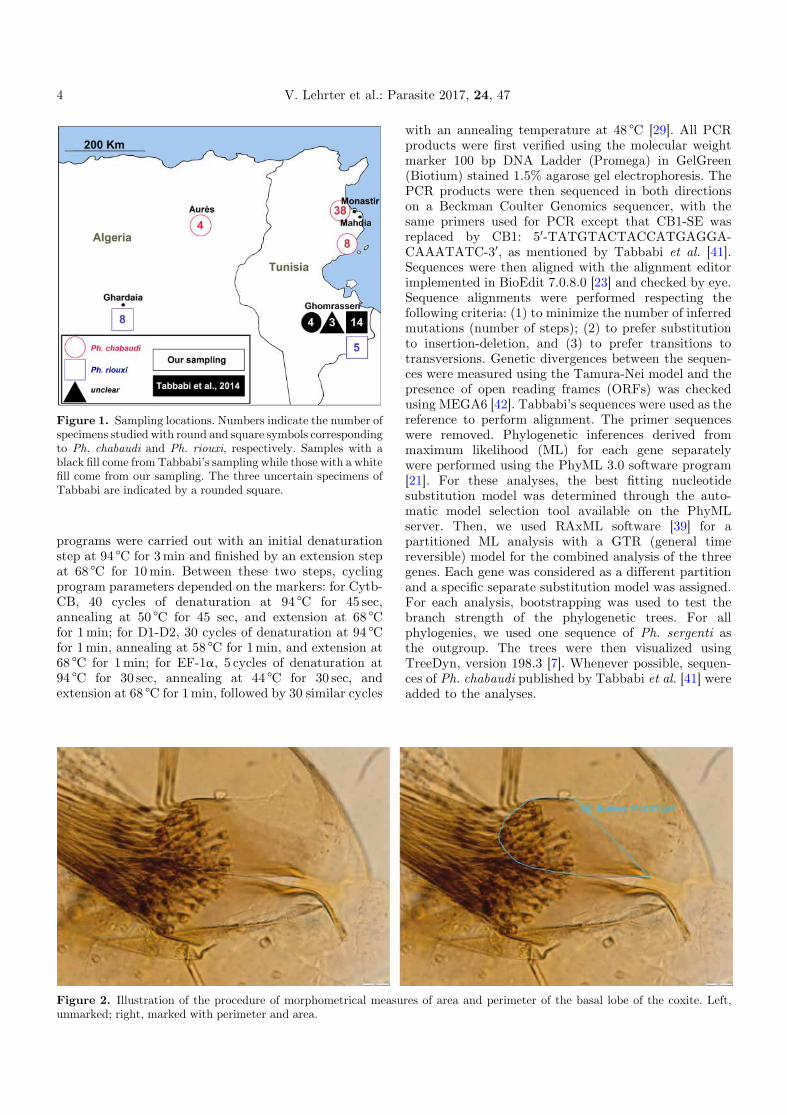
Figure 1. Sampling locations. Numbers indicate the number ofspecimens studied with round and square symbols correspondingto Ph. chabaudi and Ph. riouxi, respectively. Samples with ablack fill come from Tabbabi’s sampling while those with a whitefill come from our sampling. The three uncertain specimens ofTabbabi are indicated by a rounded square.
4 V. Lehrter et al.: Parasite 2017, 24, 47
programs were carried out with an initial denaturationstep at 94 °C for 3min and finished by an extension stepat 68 °C for 10min. Between these two steps, cyclingprogram parameters depended on the markers: for Cytb-CB, 40 cycles of denaturation at 94 °C for 45 sec,annealing at 50 °C for 45 sec, and extension at 68 °Cfor 1min; for D1-D2, 30 cycles of denaturation at 94 °Cfor 1min, annealing at 58 °C for 1min, and extension at68 °C for 1min; for EF-1a, 5 cycles of denaturation at94 °C for 30 sec, annealing at 44 °C for 30 sec, andextension at 68 °C for 1min, followed by 30 similar cycles
Figure 2. Illustration of the procedure of morphometrical measuunmarked; right, marked with perimeter and area.
with an annealing temperature at 48 °C [29]. All PCRproducts were first verified using the molecular weightmarker 100 bp DNA Ladder (Promega) in GelGreen(Biotium) stained 1.5% agarose gel electrophoresis. ThePCR products were then sequenced in both directionson a Beckman Coulter Genomics sequencer, with thesame primers used for PCR except that CB1-SE wasreplaced by CB1: 50-TATGTACTACCATGAGGA-CAAATATC-30, as mentioned by Tabbabi et al. [41].Sequences were then aligned with the alignment editorimplemented in BioEdit 7.0.8.0 [23] and checked by eye.Sequence alignments were performed respecting thefollowing criteria: (1) to minimize the number of inferredmutations (number of steps); (2) to prefer substitutionto insertion-deletion, and (3) to prefer transitions totransversions. Genetic divergences between the sequen-ces were measured using the Tamura-Nei model and thepresence of open reading frames (ORFs) was checkedusing MEGA6 [42]. Tabbabi’s sequences were used as thereference to perform alignment. The primer sequenceswere removed. Phylogenetic inferences derived frommaximum likelihood (ML) for each gene separatelywere performed using the PhyML 3.0 software program[21]. For these analyses, the best fitting nucleotidesubstitution model was determined through the auto-matic model selection tool available on the PhyMLserver. Then, we used RAxML software [39] for apartitioned ML analysis with a GTR (general timereversible) model for the combined analysis of the threegenes. Each gene was considered as a different partitionand a specific separate substitution model was assigned.For each analysis, bootstrapping was used to test thebranch strength of the phylogenetic trees. For allphylogenies, we used one sequence of Ph. sergenti asthe outgroup. The trees were then visualized usingTreeDyn, version 198.3 [7]. Whenever possible, sequen-ces of Ph. chabaudi published by Tabbabi et al. [41] wereadded to the analyses.
res of area and perimeter of the basal lobe of the coxite. Left,

Table 2. Morphological data of male samples: for each species, mean and standard deviation (sd) are calculated and minimal (min)and maximal (max) values are indicated.
Species Coxite lobearea (mm2)
Coxite lobe perimeter (mm) Coxite lobewidth (mm)
Number of setae per coxite lobe
Ph. riouxi mean 1154 160 27 35sd 118 7 3 5min 926 148 23 28max 1331 167 32 43
Ph. chabaudi mean 415 95 14 11sd 94 11 2 2min 209 65 10 7max 609 116 18 17
V. Lehrter et al.: Parasite 2017, 24, 47 5
ResultsMorphological identification
All of our specimens were correctly identified as Ph.chabaudi andPh. riouxi, according tomorphological criteriaof initial descriptions for males [9,10] and to the pharyngealarmature for females [6]. All females were morphologicallyidentified based on the presence or absence of anterolateralteeth in the pharynx. All measures related to the coxite lobeare listed in Table 2. Considering these data, we foundsignificant interspecific differences between all measure-ments of the basal lobe of the coxite, without significant geo-graphical variability (data not shown). We did not find anymorphological intermediate specimen. All Ph. riouxi werefound in the south of Tunisia and in the south of Algeria.
Sequence analyses
A total of 63 specimens were analyzed and compared tothe sequences of the 21 specimens from Tabbabi et al. [41](Table 1).
Cytochrome b analyses
For the 56 specimens successfully amplified, the lengthof the cytochrome b fragment Cytb-CB used for analysiswas 628 bp. The phylogenetic tree obtained by adding 17haplotypes of Tabbabi’s data [41] highlighted two distinctclades (Figure 3). The first one was only composed of Ph.chabaudi as supported by abootstrap value of 88.6%,with ageographical subdivision between Algeria (bootstrap value< 50%) andTunisia (bootstrap value=97.4%).The secondclade was composed of a mix of our own Ph. riouxi and the17 haplotypes of Tabbabi from the Ghomrassen area,identified as Ph. chabaudi, Ph. riouxi and intermediates.For this clade, the bootstrap value was lower (59.6%).Within this clade, the specimens were also subdivided intotwo clusters according to the geographic area, i.e. southernTunisia versus southern Algeria but with low bootstrapvalues < 50% and=62.2 %, respectively.
We obtained congruent results between morphologyand molecular observations in our samples, with aninterspecific genetic divergence of 12.2% (SD=1.38%) forCytb-CB between Ph. chabaudi and Ph. riouxi. Eleven
haplotypes in our 43 Ph. chabaudi samples had anintraspecific divergence of 0.60% (SD=0.13%), andinterestingly, a higher intraspecific divergence in the13Ph. riouxi with 12 haplotypes: 4.38% (SD=0.59%).
EF-1a analyses
EF-1a amplification gave a fragment of 454 bp inlength. All variable sites of 83 sequences alignment areshown in Table 3. One genotype R01 (01/01) was found inall 13 specimens we identified as Ph. riouxi. Theycorresponded to genotype 01/01 of Tabbabi’s samples(=genotype R01 according to our label). Interestingly,Ph.chabaudi showed very different genotypes (C01 to C16)compared to R01, with five different bases between thetwo clades, as seen in the phylogenetic tree based ongenotypes (Figure 4). These data led to a high bootstrapvalue for the Ph. riouxi clade (98.6%) and a lower one(51.4%) for Ph. chabaudi.
Analyses considering genotypes were performed as weobtained several heterozygous positions (double peaks),represented by ambiguous bases in the sequences, for threespecimens (Table 3). Indeed, several ambiguous positionswere detected in some specimens in the same sequence,precluding the haplotype determination. This is the case forgenotypes C12 and C14, corresponding to three Ph.chabaudi. All double peaks were located in the third positionof the codon without changing the amino acid translation.Overall, on the 46 samples for which the haplotypes could bedetermined, the frequency of the major haplotype was 30%(haplotype 05), followed by haplotype 10 and haplotype 06with 17% and 11%, respectively. Concerning our sampling,interspecific divergence ofEF-1a betweenPh. riouxi andPh.chabaudi was 1.15% (SD=0.53%), and intraspecific diver-gences were 0 and 0.009%, respectively, corresponding tonormal values for this marker [43].
Conversely to Tabbabi’s studies [40,41], the sequencesobtained in our sampling were congruent with themorphological identification.
D1-D2 analyses and concatenate analyses
D1-D2 amplification of our 55 samples gave fragmentsfrom 712 to 714 bp in length. For this marker, the

Figure 3. Phylogenetic tree inferred from cytochrome B data of Phlebotomus chabaudi and Ph. riouxi specimens. We added to theanalysis the sequences ofPh. chabaudi published by Tabbabi et al. (2014). The phylogram results from bootstrapped data sets obtainedusing the PhyML 3.0 program [21] using GTR (general time reversible)+G distribution (gamma distribution of rates with four ratecategories).Thetreewas visualizedusing theTreeDynprogram,version198.3 [7].Thepercentages above thebranchesare the frequencieswith which a given branch appeared in 500 bootstrap replications. Only bootstrap values higher than 50% on the early branches areshown. A sequence of Ph. sergenti (AF161216) was used as the outgroup. The sequences marked by * were published by Tabbabi et al.(2014);R= sequences found in specimensmorphologically characterizedasPh. riouxi.C= sequences found in specimensmorphologicallycharacterized as Ph. chabaudi. RC=sequences found in specimens morphologically characterized as Ph. chabaudi or Ph. riouxi.Int= sequences found in specimens morphologically characterized as intermediate between Ph. riouxi and Ph. chabaudi.
6 V. Lehrter et al.: Parasite 2017, 24, 47

Table 3. Base variability in EF-1a genotypes.On a grey background: four alleles found byTabbabi et al. (2014) (01 to 04=KC478288to KC478291). On a white background: 16 new genotypes found in our sample (n=62), composed of 12 new haplotypes (05 to 16);ND=Non Determinate. Samples in bold were used by Bounamous et al. and Boudabous et al. [5,6].
Species Number
of specimen
Genotype Codes
Positions of polymorphic sites 1 1 1 1 2 2 2 3 3 4 4 4 46 8 1 2 3 5 2 2 3 1 8 0 2 2 34 8 8 7 9 4 0 3 2 6 6 0 1 7 9
Ph. riouxi /
Ph. chabaudi
17 01/01 558, 1311, 571, 1520, 547, 556,
573, 624, 658, 906, 916, 954, 966, 1331, 1335, 1339, 1495
G G A T A G G G G A C C T C A
2 01/02 892, 1377 . S . . . . . . . . . . . . .1 02/03 980 . S . . . . . . . W . . . . .1 01/04 912 . . . . . . . . . . . . . Y .
Ph. riouxi 13 R01 (01/01)
RX1, RX2, RX3, RX5, RX6, RX7, RX8, RX9, TAT186,
TAT23, TAT24, TAT63, TAT74. . . . . . . . . . . . . . .
Ph. chabaudi 8 C01 (05/05)
SMA60, SMO1022, SMO194, SMO430, SMO554, SMO873,
SMO887, SMO911 . . G C C . . . . . T G . . .
2 C02 (06/06) CB1, SMA149 . . G C C . . . . . T G . . T1 C03 (07/07) SMO927 . . G C C . . . . . T G C . T1 C04 (05/08) CB2 . . G C C . R . . . T G . . .2 C05 (05/09) SMO919, SMO64 . . G C C . . R . . T G . . .
4 C06 (10/10) CBZAT583, SMA69, SMO310, SMO565 . . G C C T . . . . T G . . .
3 C07 (11/11) SMO892, SMO897, SMO929 . . G C C T . . . . T G . . T
6 C08 (06/11) CB3, SMA161, SMO1024, SMO575, SMO893, SMO931 . . G C C K . . . . T G . . T
2 C09 (12/12) SMO1021, SMO443 A . G C C T . . . . T G . . T1 C10 (13/13) SMA163 A . G C C T . . . . T G . . .
6 C11 (13/14) SMA160, SMO112, SMO569, SMO578, SMO895, SMO898 A . G C C K . . . . T G . . .
1 C12 (ND) SMO891 A . G C C K . . K . T G . . .1 C13 (15/15) SMO562 A . G C C . . . . . T G . . T2 C14 (ND) SMO560, SMO899 . . G C C K . . K . T G . . .1 C15 (05/16) SMO921 . . G C C . . . K . T G . . .
8 C16 (05/10) SMA162, SMO419, SMO559, SMO886, SMO888, SMO918,
SMO926, SMO941 . . G C C K . . . . T G . . .
V. Lehrter et al.: Parasite 2017, 24, 47 7
interspecific genetic divergence between Ph. chabaudi andPh. riouxi was 0.50% (SD=0.26%) and the intraspecificdivergence was 0 and 0.1%, respectively. This value(0.50%) was close to the genetic divergence observedbetween the two well-separated species Ph. chabaudi andPh. sergenti (0.74%, SD=0.31%). The lack of or the verylow intraspecific divergence can be explained by the lowmutation rate of this conserved marker [19,22,38].Phylogenetic analysis based on the sequences of the D1-D2 domain of 28S rDNAallowed us to differentiate the twospecies by their clustering into twomain clades (Figure 5).In spite of a low genetic divergence, the bootstrap valuewas strong for the Ph. riouxi clade (89.6%) and 68.6% forPh. chabaudi.
Concatenated analyses of the three loci using apartitioned ML model also showed clear clustering intwo clades corresponding to the morphological identifica-tions of Ph. chabaudi and Ph. riouxi (Figure 6). Thebootstrap values were 70.6% for the Ph. chabaudi clade
and 92.8% for the Ph. riouxi clade. As indicated by thecomparison of the trees, we did not find any signs ofintrogression.
Discussion
Phlebotomus chabaudi was described for the first timein Tunisia [9] as Paraphlebotomus with a sharply pointedaedeagus (Figure 7, A and D). The same year, this specieswas also recorded in Algeria [33] and was described with alarger and more tufted basal lobe (Figure 7, E and F). Theauthors linked this observation to variability due to thegeographically segregated populations. These two morphshave been found in sympatry without intermediatespecimens [10], justifying the description of a new species:Ph. riouxi (Figure 7, B, C, E, F).
Themorphological identification of the female remainsvery difficult. It is clearly not possible to differentiate thespermathecae of the two species (Figure 8, C and D).

Figure 4. Phylogenetic tree inferred from Phlebotomus chabaudi and Ph. riouxi specimens using the data of elongation factor 1-a gene.Sequences ofPh. chabaudipublishedbyTabbabi et al. (2014)wereadded to theanalyses.Thephylogramresults frombootstrappeddata setsobtainedusing thePhyML3.0program [21] using theHKY85 [25]+ I (proportion of invariant sites)model.The treewas visualizedusing theTreeDyn program, version 198.3 [7]. Percentages shown above the branches are the frequencies at which a given branch appeared in 500bootstrap replications. Only bootstrap values higher than 50% on the early branches are shown.A sequence ofPh. sergenti (EF416841)wasused as the outgroup. The sequences marked by * were published by Tabbabi et al. (2014); R=sequences found in specimensmorphologically characterized as Ph. riouxi. RC=sequences found in specimens morphologically characterized as Ph. chabaudi orPh. riouxi. Int= sequences found in specimens morphologically characterized as intermediate between Ph. riouxi and Ph. chabaudi.RCint=sequences found in specimens morphologically characterized as Ph. riouxi, Ph. chabaudi and intermediate specimens between thetwo species.
8 V. Lehrter et al.: Parasite 2017, 24, 47

Figure 5. Phylogenetic tree inferred from Phlebotomus chabaudi and Ph. riouxi specimens using the data of D1-D2 domain of 28SrDNA. Sequences of Ph. chabaudi published by Tabbabi et al. (2014) were added to the analyses. The phylogram results frombootstrapped data sets obtained using the PhyML 3.0 program [21] using the HKY85 model [25]. The tree was visualized using theTreeDyn program, version 198.3 [7]. The percentages above the branches are the frequencies withwhich a given branch appeared in 500bootstrap replications.Only bootstrap values higher than 50%on the early branches are shown.A sequence ofPh. sergenti (KY764627)was used as the outgroup.
V. Lehrter et al.: Parasite 2017, 24, 47 9

Figure 6. Phylogenetic tree inferred by concatenation of thethree loci under study. The phylogram was obtained by apartitioned ML analysis with a GTR (general time reversible)+G (gamma distribution of rates with four rate categories) + I(proportion of invariant sites) model using RAxML software [39].The tree was visualized using the TreeDyn program, version 198.3[7]. The percentages above the branches are the frequencies withwhich a given branch appeared in 500 bootstrap replications. Onlybootstrap values higher than 50% on branches are shown.Concatenated sequences of Ph. sergentiwere used as the outgroup.
10 V. Lehrter et al.: Parasite 2017, 24, 47
Although Depaquit et al. [10] suggested examining theappearance of the armature in the genital atrium, thiscriterion remains uncertain. Regarding the pharynx,Bounamous et al. [6] noted the presence of anterolateral
teeth in Ph. chabaudi, a character not found in Ph. riouxi(Figure 8, A and B). They suggested the use of thischaracter to identify these two species, pending a largersampling. Nevertheless, it seems that the individualvariability of the pharyngeal armature of Ph. chabaudimakes this distinction hazardous for a non-trainedentomologist.
Consequently, we selected a majority of males in thepresent study in order to reduce the risk of misidentifica-tion. Only a few females for which the morphologicalidentifications were congruent with molecular analyseswere included (those previously processed by Bounamouset al. and Boudabous et al. [5,6]) (Table 1). In the study byTabbabi et al. [41], out of 21 specimens, two-thirds(n=14) were morphologically identified as Ph. riouxi outof which 6 were males and 8 were females. Interestingly,despite the difficulty in identifying the females, allambiguous specimens were males. Unfortunately, wecould not include the specimens processed by Tabbabiin the present study and we did not find any intermediatespecimens in our collection. Consequently, we performed adetailed phylogenetic analysis of Ph. chabaudi and Ph.riouxi specimens selected from our collection, and weadded the sequences published by Tabbabi et al. [41]. Ourmolecular study included three independent markers. Thelong fragment of Cytb-CB codes for a partial proteinsequence of this gene, whereas the shorter fragment (Cytb-CB3) is more frequently used for taxonomic studies [17].Tabbabi et al. [40,41] analyzed theCytb-CB fragment thatthey considered more informative than the Cytb-CB3fragment. The interspecific (between closely related and/or vicariant species) and intraspecific divergence valuesobserved for the mitochondrial cytochrome b (Cytb-CBand Cytb-CB3) in Phlebotomus spp. are 2.7-11% and 0.1–2.5%, respectively [12,17,27]. The 12.2% interspecificvalue between Ph. chabaudi and Ph. riouxi supports thegenetic differentiation of these two taxa. Regarding theintraspecific values, the value for Ph. chabaudi (0.6%) wasin the commonly accepted range. For Ph. riouxi, the value(4.38%) is higher than the accepted range but close to thevalue observed in Sergentomyia clydei Sinton 1928,displaying a value of 5.5% due to a divergent populationfrom the Seychelles [32].
The nuclear EF-1a is known to be a good phylogeneticmarker in Metazoa [37] and was previously used in severalmolecular studies in Phlebotomine sandflies[1,16,18,29,43]. Its utility in other groups has also beendemonstrated in heliothine moths [8] and in Triatominae[14]. We selected this marker to compare our data withthose of Tabbabi et al. [41]. Several studies successfullycompared haplotypes of EF-1a [41,44], although EF-1aalso showed considerable diversity of haplotypes for asame specimen, thus complicating the analyses [29,43]. Inthe present study, we also noted significant haplotypediversity. Ribosomal marker D1-D2 does not have thisdisadvantage. Indeed, this marker is present in manyhomogeneous copies in the genome, thus providing a goodsignal that is easy to use as a genetic marker [38]. Howeverthe nuclear ribosomal DNAmay provide only a short-term

Figure 7. Differentiation criteria ofmales (A to F) 100X.A andD: aedeagus and basal lobe of coxite ofPh. chabaudi (SMO562); B andE: aedeagus and basal lobe of coxite of Ph. riouxi from Algeria (RX2); C and F: aedeagus and basal lobe of coxite of Ph. riouxi fromTunisia (TAT63). All photographs are set on the same scale.
V. Lehrter et al.: Parasite 2017, 24, 47 11
marker for introgression, because of the homogenizationof the multi-copy genes at this locus [15]. This marker isindependent of the two previous ones and is moreconserved [13]. We used this marker in previous studiesand showed its usefulness for phylogenetic analysis[11,31,32].
Several ambiguous bases were observed in EF1-asequences. Nevertheless, these ambiguous positions donot correspond to intermediate profiles between the twospecies. All our Ph. riouxi specimens from Algeria andTunisia revealed the genotype R01 (homozygotes)corresponding to the EF_chab01 haplotype defined by

Figure 8. Differentiation criteria of females (A toD), 100X.A andC: pharynx and spermathecae ofPh. chabaudi (CBZAT583); B andD: pharynx and spermathecae of Ph. riouxi (TAT186 and TAT24). All photographs are set on the same scale.
12 V. Lehrter et al.: Parasite 2017, 24, 47
Tabbabi et al. [41] (Table 3), in agreement with theresults of Boubidi et al. [4]. In contrast, we obtainedmany new sequences of EF-1a in Ph. chabaudi calledC01 to C16 (Table 3), providing five synapomorphicnucleotide substitutions that distinguished the twospecies.
Cytb and EF-1a have already been combined todemonstrate mitochondrial introgression in New WorldPhlebotomine sandflies [43]. Our study did not find anyintrogression between the two species under examination,as confirmed by the ribosomal D1-D2 analyses.
The independent phylogenetic analyses of the threegenes (Figures 3, 4 and 5) underlined the subdivision ofPh.chabaudi and Ph. riouxi specimens into two independentclades. Nevertheless, our data support low geneticdivergence between the two species, suggesting recentdifferentiation between these two taxa. This low diver-gence is confirmed by the low bootstrap values observed inCytb phylogeny for the Ph. riouxi branch (Figure 3), inEF-1a phylogeny for the Ph. chabaudi branch (Figure 4),and in D1D2 phylogeny for the Ph. chabaudi branch(Figure 5). The phylogeny of the concatenated genes
revealed bootstrap values above 70% for the two branches,suggesting that added data can only increase thedifferentiation between the two species.
When we consider the two phylogenies including thesequences published by Tabbabi et al. [41], i.e. Cytb andEF-1a phylogenies (Figures 3 and 4), all Tabbabi’ssequences are included in the Ph. riouxi branch, withoutclear distinction between our specimens and Tabbabi’sspecimens. From these data, it is difficult to explain thedisagreement between morphological characters andmolecular data observed by Tabbabi et al. It would thusappear essential to further investigate these samples ongenetic and morphological grounds to make a comparisonwith Ph. chabaudi and Ph. riouxi specimens from Algeriaand North Tunisia.
It is worth noting that the specimens in the Ph. riouxibranch were only collected in Southern Algeria andSouthern Tunisia, and that the specimens in the Ph.chabaudi branch were only collected in Northern Algeriaand Northern Tunisia. This suggests a related evolution ofthese two taxa between the South of these two countriesfor Ph. riouxi and between the North of these two

V. Lehrter et al.: Parasite 2017, 24, 47 13
countries for Ph. chabaudi. The molecular clock of Cytbhas been calculated for Ph. papatasi (Scopoli, 1786) andPh. duboscqi Neveu-Lemaire 1906, two vicariant speciesseparated by the Sahara. Its estimated calibration rangedfrom 1 to 2.5% variability per million years [17] or from1.34 to 2.64% per million years [18]. Ph. chabaudi andPh. riouxi exhibit a Cytb mean interspecific geneticdivergence of 12.2%. If we apply this calibration to thelatter species, we hypothesized their speciation startedbetween 12.2 and 4.62 Mya. This period corresponds tothe aridification of the Sahara desert (10 to 6 Mya). Thevicariance ofPh. chabaudi andPh. riouxi could result fromthe same event as the vicariance of Ph. papatasi andPh. duboscqi. The presence of intermediate specimens asdescribed by Tabbabi et al. [41], as well as specimens withmorphological criteria corresponding to Ph. chabaudi inthe South of Tunisia suggests the sympatry of the twospecies in Ghomrassen, which could be explained by amixing of the two species. Only the investigation ofsympatric populationswill answer the unresolved questionof whether or not the two lineages usually behave as truebiological species when they meet. Further morphologicalandmolecular studies on a larger sample ofPh. riouxi (e.g.from Ghomrassen) and on more genes remain necessary tohelp in determining the evolutionary history of these twospecies.
Finally, these results still support the existence of twospecies, and their typological validity, thus refuting Ph.riouxi as a junior synonym. The close genetic relationshipsand the intermediate specimens detected by Tabbabi et al.[41], however, suggest a recent speciation phenomenonfollowed by several migration events. Further genetic andmorphological studies of specimens from Algeria, Tunisiaand Morocco will help to better understand the evolutionof these two species in North Africa.
Acknowledgments. We wish to thank our colleagues fromAlgeria and Tunisia who caught the specimens processed and Pr.Matthieu Kaltenbach for proofreading this manuscript.
References
1. Absavaran A, Rassi Y, Parvizi P, Oshaghi MA, Abaie MR,Rafizadeh S, Mohebali M, Zarea Z, Javadian E. 2009.Identification of sand flies of the subgenusLarroussius basedon molecular and morphological characters in NorthWestern Iran. Journal of Arthropod-Borne Diseases, 3(2),22.
2. Al-Zahrani MA, Peters W, Evans DA, Chin C, Smith V,Lane RP. 1988. Phlebotomus sergenti, a vector of Leish-mania tropica in Saudi Arabia. Transactions of the RoyalSociety of Tropical Medicine and Hygiene, 82(3), 416.
3. Aoun K, Bouratbine A. 2014. Cutaneous Leishmaniasis inNorth Africa: a review. Parasite, 21, 14.
4. Boubidi SC, Benallal K, Boudrissa A, Bouiba L, BoucharebB, Garni R, Bouratbine A, Ravel C, Dvorak V, Votypka J,Volf P, Harrat Z. 2011. Phlebotomus sergenti (Parrot, 1917)identified as Leishmania killicki host in Ghardaïa, southAlgeria. Microbes and Infection, 13(7), 691–696.
5. Boudabous R, Bounamous A, Jouet D, Depaquit J, AugotD, Ferté H, Berchi S, Couloux A, Veuille M, Babba H. 2009.Mitochondrial DNA differentiation between two closelyrelated species, Phlebotomus (Paraphlebotomus) chabaudiand Phlebotomus (Paraphlebotomus) riouxi (Diptera:Psychodidae), based on direct sequencing and PolymeraseChain Reaction-Restriction Fragment Length Polymor-phism. Annals of Entomological Society of America, 102(3),347–353.
6. Bounamous A, Boudabous R, Jouet D, Augot D, Ferté H,Babba H, Berchi S, Depaquit J. 2008. Caractérisationmoléculaire et morphologique de deux espèces affines deParaphlebotomus: Phlebotomus chabaudi Croset, Abonnenc& Rioux, 1970 et Ph. riouxi. Depaquit, Killick-Kendrick &Léger, 1998 (Diptera: Psychodidae). Parasite, 15(4), 565–571.
7. Chevenet F, BrunC, Bañuls A-L., Jacq B, Christen R. 2006.TreeDyn: towards dynamic graphics and annotations foranalyses of trees. BMC bioinformatics, 7(1), 439.
8. Cho S, Mitchell A, Regier JC, Mitter C, Poole RW,Friedlander TP, Zhao S. 1995. A highly conserved nucleargene for low-level phylogenetics: elongation factor-1 alpharecovers morphology-based tree for heliothine moths.Molecular Biology and Evolution, 12(4), 650–656.
9. Croset H, Abonnenc E, Rioux JA. 1970. Phlebotomus(Paraphlebotomus) chabaudi n. sp. (Diptera-Psychodidae).Annales de Parasitologie Humaine et Comparée, 45(6), 863–873.
10. Depaquit J, Léger N, Killick-Kendrick R. 1998. Descrip-tion de Phlebotomus (Paraphlebotomus) riouxi n. sp.(Diptera-Psychodidae) d’Afrique du Nord. Parasite, 5,151–158.
11. Depaquit J, Perrotey S, Lecointre G, Tillier A, Tillier S,Ferté H, Kaltenbach M, Léger N. 1998. Molecularsystematics of Phlebotominae: a pilot study. Paraphyly ofthe genus Phlebotomus. Comptes Rendus de l’Académie desSciences, Paris 321, 849 55.
12. Depaquit J, Bounamous A, Akhoundi M, Augot D,Sauvage F, Dvorak V, Chaibullinova A, Pesson B, VolfP, Léger N. 2013. A taxonomic study of Phlebotomus(Larroussius) perfiliewi s. l. Infection, Genetics andEvolution 20, 500–508.
13. Depaquit J. 2014. Molecular systematics applied toPhlebotomine sandflies: Review and perspectives. Infection,Genetics and Evolution, 28, 744–756.
14. Díaz S, Triana-Chávez O, Gómez-Palacio A. 2016. Thenuclear elongation factor-1a gene: a promising marker forphylogenetic studies of Triatominae (Hemiptera: Reduvii-dae). Infection, Genetics and Evolution, 43, 274–280.
15. Dover G. 1982. Molecular drive: a cohesive mode of speciesevolution. Nature 299, 111–117.
16. Ebrahimi S, Bordbar A, Parvizi P. 2016. Genetic dynamicsin the sand fly (Diptera: Psychodidae) nuclear andmitochondrial genotypes: evidence for vector adaptationat the border of Iran with Iraq. Parasites & Vectors, 9(1).
17. Esseghir S, Ready PD, Killick-Kendrick R, Ben-Ismail R.1997. Mitochondrial haplotypes and phylogeography ofPhlebotomus vectors of Leishmania major. Insect MolecularBiology, 6(3), 211–225.
18. Esseghir S, Ready PD, Ben-Ismail R. 2000. Speciation ofPhlebotomus sandflies of the subgenus Larroussius coincid-ed with the late Miocene-Pliocene aridification of theMediterranean subregion. Biological Journal of the LinneanSociety, 70(2), 189–219.
19. FriedrichM, Tautz D. 1997. Evolution and phylogeny of theDiptera: a molecular phylogenetic analysis using 28S rDNAsequences. Systematic Biology, 46(4), 674–698.

14 V. Lehrter et al.: Parasite 2017, 24, 47
20. Guilvard E, Rioux JA, Gallego M, Pratlong F, Mahjour J,Martinez-Ortega E, Dereure J, Saddiki A, Martini A. 1991.Leishmania tropica au Maroc. III. Rôle vecteur dePhlebotomus sergenti. Annales de Parasitologie Humaineet Comparée, 66, 96–99.
21. Guindon S, Dufayard JF, Lefort V, Anisimova M, HordijkW, Gascuel O. 2010. New algorithms and methods toestimate maximum-likelihood phylogenies: assessing theperformance of PhyML 3.0. Systematic Biology, 59(3), 307–321.
22. Hadj-Henni L, De Meulemeester T, Mathieu B, Depaquit J,Augot D. 2015. Taxonomic assessment of Culicoidesbrunnicans, C. santonicus and C. vexans (Diptera: Cerato-pogonidae) in France: Implications in systematics. Infec-tion, Genetics and Evolution, 33, 324–331.
23. Hall TA. 1999. BIOEDIT: a user-friendly biologicalsequence alignment editor and analysis program forWindows 95 ⁄ 98 ⁄ NT. Nucleic Acids Symposium Series41, 95–98.
24. Harrat Z, Boubidi SC, Pratlong F, Benikhlef R, Selt B,Dedet JP, Ravel C, Belkaid M. 2009. Description of adermatropic Leishmania close to L. killicki (Rioux, Lanotte& Pratlong 1986) in Algeria. Transactions of the RoyalSociety of Tropical Medicine and Hygiene, 103(7), 716–720.
25. Hasegawa M, Kishino H, Yano T. 1985. Dating of thehuman-ape splitting by a molecular clock of mitochondrialDNA. Journal of Molecular Evolution, 22(2), 160–174.
26. Izri A, Bendjaballah A, Andriantsoanirina V, Durand R.2014. Cutaneous leishmaniasis caused by Leishmaniakillicki, Algeria. Emerging Infectious Diseases, 20(3), 502–504.
27. Latrofa MS, Dantas-Torres F, Weigl S, Tarallo VD, ParisiA, Traversa D, Otranto D. 2011. Multilocus molecular andphylogenetic analysis of phlebotomine sand flies (Diptera:Psychodidae) from southern Italy. Acta Tropica, 119(2–3),91–98.
28. Maubon D, Thurot-Guillou C, Ravel C, Leccia M-T.,Pelloux H. 2009. Leishmania killicki imported fromTunisian desert. Emerging Infectious Diseases, 15, 1864–1865.
29. Parvizi P, Assmar M. 2007. Nuclear elongation factor-1alpha gene a molecular marker for iranian sandflyidentification. Iranian Journal of Public Health, 36(2),25–37.
30. Parvizi P, Taherkhani H, Ready PD. 2010. Phlebotomuscaucasicus and Phlebotomus mongolensis (Diptera: Psy-chodidae): indistinguishable by the mitochondrial cyto-chrome b gene in Iran. Bulletin of Entomological Research,100(04), 415–420.
31. Rahola N, Henni LH, Obame J, Ayala D, Makanga BK,Lehrter V, Izri A, Paupy C, Depaquit J. 2016. A molecularstudy of the genus Spelaeomyia (Diptera: Phlebotominae)with description of the male of Spelaeomyia moucheti.Parasites & Vectors, 9, 367.
32. Randrianambinintsoa FJ, Léger N, Robert V, Depaquit J.2014. Paraphyly of the subgenus Sintonius (Diptera,Psychodidae, Sergentomyia): status of the Malagasyspecies. Creation of a new subgenus and description of anew species. PLoS ONE, 9(6), e98065.
33. Rioux JA, Croset H, Guy Y. 1970. Presence of Phlebotomus(Paraphlebotomus) chabaudi Croset, Abonnenc and Rioux,1970 in Algeria. Annales de Parasitologie Humaine etComparée, 45(6), 875–880.
34. Rioux JA, Croset H, Leger N, Benmansour N, Cadi SoussiM. 1975. Presence of Phlebotomus bergeroti, Phlebotomuschabaudi, Phlebotomus chadlii and Sergentomyia christo-phersi in Morocco. Annales de Parasitologie Humaine etComparée, 50(4), 493–506.
35. Rioux JA, Croset H, Léger N. 1974. Présence en Espagne dePhlebotomus chabaudi Croset, Abonnenc and Rioux, 1970(Diptera � Psychodidae). Annales de ParasitologieHumaine et Comparée, 49(4), 505–507.
36. Rioux JA, Lanotte G, Pratlong F. 1986. Leishmania killickin.sp. (Kinetoplastida-Trypanosomatidae). In Leishmania:taxonomie et phylogénèse: applications éco-épidémiologi-ques. IMEEE, Montpellier, France. p. 139–142.
37. Roger AJ, SandblomO, DoolittleWF, Philippe H. 1999. Anevaluation of elongation factor 1 alpha as a phylogeneticmarker for eukaryotes. Molecular Biology and Evolution, 16(2), 218–233.
38. Sonnenberg R, Nolte AW, Tautz D. 2007. An evaluation ofLSU rDNA D1-D2 sequences for their use in speciesidentification. Frontiers in Zoology, 4(1), 1.
39. Stamatakis A, 2006. RAxML-VI-HPC: maximum likeli-hood-based phylogenetic analyses with thousands of taxaand mixed models. Bioinformatics 22(21), 2688–2690.
40. Tabbabi A, Ghrab J, Aoun K, Ready PD, Bouratbine A.2011. Habitats of the sandfly vectors of Leishmania tropicaand L. major in a mixed focus of cutaneous leishmaniasis insoutheast Tunisia. Acta Tropica, 119(2–3), 131–137.
41. Tabbabi A, Rhim A, Ghrab J, Martin O, Aoun K,Bouratbine A, Ready PD. 2014. Phlebotomus (Paraphlebo-tomus) riouxi: a synonym of Phlebotomus chabaudi withoutany proven vectorial role in Tunisia and Algeria. Medicaland Veterinary Entomology, 28(S1), 51–59.
42. Tamura K, Stecher G, Peterson D, Filipski A, Kumar S.2013. MEGA6: Molecular Evolutionary Genetics AnalysisVersion 6.0.MolecularBiology andEvolution, 30(12), 2725–2729.
43. Testa JM, Montoya-Lerma J, Cadena H, Oviedo M, ReadyPD. 2002. Molecular identification of vectors of Leishmaniain Colombia: mitochondrial introgression in the Lutzomyiatownsendi series. Acta Tropica, 84(3), 205–218.
44. Wardhana AH, Hall MJR, Mahamdallie SS, Muharsini S,Cameron MM, Ready PD. 2012. Phylogenetics of the OldWorld screwworm fly and its significance for planningcontrol and monitoring invasions in Asia. InternationalJournal for Parasitology, 42(8), 729–738.
Cite this article as: Lehrter V, Bañuls A-L, Léger N, Rioux J-A, Depaquit J. 2017.Phlebotomus (Paraphlebotomus) chabaudi andPhlebotomus riouxi: closely related species or synonyms? Parasite 24, 47

V. Lehrter et al.: Parasite 2017, 24, 47 15
An international open-access, peer-reviewed, online journal publishing high quality paperson all aspects of human and animal parasitology
Reviews, articles and short notes may be submitted. Fields include, but are not limited to: general, medical and veterinary parasitology; morphology,including ultrastructure; parasite systematics, including entomology, acarology, helminthology and protistology, andmolecular analyses; molecular biologyand biochemistry; immunology of parasitic diseases; host-parasite relationships; ecology and life history of parasites; epidemiology; therapeutics; newdiagnostic tools.All papers in Parasite are published in English. Manuscripts should have a broad interest andmust not have been published or submitted elsewhere. No limitis imposed on the length of manuscripts.
Parasite (open-access) continues Parasite (print and online editions, 1994-2012) and Annales de Parasitologie Humaine et Comparée (1923-1993)and is the official journal of the Société Française de Parasitologie.
Editor-in-Chief: Submit your manuscript atJean-Lou Justine, Paris https://parasite.edmgr.com/