pheromone-andrsp5 … gprotein subunitste4inyeast* ... formation of a “shmoo” morphology ... 600...
TRANSCRIPT

Pheromone- and RSP5-dependent Ubiquitination of theG Protein � Subunit Ste4 in Yeast*□S
Received for publication, April 22, 2011, and in revised form, June 2, 2011 Published, JBC Papers in Press, June 17, 2011, DOI 10.1074/jbc.M111.254193
Ming Zhu‡, Matthew P. Torres§, Joshua B. Kelley§, Henrik G. Dohlman§, and Yuqi Wang‡1
From the ‡Department of Biology, Saint Louis University, St. Louis, Missouri 63103 and the §Department of Biochemistry andBiophysics, University of North Carolina, Chapel Hill, North Carolina 27599
Ste4 is the � subunit of a heterotrimeric G protein that medi-ates mating responses in Saccharomyces cerevisiae. Here weshow that Ste4 undergoes ubiquitination in response to phero-mone stimulation. Ubiquitination of Ste4 is dependent on theE3 ligase Rsp5. Disrupting the activity of Rsp5 abolishes ubiq-uitination of Ste4 in vivo, and recombinant Rsp5 is capable ofubiquitinating Ste4 in vitro.We find also that Lys-340 is amajorubiquitination site on Ste4, as pheromone-induced ubiquitina-tion of the protein is prevented when this residue is mutated toan arginine. Functionally, ubiquitinationdoes not appear to reg-ulate the stability of Ste4, as blocking ubiquitination has noapparent effect on either the abundance or the half-life of theprotein. However, when presented with a concentration gradi-ent of pheromone, Ste4K340R mutant cells polarize significantlyfaster than wild-type cells, indicating that ubiquitination limitspheromone-directedpolarized growth.Together, these findingsreveal a novel stimulus-dependent posttranslational modifica-tion of a G� subunit, establish Ste4 as a new substrate of the E3ligase Rsp5, and demonstrate a role forG protein ubiquitinationin cell polarization.
G proteins are membrane-associated signal transducers thattransmit signals from cell surface receptors to intracellulareffectors. Without stimulation, G proteins exist as a hetero-trimer of �, �, and � subunits. G protein-coupled receptor acti-vation leads to GDP release, GTP binding, and dissociation ofG�-GTP from the G�� subunits. Depending on the system,either G� or G�� or both transmit the signal and activatedownstream effectors (1, 2).Multiplemechanisms exist to ensure thatGprotein signaling
is tightly controlled (2, 3). For instance, the cell surface expres-sion of receptors is regulated via feedback phosphorylation andinternalization (4). The duration of G protein activation is lim-ited by regulators of G protein signaling (RGS protein), whichenhance the GTPase activity of G� and promote reassociationof the heterotrimer and G protein deactivation (5). Likewise,
the activity of most effector kinases, such as mitogen-activatedprotein kinases, is regulated by the action of phosphatases (6, 7).The ubiquitin system is now recognized as an important reg-
ulator of cell function (8–10). The process of ubiquitination iscatalyzed by a cascade of enzymes composed of a ubiquitin-activating enzyme (E1), a ubiquitin-conjugating enzyme (E2),and a ubiquitin-ligating enzyme or ligase (E3) (8, 9). In moststudied examples, ubiquitin is conjugated to the �-amino groupof lysine via the formation of an isopeptide bond (9). Less com-monly, the free amino terminus of a target protein is used forconjugation (11). Specificity of ubiquitination is afforded by thelarge number of E2 and E3 enzymes, which bind protein sub-strates with a high degree of selectivity (8, 12). Most substratesare modified with a polyubiquitin chain, which targets the sub-strates for degradation by the 26 S proteasome (8). Some pro-teins, especially those that are membrane-associated, aremono-ubiquitinated (a single ubiquitin moiety instead of apolyubiquitin chain is attached to a lysine residue on the sub-strate) (13). Distinct frompolyubiquitination,monoubiquitina-tion typically does not lead to proteasome-dependent proteol-ysis of the protein. Rather, it acts as a signal to regulateintracellular protein trafficking and/or serves as a mechanismto modulate protein-protein interactions (14–16). In thisregard, monoubiquitination could function much like phos-phorylation to affect protein activity.Evidence is emerging that ubiquitination is involved in the
regulation of G protein signaling (17, 18). In mammalian cells,several heterotrimeric G protein subunits, including G� andG� but not G�, are thought to be regulated by the ubiquitina-tion pathway (17, 18). Among those, direct ubiquitination hasbeen observed for G�s (19), G�2 (20), and transducin G� (21).However, the ubiquitination site in these proteins has not beenidentified, and the mechanism and functional consequence ofthese ubiquitination events are not fully understood (18).The G protein pathway is highly conserved across evolution,
allowing the study of fundamental mechanisms of G proteinsignaling using simple model organisms such as the Baker’syeast Saccharomyces cerevisiae (22). Indeed, many principles ofG protein signaling were initially discovered through studyingthe mating pathway in yeast (23). As with other G protein sig-naling pathways, activation of the yeast mating pathway is ini-tiated by a ligand (i.e. the mating pheromone) binding to a cellsurface receptor Ste2/Ste3, which in turn promotes GTP bind-ing to the G� subunit Gpa1. GTP triggers dissociation of Gpa1from the G�� subunits Ste4/Ste18 (3, 24). Free Ste4/Ste18transmits the signal, leading to activation of multiple down-stream effectors including Far1 (a cyclin-dependent kinase
* This work was supported, in whole or in part, by National Institutes of HealthGrants R15GM093313-01 (to Y. W.), K99 GM094533-01 (to M. T.), and R01GM073180 (to H. G. D.). This work was also supported by American HeartAssociation Scientist Development Grant 0635271N (to Y. W.) and aresearch grant from Sigma Xi (to M. Z.).
□S The on-line version of this article (available at http://www.jbc.org) containssupplemental Fig. S1.
1 To whom correspondence should be addressed: Yuqi Wang, Department ofBiology, Saint Louis University, 128 Macelwane Hall, 3507 Laclede Ave, St.Louis, MO 63103. Tel.: 314-977-4178; Fax: 314-977-3658; E-mail: [email protected].
THE JOURNAL OF BIOLOGICAL CHEMISTRY VOL. 286, NO. 31, pp. 27147–27155, August 5, 2011© 2011 by The American Society for Biochemistry and Molecular Biology, Inc. Printed in the U.S.A.
AUGUST 5, 2011 • VOLUME 286 • NUMBER 31 JOURNAL OF BIOLOGICAL CHEMISTRY 27147
by guest on June 12, 2018http://w
ww
.jbc.org/D
ownloaded from

inhibitor), Cdc24 (exchange factor for a small GTPase Cdc42),and aMAP kinase cascade comprised of Ste20, Ste11, Ste7, andFus3 (3, 23). Activation of Far1 and the MAP kinase cascaderesults in growth arrest at G1 and transcription of genesrequired for mating (3).Ste4 also regulates polarized cell growth via interactionswith
Cdc24 andFar1 (3, 25).When cells are treatedwith pheromone,they reorient their cytoskeleton and initiate polarized growthtoward the highest concentration of pheromone, leading to theformation of a “shmoo” morphology (26, 27). It is possible thatthis behavior allows the yeast to mate with the best partneravailable because such yeast cells may release the strongestmating signal. It has been suggested that Ste4may play a role insensing the pheromone gradient, but direct evidence is lacking(28).Despite the pivotal roles of Ste4 in activating a multitude of
effectors that are responsible for all aspects of the pheromoneresponse, Ste4 is not an abundant protein. In fact, earlier worksuggested that Ste4 is the limiting component in the receptor/Gprotein complex. Estimates from a large scale quantitativeimmunoblotting study indicated that the number of Ste4 mol-ecules (�2050/cell) is much lower than either G� Gpa1(�9920/cell) or G� Ste18 (�5550/cell) (29). Moreover, as littleas 2-fold overexpression of Ste4 (but not Gpa1 and Ste18) issufficient to yield full activation of the pathway (30). Given thelimiting abundance of Ste4 and its crucial roles in pheromonesignaling, it is likely that a battery of mechanisms may exist toregulate its activity to ensure accurate cellular responses topheromone treatment.In this study, we examined the potential role of the ubiquiti-
nation pathway in the regulation of Ste4. We find that Ste4 ismonoubiquitinated and that ubiquitination is stimulated bypheromone treatment. Through genetic and biochemical anal-ysis, we identify Rsp5, a homologous to the E6-AP carboxylterminus type E3 ligase, as the enzyme responsible for Ste4ubiquitination. We find also that lysine 340 in Ste4 serves as amajor ubiquitination site. Finally, we find that blocking Ste4ubiquitination alters the rate of polarized growth triggered bypheromone stimulation. Together, this study reveals a novelstimulus-dependent modification of the G protein � subunitrequired for proper cell polarization.
EXPERIMENTAL PROCEDURES
Strains and Plasmids—Standard methods for the growth,maintenance, and transformation of yeast and bacteria and forthe manipulation of DNA were used throughout. The yeastS. cerevisiae strains used in this study are BY4741 (MATa leu2�met15� his3� ura3�), BY4741-derived mutants lacking STE4,FUS3, or KSS1 (Research Genetics, Huntsville, AL), MYY290(MATa leu2 his3 ura3), and MYY808 (MATa leu2 his3 ura3rsp5–1, a temperature-sensitive mutant) (from Dr. MichaelYaffe, University of California at San Diego) (31), doxycycline-sensitive yeast strains (MATaURA3::CMV-tTA his3–1 leu2–0met15–0) (Open Biosystems) (32), YPH499 (MATa ura3–52lys2–801am ade2–101oc trp1-�63 his3-�200 leu2-�1), andYPH499-derived mutants lacking STE4 (ste4::KanMX, thiswork).
Expression plasmids used in this study that have beendescribed previously are pYES-His-8-Ubiquitin (33), pYES-GAL-STE4-FLAG (34), YCp-STE4, and the YCp-STE4-derivedmutants STE4�310–346 and STE4T320A/S335A (from Dr. DavidStone, University of Illinois at Chicago) (35). YCp-STE4K340Rwas generated by site-directedmutagenesis using YCp-STE4 asthe template. Mutagenesis primers were 5�-CAA TCG CCACAA ACT TTA AGA TCA ACA AGC TCA AGC TAT CTAGAC-3� and 5�-GTC TAG ATA GCT TGA GCT TGT TGATCT TAA AGT TTG TGG CGA TTG-3�. pRS306-STE4K340Rwas constructed by subcloning the STE4 open reading frameplus 1000 base pairs of upstream promoter sequence and 472base pairs of downstream sequence from YCp-STE4K340R intothe EcoRI/NotI sites of pRS306. The PCR primers used were5�-AAG GAA AAA AGC GGC CGC ACA GAA ATA TTTGAA ATA TAT TTC C-3� and 5�-CTA GGA ATT CAA ATTCAGGCA TTT TTGAAA TTACC-3�. The resulting plasmidwas linearized with StuI and integrated at the URA3 locus ofYPH499-derived mutants lacking STE4. The transformantswere selected on SCD-Ura, and the integrations were con-firmed by immunoblotting of Ste4 protein.pRS315-His-8-Ubiquitin (CEN/ARS, LEU2,GAL1 promoter,
CYC1 terminator) was constructed by subcloning the GAL1-His-8-Ubiquitin-CYC1 fragment from pYES-His-8-Ubiquitinto the SpeI site of pRS315. The PCR primers used were 5�-GGACTA GTA CGG ATT AGA AG-3� and 5�-GGA CTA GTGCCG ATT CAT TAA TGC AGG GC-3�. For construction ofpYES-RSP5-FLAG, a triple-FLAG epitope tag was placed at theC terminus of Rsp5 (RSP5-FLAG) by PCR amplification andsubcloning into the pYES2.1/V5-His-TOPO (2 �m, URA3,GAL1 promoter, CYC1 terminator) (Invitrogen). PCR primerswere 5�-CCCAAGCTTCCAGAATGCCTTCATCCATATCCGTC-3�, and 5�-TTACTTGTCATCGTCATCTTTATAATC CTT GTC ATC GTC ATC TTT ATA ATC CTT GTCATC GTC ATC TTT ATA ATC CCC AAG CTT TTC TTGACC AAA CCC TAT GG-3�. The plasmid pDS30 (FUS1-GFPreporter) was from Daria Siekhaus, University of California,Berkeley (36).Signaling and Degradation Bioassays—The pheromone-in-
duced phosphorylation of Fus3 and Kss1 were monitored byimmunoblotting of whole cell extracts with antibodies that spe-cifically recognize phosphorylated Fus3 and Kss1 (9101, phos-phor-p44/42 MAPK antibody from Cell Signaling Technology,Inc.). Specificity of detection was established using fus3� andkss1� cell extracts as negative controls. The pheromone-de-pendent transcription assay using the green fluorescent proteinreporter (FUS1-GFP) has been described previously (30, 37). Tomonitor the loss of Ste4 over time, early log cell cultures weretreated with 3 �M �-factor for 60 min, followed by cyclohexi-mide (10 �g/ml in 0.1% ethanol, final concentrations) for up to2 h. Growth was stopped by the addition of 10 mM NaN3 andtransfer to an ice bath. Cell extracts were prepared via TCAprecipitation, separated by 8% SDS-PAGE, and transferred to anitrocellulose membrane (Bio-Rad). The membrane wasprobed with antibodies to Ste4 at 1:2000 (from Duane Jenness,University of Massachusetts), and Pgk1 at 1:75,000 (from Jer-emy Thorner, University of California, Berkeley, CA). Immu-noreactive species were visualized by enhanced chemilumines-
Stimulus-dependent G Protein Ubiquitination
27148 JOURNAL OF BIOLOGICAL CHEMISTRY VOLUME 286 • NUMBER 31 • AUGUST 5, 2011
by guest on June 12, 2018http://w
ww
.jbc.org/D
ownloaded from

cence detection (Pierce) of horseradish peroxidase-conjugatedanti-rabbit IgG (Bio-Rad) or anti-goat IgG (SantaCruz Biotech-nology, Inc.). Specificity of detection was established usingste4� cell extract as a negative control.Isolation of Ste4 Conjugated to His-tagged Ubiquitin—Ubiq-
uitination of Ste4 was detected by purifying His-tagged ubiqui-tin conjugates as described previously (33), followed by immu-noblottingwith antibodies thatwere raised against Ste4. Briefly,wild-type or ste4� mutant cells were transformed with eitherpYES-His-8-Ubiquitin or vector and were grown in syntheticgalactose medium to early log phase. Upon treatment withpheromone (3 �M) for the indicated time, about 60 A600 cellsfrom each culture were harvested by centrifugation. Extractswere prepared for metal affinity purification of His-taggedubiquitin and conjugates by methods that were adapted fromMuratani et al. (38). Each cell pellet was suspended in 650 �l ofbuffer A2 (6 M guanidine-HCl, 100 mM Na2HPO4/NaH2PO4(pH 8.0), 10 mM imidazole, 250 mMNaCl, 0.5%Nonidet P-40, 2mM N-ethylmaleimide, and 1 pellet of complete EDTA-freeprotease inhibitor (Roche) for every 50 ml of buffer). Suspen-sionswere subjected to eight cycles of glass bead vortex homog-enization of 30 s each. The lysates were solubilized bymixing at4 °C for 1 h and clarified by two rounds of centrifugation at aspeed of 12,000 � g for 5 min and 25 min at 4 °C. The resultingsupernatants were incubated with TALON Superflow metalaffinity resin (BD Biosciences, Clontech) for 2 h at room tem-perature with rocking. Following this incubation, the resin waswashed twice with buffer A2, twice with buffer A2/T2 (1 vol-ume of buffer A2 and 3 volumes of buffer T2 (50mMNa2HPO4/NaH2PO4 (pH 8.0), 250mMNaCl, 20 mM imidazole, 0.5%Non-idet P-40), and twice with buffer T2. Resin was then washedwith buffer T2 containing 50 mM histidine to reduce the reten-tion of proteins that nonspecifically bind to the resin. Finally,8His-ubiquitin and any conjugated proteins were eluted with2� SDS-PAGE loading buffer. Samples of the metal affinity-purified proteins and total extract were resolved by 7.5% SDS-PAGE and transferred to nitrocellulose. Detection of Ste4 wascarried out with anti-Ste4 antibodies followed by goat anti-rab-bit IgG-HRP (Santa Cruz Biotechnology, Inc.) at 1:6000. Visu-alization of HRP-conjugated complexes was accomplished bythe enhanced chemiluminescence Reagent Plus from Perkin-Elmer Life Sciences.Immunoprecipitation—The association of Ste4 and Rsp5was
examined by immunoprecipitation of FLAG-tagged Rsp5 andimmunoblotting with anti-Ste4 antibodies. Cells transformedwith pYES-RSP5-FLAG or empty vector were grown to earlylog phase, treated as indicated with 3 �M pheromone for 60min, harvested by centrifugation, and resuspended in 550 �l oflysis (FLAG/Triton) buffer (50 mM NaPO4 (pH 7.5), 400 mM
NaCl, 0.1% Triton X-100, 10% glycerol, 0.5 mM dithiothreitol,25mMNaF, 25mMglycerophosphate, 1mM sodiumorthovana-date, 10 mM N-ethylmaleimide, 5 mM phenylmethylsulfonylfluoride, and one pellet of complete EDTA-free protease inhib-itor mixture (Roche) for every 50 ml of buffer). This and allsubsequent manipulations were carried out at 4 °C. Cells weresubjected to glass bead vortex homogenization for 30 s,repeated eight times, and centrifuged twice at 6000 � g for 5min and 25 min. Lysates were incubated for 2 h with a bead
volume of 10 �l of anti-FLAGM2 affinity resin (Sigma) equili-brated in lysis buffer. Immunoprecipitates were collected bycentrifugation at 1000� g for 30 s, andpelletswerewashedwith1ml of lysis buffer for 3min and repeated four times before finalresuspension in 30 �l of 2� SDS-PAGE loading buffer. Eachsample was resolved by 7.5% SDS-PAGE and immunoblottingwith anti-Ste4 antibodies at 1:1000 or anti-FLAG monoclonalantibodies at 1:2000.Purification of Ste4—FLAG-tagged Ste4 was purified from
the ste4� cells transformed with plasmid pYES-GAL-STE4-FLAG. Expression of Ste4 was induced by switching early logphase cells to galactose-containing media for 8 h. Briefly, cellswere grown in glucose-containing medium to A600 0.8, har-vested by centrifugation, resuspended in galactose-containingmedium to A600 0.3, and grown for an additional 8 h to inducethe expression of Ste4-FLAG. After treatment with 3 �M pher-omone for 60 min, cells were harvested by centrifugation andresuspended in lysis (FLAG/Triton) buffer. Ste4-FLAG wasimmunoprecipitated using anti-FLAGM2 resin. The immuno-precipitates were collected by centrifugation at 1000 � g for30 s, and the pellet was washed four times with lysis buffer (10ml for the first wash and 1 ml for the remaining three washes),four times with 1 ml of 1� ubiquitination buffer (25 mM Tris(pH 7.5), 50mMNaCl, 4mMMgCl2), andwas stored in glycerol-containing 1� ubiquitination buffer (25 mM Tris (pH 7.5), 50mM NaCl, 4 mM MgCl2, 30% glycerol) prior to the ubiquitina-tion reaction.In Vitro Ubiquitination of Ste4 by Rsp5—In vitro ubiquitina-
tion reactions were conducted in compact reaction columns(USB Corp.) with Ste4-FLAG immobilized on M2 FLAG affin-ity beads. (Note: We observed that elution with FLAG/Tritonbuffer prior to the ubiquitination reaction inhibited Rsp5 cata-lytic activity). Briefly, 15 �l of bead-immobilized Ste4-FLAGrepresenting 15% of a single affinity purification from a 3,600Acell pellet was prewashed with 0.7 ml 1� ubiquitin assay buffer(25 mMTris (pH 7.5), 50 mMNaCl, 4 mMMgCl2) to remove theglycerol storage buffer. Ubiquitination assay components werethen added to the beads, including (unless otherwise indicated)1 �M human E1-activating enzyme Uba1 (Boston Biochem), 1�M yeast E2-conjugating enzyme Ubc5b, 0.2 �M yeast E3 ligaseRsp5 (or the catalytically inactive mutant Rsp5C777A), 5 �M
ubiquitin, and 4.8mMATP (Sigma-Aldrich), resulting in a 25�lfinal reaction volume (beads plus liquid). Reactions wereallowed to proceed for 30 min at room temperature and thenquenched by addition of SDS (to 2% final) and heated to 95 °Cfor 2 min followed by dilution with 6� SDS-PAGE loadingbuffer containing 100 mM DTT. Ubiquitination of Ste4-FLAGwas detected by 10% SDS-PAGE and immunoblotting withanti-FLAG antibody as described above.PolarizedGrowth—Cellswere grown toA600 0.2, treatedwith
dimethyl sulfoxide (1%, final concentration) for 1.5 h before theaddition of nocodazole (15 �g/ml in 1% dimethyl sulfoxide,final concentrations) for 2.5–3 h. Cultures were visualized bymicroscopy to confirmG2/M arrest. The cells were then loadedinto amicrofluidics device as described previously (39), and thenocodazolewaswashed out for 20min prior to stimulationwith150 nM � factor. Note that the concentration of � factorrequired to give a good response in liquid culture is much
Stimulus-dependent G Protein Ubiquitination
AUGUST 5, 2011 • VOLUME 286 • NUMBER 31 JOURNAL OF BIOLOGICAL CHEMISTRY 27149
by guest on June 12, 2018http://w
ww
.jbc.org/D
ownloaded from

higher because of accumulation of Bar1 and degradation of the� factor. Cells were imaged at 2-min intervals for 2 h. For eachcell, the time at which the first sign of polarized growth detect-able in the differential interference contrast image wasrecorded and plotted as a function of time.
RESULTS
Ste4 is the� subunit of a heterotrimeric G protein thatmedi-ates mating responses in S. cerevisiae. Earlier studies indicatethat Ste4 represents a crucial component in the pathway, and itsactivity must be tightly regulated (30, 40). Growing evidencesuggests that ubiquitination is involved in the regulation of Gprotein signaling (17, 18). Thus, we considered the possibilitythat Ste4 may be regulated via ubiquitination.Pheromone Stimulation Induces Ubiquitination of Ste4—To
examine whether Ste4 undergoes ubiquitination, we exploitedan affinity purification method for isolating ubiquitin-conju-gated proteins (38). Cells were transformed with a plasmid thatexpresses poly-His-tagged ubiquitin (His-8-Ubi)2 from theGAL1 promoter or an empty vector as a control. Cells weretreated with mating pheromone � factor over time and col-lected at the indicated time points. Extracts from collectedcell pellets were prepared for affinity purification. The puri-fications were conducted under denaturing conditions todissociate Ste4 from other ubiquitinated proteins that hap-pen to bind Ste4 via non-covalent interactions. Samples of theextracts and purified proteins were resolved by SDS-PAGE andanalyzed by immunoblotting.Immunoblot analysis of whole-cell lysates specifically identi-
fied Ste4, as this species is clearly absent in the ste4� mutant(Fig. 1, top panel). The pheromone-induced mobility-shift ofSte4 is because of phosphorylation, as documented previously(35, 41). The population of His-8-Ubi-conjugated proteins spe-cifically isolated by affinity purification is shown on the anti-ubiquitin blot (Fig. 1, bottom panel). Immunoblotting of theaffinity-purified His-8-Ubi-conjugated proteins with anti-Ste4antibodies revealed a discrete band of about 70 kDa (Fig. 1,center panel). The intensity of the 70-kDa band was signifi-cantly increased in the pheromone-treated samples, peaking 60min after pheromone addition. The 70-kDa band as well as theminor species around itwere completely absent from the vectorand ste4� control samples (Fig. 1, center panel), indicating thatthey represent ubiquitinated Ste4. Because the majority ofubiquitinated Ste4 exists as a discrete band rather than a ladderof high molecular weight bands, it is very likely that Ste4 ismodified with a single ubiquitin moiety. Taken together, thisanalysis shows that Ste4 is monoubiquitinated and that ubiq-uitination of Ste4 is promoted by pheromone treatment.Ubiquitination of Ste4 May Be Regulated via Its Prior
Phosphorylation—Next, we investigated potential mechanismsthat may regulate Ste4 ubiquitination. Phosphorylation is acommon mechanism that targets signaling proteins for ubiq-uitination (9, 42). Ste4 is rapidly phosphorylated followingpheromone stimulation (Fig. 1, top panel) (35, 41), raising thepossibility that there may be a mechanistic link between thesetwomodifications. The precise phosphorylation sites of Ste4, as
well as the kinase responsible, have not been definitively iden-tified (3). Thus, we are not able to directly test whether phos-phorylation is required for Ste4 ubiquitination. However, pre-vious work did map Ste4 phosphorylation to a small regionencompassing residues 310 to 346. Phosphorylation is com-pletely blocked in a mutant form of Ste4 that lacks this region(Ste4�310–346) (41). A mutagenesis analysis of serine and thre-onine residues within this region suggested that phosphoryla-tion of Ste4 occurs on residues Thr-320 and Ser-335, as phos-phorylation is significantly impaired in the Ste4T320A/S335Amutant (35). To test whether these residues are required forSte4 ubiquitination, we examined the ubiquitination statusof the Ste4T320A/S335A mutant. As shown in Fig. 2, theSte4T320A/S335A mutant displays a substantial decrease in thelevel of ubiquitination. The remaining residual ubiquitina-tion might be due to residual phosphorylation of the mutant(Fig. 2). These data suggest that phosphorylation is requiredfor Ste4 ubiquitination.2 The abbreviation used is: His-8-Ubi, His-8-tagged ubiquitin.
FIGURE 1. Ste4 undergoes ubiquitination in response to pheromonestimulation. Wild-type or ste4� cells were transformed with either emptyvector or a plasmid expressing His-8-tagged ubiquitin (His8-Ubi). Early logphase cells were treated with 3 �M pheromone for the indicated time (min).His-8-Ubi-conjugated proteins were purified from whole cell lysates underdenaturing conditions. The level of Ste4 in the whole cell extracts (WCE) priorto purification was detected by immunoblotting (IB) with anti-Ste4 antibod-ies (top panel). The purified samples were analyzed by immunoblotting withanti-Ste4 antibodies (center panel) and anti-ubiquitin antibodies (bottompanel). p-Ste4, phosphorylated Ste4; Ubi-Ste4, ubiquitinated Ste4.
Stimulus-dependent G Protein Ubiquitination
27150 JOURNAL OF BIOLOGICAL CHEMISTRY VOLUME 286 • NUMBER 31 • AUGUST 5, 2011
by guest on June 12, 2018http://w
ww
.jbc.org/D
ownloaded from

Ubiquitination of Ste4 Is Dependent on the E3 Ligase Rsp5—Monoubiquitination of many plasma membrane-associatedproteins, including Ste2 and Gpa1, is catalyzed by Rsp5, aHECT-type E3 ubiquitin ligase (43, 44). One unique feature ofRsp5 is that it can form strong interactions with its substrates(45). Thus, to test whether Rsp5 may be the ligase responsiblefor Ste4 ubiquitination, we first examined whether there is anydetectable interaction between Ste4 andRsp5. For this purpose,FLAG-tagged Rsp5 was expressed in either wild-type or theste4� mutant and was purified via immunoprecipitation fromcultures treated or not treated with mating pheromone. Copu-rification of Ste4 with Rsp5 was examined via immunoblottingof the purified samples with anti-Ste4 antibodies. As shown inFig. 3A, Ste4 indeed copurifies with Rsp5.Having detected interactions between Rsp5 and Ste4, we
asked whether Rsp5 is required for in vivo ubiquitination ofSte4. RSP5 is an essential gene. To determine the consequencesof Rsp5 depletion, we used a strain inwhich the promoter of theRSP5 gene has been replacedwith a tetracyclin-regulatable pro-moter (32). In this strain, addition of doxycycline will turn offtranscription of the RSP5 gene. As shown in Fig. 3B, ubiquiti-nation of Ste4 is completely blocked after turning off theexpression of the RSP5 gene. Ubiquitination of Ste4 is also
blocked in amutant strain that harbors a temperature-sensitiveallele of RSP5 (rsp5–1), grown at the restrictive temperature(31) (supplemental Fig. 1). These data indicate that Rsp5 isrequired for the ubiquitination of Ste4 in vivo.The above analysis clearly shows that Rsp5 is necessary for
ubiquitination of Ste4. To determine whether Rsp5 is suffi-cient for Ste4 ubiquitination, we conducted an in vitroubiquitination assay using purified recombinant Rsp5 in thepresence of E1 ubiquitin-activating enzyme, E2 ubiquitin-con-jugating enzyme, ubiquitin, andATP.As shown in Fig. 4, recon-stitution of the ubiquitinating enzymes with purified Ste4resulted in the formation of monoubiquitinated Ste4. Impor-
FIGURE 2. Phosphorylation may be required for Ste4 ubiquitination. Cellsexpressing either wild-type Ste4 or Ste4T322A/S335A were transformed witheither an empty vector or a plasmid expressing His-8-tagged ubiquitin (His8-Ubi). Early log phase cells were treated with 3 �M pheromone for 60 min.His-8-Ubi-conjugated proteins were purified from whole cell lysates underdenaturing conditions. The level of Ste4 in the whole cell extracts (WCE) priorto purification was detected by immunoblotting (IB) with anti-Ste4 antibod-ies (top panel). The purified samples were analyzed by immunoblotting withanti-Ste4 antibodies (center panel) and anti-ubiquitin antibodies (bottompanel). p-Ste4, phosphorylated Ste4; Ubi-Ste4, ubiquitinated Ste4.
FIGURE 3. Rsp5 is required for Ste4 ubiquitination in vivo. A, Ste4 coimmu-noprecipitates with Rsp5. Wild-type or ste4� cells were transformed witheither an empty vector or a plasmid expressing FLAG-tagged Rsp5 (Rsp5-FLAG). Early log phase cells were treated with 3 �M pheromone for 1 h. FLAG-tagged Rsp5 was immunoprecipitated (IP) using anti-FLAG M2 resin, and thecopurified Ste4 was detected by immunoblotting of the immunoprecipitates(IP: Flag) using anti-Ste4 antibodies. The relative level of input Ste4 and Rsp5-FLAG in the whole cell extracts (WCE) prior to purification is shown in thelower panels. B, turning off RSP5 expression abolishes Ste4 ubiquitination.Wild-type (TetO7-WT) or TetO7-RSP5 (RSP5 under the control of doxcyclin-regulatable promoter) cells were transformed with either an empty vector ora plasmid expressing His-8-tagged ubiquitin (His8-Ubi). Early log phase cellswere treated or not treated with 60 �g/ml doxycycline (Dox) for the indicatedtime (h), followed by treatment of 3 �M pheromone for 1 h. His-8-Ubi-conju-gated proteins were purified from whole cell lysates under denaturing con-ditions. The level of Ste4 in the whole cell extracts (WCE) prior to purificationwas detected by immunoblotting (IB) with anti-Ste4 antibodies (top panel).The purified samples were analyzed by immunoblotting with anti-Ste4 anti-bodies (center panel) and anti-ubiquitin antibodies (bottom panel). p-Ste4,phosphorylated Ste4; Ubi-Ste4, ubiquitinated Ste4.
Stimulus-dependent G Protein Ubiquitination
AUGUST 5, 2011 • VOLUME 286 • NUMBER 31 JOURNAL OF BIOLOGICAL CHEMISTRY 27151
by guest on June 12, 2018http://w
ww
.jbc.org/D
ownloaded from
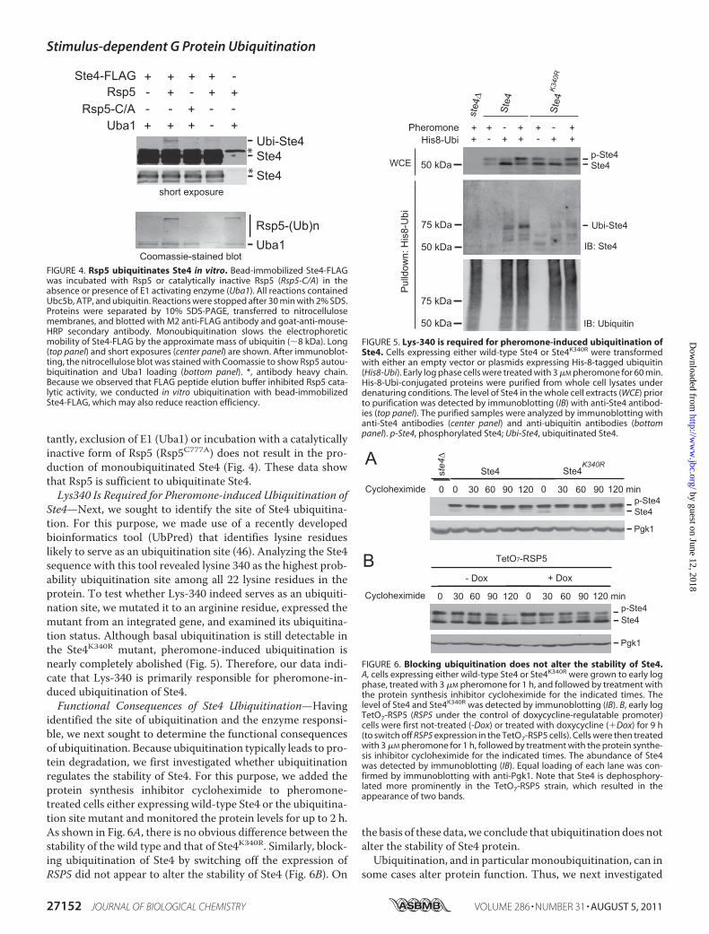
tantly, exclusion of E1 (Uba1) or incubation with a catalyticallyinactive form of Rsp5 (Rsp5C777A) does not result in the pro-duction of monoubiquitinated Ste4 (Fig. 4). These data showthat Rsp5 is sufficient to ubiquitinate Ste4.Lys340 Is Required for Pheromone-induced Ubiquitination of
Ste4—Next, we sought to identify the site of Ste4 ubiquitina-tion. For this purpose, we made use of a recently developedbioinformatics tool (UbPred) that identifies lysine residueslikely to serve as an ubiquitination site (46). Analyzing the Ste4sequence with this tool revealed lysine 340 as the highest prob-ability ubiquitination site among all 22 lysine residues in theprotein. To test whether Lys-340 indeed serves as an ubiquiti-nation site, we mutated it to an arginine residue, expressed themutant from an integrated gene, and examined its ubiquitina-tion status. Although basal ubiquitination is still detectable inthe Ste4K340R mutant, pheromone-induced ubiquitination isnearly completely abolished (Fig. 5). Therefore, our data indi-cate that Lys-340 is primarily responsible for pheromone-in-duced ubiquitination of Ste4.Functional Consequences of Ste4 Ubiquitination—Having
identified the site of ubiquitination and the enzyme responsi-ble, we next sought to determine the functional consequencesof ubiquitination. Because ubiquitination typically leads to pro-tein degradation, we first investigated whether ubiquitinationregulates the stability of Ste4. For this purpose, we added theprotein synthesis inhibitor cycloheximide to pheromone-treated cells either expressing wild-type Ste4 or the ubiquitina-tion site mutant and monitored the protein levels for up to 2 h.As shown in Fig. 6A, there is no obvious difference between thestability of the wild type and that of Ste4K340R. Similarly, block-ing ubiquitination of Ste4 by switching off the expression ofRSP5 did not appear to alter the stability of Ste4 (Fig. 6B). On
the basis of these data, we conclude that ubiquitination does notalter the stability of Ste4 protein.Ubiquitination, and in particularmonoubiquitination, can in
some cases alter protein function. Thus, we next investigated
FIGURE 4. Rsp5 ubiquitinates Ste4 in vitro. Bead-immobilized Ste4-FLAGwas incubated with Rsp5 or catalytically inactive Rsp5 (Rsp5-C/A) in theabsence or presence of E1 activating enzyme (Uba1). All reactions containedUbc5b, ATP, and ubiquitin. Reactions were stopped after 30 min with 2% SDS.Proteins were separated by 10% SDS-PAGE, transferred to nitrocellulosemembranes, and blotted with M2 anti-FLAG antibody and goat-anti-mouse-HRP secondary antibody. Monoubiquitination slows the electrophoreticmobility of Ste4-FLAG by the approximate mass of ubiquitin (�8 kDa). Long(top panel) and short exposures (center panel) are shown. After immunoblot-ting, the nitrocellulose blot was stained with Coomassie to show Rsp5 autou-biquitination and Uba1 loading (bottom panel). *, antibody heavy chain.Because we observed that FLAG peptide elution buffer inhibited Rsp5 cata-lytic activity, we conducted in vitro ubiquitination with bead-immobilizedSte4-FLAG, which may also reduce reaction efficiency.
FIGURE 5. Lys-340 is required for pheromone-induced ubiquitination ofSte4. Cells expressing either wild-type Ste4 or Ste4K340R were transformedwith either an empty vector or plasmids expressing His-8-tagged ubiquitin(His8-Ubi). Early log phase cells were treated with 3 �M pheromone for 60 min.His-8-Ubi-conjugated proteins were purified from whole cell lysates underdenaturing conditions. The level of Ste4 in the whole cell extracts (WCE) priorto purification was detected by immunoblotting (IB) with anti-Ste4 antibod-ies (top panel). The purified samples were analyzed by immunoblotting withanti-Ste4 antibodies (center panel) and anti-ubiquitin antibodies (bottompanel). p-Ste4, phosphorylated Ste4; Ubi-Ste4, ubiquitinated Ste4.
FIGURE 6. Blocking ubiquitination does not alter the stability of Ste4.A, cells expressing either wild-type Ste4 or Ste4K340R were grown to early logphase, treated with 3 �M pheromone for 1 h, and followed by treatment withthe protein synthesis inhibitor cycloheximide for the indicated times. Thelevel of Ste4 and Ste4K340R was detected by immunoblotting (IB). B, early logTetO7-RSP5 (RSP5 under the control of doxycycline-regulatable promoter)cells were first not-treated (-Dox) or treated with doxycycline (�Dox) for 9 h(to switch off RSP5 expression in the TetO7-RSP5 cells). Cells were then treatedwith 3 �M pheromone for 1 h, followed by treatment with the protein synthe-sis inhibitor cycloheximide for the indicated times. The abundance of Ste4was detected by immunoblotting (IB). Equal loading of each lane was con-firmed by immunoblotting with anti-Pgk1. Note that Ste4 is dephosphory-lated more prominently in the TetO7-RSP5 strain, which resulted in theappearance of two bands.
Stimulus-dependent G Protein Ubiquitination
27152 JOURNAL OF BIOLOGICAL CHEMISTRY VOLUME 286 • NUMBER 31 • AUGUST 5, 2011
by guest on June 12, 2018http://w
ww
.jbc.org/D
ownloaded from

whether ubiquitination of Ste4 affects its ability to transmit thepheromone signal. Upon stimulation, Ste4 activates severaleffectors leading to highly coordinated mating responses,including growth arrest, new gene transcription, and morpho-logical changes (3, 27). Ste4 activates a MAP kinase cascade byrecruiting the MAP kinase scaffold (Ste5) and the associatedkinase complex (Ste11/Ste7/Fus3) to the plasma membraneand facilitating its activation by Ste20 (3, 47). To determinewhether ubiquitination of Ste4 plays any role in regulating themagnitude and/or duration of MAPK activation, we comparedthe activation status of Fus3 in wild-type cells versus theSte4K340R mutant treated with the mating pheromone � factorover time. Fus3 activity was monitored by immunoblottingwith an antibody directed against the dually phosphorylatedform of the protein (Thr-202/Tyr-204) (p44/42) (48, 49). Asshown in Fig. 7A, no obvious difference in either themagnitudeor duration of Fus3 activation was detected between wild-typeand the Ste4K340R mutant. Consistent with this result, theSte4K340R mutant displayed the same level of pheromone-in-duced activation of gene transcription as measured by theFUS1-GFP reporter (50, 51) (Fig. 7B). This analysis indicates
that Ste4 ubiquitination plays no apparent role in regulating theMAP kinase cascade.In addition to activating a MAP kinase cascade via binding
Ste5 and Ste20, Ste4 also regulates polarized cell growth viainteractionswithCdc24 and Far1 (3, 25).When cells are treatedwith pheromone, they reorient their cytoskeleton and initiatepolarized growth toward the highest concentration of phero-mone, leading to the formation of a shmoo morphology (26,27). To determine whether ubiquitination of Ste4 may regulatethis process, we compared the polarized cell growth of the wildtype and the Ste4K340R mutant via live cell microscopy. Toensure consistent Ste4 expression from cell to cell, theSte4K340R mutant was integrated to the genome. To minimizethe potential effects of the cell cycle, the cells were synchro-nized to M phase via nocodazole treatment before pheromonestimulation. To maintain a consistent environment with con-stant replenishment of media and pheromone, washout ofnocodozole and treatment with pheromone was done in amicrofluidic chamber (39). As shown in Fig. 8, cells start toundergo polarized growth after 30 min of pheromone treat-ment. By 80 min of treatment, the majority of cells displaypolarized growth. However, the kinetics of polarized growthdisplayed by the wild type and Ste4K340Rmutant are very differ-ent. On average, the population of Ste4K340R mutants polarizessignificantly faster than thewild type (Fig. 8). Such a behavior ofthe Ste4K340R mutant indicates that ubiquitination regulatesthe process of pheromone-triggered polarized growth.
DISCUSSION
Heterotrimeric G proteins are molecular switches that con-trol many important biological processes. Thus, a clear under-standing of themechanisms that regulateGproteins is essentialfor revealing new disease mechanisms and for designing novelapproaches for pharmaceutical intervention. In this report, weshowed that the G� protein undergoes ubiquitination inresponse to pheromone stimulation. To our knowledge, this isthe first demonstration of stimulus-dependent ubiquitinationof a G protein subunit in any system.
FIGURE 7. Ubiquitination of Ste4 does not regulate the magnitude andduration of pheromone-induced activation of MAP kinases. A, whole cellextracts were prepared from the wild type and the Ste4K340R mutant treatedwith 3 �M pheromone � factor for the indicated time, resolved by 10% SDS-PAGE, and probed with anti-phospho-p44/42 (top panel) or anti-Ste4 (bottompanel) antibodies. p-Mpk1, p-Kss1, and p-Fus3 denote phosphorylated andthus activated Mpk1, Kss1, and Fus3. B, pheromone-dependent transcrip-tional induction was measured following transformation of the above cellswith a pheromone-responsive FUS1 promoter-GFP reporter. Cells were thentreated with 3 �M �-factor for 90 min, and the resulting fluorescence in eachcell was monitored by cell sorting. Pathway activation results in an increase incells with �40 fluorescence units of activity (as indicated by an M1 bar oneach graph).
FIGURE 8. Ubiquitination of Ste4 limits pheromone-induced polarizedgrowth. Cells expressing either wild-type Ste4 or Ste4K340R were grown toA600 0.2 and treated with dimethyl sulfoxide (1%, final concentration) for 1.5 hbefore the addition of nocodazole (15 �g/ml in 1% dimethyl sulfoxide, finalconcentrations) for 2.5–3 h. Cultures were visualized by microscopy to con-firm G2/M arrest. The cells were then loaded into a microfluidics device, andthe nocodazole was washed out for 20 min prior to stimulation with 150 nM �factor. Cells were imaged at 5 min intervals for 2 h. For each cell, the time atwhich the first sign of polarized growth detectable in the differential interfer-ence contrast image was recorded and plotted as a function of time. Theslope was taken from the linear portion of each curve, and the average slopesfrom three independent experiments are shown in the bar graph (right panel).The difference between Ste4 and Ste4K340R was statistically analyzed by t test.*, p � 0.05). Error bars, mean � S.E.
Stimulus-dependent G Protein Ubiquitination
AUGUST 5, 2011 • VOLUME 286 • NUMBER 31 JOURNAL OF BIOLOGICAL CHEMISTRY 27153
by guest on June 12, 2018http://w
ww
.jbc.org/D
ownloaded from

Our findings show that Rsp5 is responsible for Ste4 ubiquiti-nation. First, either turning off Rsp5 expression (the TET-RSP5strain in the presence of doxycycline) or disrupting Rsp5 func-tion (the temperature-sensitive rsp5–1 mutant) completelyabolishes Ste4 ubiquitination in vivo. Second, Ste4 physicallyinteracts with Rsp5 in yeast. Third, Rsp5 is capable of ubiquiti-nating Ste4 in vitro. Notably, earlier studies showed that Rsp5 isalso responsible for the ubiquitination of Ste2 (receptor) (44)and themonoubiquitination (but not the polyubiquitination) ofGpa1 (G�) (43). Thus, Ste4 joins a growing list of signalingproteins that serve as substrates of Rsp5. It would be interestingto examine whether there is any coordination among theseubiquitination events. Given the easily detectable interactionbetween Rsp5 and Ste4, an attractive model is that Ste4 mayserve as an adaptor protein (in addition to being a substrate) forRsp5 to facilitate its ubiquitination of other membrane-associ-ated proteins. However, monoubiquitination of Gpa1 is appar-ently not dependent on Rsp5/Ste4 interaction because similarlevel of mono-ubiquitinated Gpa1 is present in the wild typeversus a ste4� mutant3.
Although polyubiquitination is commonly regarded as adegradation signal, monoubiquitination appears to have adistinct role in protein regulation (13, 52). In accord withthis view, monoubiquitination does not appear to modulateSte4 stability. Blocking ubiquitination of Ste4, either viamutating its major ubiquitination site (Lys-340) or turningoff the expression of its ligase Rsp5, has no obvious effect oneither the abundance or the stability of Ste4. In addition, theabundance of Ste4 is not discernibly decreased in response topheromone treatment, a condition that markedly inducesSte4 ubiquitination (Fig. 1). This is in marked contrast withboth Ste2 and Gpa1. In both cases, ubiquitination by Rsp5serves to target the proteins for internalization and eventualdegradation in the vacuole (43, 44).Thus, although Ste4 is a critical determinant of the intensity
and duration of downstream MAP kinase signaling (30, 40),ubiquitination does not appear to alter the abundance of Ste4and thereby the intensity of downstreamMAP kinase signaling.Rather, ubiquitination of Ste4 appears to have a very specificrole in regulating the timing of polarized cell growth inresponse to pheromone stimulation. Compared with the wildtype, the point mutation (Ste4K340R) that blocks ubiquitinationallows cells to polarize significantly faster. On average, themutant cells initiate polarized growth 8–10min earlier than thewild type upon stimulation by pheromone. Further investiga-tion will be needed to determine how ubiquitination of Ste4selectively regulates one effector (cell polarity machinery) butnot the other (i.e. a MAP kinase cascade).Growing evidence indicates that ubiquitination, especially
monoubiquitination, can modulate protein-protein interac-tions, which consequently can lead to a very specific outcome(14). A prominent example is K-Ras, a small GTPase that con-trols cell differentiation and growth. It has been shown that avery small fraction of K-Ras is modified by mono- or diubiqui-tin (53). Interestingly, ubiquitinated K-Ras has substantially
increased affinity toward some but not all of its downstreameffectors (53). Consequently, blocking ubiquitination of K-Rasimpacts some but not all of the cellular responses controlled bythe protein. In the case of Ste4, it is entirely possible that ubiq-uitination may serve to regulate its interaction with down-stream effectors such as Far1, Cdc24, Ste5, and Ste20. However,it should be pointed out that the difference of binding could besubtle, dynamic, and limited to certain subcellular locations.Therefore, typical coimmunoprecipitation may fail to capturethose potential differences.How is ubiquitination of Ste4 itself regulated? Pheromone
stimulation induces both phosphorylation and ubiquitinationof Ste4. In agreementwith amodel that phosphorylation targetsSte4 for ubiquitination, a mutant (Ste4T320A/S335A) that signif-icantly diminishes phosphorylation of Ste4 also substantiallyabolishes ubiquitination. It is interesting to note that the majorubiquitination site Lys-340 is adjacent to the putative phos-phorylation sites (Thr-320 and Ser-335), raising a possibilitythat phosphorylationmay lead to a conformational change thathelps to expose the lysine residue to receive an activated ubiq-uitin from Rsp5. However, in consideration of this model, it isimportant to keep in mind that Thr-320 and Ser-335 wereimplicated as possible phosphorylation sites on the basis oftheir requirement for the phosphorylation-induced mobilityshift (35). Thus, it remains possible that the Ste4T320A/S335Amutant may reduce phosphorylation through an indirectmechanism.In summary, our data clearly demonstrate that the G� sub-
unit Ste4 undergoes pheromone-dependent ubiquitination andthat Rsp5 is responsible for thismodification. Ubiquitination ofSte4 occurs primarily on a single lysine residue. Significantly,ubiquitination of Ste4 selectively regulates some (e.g. polarizedcell growth) but not other aspects of pheromone responses.Together, these findings reveal a newmechanism for regulatingsignal transmission mediated by G proteins.
Acknowledgments—We thank Drs. David E. Stone, Michael P. Yaffe,and Jeremy Thorner for generously providing strains, plasmids, andantibodies, and Laura M. Hall for preliminary analysis of cellpolarization.
REFERENCES1. Sprang, S. R. (1997) Annu. Rev. Biochem. 66, 639–6782. Cabrera-Vera, T. M., Vanhauwe, J., Thomas, T. O., Medkova, M., Prein-
inger, A., Mazzoni, M. R., and Hamm, H. E. (2003) Endocr. Rev. 24,765–781
3. Dohlman, H. G., and Thorner, J. W. (2001) Annu. Rev. Biochem. 70,703–754
4. Pierce, K. L., Premont, R. T., and Lefkowitz, R. J. (2002)Nat. Rev. Mol. CellBiol. 3, 639–650
5. Dohlman, H. G., and Thorner, J. (1997) J. Biol. Chem. 272, 3871–38746. Johnson, G. L., and Lapadat, R. (2002) Science 298, 1911–19127. Martín, H., Flandez, M., Nombela, C., and Molina, M. (2005)Mol. Micro-
biol. 58, 6–168. Hershko, A., and Ciechanover, A. (1998) Annu. Rev. Biochem. 67,
425–4799. Pickart, C. M., and Eddins, M. J. (2004) Biochim. Biophys. Acta 1695,
55–7210. Ciechanover, A., Finley, D., and Varshavsky, A. (1984) J. Cell. Biochem. 24,
27–533 M. Zhu and Y. Wang, unpublished observation.
Stimulus-dependent G Protein Ubiquitination
27154 JOURNAL OF BIOLOGICAL CHEMISTRY VOLUME 286 • NUMBER 31 • AUGUST 5, 2011
by guest on June 12, 2018http://w
ww
.jbc.org/D
ownloaded from

11. Ciechanover, A., and Ben-Saadon, R. (2004)Trends Cell Biol. 14, 103–10612. Pickart, C. M. (2001) Annu. Rev. Biochem. 70, 503–53313. Hicke, L. (2001) Nat. Rev. Mol. Cell Biol. 2, 195–20114. Chen, Z. J., and Sun, L. J. (2009)Mol. Cell 33, 275–28615. Sigismund, S., Polo, S., and Di Fiore, P. P. (2004) Curr. Top Microbiol.
Immunol. 286, 149–18516. Schnell, J. D., and Hicke, L. (2003) J. Biol. Chem. 278, 35857–3586017. Wang, Y., and Dohlman, H. G. (2006) Circ. Res. 99, 1305–131418. Shenoy, S. K. (2007) Circ. Res. 100, 1142–115419. Naviglio, S., Pagano, M., Romano,M., Sorrentino, A., Fusco, A., Illiano, F.,
Chiosi, E., Spina, A., and Illiano, G. (2004) Cell. Signal. 16, 1229–123720. Hamilton, M. H., Cook, L. A., McRackan, T. R., Schey, K. L., and Hildeb-
randt, J. D. (2003) Proc. Natl. Acad. Sci. U.S.A. 100, 5081–508621. Obin, M., Lee, B. Y., Meinke, G., Bohm, A., Lee, R. H., Gaudet, R., Hopp,
J. A., Arshavsky, V. Y., Willardson, B. M., and Taylor, A. (2002) J. Biol.Chem. 277, 44566–44575
22. Dohlman, H. G., Thorner, J., Caron, M. G., and Lefkowitz, R. J. (1991)Annu. Rev. Biochem. 60, 653–688
23. Wang, Y., and Dohlman, H. G. (2004) Science 306, 1508–150924. Bardwell, L. (2005) Peptides 26, 339–35025. Bar, E. E., Ellicott, A. T., and Stone, D. E. (2003) J. Biol. Chem. 278,
21798–2180426. Madden, K., and Snyder, M. (1998) Annu. Rev. Microbiol. 52, 687–74427. Dohlman, H. G. (2002) Annu. Rev. Physiol. 64, 129–15228. Suchkov, D. V., DeFlorio, R., Draper, E., Ismael, A., Sukumar, M., Arkow-
itz, R., and Stone, D. E. (2010)Mol. Biol. Cell 21, 1737–175229. Ghaemmaghami, S., Huh, W. K., Bower, K., Howson, R. W., Belle, A.,
Dephoure, N., O’Shea, E. K., and Weissman, J. S. (2003) Nature 425,737–741
30. Hao, N., Yildirim, N., Wang, Y., Elston, T. C., and Dohlman, H. G. (2003)J. Biol. Chem. 278, 46506–46515
31. Fisk, H. A., and Yaffe, M. P. (1999) J. Cell Biol. 145, 1199–120832. Mnaimneh, S., Davierwala, A. P., Haynes, J.,Moffat, J., Peng,W. T., Zhang,
W., Yang, X., Pootoolal, J., Chua, G., Lopez, A., Trochesset, M., Morse, D.,Krogan, N. J., Hiley, S. L., Li, Z., Morris, Q., Grigull, J., Mitsakakis, N.,Roberts, C. J., Greenblatt, J. F., Boone, C., Kaiser, C. A., Andrews, B. J., andHughes, T. R. (2004) Cell 118, 31–44
33. Esch, R. K., Wang, Y., and Errede, B. (2006) Eukaryotic Cell 5, 2147–216034. Slessareva, J. E., Routt, S. M., Temple, B., Bankaitis, V. A., and Dohlman,
H. G. (2006) Cell 126, 191–20335. Li, E., Cismowski, M. J., and Stone, D. E. (1998) Mol. Gen. Genet. 258,
608–61836. Siekhaus, D. E., and Drubin, D. G. (2003) Nat. Cell Biol. 5, 231–23537. Poritz, M. A., Malmstrom, S., Kim, M. K., Rossmeissl, P. J., and Kamb, A.
(2001) Yeast 18, 1331–133838. Muratani, M., Kung, C., Shokat, K. M., and Tansey,W. P. (2005) Cell 120,
887–89939. Hao, N., Nayak, S., Behar, M., Shanks, R. H., Nagiec, M. J., Errede, B.,
Hasty, J., Elston, T. C., and Dohlman, H. G. (2008)Mol. Cell 30, 649–65640. Whiteway, M., Hougan, L., and Thomas, D. Y. (1990) Mol. Cell Biol. 10,
217–22241. Cole, G. M., and Reed, S. I. (1991) Cell 64, 703–71642. Laine, A., and Ronai, Z. (2005) Sci. STKE 2005, re543. Torres, M. P., Lee, M. J., Ding, F., Purbeck, C., Kuhlman, B., Dokholyan,
N. V., and Dohlman, H. G. (2009) J. Biol. Chem. 284, 8940–895044. Dunn, R., and Hicke, L. (2001) J. Biol. Chem. 276, 25974–2598145. Shcherbik, N., Kee, Y., Lyon, N., Huibregtse, J.M., andHaines, D. S. (2004)
J. Biol. Chem. 279, 53892–5389846. Radivojac, P., Vacic, V., Haynes, C., Cocklin, R. R., Mohan, A., Heyen,
J. W., Goebl, M. G., and Iakoucheva, L. M. (2010) Proteins 78, 365–38047. Chen, R. E., and Thorner, J. (2007) Biochim. Biophys. Acta 1773,
1311–134048. Sabbagh,W., Jr., Flatauer, L. J., Bardwell, A. J., and Bardwell, L. (2001)Mol.
Cell 8, 683–69149. Wang, Y., Abu Irqeba, A., Ayalew, M., and Suntay, K. (2009) PLoS ONE 4,
e745650. Hoffman, G. A., Garrison, T. R., and Dohlman, H. G. (2002) Methods
Enzymol. 344, 617–63151. Lan, K. L., Sarvazyan, N. A., Taussig, R., Mackenzie, R. G., DiBello, P. R.,
Dohlman,H.G., andNeubig, R. R. (1998) J. Biol. Chem. 273, 12794–1279752. Sun, L., and Chen, Z. J. (2004) Curr. Opin. Cell Biol. 16, 119–12653. Sasaki, A. T., Carracedo, A., Locasale, J. W., Anastasiou, D., Takeuchi, K.,
Kahoud, E. R., Haviv, S., Asara, J. M., Pandolfi, P. P., and Cantley, L. C.(2011) Sci. Signal 4, ra13
Stimulus-dependent G Protein Ubiquitination
AUGUST 5, 2011 • VOLUME 286 • NUMBER 31 JOURNAL OF BIOLOGICAL CHEMISTRY 27155
by guest on June 12, 2018http://w
ww
.jbc.org/D
ownloaded from

Ming Zhu, Matthew P. Torres, Joshua B. Kelley, Henrik G. Dohlman and Yuqi Wangin Yeast
Subunit Ste4βPheromone- and RSP5-dependent Ubiquitination of the G Protein
doi: 10.1074/jbc.M111.254193 originally published online June 17, 20112011, 286:27147-27155.J. Biol. Chem.
10.1074/jbc.M111.254193Access the most updated version of this article at doi:
Alerts:
When a correction for this article is posted•
When this article is cited•
to choose from all of JBC's e-mail alertsClick here
Supplemental material:
http://www.jbc.org/content/suppl/2011/06/17/M111.254193.DC1
http://www.jbc.org/content/286/31/27147.full.html#ref-list-1
This article cites 53 references, 20 of which can be accessed free at
by guest on June 12, 2018http://w
ww
.jbc.org/D
ownloaded from