pharmacology & therapeutics - uibk.ac.at · a department of pharmacology and toxicology, ......
TRANSCRIPT

Pharmacology & Therapeutics 149 (2015) 150–190
Contents lists available at ScienceDirect
Pharmacology & Therapeutics
j ourna l homepage: www.e lsev ie r .com/ locate /pharmthera
Associate editor: P. Holzer
Pharmacology of cognitive enhancers for exposure-based therapy of fear,anxiety and trauma-related disorders
N. Singewald a,⁎,1, C. Schmuckermair a,1, N. Whittle a, A. Holmes b, K.J. Ressler c
a Department of Pharmacology and Toxicology, Institute of Pharmacy and CMBI, Leopold-Franzens University of Innsbruck, Innrain 80-82, A-6020 Innsbruck, Austriab Laboratory of Behavioral and Genomic Neuroscience, National Institute on Alcohol Abuse and Alcoholism, NIH, Bethesda, MD, USAc Department of Psychiatry and Behavioral Sciences, Emory University School of Medicine, Atlanta, GA, USA
Abbreviations:5-HT, 5-hydroxytryptamine= serotonBDNF, brain-derivedneurotrophic factor; BLA, basolateral amonophosphate; Cav 1.2, L-type calcium channel alphacentral amygdala; CeM, centromedial amygdala; CeL, centrDCS, D-cycloserine; DNA, deoxyribonucleic acid; eCB, endoGABA, γ-aminobutyric acid; GABAA, GABAA receptor; GADreceptor; H3, histone H3 protein;H4, histone H4 protein;Haxis; HPC, hippocampus; icv, intracerebroventricular; IL, iMAPK, mitogen-activated protein kinase; MDMA, 3,4-memPFC, medial prefrontal cortex; mRNA, messenger ribonumethyl-D-aspartate; NMDAR, NMDA receptor; ns, not stuperiaqueductal gray; PEPA, 4-[2-(Phenylsulphonylamino)disorder; PL, prelimbic cortex; SAD, social anxiety disorderreuptake inhibitor; SSRI, selective serotonin reuptake inhibexposure; Zn2+, zinc ion.⁎ Corresponding author at: Department of Pharmacolo
Austria. Fax: +43 512 507 58889.E-mail address: [email protected] (N. Singe
1 These authors contributed equally.
http://dx.doi.org/10.1016/j.pharmthera.2014.12.0040163-7258/© 2014 The Authors. Published by Elsevier Inc
a b s t r a c t
a r t i c l e i n f oAvailable online 27 December 2014
Keywords:Fear extinctionExposure therapyAugmented relearningReconsolidationDrug developmentCognitive enhancer
Pathological fear and anxiety are highly debilitating and, despite considerable advances in psychotherapyand pharmacotherapy they remain insufficiently treated in many patients with PTSD, phobias, panic and otheranxiety disorders. Increasing preclinical and clinical evidence indicates that pharmacological treatmentsincluding cognitive enhancers, when given as adjuncts to psychotherapeutic approaches [cognitive behavioraltherapy including extinction-based exposure therapy] enhance treatment efficacy, while using anxiolyticssuch as benzodiazepines as adjuncts can undermine long-term treatment success. The purpose of this reviewis to outline the literature showing how pharmacological interventions targeting neurotransmitter systemsincluding serotonin, dopamine, noradrenaline, histamine, glutamate, GABA, cannabinoids, neuropeptides(oxytocin, neuropeptides Y and S, opioids) and other targets (neurotrophins BDNF and FGF2, glucocorticoids,L-type-calcium channels, epigenetic modifications) as well as their downstream signaling pathways, can aug-ment fear extinction and strengthen extinctionmemory persistently in preclinicalmodels. Particularly promisingapproaches are discussed in regard to their effects on specific aspects of fear extinction namely, acquisition,consolidation and retrieval, including long-term protection from return of fear (relapse) phenomena likespontaneous recovery, reinstatement and renewal of fear. We also highlight the promising translational valueof the preclinial research and the clinical potential of targeting certain neurochemical systemswith, for exampleD-cycloserine, yohimbine, cortisol, and L-DOPA. The current body of research reveals important new insights intothe neurobiology and neurochemistry of fear extinction and holds significant promise for pharmacologically-augmented psychotherapy as an improved approach to treat trauma and anxiety-related disorders in a moreefficient and persistent way promoting enhanced symptom remission and recovery.
© 2014 The Authors. Published by Elsevier Inc. This is an open access article under the CC BY-NC-ND license(http://creativecommons.org/licenses/by-nc-nd/4.0/).
in; AC, adenylate cyclase; AMPA,α-amino-3-hydroxy-5-methyl-4-isoxazolepropionic acid; AMY, amygdala; BA, basal amygdala;mygdaloid complex; BZD, benzodiazepine; CaMKII, Ca2+/calmodulin-dependent protein kinase II; cAMP, 3′-5′-cyclic adenosine1C isoform; Cav 1.3, L-type calcium channel alpha 1D isoform; CBT, cognitive behavioral therapy; CCK, cholecystokinin; CeA,olateral amygdala; CNS, central nervous system;CREB, cAMPresponse element binding; CS, conditioned stimulus;DA, dopamine;genous cannabinoiod; ERK, extracellular regulated kinase; ERP, exposure and prevention CBT; FGF2, fibroblast growth factor-2;, generalized anxiety disorder; GAD (65/67), glutamate decarboxylase 65/67; GluN, NMDA receptor subtype; GR, glucocorticoidAT, histone acetyltransferase; HDAC, histone deacetylase; HDAC2, histone deacetylase 2; HPA, hypothalamic–pituitary–adrenalnfralimbic cortex; ITC, intercalated cell masses; K, lysine; KOR, kappa opioid receptor; LA, lateral amygdala; L-DOPA, levodopa;thylenedioxy-N-methylamphetamine; Mg2+, magnesium ion; mGluR, metabotropic glutamate receptor; miRNA, micro-RNA;cleic acid; MS-275, entinostat (Pyridin-3-ylmethyl N-[[4-[(2-aminophenyl)carbamoyl] phenyl]methyl]carbamate); NMDA, N-died; NO, nitric oxide; NPS, neuropeptide S; NPY, neuropeptide Y; OCD, obsessive-compulsive disorder; OXT, oxytocin; PAG,ethylthio]-2,6-difluorophenoxyacetamide; PKC, as Ca2+ /phospholipid-dependent protein kinase C; PTSD, post-traumatic stress; SAHA, vorinostat (N-hydroxy-N′-phenyl-octanediamide); SNP, single nucleotide polymorphism; SNRI, selective noradrenalineitor; TrkB, tropomyosin-related kinase B;US, unconditioned stimulus; VGCC, voltage-gated calcium channel; VRET, virtual reality
gy & Toxicology, Center for Molecular Biosciences Innsbruck (CMBI), University of Innsbruck Innrain 80-82, A-6020 Innsbruck,
wald).
. This is an open access article under the CC BY-NC-ND license (http://creativecommons.org/licenses/by-nc-nd/4.0/).

151N. Singewald et al. / Pharmacology & Therapeutics 149 (2015) 150–190
Contents
1. Introduction. . . . . . . . . . . . . . . . . . . . . . . . . . . . . . . . . . . . . . . . . . . . . . . 1512. Neuronal substrates of fear extinction . . . . . . . . . . . . . . . . . . . . . . . . . . . . . . . . . . . 1513. Neurochemical and molecular substrates of fear extinction. . . . . . . . . . . . . . . . . . . . . . . . . . 1534. Pharmacologically enhancing extinction . . . . . . . . . . . . . . . . . . . . . . . . . . . . . . . . . . 1545. Discussion and final conclusions. . . . . . . . . . . . . . . . . . . . . . . . . . . . . . . . . . . . . . 154Conflict of interest statement . . . . . . . . . . . . . . . . . . . . . . . . . . . . . . . . . . . . . . . . . 154Acknowledgments . . . . . . . . . . . . . . . . . . . . . . . . . . . . . . . . . . . . . . . . . . . . . . 156
153154154178180180
References . . . . . . . . . . . . . . . . . . . . . . . . . . . . . . . . . . . . . . . . . . . . . . . . . . 158180
1. Introduction
Fear, anxiety and trauma-related disorders are associated with ex-cessive fear reactions triggered by specific objects, situations or internaland external cues in the absence of any actual danger, and often includean inability to extinguish learned fear and to show adequate safetylearning [(Jovanovic et al., 2012; Michael et al., 2007; Milad et al.,2009; Milad et al., 2013; Wessa & Flor, 2007) reviewed in (Holmes &Singewald, 2013) and (Kong et al., 2014)]. Pathological fear and anxietyoccur in a range of psychiatric conditions, including various types ofphobia (e.g. social phobia, agoraphobia or specific phobia), panic disor-der with/without agoraphobia, obsessive–compulsive disorder (OCD),generalized anxiety (GAD) and post-traumatic stress disorder (PTSD)(DSM-5, 2013; ICD-10, 1994). These disorders comprise the most com-mon mental disorders and are estimated to have a life-time prevalenceof up to 28% among western populations (Kessler et al., 2005; Kessleret al., 2012; Wittchen et al., 2011). In addition to the personal sufferingof patients, the economic burden caused by anxiety disorders is heavy(Gustavsson et al., 2011).
Available pharmacological and psychotherapeutic treatments(Bandelow et al., 2007) which aim to reduce fear and anxiety are asso-ciated with decreased symptom severity, but up to 40% of anxiety pa-tients show only partial long-term benefit, and a majority of them failto achieve complete remission (Bandelow et al., 2012; Hoffman &Mathew, 2008; Stein et al., 2009) clearly underlining the need for fur-ther improvement. Current pharmacological approaches either inducerapid anxiolytic effects (e.g. benzodiazepines, some antipsychotics) orrequire prolonged, chronic treatment (e.g. antidepressants) to attenu-ate symptoms of pathological fear and anxiety. Commonly employedpsychotherapeutic interventions apply cognitive behavioral strategiesand exposure techniques to help patients overcome the maladaptivebeliefs and avoidance behaviors that reinforce the pathology relatedto fear-eliciting cues. Meta-analyses show that cognitive behavioraltherapy (CBT) does have efficacy for several anxiety disorders, includingPTSD, but patients have difficulty bearing the demanding andexhausting process of therapy and many who do manage to cope withit respond only partially and often relapse with time (Choy et al., 2007).
One strategy to improve CBT is to augment psychotherapy with ad-junctive pharmacological treatments. Early attempts at combining ‘CBT’with anxiolytic medications [e.g. benzodiazepines (BZD)] showed thatthe combination was no more effective [in some instances even coun-terproductive (Marks et al., 1993; Wilhelm & Roth, 1997)] thanpsycho- or pharmacotherapy alone [for details see (Dunlop et al.,2012; Hofmann, 2012; Otto et al., 2010a,b; Rodrigues et al., 2011)].However, at least in some cases, this failuremay have reflected idiosyn-cratic effects of the drugs tested (especially BZDs) rather than utility ofthe strategy itself, and there has been an intense search to identifyagents that serve as more effective adjuncts to CBT. The preclinicalassay most frequently used in this search is fear extinction — the focusof this current review. Extinction of fear following Pavlovian fear learn-ing (Pavlov, 1927) in animals is procedurally similar to exposure-basedCBT (Milad & Quirk, 2012). We will briefly outline different aspects ofPavlovian fear learning [(which is thought to be involved in the etiology
andmaintenance of anxiety disorders, e.g. (Amstadter et al., 2009)] andextinction highlighting the key processes that could be targeted toaugment fear extinction (see Fig. 1 for an overview).
1.1. Fear and fear extinction
Experimentally, fear conditioning occurs when a previously neutralstimulus [conditioned stimulus (CS) — such as a tone or light] is pairedwith an aversive, unconditioned stimulus (US— e.g. electric shock to theforearm in humans, mild foot shock in rodents), resulting in a CS–USassociation whereby the CS alone elicits a conditioned fear response(e.g. freezing in rodents or increased skin conductance in humans).Following a successful CS–US association, fear memories require consoli-dation, a process involving a cascade ofmolecular and cellular events thatalter synaptic efficacy, aswell as a prolonged systems level interaction be-tween brain regions, to stabilize the memory (McGaugh, 2000). Onceconsolidated, fear memories, reactivated by presenting the CS, are de-stabilized to render the original fear memory liable to pharmacological/behavioral interference (see overview in Fig. 1) and this is thenfollowed by a second phase of molecular and cellular events to re-stabilize (re-consolidate) the (adapted) memory (Nader et al., 2000).
Fearmemories can be attenuated by various processes and interven-tions (including pharmacological and psychological approaches), someproducing temporary blunting of fear behaviors and others causingmore long-lasting relief. Interfering with the re-consolidation byinhibiting molecular and cellular events supporting fear memory re-stabilization [Fig. 1C, (Lee et al., 2006; Nader et al., 2000)], for examplewith β-adrenoceptor blockers (Debiec & Ledoux, 2004; Kindt et al.,2009), has been proposed as a clinical approach to alleviating fearmem-ories. For discussion on this and othermeans of reducing fear (e.g. via UShabituation) we refer the reader to some excellent prior reviews(Graham et al., 2011; Schwabe et al., 2014). Other potential ways torelieve fear include safety learning (Kong et al., 2014; Rogan et al.,2005) and erasure-like mechanisms such as destruction of erasure-preventing perineuronal nets (Gogolla et al., 2009).
Alternatively, fear memories can also be extinguished. Fear extinc-tion, a process originally described by Pavlov (Pavlov, 1927), entails re-peated exposure to anxiety-provoking cues to establish a newmemorythat counters the original fearmemory. The process is highly relevant tofear, anxiety and trauma-related disorders which are associated withnegative emotional reactions triggered by specific objects, situationsor internal and external cues that are excessive to the actual dangerposed. Moreover, extinction in animals is procedurally similar toforms of CBT that rely on exposure to anxiety-provoking cues (seeFig. 1) (Milad & Quirk, 2012), and anxiety disorders are associatedwith an inability to extinguish learned fear and to respond adequatelyto safety signals (Jovanovic et al., 2012; Michael et al., 2007; Miladet al., 2009; Milad et al., 2013; Wessa & Flor, 2007) [reviewed in(Holmes & Singewald, 2013)]. Thus, fear extinction has considerabletranslational utility. The key processes that can be targeted to pharma-cologically augment fear extinction are summarized in Fig. 1.
Extinction is a learning process driven by violation of the originalCS=US contingency (termed ‘prediction error’ for review see (Pearce

A
B
C
FEAR ALLEVIATION1
23
4
1
2
3
4
e.g. BZDs
e.g. DCS, GCs
e.g. β-Blocker
e.g. BDNF
ACUTE SUSTAINED
Without exposure With exposure
Erasure Forge�ngEx�nc�on Safety
learningReconsolida�ondisrup�on
Fig. 1. Different modes of fear alleviation and sites of possible pharmacological intervention. A, Fear alleviation is mediated via different mechanisms leading to acute or sustained fearrelief. Sustained fear relief can be obtained either with exposures to the feared cues/situations or, in some instances, also without them. For a detailed description of the involved mech-anisms, see Riebe et al. (2012). Pharmacological interventions to boost fear relief can target different mechanisms at various levels. Examples are given with numbers 1–4, for detail seetext. B, Fear conditioning represents a training phase in which a novel conditioned stimulus (CS) is paired with an unconditioned stimulus (US) (redline). Throughout fear training andtesting, fear is measured as a conditioned response (y-axis), typically freezing, fear-potentiated startle, increased heart rate, and other quantifiable behavioral measures of fear. Followingthis training period,mice undergo a consolidation phase which transfers the labile newly formed fear memory into a stable long-termmemory. Fear can be extinguished by repeated pre-sentations of the CS (without the US; red line) resulting in fear extinction. Following the extinction training session, and akin to fear learning, consolidation processes are initiated to sta-bilize this labile fear extinctionmemory into a long-termmemory. Poor extinction is evidenced by high fear responding (red bar), and successful extinction retrieval is shown by reducedfear (green) during a retrieval test. Extinguished fear can recover via three mainmechanisms: spontaneous recovery (recovery of extinguished fear responses occurs with the passage oftime in the absence of any further training), fear renewal (when the conditioned CS is presented outside the extinction context, for example the conditioning or novel contexts), and fearreinstatement (when un-signaled presentations of the US are interposed between the completion of extinction training and a subsequent retention test). Drugs (green arrows) can beadministered either immediately prior to (to induce extinction acquisition also called ‘within-session extinction’ and/or extinction consolidation) or immediately following (to rescue/boost extinction consolidation processes) the training session to modulate extinction mechanisms. A clinical aim of drug augmentation strategies is to promote good extinction retrievaland to protect against return-of-fear phenomena to provide good ‘longterm extinction’. C, Fear memory can be reactivated (red bar) by presentation of the CS, which transfers the stabi-lized memory into a labile phase which requires reconsolidation (a process by which previously consolidated memories are stabilized after fear retrieval). The fear memory can then betested during another retrieval test (red bar) to assess reconsolidation. Drugs (green arrow) can be administered either immediately prior to or after the retrieval session to modulatereconsolidation mechanisms. This effect can then be tested during subsequent retrieval tests. Evidence for successful interference with reconsolidation of the original fear memory is re-vealed in reduced freezing during the test session.
152 N. Singewald et al. / Pharmacology & Therapeutics 149 (2015) 150–190
&Bouton, 2001;McNally &Westbrook, 2006)], but it also contains otherelements including habituation and desensitization some also say era-sure/destabilization (Lin et al., 2011). Some authors therefore favorthe term ‘relearning’ over extinction [for discussion see (Riebe et al.,2012)]. As with fear learning, extinction occurs in two phases:extinction acquisition and extinction consolidation. The decrement inthe fear response during’extinction training’ is also termed ‘within-session extinction’. Similar to fearmemory, stabilization of the extinctionmemory requires both a cascade of overlapping, but dissociable, molec-ular and cellular events [for review see (Myers & Davis, 2007; Orsini &Maren, 2012)] that alter synaptic efficacy and brain systems-level inter-actions (Pape & Pare, 2010). The strength of extinction memory can beassessed at some interval (usually N1 day) after extinction training in“extinction retrieval” sessions (also termed ‘extinction retention’ or ‘ex-tinction expression’, but note that we use the term “extinction retrieval”throughout this review).
That extinction memories are prone to re-emergence (due to insuf-ficient ‘longterm extinction’) indicates that the original fear memory is
still in place and the extinction memory is weaker/more labile thanthe fear memory. This may be particularly true of older (‘remote’) fearmemories (Tsai & Graff, 2014). The re-emergence of extinguished fearoccurs under multiple circumstances: (i) renewal, when the CS ispresented in a different context to that in which extinction trainingoccurred; (ii) reinstatement, when the original US or another stress-or is given unexpectedly; and (iii) spontaneous recovery, when a sig-nificant period of time has elapsed following successful extinctiontraining (Herry et al., 2010; Myers & Davis, 2007). The likelihood offear re-emergence is dependent on the strength of the extinctionmemory which is also determined by the type of extinction protocolused see e.g. (Laborda & Miller, 2013; Li & Westbrook, 2008). Spon-taneous recovery (Rowe & Craske, 1998a,b; Schiller et al., 2008), re-instatement (Schiller et al., 2008) and renewal (Effting & Kindt,2007) are all observable in clinical settings, and can be readilyexploited in the laboratory to identify drugs and other interventionsthat can prevent fear re-emergence in animals and relapse inhumans (Vervliet et al., 2013).

Box 1Why pharmacological augmentation of extinction?
A limitation of extinction-based exposure therapy is that patients can oftenrelapse with the passage of time, with changes in context (out of the therapycontext), or under conditions of stress or other provocations, such as experiencingtrauma reminders. In the parlance of learning theory, extinction memories arelabile and fragile. A key goal for pharmacotherapy, therefore, is to identifycompounds that overcome this fragility, by bolstering the formation, persistenceand possibly context independence of extinction memories. One approach toachieving this goal is to use drugs as adjuncts to exposure therapy (cognitiveenhancers) as a way to augment the extinction learning process. In this review, wediscuss the various neurochemical and molecular signaling pathways that havebeen targeted to this end. On the one hand, there remain significant challenges toovercome, including the selective targeting of extinction-related processeswithout concurrent effects on original fear memories. On the other hand, there areclearly a plethora of potentially promising avenues to pursue, and we areoptimistic that real advances can be made in treating trauma-related disorders.
153N. Singewald et al. / Pharmacology & Therapeutics 149 (2015) 150–190
Our goal in the current review is to offer a comprehensive overviewof preclinical work on the possible pharmacological approaches (seeBox 1 for an overview) to augment fear extinction and protect againstthe re-emergence of fear, and to discuss the potential translationalvalue of these candidates as adjuncts to exposure-based CBT in anxietypatients.
2. Neuronal substrates of fear extinction
Using a number of complementary techniques (including electro-physiology, immediate-early gene mapping, tracing studies, lesioning/inactivation approaches and optogenetics) research is revealing thecomplex and interconnected brain circuitry mediating fear extinction.Several key brain areas including the amygdala (AMY), hippocampus(HPC), medial prefrontal cortex (mPFC), periaqueductal gray (PAG),bed nucleus of the stria terminalis and others have been implicated inextinction [for recent detailed reviews see (Duvarci & Pare, 2014;Ehrlich et al., 2009; Herry et al., 2010; Knapska et al., 2012; Myers &Davis, 2007; Orsini & Maren, 2012; Pape & Pare, 2010)]. Of these, wefocus on the AMY, HPC and mPFC as major, well-defined componentsof the fear circuitry (see Fig. 2).
While theAMYand themPFC are crucial for the formation andmain-tenance of fear extinctionmemories, the HPC, linkedwith themPFC andthe AMY (Pape & Pare, 2010), processes contextual information linkedwith extinction (Orsini & Maren, 2012). Altering these hippocampal
Fig. 2.Anatomy of fear extinction and expression. Fear extinction and expression rely on neurongic and GABAergic neurons, among others, are important components of connectivity and reguWhile fear neurons in the BA send excitatory projections directly to the centromedial AMY (Cecortex (IL) inhibits CeM output by driving inhibitory ITC neurons. IL inputs might also synapsewithin the central AMY (CeA) through several routes, possibly by driving inhibitory ITC or CeL (ultimate inhibition of the CeM. The hippocampus is involved in contextual aspects of extinctionmemories built following extinction are encoded by theAMY and themPFC and aremodulated bchanges in synaptic plasticity and interneuronal communication in this circuitry ultimately redurefered to recent reviews of (Duvarci & Pare, 2014; Orsini & Maren, 2012).
contributions to extinction memory or mimicking the hippocampal re-sponse within the extinction context (see below) may be mechanismsthat render extinction context-independent. The AMY is a core hub infear extinction processing. Data in rodents and humans suggestsustained AMY activity in extinction-impaired individuals, possiblyresulting from a failure to engage pro-extinction circuits in corticalareas and subregions of the AMY [reviewed in (Holmes & Singewald,2013)]. Different AMY subregions andneuronal populationshave differ-ential contributions to extinction. The centromedial AMY (CeM) is themajor output station of the AMY that drives fear via its connections tothe hypothalamus and brainstem regions (Fendt & Fanselow, 1999;LeDoux et al., 1988; Maren, 2001), and its responding is modulated fol-lowing extinction via intra-amygdala and remote inputs. Extinctiontraining has been shown to cause a rapid reduction of CS-evoked re-sponses of lateral AMY (LA) neurons possibly via depotentiation of tha-lamic inputs (Duvarci & Pare, 2014). The basal AMY (BA) containsextinction-encoding neurons which drive GABAergic cells in themedialintercalated cell masses (ITC) and neurons in the centrolateral AMY(CeL) to inhibit the CeM and the expression of fear (Duvarci & Pare,2014; Herry et al., 2008). Following from these initialfindings, addition-al studies involving optogenetic approaches, have shown that a subpop-ulation of the BA pyramidal neurons which express the Thy1 gene maymark the extinction neuron subpopulation (Jasnow et al., 2013).
It is also becoming apparent that different ITCs are part of aGABAergic feed-forward relay station interconnecting AMY nucleiwith distinct networks within the ITCs that are engaged in particularfear stages exerting different influences on extinction (Busti et al.,2011; Duvarci & Pare, 2014; Whittle et al., 2010). Separate neuronalpopulations in the CeL have been found to have contrasting rolesin fear and fear extinction. CeL-OFF neurons (PKCδ+ phenotype)inhibit fear-promoting CeM neurons, while CeL-ON (PKCδ− phenotype)cells stimulate fear-promoting CeM neurons (Ciocchi et al., 2010;Haubensak et al., 2010). Additionally, a very recent finding suggests thata subpopulation of neurons within the CeM – that of the Tac2 peptide-expressing cells – is critically involved in fear learning and fear expression(Andero et al., 2014).
Another important node within the extinction circuitry is the mPFC,which shares strong interconnections with the HPC and the AMY. Fearextinction recruits the ventral segment of the mPFC, the infralimbic(IL) subdivision [the rodent correlate of the human ventromedial PFC(vmPFC)] which projects to BA extinction and ITC neurons and canthereby inhibit the activity of CeM fear output neurons [reviewed in
al processing in an anatomical circuitry centering on theAMY,mPFC, andHPC. Glutamater-lation of fear. The AMY is critically involved in the expression of aversive (fear) memories.M) driving expression of fear (right panel), during extinction (left panel), the infralimbicdirectly on “extinction” neurons within the BA. “Extinction” neurons can influence activityoff, PKC+) neurons that limit CeM activity. There is also a BA–CeL pathway contributing tovia its projections to both the IL and the BA, among other brain regions. Hence, inhibitoryy theHPC. It is thought that extinction training and exposure therapy produce long-lastingcing fear responses via output stations including the CeM. For further details, the reader is

154 N. Singewald et al. / Pharmacology & Therapeutics 149 (2015) 150–190
(Amir et al., 2011; Senn et al., 2014)].When a conditioned cue followingextinction is presented in the extinction (therapy) context, the HPC ac-tivates the IL (an activation which is absent or less in a novel context),supporting subsequent CeM inhibition (Herry et al., 2010). The IL subdi-vision is involved in the consolidation of extinctionmemory and synap-tic plasticity in this region is crucial for successful extinction retrieval(Mueller & Cahill, 2010). Impaired extinction has been associated withrelatively low activity in IL neurons, and successful extinction with rel-atively high IL neuronal activity [reviewed in (Holmes & Singewald,2013)]. The region of the mPFC neighboring the IL [the prelimbic cortex(PL) in rodents and the dorsal anterior cingulate in humans] plays anopposite role to the IL in regulating fear and extinction [reviewed in(Milad &Quirk, 2012)]. The PL interactswith BA fear neurons, augment-ing output of the basolateral amygdaloid complex (BLA) and hence CeMactivity (Senn et al., 2014).
There is now strong evidence from both animal and human studiesthat these various components of the neural circuit underlying extinc-tion are functionally disturbed in individuals with impaired extinction(reviewed in (Holmes & Singewald, 2013) and importantly that suc-cessful extinction in animals (Whittle et al., 2010) and exposure therapyin humans (Hauner et al., 2012) can correct these abnormalities by trig-gering a lasting reorganization of this network.
3. Neurochemical and molecular substrates of fear extinction
Research is revealing that the activity of a number of specific intra-cellular signaling cascades [reviewed in (Orsini & Maren, 2012)] carry-ing biological information from the cell surface to the nucleus, is thekey to successful extinction by modulating gene transcription and ulti-mately promoting synaptic plasticity in extinction-relevant brain re-gions. The consolidation of extinction memories requires new proteinsynthesis initiated by molecular signaling cascades within the AMY,HPC and mPFC. The extinction-related molecular signaling and geneexpression modulation can differ considerably in these brain areas(Cestari et al., 2014). Key molecules in the amygdala include calcium(Ca2+) influx via NMDA-receptors [in an interaction with α-amino-3-hydroxy-5-methyl-4-isoxazolepropionic acid (AMPA) receptors] andVGCCs-mediated activation of Ca2+/calmodulin-dependent protein ki-nase II (CaMKII) and kinases such as Ca2+ /phospholipid-dependentprotein kinase (PKC), and cAMP-dependent protein kinase A (PKA).Once activated, these kinases merge into a common mitogen-activatedprotein kinase (MAPK)/extracellular regulated kinase (ERK) signalingpathway initiating cAMP response element binding (CREB) phosphory-lation and transcription of plasticity proteins [reviewed in (Orsini &Maren, 2012, see Fig. 3 for overview]. Aspects of this intracellular signal-ing seem to be disrupted in deficient extinction, as exemplified bythe correlation of impaired extinction with reduced ERK activity inextinction-relevant brain areas (Cannich et al., 2004; Herry et al., 2006;Ishikawa et al., 2012). Similarly, aberrant expression of memory-related genes in these areas is correlated with impaired extinction(Holmes & Singewald, 2013).
To enable selective therapeutic targeting, an important aim in theextinction field is to identify neural mechanisms and intracellular path-ways in fear extinction that are different from those involved in fearmemory formation. Many of the intracellular mechanisms are similar,however there are also a number of distinct processes and features infear vs fear extinction learning, including differences in protein synthe-sis dependence, in brain area- and neuronal population-specific locali-zation and recruitment of signaling pathways, in time course ofrecruitment, in distinct involvement of certain isoforms of key signalingcomponents (e.g. ERK1 vs ERK2) and resulting gene expression (Cestariet al., 2014; Guedea et al., 2011; Lattal et al., 2006; Tronson et al., 2012).While there are examples of the direct targeting of pharmacologicallyimportant components of the aforementioned intracellular signaling,e.g. the MAPK/ERK pathway (Fischer et al., 2007; Herry et al., 2006; Luet al., 2001) to modulate fear extinction, they were mainly targeted
indirectly via membrane receptors e.g. (Cannich et al., 2004; Matsudaet al., 2010) or ion channels (Davis & Bauer, 2012; Ishikawa et al.,2012). Thesefindings have stimulated the hypothesis that boosting spe-cific synaptic plasticity mechanisms by pharmacological means mayconstitute novel drug targets to promote fear extinction. Indeed, asoutlined below, systemic and brain region specific drug studies haveidentified a number of promising compounds which augment the be-havioral and neurobiological effects of fear extinction. The results ofthese studies are building a framework upon which novel therapeuticinterventions to facilitate the efficacy of CBT may be rationally devel-oped and which may ultimately help to enhance symptom remissionand recovery.
4. Pharmacologically enhancing extinction
4.1. Serotonergic system
The serotonin (5-HT) system is positioned to modulate the extinc-tion circuitry via ascending 5-HT projections arising from midbrainraphe nuclei that innervate certain brain structures including the AMY(BLA N LA≫ CeA), the HPC, the mPFC and the bed nucleus of the striaterminalis [reviewed in (Burghardt & Bauer, 2013)]. The acquisitionand expression of conditioned fear increases 5-HT release in the BLA,mPFC and dPAG (Kawahara et al., 1993; Yokoyama et al., 2005;Zanoveli et al., 2009) though possible changes in 5-HT release during ac-quisition and consolidation of fear extinction have not been reported tothe best of our knowledge.
There are 16 5-HT receptor subtypes classified into 7 receptor fami-lies (5-HT1–7) (Fig. 4). All 5-HT receptors, excluding the 5-HT3 family—
amember of a superfamily of ligand-gated ion channels, aremetabotro-pic, coupled to Gs (5-HT6 and 5-HT7), Gi/0 (5-HT1, 5-HT4 and 5-HT5) orGq (5-HT2) proteins.
To date, research has largely focused on the role of 3 of the 5-HT re-ceptors in fear extinction; namely 5 HT1A, 5-HT2 and 5-HT3 (Table 1,Fig. 4). Selective activation of 5-HT1A (Saito et al., 2013; Wang et al.,2013) or 5-HT2A receptors (Catlow et al., 2013; Zhang et al., 2013) en-hances extinction. This might occur via 5-HT1A and 5-HT2A receptorsexpressed in the mPFC and the LA (Chalmers & Watson, 1991;Cornea-Hebert et al., 1999; Santana et al., 2004). Both of these receptorsregulatemPFC and LA excitability via direct activation of pyramidal cellsand/or GABAergic interneurons (Llado-Pelfort et al., 2012; Rainnie,1999; Stutzmann & LeDoux, 1999). 5HT2A agonists increase presynapticglutamate release (Aghajanian & Marek, 1999) and increase NMDA re-ceptor sensitivity (Arvanov et al., 1999) and these are mechanismswell characterized as being important in extinction (see Section 4.4Glutamatergic system).
The roles in extinction of other 5-HT receptors or other componentsof the 5-HT system, such as melatonin ((Huang et al., 2014) have notbeen extensively explored. For example, recent work indicating thatthe 5-HT7 receptor regulates emotional memories (Eriksson et al.,2012), has not been followed up with studies on extinction. However,5-HT3 receptors have been alsoassociated with fear extinction mecha-nisms (Table 1). While 5-HT3 antagonists have been found to improveextinction (Park & Williams, 2012), constitutive deletion of 5-HT3 re-ceptors has the opposite effect, possibly due to developmental changesin the 5-HT system (Kondo et al., 2014).
Despite the limited knowledge that we have so far regarding specific5-HT receptor contributions to extinction, chronic treatment with SSRIsto enhance 5-HT availability is the first-line therapy for many anxietydisorders. Acute SSRI treatment induces anxiogenic effects, which canbe prevented with 5-HT2C antagonists in rodents and mimicked by 5-HT2C agonists administered systemically (Bagdy et al., 2001; Salchner& Singewald, 2006) or locally into the BLA (Campbell & Merchant,2003). Chronic treatment with some SSRIs (e.g., fluoxetine) but not all(e.g., citalopram) enhances extinction in rodents (Table 1) [for a recentreview see (Burghardt & Bauer, 2013)], including models of impaired

e.g. BDNF
Calcineurin
Fig. 3.Overview of pharmacological targets and signaling cascades proposed to be important inmediating synaptic plasticity underlying extinction. The formation of extinctionmemoriesrequires an intricate regulatory network of signal transduction and gene transcription and translation, leading to a complex pattern of intracellular changes and long-term structuralchanges. Various pre- and postsynaptic membrane receptors including ionotropic and metabotropic glutamate receptors, cannabinoid receptors, 5-HT, and dopamine receptors havebeen shown to be important targets. Main downstream mechanisms include calcium entry through NMDAR and VGCC in concert with AMPA receptors initiating synaptic plasticity viacalcium-dependent protein kinases (e.g. PKA, PKC, PKM and CamKII, phosphatases (e.g. calcineurin) and activation of the ERK/MAPK pathway. Subsequent interaction with transcriptionfactors, such as CREB and Zif268 within the nucleus results in a wide range of newly synthesized proteins, such as BDNF important for synaptic plasticity including LTP formation. BDNFactivated TrkB receptors further regulate the ERK/MAPK pathway. Finally, epigenetic modifications are important in translating neurotransmitter/neuromodulator signaling activity gen-erated at the synapse into activation/repression of the desired genomic response in the cell nucleus. As outlined in the text, boosting these synaptic plasticity mechanisms by pharmaco-logical means may constitute the establishment of novel drug targets to promote fear extinction.
155N. Singewald et al. / Pharmacology & Therapeutics 149 (2015) 150–190
extinction [(Camp et al., 2012; Fitzgerald et al., 2014b) see Table 1].Venlafaxine – a combined 5-HT and noradrenaline reuptake inhibitorwith weaker affinity to the noradrenaline transporter (Owens et al.,1997) – also improves extinction retrieval and protects against fear re-instatement in rodents (Yang et al., 2012). Chronic fluoxetine treatmentdoes not strengthen fear conditioning or fear expression (Camp et al.,2012) suggesting relative selectivity for extinction. Another clinicallyrelevant attribute of chronic fluoxetine treatment is the protection itprovides against the return of fear, as assayed by spontaneous recoveryor fear renewal (Deschaux et al., 2011; Deschaux et al., 2013; Karpovaet al., 2011). Furthermore, an important molecular mechanism throughwhich fluoxetine produces extinction-associated reductions in fearmay
be the transformation of adult plasticity mechanisms to a juvenile state(Karpova et al., 2011).
These encouraging preclinical discoveries will hopefully stimulatecomprehensive clinical assessment of the efficacy of SSRIs in augment-ing CBT in anxiety patients (Table 1A). There have been small-scaleclinical trials showing, for example, that paroxetine augments fear re-ductions (assessed using CAPS scores) and is associated with higher re-mission rates when combined with CBT in PTSD patients as comparedwith a placebo/CBT group. An optional treatment maintenance for anadditional 12 weeks revealed that symptomatic improvements werelong-lasting and consistent although no further improvements couldbe detected. However, this may have been due to enhanced drop-out

Fig. 4. Overview of research on the role of the serotonergic system in extinction.
156 N. Singewald et al. / Pharmacology & Therapeutics 149 (2015) 150–190
of patients who had remitted in earlier phases of the study (Schneieret al., 2012). More evidence is available in panic disorder (Table 1).There is evidence that chronic SSRI treatment (fluvoxamine, paroxetine)increases CBT-induced fear reductions in panic disorder patients(de Beurs et al., 1995; Oehrberg et al., 1995). However resultsconcerning the extinction-augmenting ability of SSRIs are not consistentas SSRIs, including fluvoxamine, fluoxetine, sertraline and paroxetine,have failed to demonstrate efficacy in primary outcomes [clinical globalimpression; (Blomhoff et al., 2001; Davidson et al., 2004; Haug et al.,2003; Koszycki et al., 2011; Stein et al., 2000). Despite their common ef-fect of serotonin transporter inhibition, SSRIs are known to differ withregard to their pharmacological properties. Along these lines, someSSRIs (e.g. paroxetine) show additional noradrenaline transporter-inhibiting properties via their metabolites (Owens et al., 1997), whileothers (e.g. citalopram) show continued selectivity for the serotonergicsystem (Deupree et al., 2007). In this respect, differential effect sizes ofSSRIs are to be expected.
Nevertheless, a number of outstanding questions remain, includingthat concerning the specificity of SSRIs in augmenting fear extinctionas opposed to fear learning (as observed with fluoxetine in preclinicalmodels; see above). In this respect, the finding that prior fear condition-ing administration of escitalopram augments extinction learning inhealthy volunteers without influencing fear acquisition (Bui et al.,
2013) may hint at a potential selectivity of SSRIs to preferentially en-gage extinction mechanisms. However, this remains to be tested.
In summary, the clinical studies performed so far have demonstratedthat SSRI augmented psychotherapy holds some benefits for panic disor-der patients and possibly for PTSD and SAD patients. However, furtherstudies of different SSRIs in larger patient cohorts will be needed, aswell as adequate follow-up time to reveal long-term effects, beforemore definite conclusions can be drawn. Until such studies, as well asstudies investigating receptor selective approaches are available, itseems rational to recommend combined treatment involving exposure-based psychotherapy together with antidepressants including SSRIssuch as fluoxetine, as there is more evidence supporting than disprovingsuch a strategy at the moment.
4.2. Dopaminergic system
An increasing amount of evidence suggests that dopamine (DA)signaling has an important role in extinction mechanisms (Fig. 5)[for a recent detailed review, see (Abraham et al., 2014)]. The DAergicsystem innervates forebrain extinction circuits through ascendingmesocortical/limbic DAergic projections from the ventral tegmentalarea targeting certain brain regions including the mPFC, HPC and AMY(Pinard et al., 2008; Pinto & Sesack, 2008; Weiner et al., 1991). There

Table 1Serotonergic signaling in fear extinction (preclinical studies).
Drug/manipulation Extinction training Extinction retrieval Longterm extinction Route Reference
Facilitating 5-HT signalingSERT KO6 ns – ns No drug (Hartley et al. 2012)
No effect – ns No drug (Wellman et al. 2007)Acute fluoxetine (SSRI) (−)## No effect ns ip (Lebron-Milad et al. 2013)Subchronic citalopram (SSRI, 9d) No effect ns ns ip (Burghardt et al. 2013)Chronic citalopram (SSRI, 22d) – (−) ns ip (Burghardt et al. 2013)Chronic (14d) fluoxetine (SSRI) (+)# (+) ns po (Lebron-Milad et al. 2013)Chronic fluoxetine (SSRI) No effect + + (Ren-A, SR, Re-in) ip (Karpova et al. 2011)
No effect ns + (Re-in) ip,po (Deschaux et al. 2011, 2013)No effect* +*4 ns po (Camp et al. 2012)No effect + ns po (Popova et al. 2014)
Acute venlafaxine (SSRI) No effect + ns ip (Yang et al. 2012)Chronic venlafaxine (SSRI) ns ns + (Re-in) ip (Yang et al. 2012)8-OH-DPAT (5HT1 ag) +*5 (−)*5 ns ip (Wang et al. 2013)Tandospirone (5HT1 ag) ns +* ns ip (Saito et al. 2013)tcb2 (5HT2A ag) + ns ns ip (Zhang et al. 2013)Psilocybin (5HT2A ag) (+) ns ns ip (Catlow et al. 2013)5-HT3 OE No effect ns ns No drug Harrell and Allan (2003)
Inhibiting 5-HT signaling5,7-Dihydroxytryptamine (+)# + ns BLA (Izumi et al. 2012)5,7-Dihydroxytryptamine No effect No effect ns IL (Izumi et al. 2012)5HT3 KO ns – ns No drug (Kondo et al. 2014)MDL11,939 (5HT2A ant) – ns ns ip (Zhang et al. 2013)Ketanserin (5HT2A/C ant) (+) ns ns ip (Catlow et al. 2013)Granisetron (5HT3 ant) No effect (+) ns ip Park and Williams (2012)
1drug administration following extinction training; 2 SERT-KO results in increased synaptic 5-HT levels; 3 7 days; 4 protection from context over-generalization; 5 valproate-inducedmodelof autism, 6 SERT-KO results in increased synaptic 5-HT levels.* facilitates rescue of impaired fear extinction; # reduced fear expression at the beginning of extinction training; ## enhanced fear expression at the beginning of extinction training.+, improved;−, impaired; (+) or (−), only minor effects; po, peroral administration; ip, intraperitoneal injection; ns, not studied; BLA, intra-basolateral amygdala administration; IL,infralimbic cortex; Ren-A, Fear renewal in conditioning context; SR, spontaneous recovery; Re-in, reinstatement; ag, agonist; ant, antagonist, KO, knock-out; OE, over-expression.In “Extinction training” (or ‘within-session extinction’), an effect on the reduction of the behavioral response (e.g. freezing) in response to repeated CS presentations is described.In “Extinction retrieval”, an effect on the recall of the extinction memory 24h following extinction training is described.In “Long-term extinction” an effect on the robustness of the extinctionmemory in terms of protection against spontaneous recovery of fear (passage of time), fear renewal (re-exposure toconditioning context or a novel context which is distinct to the conditioning and extinction context) and fear reinstatement (US re-exposure) is described.In this reviewwe aimed to summarize (druggable) systems involved in fear extinction and how pharmacological compounds have been utilized to augment fear extinction in preclinical(rodent) and small-scale clinical studies.Wedid not differentiate betweendata gained fromeithermice or rats as thiswould havegonebeyond the scope of this review. However, excellentspecies differentiation has been provided by others (e.g. (Makkar et al. 2010,Myers et al. 2011, Bowers et al. 2012, Riaza Bermudo-Soriano et al. 2012, Burghardt and Bauer 2013, Fitzgeraldet al. 2014a).
157N. Singewald et al. / Pharmacology & Therapeutics 149 (2015) 150–190
is increased dopamine release in themPFC during and following extinc-tion training (Hugues et al., 2007) and boosting DAergic signaling withDA precursors or DA-releasing drugs facilitates extinction consolidation(Abraham et al., 2012; Haaker et al., 2013) and can rescue impaired fearextinction in female rats (Rey et al., 2014). Conversely, reducingDAergic transmission by lesioning mesocortical DAergic projectionneurons (with 6-OH-DOPA) impairs extinction memory formation(Fernandez Espejo, 2003; Morrow et al., 1999) (see summary inTable 2). More recently, there have been further insights into themech-anisms of how and where DAergic signaling can influence fear extinc-tion have been made. These studies (see Table 2 and below) haverevealed a complex two-pronged feature of DAergic signaling that can(i) gate the expression of fear, and (ii) influence fear extinction consol-idation mechanisms.
Dopamine receptors are metabotropic and in the classical view,coupled to either Gs (increased cAMP, D1-like) or Gi/0 (decreasedcAMP, D2-like) proteins. However, DAergic signals can also be trans-duced via Phospholipase C (PLC) activation (enhanced DAG, IP3) medi-ated by D1-like Gα proteins, by D2-like Gβγ subunits or by receptorsforming D1/D2 heteromers (Felder et al., 1989; Lee et al., 2004) [for re-view see (Abraham et al., 2014)]. The D1-class receptor signaling mod-ulates the excitability of BLA parvalbumin-positive interneurons(Bissiere et al., 2003; Kroner et al., 2005; Loretan et al., 2004) and ITCs(Manko et al., 2011). In concert with D2-like receptors in the CeL/CeC(Perez de la Mora et al., 2012), DAergic signaling exerts tight controlover CeM-mediated expression of fear. To date, however, pharmacolog-ical manipulation of DAergic signaling in the AMY has produced
conflicting results with regard to extinction (Fiorenza et al., 2012;Hikind & Maroun, 2008) (see Table 2).
Preclinical work has aimed to clarify the role of DA receptor ac-tivity in extinction. However, research has been largely complicatedby the lack of receptor subtype-selective compounds, leaving manyinvestigations reliant on drugs acting on D1-like (D1, D5) and D2-like (D2, D3, D4) receptor subfamilies. One reliable finding, howev-er, is that DA antagonism (see Table 2) specifically in the mPFC im-pairs the retrieval of extinction memories (Hikind & Maroun,2008; Mueller et al., 2010; Pfeiffer & Fendt, 2006). In this case,DAergic effects on glutamatergic NMDA receptor signaling may beinvolved. As discussed below (Section 4.4 Glutamatergic system),NMDA receptor-mediated burst activity of mPFC neurons is associ-ated with stabilization of extinction memories (Burgos-Robleset al., 2007) and D1-like receptor agonists facilitate NMDA receptorcurrents via Gsmediated increases in adenylate cyclase (AC) activity(Snyder et al., 1998). There is also the possibility that D2 receptormechanisms in the PFC support extinction, as (Mueller et al., 2010)showed that a pre-extinction IL injection of the D2 antagonistraclopride impaired the consolidation of extinction memory. How-ever, following systemic administration of the (less selective) D2receptor antagonist sulpiride, accelerated extinction was found(Ponnusamy et al., 2005).
In summary, the results of preclinical studies do not yet fully clarifythe specific DA receptor or receptor class mediating the extinction aug-menting effect of enhanced dopaminergic signaling. Work still needs tobe done on the use of receptor-selective agonists and antagonists, as

Table 1AHuman trials: serotonergic drugs combined with CBT.
Disorder Study design Outcome (compared to placebo control group) Reference
Healthy volunteers 14 days po escitalopram pretreatment; fearconditioning paradigm
Accelerated extinction learning (skin conductance responses) (Bui et al., 2013)
Panic disorder with/withoutagoraphobia
Fluvoxamine or placebo followed by exposuretherapy; psychological panic management followedby exposure therapy or exposure therapy alone
Self-reported measuresAll treatments effective, however fluvoxamine plus CBTsuperior to all other treatments
(de Beurs et al., 1995)
Panic disorder with/withoutagoraphobia
12 weeks paroxetine or placebo plus CBT Reduced number of panic attacks in paroxetine/CBT group (Oehrberg et al., 1995)
Panic disorder with/withoutagoraphobia
10 weeks of paroxetine or placebo plus CBT in week 5and 7
No significant difference in primary CGI outcome.Secondary outcome: Higher proportion of panic-freepatients in paroxetine-CBT group.
(Stein et al., 2000)
Panic disorder with/withoutagoraphobia
12 weeks fluvoxamine or placebo with or withoutCBT
All groups except placebo without CBT improved.No difference within other groups.
(Sharp et al., 1997)
Panic disorder with/withoutagoraphobia
12 weeks of sertraline or placebo treatment plusself-administered CBT or no CBT
Reduced anticipatory anxiety in sertraline plusself-administered CBTNo significant improvements in CGI.
(Koszycki et al., 2011)
Social anxiety disorder 24 weeks of sertraline/placebo with or withoutexposure therapy (8 sessions in the first 12 weeks oftreatment)
All groups improved (also placebo without CBT)Sertraline treatment (without CBT) showed higherimprovement than CBT groups (placebo or sertraline).Placebo without CBT showed lowest benefits.
(Blomhoff et al., 2001)
Social anxiety disorder Follow-up study of (Blomhoff et al., 2001)Assessment of long-term effects 28 weeks aftercessation of medical treatment
Exposure therapy alone (without placebo or sertraline)showed a further improvement in CAPS 28 weeks aftertreatment cessation, however only reached improvementlevels comparable with those of the sertraline alone groupafter the initial 24 weeksCAPS score in the other groups (Exposure + placebo orsertraline and placebo alone) stayed constant (noimprovement compared to 24 week CAPS score)
(Haug et al., 2003)
Social anxiety disorder 14 weeks of fluoxetine or placebo plus weekly CBT orno CBTCBT consisted of group treatment combining in vivoexposure, cognitive restructuring and social skillstraining
All treatments were superior to placebo (without CBT) butno differences between groups themselves
(Davidson et al., 2004)
PTSD 10 exposure therapy sessions(1×/week) plus paroxetine CROptional 12 weeks of maintenance treatment
Greater CAPS improvement in paroxetine group vsplacebo after 10 weeksHigher rate of remission (61.5% vs 23.1% in placebo group)after 10 weeksNo changes after additional 12 weeks. CAVE drop-out ofremitters.
(Schneier et al., 2012)
CR…controlled release.
158 N. Singewald et al. / Pharmacology & Therapeutics 149 (2015) 150–190
well as biased ligands (showing functional selectivity for either ACor PLC signal transduction pathways) to reveal receptor-specificfunctions and theirmodulation of downstream signaling cascades in ex-tinction. Pharmacologically increasing the DA tone promotes extinction.L-DOPA administration following extinction training enhances fear ex-tinction in mice and healthy humans, rendering extinction resistant tofear renewal, reinstatement and spontaneous recovery (Haaker et al.,2013). L-DOPA is a precursor for all catecholamines, but preferentiallyenhances dopaminergic turnover in the frontal cortex (Dayan &Finberg, 2003), eliciting D1- and D2-like receptor responses (Trugmanet al., 1991). Indeed, L-DOPA augments extinction-related neural activ-ity in the mPFC (IL) of mice and increases mPFC functional couplingin humans (Haaker et al., 2013). These findings have immediate transla-tional potential because L-DOPA is a US Food and Drug Administration-approved compound used in the treatment of Parkinson's disease,typically in combination with carbidopa (a peripheral decarboxylaseinhibitor) to maximize central availability and minimize peripheralside effects. While there are abuse related issues with any DA en-hancer, this could be mitigated by supervised, acute treatment dur-ing CBT. Encouragingly, a small-scale clinical trial found improvedefficacy of psychotherapy in treatment-resilient PTSD patients by ad-ministration of another DA-enhancing drug, MDMA (Mithoefer et al.,2011) and a follow-up indicated robust fear reductions for up to 74months [(Mithoefer et al., 2013), see Table 2A]. A caveat here isthat it is unclear whether these effects of MDMA are solely attribut-able to its effects on DA, and not also to its effect on other targets ofMDMA, notably 5-HT. Nonetheless, boosting DA transmission in
conjunction with CBT could have significant potential translationalvalue.
4.3. Noradrenergic system
The noradrenergic system is crucial for both the formation and themaintenance of fear memories, as well as for extinction memories(reviewed in (Holmes & Quirk, 2010; Mueller & Cahill, 2010)).Noradrenaline can enhance neuronal excitability in extinction-relevant brain regions such as the IL (Mueller et al., 2008) and successfulfear extinction is associated with enhanced extracellular levels of nor-adrenaline in the mPFC (Hugues et al., 2007). Boosting noradrenalinelevels, via administration of either noradrenaline itself (Merlo &Izquierdo, 1967) or of compounds such as yohimbine (see below) ormethylphenidate (Abraham et al., 2012) enhances fear extinction(Table 3). There is evidence from preclinical studies that activatingβ-adrenoceptors, one target receptor of noradrenaline, can also facili-tate fear extinction (Table 3) [for recent review, see (Fitzgerald et al.,2014a)]. Conversely, depleting central noradrenaline or lesioning as-cending noradrenaline projections from the locus coeruleus, impairs ex-tinction, as does systemic alpha1 or β adrenoceptor blockade (Table 3)[reviewed in (Mueller & Cahill, 2010)].
There has been interest in the clinical utility of targeting noradrenergicmechanisms to augment extinction. Here, we focus onα2-adrenoceptorsgiven the recent clinical findings supporting the utility of α2-adrenoceptor antagonists in improving extinction. Based on results ofrodent studies (Cain et al., 2004), two small clinical studies (Powers

Fig. 5. Overview of research on the role of the dopaminergic system in extinction.
159N. Singewald et al. / Pharmacology & Therapeutics 149 (2015) 150–190
et al., 2009; Smits et al., 2014) have shown that combining yohimbinewith CBT can reduce anxiety in patients with social anxiety disorderand claustrophobic fear (Table 3A). A third study found no CBT-augmenting benefit of yohimbine in patientswith fear of flying; howev-er, CBTmay have exerted a ‘ceiling effect’ that occluded the drug's effect(Meyerbroeker et al., 2012).
In rodents, blocking α2-adrenoceptors (e.g., with yohimbine) thatfunction as autoreceptors on locus coeruleus neurons increases locuscoeruleus activity and noradrenaline release in terminal regions(Singewald & Philippu, 1998). Yohimbine is anxiogenic and increasesneuronal activity in widespread brain areas, including extinction-relevant areas such as the amygdala, mPFC and HPC (Singewald et al.,2003). Downstream targets of yohimbine-induced noradrenaline releaseinclude β1-adrenoceptors, and increasing activity at these receptorsin locus coeruleus terminal regions (mPFC) enhances extinction(Do-Monte et al., 2010b), possibly via facilitating extinction-relevantlong-term potentiation (Gelinas & Nguyen, 2005) in a BDNF-dependentmanner (Furini et al., 2010). Yohimbine facilitates extinctionin rodents, including extinction-impaired subjects (Hefner et al., 2008,
2008), though a more selective α2-adrenoceptor, atipamezole, doesnot (Table 3) [for further discussion, see (Holmes & Quirk, 2010)].Yohimbine also protects against spontaneous fear recovery, in amannersimilar to the effect of a noradrenaline uptake inhibitor, atomoxetine(Janak & Corbit, 2011). However, yohimbine reduces fear only in the ex-tinction context in which the drug was administered (Morris & Bouton,2007), which would be a limitation of the clinical goal of achievingcontext-independent reductions in fear. Thus, other strategies may beneeded, including combination strategies where drugs which do pro-duce context-independent extinction, for example targeting HDACs(see Section 4.10 Epigenetics), are combined with yohimbine.
4.4. Glutamatergic system
Glutamatergic signaling plays a crucial role in synaptic plasticityand many forms of learning and memory, including fear extinction(see Fig. 6 for an overview). Fast excitatory glutamatergic signaling ismediated by ionotropic receptors (NMDA and AMPA), and slower sig-naling by metabotropic (mGluR1–8) receptors. Group I (mGluR1 and

Table 2Dopaminergic signaling in fear extinction (preclinical studies).
Drug/manipulation Extinction training Extinction retrieval Longterm extinction Route Reference
Facilitating DA signalingL-DOPA ns +1 + (SR, Re-in2, Ren-A ip (Haaker et al., 2013)Amphetamine − ns ns ip (Borowski and Kokkinidis, 1998)
No effect ns ns ip (Carmack et al., 2010)(+)3 (−) ns ip (Mueller et al., 2009)
Cocaine − ns ns ip (Borowski and Kokkinidis, 1998)Methyl-phenidate + (+) ns ip (Abraham et al., 2012)
ns +1 ns ip (Abraham et al., 2012)SKF38393 (pAg D1) − ns ns ip (Borowski and Kokkinidis, 1998)
+*4 ns ns ip (Dubrovina and Zinov'eva, 2010)ns +1 ns CA1 (Fiorenza et al., 2012)ns No effect1 ns BLA (Fiorenza et al., 2012)ns No effect1 ns IL (Fiorenza et al., 2012)ns +6 ns ip (Rey et al., 2014)ns −7 ns ip (Rey et al., 2014)
Quinpirole (D2 ag) ns − ns ip (Ponnusamy et al., 2005)ns − ns ip (Nader and LeDoux, 1999)−4 ns ns ip (Dubrovina and Zinov'eva, 2010)
Inhibiting DA signaling6-OH DOPA − ns ns mPFC (Morrow et al., 1999)
− (−) ns mPFC (Fernandez Espejo, 2003)D1 knock-out − ns ns global KO (El-Ghundi et al., 2001)SCH23390 (D1 ant) ns No effect1 ns IL (Fiorenza et al., 2012)
− No effect ns BLA (Hikind and Maroun, 2008)No effect − ns IL (Hikind and Maroun, 2008)ns −1 ns CA1 (Fiorenza et al., 2012)ns No effect1 ns BLA (Fiorenza et al., 2012)
Sulpiride (D2 ant) − ns ns ip (Dubrovina and Zinov'eva, 2010)ns + ns ip (Ponnusamy et al., 2005)No effect No effect ns ip (Mueller et al., 2010)
Raclopride (D2 ant) (−) No effect ns ip (Mueller et al., 2010)No effect − ns IL (Mueller et al., 2010)
Haloperidol (D2/D3 ant) − ns ns ip (Holtzman-Assif et al., 2010)(−) ns ns icv (Holtzman-Assif et al., 2010)(−) − ns NAcb (Holtzman-Assif et al., 2010)
L-741,741 (D4 ant) No effect − ns mPFC (Pfeiffer and Fendt, 2006)
1 Drug administration following extinction training, 2 37 days, 3 increased locomotion, 4 passive avoidance paradigm, 5 reduced locomotion with 0.3 mg/kg, no effect on locomotion,extinction training and retrieval with 0.1 mg/kg, 6 rescued impaired extinction retrieval in estrus/metestrus/diestrus, 7 impairs extinction retrieval in proestrus.* Facilitates rescue of impaired fear extinction; # reduced fear expression at the beginning of extinction training; ## enhanced fear expression at the beginning of extinction training.+, Improved; −, impaired; (+) or (−), only minor effects; ip, intraperitoneal injection; ns, not studied; BLA, intra-basolateral amygdala administration; IL, infralimbic cortex; mPFC,medial prefrontal cortex; CA1, cornu ammonis 1; NAcb, nucleus accumbens; Ren-A, Fear renewal in conditioning context; SR, spontaneous recovery; Re-in, reinstatement; ag, agonist;ant, antagonist, KO, knock-out;
160 N. Singewald et al. / Pharmacology & Therapeutics 149 (2015) 150–190
mGluR5) receptors are mainly expressed postsynaptically and coupledto Gq proteins enhancing phospholipase C (PLC) activity. Group II(mGluR2, mGluR3) and Group III (mGluR4, mGluR6–mGluR8) areboth coupled to Gi proteins inhibiting adenylate cyclase (AC) activityand expressed on pre- as well as postsynaptic sites (Willard &Koochekpour, 2013).
Pharmacological potentiation of AMPA receptor activation (byPEPA) facilitates extinction learning and retrieval (Yamada et al., 2009,2011; Zushida et al., 2007), although it is ineffective in severelyextinction-impaired subjects (Whittle et al., 2013). Studies using local-ized infusion of AMPA potentiators (Zushida et al., 2007) and blockers(Falls et al., 1992; Milton et al., 2013; Zimmerman & Maren, 2010)coupled with electrophysiological recordings suggest that AMPA-mediated effects on extinction are localized to the mPFC [(Zushidaet al., 2007) see Table 4, for recent detailed reviews (Bukalo et al.,2014; Myers et al., 2011)].
The part played in extinction by themetabotropic glutamate receptorsmGluR1, mGluR5 and mGluR7 has also been evaluated. AntagonizingmGluR1 (which is expressed on ITC innervating neurons (Busti et al.,2011) with CPCCOEt (Kim et al., 2007) and antagonizing mGluR5 recep-tors in the IL (Sepulveda-Orengo et al., 2013) have been shown to impairextinction. Furthermore, gene mutations resulting in deficits in mGluR5(Xu et al., 2009) or mGluR7 (Callaerts-Vegh et al., 2006; Fendt et al.,2008; Goddyn et al., 2008) impair extinction learning, while selective ac-tivation (Dobi et al., 2013; Fendt et al., 2008; Morawska & Fendt, 2012;Rodrigues et al., 2002; Siegl et al., 2008; Toth et al., 2012b; Whittle et al.,
2013) improves extinction [see Table 4 for summary and (Bukalo et al.,2014; Myers et al., 2011) for more details].
The most extensively studied glutamate receptor in relation to fearextinction is the NMDA receptor (NMDAR). As shown in Table 4,systemic or local administration of NMDAR antagonists into the BLA ormPFC produces extinction deficits [reviewed in (Myers et al., 2011)].Due to the potential for major side-effects (e.g. excitotoxicity) a generalenhancement of NMDAR transmission is clinically undesirable andmore subtle modulation of NMDAR signaling is required. NMDARs arespecialized voltage-dependent, ligand-gated ion channels that areexpressed as heterotetramers formed by GluN1 in combination withGluN2 or GluN3 subunits. To date, eight different splice variants of theGluN1 subunit, four distinctive GluN2 (GluN2A–D) subunits and twoGluN3 (GluN3A–B) subunits have been identified [for review see(Paoletti et al., 2013)]. Activation of NMDARs requires L-glutamatebinding to GluNR2 subunits, L-glycine or D-serine binding to GluNR1subunits and membrane depolarization relocating the channel pore-blocking Mg2+ and Zn2+ ions.
The repertoire of NMDAR subunits permits the assembly of arange of NMDARs with varying dissociable signaling properties.Systemic (Dalton et al., 2008; Dalton et al., 2012; Leaderbrand et al.,2014; Sotres-Bayon et al., 2007) or localized BLA (Laurent &Westbrook, 2008; Sotres-Bayon et al., 2007; Sotres-Bayon et al., 2009)or mPFC (Laurent & Westbrook, 2008; Sotres-Bayon et al., 2009)inhibition of GluN2B-containing NMDARs (via ifenprodil or Ro25-6981) disrupts extinction. Conversely, GluN2B overexpression
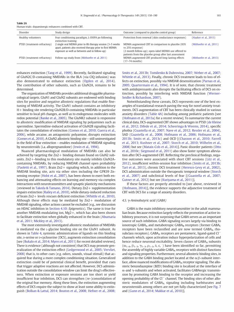
Table 2AHuman trials: dopaminergic enhancers combined with CBT.
Disorder Study design Outcome (compared to placebo control group) Reference
Healthy volunteers Fear conditioning paradigm, L-DOPA po followingextinction training
Protection from renewal (skin conductance responses) (Haaker et al., 2013)
PTSD (treatment-refractory) 2 single oral MDMA prior to 8h therapy session (3–5 weeksapart, patients also received therapy prior to first MDMAexposure as well as between and in follow-up)
MDMA augmented CBT in comparison to placebo (83%vs 25% response)(2 month follow-up); open-label MDMA use offered topatients from the placebo group after last assessment
(Mithoefer et al., 2011)
PTSD (treatment-refractory) Follow-up study from (Mithoefer et al. 2011) MDMA augmented CBT produced long-lasting effects(17–74 months)
(Mithoefer et al., 2013)
161N. Singewald et al. / Pharmacology & Therapeutics 149 (2015) 150–190
enhances extinction (Tang et al., 1999). Recently, facilitated signalingof GluN2C/D-containing NMDARs in the BLA (via CIQ infusion) wasalso demonstrated to enhance extinction (Ogden et al., 2014).The contribution of other subunits, such as GluN2A, remains to bedetermined.
The organization ofNMDARsprovides additional druggable pharma-cological targets. GluN1 and GluN2 subunits are endowed with bindingsites for positive and negative allosteric regulations that enable fine-tuning of NMDAR activity. The GluN1 subunit contains an inhibitoryH+ binding site rendering GluN2B/D-containing NMDARs in particularsensitive to local pH changes, as well as to endogenous molecules withredox potential (Banke et al., 2005). The GluNR2 subunit is responsiveto allosteric modification of NMDAR signaling by polyamines such asspermidine. Spermidine-induced activation of GluN2B signaling facili-tates the consolidation of extinction (Gomes et al., 2010; Guerra et al.,2006), while arcaine, an antagonistic polyamine, disrupts extinction(Gomes et al., 2010). A GluN2 allosteric binding site – still uninvestigatedin the field of fear extinction – enables modulation of NMDAR signalingby neurosteroids (i.a. allopregnanolone) (Irwin et al., 1994).
Nuanced pharmacological modulation of NMDARs can also beachieved by targeting the Zn2+ binding domain found on GluN2 sub-units. Zn2+-binding to this modulatory site mainly inhibits GluN2A-containing NMDARs, by reducing NMDAR channel open probability(Paoletti et al., 1997). Brain Zn2+ signaling which, in addition to theNMDAR binding site, acts via other sites including the GPR39 Zn-sensing receptor (Holst et al., 2007), has been shown to exert both en-hancing and attenuating effects on learning and memory via complexinteractionswithneurotransmitters and synaptic plasticitymechanisms(reviewed in Takeda & Tamano, 2014). Dietary Zn2+ supplementationimpairs extinction (Railey et al., 2010), while dietary-induced reductionof brain Zn2+ levels rescues deficient extinction (Whittle et al., 2010).Although these effects may be mediated by Zn2+ modulation ofNMDAR signaling, other actions cannot be excluded (e.g., see discussionon HDAC inhibition in Section 4.10: Epigenetics). The same is true foranother NMDAR-modulating ion, Mg2+, which has also been shownto facilitate extinction when globally enhanced in the brain (Abumariaet al., 2011; Mickley et al., 2013).
The most extensively investigated allosteric modulation of NMDARsis mediated via the L-glycine binding site on the GluN1 subunit. Asshown in Table 4, systemic administration of ligands on this bindingsite, D-serine or D-cycloserine (DCS), augments extinction consolidation[see (Bukalo et al., 2014;Myers et al., 2011) for recent detailed reviews].There is evidence (although not consistent) that DCSmay promote gen-eralization of the extinction effect (Ledgerwood et al., 2005; Vervliet,2008) that is, to other cues (e.g. odors, sounds, visual stimuli) that ac-quired fear during a more complex conditioning situation. Generalizedextinction could be of potential clinical benefit, provided that cuesthat trigger adaptive reactions are not affected. However, DCS adminis-tration outside the consolidation window can limit the drug's effective-ness. When extinction or exposure sessions are too short or yieldinsufficient fear inhibition, DCS can strengthen (re-)consolidation ofthe original fear memory. Along these lines, the extinction augmentingeffects of DCS require the subject to show at least some ability to extin-guish (Bolkan & Lattal, 2014; Bouton et al., 2008; Hefner et al., 2008;
Smits et al., 2013b; Tomilenko & Dubrovina, 2007; Weber et al., 2007;Whittle et al., 2013). Finally, chronic DCS treatment leads to loss of ef-fects on extinction, possibly via NMDAR desensitization (Parnas et al.,2005; Quartermain et al., 1994). It is of note, that chronic treatmentwith antidepressants also disrupts the facilitating effects of DCS on ex-tinction, possibly by interfering with NMDAR function (Werner-Seidler & Richardson, 2007).
Notwithstanding these caveats, DCS represents one of the best ex-amples of translational research paving the way for novel anxiety treat-ments. DCS augmentation of CBT has been clinically studied in variousanxiety disorders (Table 4A) including among pediatric patients [see(Hofmann et al., 2013a) for a recent review]. To summarize the currentclinical data, DCS-augmented CBT shows advantages in PTSD (de Kleineet al., 2012; Difede et al., 2014; Scheeringa & Weems, 2014), specificphobia (Guastella et al., 2007; Nave et al., 2012; Ressler et al., 2004),SAD (Guastella et al., 2008; Hofmann et al., 2006; Hofmann et al.,2013b; Smits et al., 2013c) and OCD [(Chasson et al., 2010; Farrellet al., 2013; Kushner et al., 2007; Storch et al., 2010; Wilhelm et al.,2008) but see (Mataix-Cols et al., 2014)]. Panic disorder patients (Ottoet al., 2010c; Siegmund et al., 2011) also show faster symptom allevia-tion after DCS-augmented CBT. Reflecting the preclinical findings, nega-tive outcomes were associated with short CBT sessions (Litz et al.,2012), insufficient within-session fear inhibition (Smits et al., 2013b;Tart et al., 2013), chronic DCS treatment (Heresco-Levy et al., 2002),DCS administration outside the therapeutic temporal window (Storchet al., 2007) and subclinical levels of fear [(Guastella et al., 2007;Gutner et al., 2012) but see (Kuriyama et al., 2011)].
If these factors are properly attended to [see above, reviewed in(Hofmann, 2014)], the evidence supports the adjunctive treatment ofCBT with DCS for a range of anxiety disorders.
4.5. γ-Aminobutyric acid (GABA)
GABA is the main inhibitory neurotransmitter in the adult mamma-lian brain. Because extinction largely reflects the promotion of active in-hibitory processes, it is not surprising that GABA serves as an importantsource of such inhibition. GABA signaling occurs largely via binding toionotropic GABAA and metabotropic GABAB receptors (former GABAC
receptors have been reclassified and are now termed GABAA rho-subclass receptors). GABAA receptors are pentameric, ligand-gated Cl−
channels which, upon activation induce hyperpolarization of cells andhence reduce neuronal excitability. Seven classes of GABAA subunits(α1–6, β1–3, γ1–3, ρ1–3, δ, ε, ) have been identified so far, permittingthe assembly of highly variable GABAA receptors with distinct functionsand signaling properties. Furthermore, several allosteric binding sites, inaddition to the GABA binding pocket located at the α/β-subunit inter-face, allownuancedmodifications of GABAA receptor signaling. The allo-steric benzodiazepine (BZD) binding site is localized at the interface ofα-and γ-subunits and when activated, facilitates GABAergic transmis-sion by promoting GABA binding to the receptor and increasing theopening probability of the Cl− channel. The binding sites of other allo-steric modulators of GABAA signaling including barbiturates andneurosteroids among others are not yet fully characterized [see Fig. 7and (Gunn et al., 2014; Makkar et al., 2010)].

Table 3Noradrenergic (NE) signaling in fear extinction (preclinical studies).
Drug/manipulation Extinction learning Extinction retrieval Longterm extinction Route Reference
Enhancing noradrenergic signalingNoradrenaline ns + ns icv (Merlo and Izquierdo, 1967)Methylphenidate + + ns ip (Abraham et al., 2012)Noradrenaline ns +1 ns BLA (Berlau and McGaugh, 2006)
ns No effect1 ns BLA (Fiorenza et al., 2012)ns No effect1 ns CA1 (Fiorenza et al., 2012)ns −1 ns IL (Fiorenza et al., 2012)
Atomoxetine (NE reuptake inhibitor) ns No effect6 + (SR)5 systemic (Janak and Corbit, 2011)Isoproterenol (β ag) No effect ns ns ip (subchronic) (Do-Monte et al., 2010b)
ns (+)1 ns ip (subchronic) (Do-Monte et al., 2010b)ns (+)1 ns mPFC (Do-Monte et al., 2010b)
Yohimbine (α2 ant)8 + ns ns sc (Cain et al., 2004)ns No effect1 ns sc (Cain et al., 2004)+#2 +2 ns sc (Cain et al., 2004)+ + − ip (Morris and Bouton, 2007)No effect7 No effect ns ip (Mueller et al., 2009)ns +* ns ip (Hefner et al., 2008)ns No effect6 + (SR)5 systemic (Janak and Corbit, 2011)
Atipamezole (α2 ant)8 ns No effect4 ns sc (Davis et al., 2008)
Inhibiting noradrenergic signalingPrazosine (α1 ant) ns No effect1 ns ip (subchronic) (Do-Monte et al., 2010a)
− ns ns ip (subchronic) (Do-Monte et al., 2010a)− ns ns intra-mPFC (Do-Monte et al., 2010a)
Propanolol (β ant) No effect ns ns sc (Cain et al., 2004)ns No effect1 ns sc (Cain et al., 2004)No effect2 (−)3 ns sc (Cain et al., 2004)(+)# No effect ns ip (Rodriguez-Romaguera et al., 2009)− − ns ip (subchronic) (Do-Monte et al., 2010b)ns No effect1 ns ip (subchronic) (Do-Monte et al., 2010b)
Atenolol (β ant) − ns ns mPFC (Do-Monte et al., 2010b)ns No effect1 ns mPFC (Do-Monte et al., 2010b)
Sotalol (β ant) No effect No effect ns ip (Rodriguez-Romaguera et al., 2009)Timolol (β ant) ns +1 ns BLA (Fiorenza et al., 2012)Propanolol (β ant) No effect − ns IL (Mueller et al., 2008)
ns No effect1 ns IL (Mueller et al., 2008)Timolol (β ant) ns +1 ns IL (Fiorenza et al., 2012)
ns No effect1 ns CA1 (Fiorenza et al., 2012)
1 Drug administration following extinction training; 2 spaced CS extinction, US=0.7mA; 3 spaced CS extinctionwhen US=0.4mA; no effect when US=0.7mA, 4 cocaine-conditioned placepreference, 5 4 weeks, 6 instrumental lever press response, 7 reduced fear expression at start of extinction training, if extinction is performed in the same context as conditioning, 8 note thatthe net effect of α2 blockade is enhancement of noradrenergic signaling (see text).* Facilitates rescue of impaired fear extinction; ** facilitates extinction of remotememories, *** only in older animals (7months), younger ones (2months) are not affected, # reduced fearexpression at the beginning of extinction training.+, Improved; −, impaired; (+) or (−), only minor effects; ip, intraperitoneal injection; injection; sc, subcutanous injection; icv, intracerebroventricular injection; ns, not studied; HPC,intra-hippocampal administration; BLA, intra-basolateral amygdala administration; IL, infralimbic cortex; PL, prelimbic cortex; CA1, cornu ammonis 1; Ren, Fear renewal; SR, spontaneousrecovery; Re-in, reinstatement; ag, agonist; ant, antagonist, KO, knock-out; #, reduced fear expression at start of extinction training.
162 N. Singewald et al. / Pharmacology & Therapeutics 149 (2015) 150–190
In preclinical studies, the robust inhibitory effect of full GABAA
receptor agonists at the GABA binding site (α/β-subunit interface)(e.g. muscimol), is often used to inactivate a brain region to clarify itsparticipation in certain functions including extinction. Muscimol-injections into the BLA (Laurent & Westbrook, 2008; Laurent et al.,2008; Sierra-Mercado et al., 2011), mPFC/IL (Laurent & Westbrook,2008, 2009; Sierra-Mercado et al., 2011) and HPC [(Corcoran et al.,2005; Sierra-Mercado et al., 2011), see Table 5 for a summary] disruptextinction learning and memory, supporting the crucial involvement
Table 3AYohimbine combined with CBT in small-scale clinical trials.
Disorder Study design
Social anxiety disorder (SAD) 4 sessions consisting of yohimbine being administeredprior to a CBT a session
Claustrophobic fear 1 single oral yohimbine dose prior to an exposure b session
Fear of flying 2 single oral yohimbine prior to VRET session
VRET, virtual reality exposure.a CBT involved an oral presentation on challenging topics in front of the therapists, other grb behavioral approach task,c Lack of yohimbine effectmight possibly have beendue to thepowerful influence of VRET ex
(drug condition) that ‘washed out’ or overrode any effect of manipulation (Meyerbroeker et a
of these areas. However, muscimol infusion into the BLA and IL facili-tates extinction in some studies (Akirav et al., 2006).
There is solid evidence that fear extinction learning is associatedwith upregulation of GABAergic markers in the AMY, underscoring theimportance of a certain level of GABAergic signaling in this brain areafor the successful formation of extinctionmemories. For example, levelsof gephyrin, a clustering protein facilitating postsynaptic scaffolding ofGABAA receptors, were enhanced 2 h following extinction trainingalong with enhanced surface expression of GABAA receptors in
Outcome (compared to placebo control group) Reference
Yohimbine augmented CBT-induced improvement inSAD (self-report measures; 21 day follow-up)
(Smits et al., 2014)
Yohimbine augment CBT-induced reduced fear ofenclosed spaces (7 day follow-up)
(Powers et al., 2009)
No fear augmenting effect of yohimbine c (Meyerbroeker et al., 2012)
oup members, and confederates,
posure itself leading to a greater impact on the results from this than from themanipulationl., 2012),

Fig. 6. Overview of research on the role of the glutamatergic system in extinction.
163N. Singewald et al. / Pharmacology & Therapeutics 149 (2015) 150–190
the BLA (Chhatwal et al., 2005a). Extinction learning also increasesGABAA receptor α2- and β2 subunit mRNA and levels of the GABA-synthesizing enzyme glutamate decarboxylase (GAD) and the GABAtransporter GAT1 (Heldt & Ressler, 2007). Further demonstrating theimportance of GABAergic signaling to extinction, fear extinction learn-ing is disrupted by mutation-induced deficits in the activity-dependent GAD isoform GAD65 (Sangha et al., 2009), and by viralknock-down of the constitutive GAD isoform GAD67 (Heldt et al.,2012). While these findings suggest that upregulation of GABAergicsignaling supports extinction, GABAA receptor antagonists such as pic-rotoxin or bicuculline can enhance extinctionmemorieswhen adminis-tered systemically (McGaugh et al., 1990), or when infused into the BLA(Berlau &McGaugh, 2006) or specific (fear promoting) areas (PL) of themPFC (Fitzgerald et al., 2014b; Thompson et al., 2010). The reason forthese apparent contradictions is not yet clear.
Preliminary findings from studies using genetically modified micesuggest that the development of subunit-selective GABAA receptordrugs could be a promising way to more distinctly utilize GABAergicmechanisms facilitating extinction. For example, there is reducedmRNA expression of α5 and γ1 subunits in the AMY of a mousemodel of enhanced anxiety (Tasan et al., 2011) displaying impaired ex-tinction (Yen et al., 2012). In addition, deletion of α5-containing GABAA
receptors in the HPC is sufficient to impair fear extinction recall (Yeeet al., 2004)while global deletion of theGABAAα3 subunit producesmod-est improvements in extinction learning (Fiorelli et al., 2008).
An additional possible explanation for some paradoxical findingsconcerning GABA regulation and AMY function in extinction is the factthat most of the prior studies have performed pharmacological or ge-netic manipulations that affect either the entire brain, just the AMY, oreven only AMY subregions. Despite this, we now know that GABA

Table 4Glutamatergic signaling in fear extinction (preclinical studies).
Drug/manipulation Extinctiontraining
Extinctionretrieval
Longtermextinction
Route Reference
Facilitating AMPA signalingPEPA (AMPA potentiator) No effect* No effect* ns ip (Whittle et al., 2013)
No effect* +* ns ip (Yamada et al., 2011)+# + + (Re-in) ip (Yamada et al., 2009)+ + ns ip (Zushida et al., 2007)
PEPA + NBQX (AMPA ant) No effect No effect ns ip (Zushida et al., 2007)PEPA (AMPA potentiator) (+) (+) ns PL (Zushida et al. 2007)
+ + ns BLA/ceA (Zushida et al., 2007)
Inhibiting AMPA signalingCNQX (AMPA ant) ns No effect ns BLA (Falls et al. 1992; Lin et al. 2003b; Zimmerman and
Maren 2010)
Facilitating NMDA signaling
D-Cycloserine ns + ns ip (Walker et al., 2002)ns +1 ns Systemic (Ledgerwood et al., 2003)ns +1 + (Re-in) Systemic (Ledgerwood et al., 2004)ns +1 ns Systemic (Ledgerwood et al., 2005)ns +1 ns ip (Parnas et al., 2005)ns + ns ip (Yang and Lu, 2005)ns + ns ip (Lee et al., 2006)ns + No effect sc (Woods and Bouton 2006)ns + ns sc (Werner-Seidler and Richardson, 2007)ns + ns Systemic (Weber et al., 2007)ns + ns ip (Yang et al., 2007)ns + ns ip (Yang et al., 2007)ns + ns ip (Hefner et al., 2008)ns + ns po (Yamamoto et al. 2008)ns + ns ip (Matsumoto et al., 2008)+ + ns ip (Silvestri and Root, 2008)ns + ns sc (Langton and Richardson, 2008)ns + No effect sc (Bouton et al., 2008)ns + ns ip (Lin et al., 2010)+# + + (Re-in) ip (Yamada et al., 2009)+ ns ns ip (Lehner et al., 2010)ns +1 ns Systemic (Langton and Richardson, 2010)No effect* +* ns ip (Yamada et al., 2011)No effect + ns ip (Toth et al., 2012b)ns +1 ns ip (Toth et al., 2012b)ns + ns Systemic (Gupta et al., 2013)ns + ns ip (Matsuda et al., 2010)+ + ns ip (Bai et al., 2014)ns +1* ns ip (Whittle et al., 2013)ns + ns BLA (Walker et al., 2002)ns +1 ns BLA (Ledgerwood et al., 2003)ns + ns BLA (Lee et al., 2006)ns + ns BLA (Mao et al., 2008)ns + ns BLA (Lee et al., 2006)ns + ns BLA (Akirav et al., 2009)ns No effect ns BLA (Bolkan & Lattal, 2014)ns + ns HPC (Bolkan & Lattal, 2014)ns + ns HPC (Ren et al., 2013)
Spermidine ns +1 ns HPC (Gomes et al., 2010)
Inhibiting NMDA signalingMK-801 (non-competitive NMDA ant) ns − ns Systemic (Baker & Azorlosa, 1996); (Storsve et al., 2010)
ns ns + (Re-in) sc (Johnson et al., 2000)ns −1 ns ip (Lee et al., 2006); (Liu et al., 2009)ns − ns Systemic (Langton et al., 2007); (Chan & McNally, 2009)
CPP (competitive NMDA ant) No effect −5 ns ip (Santini et al., 2001); (Sotres-Bayon et al., 2007)No effect − ns mPFC (Burgos-Robles et al., 2007)ns −1 ns mPFC (Burgos-Robles et al., 2007)− − ns BLA (Parsons et al., 2010)
AP-5 (competitive NMDA ant) ns − ns BLA (Falls et al., 1992)−## − ns BLA (Lee & Kim, 1998)ns −1 ns BLA (Lin et al., 2003a); (Laurent et al., 2008);
(Fiorenza et al., 2012)No effect − ns BLA (Lin et al., 2003a); (Zimmerman & Maren, 2010)ns − ns CA1 (Szapiro et al., 2003)ns −1 ns CA1 (Fiorenza et al., 2012)ns −1 ns mPFC (Fiorenza et al., 2012)
164 N. Singewald et al. / Pharmacology & Therapeutics 149 (2015) 150–190

Table 4 (continued)
Drug/manipulation Extinctiontraining
Extinctionretrieval
Longtermextinction
Route Reference
Inhibiting NMDA signalingifenprodil (non-comp NR2B-NMDA ant) − − ns ip (Sotres-Bayon et al., 2007)
ns −1 ns Systemic (Sotres-Bayon et al., 2009)− − ns BLA (Sotres-Bayon et al., 2007); (Laurent &
Westbrook, 2008); (Laurent et al., 2008)ns No effect1 ns BLA (Laurent et al., 2008); (Laurent & Westbrook,
2008); (Sotres-Bayon et al., 2009)No effect − ns mPFC (Laurent & Westbrook, 2008)No effect No effect ns mPFC (Sotres-Bayon et al., 2009)No effect −1 ns mPFC (Laurent & Westbrook, 2008); (Sotres-Bayon
et al., 2009)ns −1 ns HPC (Gomes et al., 2010)
Ro25-6981 (non-comp NR2B-NMDA ant) − No effect ns ip (Dalton et al., 2008, 2012)
Facilitating mGluR signalingCDPPB (mGluR5 pos modulator) +3 ns ns sc (Gass & Olive, 2009)AMN082 (mGluR7 ag) +* +* ns ip (Whittle et al., 2013)
ns + ns po (Fendt et al., 2008)− No effect ns ip (Toth et al., 2012b)ns −1 ns ip (Toth et al., 2012b)(+) No effect ns BLA (Dobi et al., 2013)− − ns mPFC (Morawska & Fendt, 2012)
Inhibiting mGluR signalingCPCCOEt (non-comp mGluR1 ant) − − ns BLA (Kim et al., 2007)
No effect No effect No effect (SR) ip (Mao et al., 2013)mGluR4 KO ns + ns No drug (Davis et al., 2013)mGluR5 KO − − ns No drug (Xu et al., 2009)MTEP (mGluR5 ant) No effect − − (SR) ip (Mao et al., 2013)
ns ns − (SR) BLA (Mao et al., 2013)MPEP (allosteric mGluR5 ant) No effect No effect ns ip (Toth et al., 2012b)
ns No effect1 ns ip (Toth et al., 2012b)No effect − ns ip (Fontanez-Nuin et al., 2011)No effect − ns IL (Fontanez-Nuin et al., 2011)− − ns IL (Sepulveda-Orengo et al., 2013)
mGluR7 KO −4 ns ns No drug (Callaerts-Vegh et al., 2006); (Goddyn et al.,2008)
+ + ns No drug (Fendt et al., 2013)siRNA knock-down of mGluR7 −2 ns ns No drug (Fendt et al., 2008)mGluR8 KO No effect# No effect ns No drug (Fendt et al., 2013)(S)-3,4-DCPG (mGluR8 ag) − No effect ns BLA (Dobi et al., 2013)
1Drug administration following extinction training; 2conditioned taste aversion; 3 cocaine conditioned place preference; 4 food-rewarded operant conditioning, 5 reduced locomotion.* Facilitates rescue of impaired fear extinction; # reduced fear expression at the beginning of extinction training; ## enhanced fear expression at the beginning of extinction training.+, Improved; −, impaired; (+) or (−), only minor effects; po, peroral administration; ip, intraperitoneal injection; sc, subcutaneous injection; ns, not studied; BLA, intra-basolateralamygdala administration; HPC, hippocampus; IL, infralimbic cortex; SR, spontaneous recovery; Re-in, reinstatement; ag, agonist; ant, antagonist, KO, knock-out;
165N. Singewald et al. / Pharmacology & Therapeutics 149 (2015) 150–190
receptors are found on most of the neuronal cell types and thus even asubregion-specific manipulation is likely to affect many different cellpopulationswhichmay have opposing functional effects. One recent at-tempt to address this complexity involved the use of an inducibleGABAa1 knockout strategy limited only to the corticotropin-releasing-hormone containing neuronal population (Gafford et al., 2012)),which revealed relatively specific effects, including enhanced anxietyand extinction deficits. It is likely that future studies aimed at cell-typespecific manipulations will help address the vast complexity of themany different cell types combined with the large number of differentGABA receptor populations.
Asmentioned above, themultiple different binding sites foundwith-in GABAA receptors can be used to elicit a more nuanced modulation ofGABAA receptor activity. Positive allosteric modulation of GABAA recep-tor activity via BZD, represents the most widely described drug-basedstrategy for acute treatment of anxiety states (Macaluso et al., 2010).The prevalence of BZD use among patients suffering frommental disor-ders is high, and is estimated to range between 40 and 70% of these pa-tients (Clark et al., 2004) However anxiolytic treatment with BZDs isrecommended only for short periods of time, due to several disadvan-tages including its sedative effects, the development of dependenceand difficulties with discontinuing chronic treatment (Otto et al.,2002). The sedating and calming effects of BZDs might in fact interferewith extinction possibly via reduced arousal and decreased release ofneurochemicals (e.g. noradrenaline) and stress hormones (e.g.
glucocorticoids) that support extinction [(Bentz et al., 2010), seeSection 4.8Glucocorticoids) for details]. BZDsmay also render extinctionmemories dependent on the drug-induced internal anxiolytic-state(state dependent learning), which upon BZD discontinuation may nolonger be accessible. These concerns are borne out by preclinical datashowing that systemic (Bouton et al., 1990; Bustos et al., 2009;Goldman, 1977; Hart et al., 2009, 2010, 2014; Pereira et al., 1989) orintra-BLA (Hart et al., 2009, 2010) BZD administration impairs extinc-tion (Table 5), presumably also through state-dependent mechanisms.Systemic treatment with a BZD inverse agonist (Harris & Westbrook,1998; Kim & Richardson, 2007, 2009) has similar effects (see Table 5for a summary]. Hence, combining BZD with CBT needs careful consid-eration. As well as further development of BZDs targeting specific sub-units of the GABAA receptor [reviewed in (Griebel & Holmes, 2013)],which provides some hope, there is preliminary evidence that putativenovel anxiolytics that do not impair extinction learning [e.g. neuropep-tide S; (Slattery et al., in press) see Section 4.7Neuropeptides] can be de-veloped to help reduce patients' aversion to the unpleasant anddemanding procedure of recalling details of their traumatic memories,thus increasing their willingness to interact with the psychotherapist.
In contrast to the relatively large number of studies of the GABAA re-ceptor, only a few studies have investigated the role of GABAB receptors.Functional GABAB receptors are heterodimers containing a GABAB1 anda GABAB2 subunit; the GABAB1 subunit has two isoforms—GABAB1a andGABAB1b (Fritschy et al., 1999). While functional deletion of GABAB

Table 4AHuman trials: D-cycloserine (DCS) combined with CBT.
Disorder Study design Outcome (compared to placebo control group) Reference
Healthy volunteers DCS or placebo 2–3 h prior extinction training No effect on fear extinction or fear recovery in healthyvolunteers
(Guastella et al., 2007)
Healthy volunteers DCS, placebo, valproic acid or combination of DCSand valproic acid 1.5 h prior extinction training
Administration of DCS, valproic acid or DCS/valproicacid combination facilitates fear extinction andprotects from reinstatement
(Kuriyama et al., 2011)
Healthy volunteers DCS or placebo 2 h prior extinction training No effect on extinction learning and retention inhealthy volunteers
(Klumpers et al., 2012)
Acrophobia DCS or placebo 1 h prior 2 sessions of virtual realityexposure3 month follow-up
Reduced fear symptoms in DCS group at all timepoints (Ressler et al., 2004)
Acrophobia 2 sessions virtual reality exposure plus DCS orplacebo after the sessions1 month follow-upRe-evaulation of (Tart et al., 2013)
No advantage of DCS over placebo augmented VREDCS effect depends on success of exposure sessions(within-session fear reduction)
(Tart et al., 2013)(Smits et al., 2013a)
Snake phobia DCS or placebo 1 h prior a single exposure session Same level of improvement with DCS, but DCS groupachieved fear reduction quicker
(Nave et al., 2012)
Panic disorder DCS or placebo 1 h prior CBT session 3–51 month follow-up
DCS group showed greater reduction in panicsymptom severity at all timepoints
(Otto et al., 2010c)
Panic disorder with agoraphobia DCS or placebo 1 h prior 3 individual exposuresessions + 8 group CBT sessions 5 monthsfollow-up
No benefits of DCS at all timepoints, but initialbenefical effects in severely symptomatic patients?
(Siegmund et al., 2011)
PTSD DCS or placebo 1 h prior 10 weekly exposuresessions
No overall enhancement of treatment effects.Higher symptom reduction in severe cases.
(de Kleine et al., 2012)
PTSD(combat-related)
DCS or placebo 30 min prior exposure session 2–6 Weaker symptom reduction compared to placebogroup
(Litz et al., 2012)
PTSD DCS or placebo 1.5 h prior 12 weekly VRE session 6months follow-up
DCS group showed earlier and greater improvement aswell as higher remission rates
(Difede et al., 2014)
pediatric PTSD DCS or placebo 12 1 h prior session 5–12 No difference in symptom reduction, DCS groupshowed trend for faster response and better retentionin 3 month follow-up
(Scheeringa & Weems, 2014)
SAD 1× psychoeducation; DCS or placebo 1 h prior to 4 hexposure session (5×, individual or group)1 month follow-up
Decreased self-reported social anxiety symptoms inDCS group after 1 month
(Hofmann et al., 2006)
SAD DCS or placebo 1 h prior to 4 h exposure therapy(5×)
Fewer social fear and avoidance in DCS group.Significant differences in DCS vs placebo following 3rdexposure session.
(Guastella et al., 2008)
SAD DCS or placebo with CBT Greater overall rates of improvement and lowerpost-treatment severity
(Smits et al., 2013b)
SAD 12 weeks; DCS or placebo 1h prior 5 exposuresessions (12 sessions at all); 6-months follow-up
Similar response and remission rates, DCS groupimproved quicker
(Hofmann et al., 2013b)
OCD D-Cycloserine (DCS) or placebo 4 h prior eachexposure session, 12 weeks (one exposuresession/week)
No statistically significant difference.High response rates in treatment and placebo group.
(Storch et al., 2007)
OCD 2×/week DCS or placebo 2 h prior exposure andresponse prevention (ERP) sessions(max 10)3 months follow-up
Faster improvements in DCS groupNo difference between DCS and placebo group(no further benefit).
(Kushner et al., 2007)
OCD 2×/week DCS or placebo 1 h prior 10 ERP sessions1 month follow-upRe-evaluation of Wilhelm et al., 2008
Faster improvements in DCS groupNo difference between DCS and placebo group(no further benefit).Specific improvements of DCS in the first 5 sessions,6× faster than placebo group
(Wilhelm et al., 2008)(Chasson et al., 2010)
Pediatric OCD DCS or placebo 1 h prior weekly session 4–10(psychoeduction, cognitive training and ERP) for 10wks
Modest reduction of obsessive symptoms (Storch et al., 2010)
Pediatric OCD DCS or placebo 1 h prior session 5–9 ERP-CBT Significant improvements in OCD severity fromposttreatment to 1-month follow-up in severe anddifficult-to-treat pediatric OCD
(Farrell et al., 2013)
Pediatric OCD DCS or placebo immediately after each of 10 CBT(ERP) sessions1 year follow-up
both groups improved;no significant advantage of DCS at any timepoint
(Mataix-Cols et al., 2014)
166 N. Singewald et al. / Pharmacology & Therapeutics 149 (2015) 150–190
receptors (GABAB1a/b knock-out) impairs extinction learning (Jacobsonet al., 2006), pharmacological GABAB receptor antagonism does not(Heaney et al., 2012; Sweeney et al., 2013). Systemic administration of(non-selective) agonists or positive allosteric modulators of GABAB
signaling also has inconsistent effects (impairing or negative) onextinction [(Heaney et al., 2012; Sweeney et al., 2013) see Table 5]. Afinal answer to this question regarding the role of GABAB receptorsmay have to await the availability of GABAB1a-and GABAB1b-selectivecompounds
Taken together, the available evidence indicates that GABA signalinghas a major, but spatially and temporally restricted role in the formationof fear extinction memories. While GABA that is engaged during extinc-tion is likely to support synaptic plasticity, increasing GABAergic toneglobally seems to limit the efficacy of extinction-based therapies bymechanisms outlined above. Therefore, it is unlikely that drugs broadlytargeting the GABA systemwill be useful in this respect. Pharmacologicaladjuncts to exposure therapy targeting the GABA system would ratherneed to strike a balance between driving and maintaining relevant

Fig. 7. Overview of research on the role of the GABAergic system in extinction.
167N. Singewald et al. / Pharmacology & Therapeutics 149 (2015) 150–190
GABA activity without over-activating the system. The evidence so farsuggests that subunit-selective or allosteric (distinct from BZD) modula-tion of GABAA receptor signaling may hold promise in this regard.
4.6. Cannabinoids
A growing body of literature demonstrates that the endogenous can-nabinoid (eCB) system modulates neuronal excitability in stressful andfearful situations (de Bitencourt et al., 2013; Gunduz-Cinar et al.,2013a). eCB signaling has been implicated in emotional and cognitiveprocessing of threatening stimuli, for instance during the consolidation
of fear and fear extinction memories (for more detailed recent reviewssee (Riebe et al., 2012; Gunduz-Cinar et al., 2013b). Upon neuronalactivation, an eCB (anandamide) and 2-arachidonoylglycerol (Fig. 8)are rapidly synthesized in the postsynaptic neuron (within minutes)and released into the synaptic cleft to modulate presynaptic signalingby the activation of Gi/0-coupled CB-1 receptors (CB-2 receptors aremainly expressed on immunecells, osteoblasts, osteoclasts and hemato-poietic cells but also on neuroglia).
Supporting a role of eCB signaling in the extinction of aversive mem-ories (see Fig. 8 for overview), there are increased eCB levels in themouse BLA following extinction training (Marsicano et al., 2002).

Table 5GABAergic signaling in fear extinction (preclinical studies).
Facilitating GABA signaling
Drug/manipulation Extinction training Extinction retrieval Longterm extinction Route Reference
GABAA agonists GABA-binding site (αβ interface)Muscimol +# No effect ns BLA (Akirav et al., 2006)
ns +1,7 ns BLA (Akirav et al., 2006)−# − ns BLA (Laurent et al., 2008), (Laurent & Westbrook, 2008)ns −1 ns BLA (Laurent & Westbrook, 2008)(+)# − ns BLA (Sierra-Mercado et al., 2011)− − ns mPFC (Laurent & Westbrook, 2008)ns −1 ns mPFC (Laurent & Westbrook, 2008)ns No effect1, ns IL (Akirav et al., 2006)+# (+) ns IL (Akirav et al., 2006)− − ns IL (Sierra-Mercado et al., 2011)No effect − ns IL (Laurent & Westbrook, 2009)(+)# No effect ns PL ((Laurent & Westbrook, 2009); (Sierra-Mercado et al.,
2011))ns ns + (Ren-A)8 dHPC (Corcoran et al., 2005)− − No effect (Ren-A) dHPC (Corcoran et al., 2005)(+)# − ns vHPC (Sierra-Mercado et al., 2011)
GABAA: agonists BZD-binding site αγ interface)Chlordiazepoxide − ns ns ip (Goldman, 1977)
ns − ns ip (Bouton et al., 1990)Diazepam ns − ns ip (Pereira et al., 1989)
ns − ns ip (Bouton et al., 1990)Midazolam ns −1 ns ip (Bustos et al., 2009)
−##6 − ns ip (Hart et al., 2009, 2010), (Hart et al., 2014)−#6 − ns BLA (Hart et al., 2009, 2010, 2014)
GABAB receptor agonistsBaclofen ns − ns ip (Heaney et al., 2012)GS39783 (positive modulator GABAB) ns No effect ns po (Sweeney et al., 2013)
Inhibiting GABA signalingDrug/manipulation Extinction learning Extinction retrieval Longterm extinction Route Reference
GAD67 KD in BLA − ns ns No drug (Heldt et al., 2012)GAD65 KO −2 −2 ns No drug (Sangha et al., 2009)α5 KD in HPC ns −3 ns No drug (Yee et al., 2004)α1 KO in CRF+ cells − − ns No drug (Gafford et al., 2012)Picrotoxin ns + ns ip (McGaugh et al., 1990)
ns +4 ns IL (Thompson et al., 2010)ns +4 ns PL (Thompson et al., 2010)ns +* ns (Fitzgerald et al., 2014b)
Bicuculline ns +1 ns BLA (Berlau & McGaugh, 2006)Bicuculline + propanolol ns No effect1 ns BLA (Berlau & McGaugh, 2006)FG7142 − − ns ip (Harris & Westbrook, 1998)
ns − ns ip (Kim & Richardson, 2007, 2009)
GABAB receptor antagonismGABAB(1b) KO −5 ns ns No drug (Jacobson et al., 2006)Phaclofen ns No effect ns ip (Heaney et al., 2012)CGP52432 ns No effect ns po (Sweeney et al., 2013)
1 Drug administration following extinction training; 2 only cuedmemory, contextual intact, 3 trace conditioning, 4 reconsolidation (injection prior reactivation of memory), 5 conditionedtaste aversion, 6 midazolam may not impair fear extinction if initial extinction occurs drug-free [see (Hart et al., 2014)], 7 short extinction training (5 CS presentations), 8 injection priorrenewal-testing.# Reduced fear expression at start of extinction training; ## enhanced immobility at start of extinction training;+, Improved; −, impaired; (+) or (−), only minor effects; ip, intraperitoneal injection; po,peroral administration; ns, not studied; HPC, intra-hippocampal administration; BLA, intra-basolateral amygdala administration; IL, infralimbic cortex; PL, prelimbic cortex; Ren-A, Fear renewal in conditioning context; ag, agonist; ant, antagonist, KO, knock-out;
168 N. Singewald et al. / Pharmacology & Therapeutics 149 (2015) 150–190
Conversely, PTSD patients show low brain anandamide levels(Neumeister et al., 2013). In rodents, facilitation of eCB signaling by CB-1 receptor agonists or exogenous cannabinoids [cannabidiol, (Bitencourtet al., 2008)], reuptake inhibitors [AM404, (Bitencourt et al., 2008;Chhatwal et al., 2005b; Pamplona et al., 2008)] or anandamide degrada-tion blockers [AM3506, URB 597, (Gunduz-Cinar et al., 2013b)] enhancesthe formation or consolidation of extinction memories. Blockade orknock-out of CB1-receptors (Cannich et al., 2004; Kamprath et al., 2006;Marsicano et al., 2002; Niyuhire et al., 2007; Pamplona et al., 2006;Plendl &Wotjak, 2010; Reich et al., 2008; Terzian et al., 2011) impairs ex-tinction and blocks the pro-extinction effects of drugs that facilitate eCBtransmission (Chhatwal et al., 2005b; Do Monte et al., 2013;Gunduz-Cinar et al., 2013b) (see Table 6). Collectively, these observations
point to CB-1 receptormediated eCB signaling as a powerfulmodulator ofextinction.
Following up on these studies, intra-cerebral infusions of CB1-receptor agonists and antagonists have revealed a potential role ofmPFC-BLA eCB signaling in extinction [see Table 6, (Do Monte et al.,2013; Ganon-Elazar & Akirav, 2013; Gunduz-Cinar et al., 2013b)]. CB1-receptor activation inhibits both presynaptic glutamate and GABArelease. The release of anandamide and concomitant CB1-receptoractivation induce long-term depression of inhibitory transmission(LTDi) in the BLA (Azad et al., 2004; Gunduz-Cinar et al., 2013b). Inaddition, a recent study demonstrated that enhanced inhibitory inputvia activation of CB1 receptor expressing cholecystokinin (CCK) positiveinterneurons selectively reduces firing of BLA fear neurons and

Fig. 8. Overview of research on the role of the cannabinoid system in extinction.
169N. Singewald et al. / Pharmacology & Therapeutics 149 (2015) 150–190
promotes extinction (Trouche et al., 2013). Of note is the fact that ithas also been shown that one potential mechanism for the CB1 effecton extinction within the AMY is via blockade of CCK release, as an intra-AMY CCK antagonist reversed the extinction blockade of systemic CB1antagonists, and CCK agonists block extinction (Chhatwal et al., 2009).
In summary, current preclinical data suggest that eCB signaling gatesBLA function through plastic changes, including a long-lasting depres-sion of inhibition, to modulate extinction-related AMY activity. Initialresults from clinical studies in healthy volunteers suggest that exoge-nous cannabinoid administration strengthens extinction and protectsagainst reinstatement [see Table 6A, (Das et al., 2013; Rabinak et al.,2013)]. Future clinical trials in extinction-impaired PTSD patients willbe needed to show whether cannabinoids (or substances which in-crease endogenous eCB signaling, such as FAAH inhibitors) mightprove useful as adjuncts to CBT-based therapy.
4.7. Neuropeptides (oxytocin, neuropeptide Y, neuropeptide S, opioids)
Various neuropeptides and their corresponding receptors are local-ized in brain regionsmediating emotional behavior and stress responseand have been considered as potential anxiolytic drug targets (Bowers
et al., 2012; Ebner et al., 2009; Griebel & Holmes, 2013). While otherneuroptides such as hypocretin/Orexin (Flores et al., 2014) may alsobe involved in fear extinction, we will here address the role of oxytocin,neuropeptide Y, neuropeptide S and opioids in extinction.
4.7.1. OxytocinThe oxytocin (OXT) system, strongly implicated in social cognition
and behavior, is posited to play a role in anxiety disorders with impairedsocial functioning (Meyer-Lindenberg et al., 2011; Neumann& Landgraf,2012). OXT is synthesized in the paraventricular (PVN) and supraopticnuclei of the hypothalamus and processed along the axonal projectionsto the posterior pituitary gland. Uponneuronal activationOXT is releasedfrom secretory vesicles into the peripheral circulation as neurohypophy-seal hormone. The central nervous system (CNS) component of OXT isderived from dendritic release of the same cell population (Ludwig &Leng, 2006), as well as from the OXT-synthesizing parvocellular PVNcells sending projections throughout the brain [reviewed in (Gimpl &Fahrenholz, 2001)]. OXT receptors are typically Gq-protein coupled andhave potential allosteric binding sites for Mg2+ and steroids like choles-terol. The distribution of OXT receptors has not been fully delineated, butOXT fibers and neurosecretory nerve endings have been described in,

Table 6Cannabinoid signaling in fear extinction (preclinical studies).
Enhanced cannabinoid signaling
Drug/manipulation Extnctiontraining
Extinctionretrieval
Longtermextnction
Route Reference
Cannabidiol + + ns icv (Bitencourt et al., 2008)+ + ns IL (Do Monte et al., 2013)
Anandamide ns +1 ns dCA1 (de Oliveira Alvares et al., 2008)AM404 (eCB uptake inhibitor) ns + ns ip (Pamplona et al., 2008)
ns + + (Re-in) ip (Chhatwal et al., 2005b)+ (+) ns icv (Bitencourt et al., 2008)No training + ns IL (Lin et al., 2009)
AM3506 (FAAH inhibitor) ns + ns ip (Gunduz-Cinar et al., 2013b)WIN 55,212-2 (low-dose CB1 Ag) +* No effect2 ns ip (Pamplona et al., 2006)WIN 55,212-2 (high-dose CB1 Ag) (−)* No effect2 ns ip (Pamplona et al., 2006)WIN 55,212-2 (CB1 Ag) +° +° ns ip (Pamplona et al., 2006)
ns + nd ip (Pamplona et al., 2008)WIN 55,212-2 (CB1 Ag) ns +4 ns BLA (Ganon-Elazar & Akirav, 2013))
ns +4 ns HPC (Ganon-Elazar & Akirav, 2013)ns No effect4 ns mPFC (Ganon-Elazar & Akirav, 2013)ns + No effect (Re-in,
SR3)IL (Lin et al., 2009)
No training + ns IL (Lin et al., 2009)ns No effect ns PL (Kuhnert et al., 2013)
HU210 No training + ns IL (Lin et al., 2009)URB597 (AEA hydrolysis inhibitor) No training + ns IL (Lin et al., 2009)AM3506 (FAAH inhibitor) + SR141716A(CB1 ant)
ns No effect ns ip + BLA (Gunduz-Cinar et al., 2013b)
AM3506 (FAAH inhibitor) ns + ns BLA (Gunduz-Cinar et al., 2013b)No training No effect ns BLA (Gunduz-Cinar et al., 2013b)
Cannabidiol + SR141716A (CB1 ant) No effect No effect ns IL + ip (Do Monte et al., 2013)
Reduced cannabinoid signalingDrug/manipulation Extnction
learningExtinctionretrieval
Longtermextinction
Administrationroute Reference
CB1 KO − − ns No drug (Cannich et al., 2004)CB1 KO − ns ns No drug ((Dubreucq et al., 2010); (Kamprath et al., 2006);
(Marsicano et al., 2002); (Plendl & Wotjak, 2010))D1-CB1 KO − ns ns No drug (Terzian et al., 2011)SR141716A CB1 ant ns No effect1 ns sc (Marsicano et al., 2002)
− ns ns sc, ip ((Kamprath et al., 2006); (Niyuhire et al., 2007);(Plendl & Wotjak, 2010)
ns −1 ns ip (Suzuki et al., 2004)− No effect ns ip (Pamplona et al., 2006, 2008)ns − ns ip (Chhatwal et al., 2005b)−## No effect ns ip (Bowers & Ressler, 2015)
AM251 (CB1 inverse ag) − ns ns ip (Reich et al., 2008)ns −1 ns dCA1 (de Oliveira Alvares et al., 2008)ns − ns IL (Lin et al., 2009)No training No effect ns IL (Lin et al., 2009)ns − ns PL (Kuhnert et al., 2013)
1 Drug administration following extinction training; 2 drug-free retrieval of contextual fear memory; 3 5 days, 4 single-prolonged-stress model: injection immediately after trauma.*, Recent memory; °, remote memory; ##, enhanced immobility at start of extinction training;+, Improved;−, impaired; (+) or (−), onlyminor effects; ip, intraperitoneal injection; ip, sc, subcutanous injection; ns, not studied; HPC, intra-hippocampal administration; BLA, intra-basolateral amygdala administration; IL, infralimbic cortex; PL, prelimbic cortex; CA1, cornu ammonis 1; Re-in, reinstatement; SR, spontaneous recovery, ag, agonist; ant, antagonist, KO,knock-out;
170 N. Singewald et al. / Pharmacology & Therapeutics 149 (2015) 150–190
among other regions, the AMY [especially CeL (Knobloch et al., 2012;Veinante & Freund-Mercier, 1997)], the dorsal and ventral HPC and themain monoamine nuclei – substantia nigra (dopamine), raphe nuclei(serotonin) and the locus coeruleus (noradrenaline) [see (Gimpl &Fahrenholz, 2001) for review).
Table 6AHuman trials: cannabinoids combined with CBT.
Disorder Study design
Healthy volunteers Fear conditioning paradigm, cannabidiol inhalation prior orfollowing extinction
Healthy volunteers Fear conditioning paradigm, tetrahydro-cannabinol prior extinctioHealthy volunteers Fear conditioning paradigm, Dronabinol prior extinction
Regulatory effects of OXT on stress-induced activation of thehypothalamic–pituitary–adrenal (HPA) axis, as well as direct OXT ef-fects on the brain and specific parts of the extinction circuitry, can affectfear extinction in variousways. Intracerebroventricular (icv) infusion ofOXTprior to fear conditioning does not affect fear learning but facilitates
Outcome (compared to placebo control group) Reference
Reduced fear in retrievalProtection against reinstatement
(Das et al., 2013)
n No effect (Klumpers et al., 2012)Reduced fear in retrieval (Rabinak et al., 2013)

171N. Singewald et al. / Pharmacology & Therapeutics 149 (2015) 150–190
later extinction acquisition and retrieval (Toth et al., 2012a). Conversely,OXT receptor antagonists impair extinction learning and retrieval whenadministered icv prior to fear acquisition (Toth et al., 2012a). More lo-calized OXY infusions into the central CeA reduce fear expression byinhibiting CeM excitatory output to fear-eliciting brainstem structures(Huber et al., 2005) (Viviani et al., 2011), suggesting one possiblemechanism for the effects of icv OXY on extinction.
Another potential mechanismmight be OXT-induced dampening ofstress-induced activation of the HPA axis (de Oliveira et al., 2012;Heinrichs et al., 2003; Quirin et al., 2011; Windle et al., 1997). Asexplained inmore detail in Section 4.8Glucocorticoids, glucocorticoid re-lease during an actual learning process facilitates cognitive processing[reviewed in (Bentz et al., 2010)], hence the release of endogenousOXT during traumatic experiences (Zoicas et al., 2012) may interferewith the consolidation of fear memories. Along these lines, OXT-mediated reduction ofHPA axis activity and subsequent lower glucocor-ticoid levels could interfere with fear acquisition, resulting in a weakerfear memory that is more susceptible to extinction training. In this con-text, OXT administration either icv or intra BLA prior to extinction train-ing disrupts extinction learning [(Toth et al., 2012a) (Lahoud &Maroun,2013) see Table 7 for summary]. OXT infusions into the dorsolateral sep-tum prior to extinction training have the opposite effect and facilitateextinction learning and retrieval in a social fear conditioning paradigmthat may recruit a somewhat different neural circuitry than non-socialforms of fear (Zoicas et al., 2012).
Considering that systemically applied neuropeptides do not crossthe blood–brain-barrier, clinical use of neuropeptides would require adirect pathway to the human brain. In the case of OXT, this may be fea-sible via intranasal administration (Born et al., 2002). Intranasal OXT ad-ministration has been demonstrated to reduce stress-induced cortisolrelease in humans (de Oliveira et al., 2012; Heinrichs et al., 2003;Quirin et al., 2011), and to attenuate AMY hyperactivity as well asAMY-hindbrain coupling in anxiety patients [(Kirsch et al., 2005;Labuschagne et al., 2010)]. In a recent study investigating the effects ofintranasal OXT treatment on fear extinction in healthy volunteers,OXT increased initial fear expression during extinction training,
Table 7Neuropeptide signaling in fear extinction (preclinical studies).
Neuropeptides
Drug/manipulation Extinction training Extinction retrieval
Oxytocin − ns+# +−*5 −*5
Chronic oxytocin (30d) ns No effect*6
NPY + +NPY KO −3⁎⁎⁎ −NPY Y1 KO − No effectNPY Y2 KO No effect No effectNPY Y1/Y2 KO − −Leu31Pro-NPY (Y1 ag) ns +BIBO 3304 (Y1 ant) ns −NPS (+)4 No effect
+ nsSHA68 (NPS-R ant) −4, ⁎⁎⁎ −Naloxone − −
ns No effect1
− −− −No effect No effect
CTAP (MOR ant) − −Dynorphin KO −3 −nor-BNI (KOR ant) ns −1
1 Drug administration following extinction training; 2 social fear conditioning;3 animals show astress model; 6 BTBR mouse autism model and C57BL6J.*** Enhanced fear expression at the beginning of extinction training, # reduced fear expression+, Improved;−, impaired; (+) or (−), only minor effects; ip, intraperitoneal injection; icv, inBLA, intra-basolateral amygdala administration; DLS, dorsolateral septum; vlPAG, ventrolatantagonist, KO, knock-out;
but this effect was gone by the end of training and there was a lowerlevel of fear in the OXT treated group in extinction retrieval (Achesonet al., 2013). These data raise the prospect of OXT-augmented CBTeffectiveness.
4.7.2. Neuropeptide YNeuropeptide Y (NPY) contains 36 amino acids and is the most
widely expressed neuropeptide in themammalian brain. There are par-ticularly high concentrations of NPY in the limbic system, including inthe AMY, the HPC and the periaqueductal gray. Six G-protein coupledNPY receptors (Y1–6) have been identified, among which Y1, Y2, Y4,and Y5 mediate central effects. Y1 mRNA is present at a high level inbrain areas involved in memory processing, namely the AMY, HPC,thalamus, hypothalamus, and the cerebral cortex. The AMY, HPC, andhypothalamus are also rich in Y2 receptors, while Y5 receptors can bedetected in HPC, cingulate cortex and thalamic and hypothalamic nuclei(Parker & Herzog, 1999) [reviewed in (Holmes et al., 2003)].
Icv administration of NPY facilitates extinction learning (Gutmanet al., 2008), while genetic deletion of NPY disrupts extinction acquisi-tion and subsequent retrieval (Verma et al., 2012). Extinction deficitscaused by NPY knock-out are rescued by AAV-mediated NPY re-expression in the BLA (Verma et al., 2012), where NPY co-localizeswith GABAergic neuronsmodifying inhibitory control of BLA projectionneurons (McDonald&Pearson, 1989). To elucidatewhichNPY receptorsmediate the BLA-associated effects on extinction, the consequencesof gene deletion via either deletion of Y1 or Y2 receptors or doubleY1/Y2 deletion have been examined. Deletion of Y1 receptors impairedextinction learning, while Y2 receptor deletion had no effect (Vermaet al., 2012). Underscoring the importance to extinction of Y1 receptorsin the BLA, local infusion of the Y1 agonist Leu31Pro34-NPY prior to ex-tinction training led to stronger extinction retrieval (Gutman et al.,2008; Lach & de Lima, 2013). Further studies are required to furtherdelineate the underlying mechanisms of Y1-mediated facilitation ofextinction, and to investigate the potential clinical use of NPY and Y1receptor agonism as adjunctive therapy to CBT.
Longterm extinction Route Reference
ns icv (Toth et al., 2012a)ns DLS (Zoicas et al., 2012)ns ip (Eskandarian et al., 2013)ns in (Bales et al., 2014)ns icv (Gutman et al., 2008)ns No drug (Verma et al., 2012)ns No drug (Verma et al., 2012)ns No drug (Verma et al., 2012)ns No drug (Verma et al., 2012)ns BLA (Lach & de Lima, 2013)ns BLA (Gutman et al., 2008)No effect BLA (Jungling et al., 2008)ns icv (Slattery et al., in press)− (Ren-A) BLA (Jungling et al., 2008)ns Systemic (McNally & Westbrook, 2003)ns Systemic (McNally & Westbrook, 2003)ns vlPAG (McNally et al., 2004)ns vlPAG (Parsons et al., 2010)ns BLA (Parsons et al., 2010)ns vlPAG (McNally et al., 2005)ns No drug (Bilkei-Gorzo et al., 2012)ns Systemic (Bilkei-Gorzo et al., 2012)
ccelerated fear learning; 4 administration 2h prior extinction training; 5 single prolonged
at the beginning of extinction training.tra-cerebroventricular injection; ns, not studied; HPC, intra-hippocampal administration;eral periaqueductal gray; Ren-A, fear renewal in conditioning context; ag, agonist; ant,

172 N. Singewald et al. / Pharmacology & Therapeutics 149 (2015) 150–190
4.7.3. Neuropeptide SNeuropeptide S (NPS) is synthesized in neurons located adjacent to
the locus coeruleus (Xu et al., 2007) and binds to G-protein coupled NPSreceptors expressed in the BLA among other areas. Intra-BLA infusion ofexogenous NPS accelerates extinction learning, while BLA NPS receptorantagonism (via SHA68) impairs extinction learning and retrieval andincreases fear renewal (Chauveau et al., 2012; Jungling et al., 2008)(Table 7). NPS treatment also rescues deficient extinction learningin hyperanxious rats (Slattery et al., in press), exhibiting aberrantcortico-AMY activation (Muigg et al., 2008). The activation ofNPS receptors in the BLA increases excitatory input to GABAergicITCs, thus increasing feed-forward inhibition to CeM neuronsand reducing fear expression (Meis et al., 2008; Meis et al., 2011;Pape et al., 2010). Exogenous NPS administration is also associatedwith enhanced DA release in the mPFC (Si et al., 2010), suggestinganother mechanism for strengthening extinction (see Section 4.2Dopaminergic system).
Underscoring the translational relevance of NPS, healthy humanvolunteers carrying the NPS receptor polymorphism rs324981,which potentiates NPS efficacy at the receptor, show enhanced extinc-tion in a virtual reality fear potentiated startle paradigm (Glotzbach-Schoon et al., 2013). Additional studies are required to assess whetherincreasing NPS signaling improves extinction in anxiety patients. Thiswould be aided by the development of small molecule NPS receptoragonists.
4.7.4. OpioidsThe opioid peptide family comprises different members including
endorphins, enkephalins, dynorphins and hemorphins. The 3 classicalopioid receptors [μ (MOR), κ (KOR) and δ (DOR)] show heterogeneousexpression throughout the brain and are located in extinction-relevantbrain areas including the AMY, mPFC and HPC. Typically, MOR, KORand DORs are coupled to Gi-proteins, reducing (in most cases) netneuronal excitability and reducing neurotransmitter release uponactivation [reviewed in (Nutt, 2014)].
There is evidence that morphine application (intended for painmanagement) following an acute trauma attenuates the incidence ofPTSD at later time points (Bryant et al., 2009; Holbrook et al., 2010;Melcer et al., 2014; Szczytkowski-Thomson et al., 2013). Aside fromthis potential PTSD preventing property of opioids, several studieshave tried to determine the importance of opioids in extinction.
Systemic administration of the opioid receptor antagonistnaloxone (which has highest affinity to MOR, but also inhibitsKOR and DOR) impairs extinction when given prior to extinctiontraining (McNally & Westbrook, 2003; McNally et al., 2004) but notwhen applied after extinction training or prior to retrieval,see Table 7). Subsequent investigations have demonstrated thatextinction-impairing effects are mediated by MORs in the PAG [asshown with the selective MOR antagonist CTAP in the PAG (McNally,2005; Parsons et al., 2010)].
The AMY ITCs express high levels of MOR (Busti et al., 2011),and lesioning of MOR-expressing neurons in the ITCs followingextinction training impairs extinction retrieval (Likhtik et al., 2008),indicating that MOR signaling in the ITCs contributes to extinction con-solidates. Probably due to the high addiction potential of MOR agonists,no clinical studies have investigated their potential in CBT. However,treatment with the opioid receptor antagonist naloxone prior toexposure therapy is associated with reduced treatment success (Arntzet al., 1993; Egan et al., 1988), which is in accordance with preclinicalresults (Table 7).
Another opioid receptor gaining attention in the field of fear ex-tinction is the KOR, activated by endogenous dynorphins (reviewedin Schwarzer, 2009). Human volunteers with single nucleotide poly-morphisms in the prodynorphin gene showed increased skin con-ductance (a physiological measure of fear) during fear extinctiontraining and blunted functional connectivity between AMY and
mPFC (Bilkei-Gorzo et al., 2012). Along these lines, genetic deletionof dynorphin or systemic antagonism of KORs in rodents impairs ex-tinction learning and reduces neuronal activity in the BLA and mPFC(Bilkei-Gorzo et al., 2012). In contrast to these extinction-impairingeffects, systemic administration of KOR antagonists protects againstfear renewal (Cole et al., 2011) — an effect mimicked by localinfusions into the ventral HPC (Cole et al., 2013). Additional studieswill help clarify these effects and their possible relevance to clinicaluse of opioidergic drugs in conjunction with exposure-basedtherapies.
4.8. Glucocorticoids
Emotionally arousing experiences activate the HPA axis and the re-lease of glucocorticoids (GCs) into the bloodstream. GCs which includecortisol in humans, and corticosterone in rodents are lipophilic andreadily cross the blood–brain-barrier to interact with glucocorticoid(GR) andmineralocorticoid receptors (MR) in the brain. GRs are widelyexpressed throughout the brain including the AMY, HPC and PFC (Bentzet al., 2010). Early pioneering studies reported that extinction-memoryformation is disrupted by adrenalectomy (Silva, 1973), as well as, morerecently, by compounds that antagonize GRs, such as mifepristone(Yang et al., 2006), or that inhibit corticosterone synthesis [metapyrone,(Barrett & Gonzalez-Lima, 2004; Blundell et al., 2011; Harrison et al.,1990; Yang et al., 2006, 2007)] (see Table 8). Conversely, systemic ad-ministration of corticosterone (Blundell et al., 2011) or the GR agonistsdexamethasone or RU28362 (Ninomiya et al., 2010; Yang et al., 2006,2007) facilitate the consolidation of aversive and appetitive extinctionmemories (see Table 8).
How can stress hormones promote the formation of extinctionmemories? Cytosolic GRs induce genomic responses by binding toglucocorticoid responsive elements within promoter regions of GC-responsive genes, and act as transcription factors inducing the expres-sion of learning-related genes. In addition, membrane-bound GRs caninduce non-genomic actions of relevance to extinction, including thesynthesis of eCBs (see Section 4.6 Cannabinoids) frommembrane phos-pholipids (Di et al., 2003; Hill et al., 2005, 2011). Further supporting alink between GRs and eCBs, and possibly of importance in GC-augmented extinction consolidation, adrenalectomy leads to reducedCB-1 receptor expression (Mailleux & Vanderhaeghen, 1993) and GC-mediated memory consolidation is prevented by CB-1 receptor antago-nists (Campolongo et al., 2009).
GRs can also induce indirect learning-relevant genomic actions viaactivation of intracellular signaling cascades such as the ERK/MAPKpathway (reviewed in Reul, 2014) in cooperation with other neuro-transmitter systems or ion channels including β-adrenoceptors(Roozendaal et al., 2008), and L-type calcium channels (Karst et al.,2002). These interrelated effects are functionally relevant, given thatthe infusion of β-adrenoceptor antagonists into the BLA blocks thememory-facilitating effects of systemically administered glucocorti-coids (Quirarte et al., 1997; Roozendaal et al., 2002).
Another important functional interaction occurs between GCs andthe glutamate system. GCs potentiate glutamatergic signaling via geno-mic and non-genomic mechanisms, by (i) increasing the readily releas-able pool of presynaptic glutamate vesicles, and (ii) enhancing NMDAand AMPA receptor surface expression through increased receptor traf-ficking (reviewed in Popoli et al., 2012). The concomitant activation ofGC and NMDA receptors, which can occur during emotionally arousingexperiences, induces downstream nuclear mechanisms that result inhistone modifications (reviewed in Reul, 2014), an epigenetic mecha-nism implicated in extinction (see Section 4.10: Epigenetics for details).Underscoring the potential importance of emotional arousal and thus ofGC release in this process, pretreatment with the anxiolytic BZD loraze-pam (resulting in reduced arousal in response to the stressor) blockedepigenetic modifications on Histone H3, while application of theanxiogenic BZD inverse agonist FG7142 (Singewald et al., 2003)

Table 8Glucocorticoid signaling in fear extinction (preclinical studies).
Facilitating glucocorticoid signaling
Drug Extinction training Extinction retrieval Long-term extinction Route Reference
Corticosterone (GR/MR ag) ns +1 No effect (Re-in) ip (Blundell et al., 2011)ns −2 ns ip (Den et al., 2014)
Dexamethasone (GR ag) ns + ns ip (Ninomiya et al., 2010); (Yang et al., 2006, 2007)RU28362 (GR ag) ns + ns AMY (Yang et al., 2006)Ganoxolone (allopregnanolone analog) +*3 +*3 ns ip (Pinna & Rasmusson, 2014)
Inhibiting glucocorticoid signalingMetyrapone (CORT/cortisol synthesis inhibitor) No effect − −* sc (Barrett & Gonzalez-Lima, 2004); (Clay et al., 2011)
ns − ns sc (Blundell et al., 2011); (Yang et al., 2006, 2007)Mifepristone (GR and progesterone ant) ns − ns AMY (Yang et al., 2006)
1 Drug administration following extinction training; 2 in adolescent but not adult rats if exposed to CORT one week prior to (drugfree) testing; adults also show impaired extinction re-trieval if they were exposed to one week of CORT in their adolescence; 3 socially isolated mice, CAVE ganoxolone was administered following a reactivation session 24h prior extinctiontraining.+, Improved;−, impaired; ip, intraperitoneal injection; sc, subcutaneous injection;ns, not studied;AMY, intra-amygdala administration; Re-in, reinstatement; ag, agonist; ant, antagonist;GR, glucocorticoid receptor; MR, mineralocorticoid receptor; CORT, corticosteroid.
173N. Singewald et al. / Pharmacology & Therapeutics 149 (2015) 150–190
induced transcription facilitating acetylation and phosphorylation onhistone tails (Papadopoulos et al., 2011). These epigenetic effectscould contribute to the aforementioned extinction impairing effects ofBZDs (see Section 4.5: γ-Aminobutyric acid (GABA) and Table 5).
In contrast to the effects of acuteGC elevations, chronic high levels ofcorticosterone reduce cell-surface NMDA and AMPA receptor expres-sion (Gourley et al., 2009). This loss of critical plasticity mechanismsmight be one explanation as to why anxiety patients with a history ofrepeated traumatic events, such as combat veterans, show greater resis-tance to treatment. However, a considerable proportion of PTSD pa-tients have reduced cortisol levels (Yehuda, 2004) and small casestudies suggest that there are beneficial effects of CBT and adjunctivecortisol administration in PTSD patients (Yehuda et al., 2010). Anumber of larger studies are under way to extend this work(NCT01108146, NCT00751855, NCT01525680). In addition, it hasbeen found that cortisol-augmented CBT has efficacy in acrophobia(de Quervain et al., 2011), arachnophobia (Soravia et al., 2006,2014) and social phobia [(Soravia et al., 2006) see Table 8A for asummary]. Whether cortisol augmented CBT for non-phobic anxietydisorders, including also GAD, facilitates fear inhibition is currentlybeing investigated in ongoing clinical studies (see Cain et al.,2012). Furthermore, future studies may implement more selectiveGC agonists than cortisol (which is also acting on MRs) to avoid non-specific side effects.
Table 8AHuman trials: glucocorticoids combined with CBT.
Disorder Study design Outc
Social phobia Cortisone or placebo administered orally 1 h before asocio-evaluative stressor
Redu
Spider phobia Cortisol or placebo administered orally 1 h beforeexposure to a spider photograph in 6 sessionsdistributed over 2 weeks
ProgRedusessi
Spider phobia Cortisol or placebo administered orally 1 h before 2exposure therapy sessionsFollow-up after 1 month
SignFearCortdurin
Acrophobia Cortisol or placebo was administered orally 1 h beforevirtual reality exposure to heightsFollow-up after 1 month
Greaacrofollo
PTSD (combat-related) Memory reactivation task followed by intravenousadministration of cortisol or salineFollow-up after 1 month
ReduNo d(init
PTSD 10 weekly sessions of prolonged exposure therapy,cortisol or placebo 30min prior session 3–10
AcceCave
4.9. Neurotrophins and miscellaneous targets
4.9.1. Fibroblast growth factor-2Fibroblast growth factor-2 (FGF2) is amulti-functional growth factor
involved in brain development and learning-relatedmolecular signalingcascades (reviewed in Graham & Richardson, 2011a). FGF2 signaling isassociated with glutamate-mediated synaptic plasticity (Numakawaet al., 2002), L-type voltage gated calcium channel expression andactivation (Shitaka et al., 1996) and phosphorylation of both MAPK(Abe & Saito, 2000) and CREB (Sung et al., 2001). FGF2 also promotesLTP in the HPC (Terlau & Seifert, 1990). Hence, FGF2 interacts with themolecular tools required for the formation and consolidation of extinc-tion memories. FGF receptors are tyrosine kinase receptors expressedwidely throughout the brain, including in areas within the extinctioncircuitry such as the HPC and the CeA, and FGF2 expression in the HPCand the mPFC is induced under stress (Molteni et al., 2001), thus sug-gesting that emotionally arousing situations requiring new learninggenerate increased FGF2 signaling which supports the formation ofemotional memories.
FGF2 has been shown to cross the blood–brain barrier (Deguchiet al., 2000) and pioneering work demonstrates that systemic adminis-tration of FGF2 prior to or following extinction training facilitates theconsolidation of extinction memories (Graham & Richardson, 2009,2010). Local infusion into the BLA replicates the extinction-facilitating
ome (compared to placebo control group) Reference
ced self-reported fear during anticipation and exposure (Soravia et al., 2006)
ressive reduction of stimulus-induced fearction of fear was maintained also 2 days followingon ending (OFF-drug)
(Soravia et al., 2006)
ificant decrease in phobic symptoms assessed with theof Spiders Questionnaireisol-treated patients reported significantly less anxietyg exposure to living spiders at follow-up
(Soravia et al., 2014)
ter reduction in fear of heights measured with thephobia questionnaire both at post-treatment and atw-up
(de Quervain et al., 2011)
ced PTSD symptomatic in cortisol-treated patientsifferences between cortisol and saline treated patientsial improvement was lost after 1 month)
(Suris et al., 2010)
lerated and greater decline in PTSD symptomsat — includes only 2 patients (1 cortisol/1 placebo)
(Yehuda et al., 2010)

Table 9Neurotrophins and miscellaneous targets in fear extinction (preclinical studies).
Neurotrophins
Drug/manipulation Extinctionlearning
Extinctionretrieval
Long-termextinction
Route Reference
FGF2 (+)# + ns sc (Graham & Richardson, 2009)ns +1 + (Re-in) (Ren-A) sc (Graham & Richardson, 2009, 2010)ns +1 + (Ren-A) BLA (Graham & Richardson, 2011b)
BDNF No training +**2 No effect (Re-in) IL (Peters et al., 2010); (Rosas-Vidal et al., 2014)No training No effect2 ns PL (Rosas-Vidal et al., 2014)
7.8-Dihydroxyflavone (+)* No effect (+) (Re-in) ip (Andero et al., 2011)No effect No effect* + (Ren-A) ip (Baker-Andresen et al., 2013)
Lentiviral transfected dominant negative form of TrkB No effect − ns BLA (Chhatwal et al., 2006)BDNF KD in HPC − ns ns No drug (Heldt et al., 2007)Val66Met BDNF SNP − ns ns No drug (Soliman et al., 2010)BDNF +/− KO −*** −*** ns No drug (Psotta et al., 2013)BDNF antibody − − ns IL (Rosas-Vidal et al., 2014)
No effect No effect ns PL (Rosas-Vidal et al., 2014)BDNF KO in forebrain ns No effect# ns PL (Choi et al., 2010)
Methylene blue, nitric oxide, histamine, and LTCCsMethylene blue ns +1 ns ip (Gonzalez-Lima & Bruchey, 2004)
ns ns +1* (Ren-A) ip (Wrubel et al., 2007)Histamine ns +1 ns CA1 (Bonini et al., 2011)Dimaprit (H2 ag) ns +1 ns CA1 (Bonini et al., 2011)Ranitidine (H2 ant) ns −1 ns CA1 (Fiorenza et al., 2012)
ns −1 ns BLA (Fiorenza et al., 2012)ns −1 ns PFC (Fiorenza et al., 2012)
SKF9188 (histamine methyl-transferase inhibitor) ns +1 ns CA1 (Fiorenza et al., 2012)ns +1 ns BLA (Fiorenza et al., 2012)ns +1 ns PFC (Fiorenza et al., 2012)
L-NAME (nNOS inhibitor)4 − − ns ip (Luo et al., 2014)CamKII-Cre Cav1.2 KO No effect ns ns No drug (McKinney et al., 2008)Cav1.3 KO No effect ns ns No drug (Busquet et al., 2008)
No effect ns ns No drug (McKinney & Murphy, 2006)Nifedipine (LTCC ant) − ns ns ip (Cain et al., 2002)
No effect ns ns icv (Busquet et al., 2008)No effect − ns BLA (Davis & Bauer, 2012)ns −1 ns HPC (de Carvalho Myskiw et al., 2014)
Verapamil (VGCC ant) No effect − ns BLA (Davis & Bauer, 2012)
1 Drug administration following extinction training; 2 drug administration 24 h prior extinction retrieval 3 drug administration 30min prior extinction retrieval 4 ABA scheme, 40mg/kg.* Facilitates rescue of impaired fear extinction; ** facilitates extinction of remotememories, *** only in older animals (7months), younger ones (2months) are not affected, # reduced fearexpression at the beginning of extinction training.+, improved; −, impaired; (+) or (−), only minor effects; ip, intraperitoneal injection; sc, subcutanous injection; icv, intracerebroventricular injection; ns, not studied; HPC, intra-hippocampal administration; BLA, intra-basolateral amygdala administration; IL, infralimbic cortex; PL, prelimbic cortex; CA1, cornu ammonis 1; Ren-A, Fear renewal in conditioning con-text; SR, spontaneous recovery; Re-in, reinstatement; ag, agonist; ant, antagonist, KO, knock-out;
174 N. Singewald et al. / Pharmacology & Therapeutics 149 (2015) 150–190
effects of systemic FGF2 (Table 9), demonstrating at least one key site ofthis neurotrophin's action (Graham&Richardson, 2011b). Of significantimportance, is the fact that FGF2-augmented extinction has been associ-ated with enhanced protection against return of fear phenomena in-cluding renewal and reinstatement [(Graham & Richardson, 2009,2010, 2011b), see Table 9 for details] making it an interesting candidatefor future clinical applications. Clinical studies investigating the poten-tial of FGF2 in CBT could be implemented in the near future as humantrials investigating FGF2 for angiogenesis have already shown somedrugs to be safe (Laham et al., 2000).
4.9.2. Brain-derived neurotrophic factorBrain-derived neurotrophic factor (BDNF) is the most abundant
neurotrophin in the CNS and a key player in synaptic plasticity(reviewed in Andero & Ressler, 2012). BDNF binds with high affinityto the tropomyosin-related kinase B (TrkB) receptor andwith low affin-ity to the p75NTR, a receptor for multiple neurotrophins (Reichardt,2006). BDNF and TrkB are present in the HPC, AMY, PFC and the hypo-thalamus, and are integral components of fear extinction mechanismsas blunted activity of BDNF-TrkB signaling is associated with deficientextinction retrieval in rats (Kabir et al., 2013). Conversely, increasedlevels of BDNF mRNA are observed in the BLA (Chhatwal et al., 2006),mPFC (Bredy et al., 2007) following successful fear extinction. The po-tential utility of BDNF-TrkB signaling as cognitive enhancing target isunderscored by the findings that stimulating TrkB signaling by
administering the TrkB agonist 7,8-dihydroxyflavone (DHF) facilitatesLTP induction in the AMY (Li et al., 2011) and can augment fear extinc-tion (Peters et al., 2010; Rosas-Vidal et al., 2014).
Haploinsufficiency of BDNF leads to deficits in extinction learningand retrieval (Psotta et al., 2013), while inhibition of the BDNF-evokedsignaling cascade via transfection of a dominant negative form of theTrkB receptor specifically disrupts extinction consolidation in a fear po-tentiated startle paradigm (Chhatwal et al., 2006). Conversely, systemicinjection of the TrkB agonist DHF promoted extinction in a stress modelof impaired fear extinction and protected against the reinstatementof fear (Andero et al., 2011) (for an overview, see Table 9). In mice andhumans, a single nucleotide polymorphism (SNP) in the BDNF gene(Val66Met), causing inefficient BDNF trafficking and reduced activity-dependent BDNF secretion, is associated with poor extinction (Table 9)and abnormal fronto-AMY activity (Soliman et al., 2010). Moreover,human carriers of the BDNF Val66Met SNP show reduced responsivityto exposure-based therapies [reviewed in (McGuire et al., 2014)],underscoring the potential role of BDNF in deficient fear extinction.
Studies aimed at identifying the site of BDNF-induced effects on ex-tinction showed that deletion of BDNF in theHPC, but not in the PL (Choiet al., 2010), impairs fear extinction learning (Heldt et al., 2007). In ad-dition, injection of BDNF antibodies (anti-BDNF) in the IL prior to extinc-tion training disrupts acquisition and retrieval of extinction memorieswhile anti-BDNF infusions in the PL cortex do not affect extinction(Rosas-Vidal et al., 2014) (Table 9). These findings indicate that BDNF

175N. Singewald et al. / Pharmacology & Therapeutics 149 (2015) 150–190
signaling in the HPC and the IL support the formation andmaintenanceof fear extinction memories. In fact, local BDNF injections in the IL pro-duced fear inhibition for recent aswell as remote fearmemories even inthe absence of extinction training (Peters et al., 2010; Rosas-Vidal et al.,2014). Given the possibility that BDNF infusions into the IL could rein-state fear by using the aforementioned extinction-independent para-digm (Peters et al., 2010), BDNF seems to induce signaling cascadescrucial for the formation of extinction memories rather than decreasingthe stability of the original fear memory. Overall these effects areconsistent with a model in which the effects of BDNF are brain-regiondependent, and BDNF appears to enhance synaptic plasticity ofthe most robust form of learning occurring in a time- and region-dependent manner.
Various targets described in this reviewmaymediate their effects onextinction via interactions with BDNF. For example, chronic treatmentwith the SSRI fluoxetine (Section 4.1: Serotonergic system) facilitates ex-tinction and enhances BDNF levels in the AMY and the HPC (Karpovaet al., 2011). Valproate, an extinction-promoting inhibitor of histonedeacetylation (see Section 4.10: Epigenetics), increases BDNF mRNA inthe mPFC (Bredy et al., 2007). Furthermore, TrkB activation triggers therelease of eCBs (Lemtiri-Chlieh & Levine, 2010) and increases the expres-sion of CB1 receptors (Maison et al., 2009), hence potentially contributingto extinction memory formation (see Section 4.6: Cannabinoids). Finally,BDNF Met66 knock-in mice exhibit dysfunctional NMDA receptor-dependent synaptic plasticity in addition to reduced extinctionacquisition (Ninan et al., 2010) that is rescued by systemic administra-tion of D-cycloserine (Yu et al., 2009), underscoring an additional stronginteraction between BDNF and the glutamatergic system [see (Andero& Ressler, 2012) for more details].
These findings suggest that BDNF signaling may be an importantcommon signaling pathway for various systems involved in extinction.Although poor blood–brain-barrier permeability (Wu, 2005) limits thetherapeutic potential of BDNF itself, TrkB receptor agonists such asDHF (see above) or LM22A-4 (Massa et al., 2010) may hold promisefor CBT-adjunctive therapy in anxiety.
4.9.3. Nitric oxide and methylene blueNitric oxide (NO) signaling has received considerable attention in
terms of its effect on fear learning and has been shown, for example,to be implicated in the formation of LTP at thalamic input synapses inthe LA (Shin et al., 2013). One recent study did demonstrate that reduc-ingNO signaling via inhibition of nitric oxide synthase (e.g. via L-NAME)impairs extinction learning and retrieval (Luo et al., 2014), indicatingthat NO signaling may also be involved in the formation and mainte-nance of fear extinction memories.
More work has been done on the brain penetrant methylene blue,which is an inhibitor of NO synthase, aswell as a centralmonoamineox-idase (MAO) inhibitor and a cerebral metabolic enhancer (reviewed inRojas et al., 2012). A contribution of nitric oxide to the behavioral effectsof this and related compounds was suggested in relation to their ob-served antidepressant-like activity (Harvey et al., 2010).
In rodents, subchronic administration of methylene blue facilitatesextinction retrieval (Gonzalez-Lima & Bruchey, 2004) (Table 9) and in-creases metabolic activity in the IL subdivision of the mPFC. Methyleneblue administration also rendered extinctionmemories resistant to fearrenewal when administered following extinction training in a learnedhelplessness model exhibiting impaired extinction (Wrubel et al.,2007).). Because methylene blue is used to treat metemoglobinemiaand has a well-described safety profile (Ohlow & Moosmann, 2011),these preclinical data could be quickly moved forward to clinical inves-tigations. Indeed, the first clinical study has recently reported thatmethylene blue treatment during CBT led to greater fear inhibitionthat was persistent for at least one month [(Telch et al., 2014) seeTable 9A for details]. However, similar to the profile of DCS discussedearlier, only those patients capable of showing some extinction
benefited from themethylene blue treatment. A clinical trial investigat-ing the potential of methylene blue in PTSD (NCT01188694) is nowunder way.
4.9.4. HistamineThe tuberomammillary nucleus is the main source of histamine in
the brain. Histaminergic projections innervate main monoaminergicnuclei, such as raphe nuclei and locus coeruleus (Lee et al., 2005), aswell as extinction relevant brain areas, including the AMY, mPFC andhippocampus (Kohler et al., 1985). To date, 4 histamine receptor sub-types (H1–4) have been identified — H1–3 are expressed in the CNS,while H4 is involved in chemotactic processes of the immune system.Local histamine infusions into the CA1 region of the HPC have been as-sociated with improved extinction consolidation (Bonini et al., 2011).This effect is H2 receptor mediated, since the H2 agonist dimapritfacilitates extinction consolidation while the histamine H2 antagonistranitidine impairs it, when injected into the CA1, BLA or IL following ex-tinction training (Bonini et al., 2011; Fiorenza et al., 2012). Furthermore,both histamine and the H2 agonist dimaprit facilitate extinction-induced phosphorylation of Erk1, suggesting a possible molecular path-way mediating the augmenting effects of H2 receptor agonism on ex-tinction consolidation (Bonini et al., 2011). However, although the H1antagonist hydroxyzine has been used in some anxiety therapies(Guaiana et al., 2010), the potential clinical use of histaminergic drugsin particular targeting the H2 receptor for CBT augmentation has notbeen investigated.
4.9.5. Voltage gated calcium channelsVoltage-gated calcium channels (VGCCs) are classified into L-, P/Q-,
R-, N- and T-type VGCCs (for a review see Catterall et al., 2005). A rise inintracellular calcium via NMDA receptor or VGCC activation initiatesvarious secondmessenger signaling cascades that play a role in LTP for-mation. Due to their neuronal expression as well as their associationwith gene transcription the L-type calcium channels (LTCCs) Cav1.2and Cav1.3 (Striessnig et al., 2006, 2014) have attracted most attentionin the fear conditioning field.
Early studies have shown that systemic injections of LTCC blockers,e.g. nifedipine (that inhibits Cav1.2 and Cav1.3 channels), impair fearextinction acquisition (Cain et al., 2002). However, whole‐brain geneknockout of Cav1.3 channels (Busquet et al., 2008; McKinney &Murphy, 2006) as well as conditional knockout of Cav1.2 on CaMKII-expressing principal neurons (McKinney et al., 2008) failed to replicatedeleterious effects of LTCC blockers on extinction training. Pre-trainingicv infusion of nifedipine has revealed no effect on extinction learning(Busquet et al., 2008), suggesting that the extinction-impairing effectsof systemic nifedipine may be due to peripheral actions (Waltereitet al., 2008).
However, pre-training intra-BLA or intra-HPC infusion of nifedipine,or of another LTCC blocker, verapamil, impairs extinction retrieval 24hlater (Davis & Bauer, 2012; de Carvalho Myskiw et al., 2014), indicatinga role for LTCCs within these regions in the consolidation of extinction.This effect could be related to Cav1.3 mediation of synaptic plasticityand neuronal excitability in the AMY (McKinney et al., 2009) or toaltered neuronal firing in the CA1 region of the HPC (Gamelli et al.,2011). However, the effect on extinction consolidation of selectiveCav1.3 activation has not been assessed so far.
While activation of Cav1.2 channels produces severe neurological ef-fects and germline gain of Cav1.3 function is deleterious (Scholl et al.,2013), selective Cav1.3 activation in adulthood has been shown to betolerated at least in mice (Hetzenauer et al., 2006; Sinnegger-Braunset al., 2004). Hence, short term use of such compounds, as would typi-cally be required in combination with exposure therapy, should be pos-sible. Taken together, these preclinical findings raise the prospect oftargeting LTCCs, and Cav1.3 in particular, for the treatment of anxiety.

176 N. Singewald et al. / Pharmacology & Therapeutics 149 (2015) 150–190
4.10. Epigenetics
The formation of extinctionmemories requires an intricate regulato-ry network of signal transduction, gene transcription and translation.A key step in this process involves the coordinated transcription of spe-cific genes coding for learning-associated transcription factors, synapticplasticity factors, neurotransmitter receptors, cytoskeletal proteinsand other cellular substrates [reviewed in (Monti, 2013; Whittle &Singewald, 2014)]. Gene transcription is in turn tightly controlled byepigenetic mechanisms (Callinan & Feinberg, 2006). Epigenetic mecha-nisms regulate the accessibility of deoxyribonucleic acid (DNA) totranscription-initiatingmachinery via certainmolecular events includinghistone tail post-translational modifications and DNA methylation. Inthis section we will focus on histone acetylation and DNA methylation,with emphasis on their dynamic interactions within the chromatin envi-ronment that orchestrate the regulation of genes at the molecular level,and review accumulating evidence that modulation of extinction-relevant receptors at the synapse can initiate epigenetic programswithinthe nucleus and modulate extinction in a persistent and context-independent manner. We will also review data showing that drugstargeting epigenetic mechanisms may serve to strengthen extinction.
4.10.1. Histone modificationsTo date, a main focus of research is on understanding the interaction
between histonemodifications and the strengthening of fear extinctionmemories. In eukaryotic cells, DNA is organized in a highly conservedstructural polymer, termed chromatin. The basic building block of chro-matin is the nucleosome, which consists of 146 base pairs (bp) of DNAwrapped around an octamer consisting of (dimers of) core histone pro-teins (H2A, H2B, H3, and H4) held together by a H1 linker protein(Kornberg& Lorch, 1999). Histones contain a globular domainwith a co-valently modifiable N-terminal tail consisting of lysine and/or arginineresidues. Histone modifications constitute a signal that is read aloneor, in combination with other modifications and interactions withneighboring histones — a ‘histone code’ (Kouzarides, 2007). At least 9different posttranslational modifications, including acetylation, phos-phorylation, methylation, biotinylation, SUMOylation, ADP ribosylation,and ubiquitination, influence chromatin condensation, resulting in genetranscription by coordinating protein and enzyme complex accessibilityto DNA (Ruthenburg et al., 2007). Here, we focus primarily on the mostwell-studied histone modification in extinction— acetylation of histoneproteins.
Histone acetylation on lysine (K) residues leads to transcriptionalactivation by neutralizing the positive charge of the K ε-amino groupof histone tails, which in-turn relaxes chromatin by reducing the elec-trostatic bonding of histone with negatively charged DNA (Graysonet al., 2010). The enzymes involved in regulating this process are 1) his-tone acetyltransferases (HATs), also known as lysine acetyltransferases(KATs) (Allis et al., 2007) or ‘writers’ (Monti, 2013) and 2) histonedeacetylases (HDACs), also known as lysine deacetylases (KDACs)(Choudhary et al., 2009) or ‘erasers’ (Monti, 2013). HATs acetylate Kresidues to promote gene transcription, whereas HDACs, which arepresent in co-repressor complexes, remove K residues to impair genetranscription (see Fig. 9.) [for greater detail, see (Fischer et al., 2010)].
There is some evidence that extinction is associated with alterationsin HAT activity and that drugs increasing HAT activity could constitute anovel approach to augmenting extinction (Fig. 9.). Following extinction,there is increased expression of the HAT p300/CBP-associated factor(PCAF) in the rodent IL, and intra-IL infusion of the PCAF activator,SPV106, facilitates extinction and protects against fear renewal (Weiet al., 2012). However, inhibiting, rather than activating, the activity ofanother HAT, p300, in the IL also strengthens extinction (Marek et al.,2011). One possible explanation of this finding is that rather thanstrengthening extinction, p300 may bolster fear memoryreconsolidation via transcription of genes including nuclear factor κB(NF-κB) (Furia et al., 2002; Perkins et al., 1997) and yin-yang 1 (YY1)
(Yao et al., 2001), known to be involved in reconsolidation (Gao et al.,2010; Lubin & Sweatt, 2007). These two studies that demonstrate howtargeting HATs affects fear in different ways indicate the need for addi-tional work to determine the optimal approach to facilitate extinctionwith HAT modulators.
There is an increasing amount of evidence to suggest that histoneacetylation is an important mechanism promoting successful fear ex-tinction (reviewed in (Whittle & Singewald, 2014)] (Fig. 9.). An impor-tant early study found that histone extinction is associated withincreased acetylation of histone H4 in the promoter region of bdnfexon IV (Bredy et al., 2007). Subsequent work showed that administra-tion of various HDAC inhibitors including TSA (trichostatin A), sodiumbutyrate, entinostat (MS-275), vorinostat (SAHA, suberoylanilidehydroxamic acid), VPA (valproic acid) and Cl-944 can augment fear ex-tinction (Table 10). The potential clinical utility of using HDAC inhibitorshas been further strengthened by showing that Cl-944, MS-275 andSAHA rescue extinction deficits in various rodent models (Graff et al.,2014; Hait et al., 2014; Matsumoto et al., 2013; Whittle et al., 2013)(see Table 10).
An additional attribute of HDAC inhibitors is their ability toextinguish remote (aged) fear memories that are normally resistantto extinction. Extinction of recent fear memories (1 day after condi-tioning), using a memory update-consolidation paradigm, increasesHDAC2 nitrosylation (Graff et al., 2014), which facilitates the dissoci-ation of HDAC2 from chromatin (Nott et al., 2008), leading to histonehyper-acetylation and HPC expression of extinction-relevant genesincluding c-Fos. By contrast, nitrosylation of HDAC2 and c-Fos ex-pression do not increase after extinction training of remote (1month post conditioning) fear memories (Graff et al., 2014)(Fig. 9.). Systemic administration of the HDAC inhibitor Cl-994 wasable to rescue remote extinction and the associated gene expressiondeficits. Rescue of impaired remote extinction has been observedwith dietary zinc restriction, which inhibits HDAC in addition toaffecting other systems (Holmes & Singewald, 2013; Whittle et al.,2010). Collectively, these two studies support the potential clinicalutility of HDAC inhibitors as adjuncts to CBT treatment of fearfulmemories that started long after the exposure to trauma.
Further supporting a role of HDAC activity in the modulation of fearextinction mechanisms is the observation that reduced HDAC activity isassociated with improvements in fear extinction (Hait et al., 2014). Atpresent 18 different mammalian HDAC isoforms have been identified,and they are divided into four classes (Classes I–IV). Targeting individualHDAC isoforms is an important aim of drug development (Grayson et al.,2010). Themajority of currently available HDAC inhibitors target multi-ple isoforms of the HDAC family (e.g. classes I, II, and IV), while some ofthe HDAC inhibitors that have proved to be successful in augmentingfear extinction (Table 10) exhibit preferential selectivity towardsclass-I HDACs (encompassing HDAC1, HDAC2, HDAC3 and HDAC8)(Haggarty & Tsai, 2011).
Studies using gene knockdown or over-expression suggest thattargeting specific HDAC isoforms could indeed be a promising approachto modulating extinction [reviewed in (Whittle & Singewald, 2014)].This is exemplified by the recent finding that gene silencing of HDAC2,but not HDAC1, in forebrain neurons promotes extinction (Morriset al., 2013). However, the effect of specifically inhibiting other HDACisoforms, including HDAC3 [recently shown to facilitate the extinctionof drug-seekingmemories (Malvaez et al., 2013)], remains to be tested.
It is likely that the extinction-facilitating effects of HDAC inhibitorsare due to activation of extinction-related gene transcription programs.The HDAC inhibitors SAHA and VPA increase, respectively histone acet-ylation in the promoter region of grin2b (NMDA receptor subunit 2B)(Fujita et al., 2012), and histone H4 acetylation in promoter IV of bdnf,leading to increased BDNF exon IV mRNA expression (Bredy et al.,2007). In addition, the HDAC inhibitor Cl-994 increases histoneH3 acet-ylation in the promoter region of plasticity associated genes includingigf2, adcy6, c-Fos, npas4, and arc (Graff et al., 2014). Indeed, a range of

Fig. 9. Schematic representation depicting the epigenetic regulation of fear extinction. DNA methylation is associated with suppression of gene transcription. The precise molecular pro-cesses through which this occurs are complex. In brief, methylation of cytosines at CpG dinucleotides recruits methyl-DNA binding proteins, at specific sites in the genome. Proteins thatbind tomethylated DNA, includingmethyl CpG-binding protein 2 (MeCP2) andmethyl CpG-binding domain protein 1 (MBD1), have both amethyl-DNA binding domain and a transcrip-tion-regulatory domain. The transcription-regulatory domain recruits adapter/scaffolding proteins, which in turn recruit HDACs, includingHDAC2, to repress gene transcription [reviewedin (Sweatt, 2009). Following extinction related neuronal activity a number of molecular mechanisms are invoked, ultimately leading to facilitated histone acetylation and subsequent en-hancement of gene transcription. These mechanisms include enhancement of HAT activity, such as PCAF (see text) and a reduction in HDAC activity by mechanisms which includenitrosylation (NO) of HDACs, including HDAC2, which facilitates dissociation of HDAC from the chromatin. Moreover, enhanced histone acetylation may also be mediated by the conver-sion of DNA demethylation which is mediated by the Tet family of methylsytosine dioxygenases. Emerging pharmaceutical evidence is showing that HDAC inhibitors can facilitate genetranscription by enhancing histone acetylation in the promoter region of extinction-relevant genes (see text).
177N. Singewald et al. / Pharmacology & Therapeutics 149 (2015) 150–190
neurotransmitter/neuromodulator systems that are known to mediateextinction, induce transcription of relevant genes via enhancement ofhistone acetylation, including histone H3 acetylation in the promoterregion of bdnf, camk2a, creb and the NMDA receptor subunit genesgrin2a and grin2b (Shibasaki et al., 2011; Tian et al., 2009, 2010;
Table 10Studies showing that HDAC inhibitors augment exposure-based fear extinction and rescue ext
Drug/manipulat Extinction training Extinction retr
HDAC1 KO (HPC) ns −#
HDAC2 KO in forebrain CamKII neurons ns + *#
Vorinostat/SAHA ns +ns + **ns + **
TSA (trichostatin A) ns + *Sodium butyrate − +
ns +ns +
Valproic acid − + *+ *
+ ** + **MS-275 (entinostat) − + **Cl-994 ns + ***FTY720 (fingolimod) − + **
* Partial extinction training: reduction of fear during the extinction training sessionwas not to pmemories combined with memory re-consolidation update paradigm; # extinction protocol b+, Improved;−, impaired; ip, intraperitoneal injection; ns, not studied;HPC, intra-hippocampanovel context; SR, spontaneous recovery; # reduced freezing levels at the beginning of extinct
Tsankova et al., 2006). Concerning a possible convergent downstreammechanism for these effects, serotonin via activation of PKA signalingpathways (Guan et al., 2002), L-type calcium channel activation(Dolmetsch et al., 2001) and BDNF/TrkB activation (Correa et al.,2012) all activate the MAPK/ERK signaling pathway, which is crucial
inction learning deficits.
ieval Longterm extinction Route Reference
ns No drug (Bahari-Javan et al., 2012)ns No drug (Morris et al., 2013)ns ip (Fujita et al., 2012)ns ip (Matsumoto et al., 2013)ns ip (Hait et al., 2014)ns ip, HPC (Lattal et al., 2007)+ (SR) ip, HPC (Stafford et al., 2012)ns ip (Itzhak et al., 2012)ns ip, HPC (Lattal et al., 2007)ns ip (Bredy et al., 2007)+ (Ren-A) ip (Bredy & Barad, 2008)+ (SR, Ren-N) ip (Whittle et al., 2013)+ (SR, Ren-N) ip (Whittle et al., 2013)+ (SR) ip (Graff et al., 2014)ns ip (Hait et al., 2014)
re-conditioning levels, ** facilitates rescue of impaired fear extinction; *** facilitate remoteased on a re-consolidation/update paradigml administration; Ren-A, Fear renewal in the conditioning context; Ren-N, Fear renewal in aion training

178 N. Singewald et al. / Pharmacology & Therapeutics 149 (2015) 150–190
for fear extinction (Pape & Pare, 2010) (see also Fig. 3). MAPK/ERK sig-naling to the nucleus could increase histone acetylation by enhancingactivity of the HAT CREB-binding protein (CBP) (Chrivia et al., 1993).
4.10.2. DNA methylationDNA methylation is a crucial mechanism for controlling chromatin
remodeling in the adult mammalian nervous system (Levenson et al.,2006; Nelson et al., 2008) and can occur at multiple sites within agene. DNA methylation is a direct chemical modification of a cytosineside-chain that adds a methyl (–CH3) group through a covalent bond,catalyzed by a class of enzymes known as DNA methyltransferases(DNMTs) (Okano et al., 1998). The DNMTs transfermethyl groups to cy-tosine residueswithin a continuous stretch of DNA, specifically at the 5′-position of the pyrimidine ring (5-methylcytosine) (Chen et al., 1991).Not all cytosines can be methylated; typically, cytosines are followedby a guanine in order to be methylated (Day & Sweatt, 2011) (Fig. 9.).However, given the recent finding that methylation can occur outsideCpG islands and in CpH (where H= adenine/cytosine/thymine) islandsand that these CpH islands can repress (Guo et al., 2014) substantiallyexpands and adds further complexity to how methylation can regulategene transcription in the central nervous system. Furthermore, thisfinding raises the open question of whether CpH methylation is a mo-lecular mechanism regulating fear extinction.
In a pioneering study, linking DNA methylation and extinction,extinction-resistant female mice were found to exhibit increased DNAmethylation of Bdnf exon IV and a concomitant decrease in BDNFmRNA expression in the PFC (Baker-Andresen et al., 2013). The factthat DNA methylation is reversible suggests that enhancement of genetranscription by DNA demethylation could represent a novel mecha-nism for strengthening extinction that has the potential to be pharma-ceutically exploited. Along these lines, the ten-eleven translocation(Tet) family of methylcytosine dioxygenases, which includes Tet1,Tet2, and Tet3 enzymes, catalyze oxidation of 5-methylcytosine to 5-hydroxymethylcytosine and thereby promotes DNA demethylation(Ito et al., 2011; Tahiliani et al., 2009).
Two recent studies demonstrate the importance of these Tet en-zymes in extinction. Extinction leads to a genome-wide redistributionof 5-hydroxymethylcytosine and Tet3 occupancy to extinction-relevant genes including the GABAA clustering protein, gephyrin (seeSection 4.5: γ-aminobutyric acid (GABA)), and to enhanced gephyrinmRNA expression in the IL (Li et al., 2014). Furthermore, gene knock-down of Tet1 impairs extinction (Rudenko et al., 2013). While agentstargeting Tet enzymes are not currently available, these emerging pre-clinical findings may provide the impetus to develop such compounds.
4.10.3. Non-coding RNAIn addition to histone modifications and DNAmethylation, gene ex-
pression is also regulated by non-coding RNAs including micro-RNAs(miRNAs) (Spadaro & Bredy, 2012). miRNAs represent classes of non-coding RNAs that mediate post-transcriptional regulation of gene ex-pression, including genes associated with synaptic plasticity, learningand memory (Barry, 2014). Although miRNAs are usually associatedwith preventing mRNA translation and thereby with suppressing newprotein synthesis, a specific miRNA was recently shown to augmentfear extinction. Fear extinction enhanced the expression of miR-128bin the IL associated with down-regulation of plasticity-related genes in-cluding creb1 and sp1 in tandem with inhibition of the original fearmemory (Lin et al., 2011). Clearly, further clarification of the role ofnon-codingRNAs during fear extinction iswarranted, including throughthe development of tools by which specific non-coding RNAs can bemodulated.
5. Discussion and final conclusions
In this reviewwe aimed to summarize (druggable) systems involvedin fear extinction and how pharmacological compounds have been
utilized to augment fear extinction in preclinical and small-scale clinicalstudies. It is anticipated that these findings are paving the way for clin-ical use of such approaches given that extinction training is procedurallysimilar to exposure-based psychotherapy and the underlying fear/ex-tinction circuitries and molecular mechanisms are well conservedacross species (Milad & Quirk, 2012). As mentioned in the introduction,the main problems with current exposure-based CBT treatment of fear,anxiety and trauma-related disorders are that a significant proportion ofpatients either: 1) have difficulties bearing the demanding andexhaustingprocess of this therapy, 2) do not, or not sufficiently, respondto the therapy — failing to substantially reduce their fear response, or3) responds initially, but suffer from return of fear phenomena hamper-ing remission and full recovery. In this reviewwe summarize the phar-macological approaches that have been investigated and utilized toimprove these shortcomings. Initial attempts to combine exposure-based CBT with pharmacotherapy included benzodiazepines thatdampened anxiety but also reduced the learning effects of this proce-dure hence provoking return of fear symptoms upon discontinuation(Hofmann, 2012; Otto et al., 2002, 2010a). A complicating issue is theobservation that the interoceptive state induced by such anxiolyticscan formpart of a “context”, and consequentlymay promote fear in sub-sequent testing in an “off drug state”, leading to state-dependent learn-ing due to a change in interoceptive context (Maren et al., 2013).However, there is evidence that targeting novel anxiolytic systemssuch as theNPS systemor fibroblast growth factor-2may elicit acute an-xiolytic effects without producing sedation, state-dependency and/orinterference with the formation of extinction memories. These findingsare particularly exciting as they are beginning to address one of the im-portant problems associated with CBT (i.e. intolerability to CBT expo-sure). However, more research is necessary to prove the utility of suchapproaches. As well as improving tolerability to exposure, improve-ments concerning onset, magnitude and duration of the therapeuticeffect of exposure-based CBT are also in themain focus of drug develop-ment in this field. As outlined in this review, the augmentation of fearextinction/exposure therapy with a range of different drugs acting ascognitive enhancers to strengthen extinction memories represents animportant approach to enhancing therapy efficacy. This has beenmade possible by revealing the neurochemical/neurobiological re-sponses within the neural circuitry whose activity is required for fearextinction and, by extension, to rescue fear extinction deficits.
Considerable progress has been made in defining the neural basis offear extinction and fear extinction deficits (for recent reviews see (Milad&Quirk, 2012; Holmes & Singewald, 2013; Parsons & Ressler, 2013). Thesteadily growing knowledge concerning where and how fear extinctionis processed and how it can be pharmacologically augmented is anessential platform from which to identify promising candidates forextinction-promoting therapeutics. Indeed, by successfully translatingfindings from animal models, progress has been made in enhancingthe outcomes of exposure-based CBT using compounds such as DCS, yo-himbine and glucocorticoids as cognitive enhancers in the treatment ofpatientswith anxiety disorders, PTSD andOCD (Hofmann& Smits, 2008;McGuire et al., 2014; Norberg et al., 2008).
As outlined in this review, an impressive number of additional ex-tinction augmenting drugs, acting on a wide variety of pharmacologicaltargets, have indeed been identified. Data arising from the use of animalmodels displaying deficient fear extinction, reflecting patients resistantto exposure therapy, indicate that different neurotransmitter/neurobio-logical signaling pathways are involved in different temporal phases ofextinction. Hence, it is likely that different pharmacological approacheswill be required to either induce fear reductions during extinction train-ing or rescue/boost the consolidation of fear extinction (reviewed in(Holmes & Singewald, 2013). The important finding that within-session fear reduction/habituation favors drug-augmented extinctionrather than facilitating reconsolidation of existing fear memories(Graham et al., 2011; Merlo et al., 2014) has also been demonstratedin clinical trials using DCS ((Hofmann, 2014) and yohimbine (Smits

179N. Singewald et al. / Pharmacology & Therapeutics 149 (2015) 150–190
et al., 2014). Moreover, multi-target approaches that simultaneouslymodulate the activity of diverse neurotransmitter/neurobiological sys-tems seem to be an important option to pursue, in particular for individ-uals with severe extinction deficit/resistance to exposure therapy.Interestingly, in the much more well established medical fields ofinfectious disease, hypertension, oncology, and similar areas, targetingmultiple differentmechanisms of action is also the norm, not the excep-tion and it is also considered promising in other fields of psychiatry(Millan, 2014).
One of the most important aspects of extinction-promoting drugs istheir ability to bolster extinction by enhancingmemory consolidation. Itis likely that this extinction enhancement occurs via the enhancementof cortico-AMY synaptic plasticity through multiple mechanisms(reviewed in (Myers & Davis, 2007; Sierra-Mercado et al., 2011; Orsini& Maren, 2012; Duvarci & Pare, 2014), ultimately providing lasting ef-fects and better protection from return of fear phenomena. If translatedinto clinical use, these effects would support sustained symptomremission and recovery. Some of the drug targets outlined in this review[e.g. epigenetic targets including histone modification, BDNF andfacilitated dopaminergic and fibroblast-growth-factor-2 signaling) andalso cholinergic targets — see (Zelikowsky et al., 2013)] seem to bepromising in this respect, as they promote long-term and context-independent fear inhibition when combined with fear extinction.Context-independent extinction learning (extinction generalization)offering enhanced protection against fear return phenomena such asrenewal, may be supported not only by targeting specific pharmacolog-ical mechanisms, but also by behavioral approaches (see below).Fortunately, some of the promising substances in that respect are ap-proved for human use (e.g. Valproate, L-DOPA, FGF2) and thus, theirextinction-augmenting capacity could be immediately examined inanxiety and trauma-focused clinical trials. Although some of thediscussed agents may share important common downstream signalingpathways, it seems clear that there are a number of different pharmaco-logical pathways to access the circuits that underlie fear extinctionlearning and memory (see Fig. 3), which will enable even morespecific targeting of the essential extinction-promoting mechanisms inthe future.
Despite this clear progress there remain major challenges for thedevelopment of medications to improve the effectiveness ofexposure-based therapies, including challenges related to the designof safe, brain-penetrant molecules with limited adverse side-effects.Individual differences in treatment tolerability and efficacy, causedby genetic variation, and prior medication history are further com-plicating concerns. For example, the careful dissection of the brainregions mediating fear extinction including the AMY, PFC, HPC andother regions has also shown that some systems can promote bothextinction-facilitating and extinction-impairing effects dependingon the brain area/cellular substrate they are acting on. An exampleis the β-adrenoceptor, blockade of which interferes with extinctionwhen restricted to the IL, but promotes extinction when the BLA istargeted — hence it is difficult to predict the net effect of medicatinga patient with a β-adrenoceptor blocker or agonist during exposuretherapy. The targeting of specific intracellular signaling cascadesmight be a solution for future, more selective approaches in thisdirection. Research in the last few years has revealed that the specificligands can bias the signal output of G-protein coupled receptors bystabilizing the receptor structure in a distinct way to the naturalligand. By providing a means to produce functional selectivity,biased agonists and antagonists may be utilized to activate preferen-tial, even unnatural, signaling cascades mediating the therapeutical-ly favored downstream biological consequences [for a recent reviewsee (Luttrell, 2014)].
Another important aspect for future clinical implementation ofdrug-induced facilitation of exposure therapy outcome concerns thetime course of drug administration. While current drug treatment foranxiety and trauma-related disorders involves mainly chronic
treatment (e.g. with SSRIs, SNRIs, TCA, MAOI) the compounds discussedin this review are (with the exception of antidepressants) recommend-ed to be applied acutely in combination with psychotherapeutic inter-ventions for a limited number of exposure trials. This strategy includesseveral advantages: First, chronic application of drugs can induce verydifferent biological effects from those observed with acute administra-tion. It is extremely challenging to predict how and at which stage(recall of fear memory, learning, reconsolidation, consolidation, retrievalof extinction memory) chronically applied drugs will enhance/interferewith fear extinction. For example, it is likely that facilitation of extinctionand disruption of fear reconsolidation, both leading to fear reduction,depend on opposingmolecular processes, such that chronic administra-tion of the same drug may lead to very different therapeutic outcomesdepending on which process (i.e. extinction or reconsolidation) it en-hances (Graham et al., 2011). Second, acute administration implies areduced side effect potential. One example is the addiction potentialof some of the compounds currently examined for extinction augmen-tation (e.g. MDMA). Indeed, first clinical trials reported that none ofthe involved patients developed a substance abuse problem whenMDMA was administered acutely to assist psychotherapy (Mithoeferet al., 2013).
In addition to pharmacological parameters, the efficacy and durationof exposure therapy in humans may also depend (as shown in animalstudies) on manipulation of specific behavioral extinction training pa-rameters [see also e.g. (de Carvalho Myskiw et al., 2014; Schiller et al.,2012)] such as providing sufficient extinction training, enhancing thenumber of contexts for such training, trial spacing, and other ap-proaches (reviewed in Herry et al., 2010; Fitzgerald et al., 2014a).It will be an important future line of research to test the optimal combi-nations between such behavioral interventions and the outlined phar-macological approaches in order to maximize treatment outcome. It isalso important to note that thought needs to be given to a variety of fac-tors concerning how to optimally utilize adjunctive and novel treatments.Such factors include consideration of monotherapy vs. add-on adjunctivetreatment; use of medications in drug-naïve vs. already medicatedsubjects; combined use ofmedication explicitlywith exposure/extinctionpsychotherapy vs. medication taken without explicit psychotherapy;timing of medication — e.g. at night to aid memory consolidation duringsleep vs. immediately before or after exposure; and use of medicationsin countries with well-developed vs. poorly developed health systems.As the field of psychopharmacotherapy matures beyond the modelsthat it has followed for decades, all of these considerations are importantfor newdrug development. For example somemedicationsmay not be asefficacious in subjects with a history of antidepressant use, as has beensuggested for D-cycloserine (Werner-Seidler & Richardson, 2007). Fur-thermore, as sleep is increasingly thought to be associatedwith extinctionlearning, targetingmedication to enhance thememory consolidation pro-cess during sleep may be important (Pace-Schott et al., 2012). Finally,medications which are available readily in generic form and that do notrequire specialized adjunctive psychotherapy may be more readily dis-seminated to locations with less well-developed mental health care sys-tems than those that are novel, or more expensive, or that may requirespecialized combined therapy. Taken together, these considerations re-mind us that identifying molecular mechanisms and novel drug ap-proaches to fear regulation is only part of the challenge, and that thecontext of the dissemination of such new therapeutics is also of criticalconcern.
In summary, based on the evidence outlined in this review, the nextfewyears promise to produce several novel clinical approaches to treatingtrauma- and anxiety-related disorders in a more efficient and persistentway that will most likely lead to enhanced symptom remission. Progresswill depend on translational research approaches and a close interactionbetween preclinical and clinical researchers. Fig. 10 outlines how the dif-ferent levels of understanding, from genetics to epigenetics to neural cir-cuits, can inform each other as well as how they can be informed acrossspecies due to the high level of convergence of shared fear-related

Human Disorders of Fear Regula�onPTSD, Panic Disorder, OCD, Phobias
Rodent Models of Fear Regula�onFear Condi�oning, Stress, Ex�nc�on
Impairment innatural recovery
Exposure basedpsychotherapy
Genes which modulate fear ex�nc�onCandidate Gene, GWAS, and Epigene�cs
Neural Circuits governing fear ex�nc�on
Gene – Environment Interac�ons
Clinical Trials ofDrugs/Methods to
Enhance exposure-based psychotherapy
Gene�c Strains & MutantLines with enhanced fearor impaired ex�nc�on
Behavior modelsof impairedex�nc�on
Manipulate genes to understand mechanism:Knockout, knockin, siRNA, inducible viral
approaches + drug targets
Examine epigene�c pathwaysDNA Methyla�on, Histone Modifica�on
Alter epigene�cs pharmacologically and gene�cally
Neurobiology of Ex�nc�on Circuits:Gene-circuit interac�ons; optogene�cs; DREADDS
iden�fica�on of site-selec�ve drug targets
Enhancing ex�nc�on in gene�c & behavioral mousemodels of ex�nc�on deficits and enhanced fear
Epigene�c correlates to human fear disorders
Fig. 10. Strategy for translating research from humans to mice and back, focusing on human disorders of fear dysregulation. Rodentmodels of enhanced fear and impaired extinction (in-duced by genetic or environmental manipulations) closely resemble human symptomatology, particularly the fear dysregulation that occurs in PTSD, panic, and phobic disorders. Usingthese models increases the chances of identifying candidate genes for human anxiety disorders by reducing the problems of genetic heterogeneity and a variable environment. It is alsoimportant to study the involvement of these candidate genes in patients, as well as using unbiased genome-wide association studies and genome-wide epigenetic approaches to identifypreviously unknown genetic pathways contributing to risk in humans. Subsequently, elucidating the function of the identified gene and its epigenetic regulation using rodent models aswell as the human population increases our knowledge of the neurobiology of fear disorders, which, at the same time, is necessary to improve themodels. It is also necessary to use rodentmodels to test the ability to pharmacologically enhance extinction of fear and diminish fear expression prior to clinical test phases. Arrows demonstrate how the different levels of under-standing, from genetics to epigenetics to neural circuits, can inform each other, as well as how they can be informed across species due to the high level of convergence of shared fear-related processing across mammals.
180 N. Singewald et al. / Pharmacology & Therapeutics 149 (2015) 150–190
processing across mammals. This research and the considerable numberof compounds to choose from in the growing pharmacological tool boxin this field, many of which display minimal side effects, may enable psy-chiatrists/psychologists to individualize drug treatment according to spe-cific symptoms of anxiety disorders, PTSD and OCD. Such options areexpected to further improve treatment outcome and to reduce theamount of psychotherapy sessions needed, or the duration or intensitythat is required for long-lasting treatment success. As outlined above, ev-idence is beginning to shape the hypothesis that specific classes of com-pounds and cognitive enhancers may be superior to others in theirability to support long-term fear-inhibiting effects. There is evidencethat the efficacy of different classes of drugs may be distinctly influencedby a number of factors including age, gender and other examples of indi-vidual differences. Hence, future research is warranted in order to inves-tigate whether specific drugs used as cognitive enhancers in this fieldaremore or less appropriate for specific individuals, conditions and treat-ment durations. In conclusion, cognitive enhancers present a promisingoption to safely augment extinction-based anxiety- and trauma therapyand to reduce treatment duration, thereby facilitating treatment coher-ence and outcomes.
Conflict of interest statement
Drs. Singewald, Schmuckermair, Whittle and Holmes declare noconflict of interest. Dr. Ressler is a founding member of ExtinctionPharmaceuticals/Therapade Technologies. He has received no equi-ty or income from this relationship within the last 5 years. Theterms of these arrangements have been reviewed and approvedby Emory University in accordance with its conflict of interestpolicies.
Acknowledgments
We acknowledge the Austrian Science Fund [FWF grant numbersSFB F4410 and P25375-B24] for funding our research related to thisreview (NS).
References
Abe, K., & Saito, H. (2000). Neurotrophic effect of basic fibroblast growth factor is mediat-ed by the p42/p44 mitogen-activated protein kinase cascade in cultured rat corticalneurons. Brain Res Dev Brain Res 122, 81–85.
Abraham, A.D., Cunningham, C.L., & Lattal, K.M. (2012). Methylphenidate enhances ex-tinction of contextual fear. Learn Mem 19, 67–72.
Abraham, A.D., Neve, K.A., & Lattal, K.M. (2014). Dopamine and extinction: a convergenceof theory with fear and reward circuitry. Neurobiol Learn Mem 108, 65–77.
Abumaria, N., Yin, B., Zhang, L., Li, X.Y., Chen, T., Descalzi, G., et al. (2011). Effects of eleva-tion of brain magnesium on fear conditioning, fear extinction, and synaptic plasticityin the infralimbic prefrontal cortex and lateral amygdala. J Neurosci 31, 14871–14881.
Acheson, D., Feifel, D., de Wilde, S., McKinney, R., Lohr, J., & Risbrough, V. (2013). Theeffect of intranasal oxytocin treatment on conditioned fear extinction and recall ina healthy human sample. Psychopharmacology (Berl) 229, 199–208.
Aghajanian, G.K., &Marek, G.J. (1999). Serotonin, via 5-HT2A receptors, increases EPSCs inlayer V pyramidal cells of prefrontal cortex by an asynchronous mode of glutamaterelease. Brain Res 825, 161–171.
Akirav, I., Raizel, H., & Maroun, M. (2006). Enhancement of conditioned fear extinction byinfusion of the GABA(A) agonistmuscimol into the rat prefrontal cortex and amygdala.Eur J Neurosci 23, 758–764.
Akirav, I., Segev, A., Motanis, H., & Maroun, M. (2009). D-cycloserine into the BLA reversesthe impairing effects of exposure to stress on the extinction of contextual fear, but notconditioned taste aversion. Learn Mem 16, 682–686.
Allis, C.D., Berger, S.L., Cote, J., Dent, S., Jenuwien, T., Kouzarides, T., et al. (2007). Newnomenclature for chromatin-modifying enzymes. Cell 131, 633–636.
Amir, A., Amano, T., & Pare, D. (2011). Physiological identification and infralimbic respon-siveness of rat intercalated amygdala neurons. J Neurophysiol 105, 3054–3066.
Amstadter, A.B., Nugent, N.R., & Koenen, K.C. (2009). Genetics of PTSD: Fear conditioningas a model for future research. Psychiatr Ann 39, 358–367.
Andero, R., Dias, B.G., & Ressler, K.J. (2014). A role for Tac2, NkB, and Nk3 receptor in nor-mal and dysregulated fear memory consolidation. Neuron 83, 444–454.

181N. Singewald et al. / Pharmacology & Therapeutics 149 (2015) 150–190
Andero, R., Heldt, S.A., Ye, K., Liu, X., Armario, A., & Ressler, K.J. (2011). Effect of 7,8-dihydroxyflavone, a small-molecule TrkB agonist, on emotional learning. Am J Psychi-atry 168, 163–172.
Andero, R., & Ressler, K.J. (2012). Fear extinction and BDNF: translating animal models ofPTSD to the clinic. Genes Brain Behav 11, 503–512.
Arntz, A., Merckelbach, H., & de Jong, P. (1993). Opioid antagonist affects behavioraleffects of exposure in vivo. J Consult Clin Psychol 61, 865–870.
Arvanov, V.L., Liang, X., Magro, P., Roberts, R., & Wang, R.Y. (1999). A pre- and postsynapticmodulatory action of 5-HT and the 5-HT2A, 2C receptor agonist DOB on NMDA-evokedresponses in the rat medial prefrontal cortex. Eur J Neurosci 11, 2917–2934.
Azad, S.C., Monory, K., Marsicano, G., Cravatt, B.F., Lutz, B., Zieglgansberger, W., et al.(2004). Circuitry for associative plasticity in the amygdala involves endocannabinoidsignaling. J Neurosci 24, 9953–9961.
Bagdy, G., Graf, M., Anheuer, Z.E., Modos, E.A., & Kantor, S. (2001). Anxiety-like effects in-duced by acute fluoxetine, sertraline or m-CPP treatment are reversed by pretreat-ment with the 5-HT2C receptor antagonist SB-242084 but not the 5-HT1A receptorantagonist WAY-100635. Int J Neuropsychopharmacol 4, 399–408.
Bahari-Javan, S., Maddalena, A., Kerimoglu, C., Wittnam, J., Held, T., Bahr, M., et al. (2012).HDAC1 regulates fear extinction in mice. J Neurosci 32, 5062–5073.
Bai, Y., Zhou, L., Wu, X., & Dong, Z. (2014). d-Serine enhances fear extinction by increasingGluA2-containing AMPA receptor endocytosis. Behav Brain Res 270, 223–227.
Baker, J.D., & Azorlosa, J.L. (1996). The NMDA antagonist MK-801 blocks the extinction ofPavlovian fear conditioning. Behav Neurosci 110, 618–620.
Baker-Andresen, D., Flavell, C.R., Li, X., & Bredy, T.W. (2013). Activation of BDNF signalingprevents the return of fear in female mice. Learn Mem 20, 237–240.
Bales, K.L., Solomon, M., Jacob, S., Crawley, J.N., Silverman, J.L., Larke, R.H., et al. (2014).Long-term exposure to intranasal oxytocin in a mouse autism model. TranslPsychiatry 4, e480.
Bandelow, B., Seidler-Brandler, U., Becker, A., Wedekind, D., & Ruther, E. (2007).Meta-analysis of randomized controlled comparisons of psychopharmacologicaland psychological treatments for anxiety disorders. World J Biol Psychiatry 8,175–187.
Bandelow, B., Sher, L., Bunevicius, R., Hollander, E., Kasper, S., Zohar, J., et al. (2012).Guidelines for the pharmacological treatment of anxiety disorders, obsessive-compulsive disorder and posttraumatic stress disorder in primary care. Int J PsychiatryClin Pract 16, 77–84.
Banke, T.G., Dravid, S.M., & Traynelis, S.F. (2005). Protons trap NR1/NR2B NMDA receptorsin a nonconducting state. J Neurosci 25, 42–51.
Barrett, D., & Gonzalez-Lima, F. (2004). Behavioral effects of metyrapone on Pavlovian ex-tinction. Neurosci Lett 371, 91–96.
Barry, G. (2014). Integrating the roles of long and small non-coding RNA in brain functionand disease. Mol Psychiatry 19, 410–416.
Bentz, D., Michael, T., de Quervain, D.J., & Wilhelm, F.H. (2010). Enhancing exposure ther-apy for anxiety disorders with glucocorticoids: from basic mechanisms of emotionallearning to clinical applications. J Anxiety Disord 24, 223–230.
Berlau, D.J., & McGaugh, J.L. (2006). Enhancement of extinction memory consolidation:the role of the noradrenergic and GABAergic systems within the basolateral amygda-la. Neurobiol Learn Mem 86, 123–132.
Bilkei-Gorzo, A., Erk, S., Schurmann, B., Mauer, D., Michel, K., Boecker, H., et al. (2012).Dynorphins regulate fear memory: from mice to men. J Neurosci 32, 9335–9343.
Bissiere, S., Humeau, Y., & Luthi, A. (2003). Dopamine gates LTP induction in lateral amyg-dala by suppressing feedforward inhibition. Nat Neurosci 6, 587–592.
Bitencourt, R.M., Pamplona, F.A., & Takahashi, R.N. (2008). Facilitation of contextual fearmemory extinction and anti-anxiogenic effects of AM404 and cannabidiol in condi-tioned rats. Eur Neuropsychopharmacol 18, 849–859.
Blomhoff, S., Haug, T.T., Hellstrom, K., Holme, I., Humble, M., Madsbu, H.P., et al. (2001).Randomised controlled general practice trial of sertraline, exposure therapy andcombined treatment in generalised social phobia. Br J Psychiatry 179, 23–30.
Blundell, J., Blaiss, C.A., Lagace, D.C., Eisch, A.J., & Powell, C.M. (2011). Block of glucocorti-coid synthesis during re-activation inhibits extinction of an established fear memory.Neurobiol Learn Mem 95, 453–460.
Bolkan, S.S., & Lattal, K.M. (2014). Opposing effects of d-cycloserine on fear despite a com-mon extinction duration: Interactions between brain regions and behavior. NeurobiolLearn Mem 113, 25–34.
Bonini, J.S., Da Silva,W.C., Da Silveira, C.K., Kohler, C.A., Izquierdo, I., & Cammarota, M. (2011).Histamine facilitates consolidation of fear extinction. Int J Neuropsychopharmacol 14,1209–1217.
Born, J., Lange, T., Kern, W., McGregor, G.P., Bickel, U., & Fehm, H.L. (2002). Sniffing neu-ropeptides: a transnasal approach to the human brain. Nat Neurosci 5, 514–516.
Borowski, T.B., & Kokkinidis, L. (1998). The effects of cocaine, amphetamine, and the do-pamine D1 receptor agonist SKF 38393 on fear extinction as measured with potenti-ated startle: implications for psychomotor stimulant psychosis. Behav Neurosci 112,952–965.
Bouton, M.E., Kenney, F.A., & Rosengard, C. (1990). State-dependent fear extinction withtwo benzodiazepine tranquilizers. Behav Neurosci 104, 44–55.
Bouton, M.E., Vurbic, D., & Woods, A.M. (2008). D-cycloserine facilitates context-specificfear extinction learning. Neurobiol Learn Mem 90, 504–510.
Bowers, M.E., Choi, D.C., & Ressler, K.J. (2012). Neuropeptide regulation of fear andanxiety: Implications of cholecystokinin, endogenous opioids, and neuropeptide Y.Physiol Behav 107, 699–710.
Bowers, M.E., & Ressler, K.J. (2015). Interaction between the cholecystokinin and endog-enous cannabinoid systems in cued fear expression and extinction retention.Neuropsychopharmacology 40, 688–700.
Bredy, T.W., & Barad, M. (2008). The histone deacetylase inhibitor valproic acid en-hances acquisition, extinction, and reconsolidation of conditioned fear. LearnMem 15, 39–45.
Bredy, T.W., Wu, H., Crego, C., Zellhoefer, J., Sun, Y.E., & Barad, M. (2007). Histone modifi-cations around individual BDNF gene promoters in prefrontal cortex are associatedwith extinction of conditioned fear. Learn Mem 14, 268–276.
Bryant, R.A., Creamer, M., O'Donnell, M., Silove, D., Clark, C.R., & McFarlane, A.C. (2009).Post-traumatic amnesia and the nature of post-traumatic stress disorder after mildtraumatic brain injury. J Int Neuropsychol Soc 15, 862–867.
Bui, E., Orr, S.P., Jacoby, R.J., Keshaviah, A., LeBlanc, N.J., Milad, M.R., et al. (2013). Twoweeks of pretreatment with escitalopram facilitates extinction learning in healthy in-dividuals. Hum Psychopharmacol 28, 447–456.
Bukalo, O., Pinard, C., & Holmes, A. (2014). Mechanisms to medicines: elucidating neuraland molecular substrates of fear extinction to identify novel treatments for anxietydisorders. Br J Pharmacol 171, 4690–4718.
Burghardt, N.S., & Bauer, E.P. (2013). Acute and chronic effects of selective serotoninreuptake inhibitor treatment on fear conditioning: implications for underlying fearcircuits. Neuroscience 247, 253–272.
Burghardt, N.S., Sigurdsson, T., Gorman, J.M., McEwen, B.S., & LeDoux, J.E. (2013). Chronicantidepressant treatment impairs the acquisition of fear extinction. Biol Psychiatry 73,1078–1086.
Burgos-Robles, A., Vidal-Gonzalez, I., Santini, E., & Quirk, G.J. (2007). Consolidation of fearextinction requires NMDA receptor-dependent bursting in the ventromedial prefron-tal cortex. Neuron 53, 871–880.
Busquet, P., Hetzenauer, A., Sinnegger-Brauns, M.J., Striessnig, J., & Singewald, N. (2008).Role of L-type Ca2+ channel isoforms in the extinction of conditioned fear. LearnMem 15, 378–386.
Busti, D., Geracitano, R., Whittle, N., Dalezios, Y., Manko, M., Kaufmann, W., et al. (2011).Different fear states engage distinct networks within the intercalated cell clusters ofthe amygdala. J Neurosci 31, 5131–5144.
Bustos, S.G., Maldonado, H., & Molina, V.A. (2009). Disruptive effect of midazolam on fearmemory reconsolidation: decisive influence of reactivation time span and memoryage. Neuropsychopharmacology 34, 446–457.
Cain, C.K., Blouin, A.M., & Barad, M. (2002). L-type voltage-gated calcium channels are re-quired for extinction, but not for acquisition or expression, of conditional fear inmice.J Neurosci 22, 9113–9121.
Cain, C.K., Blouin, A.M., & Barad, M. (2004). Adrenergic transmission facilitates extinctionof conditional fear in mice. Learn Mem 11, 179–187.
Cain, C.K., Maynard, G.D., & Kehne, J.H. (2012). Targeting memory processes with drugs toprevent or cure PTSD. Expert Opin Investig Drugs 21, 1323–1350.
Callaerts-Vegh, Z., Beckers, T., Ball, S.M., Baeyens, F., Callaerts, P.F., Cryan, J.F., et al. (2006).Concomitant deficits in working memory and fear extinction are functionally dissoci-ated from reduced anxiety in metabotropic glutamate receptor 7-deficient mice. JNeurosci 26, 6573–6582.
Callinan, P.A., & Feinberg, A.P. (2006). The emerging science of epigenomics. Hum MolGenet 15(Spec No 1), R95–R101.
Camp, M.C., Macpherson, K.P., Lederle, L., Graybeal, C., Gaburro, S., Debrouse, L.M.,et al. (2012). Genetic strain differences in learned fear inhibition associatedwith variation in neuroendocrine, autonomic, and amygdala dendritic pheno-types. Neuropsychopharmacology 37, 1534–1547.
Campbell, B.M., & Merchant, K.M. (2003). Serotonin 2C receptors within the basolateralamygdala induce acute fear-like responses in an open-field environment. Brain Res993, 1–9.
Campolongo, P., Roozendaal, B., Trezza, V., Hauer, D., Schelling, G., McGaugh, J.L., et al.(2009). Endocannabinoids in the rat basolateral amygdala enhance memory consol-idation and enable glucocorticoidmodulation of memory. Proc Natl Acad Sci U S A 106,4888–4893.
Cannich, A., Wotjak, C.T., Kamprath, K., Hermann, H., Lutz, B., & Marsicano, G. (2004). CB1cannabinoid receptors modulate kinase and phosphatase activity during extinction ofconditioned fear in mice. Learn Mem 11, 625–632.
Carmack, S.A., Wood, S.C., & Anagnostaras, S.G. (2010). Amphetamine and extinction ofcued fear. Neurosci Lett 468, 18–22.
Catlow, B.J., Song, S., Paredes, D.A., Kirstein, C.L., & Sanchez-Ramos, J. (2013). Effects of psi-locybin on hippocampal neurogenesis and extinction of trace fear conditioning. ExpBrain Res 228, 481–491.
Catterall, W.A., Perez-Reyes, E., Snutch, T.P., & Striessnig, J. (2005). International Union ofPharmacology. XLVIII. Nomenclature and structure–function relationships of voltage-gated calcium channels. Pharmacol Rev 57, 411–425.
Cestari, V., Rossi-Arnaud, C., Saraulli, D., & Costanzi, M. (2014). The MAP(K) of fear: frommemory consolidation to memory extinction. Brain Res Bull 105, 8–16.
Chalmers, D.T., & Watson, S.J. (1991). Comparative anatomical distribution of 5-HT1A re-ceptor mRNA and 5-HT1A binding in rat brain—A combined in situ hybridisation/in vitro receptor autoradiographic study. Brain Res 561, 51–60.
Chan, W.Y., & McNally, G.P. (2009). Conditioned stimulus familiarity determines effects ofMK-801 on fear extinction. Behav Neurosci 123, 303–314.
Chasson, G.S., Buhlmann, U., Tolin, D.F., Rao, S.R., Reese, H.E., Rowley, T., et al. (2010). Needfor speed: Evaluating slopes of OCD recovery in behavior therapy enhanced with d-cycloserine. Behav Res Ther 48, 675–679.
Chauveau, F., Lange, M.D., Jungling, K., Lesting, J., Seidenbecher, T., & Pape, H.C. (2012).Prevention of stress-impaired fear extinction through neuropeptide s action in thelateral amygdala. Neuropsychopharmacology 37, 1588–1599.
Chen, L., MacMillan, A.M., Chang, W., Ezaz-Nikpay, K., Lane, W.S., & Verdine, G.L. (1991).Direct identification of the active-site nucleophile in a DNA (cytosine-5)-methyl-transferase. Biochemistry 30, 11018–11025.
Chhatwal, J.P., Davis, M., Maguschak, K.A., & Ressler, K.J. (2005a). Enhancingcannabinoid neurotransmission augments the extinction of conditioned fear.Neuropsychopharmacology 30, 516–524.
Chhatwal, J.P., Gutman, A.R., Maguschak, K.A., Bowser, M.E., Yang, Y., Davis, M., et al.(2009). Functional interactions between endocannabinoid and CCK neurotransmitter

182 N. Singewald et al. / Pharmacology & Therapeutics 149 (2015) 150–190
systems may be critical for extinction learning. Neuropsychopharmacology 34,509–521.
Chhatwal, J.P., Myers, K.M., Ressler, K.J., & Davis, M. (2005b). Regulation of gephyrinand GABAA receptor binding within the amygdala after fear acquisition and extinc-tion. J Neurosci 25, 502–506.
Chhatwal, J.P., Stanek-Rattiner, L., Davis, M., & Ressler, K.J. (2006). Amygdala BDNF signal-ing is required for consolidation but not encoding of extinction. Nat Neurosci 9,870–872.
Choi, D.C., Maguschak, K.A., Ye, K., Jang, S.W., Myers, K.M., & Ressler, K.J. (2010). Prelimbiccortical BDNF is required for memory of learned fear but not extinction or innate fear.Proc Natl Acad Sci U S A 107, 2675–2680.
Choudhary, C., Kumar, C., Gnad, F., Nielsen, M.L., Rehman, M., Walther, T.C., et al. (2009).Lysine acetylation targets protein complexes and co-regulates major cellular func-tions. Science 325, 834–840.
Choy, Y., Fyer, A.J., & Lipsitz, J.D. (2007). Treatment of specific phobia in adults. Clin PsycholRev 27, 266–286.
Chrivia, J.C., Kwok, R.P., Lamb, N., Hagiwara, M., Montminy, M.R., & Goodman, R.H. (1993).Phosphorylated CREB binds specifically to the nuclear protein CBP. Nature 365,855–859.
Ciocchi, S., Herry, C., Grenier, F., Wolff, S.B., Letzkus, J.J., Vlachos, I., et al. (2010). Encodingof conditioned fear in central amygdala inhibitory circuits. Nature 468, 277–282.
Clark, R.E., Xie, H., & Brunette, M.F. (2004). Benzodiazepine prescription practicesand substance abuse in persons with severe mental illness. J Clin Psychiatry 65,151–155.
Clay, R., Hebert, M., Gill, G., Stapleton, L.A., Pridham, A., Coady, M., et al. (2011). Glucocor-ticoids are required for extinction of predator stress-induced hyperarousal. NeurobiolLearn Mem 96, 367–377.
Cole, S., Richardson, R., & McNally, G.P. (2011). Kappa opioid receptors mediate wherefear is expressed following extinction training. Learn Mem 18, 88–95.
Cole, S., Richardson, R., & McNally, G.P. (2013). Ventral hippocampal kappa opioid re-ceptors mediate the renewal of fear following extinction in the rat. PLoS One 8,e58701.
Corcoran, K.A., Desmond, T.J., Frey, K.A., &Maren, S. (2005). Hippocampal inactivation dis-rupts the acquisition and contextual encoding of fear extinction. J Neurosci 25,8978–8987.
Cornea-Hebert, V., Riad,M., Wu, C., Singh, S.K., & Descarries, L. (1999). Cellular and subcel-lular distribution of the serotonin 5-HT2A receptor in the central nervous system ofadult rat. J Comp Neurol 409, 187–209.
Correa, S.A., Hunter, C.J., Palygin, O., Wauters, S.C., Martin, K.J., McKenzie, C., et al. (2012).MSK1 regulates homeostatic and experience-dependent synaptic plasticity. J Neurosci32, 13039–13051.
Dalton, G.L., Wang, Y.T., Floresco, S.B., & Phillips, A.G. (2008). Disruption of AMPA receptorendocytosis impairs the extinction, but not acquisition of learned fear.Neuropsychopharmacology 33, 2416–2426.
Dalton, G.L., Wu, D.C., Wang, Y.T., Floresco, S.B., & Phillips, A.G. (2012). NMDA GluN2A andGluN2B receptors play separate roles in the induction of LTP and LTD in the amygdalaand in the acquisition and extinction of conditioned fear. Neuropharmacology 62,797–806.
Das, R.K., Kamboj, S.K., Ramadas, M., Yogan, K., Gupta, V., Redman, E., et al. (2013).Cannabidiol enhances consolidation of explicit fear extinction in humans.Psychopharmacology (Berl) 226, 781–792.
Davidson, J.R., Foa, E.B., Huppert, J.D., Keefe, F.J., Franklin, M.E., Compton, J.S., et al. (2004).Fluoxetine, comprehensive cognitive behavioral therapy, and placebo in generalizedsocial phobia. Arch Gen Psychiatry 61, 1005–1013.
Davis, S.E., & Bauer, E.P. (2012). L-type voltage-gated calcium channels in the basolateralamygdala are necessary for fear extinction. J Neurosci 32, 13582–13586.
Davis, M.J., Iancu, O.D., Acher, F.C., Stewart, B.M., Eiwaz, M.A., Duvoisin, R.M., et al. (2013).Role of mGluR4 in acquisition of fear learning and memory. Neuropharmacology 66,365–372.
Davis, A.R., Shields, A.D., Brigman, J.L., Norcross, M., McElligott, Z.A., Holmes, A.,et al. (2008). Yohimbine impairs extinction of cocaine-conditioned place pref-erence in an alpha2-adrenergic receptor independent process. Learn Mem 15,667–676.
Day, J.J., & Sweatt, J.D. (2011). Epigenetic mechanisms in cognition. Neuron 70,813–829.
Dayan, L., & Finberg, J.P. (2003). L-DOPA increases noradrenaline turnover in central andperipheral nervous systems. Neuropharmacology 45, 524–533.
de Beurs, E., van Balkom, A.J., Lange, A., Koele, P., & van Dyck, R. (1995). Treatment ofpanic disorder with agoraphobia: Comparison of fluvoxamine, placebo, and psycho-logical panic management combined with exposure and of exposure in vivo alone.Am J Psychiatry 152, 683–691.
de Bitencourt, R.M., Pamplona, F.A., & Takahashi, R.N. (2013). A current overview of can-nabinoids and glucocorticoids in facilitating extinction of aversive memories: Poten-tial extinction enhancers. Neuropharmacology 64, 389–395.
de CarvalhoMyskiw, J., Furini, C.R., Benetti, F., & Izquierdo, I. (2014). Hippocampal molec-ular mechanisms involved in the enhancement of fear extinction caused by exposureto novelty. Proc Natl Acad Sci U S A 111, 4572–4577.
de Kleine, R.A., Hendriks, G.J., Kusters, W.J., Broekman, T.G., & van Minnen, A. (2012). Arandomized placebo-controlled trial of D-cycloserine to enhance exposure therapyfor posttraumatic stress disorder. Biol Psychiatry 71, 962–968.
de Oliveira Alvares, L., Pasqualini Genro, B., Diehl, F., Molina, V.A., & Quillfeldt, J.A. (2008).Opposite action of hippocampal CB1 receptors in memory reconsolidation and ex-tinction. Neuroscience 154, 1648–1655.
de Oliveira, D.C., Zuardi, A.W., Graeff, F.G., Queiroz, R.H., & Crippa, J.A. (2012). Anxiolytic-like effect of oxytocin in the simulated public speaking test. J Psychopharmacol 26,497–504.
de Quervain, D.J., Bentz, D., Michael, T., Bolt, O.C., Wiederhold, B.K., Margraf, J., et al.(2011). Glucocorticoids enhance extinction-based psychotherapy. Proc Natl Acad SciU S A 108, 6621–6625.
Debiec, J., & Ledoux, J.E. (2004). Disruption of reconsolidation but not consolidation of au-ditory fear conditioning by noradrenergic blockade in the amygdala. Neuroscience129, 267–272.
Deguchi, Y., Naito, T., Yuge, T., Furukawa, A., Yamada, S., Pardridge, W.M., et al. (2000).Blood–brain barrier transport of 125I-labeled basic fibroblast growth factor. PharmRes 17, 63–69.
Den, M.L., Altmann, S.R., & Richardson, R. (2014). A comparison of the short- and long-term effects of corticosterone exposure on extinction in adolescence versus adult-hood. Behav Neurosci 128, 722–735.
Deschaux, O., Spennato, G., Moreau, J.L., & Garcia, R. (2011). Chronic treatment with flu-oxetine prevents the return of extinguished auditory-cued conditioned fear.Psychopharmacology (Berl) 215, 231–237.
Deschaux, O., Zheng, X., Lavigne, J., Nachon, O., Cleren, C., Moreau, J.L., et al. (2013). Post-extinction fluoxetine treatment prevents stress-induced reemergence ofextinguished fear. Psychopharmacology (Berl) 225, 209–216.
Deupree, J.D., Montgomery, M.D., & Bylund, D.B. (2007). Pharmacological properties ofthe active metabolites of the antidepressants desipramine and citalopram. Eur JPharmacol 576, 55–60.
Di, S., Malcher-Lopes, R., Halmos, K.C., & Tasker, J.G. (2003). Nongenomic glucocorticoidinhibition via endocannabinoid release in the hypothalamus: A fast feedback mecha-nism. J Neurosci 23, 4850–4857.
Difede, J., Cukor, J., Wyka, K., Olden, M., Hoffman, H., Lee, F.S., et al. (2014). D-cycloserineaugmentation of exposure therapy for post-traumatic stress disorder: A pilot ran-domized clinical trial. Neuropsychopharmacology 39, 1052–1058.
DoMonte, F.H., Souza, R.R., Bitencourt, R.M., Kroon, J.A., & Takahashi, R.N. (2013). Infusionof cannabidiol into infralimbic cortex facilitates fear extinction via CB1 receptors.Behav Brain Res 250, 23–27.
Dobi, A., Sartori, S.B., Busti, D., Van der Putten, H., Singewald, N., Shigemoto, R., et al.(2013). Neural substrates for the distinct effects of presynaptic group III metabotropicglutamate receptors on extinction of contextual fear conditioning in mice.Neuropharmacology 66, 274–289.
Dolmetsch, R.E., Pajvani, U., Fife, K., Spotts, J.M., & Greenberg, M.E. (2001). Signaling to thenucleus by an L-type calcium channel–calmodulin complex through the MAP kinasepathway. Science 294, 333–339.
Do-Monte, F.H., Allensworth, M., & Carobrez, A.P. (2010a). Impairment of contextual con-ditioned fear extinction after microinjection of alpha-1-adrenergic blocker prazosininto the medial prefrontal cortex. Behav Brain Res 211, 89–95.
Do-Monte, F.H., Kincheski, G.C., Pavesi, E., Sordi, R., Assreuy, J., & Carobrez, A.P. (2010b).Role of beta-adrenergic receptors in the ventromedial prefrontal cortex during con-textual fear extinction in rats. Neurobiol Learn Mem 94, 318–328.
DSM-5 (2013). Diagnostic and Statistical Manual of Mental Disorders (4th ed.). Washing-ton, DC: APA Press.
Dubreucq, S., Koehl, M., Abrous, D.N., Marsicano, G., & Chaouloff, F. (2010). CB1 receptordeficiency decreases wheel-running activity: Consequences on emotional behavioursand hippocampal neurogenesis. Exp Neurol 224, 106–113.
Dubrovina, N.I., & Zinov'eva, D.V. (2010). Effects of activation and blockade of dopaminereceptors on the extinction of a passive avoidance reaction in mice with adepressive-like state. Neurosci Behav Physiol 40, 55–59.
Dunlop, B.W., Mansson, E., & Gerardi, M. (2012). Pharmacological innovations for post-traumatic stress disorder and medication-enhanced psychotherapy. Curr Pharm Des18, 5645–5658.
Duvarci, S., & Pare, D. (2014). Amygdala microcircuits controlling learned fear. Neuron 82,966–980.
Ebner, K., Sartori, S.B., & Singewald, N. (2009). Tachykinin receptors as therapeutic targetsin stress-related disorders. Curr Pharm Des 15, 1647–1674.
Effting, M., & Kindt, M. (2007). Contextual control of human fear associations in a renewalparadigm. Behav Res Ther 45, 2002–2018.
Egan, K.J., Carr, J.E., Hunt, D.D., & Adamson, R. (1988). Endogenous opiate system and sys-tematic desensitization. J Consult Clin Psychol 56, 287–291.
Ehrlich, I., Humeau, Y., Grenier, F., Ciocchi, S., Herry, C., & Luthi, A. (2009). Amygdala inhib-itory circuits and the control of fear memory. Neuron 62, 757–771.
El-Ghundi, M., O'Dowd, B.F., & George, S.R. (2001). Prolonged fear responses in mice lack-ing dopamine D1 receptor. Brain Res 892, 86–93.
Eriksson, T.M., Holst, S., Stan, T.L., Hager, T., Sjogren, B., Ogren, S.O., et al. (2012). 5-HT1Aand 5-HT7 receptor crosstalk in the regulation of emotional memory: Implicationsfor effects of selective serotonin reuptake inhibitors. Neuropharmacology 63,1150–1160.
Eskandarian, S., Vafaei, A.A., Vaezi, G.H., Taherian, F., Kashefi, A., & Rashidy-Pour, A.(2013). Effects of systemic administration of oxytocin on contextual fear extinc-tion in a rat model of post-traumatic stress disorder. Basic Clin Neurosci 4,315–322.
Falls, W.A., Miserendino, M.J., & Davis, M. (1992). Extinction of fear-potentiated startle:Blockade by infusion of an NMDA antagonist into the amygdala. J Neurosci 12,854–863.
Farrell, L.J., Waters, A.M., Boschen, M.J., Hattingh, L., McConnell, H., Milliner, E.L., et al.(2013). Difficult-to-treat pediatric obsessive–compulsive disorder: Feasibility andpreliminary results of a randomized pilot trial of D-cycloserine-augmented behaviortherapy. Depress Anxiety 30, 723–731.
Felder, C.C., Jose, P.A., & Axelrod, J. (1989). The dopamine-1 agonist, SKF 82526, stimulatesphospholipase-C activity independent of adenylate cyclase. J Pharmacol Exp Ther 248,171–175.
Fendt, M., & Fanselow, M.S. (1999). The neuroanatomical and neurochemical basis of con-ditioned fear. Neurosci Biobehav Rev 23, 743–760.

183N. Singewald et al. / Pharmacology & Therapeutics 149 (2015) 150–190
Fendt, M., Imobersteg, S., Peterlik, D., Chaperon, F., Mattes, C., Wittmann, C., et al. (2013).Differential roles of mGlu(7) and mGlu(8) in amygdala-dependent behavior andphysiology. Neuropharmacology 72, 215–223.
Fendt, M., Schmid, S., Thakker, D.R., Jacobson, L.H., Yamamoto, R., Mitsukawa, K., et al.(2008). mGluR7 facilitates extinction of aversive memories and controls amygdalaplasticity. Mol Psychiatry 13, 970–979.
Fernandez Espejo, E. (2003). Prefrontocortical dopamine loss in rats delays long-termextinction of contextual conditioned fear, and reduces social interaction withoutaffecting short-term social interaction memory. Neuropsychopharmacology 28,490–498.
Fiorelli, R., Rudolph, U., Straub, C.J., Feldon, J., & Yee, B.K. (2008). Affective and cognitiveeffects of global deletion of alpha3-containing gamma-aminobutyric acid-A recep-tors. Behav Pharmacol 19, 582–596.
Fiorenza, N.G., Rosa, J., Izquierdo, I., & Myskiw, J.C. (2012). Modulation of the extinction oftwo different fear-motivated tasks in three distinct brain areas. Behav Brain Res 232,210–216.
Fischer, A., Radulovic, M., Schrick, C., Sananbenesi, F., Godovac-Zimmermann, J., &Radulovic, J. (2007). Hippocampal Mek/Erk signaling mediates extinction of contex-tual freezing behavior. Neurobiol Learn Mem 87, 149–158.
Fischer, A., Sananbenesi, F., Mungenast, A., & Tsai, L.H. (2010). Targeting the correctHDAC(s) to treat cognitive disorders. Trends Pharmacol Sci 31, 605–617.
Fitzgerald, P.J., Seemann, J.R., & Maren, S. (2014a). Can fear extinction be enhanced? A re-view of pharmacological and behavioral findings. Brain Res Bull 105, 46–60.
Fitzgerald, P.J., Whittle, N., Flynn, S.M., Graybeal, C., Pinard, C.R., Gunduz-Cinar, O., et al.(2014b). Prefrontal single-unit firing associated with deficient extinction in mice.Neurobiol Learn Mem 113, 69–81.
Flores, A., Valls-Comamala, V., Costa, G., Saravia, R., Maldonado, R., & Berrendero, F.(2014). The hypocretin/orexin system mediates the extinction of fear memories.Neuropsychopharmacology 39, 2732–2741.
Fontanez-Nuin, D.E., Santini, E., Quirk, G.J., & Porter, J.T. (2011). Memory for fear extinc-tion requires mGluR5-mediated activation of infralimbic neurons. Cereb Cortex 21,727–735.
Fritschy, J.M., Meskenaite, V., Weinmann, O., Honer, M., Benke, D., & Mohler, H. (1999).GABAB-receptor splice variants GB1a and GB1b in rat brain: Developmental regula-tion, cellular distribution and extrasynaptic localization. Eur J Neurosci 11, 761–768.
Fujita, Y., Morinobu, S., Takei, S., Fuchikami, M., Matsumoto, T., Yamamoto, S., et al.(2012). Vorinostat, a histone deacetylase inhibitor, facilitates fear extinction andenhances expression of the hippocampal NR2B-containing NMDA receptor gene.J Psychiatr Res 46, 635–643.
Furia, B., Deng, L., Wu, K., Baylor, S., Kehn, K., Li, H., et al. (2002). Enhancement of nuclearfactor-kappa B acetylation by coactivator p300 and HIV-1 Tat proteins. J Biol Chem277, 4973–4980.
Furini, C.R., Rossato, J.I., Bitencourt, L.L., Medina, J.H., Izquierdo, I., & Cammarota, M.(2010). Beta-adrenergic receptors link NO/sGC/PKG signaling to BDNF expressionduring the consolidation of object recognition long-term memory. Hippocampus 20,672–683.
Gafford, G.M., Guo, J.D., Flandreau, E.I., Hazra, R., Rainnie, D.G., & Ressler, K.J. (2012). Cell-type specific deletion of GABA(A)alpha1 in corticotropin-releasing factor-containingneurons enhances anxiety and disrupts fear extinction. Proc Natl Acad Sci U S A 109,16330–16335.
Gamelli, A.E., McKinney, B.C., White, J.A., & Murphy, G.G. (2011). Deletion of the L-typecalcium channel Ca(V) 1.3 but not Ca(V) 1.2 results in a diminished sAHP in mouseCA1 pyramidal neurons. Hippocampus 21, 133–141.
Ganon-Elazar, E., & Akirav, I. (2013). Cannabinoids and traumatic stress modulation ofcontextual fear extinction and GR expression in the amygdala-hippocampal-prefrontal circuit. Psychoneuroendocrinology 38, 1675–1687.
Gao, J., Wang, W.Y., Mao, Y.W., Graff, J., Guan, J.S., Pan, L., et al. (2010). A novel pathwayregulates memory and plasticity via SIRT1 and miR-134. Nature 466, 1105–1109.
Gass, J.T., & Olive, M.F. (2009). Positive allosteric modulation of mGluR5 receptorsfacilitates extinction of a cocaine contextual memory. Biol Psychiatry 65,717–720.
Gelinas, J.N., & Nguyen, P.V. (2005). Beta-adrenergic receptor activation facilitates induc-tion of a protein synthesis-dependent late phase of long-term potentiation. J Neurosci25, 3294–3303.
Gimpl, G., & Fahrenholz, F. (2001). The oxytocin receptor system: Structure, function, andregulation. Physiol Rev 81, 629–683.
Glotzbach-Schoon, E., Andreatta, M., Reif, A., Ewald, H., Troger, C., Baumann, C., et al.(2013). Contextual fear conditioning in virtual reality is affected by 5HTTLPR andNPSR1 polymorphisms: Effects on fear-potentiated startle. Front Behav Neurosci7, 31.
Goddyn, H., Callaerts-Vegh, Z., Stroobants, S., Dirikx, T., Vansteenwegen, D., Hermans, D.,et al. (2008). Deficits in acquisition and extinction of conditioned responses inmGluR7 knockout mice. Neurobiol Learn Mem 90, 103–111.
Gogolla, N., Caroni, P., Luthi, A., & Herry, C. (2009). Perineuronal nets protect fear memo-ries from erasure. Science 325, 1258–1261.
Goldman,M.S. (1977). Effect of chlordiazepoxide administered early in extinction on sub-sequent extinction of a conditioned emotional response in rats: Implications forhuman clinical use. Psychol Rep 40, 783–786.
Gomes, G.M., Mello, C.F., da Rosa, M.M., Bochi, G.V., Ferreira, J., Barron, S., et al. (2010).Polyaminergic agents modulate contextual fear extinction in rats. Neurobiol LearnMem 93, 589–595.
Gonzalez-Lima, F., & Bruchey, A.K. (2004). Extinction memory improvement by the met-abolic enhancer methylene blue. Learn Mem 11, 633–640.
Gourley, S.L., Kedves, A.T., Olausson, P., & Taylor, J.R. (2009). A history of corticosteroneexposure regulates fear extinction and cortical NR2B, GluR2/3, and BDNF.Neuropsychopharmacology 34, 707–716.
Graff, J., Joseph, N.F., Horn, M.E., Samiei, A., Meng, J., Seo, J., et al. (2014). Epigenetic prim-ing of memory updating during reconsolidation to attenuate remote fear memories.Cell 156, 261–276.
Graham, B.M., Langton, J.M., & Richardson, R. (2011). Pharmacological enhancement offear reduction: Preclinical models. Br J Pharmacol 164, 1230–1247.
Graham, B.M., & Richardson, R. (2009). Acute systemic fibroblast growth factor-2 en-hances long-term extinction of fear and reduces reinstatement in rats.Neuropsychopharmacology 34, 1875–1882.
Graham, B.M., & Richardson, R. (2010). Fibroblast growth factor-2 enhances extinctionand reduces renewal of conditioned fear. Neuropsychopharmacology 35, 1348–1355.
Graham, B.M., & Richardson, R. (2011a). Memory of fearful events: The role of fibroblastgrowth factor-2 in fear acquisition and extinction. Neuroscience 189, 156–169.
Graham, B.M., & Richardson, R. (2011b). Intraamygdala infusion of fibroblast growth fac-tor 2 enhances extinction and reduces renewal and reinstatement in adult rats. JNeurosci 31, 14151–14157.
Grayson, D.R., Kundakovic, M., & Sharma, R.P. (2010). Is there a future for histonedeacetylase inhibitors in the pharmacotherapy of psychiatric disorders? MolPharmacol 77, 126–135.
Griebel, G., & Holmes, A. (2013). 50 years of hurdles and hope in anxiolytic drug discov-ery. Nat Rev Drug Discov 12, 667–687.
Guaiana, G., Barbui, C., & Cipriani, A. (2010). Hydroxyzine for generalised anxiety disorder.Cochrane Database Syst Rev, CD006815.
Guan, Z., Giustetto, M., Lomvardas, S., Kim, J.H., Miniaci, M.C., Schwartz, J.H., et al. (2002).Integration of long-term-memory-related synaptic plasticity involves bidirectionalregulation of gene expression and chromatin structure. Cell 111, 483–493.
Guastella, A.J., Lovibond, P.F., Dadds, M.R., Mitchell, P., & Richardson, R. (2007). A random-ized controlled trial of the effect of D-cycloserine on extinction and fear conditioningin humans. Behav Res Ther 45, 663–672.
Guastella, A.J., Richardson, R., Lovibond, P.F., Rapee, R.M., Gaston, J.E., Mitchell, P., et al.(2008). A randomized controlled trial of D-cycloserine enhancement of exposuretherapy for social anxiety disorder. Biol Psychiatry 63, 544–549.
Guedea, A.L., Schrick, C., Guzman, Y.F., Leaderbrand, K., Jovasevic, V., Corcoran, K.A., et al.(2011). ERK-associated changes of AP-1 proteins during fear extinction. Mol CellNeurosci 47, 137–144.
Guerra, G.P., Mello, C.F., Sauzem, P.D., Berlese, D.B., Furian, A.F., Tabarelli, Z., et al. (2006).Nitric oxide is involved in the memory facilitation induced by spermidine in rats.Psychopharmacology (Berl) 186, 150–158.
Gunduz-Cinar, O., Hill, M.N., McEwen, B.S., & Holmes, A. (2013a). Amygdala FAAH andanandamide: Mediating protection and recovery from stress. Trends Pharmacol Sci34, 637–644.
Gunduz-Cinar, O., MacPherson, K.P., Cinar, R., Gamble-George, J., Sugden, K., Williams, B.,et al. (2013b). Convergent translational evidence of a role for anandamide inamygdala-mediated fear extinction, threat processing and stress-reactivity. MolPsychiatry 18, 813–823.
Gunn, B.G., Cunningham, L., Mitchell, S.G., Swinny, J.D., Lambert, J.J., & Belelli, D. (2014 Jun12). GABA receptor-acting neurosteroids: A role in the development and regulationof the stress response. Front Neuroendocrinol pii: S0091-3022(14)00055-7. http://dx.doi.org/10.1016/j.yfrne.2014.06.001. [Epub ahead of print].
Guo, J.U., Su, Y., Shin, J.H., Shin, J., Li, H., Xie, B., et al. (2014). Distribution, recognition andregulation of non-CpG methylation in the adult mammalian brain. Nat Neurosci 17,215–222.
Gupta, S.C., Hillman, B.G., Prakash, A., Ugale, R.R., Stairs, D.J., & Dravid, S.M. (2013). Effectof D-cycloserine in conjunction with fear extinction training on extracellular signal-regulated kinase activation in the medial prefrontal cortex and amygdala in rat. EurJ Neurosci 37, 1811–1822.
Gustavsson, A., Svensson, M., Jacobi, F., Allgulander, C., Alonso, J., Beghi, E., et al. (2011).Cost of disorders of the brain in Europe 2010. Eur Neuropsychopharmacol 21,718–779.
Gutman, A.R., Yang, Y., Ressler, K.J., & Davis, M. (2008). The role of neuropeptide Y inthe expression and extinction of fear-potentiated startle. J Neurosci 28,12682–12690.
Gutner, C.A., Weinberger, J., & Hofmann, S.G. (2012). The effect of D-cycloserine on sub-liminal cue exposure in spider fearful individuals. Cogn Behav Ther 41, 335–344.
Haaker, J., Gaburro, S., Sah, A., Gartmann, N., Lonsdorf, T.B., Meier, K., et al. (2013). Singledose of L-dopa makes extinction memories context-independent and prevents thereturn of fear. Proc Natl Acad Sci U S A 110, E2428–E2436.
Haggarty, S.J., & Tsai, L.H. (2011). Probing the role of HDACs and mechanisms ofchromatin-mediated neuroplasticity. Neurobiol Learn Mem 96, 41–52.
Hait, N.C., Wise, L.E., Allegood, J.C., O'Brien, M., Avni, D., Reeves, T.M., et al. (2014). Active,phosphorylated fingolimod inhibits histone deacetylases and facilitates fear extinc-tion memory. Nat Neurosci 17, 971–980.
Harrell, A.V., & Allan, A.M. (2003). Improvements in hippocampal-dependent learningand decremental attention in 5-HT(3) receptor overexpressing mice. Learn Mem 10,410–419.
Harris, J.A., & Westbrook, R.F. (1998). Evidence that GABA transmission mediatescontext-specific extinction of learned fear. Psychopharmacology (Berl) 140,105–115.
Harrison, S.A., Buxton, J.M., Clancy, B.M., & Czech, M.P. (1990). Insulin regulation of hex-ose transport in mouse 3T3-L1 cells expressing the human HepG2 glucose transport-er. J Biol Chem 265, 20106–20116.
Hart, G., Harris, J.A., & Westbrook, R.F. (2009). Systemic or intra-amygdala injection of abenzodiazepine (midazolam) impairs extinction but spares re-extinction of condi-tioned fear responses. Learn Mem 16, 53–61.
Hart, G., Harris, J.A., & Westbrook, R.F. (2010). Systemic or intra-amygdala infusion of thebenzodiazepine, midazolam, impairs learning, but facilitates re-learning to inhibitfear responses in extinction. Learn Mem 17, 210–220.

184 N. Singewald et al. / Pharmacology & Therapeutics 149 (2015) 150–190
Hart, G., Panayi, M.C., Harris, J.A., &Westbrook, R.F. (2014). Benzodiazepine treatment canimpair or spare extinction, depending on when it is given. Behav Res Ther 56, 22–29.
Hartley, C.A., McKenna, M.C., Salman, R., Holmes, A., Casey, B.J., Phelps, E.A., et al. (2012).Serotonin transporter polyadenylation polymorphism modulates the retention offear extinction memory. Proc Natl Acad Sci U S A 109, 5493–5498.
Harvey, B.H., Duvenhage, I., Viljoen, F., Scheepers, N., Malan, S.F., Wegener, G., et al.(2010). Role of monoamine oxidase, nitric oxide synthase and regional brain mono-amines in the antidepressant-like effects of methylene blue and selected structuralanalogues. Biochem Pharmacol 80, 1580–1591.
Haubensak, W., Kunwar, P.S., Cai, H., Ciocchi, S., Wall, N.R., Ponnusamy, R., et al. (2010).Genetic dissection of an amygdala microcircuit that gates conditioned fear. Nature468, 270–276.
Haug, T.T., Blomhoff, S., Hellstrom, K., Holme, I., Humble, M., Madsbu, H.P., et al. (2003).Exposure therapy and sertraline in social phobia: I-year follow-up of a randomisedcontrolled trial. Br J Psychiatry 182, 312–318.
Hauner, K.K., Mineka, S., Voss, J.L., & Paller, K.A. (2012). Exposure therapy triggers lastingreorganization of neural fear processing. Proc Natl Acad Sci U S A 109, 9203–9208.
Heaney, C.F., Bolton, M.M., Murtishaw, A.S., Sabbagh, J.J., Magcalas, C.M., & Kinney, J.W.(2012). Baclofen administration alters fear extinction and GABAergic protein levels.Neurobiol Learn Mem 98, 261–271.
Hefner, K., Whittle, N., Juhasz, J., Norcross, M., Karlsson, R.M., Saksida, L.M., et al. (2008).Impaired fear extinction learning and cortico-amygdala circuit abnormalities in acommon genetic mouse strain. J Neurosci 28, 8074–8085.
Heinrichs, M., Baumgartner, T., Kirschbaum, C., & Ehlert, U. (2003). Social support andoxytocin interact to suppress cortisol and subjective responses to psychosocial stress.Biol Psychiatry 54, 1389–1398.
Heldt, S.A., Mou, L., & Ressler, K.J. (2012). In vivo knockdown of GAD67 in the amygdaladisrupts fear extinction and the anxiolytic-like effect of diazepam in mice. TranslPsychiatry 2, e181.
Heldt, S.A., & Ressler, K.J. (2007). Training-induced changes in the expression of GABAA-associated genes in the amygdala after the acquisition and extinction of Pavlovianfear. Eur J Neurosci 26, 3631–3644.
Heldt, S.A., Stanek, L., Chhatwal, J.P., & Ressler, K.J. (2007). Hippocampus-specific deletionof BDNF in adult mice impairs spatial memory and extinction of aversive memories.Mol Psychiatry 12, 656–670.
Heresco-Levy, U., Kremer, I., Javitt, D.C., Goichman, R., Reshef, A., Blanaru, M., et al. (2002).Pilot-controlled trial of D-cycloserine for the treatment of post-traumatic stressdisorder. Int J Neuropsychopharmacol 5, 301–307.
Herry, C., Ciocchi, S., Senn, V., Demmou, L., Muller, C., & Luthi, A. (2008). Switching on andoff fear by distinct neuronal circuits. Nature 454, 600–606.
Herry, C., Ferraguti, F., Singewald, N., Letzkus, J.J., Ehrlich, I., & Luthi, A. (2010). Neuronalcircuits of fear extinction. Eur J Neurosci 31, 599–612.
Herry, C., Trifilieff, P., Micheau, J., Luthi, A., & Mons, N. (2006). Extinction of auditory fearconditioning requires MAPK/ERK activation in the basolateral amygdala. Eur JNeurosci 24, 261–269.
Hetzenauer, A., Sinnegger-Brauns, M.J., Striessnig, J., & Singewald, N. (2006). Brain activa-tion pattern induced by stimulation of L-type Ca2+-channels: Contribution ofCa(V)1.3 and Ca(V)1.2 isoforms. Neuroscience 139, 1005–1015.
Hikind, N., & Maroun, M. (2008). Microinfusion of the D1 receptor antagonist, SCH23390into the IL but not the BLA impairs consolidation of extinction of auditory fearconditioning. Neurobiol Learn Mem 90, 217–222.
Hill, M.N., Ho,W.S., Meier, S.E., Gorzalka, B.B., & Hillard, C.J. (2005). Chronic corticosteronetreatment increases the endocannabinoid 2-arachidonylglycerol in the rat amygdala.Eur J Pharmacol 528, 99–102.
Hill, M.N., McLaughlin, R.J., Pan, B., Fitzgerald, M.L., Roberts, C.J., Lee, T.T., et al. (2011). Re-cruitment of prefrontal cortical endocannabinoid signaling by glucocorticoidscontributes to termination of the stress response. J Neurosci 31, 10506–10515.
Hoffman, E.J., & Mathew, S.J. (2008). Anxiety disorders: A comprehensive review of phar-macotherapies. Mt Sinai J Med 75, 248–262.
Hofmann, S. G. (Ed.). (2012). Psychobiological approaches for anxiety disorders: treatmentcombination strategies, John Wiley & Sons.
Hofmann, S.G. (2014). D-cycloserine for treating anxiety disorders: Making good expo-sures better and bad exposures worse. Depress Anxiety 31, 175–177.
Hofmann, S.G., Meuret, A.E., Smits, J.A., Simon, N.M., Pollack, M.H., Eisenmenger, K., et al.(2006). Augmentation of exposure therapy with D-cycloserine for social anxiety dis-order. Arch Gen Psychiatry 63, 298–304.
Hofmann, S.G., & Smits, J.A. (2008). Cognitive-behavioral therapy for adult anxiety disor-ders: A meta-analysis of randomized placebo-controlled trials. J Clin Psychiatry 69,621–632.
Hofmann, S.G., Smits, J.A., Rosenfield, D., Simon, N., Otto, M.W.,Meuret, A.E., et al. (2013a).D-Cycloserine as an augmentation strategy with cognitive-behavioral therapy for so-cial anxiety disorder. Am J Psychiatry 170, 751–758.
Hofmann, S.G., Wu, J.Q., & Boettcher, H. (2013b). D-Cycloserine as an augmentation strat-egy for cognitive behavioral therapy of anxiety disorders. Biol Mood Anxiety Disord 3,11.
Holbrook, T.L., Galarneau, M.R., Dye, J.L., Quinn, K., & Dougherty, A.L. (2010). Morphineuse after combat injury in Iraq and post-traumatic stress disorder. N Engl J Med 362,110–117.
Holmes, A., Heilig, M., Rupniak, N.M., Steckler, T., & Griebel, G. (2003). Neuropeptide sys-tems as novel therapeutic targets for depression and anxiety disorders. TrendsPharmacol Sci 24, 580–588.
Holmes, A., & Quirk, G.J. (2010). Pharmacological facilitation of fear extinction and thesearch for adjunct treatments for anxiety disorders—The case of yohimbine. TrendsPharmacol Sci 31, 2–7.
Holmes, A., & Singewald, N. (2013). Individual differences in recovery from traumaticfear. Trends Neurosci 36, 23–31.
Holst, B., Egerod, K.L., Schild, E., Vickers, S.P., Cheetham, S., Gerlach, L.O., et al. (2007).GPR39 signaling is stimulated by zinc ions but not by obestatin. Endocrinology 148,13–20.
Holtzman-Assif, O., Laurent, V., & Westbrook, R.F. (2010). Blockade of dopamine activityin the nucleus accumbens impairs learning extinction of conditioned fear. LearnMem 17, 71–75.
Huang, F., Yang, Z., Liu, X., & Li, C.Q. (2014). Melatonin facilitates extinction, but not acqui-sition or expression, of conditional cued fear in rats. BMC Neurosci 15, 86.
Huber, D., Veinante, P., & Stoop, R. (2005). Vasopressin and oxytocin excite distinct neu-ronal populations in the central amygdala. Science 308, 245–248.
Hugues, S., Garcia, R., & Lena, I. (2007). Time course of extracellular catecholamine andglutamate levels in the rat medial prefrontal cortex during and after extinction ofconditioned fear. Synapse 61, 933–937.
ICD-10 (1994). World Health Organisation International classification of diseases.Irwin, R.P., Lin, S.Z., Rogawski, M.A., Purdy, R.H., & Paul, S.M. (1994). Steroid potentiation
and inhibition of N-methyl-D-aspartate receptor-mediated intracellular Ca++ re-sponses: Structure–activity studies. J Pharmacol Exp Ther 271, 677–682.
Ishikawa, S., Saito, Y., Yanagawa, Y., Otani, S., Hiraide, S., Shimamura, K., et al. (2012). Earlypostnatal stress alters extracellular signal-regulated kinase signaling in thecorticolimbic system modulating emotional circuitry in adult rats. Eur J Neurosci 35,135–145.
Ito, S., Shen, L., Dai, Q., Wu, S.C., Collins, L.B., Swenberg, J.A., et al. (2011). Tet proteins canconvert 5-methylcytosine to 5-formylcytosine and 5-carboxylcytosine. Science 333,1300–1303.
Itzhak, Y., Anderson, K.L., Kelley, J.B., & Petkov, M. (2012). Histone acetylation rescues con-textual fear conditioning in nNOS KO mice and accelerates extinction of cued fearconditioning in wild type mice. Neurobiol Learn Mem 97, 409–417.
Izumi, T., Ohmura, Y., Futami, Y., Matsuzaki, H., Kubo, Y., Yoshida, T., et al. (2012). Effectsof serotonergic terminal lesion in the amygdala on conditioned fear and innate fear inrats. Eur J Pharmacol 696, 89–95.
Jacobson, L.H., Kelly, P.H., Bettler, B., Kaupmann, K., & Cryan, J.F. (2006). GABA(B(1)) re-ceptor isoforms differentially mediate the acquisition and extinction of aversivetaste memories. J Neurosci 26, 8800–8803.
Janak, P.H., & Corbit, L.H. (2011). Deepened extinction following compound stimulus pre-sentation: Noradrenergic modulation. Learn Mem 18, 1–10.
Jasnow, A.M., Ehrlich, D.E., Choi, D.C., Dabrowska, J., Bowers, M.E., McCullough, K.M., et al.(2013). Thy1-expressing neurons in the basolateral amygdala maymediate fear inhi-bition. J Neurosci 33, 10396–10404.
Johnson, D.M., Baker, J.D., & Azorlosa, J.L. (2000). Acquisition, extinction, and reinstate-ment of Pavlovian fear conditioning: The roles of the NMDA receptor and nitricoxide. Brain Res 857, 66–70.
Jovanovic, T., Kazama, A., Bachevalier, J., & Davis, M. (2012). Impaired safety signal learn-ing may be a biomarker of PTSD. Neuropharmacology 62, 695–704.
Jungling, K., Seidenbecher, T., Sosulina, L., Lesting, J., Sangha, S., Clark, S.D., et al. (2008).Neuropeptide S-mediated control of fear expression and extinction: Role of interca-lated GABAergic neurons in the amygdala. Neuron 59, 298–310.
Kabir, Z.D., Katzman, A.C., & Kosofsky, B.E. (2013). Molecular mechanisms mediating adeficit in recall of fear extinction in adult mice exposed to cocaine in utero. PLoSOne 8, e84165.
Kamprath, K., Marsicano, G., Tang, J., Monory, K., Bisogno, T., Di Marzo, V., et al. (2006).Cannabinoid CB1 receptor mediates fear extinction via habituation-like processes. JNeurosci 26, 6677–6686.
Karpova, N.N., Pickenhagen, A., Lindholm, J., Tiraboschi, E., Kulesskaya, N., Agustsdottir, A.,et al. (2011). Fear erasure in mice requires synergy between antidepressant drugsand extinction training. Science 334, 1731–1734.
Karst, H., Nair, S., Velzing, E., Rumpff-van Essen, L., Slagter, E., Shinnick-Gallagher, P., et al.(2002). Glucocorticoids alter calcium conductances and calcium channel subunit ex-pression in basolateral amygdala neurons. Eur J Neurosci 16, 1083–1089.
Kawahara, H., Yoshida, M., Yokoo, H., Nishi, M., & Tanaka, M. (1993). Psychological stressincreases serotonin release in the rat amygdala and prefrontal cortex assessed byin vivo microdialysis. Neurosci Lett 162, 81–84.
Kessler, R.C., Avenevoli, S., McLaughlin, K.A., Green, J.G., Lakoma,M.D., Petukhova, M., et al.(2012). Lifetime co-morbidity of DSM-IV disorders in the US National ComorbiditySurvey Replication Adolescent Supplement (NCS-A). Psychol Med 42, 1997–2010.
Kessler, R.C., Berglund, P., Demler, O., Jin, R., Merikangas, K.R., & Walters, E.E. (2005). Life-time prevalence and age-of-onset distributions of DSM-IV disorders in the NationalComorbidity Survey Replication. Arch Gen Psychiatry 62, 593–602.
Kim, J., Lee, S., Park, H., Song, B., Hong, I., Geum, D., et al. (2007). Blockade of amygdalametabotropic glutamate receptor subtype 1 impairs fear extinction. BiochemBiophys Res Commun 355, 188–193.
Kim, J.H., & Richardson, R. (2007). A developmental dissociation of context and GABAeffects on extinguished fear in rats. Behav Neurosci 121, 131–139.
Kim, J.H., & Richardson, R. (2009). Expression of renewal is dependent on the extinction-test interval rather than the acquisition-extinction interval. Behav Neurosci 123,641–649.
Kindt, M., Soeter, M., & Vervliet, B. (2009). Beyond extinction: Erasing human fear re-sponses and preventing the return of fear. Nat Neurosci 12, 256–258.
Kirsch, P., Esslinger, C., Chen, Q., Mier, D., Lis, S., Siddhanti, S., et al. (2005). Oxytocin mod-ulates neural circuitry for social cognition and fear in humans. J Neurosci 25,11489–11493.
Klumpers, F., Denys, D., Kenemans, J.L., Grillon, C., van der Aart, J., & Baas, J.M. (2012).Testing the effects of Delta9-THC and D-cycloserine on extinction of conditionedfear in humans. J Psychopharmacol 26, 471–478.
Knapska, E., Macias, M., Mikosz, M., Nowak, A., Owczarek, D., Wawrzyniak, M., et al.(2012). Functional anatomy of neural circuits regulating fear and extinction. ProcNatl Acad Sci U S A 109, 17093–17098.

185N. Singewald et al. / Pharmacology & Therapeutics 149 (2015) 150–190
Knobloch, H.S., Charlet, A., Hoffmann, L.C., Eliava, M., Khrulev, S., Cetin, A.H., et al. (2012).Evoked axonal oxytocin release in the central amygdala attenuates fear response.Neuron 73, 553–566.
Kohler, C., Swanson, L.W., Haglund, L., &Wu, J.Y. (1985). The cytoarchitecture, histochem-istry and projections of the tuberomammillary nucleus in the rat. Neuroscience 16,85–110.
Kondo, M., Nakamura, Y., Ishida, Y., Yamada, T., & Shimada, S. (2014). The 5-HT3A recep-tor is essential for fear extinction. Learn Mem 21, 1–4.
Kong, E., Monje, F.J., Hirsch, J., & Pollak, D.D. (2014). Learning not to fear: Neural correlatesof learned safety. Neuropsychopharmacology 39, 515–527.
Kornberg, R.D., & Lorch, Y. (1999). Twenty-five years of the nucleosome, fundamentalparticle of the eukaryote chromosome. Cell 98, 285–294.
Koszycki, D., Taljaard, M., Segal, Z., & Bradwejn, J. (2011). A randomized trial of sertraline,self-administered cognitive behavior therapy, and their combination for panic disor-der. Psychol Med 41, 373–383.
Kouzarides, T. (2007). Chromatin modifications and their function. Cell 128, 693–705.Kroner, S., Rosenkranz, J.A., Grace, A.A., & Barrionuevo, G. (2005). Dopamine modulates
excitability of basolateral amygdala neurons in vitro. J Neurophysiol 93, 1598–1610.Kuhnert, S., Meyer, C., & Koch, M. (2013). Involvement of cannabinoid receptors in the
amygdala and prefrontal cortex of rats in fear learning, consolidation, retrieval andextinction. Behav Brain Res 250, 274–284.
Kuriyama, K., Honma, M., Soshi, T., Fujii, T., & Kim, Y. (2011). Effect of D-cycloserine andvalproic acid on the extinction of reinstated fear-conditioned responses and habitua-tion of fear conditioning in healthy humans: A randomized controlled trial.Psychopharmacology (Berl) 218, 589–597.
Kushner, M.G., Kim, S.W., Donahue, C., Thuras, P., Adson, D., Kotlyar, M., et al. (2007).D-cycloserine augmented exposure therapy for obsessive-compulsive disorder. BiolPsychiatry 62, 835–838.
Laborda, M.A., & Miller, R.R. (2013). Preventing return of fear in an animal model of anx-iety: Additive effects of massive extinction and extinction in multiple contexts. BehavTher 44, 249–261.
Labuschagne, I., Phan, K.L., Wood, A., Angstadt, M., Chua, P., Heinrichs, M., et al. (2010).Oxytocin attenuates amygdala reactivity to fear in generalized social anxiety disorder.Neuropsychopharmacology 35, 2403–2413.
Lach, G., & de Lima, T.C. (2013). Role of NPY Y1 receptor on acquisition, consolidation andextinction on contextual fear conditioning: Dissociation between anxiety, locomotionand non-emotional memory behavior. Neurobiol Learn Mem 103, 26–33.
Laham, R.J., Chronos, N.A., Pike, M., Leimbach, M.E., Udelson, J.E., Pearlman, J.D., et al.(2000). Intracoronary basic fibroblast growth factor (FGF-2) in patients with severeischemic heart disease: Results of a phase I open-label dose escalation study. J AmColl Cardiol 36, 2132–2139.
Lahoud, N., &Maroun, M. (2013). Oxytocinergicmanipulations in corticolimbic circuit dif-ferentially affect fear acquisition and extinction. Psychoneuroendocrinology 38,2184–2195.
Langton, J.M., Kim, J.H., Nicholas, J., & Richardson, R. (2007). The effect of the NMDAreceptor antagonist MK-801 on the acquisition and extinction of learned fear in thedeveloping rat. Learn Mem 14, 665–668.
Langton, J.M., & Richardson, R. (2008). D-cycloserine facilitates extinction the first timebut not the second time: An examination of the role of NMDA across the course of re-peated extinction sessions. Neuropsychopharmacology 33, 3096–3102.
Langton, J.M., & Richardson, R. (2010). The effect of D-cycloserine on immediate vs. de-layed extinction of learned fear. Learn Mem 17, 547–551.
Lattal, K.M., Barrett, R.M., & Wood, M.A. (2007). Systemic or intrahippocampal delivery ofhistone deacetylase inhibitors facilitates fear extinction. Behav Neurosci 121,1125–1131.
Lattal, K.M., Radulovic, J., & Lukowiak, K. (2006). Extinction: [corrected] does it or doesn'tit? The requirement of altered gene activity and new protein synthesis. Biol Psychiatry60, 344–351.
Laurent, V., Marchand, A.R., & Westbrook, R.F. (2008). The basolateral amygdala is neces-sary for learning but not relearning extinction of context conditioned fear. Learn Mem15, 304–314.
Laurent, V., & Westbrook, R.F. (2008). Distinct contributions of the basolateral amygdalaand themedial prefrontal cortex to learning and relearning extinction of context con-ditioned fear. Learn Mem 15, 657–666.
Laurent, V., & Westbrook, R.F. (2009). Inactivation of the infralimbic but not theprelimbic cortex impairs consolidation and retrieval of fear extinction. LearnMem 16, 520–529.
Leaderbrand, K., Corcoran, K.A., & Radulovic, J. (2014). Co-activation of NR2A andNR2B subunits induces resistance to fear extinction. Neurobiol Learn Mem 113,35–40.
Lebron-Milad, K., Tsareva, A., Ahmed, N., &Milad,M.R. (2013). Sex differences and estrouscycle in female rats interact with the effects of fluoxetine treatment on fear extinc-tion. Behav Brain Res 253, 217–222.
Ledgerwood, L., Richardson, R., & Cranney, J. (2003). Effects of D-cycloserine on extinctionof conditioned freezing. Behav Neurosci 117, 341–349.
Ledgerwood, L., Richardson, R., & Cranney, J. (2004). D-cycloserine and the facilitation ofextinction of conditioned fear: Consequences for reinstatement. Behav Neurosci 118,505–513.
Ledgerwood, L., Richardson, R., & Cranney, J. (2005). D-cycloserine facilitates extinction oflearned fear: Effects on reacquisition and generalized extinction. Biol Psychiatry 57,841–847.
LeDoux, J.E., Iwata, J., Cicchetti, P., & Reis, D.J. (1988). Different projections of the centralamygdaloid nucleus mediate autonomic and behavioral correlates of conditionedfear. J Neurosci 8, 2517–2529.
Lee, H., & Kim, J.J. (1998). Amygdalar NMDA receptors are critical for new fear learning inpreviously fear-conditioned rats. J Neurosci 18, 8444–8454.
Lee, H.S., Lee, B.Y., & Waterhouse, B.D. (2005). Retrograde study of projections from thetuberomammillary nucleus to the dorsal raphe and the locus coeruleus in the rat.Brain Res 1043, 65–75.
Lee, J.L., Milton, A.L., & Everitt, B.J. (2006). Reconsolidation and extinction of conditionedfear: Inhibition and potentiation. J Neurosci 26, 10051–10056.
Lee, S.P., So, C.H., Rashid, A.J., Varghese, G., Cheng, R., Lanca, A.J., et al. (2004). DopamineD1 and D2 receptor Co-activation generates a novel phospholipase C-mediated calci-um signal. J Biol Chem 279, 35671–35678.
Lehner, M., Wislowska-Stanek, A., Taracha, E., Maciejak, P., Szyndler, J., Skorzewska, A.,et al. (2010). The effects of midazolam and D-cycloserine on the release of glutamateand GABA in the basolateral amygdala of low and high anxiety rats during extinctiontrial of a conditioned fear test. Neurobiol Learn Mem 94, 468–480.
Lemtiri-Chlieh, F., & Levine, E.S. (2010). BDNF evokes release of endogenous cannabinoidsat layer 2/3 inhibitory synapses in the neocortex. J Neurophysiol 104, 1923–1932.
Levenson, J.M., Roth, T.L., Lubin, F.D., Miller, C.A., Huang, I.C., Desai, P., et al. (2006). Evi-dence that DNA (cytosine-5) methyltransferase regulates synaptic plasticity in thehippocampus. J Biol Chem 281, 15763–15773.
Li, C., Dabrowska, J., Hazra, R., & Rainnie, D.G. (2011). Synergistic activation of dopamineD1 and TrkB receptors mediate gain control of synaptic plasticity in the basolateralamygdala. PLoS One 6, e26065.
Li, X., Wei, W., Zhao, Q.Y., Widagdo, J., Baker-Andresen, D., Flavell, C.R., et al. (2014). Neo-cortical Tet3-mediated accumulation of 5-hydroxymethylcytosine promotes rapidbehavioral adaptation. Proc Natl Acad Sci U S A 111, 7120–7125.
Li, S.H., & Westbrook, R.F. (2008). Massed extinction trials produce better short-term butworse long-term loss of context conditioned fear responses than spaced trials. J ExpPsychol Anim Behav Process 34, 336–351.
Likhtik, E., Popa, D., Apergis-Schoute, J., Fidacaro, G.A., & Pare, D. (2008). Amygdalaintercalated neurons are required for expression of fear extinction. Nature 454,642–645.
Lin, C.H., Lee, C.C., & Gean, P.W. (2003a). Involvement of a calcineurin cascade in amygda-la depotentiation and quenching of fear memory. Mol Pharmacol 63, 44–52.
Lin, H.C., Mao, S.C., Su, C.L., & Gean, P.W. (2009). The role of prefrontal cortex CB1 recep-tors in the modulation of fear memory. Cereb Cortex 19, 165–175.
Lin, H.C., Mao, S.C., Su, C.L., & Gean, P.W. (2010). Alterations of excitatory transmission inthe lateral amygdala during expression and extinction of fear memory. Int JNeuropsychopharmacol 13, 335–345.
Lin, Q., Wei, W., Coelho, C.M., Li, X., Baker-Andresen, D., Dudley, K., et al. (2011). Thebrain-specific microRNA miR-128b regulates the formation of fear-extinction memo-ry. Nat Neurosci 14, 1115–1117.
Lin, C.H., Yeh, S.H., Lu, H.Y., & Gean, P.W. (2003b). The similarities and diversities of signalpathways leading to consolidation of conditioning and consolidation of extinction offear memory. J Neurosci 23, 8310–8317.
Litz, B.T., Salters-Pedneault, K., Steenkamp, M.M., Hermos, J.A., Bryant, R.A., Otto, M.W.,et al. (2012). A randomized placebo-controlled trial of D-cycloserine and exposuretherapy for posttraumatic stress disorder. J Psychiatr Res 46, 1184–1190.
Liu, J.L., Li, M., Dang, X.R., Wang, Z.H., Rao, Z.R., Wu, S.X., et al. (2009). A NMDA receptorantagonist, MK-801 impairs consolidating extinction of auditory conditioned fear re-sponses in a Pavlovian model. PLoS One 4, e7548.
Llado-Pelfort, L., Santana, N., Ghisi, V., Artigas, F., & Celada, P. (2012). 5-HT1A receptor ag-onists enhance pyramidal cell firing in prefrontal cortex through a preferential actionon GABA interneurons. Cereb Cortex 22, 1487–1497.
Loretan, K., Bissiere, S., & Luthi, A. (2004). Dopaminergic modulation of spontaneousinhibitory network activity in the lateral amygdala. Neuropharmacology 47,631–639.
Lu, K.T., Walker, D.L., & Davis, M. (2001). Mitogen-activated protein kinase cascade in thebasolateral nucleus of amygdala is involved in extinction of fear-potentiated startle. JNeurosci 21, RC162.
Lubin, F.D., & Sweatt, J.D. (2007). The IkappaB kinase regulates chromatin structureduring reconsolidation of conditioned fear memories. Neuron 55, 942–957.
Ludwig, M., & Leng, G. (2006). Dendritic peptide release and peptide-dependentbehaviours. Nat Rev Neurosci 7, 126–136.
Luo, H., Han, L., & Tian, S. (2014). Effect of nitric oxide synthase inhibitor l-NAME on fearextinction in rats: A task-dependent effect. Neurosci Lett 572, 13–18.
Luttrell, L.M. (2014). Minireview: More than just a hammer: Ligand “bias” and pharma-ceutical discovery. Mol Endocrinol 28, 281–294.
Macaluso, M., Kalia, R., Ali, F., & Khan, A.Y. (2010). The role of benzodiazepines in thetreatment of anxiety disorders: A clinical review. Psychiatr Ann 40, 605–610.
Mailleux, P., & Vanderhaeghen, J.J. (1993). Glucocorticoid regulation of cannabinoid re-ceptor messenger RNA levels in the rat caudate-putamen. An in situ hybridizationstudy. Neurosci Lett 156, 51–53.
Maison, P., Walker, D.J., Walsh, F.S., Williams, G., & Doherty, P. (2009). BDNF regulatesneuronal sensitivity to endocannabinoids. Neurosci Lett 467, 90–94.
Makkar, S.R., Zhang, S.Q., & Cranney, J. (2010). Behavioral and neural analysis of GABA inthe acquisition, consolidation, reconsolidation, and extinction of fear memory.Neuropsychopharmacology 35, 1625–1652.
Malvaez, M., McQuown, S.C., Rogge, G.A., Astarabadi, M., Jacques, V., Carreiro, S., et al.(2013). HDAC3-selective inhibitor enhances extinction of cocaine-seeking behaviorin a persistent manner. Proc Natl Acad Sci U S A 110, 2647–2652.
Manko, M., Geracitano, R., & Capogna, M. (2011). Functional connectivity of the main in-tercalated nucleus of the mouse amygdala. J Physiol 589, 1911–1925.
Mao, S.C., Chang, C.H., Wu, C.C., Orejarena, M.J., Manzoni, O.J., & Gean, P.W. (2013).Inhibition of spontaneous recovery of fear by mGluR5 after prolonged extinctiontraining. PLoS One 8, e59580.
Mao, S.C., Lin, H.C., & Gean, P.W. (2008). Augmentation of fear extinction by D-cycloserine is blocked by proteasome inhibitors. Neuropsychopharmacology 33,3085–3095.

186 N. Singewald et al. / Pharmacology & Therapeutics 149 (2015) 150–190
Marek, R., Coelho, C.M., Sullivan, R.K., Baker-Andresen, D., Li, X., Ratnu, V., et al. (2011).Paradoxical enhancement of fear extinction memory and synaptic plasticity by inhi-bition of the histone acetyltransferase p300. J Neurosci 31, 7486–7491.
Maren, S. (2001). Neurobiology of Pavlovian fear conditioning. Annu Rev Neurosci 24,897–931.
Maren, S., Phan, K.L., & Liberzon, I. (2013). The contextual brain: Implications for fear con-ditioning, extinction and psychopathology. Nat Rev Neurosci 14, 417–428.
Marks, I.M., Swinson, R.P., Basoglu, M., Kuch, K., Noshirvani, H., O'Sullivan, G., et al.(1993). Alprazolam and exposure alone and combined in panic disorder with ag-oraphobia. A controlled study in London and Toronto. Br J Psychiatry 162,776–787.
Marsicano, G., Wotjak, C.T., Azad, S.C., Bisogno, T., Rammes, G., Cascio, M.G., et al. (2002).The endogenous cannabinoid system controls extinction of aversive memories.Nature 418, 530–534.
Massa, S.M., Yang, T., Xie, Y., Shi, J., Bilgen, M., Joyce, J.N., et al. (2010). Small moleculeBDNF mimetics activate TrkB signaling and prevent neuronal degeneration in ro-dents. J Clin Invest 120, 1774–1785.
Mataix-Cols, D., Turner, C., Monzani, B., Isomura, K., Murphy, C., Krebs, G., et al. (2014).Cognitive-behavioural therapy with post-session D-cycloserine augmentationfor paediatric obsessive–compulsive disorder: Pilot randomised controlled trial. Br JPsychiatry 204, 77–78.
Matsuda, S., Matsuzawa, D., Nakazawa, K., Sutoh, C., Ohtsuka, H., Ishii, D., et al.(2010). d-serine enhances extinction of auditory cued fear conditioning via ERK1/2phosphorylation in mice. Prog Neuropsychopharmacol Biol Psychiatry 34, 895–902.
Matsumoto, Y., Morinobu, S., Yamamoto, S., Matsumoto, T., Takei, S., Fujita, Y., et al.(2013). Vorinostat ameliorates impaired fear extinction possibly via the hippocampalNMDA-CaMKII pathway in an animal model of posttraumatic stress disorder.Psychopharmacology (Berl) 229, 51–62.
Matsumoto, M., Togashi, H., Konno, K., Koseki, H., Hirata, R., Izumi, T., et al. (2008). Earlypostnatal stress alters the extinction of context-dependent conditioned fear in adultrats. Pharmacol Biochem Behav 89, 247–252.
McDonald, A.J., & Pearson, J.C. (1989). Coexistence of GABA and peptide immunore-activity in non-pyramidal neurons of the basolateral amygdala. Neurosci Lett 100,53–58.
McGaugh, J.L. (2000). Memory–a century of consolidation. Science 287, 248–251.McGaugh, J.L., Castellano, C., & Brioni, J. (1990). Picrotoxin enhances latent extinction of
conditioned fear. Behav Neurosci 104, 264–267.McGuire, J.F., Lewin, A.B., & Storch, E.A. (2014). Enhancing exposure therapy for anxiety
disorders, obsessive-compulsive disorder and post-traumatic stress disorder. ExpertRev Neurother, 1–18.
McKinney, B.C., & Murphy, G.G. (2006). The L-Type voltage-gated calcium channel Cav1.3mediates consolidation, but not extinction, of contextually conditioned fear in mice.Learn Mem 13, 584–589.
McKinney, B.C., Sze, W., Lee, B., & Murphy, G.G. (2009). Impaired long-term potentiationand enhanced neuronal excitability in the amygdala of Ca(V)1.3 knockout mice.Neurobiol Learn Mem 92, 519–528.
McKinney, B.C., Sze, W., White, J.A., & Murphy, G.G. (2008). L-type voltage-gated calciumchannels in conditioned fear: A genetic and pharmacological analysis. Learn Mem 15,326–334.
McNally, G.P. (2005). Facilitation of fear extinction by midbrain periaqueductal gray infu-sions of RB101(S), an inhibitor of enkephalin-degrading enzymes. Behav Neurosci119, 1672–1677.
McNally, G.P., Lee, B.W., Chiem, J.Y., & Choi, E.A. (2005). Themidbrain periaqueductal grayand fear extinction: Opioid receptor subtype and roles of cyclic AMP, protein kinaseA, and mitogen-activated protein kinase. Behav Neurosci 119, 1023–1033.
McNally, G.P., Pigg, M., & Weidemann, G. (2004). Opioid receptors in the midbrainperiaqueductal gray regulate extinction of pavlovian fear conditioning. J Neurosci24, 6912–6919.
McNally, G.P., & Westbrook, R.F. (2003). Opioid receptors regulate the extinction ofPavlovian fear conditioning. Behav Neurosci 117, 1292–1301.
McNally, G.P., & Westbrook, R.F. (2006). Predicting danger: The nature, consequences,and neural mechanisms of predictive fear learning. Learn Mem 13, 245–253.
Meis, S., Bergado-Acosta, J.R., Yanagawa, Y., Obata, K., Stork, O., & Munsch, T. (2008).Identification of a neuropeptide S responsive circuitry shaping amygdala activityvia the endopiriform nucleus. PLoS One 3, e2695.
Meis, S., Stork, O., & Munsch, T. (2011). Neuropeptide S-mediated facilitation of synaptictransmission enforces subthreshold theta oscillations within the lateral amygdala.PLoS One 6, e18020.
Melcer, T., Walker, J., Sechriest, V.F., II, Lebedda, M., Quinn, K., & Galarneau, M. (2014).Glasgow Coma Scores, early opioids, and posttraumatic stress disorder among com-bat amputees. J Trauma Stress 27, 152–159.
Merlo, A.B., & Izquierdo, I. (1967). The effect of catecholamines on learning in rats. MedPharmacol Exp Int J Exp Med 16, 343–349.
Merlo, E., Milton, A.L., Goozee, Z.Y., Theobald, D.E., & Everitt, B.J. (2014). Reconsolidationand extinction are dissociable and mutually exclusive processes: Behavioral and mo-lecular evidence. J Neurosci 34, 2422–2431.
Meyerbroeker, K., Powers, M.B., van Stegeren, A., & Emmelkamp, P.M. (2012). Doesyohimbine hydrochloride facilitate fear extinction in virtual reality treatmentof fear of flying? A randomized placebo-controlled trial. Psychother Psychosom81, 29–37.
Meyer-Lindenberg, A., Domes, G., Kirsch, P., & Heinrichs, M. (2011). Oxytocin and vaso-pressin in the human brain: Social neuropeptides for translational medicine. NatRev Neurosci 12, 524–538.
Michael, T., Blechert, J., Vriends, N., Margraf, J., & Wilhelm, F.H. (2007). Fear condi-tioning in panic disorder: Enhanced resistance to extinction. J Abnorm Psychol116, 612–617.
Mickley, G.A., Hoxha, N., Luchsinger, J.L., Rogers, M.M., & Wiles, N.R. (2013). Chronic die-tary magnesium-L-threonate speeds extinction and reduces spontaneous recovery ofa conditioned taste aversion. Pharmacol Biochem Behav 106, 16–26.
Milad, M.R., Furtak, S.C., Greenberg, J.L., Keshaviah, A., Im, J.J., Falkenstein, M.J., et al.(2013). Deficits in conditioned fear extinction in obsessive–compulsive disorderand neurobiological changes in the fear circuit. JAMA Psychiatry 70, 608–618(quiz 554).
Milad, M.R., Pitman, R.K., Ellis, C.B., Gold, A.L., Shin, L.M., Lasko, N.B., et al. (2009).Neurobiological basis of failure to recall extinction memory in posttraumatic stressdisorder. Biol Psychiatry 66, 1075–1082.
Milad,M.R., & Quirk, G.J. (2012). Fear extinction as amodel for translational neuroscience:Ten years of progress. Annu Rev Psychol 63, 129–151.
Millan, M.J. (2014). On ‘polypharmacy’ and multi-target agents, complementary strate-gies for improving the treatment of depression: A comparative appraisal. Int JNeuropsychopharmacol 17, 1009–1037.
Milton, A.L., Merlo, E., Ratano, P., Gregory, B.L., Dumbreck, J.K., & Everitt, B.J. (2013). Dou-ble dissociation of the requirement for GluN2B- and GluN2A-containing NMDA re-ceptors in the destabilization and restabilization of a reconsolidating memory. JNeurosci 33, 1109–1115.
Mithoefer, M.C., Wagner, M.T., Mithoefer, A.T., Jerome, L., & Doblin, R. (2011). The safetyand efficacy of {+/−}3,4-methylenedioxymethamphetamine-assisted psychothera-py in subjects with chronic, treatment-resistant posttraumatic stress disorder: Thefirst randomized controlled pilot study. J Psychopharmacol 25, 439–452.
Mithoefer, M.C., Wagner, M.T., Mithoefer, A.T., Jerome, L., Martin, S.F., Yazar-Klosinski, B.,et al. (2013). Durability of improvement in post-traumatic stress disorder symptomsand absence of harmful effects or drug dependency after 3,4-methylenedioxymethamphetamine-assisted psychotherapy: A prospective long-term follow-up study. J Psychopharmacol 27, 28–39.
Molteni, R., Fumagalli, F., Magnaghi, V., Roceri, M., Gennarelli, M., Racagni, G., et al. (2001).Modulation of fibroblast growth factor-2 by stress and corticosteroids: From develop-mental events to adult brain plasticity. Brain Res Brain Res Rev 37, 249–258.
Monti, B. (2013). Histone post-translational modifications to target memory-related dis-eases. Curr Pharm Des 19, 5065–5075.
Morawska, M.M., & Fendt, M. (2012). The effects of muscimol and AMN082 injections intothe medial prefrontal cortex on the expression and extinction of conditioned fear inmice. J Exp Biol 215, 1394–1398.
Morris, R.W., & Bouton, M.E. (2007). The effect of yohimbine on the extinction of condi-tioned fear: A role for context. Behav Neurosci 121, 501–514.
Morris, M.J., Mahgoub, M., Na, E.S., Pranav, H., & Monteggia, L.M. (2013). Loss of histonedeacetylase 2 improves working memory and accelerates extinction learning. JNeurosci 33, 6401–6411.
Morrow, B.A., Elsworth, J.D., Rasmusson, A.M., & Roth, R.H. (1999). The role ofmesoprefrontal dopamine neurons in the acquisition and expression of conditionedfear in the rat. Neuroscience 92, 553–564.
Mueller, D., Bravo-Rivera, C., & Quirk, G.J. (2010). Infralimbic D2 receptors are necessaryfor fear extinction and extinction-related tone responses. Biol Psychiatry 68,1055–1060.
Mueller, D., & Cahill, S.P. (2010). Noradrenergic modulation of extinction learning andexposure therapy. Behav Brain Res 208, 1–11.
Mueller, D., Olivera-Figueroa, L.A., Pine, D.S., & Quirk, G.J. (2009). The effects of yohimbineand amphetamine on fear expression and extinction in rats. Psychopharmacology(Berl) 204, 599–606.
Mueller, D., Porter, J.T., & Quirk, G.J. (2008). Noradrenergic signaling in infralimbic cortexincreases cell excitability and strengthens memory for fear extinction. J Neurosci 28,369–375.
Muigg, P., Hetzenauer, A., Hauer, G., Hauschild, M., Gaburro, S., Frank, E., et al. (2008).Impaired extinction of learned fear in rats selectively bred for high anxiety–evidenceof altered neuronal processing in prefrontal-amygdala pathways. Eur J Neurosci 28,2299–2309.
Myers, K.M., Carlezon, W.A., Jr., & Davis, M. (2011). Glutamate receptors in extinction andextinction-based therapies for psychiatric illness. Neuropsychopharmacology 36,274–293.
Myers, K.M., & Davis, M. (2007). Mechanisms of fear extinction. Mol Psychiatry 12,120–150.
Nader, K., & LeDoux, J.E. (1999). Inhibition of the mesoamygdala dopaminergic pathwayimpairs the retrieval of conditioned fear associations. Behav Neurosci 113, 891–901.
Nader, K., Schafe, G.E., & Le Doux, J.E. (2000). Fear memories require protein synthesis inthe amygdala for reconsolidation after retrieval. Nature 406, 722–726.
Nave, A.M., Tolin, D.F., & Stevens, M.C. (2012). Exposure therapy, D-cycloserine, and func-tional magnetic resonance imaging in patients with snake phobia: A randomizedpilot study. J Clin Psychiatry 73, 1179–1186.
Nelson, E.D., Kavalali, E.T., & Monteggia, L.M. (2008). Activity-dependent suppression ofminiature neurotransmission through the regulation of DNA methylation. J Neurosci28, 395–406.
Neumann, I.D., & Landgraf, R. (2012). Balance of brain oxytocin and vasopressin: Implica-tions for anxiety, depression, and social behaviors. Trends Neurosci 35, 649–659.
Neumeister, A., Normandin, M.D., Pietrzak, R.H., Piomelli, D., Zheng, M.Q., Gujarro-Anton,A., et al. (2013). Elevated brain cannabinoid CB1 receptor availability in post-traumatic stress disorder: A positron emission tomography study. Mol Psychiatry18, 1034–1040.
Ninan, I., Bath, K.G., Dagar, K., Perez-Castro, R., Plummer, M.R., Lee, F.S., et al. (2010). TheBDNF Val66Met polymorphism impairs NMDA receptor-dependent synaptic plastic-ity in the hippocampus. J Neurosci 30, 8866–8870.
Ninomiya, E.M., Martynhak, B.J., Zanoveli, J.M., Correia, D., da Cunha, C., & Andreatini, R.(2010). Spironolactone and low-dose dexamethasone enhance extinction of contex-tual fear conditioning. Prog Neuropsychopharmacol Biol Psychiatry 34, 1229–1235.

187N. Singewald et al. / Pharmacology & Therapeutics 149 (2015) 150–190
Niyuhire, F., Varvel, S.A., Thorpe, A.J., Stokes, R.J., Wiley, J.L., & Lichtman, A.H. (2007). Thedisruptive effects of the CB1 receptor antagonist rimonabant on extinction learning inmice are task-specific. Psychopharmacology (Berl) 191, 223–231.
Norberg, M.M., Krystal, J.H., & Tolin, D.F. (2008). A meta-analysis of D-cycloserineand the facilitation of fear extinction and exposure therapy. Biol Psychiatry 63,1118–1126.
Nott, A., Watson, P.M., Robinson, J.D., Crepaldi, L., & Riccio, A. (2008). S-Nitrosylation ofhistone deacetylase 2 induces chromatin remodelling in neurons. Nature 455,411–415.
Numakawa, T., Yokomaku, D., Kiyosue, K., Adachi, N., Matsumoto, T., Numakawa, Y., et al.(2002). Basic fibroblast growth factor evokes a rapid glutamate release through acti-vation of the MAPK pathway in cultured cortical neurons. J Biol Chem 277,28861–28869.
Nutt, D.J. (2014). The role of the opioid system in alcohol dependence. J Psychopharmacol28, 8–22.
Oehrberg, S., Christiansen, P.E., Behnke, K., Borup, A.L., Severin, B., Soegaard, J., et al.(1995). Paroxetine in the treatment of panic disorder. A randomised, double-blind,placebo-controlled study. Br J Psychiatry 167, 374–379.
Ogden, K.K., Khatri, A., Traynelis, S.F., & Heldt, S.A. (2014). Potentiation of GluN2C/DNMDA receptor subtypes in the amygdala facilitates the retention of fear and extinc-tion learning in mice. Neuropsychopharmacology 39, 625–637.
Ohlow, M.J., & Moosmann, B. (2011). Phenothiazine: The seven lives of pharmacology'sfirst lead structure. Drug Discov Today 16, 119–131.
Okano, M., Xie, S., & Li, E. (1998). Cloning and characterization of a family of novel mam-malian DNA (cytosine-5) methyltransferases. Nat Genet 19, 219–220.
Orsini, C.A., & Maren, S. (2012). Neural and cellular mechanisms of fear and extinctionmemory formation. Neurosci Biobehav Rev 36, 1773–1802.
Otto, M.W., Hong, J.J., & Safren, S.A. (2002). Benzodiazepine discontinuation difficulties inpanic disorder: Conceptual model and outcome for cognitive-behavior therapy. CurrPharm Des 8, 75–80.
Otto, M.W., McHugh, R., & Kantak, K.M. (2010a). Combined pharmacotherapy andcognitive-behavioral therapy for anxiety disorders: Medication effects, glucocorti-coids, and attenuated treatment outcomes. Clin Psychol Sci Pract 12, 72–86.
Otto, M.W., McHugh, R.K., Simon, N.M., Farach, F.J., Worthington, J.J., & Pollack, M.H.(2010b). Efficacy of CBT for benzodiazepine discontinuation in patients with panicdisorder: Further evaluation. Behav Res Ther 48, 720–727.
Otto, M.W., Tolin, D.F., Simon, N.M., Pearlson, G.D., Basden, S., Meunier, S.A., et al. (2010c).Efficacy of d-cycloserine for enhancing response to cognitive-behavior therapy forpanic disorder. Biol Psychiatry 67, 365–370.
Owens, M.J., Morgan, W.N., Plott, S.J., & Nemeroff, C.B. (1997). Neurotransmitter receptorand transporter binding profile of antidepressants and their metabolites. J PharmacolExp Ther 283, 1305–1322.
Pace-Schott, E.F., Verga, P.W., Bennett, T.S., & Spencer, R.M. (2012). Sleep promotes con-solidation and generalization of extinction learning in simulated exposure therapyfor spider fear. J Psychiatr Res 46, 1036–1044.
Pamplona, F.A., Bitencourt, R.M., & Takahashi, R.N. (2008). Short- and long-term effects ofcannabinoids on the extinction of contextual fear memory in rats. Neurobiol LearnMem 90, 290–293.
Pamplona, F.A., Prediger, R.D., Pandolfo, P., & Takahashi, R.N. (2006). The cannabinoid re-ceptor agonist WIN 55,212-2 facilitates the extinction of contextual fear memory andspatial memory in rats. Psychopharmacology (Berl) 188, 641–649.
Paoletti, P., Ascher, P., & Neyton, J. (1997). High-affinity zinc inhibition of NMDA NR1-NR2A receptors. J Neurosci 17, 5711–5725.
Paoletti, P., Bellone, C., & Zhou, Q. (2013). NMDA receptor subunit diversity: Impact on re-ceptor properties, synaptic plasticity and disease. Nat Rev Neurosci 14, 383–400.
Papadopoulos, A., Chandramohan, Y., Collins, A., Droste, S.K., Nutt, D.J., & Reul, J.M. (2011).GABAergic control of novelty stress-responsive epigenetic and gene expressionmechanisms in the rat dentate gyrus. Eur Neuropsychopharmacol 21, 316–324.
Pape, H.C., Jungling, K., Seidenbecher, T., Lesting, J., & Reinscheid, R.K. (2010). NeuropeptideS: A transmitter system in the brain regulating fear and anxiety. Neuropharmacology 58,29–34.
Pape, H.C., & Pare, D. (2010). Plastic synaptic networks of the amygdala for the acquisition,expression, and extinction of conditioned fear. Physiol Rev 90, 419–463.
Park, S.M., & Williams, C.L. (2012). Contribution of serotonin type 3 receptors in the suc-cessful extinction of cued or contextual fear conditioned responses: Interactions withGABAergic signaling. Rev Neurosci 23, 555–569.
Parker, R.M., & Herzog, H. (1999). Regional distribution of Y-receptor subtype mRNAs inrat brain. Eur J Neurosci 11, 1431–1448.
Parnas, A.S., Weber, M., & Richardson, R. (2005). Effects of multiple exposures toD-cycloserine on extinction of conditioned fear in rats. Neurobiol Learn Mem 83,224–231.
Parsons, R.G., Gafford, G.M., & Helmstetter, F.J. (2010). Regulation of extinction-relatedplasticity by opioid receptors in the ventrolateral periaqueductal gray matter. FrontBehav Neurosci 4.
Parsons, R.G., & Ressler, K.J. (2013). Implications of memory modulation for post-traumatic stress and fear disorders. Nat Neurosci 16, 146–153.
Pavlov, I.P. (1927). Conditioned Reflexes. Oxford University Press.Pearce, J.M., & Bouton, M.E. (2001). Theories of associative learning in animals. Annu Rev
Psychol 52, 111–139.Pereira, M.E., Rosat, R., Huang, C.H., Godoy, M.G., & Izquierdo, I. (1989). Inhibition by
diazepam of the effect of additional training and of extinction on the retention ofshuttle avoidance behavior in rats. Behav Neurosci 103, 202–205.
Perez de la Mora, M., Gallegos-Cari, A., Crespo-Ramirez, M., Marcellino, D., Hansson, A.C.,& Fuxe, K. (2012). Distribution of dopamine D(2)-like receptors in the rat amygdalaand their role in the modulation of unconditioned fear and anxiety. Neuroscience201, 252–266.
Perkins, N.D., Felzien, L.K., Betts, J.C., Leung, K., Beach, D.H., & Nabel, G.J. (1997). Regulationof NF-kappaB by cyclin-dependent kinases associated with the p300 coactivator.Science 275, 523–527.
Peters, J., Dieppa-Perea, L.M., Melendez, L.M., & Quirk, G.J. (2010). Induction of fearextinction with hippocampal-infralimbic BDNF. Science 328, 1288–1290.
Pfeiffer, U.J., & Fendt, M. (2006). Prefrontal dopamine D4 receptors are involved inencoding fear extinction. Neuroreport 17, 847–850.
Pinard, C.R., Muller, J.F., Mascagni, F., & McDonald, A.J. (2008). Dopaminergic innervationof interneurons in the rat basolateral amygdala. Neuroscience 157, 850–863.
Pinna, G., & Rasmusson, A.M. (2014). Ganaxolone improves behavioral deficits in a mousemodel of post-traumatic stress disorder. Front Cell Neurosci 8, 256.
Pinto, A., & Sesack, S.R. (2008). Ultrastructural analysis of prefrontal cortical inputs to therat amygdala: Spatial relationships to presumed dopamine axons and D1 and D2receptors. Brain Struct Funct 213, 159–175.
Plendl, W., & Wotjak, C.T. (2010). Dissociation of within- and between-session extinctionof conditioned fear. J Neurosci 30, 4990–4998.
Ponnusamy, R., Nissim, H.A., & Barad, M. (2005). Systemic blockade of D2-like dopaminereceptors facilitates extinction of conditioned fear in mice. Learn Mem 12, 399–406.
Popoli, M., Yan, Z., McEwen, B.S., & Sanacora, G. (2012). The stressed synapse: The impactof stress and glucocorticoids on glutamate transmission. Nat Rev Neurosci 13, 22–37.
Popova, D., Agustsdottir, A., Lindholm, J., Mazulis, U., Akamine, Y., Castren, E., et al. (2014).Combination of fluoxetine and extinction treatments forms a unique synapticprotein profile that correlates with long-term fear reduction in adult mice. EurNeuropsychopharmacol 24, 1162–1174.
Powers, M.B., Smits, J.A., Otto, M.W., Sanders, C., & Emmelkamp, P.M. (2009). Facilitationof fear extinction in phobic participants with a novel cognitive enhancer: Arandomized placebo controlled trial of yohimbine augmentation. J Anxiety Disord23, 350–356.
Psotta, L., Lessmann, V., & Endres, T. (2013). Impaired fear extinction learning in adult het-erozygous BDNF knock-out mice. Neurobiol Learn Mem 103, 34–38.
Quartermain, D., Mower, J., Rafferty, M.F., Herting, R.L., & Lanthorn, T.H. (1994). Acute butnot chronic activation of the NMDA-coupled glycine receptor with D-cycloserine facilitates learning and retention. Eur J Pharmacol 257, 7–12.
Quirarte, G.L., Roozendaal, B., & McGaugh, J.L. (1997). Glucocorticoid enhancement ofmemory storage involves noradrenergic activation in the basolateral amygdala. ProcNatl Acad Sci U S A 94, 14048–14053.
Quirin, M., Kuhl, J., & Dusing, R. (2011). Oxytocin buffers cortisol responses to stress in in-dividuals with impaired emotion regulation abilities. Psychoneuroendocrinology 36,898–904.
Rabinak, C.A., Angstadt, M., Sripada, C.S., Abelson, J.L., Liberzon, I., Milad, M.R., et al.(2013). Cannabinoid facilitation of fear extinction memory recall in humans.Neuropharmacology 64, 396–402.
Railey, A.M., Micheli, T.L., Wanschura, P.B., & Flinn, J.M. (2010). Alterations in fearresponse and spatial memory in pre- and post-natal zinc supplemented rats:Remediation by copper. Physiol Behav 100, 95–100.
Rainnie, D.G. (1999). Serotonergic modulation of neurotransmission in the rat basolateralamygdala. J Neurophysiol 82, 69–85.
Reich, C.G., Mohammadi, M.H., & Alger, B.E. (2008). Endocannabinoid modulation of fearresponses: Learning and state-dependent performance effects. J Psychopharmacol 22,769–777.
Reichardt, L.F. (2006). Neurotrophin-regulated signalling pathways. Philos Trans R SocLond B Biol Sci 361, 1545–1564.
Ren, J., Li, X., Zhang, X., Li, M., Wang, Y., & Ma, Y. (2013). The effects of intra-hippocampalmicroinfusion of D-cycloserine on fear extinction, and the expression of NMDAreceptor subunit NR2B and neurogenesis in the hippocampus in rats. ProgNeuropsychopharmacol Biol Psychiatry 44, 257–264.
Ressler, K.J., Rothbaum, B.O., Tannenbaum, L., Anderson, P., Graap, K., Zimand, E., et al.(2004). Cognitive enhancers as adjuncts to psychotherapy: Use of D-cycloserine inphobic individuals to facilitate extinction of fear. Arch Gen Psychiatry 61, 1136–1144.
Reul, J.M. (2014). Making memories of stressful events: A journey along epigenetic, genetranscription, and signaling pathways. Front Psychiatry 5, 5.
Rey, C.D., Lipps, J., & Shansky, R.M. (2014). Dopamine D1 receptor activation rescues ex-tinction impairments in low-estrogen female rats and induces cortical layer-specificactivation changes in prefrontal-amygdala circuits. Neuropsychopharmacology 39,1282–1289.
Riaza Bermudo-Soriano, C., Perez-Rodriguez, M.M., Vaquero-Lorenzo, C., & Baca-Garcia, E.(2012). New perspectives in glutamate and anxiety. Pharmacol Biochem Behav 100,752–774.
Riebe, C.J., Pamplona, F.A., Kamprath, K., & Wotjak, C.T. (2012). Fear relief-toward a newconceptual frame work and what endocannabinoids gotta do with it. Neuroscience204, 159–185.
Rodrigues, S.M., Bauer, E.P., Farb, C.R., Schafe, G.E., & LeDoux, J.E. (2002). The group I me-tabotropic glutamate receptor mGluR5 is required for fear memory formation andlong-term potentiation in the lateral amygdala. J Neurosci 22, 5219–5229.
Rodrigues, H., Figueira, I., Goncalves, R., Mendlowicz, M., Macedo, T., & Ventura, P. (2011).CBT for pharmacotherapy non-remitters—A systematic review of a next-step strate-gy. J Affect Disord 129, 219–228.
Rodriguez-Romaguera, J., Sotres-Bayon, F., Mueller, D., & Quirk, G.J. (2009). Systemic pro-pranolol acts centrally to reduce conditioned fear in rats without impairing extinc-tion. Biol Psychiatry 65, 887–892.
Rogan, M.T., Leon, K.S., Perez, D.L., & Kandel, E.R. (2005). Distinct neural signaturesfor safety and danger in the amygdala and striatum of the mouse. Neuron 46,309–320.
Rojas, J.C., Bruchey, A.K., & Gonzalez-Lima, F. (2012). Neurometabolic mechanisms formemory enhancement and neuroprotection of methylene blue. Prog Neurobiol 96,32–45.

188 N. Singewald et al. / Pharmacology & Therapeutics 149 (2015) 150–190
Roozendaal, B., Quirarte, G.L., & McGaugh, J.L. (2002). Glucocorticoids interact with thebasolateral amygdala beta-adrenoceptor–cAMP/cAMP/PKA system in influencingmemory consolidation. Eur J Neurosci 15, 553–560.
Roozendaal, B., Schelling, G., & McGaugh, J.L. (2008). Corticotropin-releasing factor in thebasolateral amygdala enhances memory consolidation via an interaction with thebeta-adrenoceptor-cAMP pathway: Dependence on glucocorticoid receptor activa-tion. J Neurosci 28, 6642–6651.
Rosas-Vidal, L.E., Do-Monte, F.H., Sotres-Bayon, F., & Quirk, G.J. (2014). Hippocampal-prefrontal BDNF and memory for fear extinction. Neuropsychopharmacology 39,2161–2169.
Rowe, M.K., & Craske, M.G. (1998a). Effects of an expanding-spaced vs massed exposureschedule on fear reduction and return of fear. Behav Res Ther 36, 701–717.
Rowe, M.K., & Craske, M.G. (1998b). Effects of varied-stimulus exposure training on fearreduction and return of fear. Behav Res Ther 36, 719–734.
Rudenko, A., Dawlaty, M.M., Seo, J., Cheng, A.W., Meng, J., Le, T., et al. (2013). Tet1 is crit-ical for neuronal activity-regulated gene expression and memory extinction. Neuron79, 1109–1122.
Ruthenburg, A.J., Li, H., Patel, D.J., & Allis, C.D. (2007). Multivalent engagement ofchromatin modifications by linked binding modules. Nat Rev Mol Cell Biol 8,983–994.
Saito, Y., Matsumoto, M., Yanagawa, Y., Hiraide, S., Inoue, S., Kubo, Y., et al. (2013). Facil-itation of fear extinction by the 5-HT(1A) receptor agonist tandospirone: Possible in-volvement of dopaminergic modulation. Synapse 67, 161–170.
Salchner, P., & Singewald, N. (2006). 5-HT receptor subtypes involved in the anxiogenic-like action and associated Fos response of acute fluoxetine treatment in rats.Psychopharmacology (Berl) 185, 282–288.
Sangha, S., Narayanan, R.T., Bergado-Acosta, J.R., Stork, O., Seidenbecher, T., & Pape, H.C.(2009). Deficiency of the 65 kDa isoform of glutamic acid decarboxylase impairs ex-tinction of cued but not contextual fear memory. J Neurosci 29, 15713–15720.
Santana, N., Bortolozzi, A., Serrats, J., Mengod, G., & Artigas, F. (2004). Expression ofserotonin1A and serotonin2A receptors in pyramidal and GABAergic neurons of therat prefrontal cortex. Cereb Cortex 14, 1100–1109.
Santini, E., Muller, R.U., & Quirk, G.J. (2001). Consolidation of extinction learning involvestransfer from NMDA-independent to NMDA-dependent memory. J Neurosci 21,9009–9017.
Scheeringa, M.S., & Weems, C.F. (2014). Randomized placebo-controlled D-cycloserinewith cognitive behavior therapy for pediatric posttraumatic stress. J Child AdolescPsychopharmacol 24, 69–77.
Schiller, D., Cain, C.K., Curley, N.G., Schwartz, J.S., Stern, S.A., Ledoux, J.E., et al. (2008). Ev-idence for recovery of fear following immediate extinction in rats and humans. LearnMem 15, 394–402.
Schiller, D., Raio, C.M., & Phelps, E.A. (2012). Extinction training during thereconsolidation window prevents recovery of fear. J Vis Exp, e3893.
Schneier, F.R., Neria, Y., Pavlicova, M., Hembree, E., Suh, E.J., Amsel, L., et al. (2012).Combined prolonged exposure therapy and paroxetine for PTSD related to theWorld Trade Center attack: A randomized controlled trial. Am J Psychiatry 169, 80–88.
Scholl, U. I., Goh, G., Stölting, G., de Oliveira, R. C., Choi, M., Overton, J. D., et al. (2013). So-matic and germline CACNA1D calcium channel mutations in aldosterone-producingadenomas and primary aldosteronism. Nat Genet 45, 1050–1054.
Schwabe, L., Nader, K., & Pruessner, J.C. (2014). Reconsolidation of human memory: Brainmechanisms and clinical relevance. Biol Psychiatry 76, 274–280.
Schwarzer, C. (2009). 30 years of dynorphins—New insights on their functions in neuro-psychiatric diseases. Pharmacol Ther 123, 353–370.
Senn, V., Wolff, S.B., Herry, C., Grenier, F., Ehrlich, I., Grundemann, J., et al. (2014). Long-range connectivity defines behavioral specificity of amygdala neurons. Neuron 81,428–437.
Sepulveda-Orengo, M.T., Lopez, A.V., Soler-Cedeno, O., & Porter, J.T. (2013). Fear extinc-tion induces mGluR5-mediated synaptic and intrinsic plasticity in infralimbic neu-rons. J Neurosci 33, 7184–7193.
Sharp, D.M., Power, K.G., Simpson, R.J., Swanson, V., & Anstee, J.A. (1997). Global measuresof outcome in a controlled comparison of pharmacological and psychological treat-ment of panic disorder and agoraphobia in primary care. Br J Gen Pract 47, 150–155.
Shibasaki, M., Mizuno, K., Kurokawa, K., & Ohkuma, S. (2011). L-type voltage-dependentcalcium channels facilitate acetylation of histone H3 through PKCgamma phosphory-lation in mice with methamphetamine-induced place preference. J Neurochem 118,1056–1066.
Shin, R.M., Higuchi, M., & Suhara, T. (2013). Nitric oxide signaling exerts bidirectionaleffects on plasticity inductions in amygdala. PLoS One 8, e74668.
Shitaka, Y., Matsuki, N., Saito, H., & Katsuki, H. (1996). Basic fibroblast growth factorincreases functional L-type Ca2+ channels in fetal rat hippocampal neurons:Implications for neurite morphogenesis in vitro. J Neurosci 16, 6476–6489.
Si, W., Aluisio, L., Okamura, N., Clark, S.D., Fraser, I., Sutton, S.W., et al. (2010). Neuropep-tide S stimulates dopaminergic neurotransmission in the medial prefrontal cortex. JNeurochem 115, 475–482.
Siegl, S., Flor, P.J., & Fendt, M. (2008). Amygdaloid metabotropic glutamate receptorsubtype 7 is involved in the acquisition of conditioned fear. Neuroreport 19,1147–1150.
Siegmund, A., Golfels, F., Finck, C., Halisch, A., Rath, D., Plag, J., et al. (2011). D-cycloserinedoes not improve but might slightly speed up the outcome of in-vivo exposure ther-apy in patients with severe agoraphobia and panic disorder in a randomized doubleblind clinical trial. J Psychiatr Res 45, 1042–1047.
Sierra-Mercado, D., Padilla-Coreano, N., & Quirk, G.J. (2011). Dissociable roles of prelimbicand infralimbic cortices, ventral hippocampus, and basolateral amygdala in the ex-pression and extinction of conditioned fear. Neuropsychopharmacology 36, 529–538.
Silva, M.T. (1973). Extinction of a passive avoidance response in adrenalectomized anddemedullated rats. Behav Biol 9, 553–562.
Silvestri, A.J., & Root, D.H. (2008). Effects of REM deprivation and an NMDA agonist on theextinction of conditioned fear. Physiol Behav 93, 274–281.
Singewald, N., & Philippu, A. (1998). Release of neurotransmitters in the locus coeruleus.Prog Neurobiol 56, 237–267.
Singewald, N., Salchner, P., & Sharp, T. (2003). Induction of c-Fos expression in specificareas of the fear circuitry in rat forebrain by anxiogenic drugs. Biol Psychiatry 53,275–283.
Sinnegger-Brauns, M.J., Hetzenauer, A., Huber, I.G., Renstrom, E., Wietzorrek, G., Berjukov,S., et al. (2004). Isoform-specific regulation of mood behavior and pancreatic beta celland cardiovascular function by L-type Ca 2+ channels. J Clin Invest 113, 1430–1439.
Slattery, D.A., Naik, R.R., Yen, Y.C., Sartori, S.B., Fuechsl, A., Grund, T., et al. (2014s).Selective breeding for high anxiety introduces a synonymous SNP that increases neu-ropeptide S receptor activity. J Neurosci (in press).
Smits, J.A., Hofmann, S.G., Rosenfield, D., DeBoer, L.B., Costa, P.T., Simon, N.M., et al.(2013a). D-cycloserine augmentation of cognitive behavioral group therapy of socialanxiety disorder: Prognostic and prescriptive variables. J Consult Clin Psychol 81,1100–1112.
Smits, J.A., Rosenfield, D., Davis, M.L., Julian, K., Handelsman, P.R., Otto, M.W., et al. (2014).Yohimbine enhancement of exposure therapy for social anxiety disorder: A random-ized controlled trial. Biol Psychiatry 75, 840–846.
Smits, J.A., Rosenfield, D., Otto, M.W., Marques, L., Davis, M.L., Meuret, A.E., et al. (2013b).D-cycloserine enhancement of exposure therapy for social anxiety disorder dependson the success of exposure sessions. J Psychiatr Res 47, 1455–1461.
Smits, J.A., Rosenfield, D., Otto, M.W., Powers, M.B., Hofmann, S.G., Telch, M.J., et al.(2013c). D-cycloserine enhancement of fear extinction is specific to successful expo-sure sessions: Evidence from the treatment of height phobia. Biol Psychiatry 73,1054–1058.
Snyder, G.L., Fienberg, A.A., Huganir, R.L., & Greengard, P. (1998). A dopamine/D1 receptor/protein kinase A/dopamine- and cAMP-regulated phosphoprotein (Mr 32 kDa)/proteinphosphatase-1 pathway regulates dephosphorylation of the NMDA receptor. J Neurosci18, 10297–10303.
Soliman, F., Glatt, C.E., Bath, K.G., Levita, L., Jones, R.M., Pattwell, S.S., et al. (2010). A genet-ic variant BDNF polymorphism alters extinction learning in both mouse and human.Science 327, 863–866.
Soravia, L.M., Heinrichs, M., Aerni, A., Maroni, C., Schelling, G., Ehlert, U., et al. (2006).Glucocorticoids reduce phobic fear in humans. Proc Natl Acad Sci U S A 103,5585–5590.
Soravia, L.M., Heinrichs, M., Winzeler, L., Fisler, M., Schmitt, W., Horn, H., et al. (2014).Glucocorticoids enhance in vivo exposure-based therapy of spider phobia. DepressAnxiety 31, 429–435.
Sotres-Bayon, F., Bush, D.E., & LeDoux, J.E. (2007). Acquisition of fear extinction requiresactivation of NR2B-containing NMDA receptors in the lateral amygdala.Neuropsychopharmacology 32, 1929–1940.
Sotres-Bayon, F., Diaz-Mataix, L., Bush, D.E., & LeDoux, J.E. (2009). Dissociable roles for theventromedial prefrontal cortex and amygdala in fear extinction: NR2B contribution.Cereb Cortex 19, 474–482.
Spadaro, P.A., & Bredy, T.W. (2012). Emerging role of non-coding RNA in neural plasticity,cognitive function, and neuropsychiatric disorders. Front Genet 3, 132.
Stafford, J.M., Raybuck, J.D., Ryabinin, A.E., & Lattal, K.M. (2012). Increasing histone acety-lation in the hippocampus-infralimbic network enhances fear extinction. BiolPsychiatry 72, 25–33.
Stein, D.J., Ipser, J., & McAnda, N. (2009). Pharmacotherapy of posttraumatic stress disor-der: A review of meta-analyses and treatment guidelines. CNS Spectr 14, 25–31.
Stein, M.B., Ron Norton, G., Walker, J.R., Chartier, M.J., & Graham, R. (2000). Do selectiveserotonin re-uptake inhibitors enhance the efficacy of very brief cognitive behavioraltherapy for panic disorder? A pilot study. Psychiatry Res 94, 191–200.
Storch, E.A., Merlo, L.J., Bengtson, M., Murphy, T.K., Lewis, M.H., Yang, M.C., et al. (2007).D-cycloserine does not enhance exposure-response prevention therapy in obses-sive–compulsive disorder. Int Clin Psychopharmacol 22, 230–237.
Storch, E.A., Murphy, T.K., Goodman, W.K., Geffken, G.R., Lewin, A.B., Henin, A., et al.(2010). A preliminary study of D-cycloserine augmentation of cognitive-behavioral therapy in pediatric obsessive–compulsive disorder. Biol Psychiatry68, 1073–1076.
Storsve, A.B., McNally, G.P., & Richardson, R. (2010). US habituation, like CS extinction,produces a decrement in conditioned fear responding that is NMDA dependent andsubject to renewal and reinstatement. Neurobiol Learn Mem 93, 463–471.
Striessnig, J., Koschak, A., Sinnegger-Brauns, M.J., Hetzenauer, A., Nguyen, N.K., Busquet, P.,et al. (2006). Role of voltage-gated L-type Ca2+ channel isoforms for brain function.Biochem Soc Trans 34, 903–909.
Striessnig, J., Pinggera, A., Kaur, G., Bock, G., & Tuluc, P. (2014). L-type Ca channels in heartand brain. Wiley Interdiscip Rev Membr Transp Signal 3, 15–38.
Stutzmann, G.E., & LeDoux, J.E. (1999). GABAergic antagonists block the inhibitory effectsof serotonin in the lateral amygdala: A mechanism for modulation of sensory inputsrelated to fear conditioning. J Neurosci 19, RC8.
Sung, J.Y., Shin, S.W., Ahn, Y.S., & Chung, K.C. (2001). Basic fibroblast growth factor-induced activation of novel CREB kinase during the differentiation of immortalizedhippocampal cells. J Biol Chem 276, 13858–13866.
Suris, A., North, C., Adinoff, B., Powell, C.M., & Greene, R. (2010). Effects of exogenous glu-cocorticoid on combat-related PTSD symptoms. Ann Clin Psychiatry 22, 274–279.
Suzuki, A., Josselyn, S.A., Frankland, P.W., Masushige, S., Silva, A.J., & Kida, S. (2004). Mem-ory reconsolidation and extinction have distinct temporal and biochemical signa-tures. J Neurosci 24, 4787–4795.
Sweatt, J.D. (2009). Experience-dependent epigenetic modifications in the central ner-vous system. Biol Psychiatry 65, 191–197.
Sweeney, F.F., O'Leary, O.F., & Cryan, J.F. (2013). GABAB receptor ligands do not modifyconditioned fear responses in BALB/c mice. Behav Brain Res 256, 151–156.

189N. Singewald et al. / Pharmacology & Therapeutics 149 (2015) 150–190
Szapiro, G., Vianna, M.R., McGaugh, J.L., Medina, J.H., & Izquierdo, I. (2003). The role ofNMDA glutamate receptors, PKA, MAPK, and CAMKII in the hippocampus in extinc-tion of conditioned fear. Hippocampus 13, 53–58.
Szczytkowski-Thomson, J.L., Lebonville, C.L., & Lysle, D.T. (2013). Morphine prevents thedevelopment of stress-enhanced fear learning. Pharmacol Biochem Behav 103,672–677.
Tahiliani, M., Koh, K.P., Shen, Y., Pastor, W.A., Bandukwala, H., Brudno, Y., et al. (2009).Conversion of 5-methylcytosine to 5-hydroxymethylcytosine in mammalian DNAby MLL partner TET1. Science 324, 930–935.
Takeda, A., & Tamano, H. (2014). Cognitive decline due to excess synaptic Zn(2+) signal-ing in the hippocampus. Front Aging Neurosci 6, 26.
Tang, Y.P., Shimizu, E., Dube, G.R., Rampon, C., Kerchner, G.A., Zhuo, M., et al. (1999). Ge-netic enhancement of learning and memory in mice. Nature 401, 63–69.
Tart, C.D., Handelsman, P.R., Deboer, L.B., Rosenfield, D., Pollack, M.H., Hofmann, S.G., et al.(2013). Augmentation of exposure therapy with post-session administration of D-cycloserine. J Psychiatr Res 47, 168–174.
Tasan, R.O., Bukovac, A., Peterschmitt, Y.N., Sartori, S.B., Landgraf, R., Singewald, N., et al.(2011). Altered GABA transmission in a mouse model of increased trait anxiety.Neuroscience 183, 71–80.
Telch, M.J., Bruchey, A.K., Rosenfield, D., Cobb, A.R., Smits, J., Pahl, S., et al. (2014). Effects ofpost-session administration of methylene blue on fear extinction and contextualmemory in adults with claustrophobia. Am J Psychiatry 171, 1091–1098.
Terlau, H., & Seifert, W. (1990). Fibroblast growth factor enhances long-term potentiationin the hippocampal slice. Eur J Neurosci 2, 973–977.
Terzian, A.L., Drago, F., Wotjak, C.T., & Micale, V. (2011). The dopamine and cannabinoidinteraction in themodulation of emotions and cognition: Assessing the role of canna-binoid CB1 receptor in neurons expressing dopamine D1 receptors. Front BehavNeurosci 5, 49.
Thompson, B.M., Baratta, M.V., Biedenkapp, J.C., Rudy, J.W., Watkins, L.R., & Maier, S.F.(2010). Activation of the infralimbic cortex in a fear context enhances extinctionlearning. Learn Mem 17, 591–599.
Tian, F., Hu, X.Z., Wu, X., Jiang, H., Pan, H., Marini, A.M., et al. (2009). Dynamic chromatinremodeling events in hippocampal neurons are associated with NMDA receptor-mediated activation of Bdnf gene promoter 1. J Neurochem 109, 1375–1388.
Tian, F., Marini, A.M., & Lipsky, R.H. (2010). NMDA receptor activation induces differentialepigenetic modification of Bdnf promoters in hippocampal neurons. Amino Acids 38,1067–1074.
Tomilenko, R.A., & Dubrovina, N.I. (2007). Effects of activation and blockade of NMDA re-ceptors on the extinction of a conditioned passive avoidance response in mice withdifferent levels of anxiety. Neurosci Behav Physiol 37, 509–515.
Toth, I., Dietz, M., Peterlik, D., Huber, S.E., Fendt, M., Neumann, I.D., et al. (2012a). Pharma-cological interference with metabotropic glutamate receptor subtype 7 but not sub-type 5 differentially affects within- and between-session extinction of Pavlovianconditioned fear. Neuropharmacology 62, 1619–1626.
Toth, I., Neumann, I.D., & Slattery, D.A. (2012b). Central administration of oxytocin recep-tor ligands affects cued fear extinction in rats and mice in a timepoint-dependentmanner. Psychopharmacology (Berl) 223, 149–158.
Tronson, N.C., Corcoran, K.A., Jovasevic, V., & Radulovic, J. (2012). Fear conditioning andextinction: Emotional states encoded by distinct signaling pathways. TrendsNeurosci 35, 145–155.
Trouche, S., Sasaki, J.M., Tu, T., & Reijmers, L.G. (2013). Fear extinction causes target-specific remodeling of perisomatic inhibitory synapses. Neuron 80, 1054–1065.
Trugman, J.M., James, C.L., & Wooten, G.F. (1991). D1/D2 dopamine receptor stimulationby L-dopa. A [14C]-2-deoxyglucose autoradiographic study. Brain 114(Pt 3),1429–1440.
Tsai, L.H., & Graff, J. (2014). On the resilience of remote traumatic memories against expo-sure therapy-mediated attenuation. EMBO Rep 15, 853–861.
Tsankova, N.M., Berton, O., Renthal, W., Kumar, A., Neve, R.L., & Nestler, E.J. (2006).Sustained hippocampal chromatin regulation in a mouse model of depression andantidepressant action. Nat Neurosci 9, 519–525.
Veinante, P., & Freund-Mercier, M.J. (1997). Distribution of oxytocin- and vasopressin-binding sites in the rat extended amygdala: A histoautoradiographic study. J CompNeurol 383, 305–325.
Verma, D., Tasan, R.O., Herzog, H., & Sperk, G. (2012). NPY controls fear conditioning andfear extinction by combined action on Y(1) and Y(2) receptors. Br J Pharmacol 166,1461–1473.
Vervliet, B. (2008). Learning and memory in conditioned fear extinction: Effects ofD-cycloserine. Acta Psychol (Amst) 127, 601–613.
Vervliet, B., Baeyens, F., Van den Bergh, O., & Hermans, D. (2013). Extinction, generaliza-tion, and return of fear: A critical review of renewal research in humans. Biol Psychol92, 51–58.
Viviani, D., Charlet, A., van den Burg, E., Robinet, C., Hurni, N., Abatis, M., et al. (2011).Oxytocin selectively gates fear responses through distinct outputs from the centralamygdala. Science 333, 104–107.
Walker, D.L., Ressler, K.J., Lu, K.T., & Davis, M. (2002). Facilitation of conditioned fear ex-tinction by systemic administration or intra-amygdala infusions of D-cycloserine asassessed with fear-potentiated startle in rats. J Neurosci 22, 2343–2351.
Waltereit, R., Mannhardt, S., Nescholta, S., Maser-Gluth, C., & Bartsch, D. (2008). Selectiveand protracted effect of nifedipine on fear memory extinction correlates with in-duced stress response. Learn Mem 15, 348–356.
Wang, C.C., Lin, H.C., Chan, Y.H., Gean, P.W., Yang, Y.K., & Chen, P.S. (2013). 5-HT1A-receptor agonist modified amygdala activity and amygdala-associated social be-havior in a valproate-induced rat autism model. Int J Neuropsychopharmacol 16,2027–2039.
Weber, M., Hart, J., & Richardson, R. (2007). Effects of D-cycloserine on extinction oflearned fear to an olfactory cue. Neurobiol Learn Mem 87, 476–482.
Wei, W., Coelho, C.M., Li, X., Marek, R., Yan, S., Anderson, S., et al. (2012). p300/CBP-Associated Factor Selectively Regulates the Extinction of Conditioned Fear. JNeurosci 32, 11930–11941.
Weiner, D.M., Levey, A.I., Sunahara, R.K., Niznik, H.B., O'Dowd, B.F., Seeman, P., et al.(1991). D1 and D2 dopamine receptor mRNA in rat brain. Proc Natl Acad Sci U S A88, 1859–1863.
Wellman, C.L., Izquierdo, A., Garrett, J.E., Martin, K.P., Carroll, J., Millstein, R., et al. (2007).Impaired stress-coping and fear extinction and abnormal corticolimbic morphologyin serotonin transporter knock-out mice. J Neurosci 27, 684–691.
Werner-Seidler, A., & Richardson, R. (2007). Effects of D-cycloserine on extinction: Conse-quences of prior exposure to imipramine. Biol Psychiatry 62, 1195–1197.
Wessa, M., & Flor, H. (2007). Failure of extinction of fear responses in posttraumaticstress disorder: Evidence from second-order conditioning. Am J Psychiatry 164,1684–1692.
Whittle, N., Hauschild, M., Lubec, G., Holmes, A., & Singewald, N. (2010). Rescue of im-paired fear extinction and normalization of cortico-amygdala circuit dysfunction ina genetic mouse model by dietary zinc restriction. J Neurosci 30, 13586–13596.
Whittle, N., Schmuckermair, C., Gunduz Cinar, O., Hauschild, M., Ferraguti, F., Holmes, A.,et al. (2013). Deep brain stimulation, histone deacetylase inhibitors and glutamater-gic drugs rescue resistance to fear extinction in a genetic mouse model.Neuropharmacology 64, 414–423.
Whittle, N., & Singewald, N. (2014). HDAC inhibitors as cognitive enhancers in fear, anx-iety and trauma therapy: Where do we stand? Biochem Soc Trans 42, 569–581.
Wilhelm, S., Buhlmann, U., Tolin, D.F., Meunier, S.A., Pearlson, G.D., Reese, H.E., et al.(2008). Augmentation of behavior therapywith D-cycloserine for obsessive–compul-sive disorder. Am J Psychiatry 165, 335–341 (quiz 409).
Wilhelm, F.H., & Roth, W.T. (1997). Acute and delayed effects of alprazolam on flight pho-bics during exposure. Behav Res Ther 35, 831–841.
Willard, S.S., & Koochekpour, S. (2013). Glutamate, glutamate receptors, and downstreamsignaling pathways. Int J Biol Sci 9, 948–959.
Windle, R.J., Shanks, N., Lightman, S.L., & Ingram, C.D. (1997). Central oxytocin adminis-tration reduces stress-induced corticosterone release and anxiety behavior in rats.Endocrinology 138, 2829–2834.
Wittchen, H.U., Jacobi, F., Rehm, J., Gustavsson, A., Svensson, M., Jonsson, B., et al. (2011).The size and burden of mental disorders and other disorders of the brain in Europe2010. Eur Neuropsychopharmacol 21, 655–679.
Woods, A.M., & Bouton, M.E. (2006). D-cycloserine facilitates extinction but does noteliminate renewal of the conditioned emotional response. Behav Neurosci 120,1159–1162.
Wrubel, K.M., Barrett, D., Shumake, J., Johnson, S.E., & Gonzalez-Lima, F. (2007).Methylene blue facilitates the extinction of fear in an animal model of susceptibilityto learned helplessness. Neurobiol Learn Mem 87, 209–217.
Wu, D. (2005). Neuroprotection in experimental stroke with targeted neurotrophins.NeuroRx 2, 120–128.
Xu, Y.L., Gall, C.M., Jackson, V.R., Civelli, O., & Reinscheid, R.K. (2007). Distribution of neu-ropeptide S receptor mRNA and neurochemical characteristics of neuropeptide S-expressing neurons in the rat brain. J Comp Neurol 500, 84–102.
Xu, J., Zhu, Y., Contractor, A., & Heinemann, S.F. (2009). mGluR5 has a critical role in inhib-itory learning. J Neurosci 29, 3676–3684.
Yamada, D., Wada, K., & Sekiguchi, M. (2011). Facilitating actions of an AMPA receptor po-tentiator upon extinction of contextually conditioned fear response in stressed mice.Neurosci Lett 488, 242–246.
Yamada, D., Zushida, K., Wada, K., & Sekiguchi, M. (2009). Pharmacological discriminationof extinction and reconsolidation of contextual fear memory by a potentiator ofAMPA receptors. Neuropsychopharmacology 34, 2574–2584.
Yamamoto, S., Morinobu, S., Fuchikami, M., Kurata, A., Kozuru, T., & Yamawaki, S. (2008). Ef-fects of single prolonged stress and D-cycloserine on contextual fear extinction and hip-pocampal NMDA receptor expression in a rat model of PTSD. Neuropsychopharmacology33, 2108–2116.
Yang, Y.L., Chao, P.K., & Lu, K.T. (2006). Systemic and intra-amygdala administration ofglucocorticoid agonist and antagonist modulate extinction of conditioned fear.Neuropsychopharmacology 31, 912–924.
Yang, Y.L., Chao, P.K., Ro, L.S., Wo, Y.Y., & Lu, K.T. (2007). Glutamate NMDA receptors with-in the amygdala participate in the modulatory effect of glucocorticoids on extinctionof conditioned fear in rats. Neuropsychopharmacology 32, 1042–1051.
Yang, Y.L., & Lu, K.T. (2005). Facilitation of conditioned fear extinction by d-cycloserine ismediated by mitogen-activated protein kinase and phosphatidylinositol 3-kinasecascades and requires de novo protein synthesis in basolateral nucleus of amygdala.Neuroscience 134, 247–260.
Yang, C.H., Shi, H.S., Zhu, W.L., Wu, P., Sun, L.L., Si, J.J., et al. (2012). Venlafaxine facilitatesbetween-session extinction and prevents reinstatement of auditory-cue conditionedfear. Behav Brain Res 230, 268–273.
Yao, Y.L., Yang, W.M., & Seto, E. (2001). Regulation of transcription factor YY1 by acetyla-tion and deacetylation. Mol Cell Biol 21, 5979–5991.
Yee, B.K., Hauser, J., Dolgov, V.V., Keist, R., Mohler, H., Rudolph, U., et al. (2004). GABA recep-tors containing the alpha5 subunit mediate the trace effect in aversive and appetitiveconditioning and extinction of conditioned fear. Eur J Neurosci 20, 1928–1936.
Yehuda, R. (2004). Risk and resilience in posttraumatic stress disorder. J Clin Psychiatry.65(Suppl 1), 29–36 Review.
Yehuda, R., Bierer, L.M., Pratchett, L., & Malowney, M. (2010). Glucocorticoid augmenta-tion of prolonged exposure therapy: Rationale and case report. Eur J Psychotraumatol1.
Yen, Y.C., Mauch, C.P., Dahlhoff, M., Micale, V., Bunck, M., Sartori, S.B., et al. (2012). In-creased levels of conditioned fear and avoidance behavior coincide with changes inphosphorylation of the protein kinase B (AKT) within the amygdala in a mousemodel of extremes in trait anxiety. Neurobiol Learn Mem 98, 56–65.

190 N. Singewald et al. / Pharmacology & Therapeutics 149 (2015) 150–190
Yokoyama, M., Suzuki, E., Sato, T., Maruta, S., Watanabe, S., & Miyaoka, H. (2005).Amygdalic levels of dopamine and serotonin rise upon exposure to conditionedfear stress without elevation of glutamate. Neurosci Lett 379, 37–41.
Yu, H., Wang, Y., Pattwell, S., Jing, D., Liu, T., Zhang, Y., et al. (2009). Variant BDNFVal66Met polymorphism affects extinction of conditioned aversive memory. JNeurosci 29, 4056–4064.
Zanoveli, J.M., Carvalho, M.C., Cunha, J.M., & Brandao, M.L. (2009). Extracellular serotoninlevel in the basolateral nucleus of the amygdala and dorsal periaqueductal gray underunconditioned and conditioned fear states: An in vivo microdialysis study. Brain Res1294, 106–115.
Zelikowsky, M., Hast, T.A., Bennett, R.Z., Merjanian, M., Nocera, N.A., Ponnusamy, R., et al.(2013). Cholinergic blockade frees fear extinction from its contextual dependency.Biol Psychiatry 73, 345–352.
Zhang, G., Asgeirsdottir, H.N., Cohen, S.J., Munchow, A.H., Barrera, M.P., & Stackman, R.W.,Jr. (2013). Stimulation of serotonin 2A receptors facilitates consolidation and extinc-tion of fear memory in C57BL/6J mice. Neuropharmacology 64, 403–413.
Zimmerman, J.M., & Maren, S. (2010). NMDA receptor antagonism in the basolateral butnot central amygdala blocks the extinction of Pavlovian fear conditioning in rats. Eur JNeurosci 31, 1664–1670.
Zoicas, I., Slattery, D.A., & Neumann, I.D. (2012). Brain oxytocin in social fear conditioningand its extinction: Involvement of the lateral septum. Neuropsychopharmacology 98,56–65.
Zushida, K., Sakurai, M., Wada, K., & Sekiguchi, M. (2007). Facilitation of extinction learn-ing for contextual fear memory by PEPA: A potentiator of AMPA receptors. J Neurosci27, 158–166.