pharmacognostic and phytochemical investigations of … and... · glycerin mounted temporary...
TRANSCRIPT

Pharmacognosy Journal | October 2010 | Vol 2 | Issue 15 21
O R I G I N A L A R T I C L EP H C O G J .
Address for correspondence:*E-mail: [email protected]
DOI: ****
INTRODUCTION
Human population in countries around the world has been using plants from thousands of years for treating/ameliorating various ailments of humans and animals. This traditional knowledge about the plants can be transferred to several generations only by proper documentation of their botanical, physicochemical, phytochemical characters and along with their medicinal uses in the form of monographs as per the WHO guidelines and presented as herbal Pharmacopoeia. These monographs enable to identify, authenticate, detect adulterants and standardize and use the plant material for therapeutic purposes.
Hibiscus micranthus Linn, is a shrubby, erect, branched, slender and stellately hairy plant. It is widely distributed in hotter
Pharmacognostic and Phytochemical Investigations of Stems of Hibiscus micranthus Linn.K. Ashok kumar*, S. Ramachandra Setty1 and Laxmi Narsu2.
*Bharat institute of technology, Ibrahimpatnam, R.R Dist, Hyderabad-501510, India (AP). 1SCS college of pharmacy, Harpanahalli Karnataka, 2Institute of science & technology, JNTU, Hyderabad.
A B S T R A C T
The present investigation deals with Pharmacognostical, physicochemical and phytochemical analysis of Hibiscus micranthus Linn.,. The macroscopic and microscopic characters, physical constant values, extractive values, ash values, micro chemical analysis and fluorescence analysis were performed. The presence of lignified, thick wall, libiform type of fibres with pointed tips & vessel elements with oblique perforations plates having short, pointed tails as seen in the powder of stem were the distinguishing microscopic features and can be used as anatomical markers. Chemomicroscopic characters present included lignin, starch, suberin, mucilage, cellulose, protein bodies and calcium oxalate crystals. Physical constants performed were loss on drying, ash content, acid insoluble ash and water soluble ash. Extractive values in pet. ether (60-80oC), chloroform, alcohol and hydroalcoholic were determined. Fluorescence studies of the powder were carried in ordinary light and UV light with various solvents. Phytochemical screening of successive extracts showed positive reactions for steroids, flavonoids, carbohydrates, phenols and tannins. The fingerprints of the hydroalcoholic extract were obtained by HPTLC technique in three best mobile phase solvent systems. The flavonoid content of hydroalcoholic extract was determined by colorimetric method. Chemical profiling of hydroalcoholic extract was also performed by GC-MS analysis. Further a HPLC method with photodiode array detector was followed to quantify rutin in hydroalcoholic extract of Hibiscus micranthus ( HEHM). The present study provides details to characterize the Pharmacognostical, physicochemical and phytochemical parameters. An accurate and rapid HPLC quantification method has also been developed for quality control determination of rutin from Hibiscus micranthus stem.
Key words: Hibiscus micranthus, Pharmacognostical analysis, Chemical profiling, HPTLC, HPLC, GC-MS.
parts of India, Ceylon, Saudi Arabia and tropical Africa. In India, the plant is known by different vernacular names in different regions as Chalabharate in telugu, sittamutti in tamil, chanakbhindo in gujrati and as okder in Sanskrit. Traditionally the plant is considered a valuable febrifuge in india, Ceylon, Saudi Arabia and tropical Africa.[1] In India certain parts of Gujarat, the fruits and flowers of this plant is used as hypoglycemic agent.[2] The plant has been scientifically validated for its antipyretic, anti-inflammatory, hematological effects[3] antimicrobial, antiviral, antitumor,[4] female antifertility, viralizing[5] and anabolizing[6] activities. Few compounds like Phenolic acids, flavonoids, β-sitosterol, alkanes, fatty alcohols and acids have been reported on carrying out conventional column chromatographic analysis. Upon literature survey, it was revealed that, no work as been reported on its pharmacognostic diagnostic features and chemical analysis by modern analytic tool like GC-MS, HPLC which reveals more details of its chemical composition. The present study deals with complete pharmacognostical and chemical profiling by using HPLC, HPTLC, GC-MS analysis.

Kumar, et al.: Pharmacognostic and Phytochemical Investigations of Stems of Hibiscus micranthus Linn.
22 Pharmacognosy Journal | October 2010 | Vol 2 | Issue 15
TBA solution attained super saturation. The specimens were cast into paraffin blocks.[7]
SectioningThe paraffin embedded specimens were sectioned with the help of rotary microtome. The thickness of the sections was 10-12 µm. dewaxing of the sections was by customary procedure.[8] The sections were stained with Toluidine blue. Glycerin mounted temporary preparations were made for acerated/cleared materials.[9]
Powder microscopyPowdered material of stem part was cleared with sodium hydroxide and mounted in glycerin medium after staining. Different cell component were studied and measured.[10]
Histo chemical testsExamination of the powder for starch grains, lignin, mucilage, calcium oxalate crystals, cutin and suberin were carried out using standard techniques.[11]
PhotomicrographsMicroscopic descriptions of tissues are supplemented with micrographs wherever necessary. Photographs of different magnifications were taken with Nikon lab photo 2 microscopic unit. For normal observations bright field was used. For the study of crystals, starch grains and lignified cells, polarized light was employed. Since these structures have birefringent property, under polarized light they appear bright against dark background. Descriptive terms of the anatomical features are as given in the standard anatomy books.[12]
Physicochemical studiesThe Loss on drying, total ash, acid insoluble, water soluble ash and successive soxhlet extractives values were assayed according to standard Indian pharmaco poeia methods. For fluorescence analysis of the powder sample it was treated with different chemical reagents to observe various colour reactions in ordinary and UV light.[13]
Phytochemical investigationChemical tests were employed in the preliminary phytochemical screening for various secondary metabolites such as tannins, phenols, steroids, carbohydrates, proteins, alkaloids, saponins, anthracene derivatives, flavonoid glycosides, and cyanogenetic glycosides.[14]
EXTRACTION
The powdered stems were exhaustively extracted with 70% Hydroalcoholic for 1 week in a soxhlet extractor. The collected extracts were filtered and evaporated under
MATERIALS
Plant materialThe whole plant parts were collected in bharat institute of technology, mangalpally, Ibrahimpatnam & were authenticated by Taxonomist Jayaraman at the National Institute of Herbal science, Chennai, India. In order to ensure the sample used was from the same source throughout the experiment, the sample was collected in sufficient quantities at a time.
The plant Hibiscus micranthus Linn., was washed thoroughly with running tap water, followed by rinsing with distilled water and then leaves, stem & roots were separated and cut into small pieces. The leaves and stems were shade dried at room temperature, while roots were dried in oven at 45° C for two weeks. The dried parts of the plant were powdered in mill to a mesh size of 150 and stored in an air tight container till further use.
Chemicals and equipmentsAll the chemicals used in the study were of analytical grade (SD fine chemicals pvt ltd. Mumbai) obtained from the central store house of the institution. Rutin was obtained from lobei chem. Pvt ltd Mumbai. Microtome (secor, India) UV spectrophotometer 1801 shizadzu, Muffle furnace (Biotechnics, India) , Nikon camera, HPLC (waters), HPTLC (Camag, Switzerland), GCMS shimadzu.
METHODS
Pharmacognostic studies
Macroscopic: The following macroscopic characters for the fresh stems were noted with the help of organs of senses: size and shape, color, odor and taste whether herbaceous or woody, upright or creeping, smooth or ridged, hairs present or not if so whether of the glandular or covering form.
MicroscopyPlant Collection and preparation for anatomical studies The plant specimens for the anatomical study were collected from Bharat institute of technology, mangalpally, Ibrahimpatnam. Care was taken to select healthy plants and normal organs. The required samples of different organs were cut and removed from the plant and fixed in FAA (Formalin-5 ml + Acetic acid-5 ml + 70% ethyl alcohol- 90 ml). After 24 hrs of fixing, the specimens were dehydrated with graded series of tertiary-butyl alcohol. Infiltration of the specimens was carried by gradual addition of paraffin wax (melting point 58-60 C) until

Pharmacognosy Journal | October 2010 | Vol 2 | Issue 15 23
Kumar, et al.: Pharmacognostic and Phytochemical Investigations of Stems of Hibiscus micranthus Linn.
Preparation of Sample solutionThe sample solution of HEHM extract 1 mg/ml was prepared using methanol as solvent. The solution is passed through a vacuum filter containing whatman filter paper of pore size 0.45 µ to get particulate free sample.
ProcedureHEHM extract/standard rutin (1 ml) was mixed with 2 ml of methanol, 1 ml of 10% aluminum chloride, 1 ml of 1M potassium acetate and 1 ml of distilled water. The mixture was incubated at room temperature for 30 min. Blank sample was prepared by omitting the standard/HEHM extract. The absorbance of the mixture was measured at 415 nm with a Shimadzu UV-1801 spectrophotometer.
HPLC ANALYSIS OF HEHM
Preparation of Sample solution100 mg of the HEHM extract was dissolved in methanol and suitably diluted to get a concentration of 10 µg/ml. The solution was subjected to sonication for degassing and later passed through a vacuum filter containing whatman filter paper of pore size 0.45 µ to get clear sample solution.
Preparation of standard solutionThe procedure followed was same as that of sample solution, except the standard used was rutin 10 µg/ml.
Testing procedureTest solution and standard solution are subjected to HPLC separately.
HPLC operating conditionsShimadzu chromatographic system with two LC-10AT VP pumps, variable wavelength programmable UV–vis detector SPD-10A, VP CTO, -10 AS VP column oven (Shimadzu) A reversed phase C18 column (25 cm × 4.6 mm i.d., particle size 5 _m; YMC, IMC, Wilmington, NC, 28403, U.S.A.) and the HPLC system was monitored by software “Class-VP series version 5.03 (Shimadzu)”. Mobile Phase: Methanol: 2% acetic acid in water (70:30), Flow rate: 1 ml/minute, Injection volume: 20 µl, Detection: 264 nm
CHARACTERIZATION OF HEHM BY GCMS ANALYSIS
Sample Preparation200 mg of the sample was dissolved in 1 ml of the n-hexane. The mixture was sonicated for 15 minutes. 3 µl of the test solution was directly injected into the system.
vacuum, yielded thick green residue. The residues was dried and stored in air tight container for further use.
HPTLC FINGERPRINTING ANALYSIS
The TLC fingerprint profile of Hydroalcoholic Extract of Hibiscus micranthus (HEHM) was carried out by HPTLC technique.
Preparation of sample5 gms of H. micranthus stem powder sample was extracted with 25 ml 70% ethanol for 8 h under reflux, filtered the extract and repeated the process thrice. Pooled the filtered extracts and evaporated to dryness. Dissolve the residue in 50 ml 70% methanol. Aliquot of the extract was taken for TLC analysis.
Chromatographic development
Apply 10 µl to the chromatographic plate using a suitable applicator and place the plate in twin trough chamber, add mobile phase in one trough and plate in another. Allow the plate to equilibrate for about 20 minutes, and develop the plate to 8 cm. Remove the plate from the chamber and dry in air.
Colour development[15]
Plates were derivatized with anisaldehyde-sulphuric acid and Lieberman burchard reagent.
Thin Layer Chromatographic conditionsStationary phase- Silica Gel 60 F254, Solvent front: 7 cm, Detection: 254 nm and 366 nm. Instrument used: High Performance Thin Layer Chromatography (CAMAG, Switzerland). Applicator: Linomat V, Derivatisation: 1. Anisaldehyde-sulphuric acid reagent, 2. Liebermann-Burchard reagent, Mobile Phase – I: Ethyl acetate: methanol – glacial acetic Acid (10:1.35:1), Mobile phase – II: Chloroform – methanol (9.9:0.1), Mobile phase – III: Ethyl acetate: formic acid – glacial acetic acid – water (10:1.1:1.1:2.6).
SPECTROPHOTOMETRIC ANALYSIS OF TOTAL FLAVONOIDAL CONTENT OF HEHM
The total flavonoidal content of HEHM was determined by aluminum chloride colorimetric method.[16]
Preparation of StandardThe standard curve was prepared using rutin with methanol as solvent. The total flavonoidal content of the extract was obtained using the standard curve.

Kumar, et al.: Pharmacognostic and Phytochemical Investigations of Stems of Hibiscus micranthus Linn.
24 Pharmacognosy Journal | October 2010 | Vol 2 | Issue 15
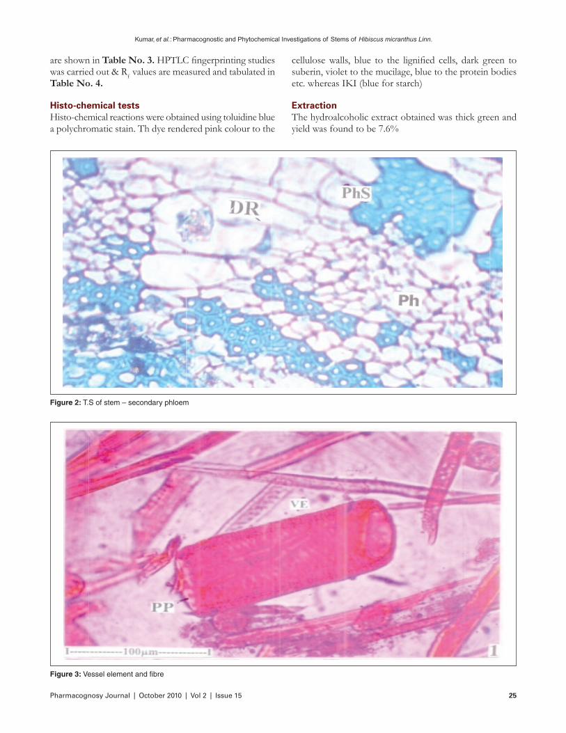
cortex and four or five layers of parenchymatous inner cortex. Secondary phloem is wide and continuous all around the stem. It has wide dilated funnel shaped rays at certain places (Fig No. 2). In other regions, the secondary phloem has tangential blocks of phloem fibres alternating with narrow segments of phloem elements. Secondary xylem is a thick hollow cylinder and consists dense xylem fibres and radial files of vessels which are separated by wide gaps. The vessels are circular, thin walled and diffuse in distribution; they include both wide and narrow vessels, the wide vessels are 40 µm in diameter; the narrow vessels are 20 µm wide. The pith is wide and parenchymatous. It consists of angular, thick walled parenchymatous cells.
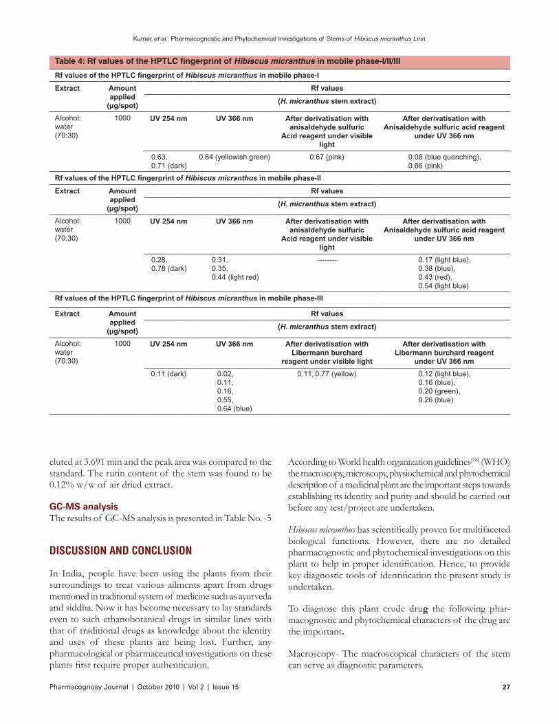
Stem powder analysis (Fig No. 3 & 4) revealed the presence of Fibres and vessel elements abundant in the powder. The fibres are libriform type with lignified thick walls and pointed tips. They are 500-650 µm long. The vessel elements are cylindrical and elongated. They have perforations plate which may be horizontal or oblique. The vessel elements with oblique perforations plates have short, pointed tails. The lateral wall pits elliptical, multiseriate and alternate. The vessel elements are 200 µm long and 40 µm wide.
Physicochemical studiesPhysical constants like Ash values, Extractive values and Loss on Drying at 110o C were determined and results are shown in Table No. 1. The behavior of powdered drug in different solutions towards ordinary and UV light were observed and the results are recorded in Table No. 2. The Preliminary Phytochemical tests of different extracts were performed, identified with using specific reagents and results
Chromatographic analysisThe sample was analyzed using Shimadzu GC-MS-QP2010 Plus apparatus equipped with quadrapole detector and split injection system. The GC was fitted with a ZP-624 capillary column (30 mm × 1.4 mm, film thickness 0.25 µm). The temperature programmed was as follows: injector temperature 220° C, initial oven temperature at 120° C for 2 minutes, then rises to 250° at the rate of 10° C per minute at 250° C for 25 minutes, transfer line temperature 220° C. Helium was used as carrier gas at 35.6 Kpa pressure with flow 2.5 ml/min and electronic pressure control on. The EM voltage was 952.9 V with lower and upper mass limits set at 30 & 350 m/z. Samples were solved in n-hexane and injected automatically. MS spectra of separated compounds were compared with one from Wiley 7 Nist 05 mass spectral database. The identity of the spectra above 95% was needed for the identification of compounds.
RESULTS
Pharmacognostic StudiesMacroscopical study revealed the dried stems are generally cylindrical, up to 1 cm thick, woody, upright, outer surface smooth in young stems and rough in old stems, greenish externally, yellowish internally, fracture splintery, taste astringent and slightly bitter, odour without any characteristic aroma, agreeable.[17]
Microscopical characters are the stem has epidermal layer of squarish cells with thick cuticle; it is broken at certain places due to growth in thickness of the stem (Fig No. 1). The epidermis is followed by a narrow zone of chlorenchymatous
Figure 1: T.S of stem – a sector enlarged
Pharmacognosy Journal | October 2010 | Vol 2 | Issue 15 25
Kumar, et al.: Pharmacognostic and Phytochemical Investigations of Stems of Hibiscus micranthus Linn.
are shown in Table No. 3. HPTLC fingerprinting studies was carried out & Rf values are measured and tabulated in Table No. 4.
Histo-chemical testsHisto-chemical reactions were obtained using toluidine blue a polychromatic stain. Th dye rendered pink colour to the
Figure 2: T.S of stem – secondary phloem
Figure 3: Vessel element and fibre
cellulose walls, blue to the lignified cells, dark green to suberin, violet to the mucilage, blue to the protein bodies etc. whereas IKI (blue for starch)
ExtractionThe hydroalcoholic extract obtained was thick green and yield was found to be 7.6%

Kumar, et al.: Pharmacognostic and Phytochemical Investigations of Stems of Hibiscus micranthus Linn.
26 Pharmacognosy Journal | October 2010 | Vol 2 | Issue 15
SPECTROPHOTOMETRIC ANALYSIS TOTAL FLAVONOIDAL CONTENT OF HEHM
Spectrophotometric analysis of HEHM for total flavonol content was determined by using standard curve prepared by using rutin. The linearity was found in the range of 10-to200 µg/ml. The total flavonol content was expressed as rutin equivalent in % w/w of the extract. The flavonoidal content was found to be 3.86 mg/100 gm of the extract.
HPLC analysis of HEHMThe rutin content of the HEHM of stem was determined by HPLC method. The analysis was performed by the injection of 20 µl of extract on a lichrospher 100RP-18(5 µm) column (250 × 4 mm), elution using mobile phase as methanol and 2% acetic acid in water (70:30) with runtime of 10min and detection by UV detector at 355 nm. Rutin
HPTLC fingerprinting analysisHPTLC fingerprinting studies was carried out & Rf values are recorded and tabulated in Table No. 4.
Table 2: Fluorescence studies of Hibiscus micranthus
Treatment Ordinary light UV 254 nm
Powder as such Pale Yellowish Yellowish fluorescence
Powder + concentrated Hydrochloric acid
Light Brown –
Powder + concentrated sulphuric acid
Deep reddish brown
–
Powder + concentrated nitric acid
Orange –
Powder + 10% sodium hydroxide
Orange red Dark green colour
Powder + glacial acetic acid Yellowish green Pale yellowPowder + chloroform Greenish
yellowGreenish fluorescence
Powder + Distilled water Pale yellow Pale yellow fluorescence
Table 3: Results of phytochemical screenings of successive extracts of stem of H. micranthus
Constituents Pet ether chloroform alcohol water
Steroid +++ +++ ------- -------Triterpenoid ------- ------- ------- -------Flavonoid ------- ------- +++ -------Phenols ------- ------- +++ -------Tannins ------- ------- +++ -------Alkaloids ------- ------- ------- -------Saponins ------- ------- ------- -------Sugars ------- ------- ------- +++prteins ------- ------- ------- +++
+++ Present ------- Absent
Table 1: Physico-chemical parameters of Hibiscus micranthus
Parameters % constituents
% LOD at 110o C 1-2.0%% Ash content 2.5%% Acid insoluble ash 0.5%% water soluble ash 2.0%% Extractive values Pet. Ether (60-80o C) 1.5 Chloroform 3.4 Alcohol 6.6 Water 4.10
Figure 4: Tailed vessel element showing lateral wall pits and fibresAbbreviations: VE- vessel, PP-perforation plate, LWP-Lateral wall pits, Fi-Fibre, Ta-Tail, Ph: Phloem, PhS: Secondary phloem, SX: Secondary xylem, DR: Druses, Pi: Pith, Co: Cortex, Ep: Epidermis
Pharmacognosy Journal | October 2010 | Vol 2 | Issue 15 27
Kumar, et al.: Pharmacognostic and Phytochemical Investigations of Stems of Hibiscus micranthus Linn.
According to World health organization guidelines[18] (WHO) the macroscopy, microscopy, physiochemical and phytochemical description of a medicinal plant are the important steps towards establishing its identity and purity and should be carried out before any test/project are undertaken.
Hibiscus micranthus has scientifically proven for multifaceted biological functions. However, there are no detailed pharmacognostic and phytochemical investigations on this plant to help in proper identification. Hence, to provide key diagnostic tools of identification the present study is undertaken.
To diagnose this plant crude drug the following phar-macognostic and phytochemical characters of the drug are the important.
Macroscopy- The macroscopical characters of the stem can serve as diagnostic parameters.
eluted at 3.691 min and the peak area was compared to the standard. The rutin content of the stem was found to be 0.12% w/w of air dried extract.
GC-MS analysisThe results of GC-MS analysis is presented in Table No. -5
DISCUSSION AND CONCLUSION
In India, people have been using the plants from their surroundings to treat various ailments apart from drugs mentioned in traditional system of medicine such as ayurveda and siddha. Now it has become necessary to lay standards even to such ethanobotanical drugs in similar lines with that of traditional drugs as knowledge about the identity and uses of these plants are being lost. Further, any pharmacological or pharmaceutical investigations on these plants first require proper authentication.
Table 4: Rf values of the HPTLC fingerprint of Hibiscus micranthus in mobile phase-I/II/III
Rf values of the HPTLC fingerprint of Hibiscus micranthus in mobile phase-I
Extract Amount applied
(μg/spot)
Rf values
(H. micranthus stem extract)
Alcohol: water (70:30)
1000 UV 254 nm UV 366 nm After derivatisation with anisaldehyde sulfuric
Acid reagent under visible light
After derivatisation with Anisaldehyde sulfuric acid reagent
under UV 366 nm
0.63, 0.71 (dark)
0.64 (yellowish green) 0.67 (pink) 0.08 (blue quenching),0.66 (pink)
Rf values of the HPTLC fingerprint of Hibiscus micranthus in mobile phase-II
Extract Amount applied
(μg/spot)
Rf values
(H. micranthus stem extract)
Alcohol: water (70:30)
1000 UV 254 nm UV 366 nm After derivatisation with anisaldehyde sulfuric
Acid reagent under visible light
After derivatisation with Anisaldehyde sulfuric acid reagent
under UV 366 nm
0.28, 0.78 (dark)
0.31, 0.35, 0.44 (light red)
-------- 0.17 (light blue), 0.38 (blue), 0.43 (red), 0.54 (light blue)
Rf values of the HPTLC fingerprint of Hibiscus micranthus in mobile phase-III
Extract Amount applied
(μg/spot)
Rf values
(H. micranthus stem extract)
Alcohol: water (70:30)
1000 UV 254 nm UV 366 nm After derivatisation with Libermann burchard
reagent under visible light
After derivatisation with Libermann burchard reagent
under UV 366 nm
0.11 (dark) 0.02,0.11,0.16,0.55,0.64 (blue)
0.11, 0.77 (yellow) 0.12 (light blue),0.16 (blue),0.20 (green),0.26 (blue)

Kumar, et al.: Pharmacognostic and Phytochemical Investigations of Stems of Hibiscus micranthus Linn.
28 Pharmacognosy Journal | October 2010 | Vol 2 | Issue 15
Table 5: Volatile compounds from methanolic extract of stem of H. micranthus Linn. as detected by GC-MS
peak Retention Time Compound % matching with Wiley library
1 0.94 Ethyl-D5 ethyl ether 942 1.86 Hexane, 1-chloro 963 4.53 2-Methylpropane-1, 2-diol 944 5.20 Octane, 4-ethyl 985 5.42 Alpha-D-Galactopyranoside, methyl 726 6.38 Methyl ethane-2, 2, 2-D3-Sulfonate 937 6.61 Butanedioic acid monomethyl ester 998 7.12 2-n-propylthiane 999 7.51 Benzoic acid 9910 7.79 Formic acid, pentyl ester 9911 8.38 Nonanoic acid 9812 8.52 Benzofuran, 2,3-dihydro- 9713 8.67 1, 3, 5-cycloheptatriene 9714 8.76 3-pyridinecarboxylic acid 9815 8.95 3, 3-Dimethylthietane 9716 9.19 2-Methoxy-4-vinylphenol 9317 9.29 Methyl-beta-D-arabinopyranoside 8818 9.39 2, 6-Dimethyl-3-trans-propenylpyrazine 9919 9.61 1-Di(t-butyl)silyloxypropane 9520 9.82 1, 10-Decane-1, 1, 10, 10 d4-diol 9721 9.97 Propanoic acid, 2-methyl-, methyl ester 9522 10.11 2-cyclopenten-1-one, 2-methyl 9723 10.25 Benzamide 9824 10.55 2-ethyl-2’, 2’, 2’-D3-Cyclopentanone 8725 10.80 (1, 1’-Bicyclopropyl)-2-octanoic acid, 2’-hexyl-, methyl ester 9326 10.97 Benzene, methyl- 9627 11.38 Suberic acid monomethyl ester 9528 11.47 Thiacyclohexan-4-ol 9729 11.56 2-allylpent-4-enoic acid, methyl ester 4130 11.68 L-Menthol 9731 11.82 Dodecanoic acid 9932 11.95 Ethanone, 1-(4-hydroxyphenyl)- 9833 12.09 2-Octenal 9834 12.20 3’, 5’-Dimethoxyacetophenone 3335 12.27 2-Butynedioic acid, diethyl ester 9736 12.37 Octanedioic acid 9837 12.37 Octanedioic acid 9538 12.52 1, 6-anhydro-beta-D-Glucopyranose 8439 12.68 Alpha-Methyl-alpha-propylsuccinimide 9940 12.76 1, 2.4-Trimethoxybenzene 9141 12.83 Acetohydrazide, 2-hydroxy-2-phenyl-N2-but-2-enylideno 9842 13.04 2-cyclohexyldimethylsilyloxybut-3-yne 9443 12.12 4-pyridinecarboxylic acid, 3-hydroxy-5-(hydroxymethyl)-2-methyl- 10044 13.19 Alpha-Methyl-D-mannopyranoside 9245 13.40 Octahydro-Naphthalene-1,8A-diol 9946 13.57 1-Methyl-4-Phenyl-1, 2, 3, 6-tetrahydropyridine 8747 13.74 4-Methyl-5-imidazolemethanol 9748 13.86 Dihydrojasmone 9949 13.98 Eicosanoic acid 9850 14.13 3-Fluorobenzoic acid, dodec-9-ynyl ester 9951 14.76 Oxirane, hexadecyl 9752 14.93 Eicosanoic acid 9053 15.01 Mome inositol 9854 15.29 Heptadecanoic acid 9755 15.55 Hexadecanoic acid, methyl ester 9956 16.02 2-Butenal, 2-methyl-4-(2, 6, 6-trimethyl-1-cyclohexen-1-yl) 9957 16.37 2H-Pyran-2-one, 5-ethylidenetetrahydro-4-(2-hydroxyethyl)- 9658 16.50 Pluchidiol 9959 16.84 1,10-Dimethyl-2-methylene-trans-decalin 10060 16.92 1-Methyl-1-n-decyloxy-1-silacyclobutane 9661 18.97 Cyclopropanebutyric acid-2[ (2-nonylcyclopropyl)methyl]-, methyl ester 9962 19.24 9, 12, 15-Octadecatrienoic acid, methyl ester 9963 19.65 1-Docosanol 99

Pharmacognosy Journal | October 2010 | Vol 2 | Issue 15 29
Kumar, et al.: Pharmacognostic and Phytochemical Investigations of Stems of Hibiscus micranthus Linn.
Secondary metabolites- The phytochemical screening of successive extracts revealed the presence of phenols, tannins, steroids, carbohydrates and flavonoids. Thus, the preliminary phytochemical tests are helpful in finding chemical constituents in the plant material that may lead to their quantitative estimation and in locating the source of pharmacologically active chemical compounds.[14,21-23]
HPTLC fingerprinting- helps in quantification, identification and checking the purity of the crude drug. The hydroalcholic extract of Hibiscus micranthus produces three different patterns of bands in Mobile Phase – I: Ethyl acetate: methanol – glacial acetic Acid (10:1.35:1), Mobile phase – II: Chloroform – methanol (9.9:0.1), Mobile phase – III: Ethyl acetate: formic acid – glacial acetic acid – water (10:1.1:1.1:2.6). The separation efficiency of mobile phase-III was maximum and total of five spots with Rf values 0.02, 0.11, 0.16, 0.55, 0.64 (blue) where detected under UV 366 nm. For identification of the drug, fingerprints in all three mobile phases will be helpful.
Estimation of rutin by HPLC- The Hydroalcoholic extract was quantified by HPLC studies which showed the presence of selected marker compound rutin and its retention time and λ max were similar to standard. The Photo diode array detector was used and set in the range of 200-780 nm. The rutin content was found to cotain 0.12% w/w of air dried extract. Thus, the proposed method is rapid, selective, requires a simple sample preparation procedure, and represents a good procedure for their quantification in plant material and in routine quality control of herbal drugs.
GC-MS analysis of HEHM- Further HEHM was analyzed by GC-MS to detect for volatile components. A total of 56 compounds were identified from the methanolic extract of the stem parts. The identified compounds represented 89.41% of the extract. The main components of the methanolic extract of stem parts were 7-Hexdecyn-1-ol(11.32%), 9,12,15-Octadecatrienoic acid methyl ester (7.88%), triacontanoic acid methyl ester (5.21%), octadecane (3.74%), 1-docosanol (3.61%), cyclopropanebutyric acid 2-( (2-nonylcyclopropyl)methyl)-methyl ester (3.95%),, 1-methyl-1-n-decyloxy-1-silacyclobutane (5.82%), Hexadecanoic acid methyl ester
Microscopy- The microscopical studies of the transverse section and powder showed presence of abundant lignified, thick wall, libiform type of fibres with pointed tips & vessel elements with oblique perforations plates having short, pointed tails, which are distinguishing microscopic features and serve as anatomical markers.[19]
Histochemical- plant metabolites are generally located in vegetative or reproductive organs. These chemicals have several uses. In Pharmacognosy discipline, these are also been utilized for identification and detection of purity of the crude drug.[14]
Ash values- are measure of inorganic content of the drug. These values are constant for pure drugs and increased/decreased when contaminated with soil and adulterants.[14]
Extractive values- Based on extractive values the correct time of collection of drug, type and conditions of extraction process and nature of chemical constituents present in the drug can be determined. These are also useful for the evaluation especially when the constituents of drug cannot be readily estimated by any other means. The drug under investigation found to contain more of polar constituents.[14]
Loss on drying- is determination of moisture content of the drug. Presence of moisture in the crude drugs serves has suitable media for bacterial growth, causes degradation of moisture sensitive chemical constituents and gives information about moisture absorbing chemical constituents of plant drugs. The crude drug powder contains less than 5%.[14]
Fluorescence studies- The drug emits visible radiations of different wavelength when observed in various solvents at 254nm. The stem powder exhibited Yellowish fluorescence (powder as such), Dark green colour (10% sodium hydroxide solution), pale yellow (glacial acetic acid), Greenish fluorescence (chloroform), yellow fluorescence (methanol), yellow fluorescence (distilled water) and green colour (10% potassium hydroxide). These tests may consider as one of the parameters for characterization of the genuine drug samples.[20]
peak Retention Time Compound % matching with Wiley library
64 21.20 7-Hexadecyn-1-ol 9865 21.56 9, 12, 15-Octadecatrienoic acid, methyl ester 9966 24.36 Capsaicin 9667 26.54 Triacontanoic acid, methyl ester 9968 30.56 Octadecane 9969 31.18 4, 8, 12, 16-Tetramethylheptadecan-4-olide 9870 31.80 Cycloheptanon, 3-butyl- 9771 32.01 Tetratriacontane 9772 36.03 Cyclopentanetridecanoic acid, methyl ester 97

Kumar, et al.: Pharmacognostic and Phytochemical Investigations of Stems of Hibiscus micranthus Linn.
30 Pharmacognosy Journal | October 2010 | Vol 2 | Issue 15
and Basella alba in mature rat testis function. J.Ethnopharmacol65:133-139.
7. Sass, J.E., 1940. Elements of Botanical Microtechnique. McGraw Hill Book Co; New York. Pp.222.
8. Johansen, D.A., 1940. Plant Microtechnique. McGraw Hill Book Co; New York. Pp.523.
9. O’Brein, T.P; Feder, N. and Mc Cull, M.E. 1964. Polychromatic staining of plant cell walls by toluidine blue-O. Protoplasma; 59:364-373.
10. Easu, K. 1964. Plant anatomy John Wiley and sons. New York. Pp.767. Easu, K. 1979.
11. Anatomy of seed plants. John Wiley and sons. New York, 550.
12. Wallis, T.E., 1990. Text book of pharmacognosy. CBS publishers Delhi; India. Pg 572-575.
13. Khandelwal, K.R., 1998. Practical pharmacognosy: Techniques & experiments. 4th edition; Nirali prakashan, India.
14. Wagner, H. and Bladt, S., 1996. Plant drug analysis-A thin layer chromatographic atlas, springer-verlog. Berlin Heidel berg.
15. Samo Kreft., Borut strukelf., Alanka Gaberscik., Ivan kreft., 2002. .Rutin in buckwheat herbs grown at different UV-B radiation levels: Comparison of two UV spectrophotometric & on HPLC method. Journal of experimental biology, Vol. 53, No. 375, pg. 1801-1804.
16. Woisky, R., Salatino, A.. 1998. Analysis of propolis; Some parameter & procedures for chemical quality control. J.Agric res. 37:99-105.
17. WHO/PHARM/92.559/rev.1., 1992.Quality control methods for medicinal plant materials, organization mondiale De La Sante, Geneva, p 9, 22-34.
18. Evans, W.C., 1996. Trease & Evan’s pharmacognosy fourteenth edition, WB Saunders co., Ltd. London, Pg 545-546.
19. Chase, C.R and Pratt., 1949. Fluorescence of powdered vegetable drugs with particular reference to development of a system of identification. J. Am. Pharmacol. Assoc. 324, 333.
20. Indian pharmacopoeia, 4th edn., vol-II, 1996. Government of India, Ministry of Health & welfare, controller of publications, New Delhi, p A53-A54.
21. Brain, K.R and Turner, T.D., 1975. The practical evaluation of phytopharmaceuticals, Wright- scientechnica, Bristol Pg 81-82.
22. Harborne, J.B., 1992. Phytochemical methods. A guide to modern technique of plant analysis. Chapman & Hill, London. 279.
(2.16%), oxirane hexadecyl-(2.60%), octahydro-naphthalene-1,8A-diol(2.19%), and octanedioic acid(4.13%). The GC-MS analysis revealed that the methanolic extract is mainly composed of fatty acid esters.
The pharmacognostic and phyto-chemical investigations of the Hibiscus micranthus L. stem has been carried out for the first time. Chemo profiling by Spectophotometric, HPLC, HPTLC and GC-MS analysis can be utilized for identification, quantification and characterization of chemical markers present in Hibiscus micranthus Linn. This could also serve in the establishing data for preparation of monograph of this plant.
REFERENCES1. Kirtikar, K.R., and Basu, B.D. 1984. In Indian medicinal plants, Vol.1,
2nd edition, Periodical Expert Book Agency. Delhi. p.293.
2. Kakrani, H.N., Bhanu, H., Kakrani., Ajay K. Saluja., 2005. Traditional treatment of Diabetes through herbs in Kutch district, Gujarat state. Planta indica, Vol 1, No. 1, Pg16-21.
3. Al-yahya, M.A., Tariq, M., Parmar, N.S., Ageel, A.M., 1987. Pharmacological investigations of Hibiscus micranthus Linn., a Febrifuge used in Saudi Arabian Folk Medicine. Phytotherapy Research, Vol 1, No. 2. Pg73-75.
4. Jain, R., Arora, R., Jain, S,C., ,1997. Chemical constituents and bioactivity studies of Hibiscus micranthus Linn. Indian J Pharm Sci Mar–Apr:91-93.
5. Telefo, P,B., Moundipa, P.F., Tehana, A.N., Tchouanguep, C.D., Mbiapo FT (1998): Effects of an aqueous extract of Aloe buettneri, Justica insularis, Hibiscus macranthus, Dicliptera verticillata on some physiological and biochemical parameters of reproduction in immature female rats. J. Ethnopharmacol 63:193-200.
6. Moundipa, F.P., Kamtchouing, P., Koueta, N., Tantchou, J., Foyang, N.P., Mbiapo, F.T., 1999. Effects of aqueous extracts of Hibiscus macranthus