pharmacognostic studies - inflibnetshodhganga.inflibnet.ac.in/bitstream/10603/14136/10/10_chapter...
TRANSCRIPT

Pharmacognostic Studies
25
CHAPTER-III - PHARMACOGNOSTIC STUDIES
3.1 INTRODUCTION
As the crude drugs form the basis for the manufacture of wide range of medicinal
preparations needed by people, the development of pharmacognostical research has
become indispensable for procuring therapeutically potent medicine prepared from
genuine drug material. The pharmacognosists have a serious responsibility, to take the
initiative not only in correctly locating the plant mentioned in old treatises and
pharmacopeias but also making them available to scientists in other disciplines to test the
use for which they are acclaimed1.
3.2 EXPERIMENTAL WORK
3.2.1 Collection and authentification of plant materials
Based on exhaustive literature survey done on medicinal plants of Annonaceae,
leaves of three plants known as Annona squamosa Linn, Annona reticulata Linn,
Annona muricata Linn were selected for the study.
Leaves of the Annona squamosa Linn, Annona reticulataLinn were collected
from the Tirumala hills, Chittor district, Andhra Pradesh. Leaves of Annona muricata
Linn were collected from the Guntur district region, Andhra Pradesh and were
authentified by Dr.D.Ramakanth raju, retire botanist, S.V. University, Tirupati and a
voucher specimen for Annona squamosa Linn, (T.S.N-007, 21/04/2011) Annona
reticulata Linn(T.S.N-005, 12/08/2010), Annona muricata Linn (T.S.N-001, 12/06
/2011) has been deposited in Pharmacognosy Department, Andhra university.

Pharmacognostic Studies
26
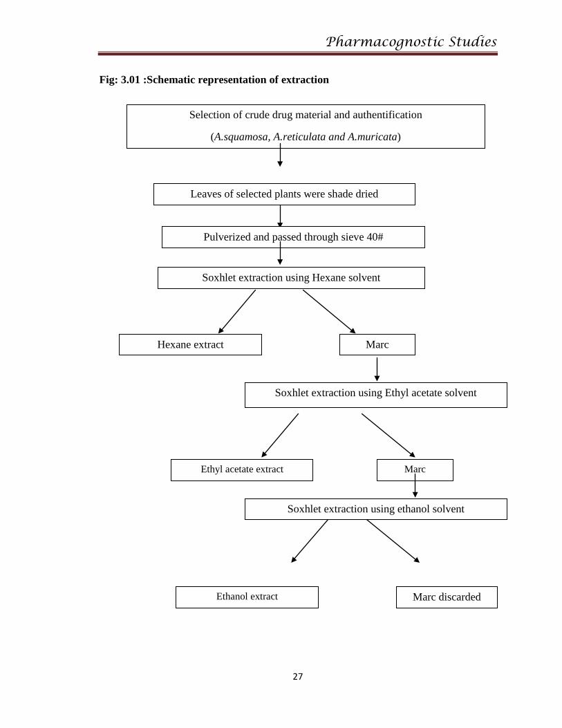
3.2.2 Extraction:
All the plant materials obtained were shade dried, made into coarse powder and passed
through sieve#40, were successively extracted with Hexane, Ethyl acetate and Ethanol
by Soxhlet extraction method2(Fig:3.01).
Procedure:
Collected leaves were shade dried, pulverized to a coarse powder in a mechanical
grinder, passed through 40# mesh sieve. 1kg of plant material was extracted in Soxhlet
extractor consecutively using solvents of non polar to polar grade (hexane, Ethyl acetate
and Ethanol) obtained crude extracts were evaporated to dryness in a rotary evaporator.
Pharmacognostic Studies
27
Fig: 3.01 :Schematic representation of extraction
Selection of crude drug material and authentification
(A.squamosa, A.reticulata and A.muricata)
Leaves of selected plants were shade dried
Pulverized and passed through sieve 40#
Soxhlet extraction using Hexane solvent
Hexane extract
Marc
Soxhlet extraction using Ethyl acetate solvent
Marc Ethyl acetate extract
Soxhlet extraction using ethanol solvent
Ethanol extract Marc discarded

Pharmacognostic Studies
28
3.2.3 Macroscopic characterization of plants:
Macroscopic evaluation of the selected plants were recorded as per visual
observation organoleptic evaluation of the selected plants, colour, odour, taste, size and
shape were recorded separately results were given in the Table: 3.01- 3.03.
3.2.4 Microscopical characterization
Healthy organs were collected for Microscopic evaluation.
Instruments used for microscopic evaluation:
• Electron microscope of Quantum model
• Camera Lucida
• Stage micrometer
• Eye piece micrometer
3.2.4.1 Transverse Section Studies
Sectioning:
Numerous free hand sections were taken, stained and mounted following the usual
micro technique described by Brain (1975) 3 and photographs of different magnifications
were taken using Electron microscope and results were given in (Fig:3.05-3.16).

Pharmacognostic Studies
29
Staining:
The following reagents were used for staining the transverse sections
• Toludine blue
• Phloroglucinol
• Methyl orange
• Iodine
• 5% sodium hydroxide
• Dilute hydrochloric acid
• 1% Chloral hydrate
• Conc. Nitric acid
3.2.4.2 Powder microscopic studies4:
Leaf powders of the selected plants were observed under microscope with
distilled water, stained with phloroglucinol and Hydrochloride, pictures were given in
(Fig: 3.17-3.19).
3.2.5 Fluorescence analysis of powders5:
Obtained leaves of selected plants were shade dried, made into powder and
observed under normal daylight, UV light at 2 different wavelengths one is at 254nm, and
other is 365nm. Obtained results were given in (Table :3.04 - 3.06).

Pharmacognostic Studies
30
3.2.6 Fluorescence analysis of extracts5:
Obtained plant extracts were analyzed under day light, short wavelength and in
long wavelength region, results were given in (Table: 3.07-3.09).
3.2.7 Quantitative microscopy - Determination of leaf constants6:
Leaf surfaces are studied by scrapping and by peeling of the upper and lower
epidermal surfaces of the leaves and then washed with chloral hydrated and observed
under microscope for its stomatal structure, epidermal pattern, veiniselt pattern, vein
termination pattern and palisade ratio.
3.2.7.1 Stomatal index: It is the percentage, which no. of stomata formed to the total
no.of epidermal cells; each stoma being considered as one cell.
3.2.7.2 Stomatal Number: It is the average number of stomata per square mm of the
epidermis of the leaf.
3.2.7.3 Palisade ratio: It is the average number of palisade cells beneath each epidermal
cell.
Procedure:
Middle part of the leaf was cleared by boiling with chloral hydrate solution. Upper
and lower epidermis were peeled out separately with the help of forceps & kept it on slide
and mounted in glycerin water. With the help of micrometer, 1mm square was drawn.
Number of stomata and epidermal cell which were present in the area of 1 sq.mm were
counted.

Pharmacognostic Studies
31
Stomatal Index: It is calculated by using this formula: S. I = S/E + S x 100
S. I = Stomatal Index,
S = No. of stomata per unit area,
E = No. of epidermal cells in the same unit area
3.2.7.4 Vein termination number & vein islet number
Veinlet termination number is defined as the number of veinlet terminations
per square mm of the leaf surface, midway between midrib of the leaf and its margin.
A vein-islet is the small area of green tissue surrounded by the vein-islets. The
veinislet number is the average number of vein-islets per square mm of a leaf surface. It
is determined by counting the no.of vein-islets in an area of 4 square mm of the central
part of the leaf between the midrib and the margin. Results were given in (Table :3.10)
3.2.8 Physicochemical parameter studies on selected plants
3.2.8.1 Determination of Foreign organic matter7
Collected plant material was spread in a thin layer and sort the foreign matter into
groups either by visual inspection, using a magnifying lens (6x or 10x), or with the help
of a suitable sieve, according to the requirements for the specific plant material.
Remainder of the sample was sifted through a No.250 sieve; dust is regarded as mineral
admixture. Weigh the portions of this sorted foreign matter to within 0.05g. Calculate the
content of each group in grams per 100g of air-dried sample.
For some medicinal plant materials where the foreign matter may closely
resemble the material itself, it may be necessary to take a pooled sample of the plant
material and apply a critical test, either chemical, physical, or by microscopy. The

Pharmacognostic Studies
32
proportion of foreign matter is calculated from the sum of the portions that fail to respond
to the test.
3.2.8.2 Determination of ash value8
The ash remaining after complete ignition of the medicinal plant materials is
determined by three different methods known as Total ash, Acid-insoluble ash and water-
soluble ash.
Acid-insoluble ash is the residue obtained after boiling the total ash with dilute
hydrochloric acid and igniting the remaining insoluble matter. This measures the amount
of silica present, especially as sand and siliceous earth.
Water-soluble ash is the difference in weight between the total ash and the residue
after treatment of the total ash with water.
Procedure for Total ash:
Accurately weighed 3 g of air dried powdered drug was taken in a tarred silica
crucible and incinerated by gradually increasing the temperature to 500-6000C until it is
white, indicating the absence of Carbon, Cool and weigh, this process repeated till
constant weight is obtained. Then the percentage of total ash was calculated with
reference to the air dried drug.
a. Procedure for Acid insoluble ash:
The total ash was boiled with 25 ml of 2 N HCl for 5 minutes. The insoluble
matter was collected on an ash less filter paper, washed with hot Water, ignited and
weighed, then calculated the percentage of acid insoluble ash with reference to the air
dried drug.

Pharmacognostic Studies
33
b. Procedure for water insoluble ash:
The total ash was boiled with 25 ml. of water for 5 minutes. The insoluble matter
was collected on an ash less filter paper, washed with hot water and ignited for 15
minutes at a temperature not exceeding 4500C. The weight of insoluble matter was
subtracted from the weight of total ash. The difference in weight represents the water
soluble ash. The percentage of water soluble ash was calculated with reference to the air
dried drug.
3.2.8.3 Determination of moisture content by loss on drying9
Moisture content determination is important, not only to know excess water, but
also in conjunction with suitable temperature moisture will lead to the activation of
enzymes and gives suitable conditions to the proliferation of living organism. As most
vegetable drugs contain all the essential food requirements for mould, insects and mites,
deterioration can be very rapid, once infestation has taken place. Various methods for
moisture determination are loss on drying, separation and measurement of moisture,
chemical methods, electrometric methods, and spectroscopic methods as per IP.
• 10gm of powder was weighed and placed it in a moisture content
apparatus.
• Temperature was adjusted to 100-1100c till weight get constant and
collected in desiccators and weighed.
• The loss of weight was regarded as a measure of moisture content as per
IP.

Pharmacognostic Studies
34
3.2.8.4 Determination of Foaming index10
The foaming ability of an aqueous decoction of plant materials & their extracts is
measured in terms of a foaming index.
Weighed accurately about 1 g of coarsely powdered drug and transferred to 500
ml conical flask containing 100 ml of boiling water maintained at moderate boiling at 80-
900C for about 30 mins . Then made it cold, filtered into a volumetric flask and added
sufficient water through the filter to make the volume up to 100 ml (V1). Cleaned 10
stopper test tubes were taken and marked with 1 to 10. The successive portions of 1, 2 ml
up to 10 ml drug was taken in separate tubes and adjusted remaining volume with the
liquid up to 10 ml in each. After closing the tubes with stoppers, Shook them for 15
seconds and allowed to stand for 15 mins then measured the height. If the height of the
foam in each tube is less than 1cm, the foaming index is less than 100(not significant).
Here, if the foam is more than 1cm height after the dilution of plant material in the sixth
tube, then corresponding number of the test tube was the index sought. If the height of the
foam in every tube is more than 1cm, the foaming index is more than 1000. In this case,
10ml of the first decoction of the plant material needs to be measured and transferred to a
100ml volumetric flask (V2) and volume is to be maintained up to 100ml and follow the
same procedure. Foaming Index was calculated by using this formula
Foaming Index = 1000/a in case of V1
Foaming Index = 1000 × 10/a in case of V2
Where, a = Volume (ml) of decoction used for preparing the dilution in the tube where
exactly 1 cm or more foam was observed.

Pharmacognostic Studies
35
3.2.8.5 Determination of Swelling index11.
Many medicinal plant materials are of specific therapeutic or pharmaceutical
utility because of their swelling properties, especially gums containing an appreciable
amount of mucilage, pectin or hemicellulose.
Procedure:
It was carried out simultaneously no fewer than three determinations for any
given material. Introduce the specified quantity of the plant material concerned,
previously reduced to the required fineness and accurately weighed 1g of plant material
into a 25 ml glass-stopper measuring cylinder. The internal diameter of the cylinder was
about 16 mm, the length of the graduated portion about 125mm, marked in 0.2 ml
divisions from 0-25 ml in an upwards direction. 25 ml of water was added and shake the
mixture thoroughly every 10 minutes for 1 h. Allowed to stand for 3 h at room
temperature. Measured the volume in ml occupied by the plant material, including any
sticky mucilage.
3.2.8.6 Determination of Extractive value12
1000g of course powder was subjected to Soxhlation with different solvents then
the remained extract was weighed and calculated its percentage of extractive value using
the formula
x = X *100 1000
X= Amount of extract obtained after complete extract in grams.

Pharmacognostic Studies
36
3.3 RESULTS AND DISCUSSION
3.3.1 Morphological characterization of the selected plants reveals the following
characters
Table : 3.01 Morphological characterization of Annona squamosa Linn :
Characters Seeds Leaves Stems Roots Fruits
Colour Black Green Green to
brown
Light
brown/Dark
brown
Greenish
outside ,
whitish pulpy
inside
Odour Odourless Characteristic
odour
Characteristic
odour
Odourless Sweetish
Taste Taste less Bitter Sight bitter Bitter Sweetish
Leaves : Ovate to lanceolate shape, simple margin, lamina measures about 10×5 cm,
they are simple, alternated to spirally arranged with zig zag pattern. Sides
some times are slightly unequal and the leaf edges are without teeth,
inconspicuously hairy when young.
Petiolate : Measures about 1-1.5 cm, twisted and channeled, stipulates linear,
Flowers : Hermaphrodite, usually somewhat fragrant, solitary or in fascicles with 2 to 4
flowers, with three green sepals and six petals arranged in two verticils. The

Pharmacognostic Studies
37
flowers have several conglomerated and spirally arranged stamens below and
around an upper globose shaped dome of numerous united carpels.
Seeds : Black colour with ovoid shape, numerous scattered over the white pulp.
Stems : Cylindrical with characteristic odour and bitter taste. Outer side thick cork cells
are found upon maturation.
Fig :3.02 Morphological characterization of some plant parts of Annona squamosa
Linn

Pharmacognostic Studies
38
Table : 3.02 Morphological characterization of Annona reticulata Linn:
Characters Seeds Leaves Stems Roots Fruits
Colour Black Green Green to
brown
Light
brown/Dark
brown
Yellow to
orange
outside ,
whitish pulpy
inside
Odour odourless Characteristic
odour
Characteristic
odour
Odourless Sweetish
Taste Taste less Bitter Sight bitter Bitter Sweet slight
sour
It reaches to a height of 6.0 to 7.5 m , with many lateral branches.
Leaves : Larger, long, narrow, glabrous are present, oblong-lanceolate and dark green
colour measuring about 25- 30 cm in length and 7 cm wide, with 10 to 20 vein
pairs and a pubescent petiole.
Flowers : They are grouped in a short inflorescence with 2 to 10 flowers, with pedicels
measuring 1.5 to 3.0 cm in length.
Stems : Cylindrical with lenticels and very short coffee- coloured hairs.
Pharmacognostic Studies
39
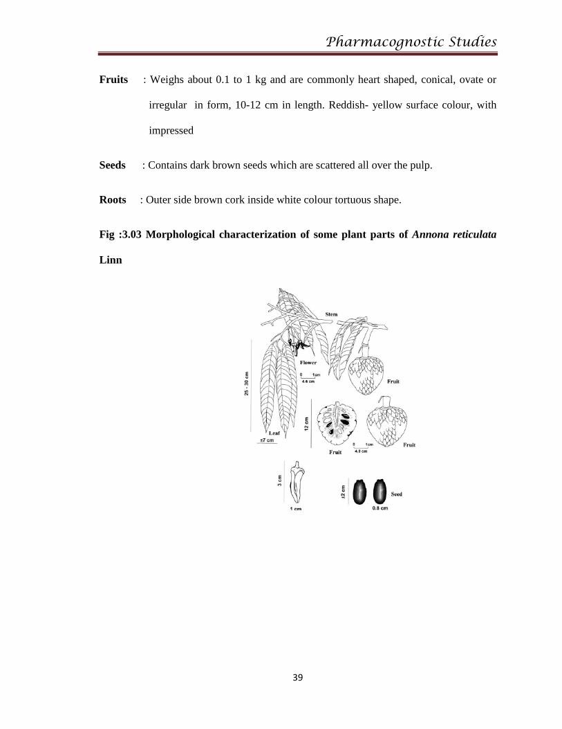
Fruits : Weighs about 0.1 to 1 kg and are commonly heart shaped, conical, ovate or
irregular in form, 10-12 cm in length. Reddish- yellow surface colour, with
impressed
Seeds : Contains dark brown seeds which are scattered all over the pulp.
Roots : Outer side brown cork inside white colour tortuous shape.
Fig :3.03 Morphological characterization of some plant parts of Annona reticulata
Linn

Pharmacognostic Studies
40
Table : 3.03 Morphological characterization of Annona muricata Linn:
Characters Seeds Leaves Stems Roots Fruits
Colour Black Green Green to
brown
Light
brown/Dark
brown
Green to orange
outside, whitish
pulpy inside
Odour Odourless Characteristic
odour
Characteristic
odour
Odourless Characteristic
Taste Taste less Bitter Slight bitter Bitter Sweetish
followed by
sour taste
It is a small, slender, evergreen tree, 4 to 8 m tall when fully mature.
Leaves : Oblong –ovate to cylindrical, 14-16cm in length, 5-7 cm in width.
Flowers : Larger than the other two species (A.squmosa, A.reticulata)
Fruits : Ovate, conical or heart-shaped fruits, dark green in unripe condition, slightly
lighter green when rip. Rind has many short, fleshy, pointed carper
protuberances and is popularly regarded as spiny. It gives largest fruit size as
compare to other species averaging 4kg.
Seeds : It contains 127 to 170 seeds, scattered throughout the pulp. Size varies from 1 to
2 cm in length, 0.33 to0.59 g in weight, black colour.
Stems : Rounded, rough and not pubescent, with a dark brown colour.
Roots : Brown colour cork is present on surface, tortuous shape, contains slight hairs.

Pharmacognostic Studies
41
Fig :3.04 Morphological characterization of some plant parts of Annona muricata
Linn
3.3.2 Microscopical characterization
a. Transverse section of Annona squamosa Linn leaf (Fig :3.05) :
Transverse section through midrib shows the upper and lower single layered
compactly arranged barrel shaped epidermis with thick cuticle and rarely simple
trichomes on lower surfaces. Lamina upper 1-2 layered palisade parenchyma and
lowers 5-6 layers of spongy parenchyma throughout the lamina lysogenous cavities are
very common, prismatic crystals, oil globules and tannin content material spread
throughout the lamina and also even in midrib. Through midrib shows vascular

Pharmacognostic Studies
42
bundles radially arranged. Vascular bundle surrounded by pericyclic fibers on both the
side, rest half consists parenchyma cells.
b. Transverse section of leaf Annona reticulata Linn leaf (Fig :30.6):
Transverse section through midrib shows the upper and lower single layered
compactly arranged rectangular to barrel shaped epidermis with thick cuticle and
multicellular trichomes filled with tannin on lower surfaces. Lamina upper single
layered palisade parenchyma and lowers 6-7 layers of spongy parenchyma lysogenous
cavities are very common, prismatic crystals, oil globules and tannin content material
spread throughout the lamina and also even in midrib. Through midrib shows vascular
bundle radially arranged. Vascular bundle surrounded by pericyclic fibers on both the
side, in center a group of stone cells are observed. In the surface study, upper and
lower epidermis of the leaf was peeled off and observed under the microscope, the
upper epidermis show only epidermal cells, lysogenous cavity and oil globules
whereas, lower epidermis shows paralytic stomata and blade edged epidermis cells,
lysogenous cavity, oil globules.
c. Transverse section of Annona muricata Linn Leaf (Fig: 3.07):
It shows dorsiventral single layer of palisade cells are present below upper
epidermis. Stomata are of paracytic type found in lower epidermis. Mesophyll consist
of 3-4 layers of spongy parenchyma with many intercellular spaces. The midrib shows
collenchymas below epidermis on both surfaces. Parenchymatous cells occupy the
space between collenchymas and vascular bundle. The vascular bundle consist of
lignified xylem and phloem that are arranged in collateral-open type (layer of

Pharmacognostic Studies
43
cambium is sperating xylem and phloem). Sclerides are present below
collenchymatous cells of upper epidermis.
d. Transverse section of Annona squamosa Linn stem (Fig: 3.08 ) :
It shows the presence of unicellular layer of epidermal cells followed by
collenchymatous cells , contains wide cortex and lignified vascular bundles,
parenchyma cells contains starch grains of ovoid shape, in centre portion of the stem
pith is present.
e. Transverse section of Annona reticulata Linn stem (Fig: 3.09)
It shows collenchymatous cells below epidermis, followed by pericyclic
fibers, xylem, phloem and parenchymatous cells. Xylem is surrounded by starch
grains and also contains lignified stone cells. Starch grains are oval or ellipsoid,
turning blue when treated with iodine.
f. Transverse section of Annona muricata Linn stem (Fig: 3.10)
Shows the presence of epidermis with cuticle followed by collenchyma cells
in 1-2 layers, contains pericyclic fibers which are lignified. T.S shows the presence
of xylem and phloem cells and are of biocollateral separated by collenchyma, starch
grains of ovate shape.
g. Transverse section of Annona squamosa Linn stem bark (Fig : 3.11 ):
It showed the presence of uniformly arranged single layered cork cells
beneath which cortex is present. Lower portion of T.S showed the presence of
uniformly dividing cells and medullary rays were also observed.

Pharmacognostic Studies
44
h. Transverse section of Annona reticulata Linn stem bark (Fig : 3.12):
It showed the presence of 5- 6 layers of cork cells followed by cortex cells,
medullary rays, stone cells were observed with radially dividing parenchyma cells.
i. Transverse section of Annona muricata Linn stem bark of (Fig : 3.13 ) :
Showed the presence of 7-8 layers of uniformly arranged cork cells followed
by cortex cells, it also contains radially dividing parenchyma cells, wood elements,
lignified fibers, flower shaped calcium oxalate crystals were observed.
j. Transverse section of Annona squamosa Linn root (Fig : 3.14)
Showed the presence of thick cork of 3 to 4 layers, followed by
collenchymatous cells and a bundle of vascular bundles containing xylem and
phloem, contains parenchyma cells which are ovoid in shape and large size of 20-30
microns.
k. Transverse section Annona reticulata Linn of root (Fig : 3.15):
Shows wide cortex with uniform arrangement of cork cells and also contains
large stone cells. Phloem shows large sieve tubes, cell inclusions interspersed with
phloem parenchyma and fibers.
l. Transverse section Annona muricata Linn of root (Fig : 3.16):
Annona muricata root transverse section exhibits less cork cell layer as
compare to Annona squamosa and Annona reticulata it shows the presence of
pericylic fibers, xylem vessels, phloem cells are present which are of bicollateral
vascular bundles. The parenchyma cells are arranged in the mesocarp and endocarp
region. In the parenchyma cells cell inclusion, sieve tubes were also observed.

Pharmacognostic Studies
45
3.3.3 Powder microscopic characters
a. Annona squamosa Linn leaf powder characters (Fig :3.17):
Paracytic stomata was observed, prism shaped calcium oxalate
crystals, unicellular covering trichome, covering trichome with bifurcate head,
glandular trichome with unicellular head and unicellular stalk.
b. Annona reticulata Linn leaf powder characters (Fig : 3.18) :
It shows paracytic stomata from lower surface, fragment of fibers with
narrow lumen, multicellular trichome filled with tannin content from epidermal
surface, microrosette crystals of calcium oxalate, pitted stone cells with wide
lumen, annular vessels.
c. Annona muricata Linn leaf powder characters (Fig : 3.19):
It shows the presence of paracytic stomata with wavy type lower
epidermal, rectangular prism type, rossette type calcium oxalate crystals, hooked
type covering trichome with bulbous base, spiral type annular vessels.

Pharmacognostic Studies
46
3.3.4 Fluorescent analysis of selected plants leaf powder
Table: 3.04 Fluorescent analysis of Leaf powder of Annona Squamosa Linn
S.no Reagents with
powder
Daylight Short wave
length
Long wave
length
1) Leaf powder Light green Light green Light green
2) Powder+water Brown Brown Brown
3) Powder+Ethanol Dark green Light red Dark red
4) Powder +dil HCl Light brown Light brown Light brown
5) Powder + dil H2So4 Dark brown Dark brown Dark brown
6) Powder + dil HNo3 Orange Orange Light green
7) Powder + aq.NaoH Dark green Dark green Dark green
8) Powder + alc.NaoH Dark green Light Red Dark Red
9) Powder + aq.KOH Light green Light green Light green
10) Powder + alc.KOH Green Light red Dark red

Pharmacognostic Studies
47
Table: 3.05 Fluorescent analysis of Leaf powder of Annona reticulata Linn
S.no Reagents with
powder
Daylight Short wave
length
Long wave
length
1) Leaf powder Green Light green Dark green
2) Powder + water Dark green Brown Brownish red
3) Powder + ethanol Dark brown Light red Dark red
4) Powder + dil HCl Light brown Light brown Light brown
5) Powder+ dil H2So4 Dark brown Dark brown Dark brown
6) Powder +dil HNo3 Red Orange red Reddish orange
7) Powder+ aq.NaoH Dark green Dark brown Dark green
8) Powder+alc.NaoH Dark green Light Red Dark Red
9) Powder + aq.KOH Light green Light green Light green
10) Powder + alc.KOH Green Light brown Dark brown

Pharmacognostic Studies
48
Table:3.06 Fluorescent analysis of Leaf powder of Annona muricata Linn
S.no Reagents with
powder
Daylight Short wave
length
Long wave
length
1) Leaf powder Dark green Green Greenish brown
2) Powder + water Brownish green Dark Brown Brownish red
3) Powder + ethanol Dark green Light red Reddish green
4) Powder + dil HCl Brown Light brown Dark brown
5) Powder + dil H2So4 Dark brown Dark brown Dark brown
6) Powder + dil HNo3 Red Reddish orange Greenish red
7) Powder + aq.NaoH Dark green Dark green Dark green
8) Powder + alc.NaoH Dark green Light Red Dark Red
9) Powder + aq.KOH Light green Light green Light green
10) Powder + alc.KOH Green Light red Dark red

Pharmacognostic Studies
49
3.4.5: Fluorescent analysis of leaf extracts of selected plants
Table:3.07 Fluorescent analysis of leaf extracts of Annona squamosa Linn
S.no Extract Nature of
extract
Appearance
in Day light
Short wave
length
Long wave
length
1 Hexane extract Semi solid Dark green Light green Dark red
2 Ethyl acetate extract Semi solid Light green Light green Dark red
3 Alcohol extract Semi solid Greenish
brown
Dark red Dark red
Table:3.08 Fluorescent analysis of leaf extracts of Annona Reticulata Linn
S.no Extract Nature of
extract
Appearance
in Day light
Short wave
length
Long wave
length
1 Hexane extract Semi solid Dark green Light green Dark red
2 Ethyl acetate extract Semi solid Light green Light green Dark red
3 Alcohol extract Semi solid Greenish
brown
Dark brown Reddish
brown

Pharmacognostic Studies
50
Table:3.09 Fluorescent analysis of leaf extracts of Annona muricata Linn
S.no Extract Nature of
extract
Appearance
in Day light
Short wave
length
Long wave
length
1 Hexane extract Semi solid Dark green Light green Dark red
2 Ethyl acetate extract Semi solid Light green Light green Dark red
3 Alcohol extract Semi solid Greenish
brown
Dark red Dark red
3.4.6 Results of Quantitative microscopic evaluation of selected plants
Table:3.10 Determination of leaf constants
Leaf constants A.squamosa A.reticulata A.muricata
Stomatal number/sqmm
( Upper epidermis)
159-167 164-178 191-198
Stomatal number/sqmm
( Lower epidermis)
175-189 182-196 205-216
Stomatal index
(Upper epidermis )
15.3-24.2 16.3-19.5 17.5-21.6
Stomatal index
( Lower epidermis )
20.2-26.9 22.5-26.4 24.8-29.8
Palisade ratio 8-10 9-11 12-14
Vein islet number/sq mm 7.3-9.2 8.5-10.6 6.4-7.9
Vein termination number/sqmm 9.6-11.5 11.3-16.4 9.5-11.5

Pharmacognostic Studies
51
3.4.7 Results of physicochemical evaluation of the selected plants
Table :3.11 Physicochemical constants results
S.no Physicochemical
properties
A.squamosa A.reticulata A.muricata
Values in
%w/w
Values in
%w/w
Values in
%w/w
1. Total ash 18.1 16.5 17..3
2. Acid insoluble ash 17.2 16.1 15.3
3. Water soluble ash 6.95 7.5 5.6
4. Extractive values
Hexane extract 7.4 5.5 4.9
5. Ethyl acetate extract 15.7 18.2 13.5
6. Alcohol extract 25.8 19.3 20.6
7. L.O.D 3 5 6
8. F.O.M 3.7 2.1 3.2
9. Swelling index 4 6 9
10. Foaming index -- - --
11. Volatile oil content 0.01% 0.0 0.04

Pharmacognostic Studies
52
Fig : 3.05 T.S of Annona squamosa Linn Leaf:
Fig : 3.06 T.S of Annona reticulata Linn leaf
Fig :3.07 T.S of Annona muricata Linn leaf

Pharmacognostic Studies
53
Fig 3.08 T.S of Annona squamosa Linn Stem
Fig :3.09 T.S of Annona reticulata Linn stem:
Fig :3.10 T.S of Annona muricata Linn stem:

Pharmacognostic Studies
54
Fig : 3.11 T.S of Annona squamosa Linn stem bark
Fig : 3.12 T.s of Annona reticulata Linn stem bark
Fig :3.13 T.S of Annona muricata Linn stem bark

Pharmacognostic Studies
55
Fig: 3.14 T.S of Annona squamosa Linn root :
Fig: 3.15 T.S of Annona reticulata Linn root:
Fig: 3.16 T.S of Annona muricata Linn root:

Pharmacognostic Studies
56
Fig: 3.17 Powder microscopic characters of Annona squamosa Linn leaf powder
Covering trichome with bifurcate end Glandular trichome

Pharmacognostic Studies
57
Fig: 3.18 Powder microscopic characters of Annona reticulata Linn leaf powder
Covering bicellular glandular trichome Annular vessels
Oil glands at mid rib region Covering trichome with tannin
Rosette shape calcium oxalate
Epidermal cells with oil glands

Pharmacognostic Studies
58
Fig: 3.19 Powder microscopic characters of Annona muricata Linn leaf powder
Lower epidermal cells
Pitted xylem vessels
Hooked shape covering trichome
with bulbous base
Fibers
Prism type calcium oxalates
Flower shape calcium oxalates

Pharmacognostic Studies
59
3.5 CONCLUSION:
From the pharmacognostic and phytochemical investigations, it is quite possible
to set the standards of the selected plants, as per the pharmacopoeial guidelines and it
will be use full for selecting the proper herb, in carrying out the research work on these
Annona squamosa, Annona reticulata, Annona muricata .
3.6 REFERENCES :
1. Sawant Laxman, Prabhakar Bala, Kachaeala Yusuf, Pandita Nancy
Pharmacognostical standardization of Enicostemma Littotale Blume. J.Pharmacog.
2010; 12 (16): 12-20.
2. Reddy Y R, Venkatesh S, Ravichandran T, Suburaju T, Surseh B. Pharmacognostical
studies of Wrightia tinctoria bark. Pharm Biol. 1999; 37:291-295.
3. Brain K R, Turner T D. The practical evaluation of Phytopharmaceuticals: Wright-
Scien technica Bristol. CBS Publishers. 1975: 4-9.
4. Mukherjee P K: Quality Control of Herbal Drugs: Business Horizon Pharmaceutical
Publication, New Delhi, 2008: 187-191, 214-215.
5. Chase C R, Pratt R, Fluorescence of powdered vegetable drug with particular
reference to development of a system of identification: J of Amer Pharm Assoc. 1948;
38: 324-331.
6. Anonymous. Macroscopic and microscopic Examination: Quality Control Methods
for medicinal Plant Materials, WHO, Geneva .1998: 169-182.
7. Quality control methods for medicinal plants material by W.H.O. Guidelines : 28.
8. Quality control methods for medicinal plants material by W.H.O. Guidelines : 96.

Pharmacognostic Studies
60
9. Quality control methods for medicinal plants material by W.H.O. Guidelines : 45.
10. Quality control methods for medicinal plants material by W.H.O. Guidelines : 34.
11. Evans W C. Trease and Evans Pharmacognosy, WB Saunders Ltd. 3rd Edn London.
2002: 32-33.
12. Gamble J S. Flora of the presidency of Madras Vol I, Botanical survey of India,
Calcutta, India. 1935: 122.