phakic intraocular lens implantation for the correction of ...€¦ · phakic intraocular lens...
TRANSCRIPT

Ophthalmic Technology Assessment
Phakic Intraocular Lens Implantation forthe Correction of MyopiaA Report by the American Academy of Ophthalmology
David Huang, MD, PhD, Steven C. Schallhorn, MD, Alan Sugar, MD, MS, Ayad A. Farjo, MD,Parag A. Majmudar, MD, William B. Trattler, MD, David J. Tanzer, MD
Objective: To review the published literature for evaluation of the safety and outcomes of phakic intraocularlens (pIOL) implantation for the correction of myopia and myopic astigmatism.
Methods: Literature searches of the PubMed and Cochrane Library databases were conducted on October7, 2007, and July 14, 2008. The PubMed search was limited to the English language; the Cochrane Library wassearched without language limitations. The searches retrieved 261 references. Of these, panel members chose85 papers that they considered to be of high or medium clinical relevance to this assessment. The panelmethodologist rated the articles according to the strength of evidence.
Results: Two pIOLs have been approved by the US Food and Drug Administration (FDA): one iris-fixatedpIOL and one posterior-chamber IOL. In FDA trials of iris-fixated pIOLs, uncorrected visual acuity (UCVA) was�20/40 in 84% and �20/20 in 31% after 3 years. In FDA trials of posterior-chamber pIOLs, UCVA was �20/40in 81% and �20/20 in 41%. Satisfaction with the quality of vision with both types of pIOLs was generally high.Toric anterior- and posterior-chamber pIOLs have shown improved clinical results in European trials comparedwith spherical pIOLs. Comparative studies showed pIOLs to provide better best spectacle-corrected visualacuity (BSCVA) and refractive predictability and stability compared with LASIK and photorefractive keratectomyand to have a lower risk of retinal detachment compared with refractive lens exchange. Reported complicationsand long-term safety concerns include endothelial cell loss, cataract formation, secondary glaucoma (pupillaryblock, pigment dispersion), iris atrophy (pupil ovalization), and traumatic dislocation.
Conclusions: Phakic IOL implantation is effective in the correction of myopia and myopic astigmatism. Incases of high myopia of �8 diopters or more, pIOLs may provide a better visual outcome than keratorefractivesurgeries and better safety than refractive lens exchange. The short-term rates of complications and loss ofBSCVA are acceptable. Comprehensive preoperative evaluation and long-term postoperative follow-up exami-nations are needed to monitor for and prevent serious complications, and to establish long-term safety.
Financial Disclosure(s): Proprietary or commercial disclosure may be found after the references.
Ophthalmology 2009;116:2244–2258 © 2009 by the American Academy of Ophthalmology.The American Academy of Ophthalmology prepares Oph-thalmic Technology Assessments to evaluate new and ex-isting procedures, drugs, and diagnostic and screening tests.The goal of an assessment is to systematically review theavailable research for clinical evidence of efficacy andsafety. After review by members of the Ophthalmic Tech-nology Assessment Committee, other Academy commit-tees, relevant subspecialty societies, and legal counsel, as-sessments are submitted to the Academy’s Board ofTrustees for consideration as official Academy statements.
Background
Keratorefractive surgeries, such as photorefractive keratec-tomy and LASIK, have limitations when used for the cor-
rection of high refractive errors.1–4 Wound healing and2244 © 2009 by the American Academy of OphthalmologyPublished by Elsevier Inc.
biomechanical responses can occasionally lead to poor re-fractive predictability, prolonged visual recovery, instabilityof refraction, and loss of vision from corneal irregularity orscarring. Removing too much corneal tissue with the lasercan induce progressive ectasia. Transient or permanentsymptoms can occur, including night vision disturbancesand dry eyes. As our understanding of these limitations hasexpanded, indications for corneal refractive surgery havenarrowed.
Intraocular refractive procedures offer many potentialadvantages: a broader range of treatable ametropia, fastervisual recovery, more stable refraction, and better visualquality.5–7 Two basic intraocular refractive procedures ex-ist: phakic intraocular lens (pIOL) implantation and clearlens extraction with lens implantation, also called refractive
lens exchange. Refractive lens exchange may increase theISSN 0161-6420/09/$–see front matterdoi:10.1016/j.ophtha.2009.08.018

Huang et al � Ophthalmic Technology Assessment
risk for retinal detachment and is generally not consideredin myopic pre-presbyopic patients who can still accommo-date. Retinal detachment after refractive lens exchange forhigh myopia has been described to occur in 2% to 8% ofpatients.8
The risks and benefits of pIOL implantation in appropri-ate patients may be more favorable than other refractivesurgery techniques. The pIOL is removable surgically,which makes the refractive result potentially reversible.Visual recovery is fast, and accommodation is preserved.Implantation of a pIOL utilizes operative techniques fa-miliar to most cataract surgeons and does not requireexpensive or specialized devices, such as an excimerlaser or microkeratome. However, it is important to re-alize that complications relating to pIOLs can be moredisabling than those from keratorefractive surgery. Glau-coma, angle closure, cataract formation, corneal decom-pensation, pupil ovalization, uveitis, and endophthalmitisare potential complications after pIOL insertion. Thepurpose of this assessment was to review the safety andoutcomes of pIOL implantation.
Types of Phakic Intraocular Lenses
Phakic intraocular lenses may be classified according to thesite of implantation within the eye: anterior chamber orposterior chamber. Anterior-chamber pIOLs are further sub-divided based on the method of fixation to the ocular struc-tures: angle fixated or iris fixated.
The first pIOLs were designed to be placed into theanterior chamber and supported by the angle. Strampelliimplanted the first pIOL in 1953 by placing a minus-powered lens into the anterior chamber to correct myopia.9
Use of this lens was associated with severe complications,including endothelial decompensation, pupillary ovaliza-tion, and angle fibrosis that were attributed to the coarsematerial of the pIOL, thick and poorly polished haptics, aswell as inappropriate sizing that led to significant physicalcontact with iris and angle structures. Unfortunately, sub-sequent attempts at pIOL design by Barraquer as well asChoyce in the late 1950s met with similar setbacks.10 Infact, many of the pIOLs implanted during that period re-quired explantation, and the idea of a pIOL was largelyabandoned. It was nearly 30 years before pIOL design wasrevisited and �40 years before pIOLs became more widelyaccepted.11
The pIOLs designed by Dvali and Baikoff in 1986 weresignificantly more advanced compared with their counter-parts from the 1950s.10 Modern pIOL materials and hapticdesign, with thinner, more flexible, and highly polishedhaptics, allowed for rapid progress in the development ofpIOLs. A number of pIOL designs were developed, startingwith the Baikoff ZB lens, progressing to the ZB5M, theZB5MF, and the NuVita lens. The ZB lens was an all-polymethylmethacrylate (PMMA) pIOL that had a Z-flexhaptic design with four support points, haptic angulation of25°, and a 4.5-mm optic.12 In 1990, the pIOL was modifiedto the ZB5M, which had a smaller vaulting angle, thinner
optic, and greater haptic flexibility. The ZB5MF addedfluorine plasma to the surface of the pIOL to increasebiocompatibility. The final variant of the Baikoff pIOLs wasthe NuVita MA20, a single-piece PMMA pIOL with a5.0-mm optical zone.13 This pIOL was associated with anumber of pupillary and angle abnormalities, and it waseventually taken off the market in Europe owing to nightvision problems.
A number of anterior-chamber, angle-supported pIOLsare or were available internationally. The ZSAL4 andZSAL4-Plus (Morcher GmbH, Stuttgart, Germany) and thePhakic 6 IOL (Ophthalmic Innovations International, Inc.,Ontario, CA) feature rigid optics and footplates. TheZSAL4 IOL, developed in 1994, avoided some of the com-plications of the Baikoff IOLs, but pupillary ovalization anddecentration remained significant problems.14,15 It was aplano-concave, single piece PMMA IOL (overall length12.5–13.0 mm) with a 3-sided-edge design optic (5.5 mm)that was thought to decrease glare and night vision prob-lems.9,15–17 The z-shaped haptics, with an angulation of 19°,were made longer to decrease compressive forces againstthe angle structures. The pIOL is available from �6 to �20diopters (D). The Phakic 6 is also an angle-supported pIOLwith an optic diameter of 4.5 to 5.5 mm and an overalllength of 11.5 to 13.0 mm.18 The size of the pIOL implantedis determined by the horizontal white-to-white corneal-diameter measurement. The Kelman Duet Implant PhakicIOL (Tekia, Inc., Irvine, CA) is another angle-supportedIOL featuring rigid optic and haptics but with a unique2-part design.19 This IOL features a separate optic andhaptic, which can be implanted via a 2.5-mm incision. ThePMMA haptic has 3 points of support and is available in 3sizes: 12, 12.5, and 13 mm. The separate optic and hapticsallow for exchangeability of the optic if the refractionchanges or the haptic is sized inappropriately. Other angle-supported lens types include the GBR IOL (IOLtech Labo-ratoires Co., La Rochelle, France) and the Vivarte (CIBAVision, Duluth, GA).12,20 These lenses feature a flexibleoptic and rigid haptics. The Vivarte is no longer marketedowing to concerns over endothelial cell loss. The ICAREIOL (Cornéal Laboratoires, Paris, France) has a flexibleoptic and haptics, and is a single-piece hydrophobic acrylicpIOL with an optic diameter of 5.75 mm and an overalldiameter of 12 to 13.5 mm.21 Four haptics prevent rotationof the pIOL and minimize pressure against the angle struc-tures. Another angle-supported pIOL with flexible opticsand flexible haptics is the AcrySof pIOL (Alcon Laborato-ries, Inc., Fort Worth, TX). The AcrySof pIOL is manufac-tured in a 5.5- or 6.0-mm diameter meniscus optic with anoverall length of 12.5 to 14.0 mm and a dioptric range of�6.00 to �16.50 D. The lens material is the same as forother AcrySof IOLs and has a long track record of excellentbiocompatibility.22,23 This lens is undergoing US Food andDrug Administration (FDA) clinical investigation.
In the 1980s, as an increasing number of reports indi-cated complications from use of the angle-supported pIOLs,a new type of anterior-chamber pIOL was developed basedon the 1977 design of Jan Worst’s iris-fixated “iris-claw”lens.9,10,24 This pIOL had a biconcave optic design and wascompression molded and lathe cut from PMMA. It was
initially developed for the correction of aphakia. Anterior-2245

Ophthalmology Volume 116, Number 11, November 2009
chamber, iris-fixated pIOLs have the advantages of “onesize fits all” sizing, optimal distance from the crystallinelens and corneal endothelium, and excellent and stable lenscentration. In addition, the integrity of the iris vascularsupply is maintained, and there is relatively unrestrictedpupil dilation. In 1986, the Worst-Fechner iris-claw pIOLwas initially developed with a biconcave, PMMA optic, andin 1991 the design was modified to a 5-mm convex/concaveoptic with an overall length of 8.5 mm. In 1998, the designwas modified to incorporate an 0.87-mm vault anterior tothe iris, and the option of a 6-mm optic was added.24 Thename of the pIOL was changed to Artisan (Ophtec BV,Groningen, The Netherlands), and it became available inpowers from �3 to �23.5 D. In 2004, the pIOL gainedFDA approval under the name Verisyse Phakic IOL (AbbottMedical Optics, Inc., Santa Ana, CA), with powers from �5to �20 D. A toric Artisan model is available in Europe withparameters similar to the Artisan, but with cylindrical pow-ers up to 7.5 D.25–27 The Artiflex IOL was developed basedon the Artisan platform, with a flexible, convex–concave,6.0-mm silicone optic, PMMA haptics, and overall length of8.5 mm.28 The IOL is available in powers of �2 to �14.5D, and it utilizes a small (3.2 mm), self-sealing incision,thereby allowing for more rapid recovery of visual acuity.The Artiflex has Conformité Européene marking in theEuropean Union and is undergoing FDA clinical trials as theVeriflex lens (Abbott Medical Optics, Inc.).
Posterior-chamber pIOLs are cosmetically appealing be-cause they are only visible by careful examination and areplaced far from the anterior-chamber angle and the cornealendothelium.
The first posterior-chamber pIOLs were developed byFyodorov in 1986.29 They originally had a collar-buttonconfiguration with the optic in the anterior chamber and thehaptics behind the iris. The Chiron Adatomed pIOL wasdesigned as a rectangular, silicone-plate IOL with a lengthof 10.5 to 12.5 mm and a circular optical zone with adiameter of 5.5 mm.30 Use of this pIOL resulted in a veryhigh incidence of anterior-chamber inflammation and cata-ract, and so it was ultimately discontinued.9 The PRL Pha-kic Refractive Lens (CIBA Vision) is a nonfixated, 1-piece,hydrophobic silicone elastomer designed to “float” abovethe crystalline lens surface, with the haptics resting on thezonules.9,31–33 The IOL was available in 2 lengths in powersfrom �3 to �20 D. This pIOL was also discontinued due toa tendency to create zonular dehiscence and subluxation intothe vitreous cavity.34 The STAAR Surgical Co. (Monrovia,CA) Visian ICL is currently the only posterior-chamber pIOLapproved for use in the United States.9,29,35,36 It, too, hasundergone a number of modifications in design since 1993,culminating in the V4 (version 4) design in 1999 thatincreased the vaulting over the anterior lens capsule.37 TheIOL material is described as “collamer,” a copolymer ofhydroxyethyl methacrylate and porcine collagen. The lensname was originally intended to be marketed as the “im-plantable contact lens.” However, the FDA advised againstuse of this phrase because of the potential for consumers toconfuse it with corneal contact lenses, and the companychanged the name to “implantable collamer lens.”9 The
pIOL is approved for the correction of myopia ranging from2246
�3 to �15 D and for the reduction of myopia ranging from�15 to �20 D, with �2.5 D astigmatism at the spectacleplane. STAAR Surgical has submitted a Pre-Market Ap-proval supplement to the FDA for the Visian Toric ICL, atoric implantable collamer lens, seeking an indication of �3to �20 D of myopia with astigmatism of 1 to 4 D.
Food and Drug Administration Status
Although a number of pIOL designs and modifications havebeen implemented worldwide, currently only 2 pIOLs areapproved by the FDA. The Verisyse Phakic IOL, marketedinternationally as the Artisan lens by Ophtec and distributedin the United States by Advanced Medical Optics, Inc., wasthe first pIOL to gain approval by the FDA, in 2004. TheVisian ICL, manufactured by STAAR Surgical Company,gained FDA approval in December 2005. Two pIOLs arecurrently undergoing FDA-approved phase 3 clinical trials:the iris-fixated Veriflex anterior-chamber pIOL (marketedinternationally by Ophtec as the Artiflex lens) and theangle-supported ACRYSOF anterior-chamber pIOL. TheACRYSOF pIOL received European Union Conformité Eu-ropéene Marking in August 2008 based on clinical trial datainvolving 360 patients whose average preoperative refrac-tive error was approximately �10.5 D.
This assessment focuses primarily on the pIOLs that areFDA approved or are in the process of gaining FDA ap-proval. Other pIOL designs are briefly reviewed to gaininsight on the design characteristics and how they relate tothe complications that were encountered.
Table 1 lists the pIOLs that have been approved by theFDA for the correction of myopia. Table 2 lists the contra-indications for the FDA-approved pIOLs. Table 3 lists theincidences of complications encountered in the FDA trials.
Preoperative Evaluation for Implantation
The preoperative evaluation of a patient for a pIOL is morecomprehensive than is required for keratorefractive surgery.It consists of a complete ophthalmologic examination, in-cluding a medical and ophthalmologic history, as well asspecialized testing to detect any pathology that may be acontraindication to using a pIOL.38 As with every operativeprocedure, the surgeon should ensure that the patient re-ceives proper informed consent.39,40 A manifest and, whereappropriate, cycloplegic refraction should be performed toaccurately determine the refractive state of the eye. Forpatients who wear contact lenses, especially rigid contactlenses, any evidence of corneal warpage requires that cor-neal stability be confirmed by serial measurements. As ageneral guideline,38 spherical soft contact lenses should bediscontinued for approximately 1 week.41 Toric soft contactlenses and rigid contact lenses should be discontinued for alonger period, because they are associated with a greaterpotential for corneal warpage and refractive instability.Documentation of refractive stability, usually �0.5 D ofchange over 6 months to 1 year or more, is also advised tohelp ensure that the correction will be appropriate in the
future.
atfda/
Huang et al � Ophthalmic Technology Assessment
The preoperative examination includes best spectacle-corrected visual acuity (BSCVA), slit-lamp biomicroscopy,central corneal thickness measurement, endothelial cellcount, keratometry, axial eye length measurement, tonom-etry, measurement of mesopic pupil diameter, and indirectophthalmoscopy. A thorough peripheral retinal examinationis necessary to rule out retinal tears, especially in highlymyopic eyes. Because the refractive error and wound heal-ing may be altered during pregnancy and lactation, theseconditions are contraindications to pIOL implantation.
The anterior-chamber depth is a critical component tothe safety of a pIOL procedure and should be assessedbefore surgery. A shallow anterior chamber can complicatethe insertion and placement of the pIOL as well as increasethe loss of endothelial cells. In a study of 318 eyes of 173myopic patients treated with an iris-fixated pIOL, a signif-icant correlation was found between lower anterior-chamberdepth and endothelial cell loss.42 The minimum anterior-chamber depth for pIOL eligibility is generally between 3.0and 3.2 mm as measured between the central anterior lenscapsule and the endothelium. A variety of devices are avail-able to assess anterior-chamber depth, including ultrasoundimaging, the rotating Scheimpflug camera, scanning slittomography, partial coherence interferometry, and optical
Table 1. Phakic Intraocular Lenses Approved by the US
Model Co
Visian ICL (Implantable Collamer Lens)(P030016; 12/22/05)
STAAR Surgical
Artisan (Model 206 and 204) Phakic IntraocularLens/Verisyse (VRSM5US and VRSM6US)Phakic Intraocular Lens (P030028; 9/10/04)
Ophtec BV (GronNetherlands)/O(Boca Raton, F
D � diopter.Source: Available from http://www.accessdata.fda.gov/scripts/cdrh/devices
Table 2. Contraindications for Implantation of Phakic Intrao
Contraindications Visian Implantable Collamer
Anterior chamber depth (mm) �3.0Anterior chamber angle Less than grade II determined by g
examinationPregnant or nursing YesEndothelial density Age-dependent minimum* (range
cells/mm2)Iris details No
Source: Available from http://www.accessdata.fda.gov/scripts/cdrh/devices*The minimum endothelial cell density was determined by the upperanterior-chamber depth in the FDA-authorized clinical trials. This was ba
2
should result in �1000 cells/mm at 75 years of age.coherence tomography. All have shown reasonable interde-vice agreement in comparative studies.43–46
An assessment of the anterior-chamber angle configura-tion is necessary for the placement of anterior-chamberlenses. Gonioscopy, ultrasound, or optical coherence to-mography can be used for this evaluation. In addition, acareful examination of the iris should be a part of thepreoperative workup.
An evaluation of the endothelium is a necessary part ofthe preoperative evaluation for pIOL patients, because pIOLinsertion has the potential to reduce the number of viableendothelial cells.29,47 This can result in an immediate endo-thelial loss owing to surgical trauma and/or a chronic, andpossibly progressive, reduction of cells as a result of theimplanted pIOL. All FDA-approved pIOLs have a mini-mum preoperative endothelial cell count requirement re-lated to patient age. This minimum provides added safetyfor long-term corneal clarity by accounting for the naturalendothelial loss that occurs with age. This is especiallyimportant, because pIOLs are generally implanted in pa-tients younger than the population with cataract. Thus, thereis a need to preserve an adequate endothelial cell density(ECD) to accommodate aging. Accordingly, evaluation ofthe endothelium can be performed by manual specular,
and Drug Administration for the Correction of Myopia
y Indications
Monrovia, CA) To correct myopia from �3 to �15 D and reducemyopia from �15 to �20 D with �2.5 D ofastigmatism at the spectacle plane
Age 21–45 yearsAnterior chamber depth 3.0 mmRefractive stability: within 0.5 D for 1 year before
implantation, TheUSA Inc.
To correct myopia from �5 to �20 D with �2.5D of astigmatism at the spectacle plane
Age �21 yearsAnterior chamber depth 3.2 mmRefractive stability: within 0.5 D for 6 months
before implantation
index.cfm. Accessed December 5, 2008.
Lenses Approved by the US Food and Drug Administration
Artisan (Model 206 and 204) Phakic Intraocular Lens/Verisyse (VRSM5US and VRSM6US) Phakic
Intraocular Lens
�3.2copic Any angle abnormalities
Yes3875 Age-dependent minimum* (2000–3550 cells/mm2)
Abnormal iris, such as peaked pupil or elevated iris margin
index.cfm. Accessed December 5, 2008.confidence interval of the average cell loss for eyes with a specified
n the minimum endothelial cell density criteria as a function of age that
Food
mpan
Co. (
ingenphtecL)
cular
Lens
onios
1900–
atfda/90%sed o
2247

67. S
Ophthalmology Volume 116, Number 11, November 2009
automated contact and noncontact specular, and confocalmicroscopy.48–50
Correct sizing of the pIOL is important to avoid postop-erative complications such as spinning, decentration, andcataract formation (too little vault for a posterior-chamberpIOL). This typically involves measurement of the horizon-tal white-to-white corneal diameter using a caliper or topo-graphic device.
An important element of the preoperative workup is thepIOL power calculation. The significant variables are re-fractive error, corneal curvature, and anterior-chamberdepth. Of these three, the least accurately measured variableis anterior-chamber depth. This depth essentially accountsfor the vertex distance of the lens. Newer techniques (suchas optical coherence tomography) for measuring anterior-chamber depth may improve the overall refractive accuracy.Each manufacturer provides software to calculate the IOLoptic power. This calculation is typically based on theformula developed by van der Heijde51:
Power � n⁄�n⁄k � Ps � d� � n⁄�n⁄k � d� where n is therefractive index of the aqueous (1.336), d is the distancebetween the anterior corneal vertex and the principal planeof the IOL in meters (depth of the anterior chamber minus0.8 mm), k is the dioptric power of the cornea, and Ps is theequivalent power of the eye’s spectacle correction at thecorneal plane.
Operative Technique
Laser or operative peripheral iridotomies are required be-fore both anterior- and posterior-chamber pIOL implanta-tions to prevent pupillary block.
For eyes undergoing implantation of anterior-chamber,iris-fixated pIOLs, the pupil is constricted with miotic dropsand the procedure is performed under either topical, perib-ulbar, or retrobulbar anesthesia. Two paracenteses are cre-ated and the anterior chamber is filled with an ophthalmicviscosurgical device (OVD). A scleral tunnel, limbal inci-sion, or corneal incision is made, usually in the steepestcorneal meridian, which is approximately equal to the lens
Table 3. Incidence of Complications with Phakic Intraoc
ModelNo. ofEyes Glare/Halos Hyph
Artisan (Model 206 And 204)Phakic Intraocular Lens/Verisyse (VRSM5US andVRSM6US) PhakicIntraocular Lens (P030028;9/10/04)
662 18.2% (n � 472) 0.2%
Visian ICL (ImplantableCollamer Lens) (P030016;12/22/05)[5-year specularmicroscopy data (2007)]
526 3 years glare: worse9.7%; better 12.0%
Halos worse 11.4%;better 9.1%
0%
ASC � anterior subcapsular cataract; IOP � intraocular pressure; NR �Source: Adapted with permission from the American Academy of OphthScience Course. Refractive Surgery: Section 13, 2008–9. Table 8–3, p. 1
optic diameter. The pIOL is inserted and rotated into a
2248
horizontal position. A fold of the peripheral iris is thencaptured by the pincherlike lens haptics in a process calledenclavation. A peripheral surgical iridotomy can be per-formed. The incision is closed with an appropriate sutureand the OVD is removed.
For eyes undergoing implantation of posterior-chamberpIOLs, the pupil is dilated with mydriatic drops and theprocedure is performed under topical anesthesia. A 3.2-mmtemporal clear corneal incision is created as well as 1 or 2paracenteses. The anterior chamber is filled with an OVD.The pIOL is then injected into the anterior chamber, anteriorand parallel to the iris plane, and allowed to unfold. Eachcorner of the footplates is gently tucked beneath the iris.Once the pIOL is well positioned, the OVD is removed andthe corneal wound checked for integrity. Generally, theprocedure on the other eye follows in 1 or 2 weeks.
Postoperative Management
Follow-up examinations are typically scheduled at 1 day, 1week, 1 month, 2 months, 6 months, and 1 year after surgeryand yearly thereafter. Postoperative examinations shouldinclude slit-lamp biomicroscopy, keratometry, applanationtonometry, subjective and objective refraction, and mea-surement of uncorrected visual acuity (UCVA), BSCVA,and ECD (beginning at 6 months after surgery). Within thefirst 6 postoperative weeks, the suture is cut or removed ifit has created undesirable corneal astigmatism.
Resource Requirements
The implantation of a pIOL requires instruments commonlyavailable for cataract surgery. In addition, a few specialinstruments are needed for the enclavation of the iris-fixatedpIOL and the manipulation of the posterior-chamber pIOL.The preoperative and postoperative evaluation of pIOL pa-tients requires standard equipment for refraction, visiontesting, and slit-lamp biomicroscopy. In addition, special-ized instruments are used for anterior-segment biometry and
Lenses in US Food and Drug Administration Submission
Mean EndothelialCell Loss Cataract Iritis IOP Elevation
4.75% at 3 years(n � 353)
5.2% (12/232) 0.5% 0%
Cumulative loss of12.8% approachingstability at 5 years
Visually significantASC 0.4%; NS
1.0%
NR 0.4%No cases of visual
field loss ornerve damage
reported; NS � nuclear sclerosis.ogy Basic and Clinical Science Course Subcommittee. Basic Clinical andan Francisco, CA: American Academy of Ophthalmology, 2008.
ular
ema
notalmol
endothelial cell counting.

Huang et al � Ophthalmic Technology Assessment
Question for Assessment
This assessment addresses the following question: What arethe safety and outcomes of pIOL implantation for the cor-rection of myopia and myopic astigmatism?
Description of Evidence
Literature searches of the PubMed and Cochrane Librarydatabases were conducted on October 7, 2007, and July 14,2008 using the MeSH terms lenses, intraocular, myopia/prevention and control, myopia/rehabilitation, myopia/surgery,myopia/therapy, treatment outcome and key words phakic,refractive, angle-fixated, angle-supported, iris-fixated, toric,implantable contact lens, implantable Collamer lens, BaikoffZBM5, NuVita, Visian, ICL, Artisan, Verisyse, Artiflex,Veriflex. The PubMed search was limited to the Englishlanguage; the Cochrane Library was searched without lan-guage limitations. The searches retrieved 261 references.
The first author reviewed the literature searches andselected 188 papers to review in full text to consider theirrelevance to the assessment question. Of these, panel mem-bers chose 85 papers that they considered to be of high ormedium clinical relevance to this assessment. An additional8 papers were identified during preparation of the assess-ment. The panel methodologist rated the articles accordingto the strength of evidence. A level I rating was assigned towell-designed and well-conducted randomized clinical tri-als; a level II rating was assigned to well-designed case-control and cohort studies and poor-quality randomizedstudies; and a level III rating was assigned to case series,case reports, and poor-quality cohort and case-control stud-ies. Three studies29,52,53 described well-conducted random-ized controlled trials with good follow-up, although all hadrelatively small numbers, and were rated as level I. Threestudies were rated as level II; one was a small randomizedtrial with incomplete descriptions and 2 were cohort stud-ies.54–56 The remainder of the literature, most of whichdescribed uncontrolled case series, was rated as level III.
Published Results
Visual Outcomes
Phakic IOLs can provide immediate improvement in UCVA,an increase in BSCVA, and preservation of accommodation,and they can correct higher levels of both myopia andhyperopia.57 Because the FDA has approved only 2 of these,the Artisan/Verisyse IOL and the Visian ICL, discussion ofvisual acuity results are limited to these devices only. Initialresults from clinical trials on the toric versions of thesepIOLs are discussed later.
In general, best corrected and uncorrected visual acuitieshave not been reported to differ significantly between these2 lenses, although 1 long-term study found slightly bettervisual results with the Artisan than with the Visian ICLlens.58
Iris-fixated Phakic Intraocular Lenses. The Worst-
Fechner lens has been implanted in patients with myopiaranging from �5 to �31.75 D, with the majority of studiesreporting on implantation in patients with at least �7D.10,59–63 The IOL showed good postoperative visual acuityresults, but high endothelial cell count loss led to discon-tinuation.10,59–63 The second iteration of the iris-claw IOL,the Worst myopia claw, altered the optical part of the IOLinto a convex–concave model to reduce the degree of en-dothelial cell loss while maintaining visual acuity results.64
In eyes with myopia ranging from �6 to �28 D (meanpreoperative refractive error was �14.70 D), mean post-operative refraction was �0.93 D, with a high percentageof achieved near emmetropia, accuracy, stability, andpredictability.64
In FDA trials, preoperative refractive error in phase 1ranged from �8 to �20 D, but it was expanded in the phase2 and 3 trials to include �5 to �20 D.24 In an interim reporton 264 eyes, the overall preoperative UCVA was �20/400in 98% of the subjects, with best-corrected visual acuity(BCVA) �20/20 in only 54%.24 At 6 months postopera-tively, 100% of the eyes had �20/40 BCVA, and 83% had�20/40 UCVA, without regard to astigmatism. Further,72% of the subjects gained �1 lines, with 22% gaining �2lines; 90% were within 1 D of intended correction.24 TheFDA trial results concurred with earlier short-term studyresults.65,66 Longer term studies also found stable refraction,which increased both UCVA and BCVA.5,21,52,54,67–70
Longer term FDA clinical trial results found UCVA was�20/40 in 84%, �20/25 in 52%, and �20/20 in 31% of the3-year cohort (n � 231).47 At 5 years postoperatively,UCVA was �20/40 in 95% in 1 study,71 but it was only65% in a second study.72 At 10 years, refraction was stillstable, with 93.3% reaching a BCVA of �20/40 and 82%achieving a UCVA of �20/40 (n � 89).73
When compared prospectively with LASIK for the cor-rection of myopia between �8 and �12 D, the pIOL had asuperior safety index and was preferred by more patients.74
Neither the predictability of the refractive outcomes norUCVA was significantly different between these 2 groups.
In a prospective, randomized trial with paired-eye con-trol for the correction of myopia between �8 and �12 D,the Artisan-treated eye was found to be superior to LASIKin terms of the safety index (postoperative BCVA/preoper-ative BCVA; P�0.02 at 1 year) and subjective preference(4/25 preferred LASIK, 11/25 preferred Artisan).74 Predict-ability of refractive outcomes and UCVA did not differsignificantly.
Patient satisfaction with visual acuity results has alsobeen highly favorable, even when fewer patients achieve�20/40 UCVA.75 The Artisan IOL has been shown toincrease contrast sensitivity after implantation.55 UsingLASIK as an enhancement treatment for residual refractiveerror after IOL implantation was found to be an effectivetreatment for patients with myopia greater than �15 D.76
The Veriflex/Artiflex IOLs differ from the Verisyse/Artisan IOLs in that the former are foldable pIOLs madefrom flexible hydrophobic polysiloxane material, and thelatter are not foldable and made from rigid PMMA material.European clinical trials on the foldable version indicate that
it can provide a faster visual recovery and better UCVA2249

dens
Ophthalmology Volume 116, Number 11, November 2009
than the rigid lens, with overall efficacious and predictableresults.28,77–79
Posterior Chamber Phakic Intraocular Lenses. Thecurrent STAAR model available in the United States is theVisian ICL. Studies on all versions of this pIOL have includedpatients with myopia from �5.0 to �24.75 D37,80 and hyper-opia up to �11.75 D,81,82 with the majority including patientsbetween �9.00 and �20.00 D.29,35,36,61,83,84 European stud-ies on the various STAAR lenses have included patientswith myopia of at least �5.0 D80 and hyperopia up to�11.75 D.81,82 The majority of studies have includedpatients whose myopia was between �9.00 and �20.00D,29,35,36,61,83,84 but 1 study included eyes with myopicerrors up to �24.75 D.37
European studies on the earlier iterations of the collamerlens had excellent visual results, although complications didexist. Three studies published results on more than 1 versionof the collamer lens, with a UCVA of �20/40 in 76% ofeyes at 18 months,58 �20/40 in 72% of eyes at 1 month,82
and �20/40 in 75% of the eyes.85 One study compared 2versions of the lens; 6 eyes were implanted with the V3 and12 with the V4.83 The combined UCVA results found that 8eyes (44%) were �20/40 at �1 year postoperatively. Otherstudies do not specify which version of the pIOL was used,but results are similar.29,36,80,81,84
In the United States, the clinical trial for FDA approvalreported on 526 eyes of 294 subjects implanted with the V4to treat preoperative myopia ranging from �3.0 to �20.0 D,and 3-year results were reported on 369 eyes examined.86
The mean preoperative refractive error in this cohort was
Study IOLNo. ofEyes
No. ofPatients
Follow-up(mos)
Alio et al19 Kelman Duet 169 110 12Allemann et al13 NuVita 21 12 24Javaloy et al12 ZB5M 225 146 12–144Leccisotti and Fields15 ZSAL-4 190 115 12–24Perez-Santonja et al14 ZSAL-4 23 16 24
BSCVA � best spectacle-corrected visual acuity; ECD � endothelial cell
Study IOLNo. ofEyes
No. ofPatients
Follow(mos
Benedetti et al67 Artisan 93 60 24Budo et al54 Artisan 518 335 6–3Dick et al26 Artisan-toric 70 53 6Landesz et al68 Artisan 67 36 NRLandesz et al65 Artisan 78 39 6–2Menezo et al61 Worst-Fechner 94 62 36–7Perez-Santonja et al63 Worst-Fechner 32 19 6–2Silva et al71 Artisan 26 15 6–6Stulting et al47 Artisan 684 684 35 [at 36Tahzib et al73 Artisan 89 49 120Tehrani and Dick28 Artiflex 41 22 6
BSCVA � best spectacle-corrected visual acuity; ECD � endothelial cell dens
2250
�10.06�3.74 D. Uncorrected visual acuity was �20/20 in41% of eyes and �20/40 in 81%. Overall, UCVA was betterin those with lower levels of preoperative myopia: 97% hada UCVA of �20/40 if preoperative myopia was no morethan �7 D, compared with 70% of eyes with preoperativemyopia more than �10 D.
Fifteen eyes (3%) required an additional refractive pro-cedure. Patient satisfaction with visual outcomes study wasfavorable, with 92% claiming to be “very satisfied” or“extremely satisfied.” Those in the highest myopic preop-erative group were less satisfied than the mid- or low-myopic groups; 1% (2 eyes) in the highest preoperativemyopic group was dissatisfied, compared with none in theother 2 groups.
In terms of contrast sensitivity, the ICL fares well, withno loss reported in the FDA study at any spatial frequency.Overall complications were minor and occurred in the groupwith the highest preoperative myopia levels. These arediscussed later.
Results in highly myopic Asian eyes are similar to thosein the FDA study, without initial overcorrection or gradualregression of myopia. Modifying the horizontal white-to-white corneal diameter measurement nomogram in thesesmaller Asian eyes increased the likelihood of success.37
When compared with visual outcomes of LASIK inmyopes up to �12.00 D, the ICL offered better safety,efficacy, predictability, and stability.6,86–88 At 1 year, 90%of those implanted with the V4 IOL (n � 184) during anFDA study had a BCVA of 20/20, compared with 82% (n �94) of those who had undergone LASIK. There were 96
Table 4. Complications Associated with Anterior-Chamber
oss of BSCVA2 Lines (%) ECD Loss (%) Increased IOP (%)
None 5.43 � 11.19 Yes for first 3 mosNone 12 [at 2 yrs] NR3.50 30.31 [at 12 yrs] 2.2 [required prolonged treatment]
None 6.2 [at 1 yr] 0.5 [required prolonged treatment]None 4.18 [at 2 yrs] 13 [first month]
ity; IOL � intraocular lens; IOP � intraocular pressure; NR � not rated.
Table 5. Complications Associated with Anterior-Chamber
Loss of BSCVA>2 Lines (%) ECD Loss (%) Increased IOP (%)
0 5.4 [at 24 mos] 4.301.2 0.7 [at 36 mos] NR0 4.5 [at 6 mos] 02.99 10.9 [at 36 mos] 2.99 (early postoperatively)2.56 Inconsistent results NR0 17.9 [at 60 mos] 5.3 (early postoperatively)0 17.6 [at 24 mos] 15.6 (early postoperatively)0 14.05 [at 60 mos] 00 4.76 [at 36 mos] None after 1 month2.6 8.86 [at 120 mos] 00 2.3 [at 6 mos] Short term
L>
-up)
6
4240mos]
ity; IOL � intraocular lens; IOP � intraocular pressure; NR � not rated.

Huang et al � Ophthalmic Technology Assessment
patients (52%, n � 185) who attained 20/20 UCVA at 1year with the ICL, compared with 36 patients (36%, n �100) in the LASIK group.88 Similar results were found forlow myopes when compared with LASIK outcomes aswell.87 The pIOL also performed better than photorefractivekeratectomy in terms of safety, efficacy, predictability, andstability.7
Outcomes of Toric Lenses and Other AdjunctiveProcedures for Astigmatism
Spherical pIOLs are approved by the FDA for use in theUnited States, and they have been proven to be efficaciousin the reduction or elimination of myopia. One major dif-ficulty with currently approved technology is that astigmaticrefractive error, present in nearly every ametropic eye, is notimproved by a spherical pIOL. The patient’s inability totolerate astigmatic blur after spherical pIOL insertion re-quires the surgeon to address it by prescribing eyeglasses orcontact lenses, or by following up with additional operativeprocedures such as limbal relaxing incisions, astigmatickeratotomy, or the more popular application of the excimerlaser through either photorefractive keratectomy (PRK) orLASIK.
The concept of bioptics was first reported by Zaldivarin 1998 to describe using LASIK in combination withposterior-chamber pIOLs for high levels of myopia andastigmatism.36 This concept was further elucidated in theliterature by Guell in 1998 and again in 2001 when hedescribed the concept of adjustable refractive surgery, com-
Angle-Supported IOLs Reported in Clinical Trials
Uveitis (%)Pupil
Ovalization (%)Iris
Synechiae (%) De
0.59 11.24 0.59NR 40 NR1.33 34.7 NR1 11 NR
8.7 [early postoperatively] 17.4 NR
Iris-Supported IOLs Reported in Clinical Trials
Uveitis/Hyphema (%)Pupil
Ovalization (%)Iris
Synechiae/At
NR NR 11.81.6 0 0.40 0 0
1.49 (early postoperatively) 0 03.85 (early postoperatively) NR NR3.2 (early postoperatively) NR 4.29.3 (generally early postoperatively) NR 90.6
NR 0 0None after 3 mos 1.7 NR
NR NR NRShort term NR NR
bining iris-fixated pIOLs and LASIK.76 In 2003, Munoz etal described combining angle-supported pIOLs and LASIKfor the correction of high myopia.89
The Artisan/Verisyse toric pIOL is being studied in Eu-rope and the United States. The pIOL’s firm fixation to theiris stroma reduces the likelihood of rotation.9 The firstpublished report on the results of the surgical implantationof a toric pIOL came from the European Multicenter Studypublished by Dick et al in 2003.26 Safety, efficacy, predict-ability, stability, complications, and patient satisfactionwere reported after implantation of iris-fixated pIOLs formyopia and hyperopia with astigmatism. Forty-eight my-opic eyes with a mean spherical equivalent of �8.90 Dand 22 hyperopic eyes with a mean spherical equivalentof �3.25 D were included. Mean sphere and cylindercorrected in the myopic group were �7.03�4.65 D(range, �19.0 to 1.75) and �3.74�1.09 D (range, �7.25to �1.75), respectively, and in the hyperopic group theywere �5.2�1.93 D (range, �2 to �8) and �3.7�1.05 D(range, �6 to �1.5 D), respectively. By 6 months postop-eratively, no eye in either group experienced a loss ofBSCVA, and 46 eyes gained �1 lines of BSCVA from theirpreoperative level. Uncorrected visual acuity was �20/40 in85.4% of myopic and 95.5% of hyperopic eyes, respec-tively. All eyes were within �1.00 D of intended refractionand 83.3% of myopic and 50% of hyperopic eyes werewithin �0.50 D of intended correction. There was a 4.5%decrease in mean endothelial cell count by 6 months afterimplantation; otherwise, no serious complications were re-
ation (%)Night
Halos/Glare (%) Cataract (%) Reoperations (%)
.51 1.18 [at 12 mos] None 7.1R 20 “moderate” None 4.8R NR 10.7 6.41R 18 NR 1
.30 26.1 None NR
(%)Decentration
(%)Night
Halos/Glare (%)Cataract
(%)Reoperations
(%)
NR 6.4 NR 08.8 13.66 2.4 8.8NR 1.4 0 3.771.49 22.2 2.99 1.49NR 12.8 2.56 5.133.2 23.4 NR 3.2
15.63 56.25 0 3.13NR 3.85 3.85 7.69NR NR NR 3.48NR 4.49 2.25 3.37NR NR 0 NR
centr
6NNN4
rophy
43
2251

with
Ophthalmology Volume 116, Number 11, November 2009
ported. Overall patient satisfaction was very high.26 Similarresults were found in other studies for the correction ofmyopia.25
This landmark study was followed by the report fromGuell et al that discussed 5-year follow-up of 399 iris-fixated pIOLs, including 84 toric pIOLs.72 Preoperativemean spherical equivalent of �6.82 D was reduced to�0.09 D by the final examination and preoperative cylinderof �3.24 D was reduced to �0.83 D. Endothelial cell countwas reduced 3.6% by the final examination.72
Quality of vision was specifically addressed in anarticle published by Dick et al in 2004 evaluating thechange in contrast sensitivity with glare after the implan-tation of iris-fixated toric pIOLs.27 Using the CVS-1000HGT (VectorVision, Greenville, OH), which tests 4 sepa-rate spatial frequencies with sine wave gratings, the inves-tigators determined that 3 months after implantation ofiris-fixated toric pIOLs for myopia and astigmatism, themean contrast sensitivity improved at all spatial frequenciestested and the extent of improvement was statistically sig-nificant at the 6 and 12 cycle per degree frequencies.27
Bartels et al25 acknowledged the effect of incision-induced astigmatism as a significant variable for theaccuracy of toric pIOL implantation, especially in theiris-fixated device, which currently requires a largercorneal–scleral wound for implantation. The authors recom-mended a systematic undercorrection of 0.50 D for attemptedcylindrical outcome to account for the induced astigmatismassociated with a 5.5-mm incision.25
A toric model of the Visian ICL for spherocylindricalcorrection is also in trials.9,88 The clinical outcomes of theFDA trial evaluating efficacy of the posterior-chamber toricpIOL for moderate to high myopic astigmatism were re-ported by Sanders et al in 2007.88 This was a prospectiveclinical trial involving 210 eyes followed for 12 months. By12 months postoperatively, 83% of eyes had a UCVA of�20/20 and 96% were �20/40. Seventy-six percent of eyeshad postoperative UCVA better than or equal to their pre-operative BSCVA. Mean spherical equivalent was reducedfrom �9.36 D preoperatively to 0.05 D postoperatively.Astigmatism was reduced by 74%, from a mean of 1.93 Dpreoperatively to 0.51 D postoperatively. As a measure ofaccuracy, 97% of eyes were within �1.0 D and 77% were
Study IOLNo. ofEyes
No. ofPatients
Follow-up(mos)
Chang and Meau37 Visian ICL 61 40 1–32Donoso and Castillo34 PRL 53 39 8�9.4 (SD)Jimenez-Alfaro et al29 ICL 20 10 12–24Jongsareejit31 PRL 50 31 NRKoivula et al32 PRL 20 20 24Lackner et al80 Visian ICL 76 46 36Sanders et al88 Toric ICL 210 124 1 week–12 moVerde et al33 PRL 90 51 12
BSCVA � best spectacle-corrected visual acuity; ECD � endothelial cell*The loss of ECD was 6.4% in those with clear lens and 26.1% in those
within �0.50 D of intended refraction by the 12-month
2252
postoperative examination. There was a mean improvementin BSCVA of 0.88 lines, a 2% incidence of eyes losing �2lines of BSCVA, and a 19% incidence of eyes gaining �2lines of BSCVA from the preoperative to 12-month post-operative examination. Additionally, BCVA of �20/12.5was achieved in 38% of patients, and 99% were �20/20.Three toric pIOLs were removed without significant loss ofBSCVA, and 1 clinically significant lens opacity was ob-served and treated with no loss in BSCVA. This importantstudy concluded that the treatment of moderate to highmyopic astigmatism supported efficacy and predictability ofthe posterior-chamber toric pIOL, with no significant safetyconcerns identified.88 As of this writing, no toric pIOL isapproved for use in the United States, and the FDA iscontinuing to evaluate the results of this clinical trial.
Safety
Although multiple factors influence the side effect profilesof pIOLs, the majority of complications can generally bepredicted by the design and location of the pIOL within theanterior segment. The closer the pIOL comes to the cornealendothelium, angle structures, or crystalline lens, the greaterthe risk of endothelial cell loss, iris complications, andcataract, respectively. In addition to the inherent problemsfrom pIOL designs, appropriate sizing of the pIOL, surgeoninexperience, and surgical trauma as well as other patient-specific factors can contribute to intraoperative and postop-erative complications. A comprehensive review and groupedanalysis of pIOL complications and possible causes can befound elsewhere.9
The most significant, and suspected, concerns withanterior-chamber pIOLs are elevated intraocular pressureand endothelial cell loss. This is in contrast with posterior-chamber pIOLs, where cataract formation and lens sublux-ation are greater concerns. Given the risk of pupillary block,peripheral iridectomies or iridotomies are placed preventa-tively in all pIOL patients, yet this, too, may lead to com-plications including secondary images, hyphema, localizedcataract, and iris synechiae. With modern pIOL designs,increased IOP seems to be relatively uncommon after 3months postoperatively and is typically thought to be relatedto corticosteroid response.59 However, there have been case
Table 6. Complications Associated with
oss of BSCVA>2 Lines (%) ECD Loss (%) Increased IOP (%)
0 NR 0 [at 3 mos]1.89 NR 00 6.57 [at 24 mos] 0 [at 3 mos]0 NR 2 (angle closure)0 7.7 [at 24 mos] Early postoperatively only0 6.4–26.1 [at 36 mos*] NR0.50 NR NR0 Not significant at 12 mos Increased up to 1 year
ity; IOL � intraocular lens; IOP � intraocular pressure; NR � not rated;lens opacification.
L
s
dens
reports of malignant glaucoma90 and intractable elevation of
Huang et al � Ophthalmic Technology Assessment
IOP requiring filtration surgery.91 Similarly, late angle-closure glaucoma can occur owing to pupillary block withor without90 closure of the original iridotomy.
Angle-supported pIOLs may be more prone to endothe-lial cell loss, because they are more difficult to size appro-priately and are more prone to postoperative rotation thaniris-fixated pIOLs. However, 1 study with older versions ofthis style found the opposite.92 In initial FDA studies of theArtisan lens, endothelial cell loss was not found to bemarked,66 but ECD measurement was not standardizedacross each study center. A post hoc analysis of the FDAstudy for the Artisan lens47 performed the ECD analysis ina more standardized fashion from 12 of 25 sites incorporat-ing 353 eyes of 684 recruited and did not find marked cellloss at 3 years postoperatively. However, 1 center from thesame FDA study found significant and progressive cell lossat 5 years postoperatively.71 A separate report also notedprogressive loss of ECD at 5 years.52
Given the proximity of pIOLs to the iris, uveitis has beena concern, but it does not seem to be a significant long-termcomplication with modern designs. However, 1 group hasfound low-grade subclinical inflammation by laser flare cellmeter relative to controls as long as 2 years postoperativelywith an iris-fixated and angle-supported anterior chamberpIOL.56,63 Increased flare has also been found after theimplantation of a posterior chamber pIOL.29 One group hasfound an association between increased ECD loss and lensopacification 3 years after posterior-chamber pIOL implan-tation, and attributed it to chronic inflammation.80 There isa need to study uveitis in a systematic and standardizedfashion after pIOL implantation.
The influence of operative technique and the surgeon’sability on complication rates is also difficult to ascertain, butthere seems to be a typical learning curve. With iris-fixatedpIOLs, difficulty with enclavation of the iris can lead to irisatrophy and decentration of the implant. Surgical traumaduring implantation of posterior-chamber pIOLs may leadto cataract formation, typically of the anterior subcapsularvariety. Alternatively, chronic trauma from contact of theIOL with the crystalline lens can also result in cataractformation.82 Similarly, chronic zonular trauma can lead tomild IOL decentration or to complete subluxation of theIOL into the posterior segment.34 Blunt external trauma in
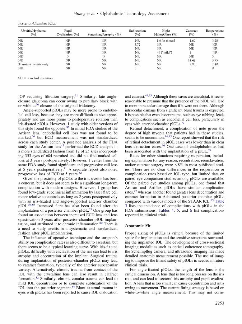
Posterior-Chamber IOLs
Uveitis/Hyphema(%)
PupilOvalization (%)
IrisSynechiae/Atrophy (%
NR NR NRNR NR NRNR NR NRNR NR NRNR 5 5NR NR NRTransient uveitis only NR NRNR NR NR
SD � standard deviation.
eyes with pIOLs has been reported to cause IOL dislocation
and cataract.49,93 Although these cases are anecdotal, it seemsreasonable to presume that the presence of the pIOL will leadto more intraocular damage than if it were not there. Althoughintraocular damage from significant blunt trauma is expected,it is possible that even lesser trauma, such as eye rubbing, leadsto complications such as endothelial cell loss, particularly ineyes with anterior-chamber pIOLs.72
Retinal detachment, a complication of note given thedegree of high myopia that patients had in these studies,seems to be uncommon.94,95 One report showed that the riskof retinal detachment in pIOL cases was lower than in clearlens extraction cases.96 One case of endophthalmitis hadbeen associated with the implantation of a pIOL.97
Rates for other situations requiring reoperation, includ-ing explantation for any reason, recentration, reenclavation,and/or cataract surgery were �8% in most published stud-ies. There are no clear differences in the reoperation orcomplication rates based on IOL type, but limited data onpaired eye comparison studies among pIOLs are available.Of the paired eye studies among pIOLs, one found thatArtisan and Artiflex pIOLs have similar complicationrates,77 whereas another found greater lens decentration andcataract formation in Adatomed posterior-chamber pIOLscompared with various models of the STAAR ICL.85 Table3 lists the incidence of complications with pIOLs in theFDA submissions. Tables 4, 5, and 6 list complicationsreported in clinical trials.
Anatomic Fit
Proper sizing of pIOLs is critical because of the limitedspace for implantation and the sensitive structures surround-ing the implanted IOL. The development of cross-sectionalimaging modalities such as optical coherence tomography,the Scheimpflug camera, and ultrasound imaging has madedetailed anatomic measurement possible. The use of imag-ing to improve the fit and safety of pIOLs is needed in futureclinical trials.
For angle-fixated pIOLs, the length of the lens is thecritical dimension. A lens that is too long presses on the irisroot and can lead to sectoral iris atrophy and pupil ovaliza-tion. A lens that is too small can cause decentration and iritisowing to movement. The current fitting strategy is based on
Subluxation(%)
NightHalos/Glare (%)
Cataract(%)
Reoperations(%)
NR 1.6 [at 6 mos] 1.60 3.283.77 NR NR NRNR NR NR NRNR 46 (“mild”) 2 NRNR NR NR 5NR NR 14.47 3.95NR NR 2.90 2.400 NR 0 0
)
white-to-white angle measurement. This may not corre-
2253

Ophthalmology Volume 116, Number 11, November 2009
spond well with internal anterior-chamber width. Directmeasurement of the anterior-chamber width from recess torecess using optical coherence tomography98,99 or ultra-sound imaging100 may improve the fit of the lens. Three-dimensional imaging of the anterior chamber can determinethe widest meridian and help to determine the most stableorientation for IOL implantation.98 Later generations ofangle-fixated pIOLs that use more flexible haptic architec-ture or material may further improve the fit and safety of theIOL.
For iris-fixated pIOLs, the crystalline lens rise—the axialdistance between the crystalline lens apex and the linejoining the 2 opposite angles—has been found to be anotherimportant anatomic parameter to consider, in addition to theanterior-chamber depth. A crystalline lens rise of �0.6 mmhas been found to cause a high incidence of pigment dis-persion.101 The crystalline lens rise can be measured bycross-sectional imaging modalities such as optical coher-ence tomography, the Scheimpflug camera, and ultrasound.Using the cross-sectional images, lens implantation can besimulated and the resulting clearance from the cornea andthe crystalline lens can be measured.102
For angle-fixated and iris-fixated pIOLs, the most likelysite of endothelium–IOL touch may be at the periphery ofthe optic, where the negatively powered lens is thickest.Thus, safety assessment may be improved by using a cross-sectional imaging modality to model the corneal clearanceof the peripheral optic preoperatively and measure it postop-eratively. Because the loss of corneal endothelial cells mayoccur primarily in the periphery, central endothelial cell countmay not detect progressive loss until several years later.13
Peripheral endothelial cell counting may be able to detectprogressive loss earlier. Case reports of corneal decompensa-tion despite uncomplicated pIOL implantation,103 sometimesmany years later,104 suggest that long-term monitoring of cor-neal clearance and endothelial cell count are prudent.
For posterior-chamber pIOLs, the size of the lens relativeto the distance between opposite ciliary sulci determines thevault of the IOL and clearance over the crystalline lens.Ultrasound imaging to directly measure the sulcus-to-sulcuswidth may improve the predictability of the crystalline lensvault and reduce the incidence of cataract from inadequateclearance and pigment dispersion from too much vault.
Aging changes are a concern for the long-term safety ofpIOLs. The human crystalline lens increases in thicknesswith age, with corresponding shallowing of the anteriorchamber.105,106 Thus, a pIOL that has adequate clearanceover the crystalline lens may come into contact with it whenthe patient reaches an older age. The sulcus-to-sulcus widthof the posterior chamber also narrows with age107,108; thus,the vault of a posterior-chamber pIOL may increase withage and eventually cause pigment dispersion. The effect ofthese aging changes on the tolerance of pIOLs deservesfurther study.
In conclusion, phakic IOL surgery is an efficacious tech-nique for correcting refractive error in patients who wouldotherwise be poor candidates for corneal refractive surgeryowing to high myopia. The designs of pIOLs have evolvedover many years. Most early designs have been abandoned
because of high rates of complications. At present, 1 iris-2254
fixated pIOL and 1 posterior-chamber pIOL have receivedFDA approval in the United States, but several other lensesare undergoing trials. The newer designs aim to improve theease of implantation and to correct astigmatism.
Visual outcomes are, in the aggregate, very encouraging.By retrospective comparison, pIOL surgery seems to offerdistinct predictability and stability advantages over LASIKfor patients with high and perhaps even moderate myopia. Arelatively high percentage of patients had increased BSCVAand increased contrast sensitivity. Satisfaction with thequality of vision was generally high. Residual refractiveerror has been successfully addressed with a bioptics ap-proach involving secondary LASIK109 or PRK.110
The main concerns with pIOL implantation relate to itssafety. In addition to the rare catastrophic risks of intraoc-ular surgery, such as endophthalmitis and hemorrhage (risksthat are absent in LASIK and with contact lens or eyeglasscorrection), there are potential long-term risks with pIOLs.Chief among safety concerns are long-term endothelial cellloss and cataract formation. Although the FDA-approvedpIOLs have acceptable rates of complications and loss ofBSCVA through the 3- to 5-year duration of the trials,longer term problems cannot be ruled out. There is evidencethat endothelial cell loss continues at a higher than normalrate even beyond 5 years for pIOLs located in the anteriorchamber, potentially leading to corneal edema that requireskeratoplasty. The rate of cataract formation may becomehigher as patients age.
Secondary glaucoma (pupillary block, pigment disper-sion), iris atrophy (pupil ovalization), and traumatic dislo-cation are also concerns. Patients should be informed ofthese long-term risks before surgery and be advised tomaintain regular follow-up after the surgery. Endothelialcell count and intraocular pressure should be measuredregularly. Routine slit-lamp biomicroscopy should be per-formed to detect possible corneal edema, pigment disper-sion, pupil ovalization, closure of peripheral iridotomy, lensdislocation, and cataract formation. Regular dilated fundusexaminations are also needed to screen for retinal breaksand detachment in these highly myopic patients.
Despite the risks of pIOL implantation for high myopia,it remains an attractive method compared with alternativeoperative treatments. LASIK and PRK for high myopia areless predictable and stable than keratorefractive surgery forlower degrees of myopia. Both LASIK and PRK can inducehigher order aberrations that decrease the quality of vision.Post-LASIK ectasia and post-PRK haze are also significantrisks for high-dioptric laser treatments. Another alternativeis refractive lens exchange, which carries an increased riskof retinal detachment. The reasonable alternatives for eachpatient depend on the degree of myopia, corneal thickness,anatomy, and other individual factors. The surgeon mustadvise patients on the pros and cons of the variousreasonable operative alternatives to eyeglass or contact lenswear, and help them to make the proper choice consistentwith their visual goals and occupational needs.
The information in this assessment is current as of thedate it was prepared, but we expect new data to becomeavailable rapidly in this area of active research on pIOLs.
Furthermore, there may be new pIOLs under development
Huang et al � Ophthalmic Technology Assessment
that were not yet in clinical trials and thus not included inthis assessment. Interested readers are advised to updatesearches to remain current with developments in this field.
Future Research
Future research should be directed at prospective and ret-rospective studies of the long-term (�10 years) efficacy andcomplications of pIOLs, imaging studies to evaluate thesizing and anatomic fit of pIOLs before surgery and assessthe results after surgery, and randomized, controlled clinicaltrials on the merits of different lens models and types.
References
1. American Academy of Ophthalmology. Excimer laser pho-torefractive keratectomy (PRK) for myopia and astigmatism.Ophthalmology 1999;106:422–37.
2. Schallhorn SC, Farjo AA, Huang D, et al, Ophthalmic Tech-nology Assessment Committee Refractive Management/In-tervention Panel. Wavefront-guided LASIK for the correc-tion of primary myopia and astigmatism: a report by theAmerican Academy of Ophthalmology. Ophthalmology 2008;115:1249–61.
3. Sugar A, Rapuano CJ, Culbertson WW, et al, OphthalmicTechnology Assessment Committee 2000–2001 RefractiveSurgery Panel. Laser in situ keratomileusis for myopia andastigmatism: safety and efficacy: a report by the AmericanAcademy of Ophthalmology. Ophthalmology 2002;109:175– 87.
4. Varley GA, Huang D, Rapuano CJ, et al, Ophthalmic Tech-nology Assessment Committee Refractive Surgery Panel.LASIK for hyperopia, hyperopic astigmatism, and mixedastigmatism: a report by the American Academy of Ophthal-mology. Ophthalmology 2004;111:1604–17.
5. El Danasoury MA, El Maghraby A, Gamali TO. Comparisonof iris-fixed Artisan lens implantation with excimer laser insitu keratomileusis in correcting myopia between �9.00 and�19.50 diopters: a randomized study. Ophthalmology 2002;109:955–64.
6. Sanders DR. Matched population comparison of the Visianimplantable collamer lens and standard LASIK for myopia of�3.00 to �7.88 diopters. J Refract Surg 2007;23:537–53.
7. Schallhorn S, Tanzer D, Sanders DR, Sanders ML. Random-ized prospective comparison of Visian toric implantable col-lamer lens and conventional photorefractive keratectomy formoderate to high myopic astigmatism. J Refract Surg 2007;23:853–67.
8. Colin J, Robinet A, Cochener B. Retinal detachment afterclear lens extraction for high myopia: seven-year follow-up.Ophthalmology 1999;106:2281–4; discussion 85.
9. Chen LJ, Chang YJ, Kuo JC, et al. Metaanalysis of cataractdevelopment after phakic intraocular lens surgery. J CataractRefract Surg 2008;34:1181–200.
10. Fechner PU, Strobel J, Wichmann W. Correction of myopiaby implantation of a concave Worst-iris claw lens into phakiceyes. Refract Corneal Surg 1991;7:286–98.
11. Praeger DL, Momose A, Muroff LL. Thirty-six monthfollow-up of a contemporary phakic intraocular lens for thesurgical correction of myopia. Ann Ophthalmol 1991;23:
6–10.12. Javaloy J, Alio JL, Iradier MT, et al. Outcomes of ZB5Mangle-supported anterior chamber phakic intraocular lensesat 12 years. J Refract Surg 2007;23:147–58.
13. Allemann N, Chamon W, Tanaka HM, et al. Myopic angle-supported intraocular lenses: two-year follow-up. Ophthal-mology 2000;107:1549–54.
14. Perez-Santonja JJ, Alio JL, Jimenez-Alfaro I, Zato MA.Surgical correction of severe myopia with an angle-supported phakic intraocular lens. J Cataract Refract Surg2000;26:1288–302.
15. Leccisotti A, Fields SV. Clinical results of ZSAL-4 angle-supported phakic intraocular lenses in 190 myopic eyes. JCataract Refract Surg 2005;31:318–23.
16. Jimenez-Alfaro I, Garcia-Feijoo J, Perez-Santonja JJ, CuinaR. Ultrasound biomicroscopy of ZSAL-4 anterior chamberphakic intraocular lens for high myopia. J Cataract RefractSurg 2001;27:1567–73.
17. Leccisotti A. Bioptics by angle-supported phakic lenses andphotorefractive keratectomy. Eur J Ophthalmol 2005;15:1–7.
18. Yu A, Wang Q, Xue A, et al. Comparison of contrastsensitivity after angle-supported, iris-fixated and posteriorchamber phakic intraocular lens implantation for high myo-pia. Ophthalmologica 2008;222:53–7.
19. Alio JL, Pinero D, Bernabeu G, et al. The Kelman Duetphakic intraocular lens: 1-year results. J Refract Surg 2007;23:868–79.
20. Van Cleynenbreugel H. Late postoperative complications ofbackward implantation of a Vivarte phakic intraocular lens. JCataract Refract Surg 2007;33:1474–6.
21. Gierek-Ciaciura S, Gierek-Lapinska A, Ochalik K, Mrukwa-Kominek E. Correction of high myopia with different phakicanterior chamber intraocular lenses: ICARE angle-supportedlens and Verisyse iris-claw lens. Graefes Arch Clin ExpOphthalmol 2007;245:1–7.
22. Oshika T, Nagata T, Ishii Y. Adhesion of lens capsule tointraocular lenses of polymethylmethacrylate, silicone, andacrylic foldable materials: an experimental study. Br J Oph-thalmol 1998;82:549–53.
23. Linnola RJ, Sund M, Ylonen R, Pihlajaniemi T. Adhesionof soluble fibronectin, laminin, and collagen type IV tointraocular lens materials. J Cataract Refract Surg 1999;25:1486 –91.
24. Alexander L, John M, Cobb L, et al. U.S. clinical investiga-tion of the Artisan myopia lens for the correction of highmyopia in phakic eyes: report of the results of phases 1 and2, and interim phase 3. Optometry 2000;71:630–42.
25. Bartels MC, Saxena R, van den Berg TJ, et al. The influenceof incision-induced astigmatism and axial lens position onthe correction of myopic astigmatism with the Artisan toricphakic intraocular lens. Ophthalmology 2006;113:1110–7.
26. Dick HB, Alio J, Bianchetti M, et al. Toric phakic intraocularlens: European multicenter study. Ophthalmology 2003;110:150–62.
27. Dick HB, Tehrani M, Aliyeva S. Contrast sensitivity afterimplantation of toric iris-claw lenses in phakic eyes. J Cat-aract Refract Surg 2004;30:2284–9.
28. Tehrani M, Dick HB. Short-term follow-up after implanta-tion of a foldable iris-fixated intraocular lens in phakic eyes.Ophthalmology 2005;112:2189–95.
29. Jimenez-Alfaro I, Benitez del Castillo JM, Garcia-Feijoo J, etal. Safety of posterior chamber phakic intraocular lenses forthe correction of high myopia: anterior segment changes afterposterior chamber phakic intraocular lens implantation. Oph-
thalmology 2001;108:90–9.2255

Ophthalmology Volume 116, Number 11, November 2009
30. Marinho A, Neves MC, Pinto MC, Vaz F. Posterior cham-ber silicone phakic intraocular lens. J Refract Surg 1997;13:219 –22.
31. Jongsareejit A. Clinical results with the Medennium phakicrefractive lens for the correction of high myopia. J RefractSurg 2006;22:890–7.
32. Koivula A, Taube M, Zetterstrom C. Phakic refractive lens:two-year results. J Refract Surg 2008;24:507–15.
33. Verde CM, Teus MA, Arranz-Marquez E, Cazorla RG. Me-dennium posterior chamber phakic refractive lens to correcthigh myopia. J Refract Surg 2007;23:900–4.
34. Donoso R, Castillo P. Correction of high myopia with thePRL phakic intraocular lens. J Cataract Refract Surg 2006;32:1296–300.
35. Jimenez-Alfaro I, Gomez-Telleria G, Bueno JL, Puy P. Con-trast sensitivity after posterior chamber phakic intraocularlens implantation for high myopia. J Refract Surg 2001;17:641–5.
36. Zaldivar R, Davidorf JM, Oscherow S. Posterior chamberphakic intraocular lens for myopia of �8 to �19 diopters. JRefract Surg 1998;14:294–305.
37. Chang JS, Meau AY. Visian collamer phakic intraocular lensin high myopic Asian eyes. J Refract Surg 2007;23:17–25.
38. American Academy of Ophthalmology Refractive Manage-ment/Intervention Panel. Preferred Practice Pattern. Refrac-tive Errors and Refractive Surgery. San Francisco: AmericanAcademy of Ophthalmology; 2007:10. Available at: http://one.aao.org/CE/PracticeGuidelines/PPP.aspx?p�1. AccessedDecember 16, 2008.
39. American Academy of Ophthalmology. Policy Statement.Pretreatment assessment: responsibilities of the ophthalmol-ogist. San Francisco: American Academy of Ophthalmology;2006. Available at: http://one.aao.org/CE/PracticeGuidelines/ClinicalStatements_Content.aspx?cid�0b059383-f91a-4f23-9e16-09c05bcdf11b. Accessed December 16, 2008.
40. American Academy of Ophthalmology. Policy Statement.An ophthalmologist’s duties concerning postoperative care.San Francisco: American Academy of Ophthalmology; 2006.Available at: http://one.aao.org/CE/PracticeGuidelines/ClinicalStatements_Content.aspx?cid�219fce14-3bee-499b-aeea-b740c637e534. Accessed December 16, 2008.
41. Nourouzi H, Rajavi J, Okhovatpour MA. Time to resolutionof corneal edema after long-term contact lens wear. Am JOphthalmol 2006;142:671–3.
42. Saxena R, Boekhoorn SS, Mulder PG, et al. Long-termfollow-up of endothelial cell change after Artisan phakicintraocular lens implantation. Ophthalmology 2008;115:608 –13.
43. Frisch IB, Rabsilber TM, Becker KA, et al. Comparison ofanterior chamber depth measurements using Orbscan II andIOLMaster. Eur J Ophthalmol 2007;17:327–31.
44. Rabsilber TM, Becker KA, Frisch IB, Auffarth GU. Anteriorchamber depth in relation to refractive status measured withthe Orbscan II topography system. J Cataract Refract Surg2003;29:2115–21.
45. Lavanya R, Teo L, Friedman DS, et al. Comparison ofanterior chamber depth measurements using the IOLMaster,scanning peripheral anterior chamber depth analyser, andanterior segment optical coherence tomography. Br J Oph-thalmol 2007;91:1023–6.
46. Dada T, Sihota R, Gadia R, et al. Comparison of anteriorsegment optical coherence tomography and ultrasoundbiomicroscopy for assessment of the anterior segment. JCataract Refract Surg 2007;33:837–40.
47. Stulting RD, John ME, Maloney RK, et al, U.S. Verisyse
Study Group. Three-year results of Artisan/Verisyse phakic2256
intraocular lens implantation: results of the United StatesFood and Drug Administration clinical trial. Ophthalmology2008;115:464–72.
48. Modis L Jr, Langenbucher A, Seitz B. Corneal endothelialcell density and pachymetry measured by contact and non-contact specular microscopy. J Cataract Refract Surg 2002;28:1763–9.
49. de Sanctis U, Machetta F, Razzano L, et al. Corneal endo-thelium evaluation with 2 noncontact specular microscopesand their semiautomated methods of analysis. Cornea 2006;25:501–6.
50. Kitzmann AS, Winter EJ, Nau CB, et al. Comparison ofcorneal endothelial cell images from a noncontact specularmicroscope and a scanning confocal microscope. Cornea2005;24:980–4.
51. van der Heijde GL. Some optical aspects of implantation ofan IOL in a myopic eye. Eur J Implant Refract Surg 1989;1:245–8.
52. Benedetti S, Casamenti V, Benedetti M. Long-term endothe-lial changes in phakic eyes after Artisan intraocular lensimplantation to correct myopia: five-year study. J CataractRefract Surg 2007;33:784–90.
53. Tahzib NG, Eggink FA, Frederik PM, Nuijts RM. Recurrentintraocular inflammation after implantation of the Artiflexphakic intraocular lens for the correction of high myopia. JCataract Refract Surg 2006;32:1388–91.
54. Budo C, Hessloehl JC, Izak M, et al. Multicenter study of theArtisan phakic intraocular lens. J Cataract Refract Surg 2000;26:1163–71.
55. Lombardo AJ, Hardten DR, McCulloch AG, et al. Changesin contrast sensitivity after Artisan lens implantation for highmyopia. Ophthalmology 2005;112:278–85.
56. Perez-Santonja JJ, Iradier MT, Benitez del Castillo JM, et al.Chronic subclinical inflammation in phakic eyes with in-traocular lenses to correct myopia. J Cataract Refract Surg1996;22:183–7.
57. Kohnen T. Searching for the perfect phakic intraocular lens.J Cataract Refract Surg 2000;26:1261–2.
58. Menezo JL, Peris-Martinez C, Cisneros AL, Martinez-CostaR. Phakic intraocular lenses to correct high myopia:Adatomed, Staar, and Artisan. J Cataract Refract Surg 2004;30:33–44.
59. Aguilar-Valenzuela L, Lleo-Perez A, Alonso-Munoz L, et al.Intraocular pressure in myopic patients after Worst-Fechneranterior chamber phakic intraocular lens implantation. J Re-fract Surg 2003;19:131–6.
60. Fechner PU, Haubitz I, Wichmann W, Wulff K. Worst-Fechner biconcave minus power phakic iris-claw lens. JRefract Surg 1999;15:93–105.
61. Menezo JL, Avino JA, Cisneros A, et al. Iris claw phakicintraocular lens for high myopia. J Refract Surg 1997;13:545–55.
62. Menezo JL, Cisneros A, Hueso JR, Harto M. Long-termresults of surgical treatment of high myopia with Worst-Fechner intraocular lenses. J Cataract Refract Surg 1995;21:93–8.
63. Perez-Santonja JJ, Bueno JL, Zato MA. Surgical correctionof high myopia in phakic eyes with Worst-Fechner myopiaintraocular lenses. J Refract Surg 1997;13:268–81; discus-sion 81–4.
64. Landesz M, Worst JG, Siertsema JV, van Rij G. Correctionof high myopia with the Worst myopia claw intraocular lens.J Refract Surg 1995;11:16–25.
65. Landesz M, van Rij G, Luyten G. Iris-claw phakic intraoc-
ular lens for high myopia. J Refract Surg 2001;17:634–40.
Huang et al � Ophthalmic Technology Assessment
66. Maloney RK, Nguyen LH, John ME. Artisan phakic intraoc-ular lens for myopia: short-term results of a prospective,multicenter study. Ophthalmology 2002;109:1631–41.
67. Benedetti S, Casamenti V, Marcaccio L, et al. Correction ofmyopia of 7 to 24 diopters with the Artisan phakic in-traocular lens: two-year follow-up. J Refract Surg 2005;21:116 –26.
68. Landesz M, Worst JG, van Rij G. Long-term results ofcorrection of high myopia with an iris claw phakic intraoc-ular lens. J Refract Surg 2000;16:310–6.
69. Lifshitz T, Levy J, Aizenman I, et al. Artisan phakic intraoc-ular lens for correcting high myopia. Int Ophthalmol 2004;25:233–8.
70. Senthil S, Reddy KP. A retrospective analysis of the firstIndian experience on Artisan phakic intraocular lens. IndianJ Ophthalmol 2006;54:251–5.
71. Silva RA, Jain A, Manche EE. Prospective long-term eval-uation of the efficacy, safety, and stability of the phakicintraocular lens for high myopia. Arch Ophthalmol 2008;126:775–81.
72. Guell JL, Morral M, Gris O, et al. Five-year follow-up of 399phakic Artisan-Verisyse implantation for myopia, hyperopia,and/or astigmatism. Ophthalmology 2008;115:1002–12.
73. Tahzib NG, Nuijts RM, Wu WY, Budo CJ. Long-term studyof Artisan phakic intraocular lens implantation for the cor-rection of moderate to high myopia: ten-year follow-up re-sults. Ophthalmology 2007;114:1133–42.
74. Malecaze FJ, Hulin H, Bierer P, et al. A randomized pairedeye comparison of two techniques for treating moderatelyhigh myopia: LASIK and Artisan phakic lens. Ophthalmol-ogy 2002;109:1622–30.
75. Tahzib NG, Bootsma SJ, Eggink FA, Nuijts RM. Functionaloutcome and patient satisfaction after Artisan phakic intraoc-ular lens implantation for the correction of myopia. Am JOphthalmol 2006;142:31–9.
76. Guell JL, Vazquez M, Gris O. Adjustable refractive surgery:6-mm Artisan lens plus laser in situ keratomileusis forthe correction of high myopia. Ophthalmology 2001;108:945–52.
77. Coullet J, Guell JL, Fournie P, et al. Iris-supported phakiclenses (rigid vs foldable version) for treating moderately highmyopia: randomized paired eye comparison. Am J Ophthal-mol 2006;142:909–16.
78. Guell JL, Morral M, Gris O, et al. Evaluation of Verisyse andArtiflex phakic intraocular lenses during accommodation us-ing Visante optical coherence tomography. J Cataract RefractSurg 2007;33:1398–404.
79. Koss MJ, Cichocki M, Kohnen T. Posterior synechias fol-lowing implantation of a foldable silicone iris-fixated phakicintraocular lens for the correction of myopia. J CataractRefract Surg 2007;33:905–9.
80. Lackner B, Pieh S, Schmidinger G, et al. Long-term resultsof implantation of phakic posterior chamber intraocularlenses. J Cataract Refract Surg 2004;30:2269–76.
81. Pesando PM, Ghiringhello MP, Tagliavacche P. Posteriorchamber collamer phakic intraocular lens for myopia andhyperopia. J Refract Surg 1999;15:415–23.
82. Sarikkola AU, Sen HN, Uusitalo RJ, Laatikainen L. Trau-matic cataract and other adverse events with the implantablecontact lens. J Cataract Refract Surg 2005;31:511–24.
83. Pineda-Fernandez A, Jaramillo J, Vargas J, et al. Phakicposterior chamber intraocular lens for high myopia. J Cata-ract Refract Surg 2004;30:2277–83.
84. Rosen E, Gore C. Staar Collamer posterior chamber phakicintraocular lens to correct myopia and hyperopia. J Cataract
Refract Surg 1998;24:596–606.85. Menezo JL, Peris-Martinez C, Cisneros A, Martinez-CostaR. Posterior chamber phakic intraocular lenses to correcthigh myopia: a comparative study between Staar andAdatomed models. J Refract Surg 2001;17:32–42.
86. ICL in Treatment of Myopia (ITM) Study Group. UnitedStates Food and Drug Administration clinical trial of theimplantable collamer lens (ICL) for moderate to highmyopia: three-year follow-up. Ophthalmology 2004;111:1683–92.
87. Sanders D, Vukich JA. Comparison of implantable collamerlens (ICL) and laser-assisted in situ keratomileusis (LASIK)for low myopia. Cornea 2006;25:1139–46.
88. Sanders DR, Schneider D, Martin R, et al. Toric implantablecollamer lens for moderate to high myopic astigmatism.Ophthalmology 2007;114:54–61.
89. Munoz G, Alio JL, Montes-Mico R, Belda JI. Angle-supported phakic intraocular lenses followed by laser-assisted insitu keratomileusis for the correction of high myopia. Am JOphthalmol 2003;136:490–9.
90. Kodjikian L, Gain P, Donate D, et al. Malignant glaucomainduced by a phakic posterior chamber intraocular lens formyopia. J Cataract Refract Surg 2002;28:2217–21.
91. Sanchez-Galeana CA, Zadok D, Montes M, et al. Refractoryintraocular pressure increase after phakic posterior chamberintraocular lens implantation. Am J Ophthalmol 2002;134:121–3.
92. Perez-Santonja JJ, Iradier MT, Sanz-Iglesias L, et al. Endo-thelial changes in phakic eyes with anterior chamber intraoc-ular lenses to correct high myopia. J Cataract Refract Surg1996;22:1017–22.
93. Munoz G, Montes-Mico R, Belda JI, Alio JL. Cataract afterminor trauma in a young patient with an iris-fixated in-traocular lens for high myopia. Am J Ophthalmol 2003;135:890 –1.
94. Ruiz-Moreno JM, Alio JL, Perez-Santonja JJ, de la Hoz F.Retinal detachment in phakic eyes with anterior chamberintraocular lenses to correct severe myopia. Am J Ophthal-mol 1999;127:270–5.
95. Martinez-Castillo V, Boixadera A, Verdugo A, et al. Rheg-matogenous retinal detachment in phakic eyes after posteriorchamber phakic intraocular lens implantation for severe my-opia. Ophthalmology 2005;112:580–5.
96. Arne JL. Phakic intraocular lens implantation versus clearlens extraction in highly myopic eyes of 30- to 50-year-oldpatients. J Cataract Refract Surg 2004;30:2092–6.
97. Perez-Santonja JJ, Ruiz-Moreno JM, de la Hoz F, et al.Endophthalmitis after phakic intraocular lens implantation tocorrect high myopia. J Cataract Refract Surg 1999;25:1295– 8.
98. Baikoff G, Jodai HJ, Bourgeon G. Measurement of theinternal diameter and depth of the anterior chamber: IOL-Master versus anterior chamber optical coherence tomogra-pher. J Cataract Refract Surg 2005;31:1722–8.
99. Goldsmith JA, Li Y, Chalita MR, et al. Anterior chamberwidth measurement by high-speed optical coherence tomog-raphy. Ophthalmology 2005;112:238–44.
100. Lee JY, Kim JH, Kim HM, Song JS. Comparison of anteriorchamber depth measurement between Orbscan IIz and ultra-sound biomicroscopy. J Refract Surg 2007;23:487–91.
101. Baikoff G, Bourgeon G, Jodai HJ, et al. Pigment dispersionand Artisan phakic intraocular lenses: crystalline lens rise asa safety criterion. J Cataract Refract Surg 2005;31:674–80.
102. Baikoff G, Lutun E, Wei J, Ferraz C. Contact between 3phakic intraocular lens models and the crystalline lens: ananterior chamber optical coherence tomography study. J Cat-
aract Refract Surg 2004;30:2007–12.2257

Ophthalmology Volume 116, Number 11, November 2009
103. Coullet J, Mahieu L, Malecaze F, et al. Severe endothelialcell loss following uneventful angle-supported phakic in-traocular lens implantation for high myopia. J Cataract Re-fract Surg 2007;33:1477–81.
104. Alio JL, Abdelrahman AM, Javaloy J, et al. Angle-supportedanterior chamber phakic intraocular lens explantation causesand outcome. Ophthalmology 2006;113:2213–20.
105. Baikoff G, Lutun E, Ferraz C, Wei J. Static and dynamicanalysis of the anterior segment with optical coherence to-mography. J Cataract Refract Surg 2004;30:1843–50.
106. Koretz JE, Strenk SA, Strenk LM, Semmlow JL. Scheimp-flug and high-resolution magnetic resonance imaging of theanterior segment: a comparative study. J Opt Soc Am A Opt
Image Sci Vis 2004;21:346–54.Parag A. Majmudar – Consultant/Advisor – Advanced Medical Optics/
2258
107. Strenk SA, Semmlow JL, Strenk LM, et al. Age-related changesin human ciliary muscle and lens: a magnetic resonance imag-ing study. Invest Ophthalmol Vis Sci 1999;40:1162–9.
108. Strenk SA, Strenk LM, Guo S. Magnetic resonance imagingof aging, accommodating, phakic, and pseudophakic ciliarymuscle diameters. J Cataract Refract Surg 2006;32:1792–8.
109. Zaldivar R, Davidorf JM, Oscherow S, et al. Combinedposterior chamber phakic intraocular lens and laser in situkeratomileusis: bioptics for extreme myopia. J Refract Surg1999;15:299–308.
110. Sanchez-Galeana CA, Smith RJ, Rodriguez X, et al. Laser insitu keratomileusis and photorefractive keratectomy for re-sidual refractive error after phakic intraocular lens implanta-
tion. J Refract Surg 2001;17:299–304.Footnotes and Financial Disclosures
Originally received: June 17, 2009.Accepted: August 11, 2009. Manuscript no. 2009-829.
Prepared by the Ophthalmic Technology Assessment Committee Refrac-tive Management/Intervention Panel and approved by the American Acad-emy of Ophthalmology’s Board of Trustees February 21, 2009.
Financial Disclosure(s):The authors have made the following disclosures:David Huang – Consultant – Ophthalmic Systems, Optovue, Inc.; LectureFees/Equity Owner – Optovue, Inc.; Patents/Royalties – Carl Zeiss Med-itech/Optovew, Inc.; Grant Support – Optovue, Inc./Vistakon Johnson &Johnson Visioncare, Inc.
Steven C. Schallhorn – Consultant/Advisor – Acufocus, Inc./AdvancedMedical Optics
Alan Sugar – Consultant/Advisor – Lux Biosciences; Grant Support – LuxBiosciences/National Eye Institute.
Inspire; Lecture Fees – Advanced Medical Optics/Alcon Laboratories,Inc./Allergan, Inc./Bausch & Lomb Surgical/Inspire.
William B. Trattler – Consultant/Advisor – Allergan Inc./Inspire Pharma-ceuticals, Inc./Ista Pharmaceuticals, Inc./Sirion/Vistakon/Wavetec; LectureFees – Advanced Medical Optics/Allergan, Inc./Inspire Pharmaceuticals,Inc./Sirion; Equity Owner – AskLasikDocs.com; Grant Support – Ad-vanced Medical Optics/Allergan, Inc./Glaukos Corporation/Inspire Phar-maceutical, Inc./Ista Pharmaceutical/Lenstec, Inc./QLT Phototherapeu-tics, Inc./Rapid Pathogen Screenings/Sirion/Vistakon/Wavetec.
Funded without commercial support by the American Academy of Oph-thalmology.
Correspondence:Ms. Nancy Collins, Guidelines and Assessment Manager, American Acad-emy of Ophthalmology, The Eye MD Association, PO Box 7424, San
Francisco, CA 94120-7424. E-mail: [email protected].