acrysof phakic angle-supported intraocular lens for the ... · lens opacities classification...
TRANSCRIPT

AcrySof Phakic Angle-supportedIntraocular Lens for the Correction ofModerate-to-High Myopia: One-YearResults of a Multicenter European Study
Thomas Kohnen, MD,1 Michael C. Knorz, MD,2 Béatrice Cochener, MD,3 Ralf H. Gerl, MD,4
Jean-Louis Arné, MD,5 Joseph Colin, MD,6 Jorge L. Alió, MD,7 Roberto Bellucci, MD,8 Antonio Marinho, MD9
Purpose: To investigate the safety and effectiveness of the AcrySof phakic angle-supported intraocular lens(IOL) (Alcon Laboratories, Inc., Fort Worth, TX) for correction of moderate-to-high myopia in adults.
Design: One-year interim analysis of a phase 3, nonrandomized, open-label, prospective, multicenterEuropean clinical study.
Participants: A total of 190 subjects (190 eyes) with moderate-to-high myopia. The preoperative meanmanifest refraction spherical equivalent (MRSE) was �10.38 diopters (D) �2.43 standard deviation (SD).
Methods: Unilateral implantation of the AcrySof phakic angle-supported IOL.Main Outcome Measures: Best spectacle-corrected visual acuity (BSCVA), uncorrected distance visual
acuity (UCVA), predictability and stability of MRSE, adverse events, and endothelial cell density.Results: Of 190 subjects enrolled, 161 completed the 1-year postoperative visit. No subjects lost �2 lines
BSCVA. A UCVA of 20/20 or better was achieved by 57.8%; 99.4% had 20/40 or better. A BSCVA of 20/32 orbetter was achieved by 100% of subjects; 85.7% had 20/20 or better. The mean MRSE was �0.23 D (�0.50 D:�2.50 to 0.75 D). Residual refractive error was within �1.0 D from the target for 95.7% of subjects and within�0.5 D for 72.7% of subjects. The overall mean percentage change in central endothelial cell density 1 year aftersurgery was �4.77�8.04% (n � 139). No pupil ovalization, pupillary block, or retinal detachment events wereobserved.
Conclusions: The AcrySof phakic angle-supported IOL yielded excellent refractive correction and predict-ability with acceptable safety in subjects with moderate-to-high myopia. These 1-year interim analysis findingsdemonstrate preliminary support for the safety and efficacy of this IOL.
Financial Disclosure(s): Proprietary or commercial disclosure may be found after the references.Ophthalmology 2009;116:1314 –1321 © 2009 by the American Academy of Ophthalmology.
The correction of moderate-to-high myopia remains chal-lenging. Corrective options such as spectacles and contactlenses are often unsatisfactory to patients, and many of thesepatients have refractive errors outside the range of treatmentwith laser ablation refractive surgery. In an effort to developa refractive surgical option for patients with moderate-to-high myopia, phakic intraocular lenses (IOLs) of variousdesigns and materials have been placed in the anteriorchamber, fixed to the iris, and placed in the posterior cham-ber. Phakic IOLs have demonstrated excellent refractiveresults but have also been associated with safety con-cerns.1–9 A fundamental goal in phakic IOL development isto reduce potential risk while providing patients withmoderate-to-high myopia visual correction, comfort, andconvenience not attainable with spectacles or contact lenses.
The primary safety concerns associated with angle-supported anterior chamber phakic IOLs are corneal endo-
thelial cell loss and pupil ovalization.1 Published case re-1314 © 2009 by the American Academy of OphthalmologyPublished by Elsevier Inc.
ports and clinical studies of up to 12 years duration foundendothelial cell density changes ranging from significantlosses to long-term gains, with recent publications generallydescribing smaller losses or gains compared with precedingreports.1–6,10–13 A high frequency of pupil ovalization andIOL rotation has been associated with an early investiga-tional angle-supported anterior chamber phakic IOL.14 Thislens has demonstrated satisfactory refractive results but isless widely accepted because of pupil ovalization and IOLrotation associated with IOL footplate displacement.
The AcrySof phakic angle-supported IOL (Alcon Labo-ratories, Inc., Fort Worth, TX) is made of foldable hydro-phobic acrylate, permitting a small corneal incision size(�3.0 mm) using the Monarch II IOL Delivery System withthe “B” or “C” cartridge. The haptics are designed to permitcompression within the angle for IOL stability (data on file,Alcon Research, Ltd.), without creating excessive force that
could cause angle tissue damage or pupil ovalization. Mod-ISSN 0161-6420/09/$–see front matterdoi:10.1016/j.ophtha.2009.01.041
IOLs
Kohnen et al � AcrySof Phakic IOL for Correction of Moderate-to-High Myopia
els of varying diameter allow fit within a variety of anteriorchamber dimensions. The IOL is vaulted to provide optimalcentral clearance distance between the IOL and the corneaand the natural crystalline lens. These characteristics areintended to achieve predictable implantation, stable vault-ing, and low compression forces on the angle, while mini-mizing corneal endothelial cell loss, pupil ovalization, andcataract formation. This article describes the 1-year interimanalysis results of a 5-year investigation of the AcrySofphakic angle-supported IOL for the correction of moderate-to-high myopia in adult subjects.
Subjects and Methods
Subject Recruitment
The study included 190 adult subjects with good general andocular health and moderate-to-high myopia (range, �6.0 to �16.5diopters [D]) in the intended operative eye. Eligibility criteriaincluded a preoperative best spectacle-corrected visual acuity(BSCVA) of �0.3 logarithm of the minimum angle of resolution(logMAR) or better and refraction within �0.5 D at least 12months before surgery, as determined by the manifest refractionspherical equivalent (MRSE).
Exclusion criteria included an anterior chamber depth �3.2 mm(including the corneal epithelium), history of corneal or intraocularsurgery (e.g., laser coagulation of retinal defects), mesopic pupildiameter �7.0 mm, astigmatism �2.0 D, or cataract of any type ordegree. Subjects were also excluded for nonqualifying preoperativeendothelial cell density according to age-based protocol-specifiedcriteria (i.e., �2800 cells/mm2, age 18–25 years; �2600 cells/mm2, age 26–35 years; �2200 cells/mm2, age 36–45 years; and�2000 cells/mm2, age �46 years; Refractive Implants: Investiga-tional Device Exemptions and Premarket Approval Applications;U.S. Food and Drug Administration, Draft August 1, 2000).
Subjects provided written, informed consent before participat-
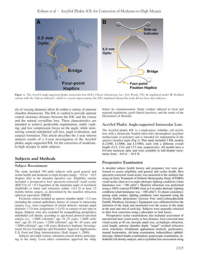
Figure 1. The AcrySof angle-supported phakic intraocular lens (IOL) (Aversion with the “side-up indicator”, which is a recent improvement; the
ing in the study. Local ethics committees approved the study
before its commencement. Study conduct adhered to local andregional regulations, good clinical practices, and the tenets of theDeclaration of Helsinki.
AcrySof Phakic Angle-supported Intraocular Lens
The AcrySof phakic IOL is a single-piece, foldable, soft acryliclens with a chemically bonded ultraviolet chromophore (acrylate/methacrylate co-polymer) and is intended for implantation in theanterior chamber angle (Fig 1). This study included 3 IOL models(L12500, L13000, and L13500), each with a different overalllength (12.5, 13.0, and 13.5 mm, respectively). All models have a6.0-mm meniscus optic and were available in half-diopter incre-ments from �6.0 to �16.5 D.
Preoperative Examination
A detailed subject health history and pregnancy test were per-formed to assess eligibility and general and ocular health. Bestspectacle-corrected visual acuity was measured to the smallest lineusing an Early Treatment of Diabetic Retinopathy Study (ETDRS)visual acuity chart at 4 m under photopic lighting conditions (chartluminance was �180 cd/m2). Manifest refraction was performedusing a 100% contrast ETDRS chart at 4 m under photopic lightingconditions (chart luminance was �180 cd/m2). To ensure consistencyamong study centers, lighting conditions were measured using theGossen Starlite photometer (Gossen Foto und-Lichtme�technikGmbH, Nürnberg, Germany). Equipment was calibrated before theinitiation of the study and monitored over the course of the studyat the start and end of each day. Subjects were manually refractedto their best correction using a phoropter or trial lenses.
Preoperative ocular examinations also included assessment ofuncorrected near visual acuity at best distance, best-corrected nearvisual acuity at 40 cm, mesopic pupil size, cycloplegic refraction,axial length, anterior chamber depth, “target” residual refractiveerror, tonometry (Goldmann applanation method), pachymetry,manual keratometry, slit-lamp examination, indirect/direct ophthal-moscopy (dilated fundus examination), gonioscopic examination, en-
Laboratories, Inc., Fort Worth, TX). A, Implanted model. B, Modifiedimplanted during this study did not have this indicator.
lcon
dothelial cell density analysis, and a crystalline lens assessment using
1315

Ophthalmology Volume 116, Number 7, July 2009
Lens Opacities Classification System III, a subjective, standard-ized cataract grading method.15
For purposes of lens size selection and eligibility determina-tion, the anterior chamber diameter was measured preoperativelyas the width of the cornea from the nasal limbus to the temporallimbus (white-to-white measurement). This was measured withcalipers, a Zeiss IOL Master (Carl Zeiss AG, Oberkochen, Germany),or an Orbscan II topographer (Bausch & Lomb, Rochester, NY).
Gonioscopic examination was performed by using the instru-ment of the physician’s choice. The examination included assess-ment for anterior chamber angle recession, angle trauma, or ana-tomic anomalies.
Endothelial images were taken at the corneal center (3 images)using the Konan Noncon-Robo specular microscope (KonanMedical, Inc., Hyogo, Japan). To minimize analysis variability, anoncontact specular microscope was used at each site, standard-ized training was provided with skill assessment to ensure qualityimages, and a centralized reading center (Alcon Research, Ltd.)performed endothelial cell analyses. Images sent to the readingcenter were analyzed using the center method (Konan analysissoftware). In the center method, the computer mouse is used to dotthe center of the cells in the digital images. At least 100 contiguouscells were marked on the image to obtain an analysis of at least 50cells. The cell counts of 2 to 3 images were averaged to calculatethe mean endothelial cell density.
Surgical Technique and Postoperative Treatment
All 9 participating surgeons received common training on thestudy protocol and surgical technique. Investigators selected pha-kic IOL power to achieve target residual refraction. Power calcu-lations were predicted using the formula originally derived by Van
Figure 3. A-D Postoperative images of the AcrySof angle-supported phanondilated and a dilated eye, respectively. C and D, gonioscopic views of
anterior chamber angle, respectively.1316
der Heijde16 and further refined by Holladay.17 Iridectomy oriridotomy at the time of surgery was not considered necessary forsuccessful implantation but could be performed at investigatordiscretion and was done in only 5 of the 190 surgeries (2.6%).
Before surgery, the pupil was constricted (pilocarpine 2% wasrecommended) to prevent potential contact with the crystallinelens. Investigators administered the anesthesia of their choice (e.g.,topical, retrobular, peribulbar, or general anesthesia). Investigatorsdetermined final lens size by confirming white-to-white measure-ment of the anterior chamber diameter with calipers, on the sedatedeye. In this study, white-to-white measurements plus 0.5 mmdetermined the overall size of the IOL. The anterior chamber wasaccessed with a corneal tunnel incision of approximately 3.0 mmoriented temporally, superiorly, or along the steepest axis.Administration of acetylcholine chloride intraocular solution 1%(Miochol-E, Novartis Ophthalmics, East Hanover, NJ) was usedwhen insufficient pupil constriction was observed intraoperatively.To inflate and maintain the chamber, sodium hyaluronate 1%(Provisc, Alcon Laboratories, Inc.) was injected tangentially intothe angle, away from the pupil. This cohesive ophthalmic vis-coelastic device (OVD) was used to achieve ease of OVD removalafter IOL implantation. The AcrySof phakic IOL was loaded intothe Monarch II IOL Delivery System with its anterior optic surfacefacing upward (Fig 2A, available at http://aaojournal.org) and wasthen folded and slowly delivered with the cartridge positioned atmidpupil to provide delivery in the area of maximum corneal depth(Fig 2B and video clip, available at http://aaojournal.org). Afterpausing for the leading haptics to unfold, delivery was continued(Fig 2C, available at http://aaojournal.org); when the leading hap-tics reached the distal angle, the cartridge was withdrawn asdelivery continued, to avoid increased compression in the distalangle. Trailing haptics were left just outside the incision and then
traocular lens (IOL). A and B, the implanted AcrySof phakic IOL in ans in the anterior chamber angle and the haptic footplate position in the
kic inthe le

Kohnen et al � AcrySof Phakic IOL for Correction of Moderate-to-High Myopia
tucked one at a time into the anterior chamber angle (Fig 2D,available at http://aaojournal.org) so that the lens was positionedwith all 4 haptics in the anterior chamber (Fig 2E, available athttp://aaojournal.org). Incision size was confirmed with the gaugeor device of the investigators’ choice. Lens position and integritywere confirmed as part of the gonioscopic examination beforewound closure.
Investigators thoroughly removed the cohesive OVD with irri-gation and aspiration devices and techniques of their choice. Pas-sive removal consisted of irrigation via injection of intraocularirrigating solution to displace the OVD through the incision. Ac-tive removal included the use of a bimanual or single port systemto simultaneously irrigate and aspirate the OVD.
Postoperative images show the implanted AcrySof phakic IOLin a nondilated (Fig 3A) and a dilated (Fig 3B) eye. Gonioscopicviews show the lens in the anterior chamber angle (Fig 3C) and thehaptic footplate position in the anterior chamber angle (Fig 3D).Closure with sutures was optional. Acetazolamide (Diamox, Led-erle Laboratories, Philadelphia, PA) or equivalent was given at theconclusion of surgery to control intraocular pressure (IOP). Theoperative eye was protected with an eye shield, and subjects wereinstructed not to rub the eye and to avoid direct eye trauma.Postoperative treatment included an ocular antibiotic and steroidregimen (e.g., prednisolone acetate ophthalmic suspension 1%[Pred Forte, Allergan, Inc., Irvine, CA]) or tobramycin 0.3%/dexamethasone 0.1% [Tobradex, Alcon Laboratories, Inc.]) for 1to 4 weeks.
Postoperative Evaluations
Subjects were examined on the first postoperative day and 1 weekafter surgery. Subsequent examinations were performed at 1, 3, 6,and 12 months and included uncorrected and BSCVA, mesopicpupil size, cycloplegic refraction, tonometry (Goldmann applana-tion method), pachymetry, manual keratometry, slit-lamp exami-nation, dilated fundus examination, gonioscopic examination, en-dothelial cell density analysis, and a crystalline lens assessmentusing Lens Opacities Classification System III. Visual acuities,including uncorrected distance visual acuity (UCVA) and BSCVA,were measured to the smallest line using an ETDRS chart at 4 munder photopic conditions. Manifest refraction was performed withthe same phoropter or trial frames as were used in preoperativeassessments, using a 100% contrast ETDRS chart at 4 m underphotopic conditions. Endothelial cell density was assessed 1 monthafter surgery and at all subsequent examinations in the samemanner as was done preoperatively. Intraocular lens position was
Figure 4. Best-corrected visual acuity 1 year after implantation with theAcrySof phakic angle-supported refractive intraocular lens (IOL).
assessed at each postoperative visit via slit-lamp examination,
using the centerline of the lens (a line along the optic diameterextending across both haptic ramps). Intraocular lens position wasrecorded in four 15-degree increment categories (i.e., 0–15, 15–30,30–45, and 45–60 degrees). As part of the ongoing study, subjectswill receive examinations every 6 months for the first 3 years aftersurgery and then annually for an additional 2 years.
Statistical AnalysisPrimary study results were calculated and summarized descrip-tively (e.g., n, %, mean, standard deviation, range). The overallmean change in endothelial cell density was calculated as thepercent change in mean values from the preoperative visit to 1 yearafter surgery. Intraocular lens position was estimated as a support-ive safety outcome. Because statistical analyses were interim de-scriptive summary results for a single treatment group, a level ofstatistical significance was not prespecified.
Results
Results are presented with conformance to the standard format forreporting refractive surgical data described by Koch et al.18
Demographics and Subject CharacteristicsSubjects had a mean age of 35.7�8.6 years (standard deviation)ranging from 18 to 53 years; 42% were male and 58% werefemale, and most were Caucasian (n � 187/190, 98.4%) (Table 1,available at http://aaojournal.org). The mean preoperative MRSEin the operative eye was �10.38 D (�2.43 D) ranging from�16.50 to �6.63 D. The mean lens power of implanted IOLswas �11.10�2.20 D (range, �16.50 to �7.50 D). A preoperativeBSCVA of 20/40 or better was achieved by 99.5% (189/190) ofsubjects, and 54.7% (104/190) had 20/20 or better.
SafetyOne year postoperatively, no subjects lost 2 or more lines ofBSCVA (Fig 4). Many subjects (44.7%, n � 72) had no change inlines of BSCVA, 31.1% (n � 50) gained 1 BSCVA line, 23.0%(n � 37) gained �2 BSCVA lines, and 1.2% (n � 2) lost 1 line.The safety index (ratio of mean postoperative BSCVA of 1.15/mean preoperative BSCVA of 0.92) was 1.25.
EfficacyOne year after surgery, 57.8% of subjects achieved a UCVA of20/20 or better and 99.4% achieved 20/40 or better (Fig 5). The
Figure 5. Uncorrected visual acuity 1 year after implantation of the
AcrySof phakic angle-supported refractive intraocular lens.1317

Ophthalmology Volume 116, Number 7, July 2009
efficacy index (ratio of mean postoperative UCVA 0.96/meanpreoperative BSCVA 0.92) was 1.04. In addition, 100% of sub-jects achieved a BSCVA of 20/32 or better, and 85.7% achieved20/20 or better (Fig 6, available at http://aaojournal.org).
Predictability and Stability
The 1-year postoperative mean MRSE was �0.23 D (�0.50 D,range �2.50 to 0.75 D). The 1-year intended versus achievedrefraction for each subject is illustrated in Figure 7. A residualrefractive error within �0.50 D of the targeted refractive error wasachieved by 72.7% (n � 117) of subjects, and a residual refractiveerror within �1.00 D of targeted refractive error was achieved by95.7% (n � 154) (Fig 8, available at http://aaojournal.org). Mostsubjects (56%) had a mean postoperative MRSE that was between0.00 D and �0.50 D. Mean MRSE during the study duration ispresented in Figure 9. Mean MRSE improved from preoperativevalues of �10.38 to �0.20 D 1 week after surgery and was stable6 months (�0.21 D) and 1 year (�0.23 D) after surgery.
Secondary Surgical Modification and Reversibility
Two subjects underwent secondary surgical interventions. Approx-imately 5 months after surgery, 1 subject had IOL replacement
Figure 7. Intended versus achieved refractive error 1 year after implan-tation with the AcrySof phakic angle-supported refractive intraocular lens.
Figure 9. Mean manifest refraction spherical equivalent (MRSE) frombefore surgery to 1 year after implantation with the AcrySof phakic
angle-supported refractive intraocular lens. SD � standard deviation.1318
with new suturing for power exchange. This subject had an initialpostoperative BSCVA of �0.04 logMAR and a postsecondarysurgery BSCVA of �0.02 logMAR. One subject had IOL removal1 day after surgery to correct upside-down lens placement. Thissubject had an initial postoperative BSCVA of �0.10 logMAR;however, the postsecondary surgery BSCVA is not known, be-cause the subject refused additional examinations and exited thestudy on the day of secondary surgery.
Adverse Events
Six subjects had increased IOP requiring treatment at �1 monthafter surgery for the following reasons: steroid response (n � 4),related to surgery (n � 1), and at an unscheduled visit for anunknown reason (n � 1) (Table 2). Five subjects had increasedIOP on the day of surgery because of retained OVD. Fivesubjects had cataract formation for the following reasons: highmyopia and age-related changes (n � 2), upside-down IOLimplantation (n � 2), and subject age and family pathology(n � 1). One subject had corneal haze due to surgery; this event
Table 3. Change in Central Endothelial Cell Density fromPreoperative Visit to One Year after Surgery
Mean Change in EndothelialCell Density Category, %
Subjectsn (%)
Overall Mean Change,%
Loss �10 21 (15.1)Loss �10 to �5 20 (14.4)Loss �5 to gain �5 92 (66.2)Gain �5 to �10 5 (3.6)Gain �10 1 (0.7)
Table 2. Adverse Event Incidence Rates
Adverse Event
IncidenceRate
N � 190
n %
Increased IOP requiring treatmenta 6 3.2Prolonged hospitalization for increased IOPb,c 5 2.6Cataract formationd 5 2.6Corneal haze 1 0.5Synechia (single-strand) 2 1.1Secondary surgical intervention
IOL replacement for power exchangee 1 0.5IOL removal because of upside-down placement 1 0.5New suturinge 1 0.5
Endophthalmitis 0 0Pupillary block 0 0Pupil ovalization 0 0Retinal detachment 0 0
IOL � intraocular lens; IOP � intraocular pressure.Incidence rates are based on the number of eyes with an event divided bythe number of eyes implanted.a�1 mo after surgery.bOn day of surgery because of retained OVD.cProlonged hospitalization was defined as a hospital stay �24 hrs beyondthe planned stay.dSecondary to concurrent ophthalmic disease (n � 2), upside-down IOLimplantation (n � 2), subject age, and family pathology (n � 1).eObserved in the same subject.
Total 139 (100) �4.77�8.04
Kohnen et al � AcrySof Phakic IOL for Correction of Moderate-to-High Myopia
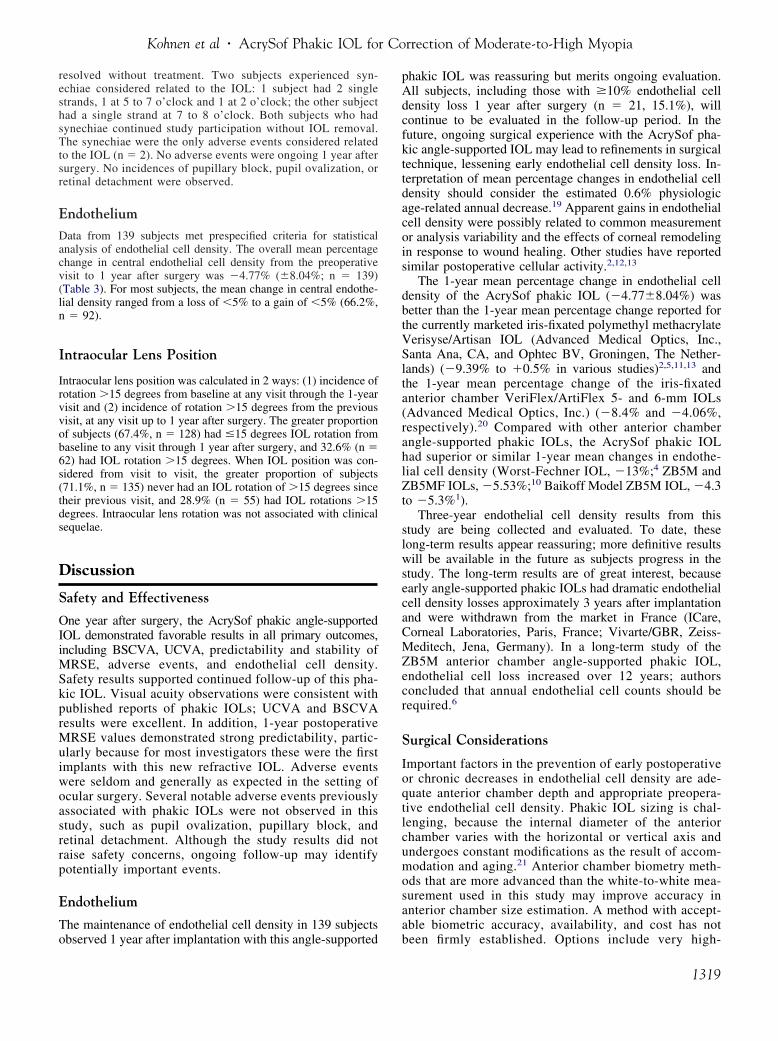
resolved without treatment. Two subjects experienced syn-echiae considered related to the IOL: 1 subject had 2 singlestrands, 1 at 5 to 7 o’clock and 1 at 2 o’clock; the other subjecthad a single strand at 7 to 8 o’clock. Both subjects who hadsynechiae continued study participation without IOL removal.The synechiae were the only adverse events considered relatedto the IOL (n � 2). No adverse events were ongoing 1 year aftersurgery. No incidences of pupillary block, pupil ovalization, orretinal detachment were observed.
EndotheliumData from 139 subjects met prespecified criteria for statisticalanalysis of endothelial cell density. The overall mean percentagechange in central endothelial cell density from the preoperativevisit to 1 year after surgery was �4.77% (�8.04%; n � 139)(Table 3). For most subjects, the mean change in central endothe-lial density ranged from a loss of �5% to a gain of �5% (66.2%,n � 92).
Intraocular Lens Position
Intraocular lens position was calculated in 2 ways: (1) incidence ofrotation �15 degrees from baseline at any visit through the 1-yearvisit and (2) incidence of rotation �15 degrees from the previousvisit, at any visit up to 1 year after surgery. The greater proportionof subjects (67.4%, n � 128) had �15 degrees IOL rotation frombaseline to any visit through 1 year after surgery, and 32.6% (n �62) had IOL rotation �15 degrees. When IOL position was con-sidered from visit to visit, the greater proportion of subjects(71.1%, n � 135) never had an IOL rotation of �15 degrees sincetheir previous visit, and 28.9% (n � 55) had IOL rotations �15degrees. Intraocular lens rotation was not associated with clinicalsequelae.
Discussion
Safety and Effectiveness
One year after surgery, the AcrySof phakic angle-supportedIOL demonstrated favorable results in all primary outcomes,including BSCVA, UCVA, predictability and stability ofMRSE, adverse events, and endothelial cell density.Safety results supported continued follow-up of this pha-kic IOL. Visual acuity observations were consistent withpublished reports of phakic IOLs; UCVA and BSCVAresults were excellent. In addition, 1-year postoperativeMRSE values demonstrated strong predictability, partic-ularly because for most investigators these were the firstimplants with this new refractive IOL. Adverse eventswere seldom and generally as expected in the setting ofocular surgery. Several notable adverse events previouslyassociated with phakic IOLs were not observed in thisstudy, such as pupil ovalization, pupillary block, andretinal detachment. Although the study results did notraise safety concerns, ongoing follow-up may identifypotentially important events.
Endothelium
The maintenance of endothelial cell density in 139 subjects
observed 1 year after implantation with this angle-supportedphakic IOL was reassuring but merits ongoing evaluation.All subjects, including those with �10% endothelial celldensity loss 1 year after surgery (n � 21, 15.1%), willcontinue to be evaluated in the follow-up period. In thefuture, ongoing surgical experience with the AcrySof pha-kic angle-supported IOL may lead to refinements in surgicaltechnique, lessening early endothelial cell density loss. In-terpretation of mean percentage changes in endothelial celldensity should consider the estimated 0.6% physiologicage-related annual decrease.19 Apparent gains in endothelialcell density were possibly related to common measurementor analysis variability and the effects of corneal remodelingin response to wound healing. Other studies have reportedsimilar postoperative cellular activity.2,12,13
The 1-year mean percentage change in endothelial celldensity of the AcrySof phakic IOL (�4.77�8.04%) wasbetter than the 1-year mean percentage change reported forthe currently marketed iris-fixated polymethyl methacrylateVerisyse/Artisan IOL (Advanced Medical Optics, Inc.,Santa Ana, CA, and Ophtec BV, Groningen, The Nether-lands) (�9.39% to �0.5% in various studies)2,5,11,13 andthe 1-year mean percentage change of the iris-fixatedanterior chamber VeriFlex/ArtiFlex 5- and 6-mm IOLs(Advanced Medical Optics, Inc.) (�8.4% and �4.06%,respectively).20 Compared with other anterior chamberangle-supported phakic IOLs, the AcrySof phakic IOLhad superior or similar 1-year mean changes in endothe-lial cell density (Worst-Fechner IOL, �13%;4 ZB5M andZB5MF IOLs, �5.53%;10 Baikoff Model ZB5M IOL, �4.3to �5.3%1).
Three-year endothelial cell density results from thisstudy are being collected and evaluated. To date, theselong-term results appear reassuring; more definitive resultswill be available in the future as subjects progress in thestudy. The long-term results are of great interest, becauseearly angle-supported phakic IOLs had dramatic endothelialcell density losses approximately 3 years after implantationand were withdrawn from the market in France (ICare,Corneal Laboratories, Paris, France; Vivarte/GBR, Zeiss-Meditech, Jena, Germany). In a long-term study of theZB5M anterior chamber angle-supported phakic IOL,endothelial cell loss increased over 12 years; authorsconcluded that annual endothelial cell counts should berequired.6
Surgical Considerations
Important factors in the prevention of early postoperativeor chronic decreases in endothelial cell density are ade-quate anterior chamber depth and appropriate preopera-tive endothelial cell density. Phakic IOL sizing is chal-lenging, because the internal diameter of the anteriorchamber varies with the horizontal or vertical axis andundergoes constant modifications as the result of accom-modation and aging.21 Anterior chamber biometry meth-ods that are more advanced than the white-to-white mea-surement used in this study may improve accuracy inanterior chamber size estimation. A method with accept-able biometric accuracy, availability, and cost has not
been firmly established. Options include very high-1319

Ophthalmology Volume 116, Number 7, July 2009
frequency ultrasound, the Scheimpflug camera, and an-terior segment optical coherence tomography.22 In par-ticular, the use of anterior chamber optical coherencetomography with defined objective measurements ispromising.21,23 Additional study of the suitability of suchmethods for clinical use in anterior chamber measure-ment for phakic IOL sizing is needed.
Improved methods of anterior chamber biometrywould also contribute to accurate sizing resulting inimproved IOL stability. In this study, IOL rotation wasnot associated with clinical sequelae 1 year postopera-tively. However, several factors limit interpretation ofIOL stability in this study. Intraocular lens position wasindicated in 15-degree increments, so observer variabilityin rounding may have led to erroneous observations.Variances in a subject’s head tilt on slit-lamp examina-tion may have also confounded IOL position results.Improved precision in IOL position reporting and stan-dardized subject position has been implemented success-fully in other studies24 and may increase the reliability ofresults in future studies of the AcrySof phakic angle-supported IOL.
Several surgical considerations were regarded as essen-tial. Accurate power calculation was necessary to ensuredesired postsurgical refractive results. Proper loading of theinjector with the anterior optic surface of the IOL facingupward must be emphasized, because 2 lenses in this studywere implanted upside-down, resulting in iatrogenic cata-ract formation. Subsequent to this study, the IOL has beenmodified to include side-up indicators that are visiblethrough the injector cartridge (Fig 1). Iridectomy, whichwas not considered necessary but was permitted, was per-formed in only 5 of the 190 surgeries (2.6%). Increased IOPoccurring soon after implantation tended to be related toretained OVD, whereas elevated IOP of a later onset wasmore often associated with prolonged steroid administra-tion. These observations underscore the importance of thor-ough OVD removal and appropriate postoperative medicalmanagement.
In conclusion, favorable study findings in all primaryoutcomes, along with advantages including a small incisionsize (�3.0 mm) and a simplified operative procedure, sup-port the potential of the AcrySof phakic angle-supportedIOL in the correction of moderate-to-high myopia. Clinicaloutcomes 1 year after surgery were promising; however,further follow-up is needed to investigate the long-termeffects of the IOL in the anterior chamber angle. Untilfurther long-term data are available, the plan of care mayneed to include endothelial cell density monitoring for theduration of the implant. Favorable long-term clinical studyresults with this hydrophobic refractive IOL, together withadvances in IOL design, materials, and anterior chamberbiometry, may supersede previous safety concerns associ-ated with other angle-supported phakic IOLs. On the basisof these early observations of excellent refractive correctionand predictability with acceptable safety, the AcrySof pha-kic angle-supported IOL represents a promising future op-tion for the reduction or correction of moderate-to-high
myopia.1320
References
1. Baikoff G, Arne JL, Bokobza Y, et al. Angle-fixated anteriorchamber phakic intraocular lens for myopia of �7 to �19diopters. J Refract Surg 1998;14:282–93.
2. Benedetti S, Casamenti V, Benedetti M. Long-term endothe-lial changes in phakic eyes after Artisan intraocular lensimplantation to correct myopia: five-year study. J CataractRefract Surg 2007;33:784–90.
3. Mimouni F, Colin J, Koffi V, Bonnet P. Damage to thecorneal endothelium from anterior chamber intraocularlenses in phakic myopic eyes. Refract Corneal Surg 1991;7:277– 81.
4. Perez-Santonja JJ, Bueno JL, Zato MA. Surgical correction ofhigh myopia in phakic eyes with Worst-Fechner myopia in-traocular lenses. J Refract Surg 1997;13:268–81.
5. Tahzib NG, Nuijts RM, Wu WY, Budo CJ. Long-term studyof Artisan phakic intraocular lens implantation for the correc-tion of moderate to high myopia: ten-year follow-up results.Ophthalmology 2007;114:1133-42.
6. Javaloy J, Alio JL, Iradier MT, et al. Outcomes of ZB5Mangle-supported anterior chamber phakic intraocular lenses at12 years. J Refract Surg 2007;23:147–58.
7. Baikoff G, Bourgeon G, Jodai HJ, et al. Pigment dispersionand Artisan implants: crystalline lens rise as a safety criterion[in French]. J Fr Ophtalmol 2005;28:590–7.
8. Brandt JD, Mockovak ME, Chayet A. Pigmentary dispersionsyndrome induced by a posterior chamber phakic refractivelens. Am J Ophthalmol 2001;131:260–3.
9. Martinez-Castillo V, Elies D, Boixadera A, et al. Siliconeposterior chamber phakic intraocular lens dislocated into thevitreous cavity. J Refract Surg 2004;20:773–7.
10. Alio JL, de la Hoz F, Perez-Santonja JJ, et al. Phakic anteriorchamber lenses for the correction of myopia: a 7-year cumu-lative analysis of complications in 263 cases. Ophthalmology1999;106:458–66.
11. Landesz M, Worst JG, van Rij G. Long-term results of cor-rection of high myopia with an iris claw phakic intraocularlens. J Refract Surg 2000;16:310–6.
12. Perez-Santonja JJ, Alio JL, Jimenez-Alfaro I, Zato MA. Sur-gical correction of severe myopia with an angle-supportedphakic intraocular lens. J Cataract Refract Surg 2000;26:1288–302.
13. Saxena R, Boekhoorn SS, Mulder PG, et al. Long-termfollow-up of endothelial cell change after Artisan phakic intraoc-ular lens implantation. Ophthalmology 2008;115:608–13.
14. Allemann N, Chamon W, Tanaka HM, et al. Myopic angle-supported intraocular lenses: two-year follow-up. Ophthal-mology 2000;107:1549–54.
15. Chylack LT Jr. Instructions for applying the Lens OpacitiesClassification System, version III (LOCS III). Boston, MA:Center for Ophthalmic Research, Brigham and Women’sHospital; 2001:112.
16. Van der Heijde GL. Some optical aspects implantation of anIOL in an myopic eye. Eur J Implant Refract Surg 1989;1:245–8.
17. Holladay JT. Standardizing constants for ultrasonic biometry,keratometry, and intraocular lens power calculations. J Cata-ract Refract Surg 1997;23:1356–70.
18. Koch DD, Kohnen T, Obstbaum SA, Rosen ES. Format forreporting refractive surgical data. J Cataract Refract Surg1998;24:285–7.
19. Bourne WM, Nelson LR, Hodge DO. Central corneal endo-thelial cell changes over a ten-year period. Invest Ophthalmol
Vis Sci 1997;38:779–82.
Kohnen et al � AcrySof Phakic IOL for Correction of Moderate-to-High Myopia
20. Guell JL, Morral M, Gris O, et al. Five-year follow-up of 399phakic Artisan-Verisyse implantation for myopia, hyperopia,and/or astigmatism. Ophthalmology 2008;115:1002–12.
21. Baikoff G. Anterior segment OCT and phakic intraocular lenses:a perspective. J Cataract Refract Surg 2006;32:1827–35.
22. Coullet J, Mahieu L, Malecaze F, et al. Severe endothelial cellloss following uneventful angle-supported phakic intraocularlens implantation for high myopia. J Cataract Refract Surg
2007;33:1477–81.The study group is available in Appendix 1 at http://aaojournal.org.
23. Kohnen T, Thomala MC, Cichocki M, Strenger A. Internalanterior chamber diameter using optical coherence tomog-raphy compared with white-to-white distances using auto-mated measurements. J Cataract Refract Surg 2006;32:1809 –13.
24. Baumeister M, Buhren J, Kohnen T. Position of angle-supported, iris-fixated, and ciliary sulcus-implanted myopicphakic intraocular lenses evaluated by Scheimpflug photogra-
phy. Am J Ophthalmol 2004;138:723–31.Footnotes and Financial Disclosures
Originally received: August 13, 2008.Final revision: January 27, 2009.Accepted: January 27, 2009.Available online: May 30, 2009. Manuscript no. 2008-973.1 Johann Wolfgang Goethe-University, Department of Ophthalmology,Frankfurt/Main, Germany.2 FreeVis LASIK Center, University Eye Clinic Mannheim, Mannheim,Germany.3 CHU Morvan, Service Ophtalmologie, Brest, France.4 Eye clinic Ahaus, Ahaus, Germany.5 Hôpital Purpan, Service d’Ophtalmologie, Toulouse, France.6 Groupe Hospitalier Pellegrin, Service d’Ophtalmologie, Bordeaux,France.7 Instituto Oftalmologico de Alicante, Alicante, Spain.8 Unità Operativa di Oculistica, Verona, Italy.9 Hospital General Santo Antonio, Porto, Portugal.
Presented in part at: the American Academy of Ophthalmology annualmeeting, November 2006, Las Vegas, Nevada.
Financial Disclosure(s):The author(s) have made the following disclosure(s):
Michael C. Knorz is a paid consultant for Alcon Inc. and AMO Inc.
Thomas Kohnen is a paid consultant for Alcon Inc.
Jean L. Arné is a paid consultant for Alcon Inc.
Beatrice Cochener is a paid consultant for Alcon Inc., AMO Inc., Bausch& Lomb, and THEA.
Joseph Colin is a paid consultant for Alcon Inc.
Jorge L. Alió is a paid consultant for Alcon Inc.
Supported by Alcon Laboratories, Inc., Fort Worth, Texas, as a clinicalinvestigational project.
Correspondence:Thomas Kohnen, MD, Professor of Ophthalmology, Johann WolfgangGoethe-University, Department of Ophthalmology, Theodor-Stern-Kai 7, 60590 Frankfurt am Main, Germany. E-mail: [email protected]
frankfurt.de.1321

Ophthalmology Volume 116, Number 7, July 2009
Appendix 1. Multicenter European StudyGroup
Magdalena Cichocki, MD, Christoph Kühne, MD, EvdoxiaTerzi, MD, Tanja Wiegand (Department of Ophthalmology,Johann Wolfgang Goethe-University, Frankfurt am Main, Ger-many); José Luis Güell (Instituto de Microcirugia Ocular,Barcelona, Spain), Stefan Häsemeyer, MD (FreeVis Lasik-Zentrum, Universitäts-Augenklinik, Mannheim, Germany);Christine Tanguy-Gueguen, Stephane Lebaillif, MD (CHU
Morvan, Brest, France); Stefanie Schmickler, MD, Diana1321.e1
Laukötter, Susanne Barte-Lohmann (Augenklinik Ahaus,Ahaus, Germany), Catherine Garabétian, Pierre Fournié, MD,Laurence Mahieu, MD, Corinne Rumebe (CHU Purpan, Tou-louse, France); Concepción de la Vega, MD, Tomas Javaloy,MD, Esperanza Sala (Vissum–IOA, Alicante, Spain); JerômeGuinguet, Sylvie Simonpoli, MD, (CHU Bordeaux–HospitalPellegrin, Bordeaux, France); Simonetta Morselli, MD, NicolaDalla Pellegrina, MD (Ospedale Borgo Trento, Verona, Italy);Fernando Vaz, MD, Teresa Pacheco, MD, Bernadette Pessoa,MD, Marta Macedo, MD, and Miguel Gomes, MD (Hospital
Geral Santo Antonio, Porto, Portugal).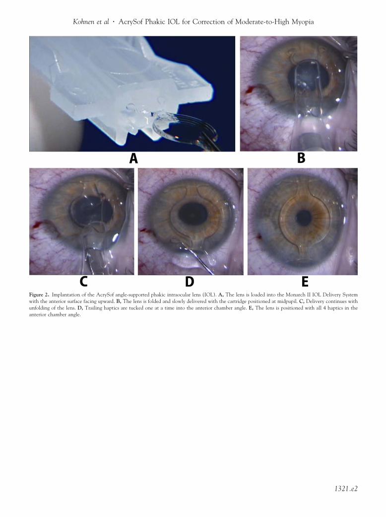
Kohnen et al � AcrySof Phakic IOL for Correction of Moderate-to-High Myopia
Figure 2. Implantation of the AcrySof angle-supported phakic intraoculawith the anterior surface facing upward. B, The lens is folded and slowly dunfolding of the lens. D, Trailing haptics are tucked one at a time into tanterior chamber angle.
r lens (IOL). A, The lens is loaded into the Monarch II IOL Delivery Systemelivered with the cartridge positioned at midpupil. C, Delivery continues withhe anterior chamber angle. E, The lens is positioned with all 4 haptics in the
1321.e2

AcrySof phakic angle-supported refractive intraocular lens.
Ophthalmology Volume 116, Number 7, July 2009
Table 1. Demographics and Subject Characteristics
Demographic/Subject Characteristic Value
No. of subjects (eyes) 190 (190)Age (yrs)
Mean�SD 35.7�8.6Range 18–53
Gender, n (%)Female 110 (58)Male 80 (42)
Race, n (%)Caucasian 187 (98)African American 1 (1)Hispanic 2 (1)
Preoperative MRSE (D)Mean�SD �10.38�2.43Range �16.50 to �6.63
Implanted IOL power (D)Mean�SD �11.1�2.2Range �16.5 to �7.50
Implanted IOL model, n (%)L12500 (12.5 mm OAL) 66 (34.7)L13000 (13.0 mm OAL) 90 (47.3)L13500 (13.5 mm OAL) 34 (17.9)
D � diopter; IOL � intraocular lens; MRSE � manifest refraction
spherical equivalent; OAL � overall length; SD � standard deviation.Figure 6. Best-corrected visual acuity 1 year after implantation of the
AcrySof phakic angle-supported refractive intraocular lens.1321.e3
Figure 8. Residual refractive error 1 year after implantation with the