pex5rp: a novel pex5p-related protein that interacts with
TRANSCRIPT

1
Potential role for Pex19p in assembly of PTS-
receptor docking complexes
Marc Fransen, Ilse Vastiau, Chantal Brees, Vanessa Brys, Guy P. Mannaerts,
Paul P. Van Veldhoven
Katholieke Universiteit Leuven, Faculteit Geneeskunde, Campus Gasthuisberg (O/N),
Departement Moleculaire Celbiologie, Afdeling Farmacologie, Herestraat 49, B-3000 Leuven,
Belgium
*Corresponding author
Dr. Marc Fransen, Katholieke Universiteit Leuven, Faculteit Geneeskunde, Campus
Gasthuisberg (O/N), Departement Moleculaire Celbiologie, Afdeling Farmacologie, Herestraat
49 (O/N), B-3000 Leuven, Belgium (Tel: 32-16-345786; Fax: 32-16-345699; Email:
Running title: Pex19p competes with Pex5p and Pex13p for binding to Pex14p
JBC Papers in Press. Published on January 10, 2004 as Manuscript M304941200
Copyright 2004 by The American Society for Biochemistry and Molecular Biology, Inc.
by guest on March 31, 2018
http://ww
w.jbc.org/
Dow
nloaded from
2
Summary
Human Pex19p binds a broad spectrum of peroxisomal membrane proteins (PMPs). It has
been proposed that this peroxin may (i) act as a cycling PMP receptor protein, (ii) facilitate the
insertion of newly synthesized PMPs into the peroxisomal membrane, or (iii) function as a
chaperone to associate and/or dissociate complexes comprising integral PMPs already in the
peroxisomal membrane. We previously demonstrated that human Pex19p binds peroxisomal
integral membrane proteins at regions distinct from their sorting sequences. Here we demonstrate
that a mutant of Pex13p that fails to bind to Pex19p nevertheless targets to and integrates into the
peroxisomal membrane. In addition, through in vitro biochemical analysis, we show that Pex19p
competes with Pex5p and Pex13p for binding to Pex14p, supporting a role for this peroxin in
regulating assembly/disassembly of membrane-associated protein complexes. To further
examine the molecular mechanism underlying this competition, six evolutionarily conserved
amino acids in the Pex5p-/Pex13p-/Pex19p-binding domain of Pex14p were subjected to site-
directed mutagenesis and the corresponding mutants functionally analyzed. Our results indicate
that the physically overlapping binding sites of Pex14p for Pex5p, Pex13p, and Pex19p are
functionally distinct, suggesting that competition occurs through induction of structural changes,
rather than through direct competition. Importantly, we also found that amino acid substitutions
resulting in a strongly reduced binding affinity for Pex13p affect the peroxisomal localization of
Pex14p.
by guest on March 31, 2018
http://ww
w.jbc.org/
Dow
nloaded from
3
Introduction
To date, peroxisome biogenesis studies in a number of evolutionarily diverse organisms
have identified 29 gene products (1), called peroxins (abbreviated ‘Pexp’ and including a number
corresponding to the order of discovery), which are essential for formation of the organelle. In
man, peroxisome biogenesis requires the concerted action of at least 16 of these peroxins (2).
Despite the fact that substantial progress has been made in peroxin identification, the function of
most of these proteins in the biogenesis process is still only partially understood.
Significant interest has developed in recent years regarding the essentially open question
of how PMPs1 find their way to the organelle (3). In a select few examples, peroxisomal
targeting signals (so-called mPTSs) have been identified (4). However, no firm conclusions may
yet be drawn about the presence or absence of consensus sequences or common structural
properties with respect to these mPTSs (5). The observation that Pex19p interacts with the
mPTSs of PpPex2p, HsPex11pβ, HsPex13p, HsPex14p, HsPex16p, HsPMP22, HsPMP34,
HsPMP70, HsALDP, and HsALDPR (6-11), and that a portion of cellular Pex19p is found
associated with the outer surface of peroxisomes (6,12), makes this peroxin a reasonable
candidate for a cycling PMP receptor protein. However, as shown by multiple independent point
mutations, the physically overlapping Pex19p binding domain and the peroxisomal sorting
signals of HsPex13p can be functionally separated (10). Moreover, for a number of PMPs
including PpPex3p, PpPex13p, PpPex17p, PpPex22p, HsPex3p, HsPex12p, HsPMP70, and
RnPex3p, the Pex19p interaction domain and the targeting domain do not overlap at all (7,10,13-
15). These results argue against a role of Pex19p as a general cycling PMP receptor protein.
by guest on March 31, 2018
http://ww
w.jbc.org/
Dow
nloaded from

4
Besides being connected with a transport function, Pex19p might function directly (i) in
maintaining the solubility of integral PMPs before insertion into the peroxisome membrane (9),
(ii) in the insertion process of PMPs into the peroxisome membrane (6) or (iii) in stabilizing
PMP interactions within the peroxisomal membrane itself (16). In addition, based on data
pointing to a role for Pex19p in interacting with already inserted PMPs, Subramani and
coworkers suggested that Pex19p may be functioning as a (dis)assembly factor, or as a
chaperone, to regulate membrane-associated protein complexes (7).
In this study, we employed a Pex13p protein displaying no detectable affinity for Pex19p
but still possessing peroxisomal targeting information to demonstrate that Pex19p is not directly
implicated in the membrane insertion process of this PMP. In addition, we provide evidence that
Pex19p can alter the binding properties of the Pex14p docking complex, supporting a role for
this peroxin in regulating assembly/disassembly of membrane-associated protein complexes.
by guest on March 31, 2018
http://ww
w.jbc.org/
Dow
nloaded from

5
Experimental Procedures
Plasmids – The plasmids encoding biotin-HsPex5p(259-639) (pXaPTS1-BP) (17), (His)6-
MmPex5pL (pKG100) (18), (His)6-HsPex13p/SH3 (pMF68) (19), HsPex13p(WT)-GFP (pMF121)
(10), HsPex13p(V178E)-GFP (pMF551) (10), Gal4pBD-HsPex14p(WT) (pMF101) (10), biotin-
HsPex14p (pMF42) (19), Gal4pBD-HsPex14p(22-81,295-377) (pMF141) (20), Gal4pBD-
HsPex14p(43-81,295-377) (pMF151) (20), Gal4pBD-HsPex19p (pMF132) (10), and DsRed-
KSKL (pMF578) (10) are described elsewhere. The plasmid encoding GST-Pex14p (pKG45)
was kindly provided by Dr. K. Ghys (Leuven, Belgium). The oligonucleotides (Invitrogen) and
plasmids constructed for this study are compiled in Tables I and II, respectively. Cloning vectors
were obtained from Amersham Bioscience (pGEX-4T-3 vector), Clontech (pEGFP-N1 vector),
Promega (PinPoint Xa vectors), and Euroscarf (pZome-1-C). Standard molecular biology
procedures were used (21). PCRs were routinely performed using Pfx DNA polymerase
(Invitrogen), and the E. coli strain Top10F’ (Invitrogen) was used for all DNA manipulations.
The identities of essential constructs were confirmed by DNA sequencing.
Cell culture, transfections, fluorescence microscopy, and fractionation of CHO cells – Chinese
hamster ovary (CHO) cells were cultured as described elsewhere (10). After transfer to
coverslips, the cells were transiently transfected by the polyethylenimine transfection method
(22), and processed for (in)direct fluorescence as described (23). The peroxisomal localization of
the GFP (direct fluorescence) and protein A (indirect fluorescence) fusion proteins was
confirmed by co-localization studies with the peroxisome-targeted DsRed-KSKL reporter protein
(10) or endogenous expressed PMP70. Fluorescence was observed under a Leica DMR
by guest on March 31, 2018
http://ww
w.jbc.org/
Dow
nloaded from

6
microscope equipped with FITC/RSGFP/Bodipy/Fluo3/DIO and Texas Red filters. To isolate a
membranous fraction containing only integral membrane proteins, transfected cells, grown in
culture dishes to 90 % confluency, were freed from these dishes by scraping, resuspended in 0.1
M Na2CO3 (pH 11.5), homogenized (20 strokes) with a Teflon-glass Potter-Elvehjem
homogenizer (Kontes), and subjected to a 100,000 x g spin for 1 h. After centrifugation, the
pellet was resuspended in 0.1 M Na2CO3 (pH 11.5) and the entire procedure was repeated. This
procedure resulted in a combined supernatant fraction yielding soluble and peripheral membrane
proteins, and a pellet fraction containing the integral membrane proteins (24). To isolate integral
membrane proteins of a purified peroxisomal fraction, the freed cells were resuspended in
homogenization buffer (0.25 M sucrose, 5 mM MOPS (pH 7.2), 1 mM EDTA (pH 7.2), 1 mM
DTT, 0.1 %(v/v) ethanol, and a protease inhibitor mixture (Roche Molecular Biochemicals)) and
homogenized (20 strokes) using a stainless steel tissue grinder (Kontes). A post-nuclear
supernatant resulting from a low speed centrifugation (500 x g, 10 min.) was further separated by
density gradient centrifugation using a Nycodenz step gradient (25). The obtained fractions were
analyzed by Western blotting and the peroxisomal peak fractions were pooled, 10-fold diluted in
0.1 M Na2CO3 (pH 11.5), and processed as described above.
Antibodies – The production and characterization of the rabbit antisera against (His)6-
Pex13p/SH3, (His)6-Pex14p, (His)6-Pex19p, and peroxisomal matrix proteins is described
elsewhere (10,19). The rabbit and mouse antisera against (His)6-GFP (encoded by pEGFPH1, a
plasmid kindly provided by Dr. Y. Sakai [Kyoto, Japan]) were generated as previously described
(10). The rabbit antisera against Pex5p and PMP70 and the mouse monoclonal anti-TY antibody
were kindly provided by Dr. M. Baes (Leuven, Belgium), Dr. S. Subramani (San Diego, USA)
by guest on March 31, 2018
http://ww
w.jbc.org/
Dow
nloaded from
7
and Dr. K. Gull (Manchester, United Kingdom), respectively. The mouse monoclonal anti-(His)6
antibody was purchased from Clontech. The rabbit antibodies raised against catalase (Rockland),
cytochrome P450 4A (Affinity Bioreagents Inc.), glutamate dehydrogenase (Rockland), human
serum albumin (Sigma), and protein A (Sigma) were also commercially obtained. The FITC-
labeled anti-rabbit IgGs were obtained from Sigma.
Expression of recombinant proteins and preparation of bacterial lysates – Top10F’ cells
transformed with plasmids encoding the appropriate GST-, biotin-, or (His)6-tagged fusion
proteins were cultured overnight. Five milliliter of each culture of E. coli was diluted in 50 ml of
yeast extract-tryptone medium containing 50 µg/ml ampicillin. After being cultured for 1 h at
37°C, the cells were further grown for 3 h at 33 °C in the presence of 0.1 mM IPTG (or 2 %
(w/v) arabinose) to induce the expression of the tagged proteins. The cells were harvested and
resuspended in 5 ml of ice-cold binding buffer consisting of 50 mM Tris-HCl (pH 7.5), 150 mM
NaCl, 1 % (w/v) Triton X-100, 10 % (w/v) glycerol, 1 mM EDTA, 1 mM dithiothreitol, and a
protease inhibitor mixture (1 µg/ml aprotinin, 0.5 µg/ml leupeptin, 1 µg/ml α2-macroglobulin,
and 1 µg/ml chymostatin). Lysates were prepared by sonicating the cells with a Branson Sonifier
B15 P cell disrupter, equipped with a microtip (output 5, duty 50 %, 10 times 15 seconds). Cell
debris was removed by centrifugation (13,000 x g for 15 min, 4°C).
In vitro binding assays – The pull-down assays employed to study the interactions between
bacterially expressed peroxins were basically performed as described by Fujiki and coworkers
(26). Briefly, in a first step Glutathione-Sepharose 4B (Amersham Biosciences) or ImmunoPure
immobilized streptavidin (Pierce) beads were coated with the appropriate fusion proteins.
by guest on March 31, 2018
http://ww
w.jbc.org/
Dow
nloaded from

8
Therefore, 30 to 500 µl of the bacterial lysates was mixed with 500 µl of a 50 % slurry of the
beads. After being rotated for 1 h at 4°C, the beads were washed 5 times with ice-cold binding
buffer, resuspended in 500 µl of this buffer, and divided into 50 µl aliquots. In a next step,
aliquots of the coated beads were incubated, by rotation for 1 h at 4 °C, with various bacterial
lysates. The beads were washed five times with the ice-cold binding buffer, and five times with
the same buffer minus glycerol. Bound proteins were eluted from the beads by boiling for 5
minutes in 200 µl of SDS-PAGE sample buffer, and, after removing the beads, 20 µl fractions
were loaded on SDS-PAGE and analyzed by immunoblotting. To study the interaction between
protein A, Pex13p(WT)-protein A, Pex13p(V178E)-protein A and Pex19p-GFP, CHO cells co-
transfected with plasmids coding for the corresponding fusion proteins (three tissue culture
dishes (150 mm x 20 mm) per condition) were freed from the dishes by scraping, lysed in 1600
µl ice-cold binding buffer, and homogenized by using a stainless steel tissue grinder (20 strokes).
After centrifugation (13,000 x g, 15 minutes), the supernatant was incubated with 50 µl of a 50
% slurry of rabbit IgG-beads (Sigma) and rotated for 1 h at 4°C. The beads were washed and
further processed as described for the bacterially expressed proteins.
by guest on March 31, 2018
http://ww
w.jbc.org/
Dow
nloaded from
9
Results
A Pex13p mutant that fails to bind to Pex19p nevertheless targets to and integrates into the
peroxisomal membrane
It has been postulated that Pex19p may be involved in the insertion process of PMPs into
the peroxisomal membrane (6). Here we have investigated whether or not Pex13p(V178E), a
mutant of Pex13p displaying a peroxisomal distribution pattern but no detectable affinity for
Pex19p in the yeast two-hybrid system (10), still achieves stable insertion into the peroxisomal
membrane. To accomplish this, first, we confirmed the peroxisomal localization of the employed
Pex13p-TY-GFP and Pex13p-protein A fusion proteins in CHO cells by fluorescence
microscopy (Fig. 1). As we could not exclude that Pex19p and Pex13p(V178E) do not interact in
mammalian cells, we incubated rabbit IgG-agarose with cell lysates derived from CHO-cells co-
transfected with plasmids coding for GFP-Pex19p and protein A, Pex13p(WT)-protein A or
Pex13p(V178E)-protein A. Analysis of the bound proteins revealed that GFP-Pex19p was retained
on the beads in the presence of Pex13p(WT)-protein A, but not in the presence of Pex13p(V178E)-
protein A (Fig. 2). These observations, which extend our previous findings obtained with the
yeast two-hybrid system (10), suggest that Pex19p does also not form a complex with
Pex13p(V178E) in mammalian cells. Next, we lysed cells transfected with plasmids encoding GFP,
Pex13p(WT)-TY-GFP, and Pex13p(V178E)-TY-GFP in 0.1 M sodium carbonate – a treatment that
releases soluble and peripheral membrane proteins (24), and immunoblotted the soluble and
insoluble fractions with the anti-TY1 epitope tag monoclonal antibody BB2 (27). Under these
conditions, the wild-type and mutated Pex13p-TY-GFP fusion proteins were carbonate-
inextractable – that is, were very tightly associated with the organelle membrane (Fig. 3A).
Similar results were obtained with extraction by Triton X-114 (data not shown). In another
by guest on March 31, 2018
http://ww
w.jbc.org/
Dow
nloaded from
10
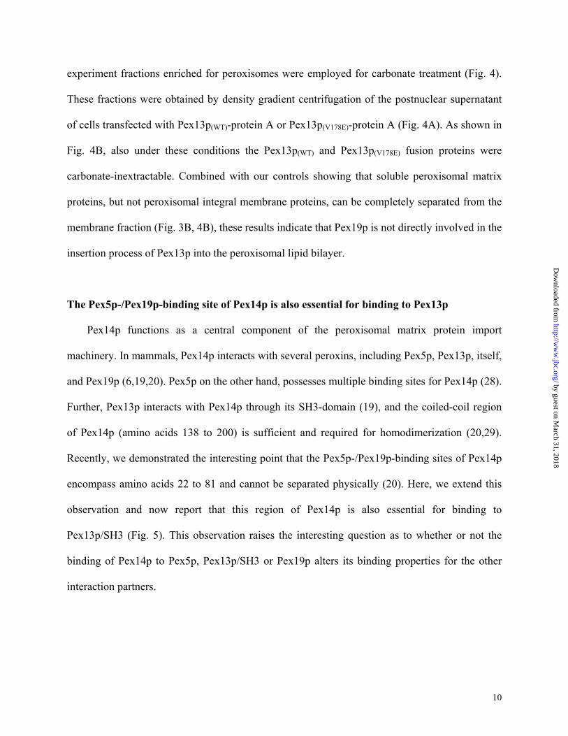
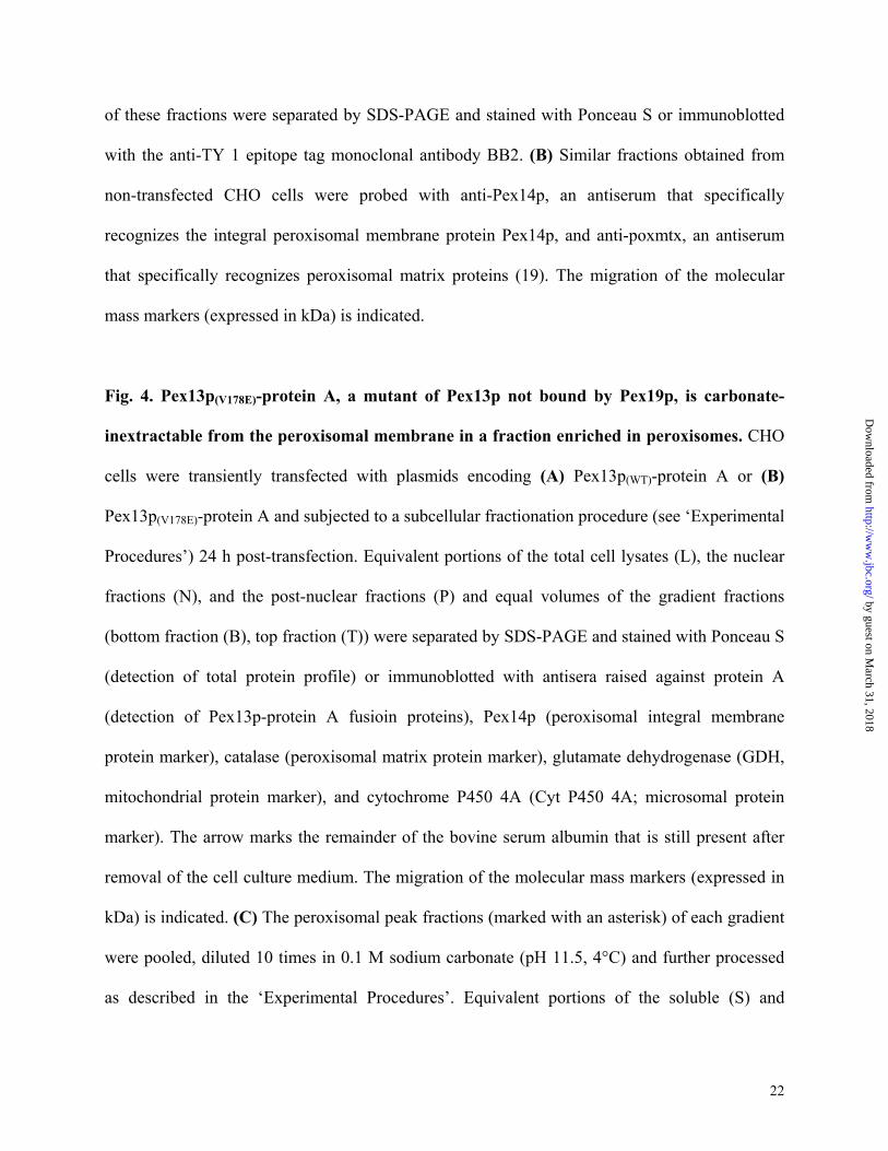
experiment fractions enriched for peroxisomes were employed for carbonate treatment (Fig. 4).
These fractions were obtained by density gradient centrifugation of the postnuclear supernatant
of cells transfected with Pex13p(WT)-protein A or Pex13p(V178E)-protein A (Fig. 4A). As shown in
Fig. 4B, also under these conditions the Pex13p(WT) and Pex13p(V178E) fusion proteins were
carbonate-inextractable. Combined with our controls showing that soluble peroxisomal matrix
proteins, but not peroxisomal integral membrane proteins, can be completely separated from the
membrane fraction (Fig. 3B, 4B), these results indicate that Pex19p is not directly involved in the
insertion process of Pex13p into the peroxisomal lipid bilayer.
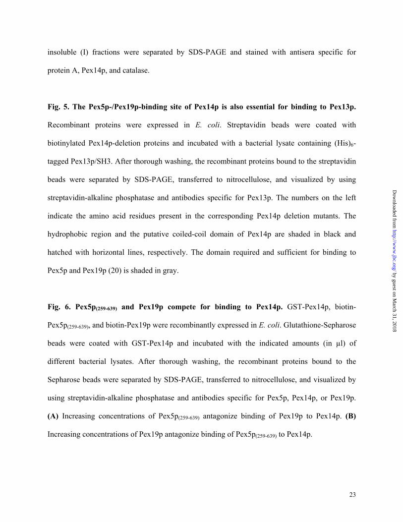
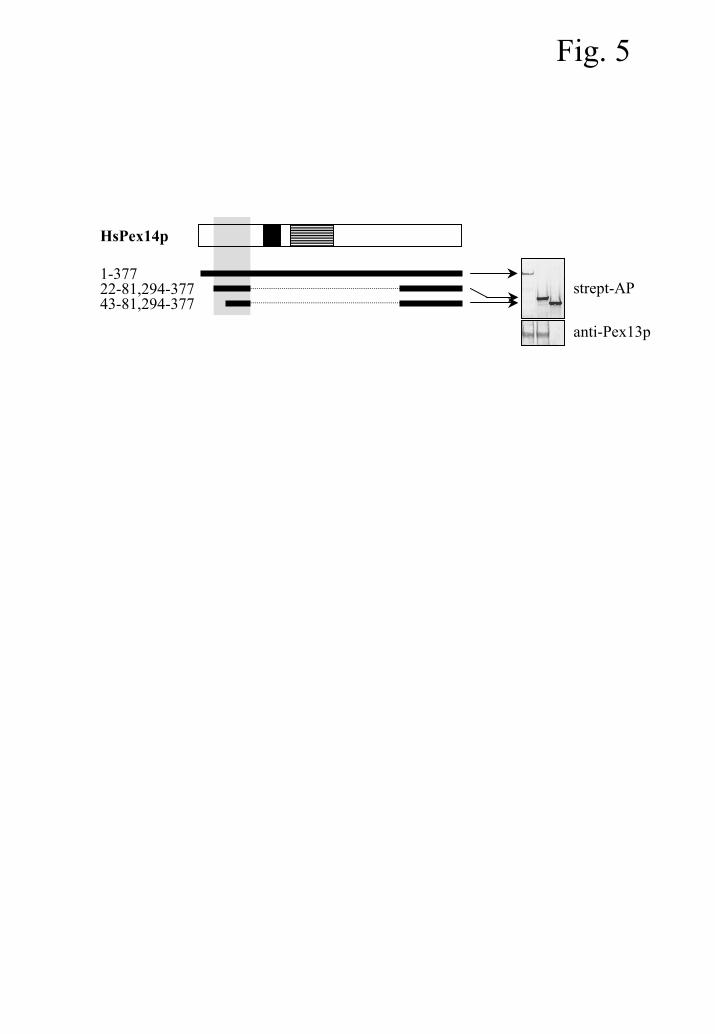
The Pex5p-/Pex19p-binding site of Pex14p is also essential for binding to Pex13p
Pex14p functions as a central component of the peroxisomal matrix protein import
machinery. In mammals, Pex14p interacts with several peroxins, including Pex5p, Pex13p, itself,
and Pex19p (6,19,20). Pex5p on the other hand, possesses multiple binding sites for Pex14p (28).
Further, Pex13p interacts with Pex14p through its SH3-domain (19), and the coiled-coil region
of Pex14p (amino acids 138 to 200) is sufficient and required for homodimerization (20,29).
Recently, we demonstrated the interesting point that the Pex5p-/Pex19p-binding sites of Pex14p
encompass amino acids 22 to 81 and cannot be separated physically (20). Here, we extend this
observation and now report that this region of Pex14p is also essential for binding to
Pex13p/SH3 (Fig. 5). This observation raises the interesting question as to whether or not the
binding of Pex14p to Pex5p, Pex13p/SH3 or Pex19p alters its binding properties for the other
interaction partners.
by guest on March 31, 2018
http://ww
w.jbc.org/
Dow
nloaded from
11
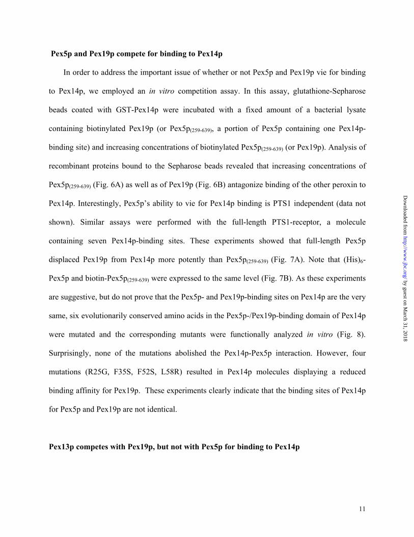
Pex5p and Pex19p compete for binding to Pex14p
In order to address the important issue of whether or not Pex5p and Pex19p vie for binding
to Pex14p, we employed an in vitro competition assay. In this assay, glutathione-Sepharose
beads coated with GST-Pex14p were incubated with a fixed amount of a bacterial lysate
containing biotinylated Pex19p (or Pex5p(259-639), a portion of Pex5p containing one Pex14p-
binding site) and increasing concentrations of biotinylated Pex5p(259-639) (or Pex19p). Analysis of
recombinant proteins bound to the Sepharose beads revealed that increasing concentrations of
Pex5p(259-639) (Fig. 6A) as well as of Pex19p (Fig. 6B) antagonize binding of the other peroxin to
Pex14p. Interestingly, Pex5p’s ability to vie for Pex14p binding is PTS1 independent (data not
shown). Similar assays were performed with the full-length PTS1-receptor, a molecule
containing seven Pex14p-binding sites. These experiments showed that full-length Pex5p
displaced Pex19p from Pex14p more potently than Pex5p(259-639) (Fig. 7A). Note that (His)6-
Pex5p and biotin-Pex5p(259-639) were expressed to the same level (Fig. 7B). As these experiments
are suggestive, but do not prove that the Pex5p- and Pex19p-binding sites on Pex14p are the very
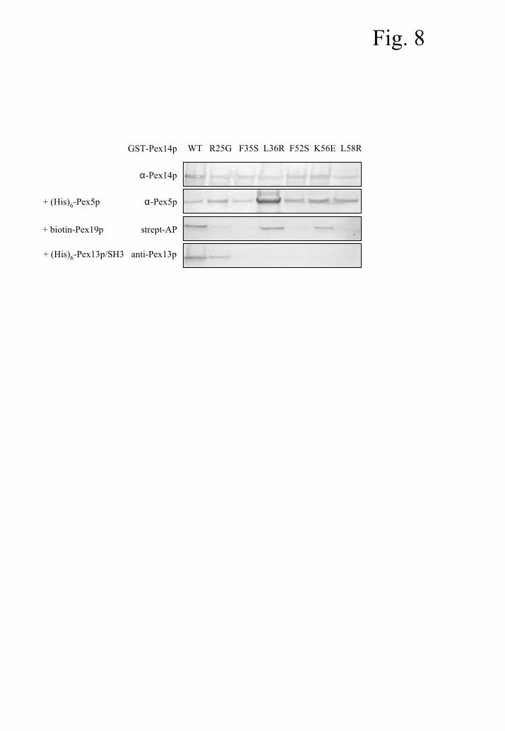
same, six evolutionarily conserved amino acids in the Pex5p-/Pex19p-binding domain of Pex14p
were mutated and the corresponding mutants were functionally analyzed in vitro (Fig. 8).
Surprisingly, none of the mutations abolished the Pex14p-Pex5p interaction. However, four
mutations (R25G, F35S, F52S, L58R) resulted in Pex14p molecules displaying a reduced
binding affinity for Pex19p. These experiments clearly indicate that the binding sites of Pex14p
for Pex5p and Pex19p are not identical.
Pex13p competes with Pex19p, but not with Pex5p for binding to Pex14p
by guest on March 31, 2018
http://ww
w.jbc.org/
Dow
nloaded from

12
In order to investigate whether or not binding of Pex13p/SH3 to Pex14p alters the binding
properties of Pex14p for Pex5p and Pex19p, we again employed the in vitro competition assay.
This time the glutathione-Sepharose beads coated with GST-Pex14p were incubated with
bacterial lysates containing (His)6-Pex13p/SH3, biotin-Pex5p(259-639) and/or biotin-Pex19p.
Analysis of recombinant proteins bound to the Sepharose beads confirmed our previous
observations (20) that Pex5p, Pex13p/SH3, and Pex14p form a ternary complex (Fig. 9A). In
addition, this figure shows that Pex13p/SH3 and Pex19p antagonize each other’s binding to
Pex14p (Fig. 9B). Moreover, as mutations within the Pex13p-/Pex19p-binding site of Pex14p
differentially affected the binding of Pex13p/SH3 and Pex19p (Fig. 8), the binding sites for both
peroxins appear to be also functionally distinct. Pex13p/SH3 and Pex5p did not affect each
other’s binding to Pex14p (Fig. 9A).
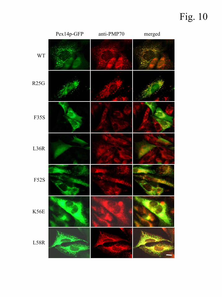
A strongly reduced binding affinity for Pex13p affects the peroxisomal localization of
Pex14p
It has been proposed that Pex19p may act as a cycling PMP receptor protein (6).
Consistent with this hypothesis, it has been reported that the targeting element of Pex14p retains
the ability to bind Pex19p (6). By using the Pex14p mutants described above, we investigated
whether or not there is a functional link between Pex19p-binding and peroxisomal localization of
Pex14p. The GFP-tagged Pex14p mutants were expressed in CHO cells and the localization of
the fusion proteins was determined by indirect fluorescence microscopy. Representative pictures
of transfected CHO cells that illustrate the observed staining patterns of the GFP-fusion proteins
are shown in Fig. 10. These results provide evidence that for Pex14p Pex19p-binding (Fig. 8)
and peroxisomal sorting (Fig. 10) are not functionally linked. This observation is difficult to
by guest on March 31, 2018
http://ww
w.jbc.org/
Dow
nloaded from
13
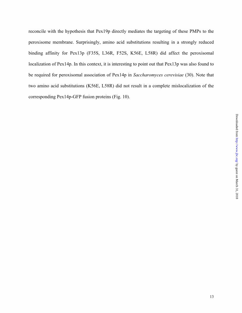
reconcile with the hypothesis that Pex19p directly mediates the targeting of these PMPs to the
peroxisome membrane. Surprisingly, amino acid substitutions resulting in a strongly reduced
binding affinity for Pex13p (F35S, L36R, F52S, K56E, L58R) did affect the peroxisomal
localization of Pex14p. In this context, it is interesting to point out that Pex13p was also found to
be required for peroxisomal association of Pex14p in Saccharomyces cerevisiae (30). Note that
two amino acid substitutions (K56E, L58R) did not result in a complete mislocalization of the
corresponding Pex14p-GFP fusion proteins (Fig. 10).
by guest on March 31, 2018
http://ww
w.jbc.org/
Dow
nloaded from

14
Discussion
Based on the observations that (i) Pex19p binds a broad spectrum of PMPs, and (ii) cells
deficient in this peroxin lack peroxisomal membrane structures, Pex19p has been implicated in
the peroxisome membrane assembly process (6,12). Although several hypotheses have been put
forward (see Introduction), there is currently no consensus as to the exact function of this
peroxin. Recently, we and others have demonstrated that Pex19p does not function as a general
mPTS receptor (7,10). In an attempt to elucidate a non-mPTS receptor role for Pex19p, we have
investigated whether or not the protein is involved in the membrane insertion of newly-
synthesized PMPs. To accomplish this, we employed human Pex13p(V178E), a molecule that
displays no detectable affinity for Pex19p but that is still associated with peroxisomes (10, this
study). Our results show that human Pex13p(V178E) expressed in CHO cells remained
sedimentable after carbonate extraction - a widely used procedure for determining if an integral
membrane protein has achieved stable insertion into the lipid bilayer (24). Thus, it appears
unlikely that Pex19p functions as a membrane insertion factor for the integral membrane peroxin
Pex13p.
Another potential non-mPTS receptor role for Pex19p is that the peroxin functions as an
assembly or disassembly factor of peroxisomal membrane protein complexes (7). Accumulating
evidence points to the existence of multiple, dynamic complexes of peroxins in vivo; however, at
this point very little is known about how the peroxisomal protein translocation apparatus is
regulated (26,29,31-34). Recently, we and others reported that human Pex19p interacts with the
critically important peroxisomal integral membrane protein Pex14p – a pivotal component of the
docking complex for the functional peroxisomal native protein import receptors Pex5p and
by guest on March 31, 2018
http://ww
w.jbc.org/
Dow
nloaded from
15
Pex7p (6,10). In order to investigate whether or not Pex19p is involved in the regulation of the
Pex14p docking complex, we employed in vitro biochemical assays. Specifically, we examined
the effect of Pex19p on the Pex5p- and Pex13p (another component of the docking complex)-
binding properties of Pex14p. Our studies revealed that increasing concentrations of Pex19p
antagonize binding of Pex5p and Pex13p to Pex14p. Further, in experiments in which increasing
concentrations of Pex5p and Pex13p were used, binding of Pex19p to Pex14p was antagonized.
As expected, the full-length PTS1-receptor, a molecule containing seven Pex14p-binding sites,
was a more effective competitor for the Pex14p-Pex19p interaction than Pex5p(259-639), a
molecule containing only one Pex14p-binding site. In addition, Pex19p appears to be a more
effective competitor for the Pex14p-Pex13p interaction than for the Pex14p-Pex5p interaction –
although the basis for this apparent selectivity is unclear. Summarized, these results show that
there is clearly a dynamic interplay between Pex14p and its interaction partners - Pex5p, Pex13p,
and Pex19p.
Recently, we showed that the Pex5p-/Pex19p-binding sites of Pex14p encompass amino
acids 22 to 81 and cannot be separated physically (20). In this study we provided evidence that
this region of Pex14p is also essential for binding to Pex13p/SH3. In addition, site-directed
mutagenesis and functional analysis of six evolutionarily conserved amino acids in this domain
of Pex14p identified five point mutations (F35S, L36R, F52S, K56E, L58R) that resulted in a
strongly reduced binding affinity for Pex13p. The observation that (i) all these mutants fully
retained the ability to bind Pex5p, and (ii) three mutations (F35S, F52S, L58R) were selectively
affected in Pex19p binding, indicate that the physically overlapping binding sites of Pex14p for
Pex5p, Pex13p, and Pex19p are functionally distinct. This suggests that competition occurs
by guest on March 31, 2018
http://ww
w.jbc.org/
Dow
nloaded from
16
through induction of structural changes, rather than through direct competition for binding. We
are currently exploring the nature of these structural changes in greater detail.
Interestingly, we observed no functional link between Pex19p-binding and peroxisomal
localization of Pex14p. This result argues against a role of Pex19p as a cycling receptor protein
for Pex14p. Remarkably, we observed that all amino acid substitutions resulting in a strongly
reduced binding affinity for Pex13p did affect the peroxisomal localization of Pex14p. In this
context, it is important to mention that these results support the observation in S. cerevisiae that
the presence of Pex13p is a prerequisite for peroxisomal membrane association of Pex14p (30).
Whether or not Pex13p in this organism is involved in targeting or required for binding or
retention of Pex14p at the peroxisome is not clear yet (30). In addition, it has been suggested that
the SH3 domain of Pex13p may not provide the only binding site for Pex14p at the peroxisomal
membrane (34).
Our observations that Pex19p can modulate the Pex5p- and Pex13p-binding properties of
Pex14p in vitro suggest that Pex19p may play an important and direct role in the import of
peroxisomal matrix proteins through a regulation of the Pex14p docking complex. In addition,
our previous and current findings indicate that Pex19p does not serve a role as a general PMP-
import receptor or Pex13p membrane insertion factor.
by guest on March 31, 2018
http://ww
w.jbc.org/
Dow
nloaded from

17
References
1. Vizeacoumar, F.J., Torres-Guzman, J.C., Tam, Y.Y., Aitchison, J.D., and Rachubinski R.A.
(2003) J. Cell Biol. 161, 321-332
2. Matsumoto, N., Tamura, S., and Fujiki, Y. (2003) Nat. Cell Biol. 5, 454-460
3. Sparkes, I.A., and Baker, A. (2002) Mol. Membr. Biol. 19, 171-185
4. Purdue, P.E., and Lazarow, P.B. (2001) Annu. Rev. Cell Dev. Biol. 1, 701-752
5. Terlecky, S.R., and Fransen, M. (2000) Traffic 1, 465-473
6. Sacksteder, K.A., Jones, J.M., South, S.T., Li, X., Liu, Y., and Gould, S.J. (2000) J. Cell
Biol. 148, 931-944
7. Snyder, W.B., Koller, A., Choy, A.J., and Subramani, S. (2000) J. Cell Biol. 149, 1171-1178
8. Gloeckner, C.J., Mayerhofer, P.U., Landgraf, P., Muntau, A.C., Holzinger, A., Gerber, J.K.,
Kammerer, S., Adamski, J., and Roscher, A.A. (2000) Biochem. Biophys. Res. Commun. 271,
144-150
9. Jones, J.M., Morrell, J.C., and Gould, S.J. (2001) J. Cell Biol. 153, 1141-1150
10. Fransen, M., Wylin, T., Brees, C., Mannaerts, G.P., and Van Veldhoven, P.P. (2001) Mol.
Cell. Biol. 21, 4413-4424
11. Brosius, U., Dehmel, T., and Gartner, J. (2002) J. Biol. Chem. 277, 774-784
12. Matsuzono, Y., Kinoshita, N., Tamura, S., Shimozawa, N., Hamasaki, M., Ghaedi, K.,
Wanders, R.J., Suzuki, Y., Kondo, N., and Fujiki, Y. (1999) Proc. Natl. Acad. Sci. USA 96,
2116-2121
13. Soukupova, M., Sprenger, C., Gorgas, K., Kunau, W.H., and Dodt, G. (1999) Eur. J. Cell
Biol. 78, 357-374
by guest on March 31, 2018
http://ww
w.jbc.org/
Dow
nloaded from
18
14. Ghaedi, K., Tamura, S., Okumoto, K., Matsuzono, Y., and Fujiki, Y. (2000) Mol. Biol. Cell
11, 2085-2102
15. Biermanns, M., and Gartner, J. (2001) Biochem. Biophys. Res. Commun. 285, 649-655
16. Lambkin, G.R., and Rachubinski, R.A. (2001) Mol. Biol. Cell 12, 3353-3364.
17. Fransen, M., Brees, C., Baumgart, E., Vanhooren, J.C., Baes, M., Mannaerts, G.P., and Van
Veldhoven, P.P. (1995) J. Biol. Chem. 270, 7731-7736
18. Ghys, K., Fransen, M., Mannaerts, G.P., and Van Veldhoven, P.P. (2002) Biochem. J. 365,
41-50
19. Fransen, M., Terlecky, S.R., and Subramani, S. (1998) Proc. Natl. Acad. Sci. USA 95, 8087-
8092
20. Fransen, M., Brees, C., Ghys, K., Amery, L., Mannaerts, G.P., Ladant, D., and Van
Veldhoven P.P. (2002) Mol. Cell. Proteomics 1, 243-252
21. Sambrook, J., Fritsch, E.F., and Maniatis, T. (1989) Molecular Cloning: A Laboratory
Manual, 2nd Ed., Cold Spring Harbor Laboratory, Cold Spring Harbor, NY
22. Boussif, O., Lezoualc'h, F., Zanta, M.A., Mergny, M.D., Scherman, D., Demeneix, B., and
Behr, J.P. (1995) Proc. Natl. Acad. Sci. USA 92, 7297-7301
23. Fransen, M., Van Veldhoven, P.P., and Subramani, S. (1999) Biochem. J. 340, 561-568
24. Fujiki, Y., Hubbard, A.L., Fowler, S., and Lazarow, P.B. (1982) J. Cell Biol. 93, 97-102
25. Verheyden, K., Fransen, M., Van Veldhoven, P.P., and Mannaerts, G.P. (1992) Biochim.
Biophys. Acta 1109, 48-54
26. Otera, H., Setoguchi, K., Hamasaki, M., Kumashiro, T., Shimizu, N., and Fujiki, Y. (2002)
Mol. Cell. Biol. 22, 1639-1655
by guest on March 31, 2018
http://ww
w.jbc.org/
Dow
nloaded from
19
27. Bastin, P., Bagherzadeh, Z., Matthews, K.R., and Gull, K. (1996) Mol. Biochem. Parasitol.
77, 235-239
28. Schliebs, W., Saidowsky, J., Agianian, B., Dodt, G., Herberg, F.W., and Kunau, W.H. (1999)
J. Biol. Chem. 274, 5666-5673
29. Oliveira, M.E., Reguenga, C., Gouveia, A.M., Guimaraes, C.P., Schliebs, W., Kunau, W.H.,
Silva, M.T., Sa-Miranda, C., and Azevedo, J.E. (2002) Biochim. Biophys. Acta 1567, 13-22
30. Girzalsky, W., Rehling, P., Stein, K., Kipper, J., Blank, L., Kunau, W.H., and Erdmann, R.
(1999) J. Cell Biol. 144, 1151-1162
31. Reguenga, C., Oliveira, M.E., Gouveia, A.M., Sa-Miranda, C., and Azevedo, J.E. (2001) J.
Biol. Chem. 276, 29935-29942
32. Stein, K., Schell-Steven, A., Erdmann, R., and Rottensteiner, H. (2002) Mol. Cell. Biol. 22,
6056-6069
33. Hazra, P.P., Suriapranata, I., Snyder, W.B., and Subramani, S. (2002) Traffic 3, 560-574
34. Agne, B., Meindl, N.M., Niederhoff, K., Einwachter, H., Rehling, P., Sickmann, A., Meyer,
H.E., Girzalsky, W., and Kunau W.H. (2003) Mol. Cell 11, 635-646
by guest on March 31, 2018
http://ww
w.jbc.org/
Dow
nloaded from

20
Footnotes
1 The abbreviations used are: ALDP, adrenoleukodystrophy protein; ALDPR,
adrenoleukodystrophy-related protein; CHO, Chinese hamster ovary; GDH, glutamate
dehydrogenase; GFP, enhanced green fluorescent protein; GST, glutathione S-
transferase; Hs, Homo sapiens; IPTG, isopropyl-β-D-thiogalactoside; Mm, Mus
musculus; mPTS, membrane peroxisomal targeting signal; PAGE, polyacrylamide gel
electrophoresis; PCR, polymerase chain reaction; Pexp, peroxin; Pp, Pichia pastoris;
PMP, peroxisomal membrane protein; PMP22, 22 kDa PMP; PMP34, 34 kDa PMP;
PMP70, 70 kDa PMP; Rn, Rattus norvegicus; SH3, src homology 3; TY, epitope tag
derived from the major structural protein of the Saccharomyces cerevisiae Ty1 virus-like
particle.
2 We are grateful to Dr. S. Terlecky (Michigan, USA) for his helpful discussions and
thank Dr. K. Gull (Manchester, United Kingdom) for the anti-TY antibodies, Dr. S.
Subramani for the anti-PMP70 antiserum, Dr. M. Baes (Leuven, Belgium) for the anti-
Pex5p antiserum, Dr. Y. Sakai (Kyoto, Japan) for the pEGFPH1 plasmid, Dr. K. Ghys
(Leuven, Belgium) for the pKG45 plasmid, and EUROSCARF (Frankfurt, Germany) for
the pZome-1-C plasmid.
3 This work was supported by grants from the Flemish government (Geconcerteerde
Onderzoeksacties, GOA/99/09) and the ‘Fonds voor Wetenschappelijk Onderzoekzoek-
Vlaanderen‘ (Krediet aan Navorsers, S/25-DP.E20).
by guest on March 31, 2018
http://ww
w.jbc.org/
Dow
nloaded from
21
Figure legends
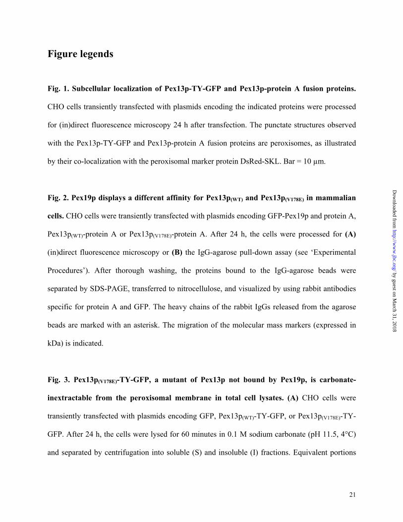
Fig. 1. Subcellular localization of Pex13p-TY-GFP and Pex13p-protein A fusion proteins.
CHO cells transiently transfected with plasmids encoding the indicated proteins were processed
for (in)direct fluorescence microscopy 24 h after transfection. The punctate structures observed
with the Pex13p-TY-GFP and Pex13p-protein A fusion proteins are peroxisomes, as illustrated
by their co-localization with the peroxisomal marker protein DsRed-SKL. Bar = 10 µm.
Fig. 2. Pex19p displays a different affinity for Pex13p(WT) and Pex13p(V178E) in mammalian
cells. CHO cells were transiently transfected with plasmids encoding GFP-Pex19p and protein A,
Pex13p(WT)-protein A or Pex13p(V178E)-protein A. After 24 h, the cells were processed for (A)
(in)direct fluorescence microscopy or (B) the IgG-agarose pull-down assay (see ‘Experimental
Procedures’). After thorough washing, the proteins bound to the IgG-agarose beads were
separated by SDS-PAGE, transferred to nitrocellulose, and visualized by using rabbit antibodies
specific for protein A and GFP. The heavy chains of the rabbit IgGs released from the agarose
beads are marked with an asterisk. The migration of the molecular mass markers (expressed in
kDa) is indicated.
Fig. 3. Pex13p(V178E)-TY-GFP, a mutant of Pex13p not bound by Pex19p, is carbonate-
inextractable from the peroxisomal membrane in total cell lysates. (A) CHO cells were
transiently transfected with plasmids encoding GFP, Pex13p(WT)-TY-GFP, or Pex13p(V178E)-TY-
GFP. After 24 h, the cells were lysed for 60 minutes in 0.1 M sodium carbonate (pH 11.5, 4°C)
and separated by centrifugation into soluble (S) and insoluble (I) fractions. Equivalent portions
by guest on March 31, 2018
http://ww
w.jbc.org/
Dow
nloaded from
22
of these fractions were separated by SDS-PAGE and stained with Ponceau S or immunoblotted
with the anti-TY 1 epitope tag monoclonal antibody BB2. (B) Similar fractions obtained from
non-transfected CHO cells were probed with anti-Pex14p, an antiserum that specifically
recognizes the integral peroxisomal membrane protein Pex14p, and anti-poxmtx, an antiserum
that specifically recognizes peroxisomal matrix proteins (19). The migration of the molecular
mass markers (expressed in kDa) is indicated.
Fig. 4. Pex13p(V178E)-protein A, a mutant of Pex13p not bound by Pex19p, is carbonate-
inextractable from the peroxisomal membrane in a fraction enriched in peroxisomes. CHO
cells were transiently transfected with plasmids encoding (A) Pex13p(WT)-protein A or (B)
Pex13p(V178E)-protein A and subjected to a subcellular fractionation procedure (see ‘Experimental
Procedures’) 24 h post-transfection. Equivalent portions of the total cell lysates (L), the nuclear
fractions (N), and the post-nuclear fractions (P) and equal volumes of the gradient fractions
(bottom fraction (B), top fraction (T)) were separated by SDS-PAGE and stained with Ponceau S
(detection of total protein profile) or immunoblotted with antisera raised against protein A
(detection of Pex13p-protein A fusioin proteins), Pex14p (peroxisomal integral membrane
protein marker), catalase (peroxisomal matrix protein marker), glutamate dehydrogenase (GDH,
mitochondrial protein marker), and cytochrome P450 4A (Cyt P450 4A; microsomal protein
marker). The arrow marks the remainder of the bovine serum albumin that is still present after
removal of the cell culture medium. The migration of the molecular mass markers (expressed in
kDa) is indicated. (C) The peroxisomal peak fractions (marked with an asterisk) of each gradient
were pooled, diluted 10 times in 0.1 M sodium carbonate (pH 11.5, 4°C) and further processed
as described in the ‘Experimental Procedures’. Equivalent portions of the soluble (S) and
by guest on March 31, 2018
http://ww
w.jbc.org/
Dow
nloaded from
23
insoluble (I) fractions were separated by SDS-PAGE and stained with antisera specific for
protein A, Pex14p, and catalase.
Fig. 5. The Pex5p-/Pex19p-binding site of Pex14p is also essential for binding to Pex13p.
Recombinant proteins were expressed in E. coli. Streptavidin beads were coated with
biotinylated Pex14p-deletion proteins and incubated with a bacterial lysate containing (His)6-
tagged Pex13p/SH3. After thorough washing, the recombinant proteins bound to the streptavidin
beads were separated by SDS-PAGE, transferred to nitrocellulose, and visualized by using
streptavidin-alkaline phosphatase and antibodies specific for Pex13p. The numbers on the left
indicate the amino acid residues present in the corresponding Pex14p deletion mutants. The
hydrophobic region and the putative coiled-coil domain of Pex14p are shaded in black and
hatched with horizontal lines, respectively. The domain required and sufficient for binding to
Pex5p and Pex19p (20) is shaded in gray.
Fig. 6. Pex5p(259-639) and Pex19p compete for binding to Pex14p. GST-Pex14p, biotin-
Pex5p(259-639), and biotin-Pex19p were recombinantly expressed in E. coli. Glutathione-Sepharose
beads were coated with GST-Pex14p and incubated with the indicated amounts (in µl) of
different bacterial lysates. After thorough washing, the recombinant proteins bound to the
Sepharose beads were separated by SDS-PAGE, transferred to nitrocellulose, and visualized by
using streptavidin-alkaline phosphatase and antibodies specific for Pex5p, Pex14p, or Pex19p.
(A) Increasing concentrations of Pex5p(259-639) antagonize binding of Pex19p to Pex14p. (B)
Increasing concentrations of Pex19p antagonize binding of Pex5p(259-639) to Pex14p.
by guest on March 31, 2018
http://ww
w.jbc.org/
Dow
nloaded from
24
Fig. 7. Full-length Pex5p displaces Pex19p from Pex14p with a higher efficacy than
Pex5p(259-639). (A) GST-Pex14p, (His)6-Pex5p, biotin-Pex5p(259-639), and biotin-Pex19p were
recombinantly expressed in E. coli. Glutathione-Sepharose beads were coated with GST-Pex14p
and incubated with the indicated amounts (in µl) of different bacterial lysates. After thorough
washing, the recombinant proteins bound to the Sepharose beads were separated by SDS-PAGE,
transferred to nitrocellulose, and visualized by using streptavidin-alkaline phosphatase and
antibodies specific for Pex14p and (His)6. (B) Biotin-Pex5p(259-639) and (His)6-Pex5p are
expressed approximately equally.
Fig. 8. Site-directed mutagenesis analysis of the Pex5p-/Pex13p-/Pex19p-binding site of
Pex14p. GST-Pex14p fusion proteins, (His)6-Pex5p, (His)6-Pex13p/SH3, and biotin-Pex19p
were recombinantly expressed in E. coli. Glutathione-Sepharose beads were coated with equal
amounts of the GST-Pex14p fusion proteins and incubated with bacterial lysates containing
(His)6-Pex5p, (His)6-Pex13p/SH3, or biotin-Pex19p. After thorough washing, the recombinant
proteins bound to the Sepharose beads were separated by SDS-PAGE, transferred to
nitrocellulose, and visualized by using streptavidin-alkaline phosphatase and antibodies specific
for Pex5p and Pex13p. GST alone did not bind to (His)6-Pex5p, (His)6-Pex13p/SH3, and biotin-
Pex19p (not shown).
Fig. 9. Pex13p/SH3 can compete with Pex19p, but not with Pex5p for binding to Pex14p.
GST-Pex14p, (His)6-Pex13p/SH3, and biotin-Pex19p were recombinantly expressed in E. coli.
Glutathione-Sepharose beads were coated with GST-Pex14p and incubated with the indicated
amounts (in µl) of different bacterial lysates. After thorough washing, the recombinant proteins
by guest on March 31, 2018
http://ww
w.jbc.org/
Dow
nloaded from

25
bound to the Sepharose beads were separated by SDS-PAGE, transferred to nitrocellulose, and
visualized by using streptavidin-alkaline phosphatase and antibodies specific for Pex13p and
Pex14p. (A) Pex5p, Pex13p/SH3, and Pex14p can form a ternary complex. (B) Pex13p/SH3 and
Pex19p both antagonize binding of the other peroxin to Pex14p.
Fig. 10. Targeting of Pex14p-GFP fusion proteins in CHO cells. CHO cells were transiently
transfected with plasmids coding for Pex14pWT (WT), Pex14pR25G (R25G), Pex14pF35S (F35S),
Pex14pL36R (L36R), Pex14pF52S (F52S), Pex14pK56E (K56E), or Pex14pL58R (L58R) N-terminally
fused to GFP. After 24 h, the cells were processed for indirect immunofluorescence using mouse
antibodies specific for GFP and rabbit antibodies specific for endogenous expressed PMP70. Bar
= 10 µm.
by guest on March 31, 2018
http://ww
w.jbc.org/
Dow
nloaded from

26
Table I. Synthetic oligonucleotide primers used in this study (introduced restriction sites are
underlined).
Name Oligonucleotide
Pex13pZomeFW
Pex13pZomeRV
Pex13-TY1
5’-GGGGGATCCATGGCGTCCCAGCCGCCA-3’
5’-GGGGGATCCAAAAAGATCTTGCTTTTCTCCATC-3’
5’-GATCTTGAGGTGCACACCAACCAGGACCCTCTGGACCTGCA-3’
Pex13-TY2 5’-GGTCCAGAGGGTCCTGGTTGGTGTGCACCTCAA-3’
Pex14.1 5’-ACGCGTCGACATGGCGTCCTCGGAGCAGG-3’
Pex14.2 5’-ATCGTAATGCGGCCGCCTAGTCCCGCTCACTCTC-3’
Pex14.3 5’-GGGAAGATCTATGGCGTCCTCGGAGCAG-3’
Pex14.4 5’-AATCTGCAGGTCCCGCTCACTCTCGTT-3’
R25Gfw 5’-GTGCTGCCTGGAGAGCCGCTGATTGCC-3’
R25Grv 5’-CAGCGGCTCTCCAGGCAGCACATTTTC-3’
F35Sfw 5’-GTGAAGTCTCTACAGAATTCCCGGGTC-3’
F35Srv 5’-ATTCTGTAGAGACTTCACTGCCGTGGC-3’
L36Rfw 5’-AAGTTTCGACAGAATTCCCGGGTCCGC-3’
L36Rrv 5’-GGAATTCTGTCGAAACTTCACTGCCGT-3’
F52Sfw 5’-AGAGCATCTCTAAAGAAGAAAGGGCTG-3’
F52Srv 5’-CTTCTTTAGAGATGCTCTCCTGGTTGC-3’
K56Efw 5’-CTAAAGAAGGAAGGGCTGACAGATGAA-3’
K56Erv 5’-CAGCCCTTCCTTCTTTAGAAATGCTCT-3’
L58Rfw 5’-AAGAAAGGGCGGACAGATGAAGAGATT-3’
L58Rrv 5’-ATCTGTCCGCCCTTTCTTCTTTAGAAA-3’
by guest on March 31, 2018
http://ww
w.jbc.org/
Dow
nloaded from

27
Table II. Plasmids constructed for this study.Name Protein Cloning vector insert
pMF792 HsPex13p-TY-GFP pMF121
Bgl II/Pst I
linker ligation (Pex13-TY1, Pex13-TY2)
pMF795 HsPex13p(V178E)-TY-GFP pMF551Bgl II/Pst I
linker ligation (Pex13-TY1, Pex13-TY2)
pMF964 HsPex13p-protein A pZome-1-CBam HI
Bam HI digest of PCR product: template pMF121(Pex13pZomeFW, Pex13pZomeRV )
pMF966 HsPex13p(V178E)-protein A pZome-1-CBam HI
Bam HI digest of PCR product: template pMF551(Pex13pZomeFW, Pex13pZomeRV )
pKG45 GST-HsPex14p pGEX-4T-3 Sal I/Not I digest of PCR product: template pMF101(Pex14.1, Pex14.2 )
pMF445 biotin-HsPex14p(22-81,295-377) PinPoint Xa1Bam HI/Eco RV
Bam HI/Eco RV fragment of pMF141 (20)
pMF444 biotin-HsPex14p(43-81,295-377) PinPoint Xa1Bam HI/Eco RV
Bam HI/Eco RV fragment of pMF151 (20)
pMF999A GST-HsPex14p(R25G) pGEX-4T-3Sal I/Not I
Sal I/Not I digest of PCR product generated bysequential PCR-steps:(1) template pKG45 (Pex14.1, R25Grv)(2) template pKG45 (R25Gfw, Pex14.2)(3) template (1) + (2) (Pex14.1, Pex14.2)
pMF999B GST-HsPex14p(F35S) pGEX-4T-3Sal I/Not I
Sal I/Not I digest of PCR product generated bysequential PCR-steps:(1) template pKG45 (Pex14.1, F35Srv)(2) template pKG45 (F35Sfw, Pex14.2)(3) template (1) + (2) (Pex14.1, Pex14.2)
pMF999C GST-HsPex14p(L36R) pGEX-4T-3Sal I/Not I
Sal I/Not I digest of PCR product generated bysequential PCR-steps:(1) template pKG45 (Pex14.1, L36Rrv)(2) template pKG45 (L36Rfw, Pex14.2)(3) template (1) + (2) (Pex14.1, Pex14.2)
pMF999D GST-HsPex14p(F52S) pGEX-4T-3Sal I/Not I
Sal I/Not I digest of PCR product generated bysequential PCR-steps:(1) template pKG45 (Pex14.1, F52Srv)(2) template pKG45 (F52S, Pex14.2)(3) template (1) + (2) (Pex14.1, Pex14.2)
pMF999E GST-HsPex14p(K56E) pGEX-4T-3Sal I/Not I
Sal I/Not I digest of PCR product generated bysequential PCR-steps:(1) template pKG45 (Pex14.1, K56Erv)(2) template pKG45 (K56Efw, Pex14.2)(3) template (1) + (2) (Pex14.1, Pex14.2)
pMF999F GST-HsPex14p(L58R) pGEX-4T-3Sal I/Not I
Sal I/Not I digest of PCR product generated bysequential PCR-steps:(1) template pKG45 (Pex14.1, L58Rrv)(2) template pKG45 (L58Rfw, Pex14.2)(3) template (1) + (2) (Pex14.1, Pex14.2)
pMF120 HsPex14p(WT)-GFP pEGFP-N1Bgl II/Pst I
Bgl II/Pst I digest of PCR product:template pMF101 (Pex14.3, Pex14.4)
pMF868 clone A clone B clone C clone D clone E clone F
HsPex14p(R25G)-GFPHsPex14p(F35S)-GFPHsPex14p(L36R)-GFPHsPex14p(F52S)-GFPHsPex14p(K56E)-GFPHsPex14p(L58R)-GFP
pEGFP-N1Bgl II/Pst I
Bgl II/Pst I digest of PCR product:template pMF999A (Pex14.3, Pex14.4)template pMF999B (Pex14.3, Pex14.4)template pMF999C (Pex14.3, Pex14.4)template pMF999D (Pex14.3, Pex14.4)template pMF999E (Pex14.3, Pex14.4)template pMF999F (Pex14.3, Pex14.4)
pMF122 biotin-HsPex19p PinPoint Xa3Bam HI/Sma I
Bam HI/Sma I fragment of pMF132 (10)
by guest on March 31, 2018
http://ww
w.jbc.org/
Dow
nloaded from

FITC filter Texas red filter merged
DsRed-SKL GFP
DsRed-SKL Pex13p(V178E)-TY-GFP
DsRed-SKL Pex13p(WT)-TY-GFP
Fig. 1
DsRed-SKL protein A
DsRed-SKL Pex13p(V178E)-protein A
DsRed-SKL Pex13p(WT)-protein A
by guest on March 31, 2018
http://ww
w.jbc.org/
Dow
nloaded from

94
67
43
30
94
67
43
Pex13pWT-protein AGFP-Pex19p
Protein AGFP-Pex19p
Pex13pV178E-protein AGFP-Pex19p
anti-protein A
anti-GFP
GFP-Pex19p
protein A
Pex13p(WT/V178E)-protein A
Fig. 2
FITCfilter
Texas redfilter
*
*
A
B
by guest on March 31, 2018
http://ww
w.jbc.org/
Dow
nloaded from

Fig. 3
anti-Pex14p
I S
anti-poxmtx
I S
I S I S I S
94
67
43
94
67
43
Ponceau S
anti-TY
A BPex13pWT Pex13pV178EGFP
by guest on March 31, 2018
http://ww
w.jbc.org/
Dow
nloaded from
A. Pex13p(WT)-protein A
anti-GDH
anti-Pex14p
anti-protein A
anti-Cyt P450 4A
anti-catalase
Ponceau S
Fig. 4
L N P B T946743302014
946743302014
L N P B T
B. Pex13p(V178E)-protein A
* *
* *
anti-GDH
anti-Pex14p
anti-protein A
anti-Cyt P450 4A
anti-catalase
Ponceau S
C. Carbonate extraction
anti-Pex14p
anti-protein A
anti-catalase
I S I S
Pex13pWT Pex13pV178E
by guest on March 31, 2018
http://ww
w.jbc.org/
Dow
nloaded from
HsPex14p
22-81,294-37743-81,294-377
1-377strept-AP
anti-Pex13p
Fig. 5
by guest on March 31, 2018
http://ww
w.jbc.org/
Dow
nloaded from
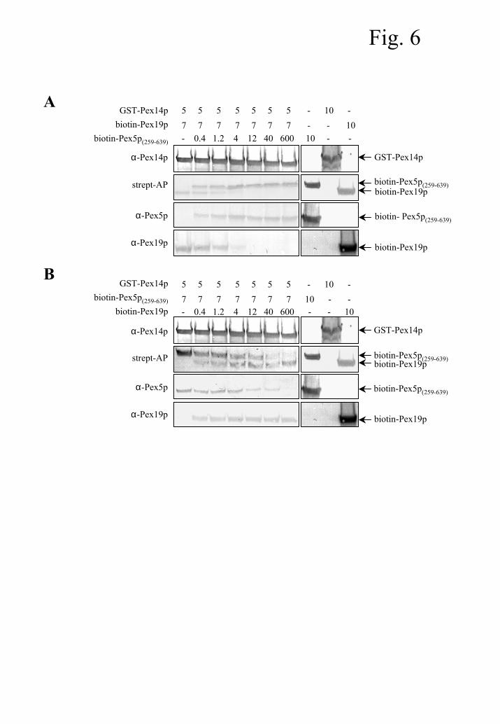
Fig. 6
A
biotin-Pex19p
GST-Pex14pα-Pex14p
strept-AP
GST-Pex14pbiotin-Pex19p
biotin-Pex5p(259-639) - 0.4 1.2 4 12 40 600 10 - - 7 7 7 7 7 7 7 - - 10 5 5 5 5 5 5 5 - 10 -
biotin- Pex5p(259-639)
biotin-Pex19p
α-Pex5p
α-Pex19p
biotin-Pex5p(259-639)
B
- 0.4 1.2 4 12 40 600 - - 10
α-Pex14p
strept-AP
GST-Pex14pbiotin-Pex5p(259-639)
biotin-Pex19p
biotin-Pex5p(259-639)biotin-Pex19p
7 7 7 7 7 7 7 10 - - 5 5 5 5 5 5 5 - 10 -
GST-Pex14p
biotin-Pex5p(259-639)
biotin-Pex19p
α-Pex5p
α-Pex19p
by guest on March 31, 2018
http://ww
w.jbc.org/
Dow
nloaded from

Fig. 7
A
- 100 100 1 - 100 100 1 - - - 10
α-Pex14p
strept-AP
GST-Pex14p(His)6-Pex5p
biotin-Pex5p(259-639)
biotin-Pex19p
biotin-Pex5p(259-639)biotin-Pex19p
1 1 - 100 - - - - - - 10 - 5 5 5 5 5 5 5 5 10 - - -
GST-Pex14p
(His)6-Pex5pα-(His)6
- - - - 1 1 - 100 - 10 - -
(His)6-Pex5p
biotin-Pex5p(259-639)
B
- - - 10
α-Pex5p
GST-Pex14p(His)6-Pex5p
biotin-Pex5p(259-639)
biotin-Pex19p
- - 10 - 10 - - -
- 10 - -
by guest on March 31, 2018
http://ww
w.jbc.org/
Dow
nloaded from
GST-Pex14p WT R25G F35S L36R F52S K56E L58R
α-Pex14p
+ biotin-Pex19p strept-AP
+ (His)6-Pex5p α-Pex5p
+ (His)6-Pex13p/SH3 anti-Pex13p
Fig. 8
by guest on March 31, 2018
http://ww
w.jbc.org/
Dow
nloaded from

Pex19p
Pex14p
Pex13p/SH3
α-Pex14p
strept-AP
GST-Pex14pbiotin-Pex19p
(His)6-Pex13p/SH3 0.3 0.3 - 50 - 50 0.3 0.3
α-Pex13p
0.3 0.3 0.3 0.3
B
biotin-Pex5p(259-639)
Pex14p
Pex13p/SH3
α-Pex14p
α-Pex13p
GST-Pex14p(His)6-Pex13p/SH3biotin-Pex5p(259-639)
0.3 0.3 - 50 - 50 0.3 0.3
strept-AP
0.3 0.3 0.3 0.3A
Fig. 9
by guest on March 31, 2018
http://ww
w.jbc.org/
Dow
nloaded from
anti-PMP70 mergedPex14p-GFP
WT
R25G
F35S
L36R
F52S
K56E
L58R
Fig. 10
by guest on March 31, 2018
http://ww
w.jbc.org/
Dow
nloaded from
Van VeldhovenMarc Fransen, Ilse Vastiau, Chantal Brees, Vanessa Brys, Guy P. Mannaerts and Paul P.
Potential role for Pex19p in assembly of PTS-receptor docking complexes
published online January 10, 2004J. Biol. Chem.
10.1074/jbc.M304941200Access the most updated version of this article at doi:
Alerts:
When a correction for this article is posted•
When this article is cited•
to choose from all of JBC's e-mail alertsClick here
by guest on March 31, 2018
http://ww
w.jbc.org/
Dow
nloaded from