human rotavirus cell attachment protein vp8* specifically interacts
TRANSCRIPT

Genotype-dependent glycan specificity in rotaviruses – evolution of a new
paradigm.
B. V. V. Prasad1,2 , L. Hu1, , S. E. Crawford2, R. Czako2, S. Ramani2, N. Cortes-Penfield2, D. F. Smith3, G. Kang4, J.
Le Pendu5, and M. K. Estes2
1Departments of Biochemistry and Molecular Biology and 2Molecular Virology and Microbiology, Baylor College of Medicine, Houston, TX,
3Department of Biochemistry, Emory University School of Medicine, GA, 4Christian Medical College, Vellore, India, 5NSERM, Nantes, France.

The key event of initial cell attachment of rotavirus is mediated by VP8*domain of the VP4 spike
VP4 (VP5*+VP8*)
VP7
VP2
VP1/VP3
VP6
VP8*
VP5*
Prasad et al., Nature (1996); Li et al., JVI (2009); Settembre et al., EMBO J. (2010)

Classification of rotavirus strains
• Rotaviruses exhibit enormous genetic and strain diversity
• Point mutations, gene rearrangements, and genetic reassortment contribute to the expanding diversity
• Based on neutralization specificity to VP7 and VP4, rotaviruses are classified into G (VP7) and P (VP4) serotypes
• Based on VP4 sequences, rotaviruses are classified into > 35 P genotypes
Sialidase sensitive rotaviruses • Virus infectivity decreases by sialidase treatment of cells • VP8* binds to glycans with terminal sialic acid (Sia) (e.g. GD1a)
Sialidase insensitive rotaviruses • Virus infectivity is not affected by sialidase treatment of cells • VP8* binds to glycans with internal sialic acid (Sia) (e.g. GM1)

Structures of VP8* of sialidase sensitive and insensitive strains
Simian RRV P[3]
Sensitive Terminal Sia
Narrow
Porcine CRW-8 P[7]
Sensitive Terminal Sia
Narrow
Sia
Sia
Human DS-1 P[4]
Insensitive Internal Sia
Wide
Human Wa P[8]
Insensitive Internal Sia
Wide
Do all sialidase-insensitive HR genotypes recognize sialoglycans?

Sialidase Insensitive Human Rotavirus HAL1166 P[14] VP8*
• This virus was first Isolated from an infant with diarrhea in Finland
• The P[14] rotavirus strains are being increasingly documented globally
• Phylogenetically distinct from the previously reported VP8*s
• These human rotaviruses are thought have jump from animal to human hosts

X-ray structure of P[14] VP8* not compatible
with Sia binding
The galectin-like fold: • Two twisted β-sheets (green & blue)
separated by a shallow cleft
Resolution: 1.5Å Space group: P21 R= 17.8%, Rfree=21.9%

Glycan array of GST-P[14] VP8*
N-terminal GST tag
611 glycans :
Sialylated glycans with
terminal or internal Sia,
Non-sialylated glycans
P[14] VP8*
N
C
Anti-GST Ab

Glycan#
Structure
Average RFU
%CV
367
GalNAcα1-3(Fucα1-2)Galβ1-4GlcNAcβ1-2Manα1-3(GalNAcα1-3(Fucα1-2)Galβ1-4GlcNAcβ1-2Manα1-6)Manβ1-4GlcNAcβ1-4GlcNAcβ-Sp20
2703
4
332 GalNAcα1-3(Fucα1-2)Galβ1-4GlcNAcβ1-3Galβ1-4GlcNAcβ-Sp0 1892 6
89 GalNAcα1-3(Fucα1-2)Galβ-Sp18 1574 18
85 GalNAcα1-3(Fucα1-2)Galβ1-4GlcNAcβ-Sp8 1324 11
88 GalNAcα1-3(Fucα1-2)Galβ-Sp8 867 16
86 GalNAcα1-3(Fucα1-2)Galβ1-4Glcβ-Sp0 807 26
390 GalNAcα1-3(Fucα1-2)Galβ1-3GalNAcα1-3(Fucα1-2)Galβ1-4GlcNAcβ-Sp0 681 17
141 Galβ1-3GalNAcβ1-4(Neu5Acα2-3)Galβ1-4Glcβ-Sp0 [GM1 with internal Sia] 5 184
409
Neu5Acα2-3Galβ1-3GalNAcβ1-4(Neu5Acα2-8Neu5Acα2-3)Galβ1-4Glcβ-Sp0
[“GD1a like” with terminal Sia] 1 272
Human rotavirus P[14] VP8* specifically binds to A-type histo-blood group antigen (HBGA)
* Relative Fluorescence Units (RFU) % Coefficient of Variation (%CV) =100Std.Dev/Mean

Sialidase-insensitive human rotavirus HAL1166 P[14] VP8* specifically binds to a non-sialylated glycan, A-type HBGA
HBGA Sia
Simian P[3] Sensitive
Terminal Sia
Narrow
Human P[14] Insensitive
HBGA
Narrow
• VP8* of a sialidase-insensitive human rotavirus also has a narrow cleft
• HBGA binding site in the P[14] VP8* remarkably overlaps with that of the Sia binding site in animal sialidase sensitive strains
Dormitzer et al., EMBO J (2002); Hu et al., Nature (2012)

Subtle changes within the same structural framework lead to altered receptor specificity
• Conformational change of R101
• Insertion of S187
• Switch the side chain orientations of Y188 and Y189
Simian RRV, P[3], sialidase-sensitive Human HAL 1166, P[14], sialidase-insensitive
Sia HBGA
Is the interaction with HBGA biologically relevant?
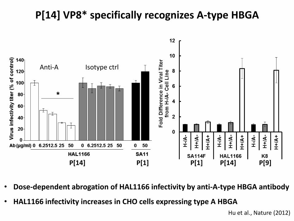
P[14] VP8* specifically recognizes A-type HBGA
• Dose-dependent abrogation of HAL1166 infectivity by anti-A-type HBGA antibody
• HAL1166 infectivity increases in CHO cells expressing type A HBGA
P[9]
Hu et al., Nature (2012)
P[14] P[1] P[1] P[14]
Anti-A Isotype ctrl

Glycan specificity varies between VP8* of different P genotypes
Sia
P[14], Insensitive, Narrow
Correlates with wide cleft
Glycan binding site
HBGA GM1 or other glycan?
P[3], Sensitive, Narrow P[8], Insensitive, Wide
?
The wide cleft correlates with a deletion at position 136, and a significant change at position 101
The amino acids at the known glycan binding site change significantly between VP8* of narrow cleft and wide cleft
?

Representative structures of VP8*: Class A: P[14] HAL1166; Class C: P[3] RRV; Class D: P[8] Wa
Structure-based classification of VP8*s
P[14] HAL1166 A-type HBGA
P[9] K8 A-type HBGA
Class A Human RV, s-i, narrow cleft
Class B Animal RV, s-i, narrow cleft
P[5] Bovine UK ?
P[12] Equine H2 ?
P[16] Murine Eb ?
P[3] Simian
RRV Sia
P[7] Porcine CRW8
Sia
Class C Animal RV, s-s, narrow cleft
Class D Human RV, s-i, wide cleft
P[4] DS-1 ?
P[8] Wa ?
P[6] McN13 ?
P[11] N155 ?

How do genotypic variations affect glycan specificity in VP8* with a wide cleft?
• What glycans do they bind? • Do glycans bind to the same region as that in the narrow cleft?

Structures of P[11] VP8* of neonate-specific sialidase insensitive strain N155
Human N155 P[11] VP8* Human Indian G2P[4] VP8*
Human N155 P[11] VP8* Cell dimensions: a=36.53Å, b=71.94Å, c=58.05Å, α=90°, β=90.02°, γ=90° Resolution: 1.66 Å Space group: P 1 21 1 Rwork= 15%, Rfree=17%

0
2
4
6
8
10
0
2
4
6
8
10
H-/A- H+/A- H+/A+
Fo
ld
d
iffe
re
nc
e in
vira
l tite
r
fro
m p
are
nta
l c
ell lin
e
H-/A- H+/A- H+/A+
P[11] VP8* specifically recognizes HBGA H type II precursor
Fo
ld
d
iffe
re
nc
e in
vira
l tite
r
fro
m p
are
nta
l c
ell lin
e
G10P[11] infectivity increases in CHO cells expressing type A or H HBGA
SA114F (G3P[1]) N155 (G10P[11])

* Relative Fluorescence Units (RFU); % Coefficient of Variation (%CV) =100Std.Dev/Mean
Glycan Structure Ave. RFU
Std % CV
Fucα1-2Galβ1-4(Fucα1-3)GlcNAcβ1-3GalNAcα-Sp14 (H-type and Lewisy)
458 20 4
Galβ1-3GalNAcβ1-4(Neu5Acα2-3)Galβ1-4Glcβ-Sp0 (GM1with internal Sia)
10 1 6
Neu5Acα2-3Galβ1-3GalNAcβ1-4(Neu5Acα2-8Neu5Acα2-3)Galβ1-4Glcβ-Sp0 (GD1a-like with terminal Sia)
30 14 46
VP8* of sialidase-insensitive human rotavirus G2P[4] (wide cleft) binds to H-type and Lewisy HBGAs.
Glycan array of G2P[4] VP8*

• Interactions with sialo-glycans for initial cell attachment is not an
obligatory requirement as previously thought.
• Rotaviruses exhibit genotype-dependent variations in glycan
specificity which may have implications in host specificity, tissue
tropism, susceptibility, pathogenesis and interspecies
transmission.
• Specific recognition of A-type HBGA may be the basis for inter-
species transmission observed in P[14] rotaviruses.
• Glycan binding site in globally dominant P[4] VP8* is distinct; in
addition to H-type P[4]VP8* can bind to Lewis HBGA – basis for
increased prevalence?
• VP8* of neonate-specific P[11] strain shows significant structural
alterations and specifically interacts with H type II precursor -
what is the significance?
Conclusions

Thank you!
We acknowledge the support from NIH grants AI36040 (to B.V.V.P.), AI 080656 and P30 DK56338 (to M.K.E.), GM62116 (to the Consortium for Functional Glycomics), and the Robert Welch foundation (Q1279) to B.V.V.P.
We thank Dr. Robert Atmar and Dr. Sreejesh
Shanker for helpful discussions and BCM X-ray core facility for data collection.
Acknowledgements

Precursor FUT3 FUT2
Lea
Leb
H type 1
FUT3 Enzyme A Enzyme B
A type 1 B type 1
A Leb B Leb
FUT3 FUT3
Keys:
Gal
GlcNAc
Fuc
GalNAc
FUT: Fucosyltransferase FUT2: Secretor Enzyme FUT3: Lewis Enzyme
1 - 3
1 - 3 1 - 3
1 - 3 1 - 3 1 - 3
1 - 3 1 - 3
2 1
2 1
2 1
2 1
2 1
4 1
4 1
4 1
4 1
2 1
1 - 3 1 - 3
1 - 3 1 - 3

Glycan#
Structure
Average RFU
%CV
367
GalNAcα1-3(Fucα1-2)Galβ1-4GlcNAcβ1-2Manα1-3(GalNAcα1-3(Fucα1-2)Galβ1-4GlcNAcβ1-2Manα1-6)Manβ1-4GlcNAcβ1-4GlcNAcβ-Sp20
2703
4
332 GalNAcα1-3(Fucα1-2)Galβ1-4GlcNAcβ1-3Galβ1-4GlcNAcβ-Sp0 1892 6
89 GalNAcα1-3(Fucα1-2)Galβ-Sp18 1574 18
85 GalNAcα1-3(Fucα1-2)Galβ1-4GlcNAcβ-Sp8 1324 11
88 GalNAcα1-3(Fucα1-2)Galβ-Sp8 867 16
86 GalNAcα1-3(Fucα1-2)Galβ1-4Glcβ-Sp0 807 26
390 GalNAcα1-3(Fucα1-2)Galβ1-3GalNAcα1-3(Fucα1-2)Galβ1-4GlcNAcβ-Sp0 681 17
141 Galβ1-3GalNAcβ1-4(Neu5Acα2-3)Galβ1-4Glcβ-Sp0 [GM1 with internal Sia] 5 184
409
Neu5Acα2-3Galβ1-3GalNAcβ1-4(Neu5Acα2-8Neu5Acα2-3)Galβ1-4Glcβ-Sp0
[“GD1a like” with terminal Sia] 1 272
Human rotavirus P[14] VP8* specifically binds to A-type histo-blood group antigen (HBGA)
* Relative Fluorescence Units (RFU) % Coefficient of Variation (%CV) =100Std.Dev/Mean