peripheral ameloblastoma of the lower molar …mj-med-u-tokai.com/pdf/370201.pdfperipheral...
TRANSCRIPT

Tokai J Exp Clin Med., Vol. 37, No. 2, pp. 30-34, 2012
―30―
INTRODUCTION
Ameloblastomas are the most common odontogenic tumor and they are clinically and histologically diverse. These tumors have several distinct clinical types, in-cluding solid, multicystic, unicystic, desmoplastic, and peripheral ameloblastomas (PAs). Each has a speci�c biological behavior and consequently a different prog-nosis and treatment [1]. Although ameloblastomas are subclassified as follicular, plexiform, granular, basal cell, acanthomatous, and desmoplastic types in histol-ogy [2], different histological patterns can coexist in the same lesion. This diversity may re�ect the complex development of dental structures. Ameloblastomas generally occur in the intraosseous region of the jaws. However, PA is a rare variant of ameloblastoma that occurs in extraosseous regions such as the gingiva and oral mucosa covering the alveolus. PAs comprise 1.3–10% of all ameloblastomas and frequently occur in the �fth through seventh decades [2, 3]. The mean age of patients with PA (males 53 years, females 51 years) is signi�cantly higher than for the intraosseous counterpart, which has a mean age of 37 years [2]. The male:female ratio is 1.9:1 and the mandible: maxilla ratio is 2.4:1 [2]. Clinically, a PA is a painless, �rm, exo-phytic growth with a smooth, granular, pebbly, warty, or papillary surface [2].
Two questions concerning PA remain: its histogenic origin and how to differentiate PA from intraoral basal cell carcinoma (BCC) [4]. Typical ameloblastomas are tumors of intraosseous odontogenic epithelial origin [5]. By contrast, PAs are thought to be derived from
the extraosseous remnants of odontogenic epithelium within the underlying connective tissue or from the basal cell layer of the oral epithelium, which is believed to have odontogenic potential [2, 6]. Continuity be-tween the tumor nests and oral epithelium is reported in about 50% of cases [5], suggesting a basal cell origin for PA. However, the tumor may simply extend into and involve the oral epithelium [4]. The histopatho-logical features of follicular PA are similar to those of intraoral BCC [4, 7, 8]. Furthermore, both PA and intraoral BCC can arise from the surface epithelium [9]. Therefore, PA and intraoral BCC may be the same entity [8]. However, distinguishing between these two neoplasms is important because their biological behav-ior and treatment differ [8].
This paper reports a patient with PA and the results of immunohistochemical studies for cytokeratins (CK7, 14, 19, AE1/AE3, CAM5.2, and 34 b E12), epithelial membrane antigen (EMA), Ber-EP4, p53, p63, and Ki-67 to further evaluate the two above-mentioned ques-tions regarding PA.
CASE REPORT
A 46-year-old man was referred to the Department of Oral and Maxillofacial Surgery, National Hospital Organization Shizuoka Medical Center with a painless mass of 5 months' duration on the lingual gingival region of the lower left third molar.
There was no swelling of the left cheek or associated lymph node enlargement. On intraoral examination, the mass was a 1.5-cm-diameter, �rm, exophytic growth with a papillary surface (Fig. 1). Plain radiographs
Peripheral Ameloblastoma of the Lower Molar Gingiva: A Case Report and Immunohistochemical Study
Hisashi KATO*1, Yoshihide OTA*1, Masashi SASAKI*2, Kazunari KARAKIDA*1, Akihiro KANEKO*1, Yasutomo SEKIDO*3 and Keiichi TSUKINOKI*4
*1Department of Oral and Maxillofacial Surgery, Tokai University School of Medicine *2Department of Oral and Maxillofacial Surgery, National Hospital Organization Shizuoka Medical Center
*3Department of Pathology, Tokai University School of Medicine *4Department of Diagnostic Science Division of Pathology, Kanagawa Dental College
(Received February 2, 2012; Accepted April 16, 2012)
Peripheral ameloblastoma (PA) is a rare extraosseous odontogenic tumor with histological characteristics similar to those of the common intraosseous ameloblastoma. Two questions regarding PA remain: its histogenic origin and how to differentiate between PA and intraoral basal cell carcinoma. We describe a patient with PA. The result of immunohistochemistry showed cytokeratin (CK) 7-, CK14+, CK19+, AE1/AE3+, CAM5.2-, 34 b E12+, epithelial membrane antigen-, Ber-EP4-, p53-, p63+, and low Ki-67, that was similar to those of 4 cases of intraosseous amelo-blastoma. Our results suggest that a PA originates from odontogenic epithelial remnants, rather than from the oral epithelium.
Key words: peripheral ameloblastoma, intraosseous ameloblastoma, intraoral basal cell carcinoma, immunohis-tochemistry, odontogenic tumor
Hisashi Kato, Department of Oral and Maxillofacial Surgery Tokai University School of Medicine, 143 Shimokasuya, Isehara, Kanagawa 259-1193, JapanTel: +81-463-93-1121 Fax: +81-463-95-7567 E-mail: [email protected]

H. KATO et al. / A Case Report of Peripheral Ameloblastoma and Immunohistochemical Study
―31―
and computed tomography (CT) showed no bone resorption (Fig. 2). There was no history of dental or other medical problems. The rest of the clinical and laboratory examination was within normal limits. With the provisional diagnosis of a gingival tumor, the biopsy was performed. The pathological examination showed a follicular type ameloblastoma and con�rmed the diagnosis of peripheral ameloblastoma. The mass was excised with a small margin of normal tissue under general anesthesia. Although the surface of the denuded bone showed no resorption, the surface was removed. Healing was uneventful and there has been no sign of recurrence for 2 years and 3 months.
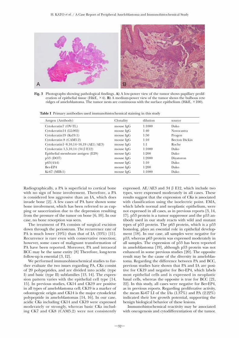
Histopathologically, the tumor demonstrated an ex-ophytic growth pattern and consisted of proliferating squamous-like cells. The tumor had the bulbous rete ridges of an ameloblastoma. These ridges consisted of peripheral rows of palisaded columnar cells and a cen-tral area of cuboidal or stellate cells. The tumor nests were continuous with the surface epithelium (Fig. 3).
We performed immunohistochemical studies for cytokeratins (CK7, 14, 19, AE1/AE3, CAM5.2, and 34 b E12), EMA, Ber-EP4, p53, p63, and Ki-67 to further evaluate the PA. Biopsy and operative specimens of four typical intraosseous ameloblastomas (IAs) and the PA from this case were examined. The IA specimens were obtained from the mandibles of two males and two females ranging in age from 17 to 53
years (mean age 39.5 years). Details of the primary antibodies and their use, and the results are presented in Tables I and II, respectively. The routine indirect immunoperoxidase method was used for immunohis-tochemistry. After deparaf�nization, the sections were immunostained in an autoanalyzer (BenchMark® XT; Roche Diagnostics K.K., Tokyo, Japan). After activat-ing the antigen, monoclonal antibodies were used as the primary antibody and an iView DAB Detection kit (Roche Diagnostics K.K.) was used for the autoimmu-nostaining reagent. Dehydration and mounting were performed after nuclear staining with hematoxylin. All cases were positive for CK14, CK19, AE1/AE3, 34 b E12, and p63, while negative for CK7, CAM5.2, EMA, Ber-EP4, and p53. (Fig. 4). The Ki-67 labeling index (Ki-67 LI) in the PA and IAs was 2.22% and 1.37% (mean), respectively (Fig. 5).
DISCUSSION
PA is difficult to diagnose based only on clinical findings, and it can be confused clinically with an epulis, �broma, gingival tumor, or carcinoma includ-ing intraoral BCC [3]. PA is frequently diagnosed after histological examination of specimens from a resected tumor or biopsy. Histologically, a PA consists of proliferating ameloblastic epithelium underlying mucosal epithelium [3]. In our case, we were not able to diagnose PA based only on clinical findings.
Fig. 1 Intraoral view of a 1.5-cm-diameter peripheral ameloblastoma on the lingual gingiva of the lower left third molar. The lesion showed exophytic growth with a papillary surface.
B
A
Fig. 2 A) Panoramic radiograph showing no bone resorption. B) CT image showing no bone resorption.
H. KATO et al. / A Case Report of Peripheral Ameloblastoma and Immunohistochemical Study
―32―
Radiographically, a PA is superficial to cortical bone with no sign of bone involvement. Therefore, a PA is considered less aggressive than an IA, which does invade bone [2]. A few cases of PA have shown some bone involvement, which has been referred to as cup-ping or saucerization due to the depression resulting from the pressure of the tumor on bone [6, 10]. In our case, no bone resorption was seen.
The treatment of PA consists of surgical excision down through the periosteum. The recurrence rate of PA is much lower (19%) than that of IA (33%) [11]. Recurrence is rare even with conservative resection; however, some cases of malignant transformation of PA have been reported. Moreover, PA and intraoral BCC may be the same entity [8] Therefore, long-term follow-up is essential [3, 12].
We performed immunohistochemical studies to fur-ther evaluate the two issues regarding PA. CKs consist of 20 polypeptides, and are divided into acidic (type I) and basic (type II) subfamilies [13, 14]. The expres-sion pattern varies with the epithelial cell type [14, 15]. In previous studies, CK14 and CK19 are positive in all types of ameloblastoma cell; CK19 is a marker of odontogenic origin and CK14 is the major cytoskeletal polypeptide in ameloblastomas [14, 16]. In our case, acidic CKs including CK14 and CK19 were expressed moderately or strongly, whereas basic CKs includ-ing CK7 and CK8 (CAM5.2) were not consistently
expressed. AE/AE3 and 34 b E12, which include two types, were expressed moderately in all cases. These results suggest that the expression of CKs is associated with classification using the isoelectric point. EMA, which labels normal and neoplastic epithelium, were not expressed in all cases, as in previous reports [3, 14, 17]. p53 protein is a tumor suppressor and the p53 an-tibody used in our study reacts with wild and mutant types of p53 protein. The p63 protein, which is a p53 homolog, plays an essential role in epithelial develop-ment [18]. In our case, all samples were negative for p53, whereas p63 protein was expressed moderately in all samples. The expression of p53 has been reported in ameloblastoma [19], although p53 protein was not enhanced in some previous studies [20]. The opposite result may be the cause of the diversity in ameloblas-toma. Regarding the difference between PA and BCC, previous studies have shown that PA and IA are posi-tive for CK19 and negative for Ber-EP4, which labels most epithelial cells and is expressed in neoplastic basal cells, whereas the opposite is true for BCC [21, 22]. In this study, all cases were negative for Ber-EP4, as in previous reports. Regarding proliferative activity, the mean Ki-67 LI of the IAs (1.37%) and PA (2.22%) indicated their low growth potential, supporting the benign biological behavior of these lesions.
Immunohistochemical reactivity may be associated with oncogenesis and cytodifferentiation of the tumor.
Table I Primary antibodies used immunohistochemical staining in this study
Antgen (Antibody) Clonality dilution source
Cytokeratin7 (OV-TL)Cytokeratin14 (LL002)Cytokeratin19 (Ks19.1)Cytokeratin 8 (CAM5.2)Cytokeratin1-8,10,14-16,19 (AE1/AE3)Cytokeratin 1,5,10,14 (34 b E12)Epithelial membrane antigen (E29)p53 (DO7)p63(4A4)Ber-EP4Ki-67 (MIB-1)
mouse IgGmouse IgGmouse IgGmouse IgGmouse IgGmouse IgGmouse IgGmouse IgGmouse IgGmouse IgGmouse IgG
1:10001:401:501:101:11:10001:2001:20001:101:2001:1000
DakoNovocastraProgenBecton DickinRocheDakoDakoDiyatoronDakoDakoDako
A B
Fig. 3 Photographs showing pathological �ndings. A) A low-power view of the tumor shows papillary prolif-eration of epithelial tissue (H&E, ×4). B) A medium-power view of the tumor shows the bulbous rete ridges of ameloblastoma. The tumor nests are continuous with the surface epithelium (H&E, ×100).

H. KATO et al. / A Case Report of Peripheral Ameloblastoma and Immunohistochemical Study
―33―
In this study, the immunohistochemical reactivity of the PA was similar to that of IAs. Although continuity between the tumor nests and oral epithelium was seen in our case, these �ndings suggest that PA and IA are tumors with a common origin, and that PA is derived from odontogenic epithelial remnants, rather than from basal cells of the oral epithelium.
REFERENCES1) Gardner DG. Some current concepts on the pathology of amelo-
blastomas. Oral Surg Oral Med Oral Pathol Oral Radiol Endod 1996; 82: 660-9.
2) Gardner DG, Heikinheimo K, Shear M, Philipsen HP, Coleman H. Ameloblastoma. In: Barnes L, Eveson J, Reichart P, Sidransky D, eds. World Health Organization classi�cation of tumours; pathol-ogy and genetics of head and neck tumours. Lyon: IARC Press, 2005: 296–300.
3) Kishino M, Murakami S, Yuki M, Iida S, Ogawa Y, Kogo M, et al. A immunohistochemical study of the peripheral ameloblastoma. Oral Dis 2007; 13: 575-80.
4) Ide F, Mishima K, Miyazaki Y, Saito I, Kusama K. Peripheral ameloblastoma in-situ: an evidential fact of surface epithelium origin. Oral Surg Oral Med Oral Pathol Oral Radiol Endod 2009; 108: 763-7.
5) el-Mofty SK, Gerard NO, Farish SE, Rodu B. Peripheral amelo-blastoma: a clinical and histologic study of 11 cases. J Oral Maxillofac Surg 1991; 49: 970-4.
6) Redman RS, Keegan BP, Spector CJ, Patterson RH. Peripheral ameloblastoma with unusual mitotic activity and conflicting evidence regarding histogenesis. J Oral Maxillofac Surg 1994; 52: 192-7.
7) Simpson HE. Basal-cell carcinoma and peripheral ameloblas-toma. Oral Surg Oral Med Oral Pathol 1974; 38: 233-40.
8) Koutlas IG, Koch CA, Vickers RA, Brouwers FM, Vortmeyer AO. An unusual ostensible example of intraoral basal cell carcinoma. J Cutan Pathol 2009; 36: 464-70.
9) Gardner DG. Peripheral ameloblastoma: a study of 21 cases, in-cluding 5 reported as basal cell carcinoma of the gingiva. Cancer 1977; 39: 1625-33.
10) Lin SC, Lieu CM, Hahn LJ, Kwan HW. Peripheral ameloblastoma with metastasis. Int J Oral Maxillofac Surg 1987; 16: 202-4.
11) Yamanishi T, Ando S, Aikawa T, Kishino M, Nakano Y, Sasai K, et al. A case of extragingival peripheral ameloblastoma in the buc-cal mucosa. J Oral Pathol Med 2007; 36: 184-6.
12) Wettan HL, Patella PA, Freedman PD. Peripheral ameloblastoma: review of the literature and report of recurrence as severe dyspla-sia. J Oral Maxillofac Surg 2001; 59: 811-5.
13) Quinlan RA, Schiller DL, Hatzfeld M, Achtstätter T, Moll R, Jorcano JL, et al. Patterns of expression and organization of cytokeratin intermediate �laments. Ann N Y Acad Sci 1985; 455: 282-306.
14) Crivelini MM, de Araújo VC, de Sousa SO, de Araújo NS. Cytokeratins in epithelia of odontogenic neoplasms. Oral Dis 2003; 9: 1-6.
15) Moll R, Franke WW, Schiller DL, Geiger B, Krepler R. The catalog of human cytokeratins: patterns of expression in normal epithelia, tumors and cultured cells. Cell 1982; 31: 11-24.
16) Kumamoto H, Yoshida M, Ooya K. Immunohistochemical detec-tion of amelogenin and cytokeratin 19 in epithelial odontogenic tumors. Oral Dis 2001; 7: 171-6.
17) Ota Y, Goto J, Sasaki J, Osamura Y. Immunohistochemical studies of ameloblastoma with special emphasis on keratin. Tokai J Exp Clin Med 1988; 13: 219-26.
18) Lo Muzio L, Santarelli A, Caltabiano R, Rubini C, Pieramici T, Giannone N, et al. p63 expression correlates with pathological features and biological behaviour of odontogenic tumours. Histopathology 2006; 49: 211-4.
19) Barboza CA, Pereira Pinto L, Freitas Rde A, Costa Ade L, Souza LB. Proliferating cell nuclear antigen (PCNA) and p53 protein expression in ameloblastoma and adenomatoid odontogenic tumor. Braz Dent J 2005; 16: 56-61.
20) Migaldi M, Sartori G, Rossi G, Cittadini A, Sgambato A. Tumor cell proliferation and microsatellite alterations in human amelo-
Tab
le I
I I
mm
unoh
isto
chem
ical
res
ults
Mar
ker
subs
tan
ceA
ge/S
exH
isto
logi
c fe
atur
eC
K7
CK
14C
K19
CA
M5.
2A
E1/
AE
334
b E
12E
MA
p53
p63
Ber
-EP4
Ki-6
7
PA IA1
IA2
IA3
IA4
46/M
17/F
44/F
44/M
53/M
Folli
cula
rPl
exif
orm
Folli
cula
rPl
exif
orm
Folli
cula
r
- - - - -
++ ++ ++ + ++
+ ++ ++ ++ ++
- - - - -
+ + + + +
+ ++ + + +
- - - - -
- - - - -
+ ++ ++ + +
- - - - -
LI:
2.2
2%L
I: 3
.65%
LI:
0.4
9%L
I: 0
.24%
LI:
1.1
%
PA: p
erip
hera
l am
elob
last
oma,
IA
: int
raos
seou
s am
elob
last
oma
++: s
tron
g-po
siti
ve r
eact
ion.
, + :
mod
erat
e-po
siti
ve r
eact
ion.
, ±: w
eak-
posi
tive
rea
ctio
n.,
- : n
on-r
eact
ion
LI:
labe
ling
inde
x

H. KATO et al. / A Case Report of Peripheral Ameloblastoma and Immunohistochemical Study
―34―
blastoma. Oral Oncol 2008; 44: 50-60.21) Latza U, Niedobitek G, Schwarting R, Nekarda H, Stein H. Ber-
EP4: new monoclonal antibody which distinguishes epithelia from mesothelial. J Clin Pathol 1990; 43: 213-9.
22) Tellechea O, Reis JP, Domingues JC, Baptista AP. Monoclonal antibody Ber EP4 distinguishes basal-cell carcinoma from squamous-cell carcinoma of the skin. Am J Dermatopathol 1993; 15: 452-5.
A B
Fig. 5 The Ki-67 labeling index is A) 2.22% in PA; and B) 0.49% in IA (case 2) (×200).
A B
Fig. 4 The expression of CK19: A) moderate immunoreactivity for CK19 in PA; and B) strong immunoreac-tivity for CK19 in IA (case 2) (×200).