perioperative management of external ventricular and ... · pdf fileperioperative management...
TRANSCRIPT

Perioperative Management of External Ventricular
(EVD) and Lumbar Drain (LD) Educational Document from the Society of Neuroscience in
Anesthesiology & Critical Care (SNACC)
SNACC Task Force for Perioperative Management of EVD & LD
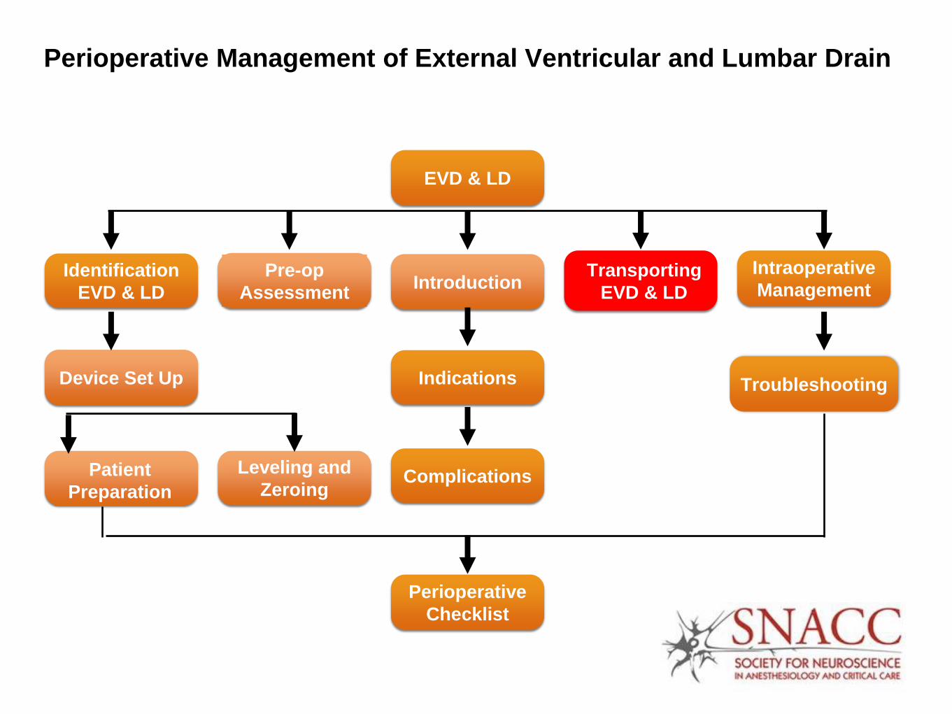
Introduction Identification
EVD & LD
Device Set Up
Pre-op
Assessment
Transporting
EVD & LD
Intraoperative
Management
Troubleshooting
Patient
Preparation
Leveling and
Zeroing
Indications
Complications
EVD & LD
Perioperative
Checklist

This Presentation is Free of
Commercial Bias
SNACC does not endorse any
particular EVD or LD system
manufacturer

Introduction Identification
EVD & LD
Device Set Up
Pre-op
Assessment
Transporting
EVD & LD
Intraoperative
Management
Patient
Preparation
Leveling and
Zeroing
Indications
Complications
EVD & LD
Perioperative
Checklist
Perioperative Management of External Ventricular and Lumbar Drain
Troubleshooting
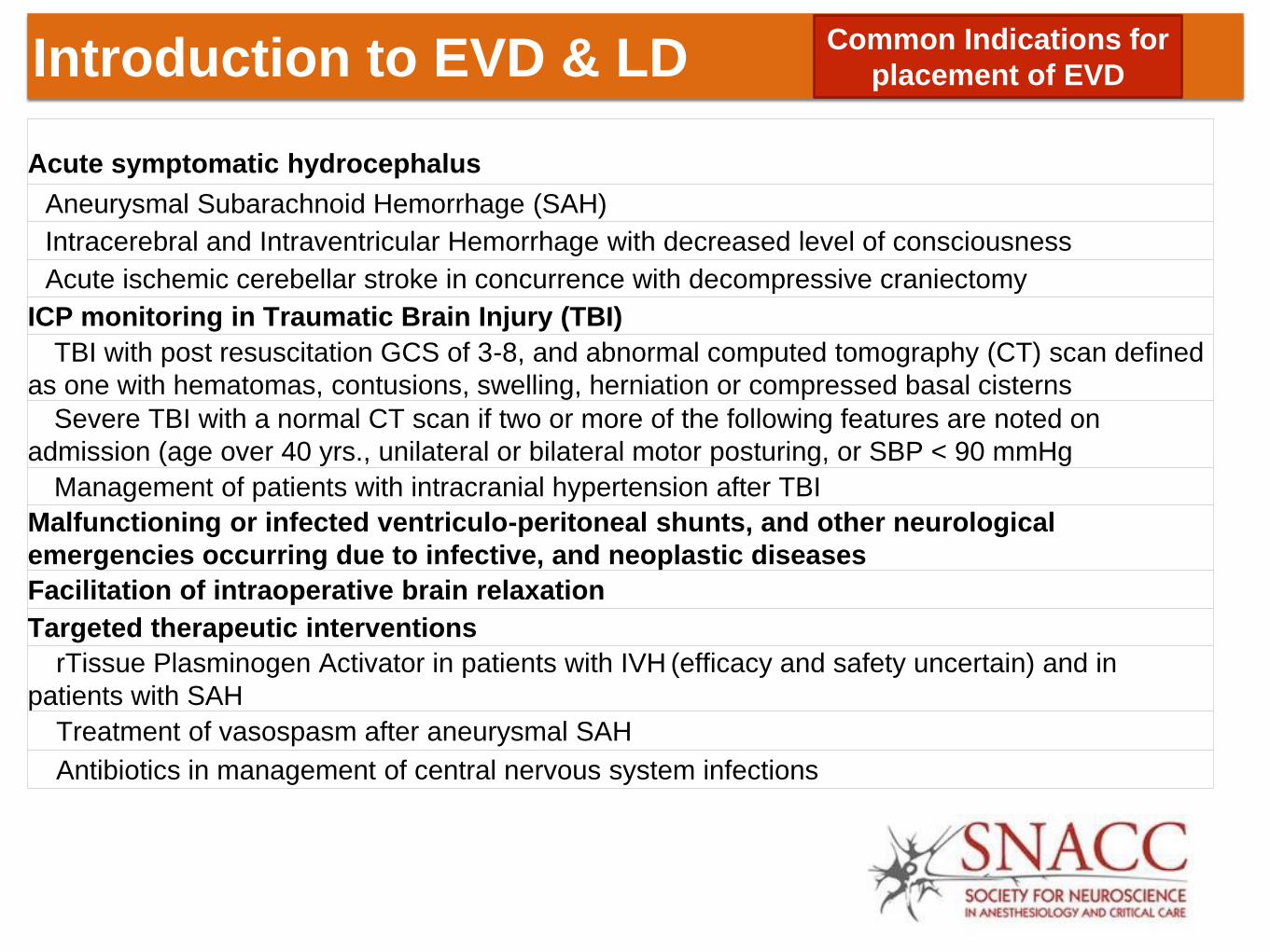
Acute symptomatic hydrocephalus
Aneurysmal Subarachnoid Hemorrhage (SAH)
Intracerebral and Intraventricular Hemorrhage with decreased level of consciousness
Acute ischemic cerebellar stroke in concurrence with decompressive craniectomy
ICP monitoring in Traumatic Brain Injury (TBI)
TBI with post resuscitation GCS of 3-8, and abnormal computed tomography (CT) scan defined
as one with hematomas, contusions, swelling, herniation or compressed basal cisterns
Severe TBI with a normal CT scan if two or more of the following features are noted on
admission (age over 40 yrs., unilateral or bilateral motor posturing, or SBP < 90 mmHg
Management of patients with intracranial hypertension after TBI
Malfunctioning or infected ventriculo-peritoneal shunts, and other neurological
emergencies occurring due to infective, and neoplastic diseases
Facilitation of intraoperative brain relaxation
Targeted therapeutic interventions
rTissue Plasminogen Activator in patients with IVH (efficacy and safety uncertain) and in
patients with SAH
Treatment of vasospasm after aneurysmal SAH
Antibiotics in management of central nervous system infections
Introduction to EVD & LD
Next Pge
Common Indications for
placement of EVD
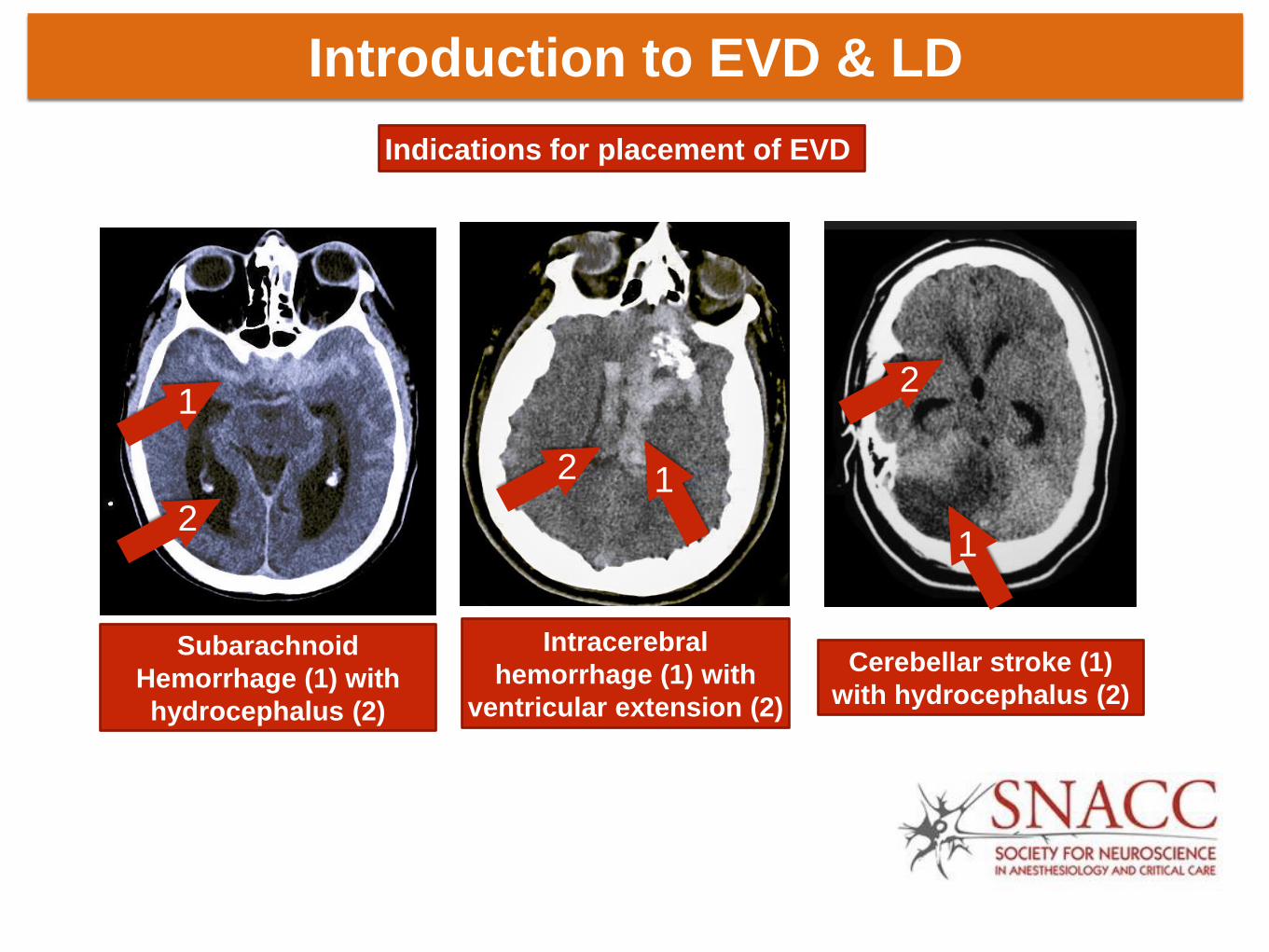
Introduction to EVD & LD
Indications for placement of EVD
Subarachnoid
Hemorrhage (1) with
hydrocephalus (2)
Intracerebral
hemorrhage (1) with
ventricular extension (2)
1
2
2 1
Cerebellar stroke (1)
with hydrocephalus (2)
1
2

Introduction to EVD & LD
Indications for placement of LD
Acute symptomatic hydrocephalus in SAH
Spinal cord protective strategy in open and endovascular thoracic aortic repair for
patients at high risk of spinal cord injury
Active CSF leak (due to craniofacial trauma) or those at risk for CSF leak during skull
base procedures, however lumbar drains do not reduce postoperative CSF leaks
Facilitate intraoperative brain relaxation and intraoperative exposure

Introduction to EVD & LD Complications associated with
placement of EVD & LD
Hemorrhage
Intracerebral hemorrhage, tract hematoma or tract hemorrhages (0-41%)
Neuraxial hematoma (0-3.2%)
Neural injury
Infection (0-28% EVD, 0-50% LD)
Malposition
Occlusion and malfunction
Over drainage of CSF
Subdural or epidural hematoma
Re-bleeding from a ruptured cerebral aneurysm
Intracranial hypotension
Cerebellar tonsillar herniation
Paradoxical herniation
Pneumocephalus
Iatrogenic vascular injury (arteriovenous fistula, cerebral pseudo aneurysm)
Fracture of catheters, with retained fragment of catheter
Inadvertent injections of drugs
Postdural puncture headache

Introduction to EVD & LD Complications associated with
placement of EVD
1
2 2
Hemorrhage (1) along EVD (2) track
Tractoma

Differences between EVD and LD
EVD LD
Intracranial pressure
monitor Yes
No
( Intraspinal pressure)
Drainage Dependent on ICP and
EVD setting
Typically drain pre-
determined amount
every hour
Leveling External auditory
meatus
Phlebostatic axis
OR
catheter insertion site

Introduction Identification
EVD & LD
Device Set Up
Pre-op
Assessment
Transporting
EVD & LD
Intraoperative
Management
Patient
Preparation
Leveling and
Zeroing
Indications
Complications
EVD & LD
Perioperative
Checklist
Perioperative Management of External Ventricular and Lumbar Drain
Troubleshooting

Identify Components of EVD
(1) (Type of EVD catheter)
Antimicrobial-impregnated EVD
Clindamycin and Rifampin Non-antimicrobial impregnated EVD

Identify Components of EVD (2)
Antibiotic Impregnated EVD
Tip of EVD
35 cm catheter
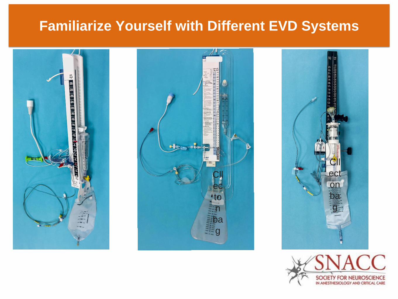
Familiarize Yourself with Different EVD Systems
Cll
ec
to
n
ba
g
Cll
ect
on
ba
g

Identify Components of EVD
(3) The Collecting System
1 3 4 2
1
2
3
4
Flushless transducer
Stopcock 2 (used to zero)
Stopcock 3 clamped to drain
Stopcock 1
5
5 EVD set at +10 cm H20 6
6 Graduated drip chamber
(burette) for collecting CSF
7 7 Stopcock 4 to stop flow of CSF in
collection bag CSF flow from patient

Identify Components of LD

Introduction Identification
EVD & LD
Device Set Up
Pre-op
Assessment
Transporting
EVD & LD
Intraoperative
Management
Patient
Preparation
Leveling and
Zeroing
Indications
Complications
EVD & LD
Perioperative
Checklist
Perioperative Management of External Ventricular and Lumbar Drain
Troubleshooting

Device Set Up (EVD)
EVD systems should be set up by personnel intimately
familiar with the devices and demonstrate appropriate
clinical competency
Devices should be set up observing standards of sterile
techniques
Only flushless transducer systems are used
EVD system is primed with sterile, preservative free saline
Setting should be expressed in cm H20
Leveling of EVD should always be made at the external
auditory meatus (EAM)

Device Set Up
Carpenter’s Level
Bubble Level Laser Level
Bubble
Laser

Device Set Up (EVD)
External Auditory Meatus
Leveling
Device Set Up (LD)

Device Set Up (LD)
LD systems should be set up by personnel intimately familiar
with the devices and demonstrate appropriate clinical
competency
Devices should be set up observing standards of sterile
techniques
Only flushless transducer systems are used
LD system is primed with sterile, preservative free saline
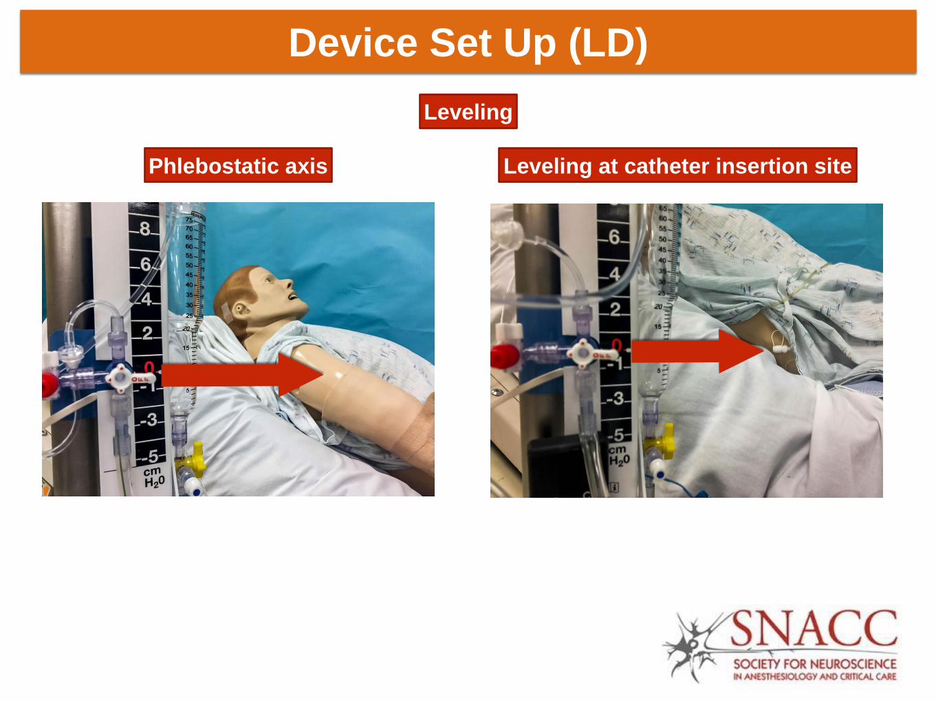
Leveling of LD can be made at the external auditory meatus
(EAM), level of catheter insertion or at the phlebostatic axis
by use of Carpenter’s bubble or laser level
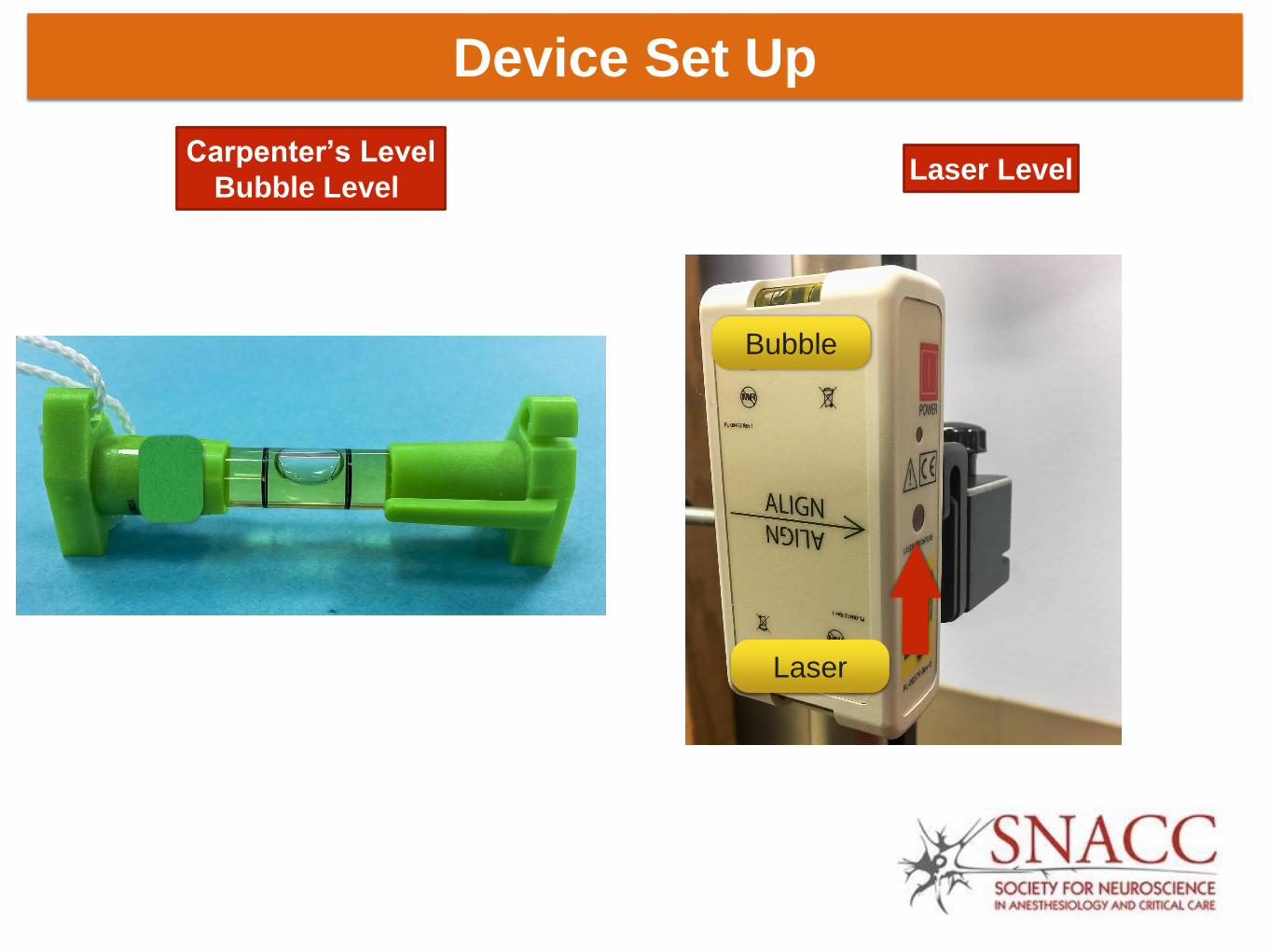
Device Set Up
Carpenter’s Level
Bubble Level Laser Level
Bubble
Laser
Device Set Up (LD)
Leveling at catheter insertion site Phlebostatic axis
Leveling

Introduction Identification
EVD & LD
Device Set Up
Pre-op
Assessment
Transporting
EVD & LD
Intraoperative
Management
Patient
Preparation
Leveling and
Zeroing
Indications
Complications
EVD & LD
Perioperative
Checklist
Perioperative Management of External Ventricular and Lumbar Drain
Troubleshooting

Zeroing EVD and LD system
Connect ventricular or lumbar
catheter under sterile
conditions
Attach pressure cable to
flushless transducer
Turn stopcock off to patient
by turning it to “3 o’clock”
position (1)
Open system to air (2) by
removing the red cap
Press “zero” on monitor
When monitor indicates “0”,
return stopcock upright
Replace injection cap (3)
1
2
3

EVD & LD Device Set up
CAUTION

EVD & LD Device Set up
Do not connect EVD or LD system to a high pressure system such as pressure bag
used for arterial or central venous catheter
CAUTION

Introduction Identification
EVD & LD
Device Set Up
Pre-op
Assessment
Transporting
EVD & LD
Intraoperative
Management
Patient
Preparation
Leveling and
Zeroing
Indications
Complications
EVD & LD
Perioperative
Checklist
Perioperative Management of External Ventricular and Lumbar Drain
Troubleshooting

Patient Preparation
Follow ASRA* guidelines (LD) & NCS **guidelines (EVD) for
prompt coagulopathy screening and reversal prior to EVD or LD
placement and maintenance
Administer antibiotics only prior to placement of EVD or LD,
and follow institutional antibiograms in selecting antibiotics
Whenever possible use antimicrobial-impregnated EVDs
Practice strict aseptic technique based on national and
institutional guidelines
*ASRA: American Society of Regional Anesthesia
**NCS: Neurocritical Care Society

Introduction Identification
EVD & LD
Device Set Up
Pre-op
Assessment
Transporting
EVD & LD
Intraoperative
Management
Patient
Preparation
Leveling and
Zeroing
Indications
Complications
EVD & LD
Perioperative
Checklist
Perioperative Management of External Ventricular and Lumbar Drain
Troubleshooting

Preoperative Assessment
Focused history and physical examination
CSF color and consistency
Hourly and daily CSF output
ICP values, ICP waveform analysis, ICP trends,
autoregulation indices, CPP and other multimodal monitoring
data
Clinical and radiological evidence of clamping trial tolerance
All pertinent data regarding EVD and LD may be incorporated
into a pre-operative handoff between intensive care/ ward
providers and anesthesia providers

Preoperative Assessment
Setting of EVD
+ 10 cm H20 + 5 cm H20 + 20 cm H20

Preoperative Assessment
Color of CSF
Hemorrhagic (Bloody) Xanthochromic Tea-colored
Intraoperative Management of EVD & LD
Normal ICP waveform
P
1 P
2 P
3
P
1
P
2
P
3
Percussion wave ~ reflections off choroid plexus
Tidal wave ~ brain compliance
Dicrotic wave ~ aortic valve closure
Normally P2 wave is 80% of P1 wave
Intraoperative Management of EVD & LD
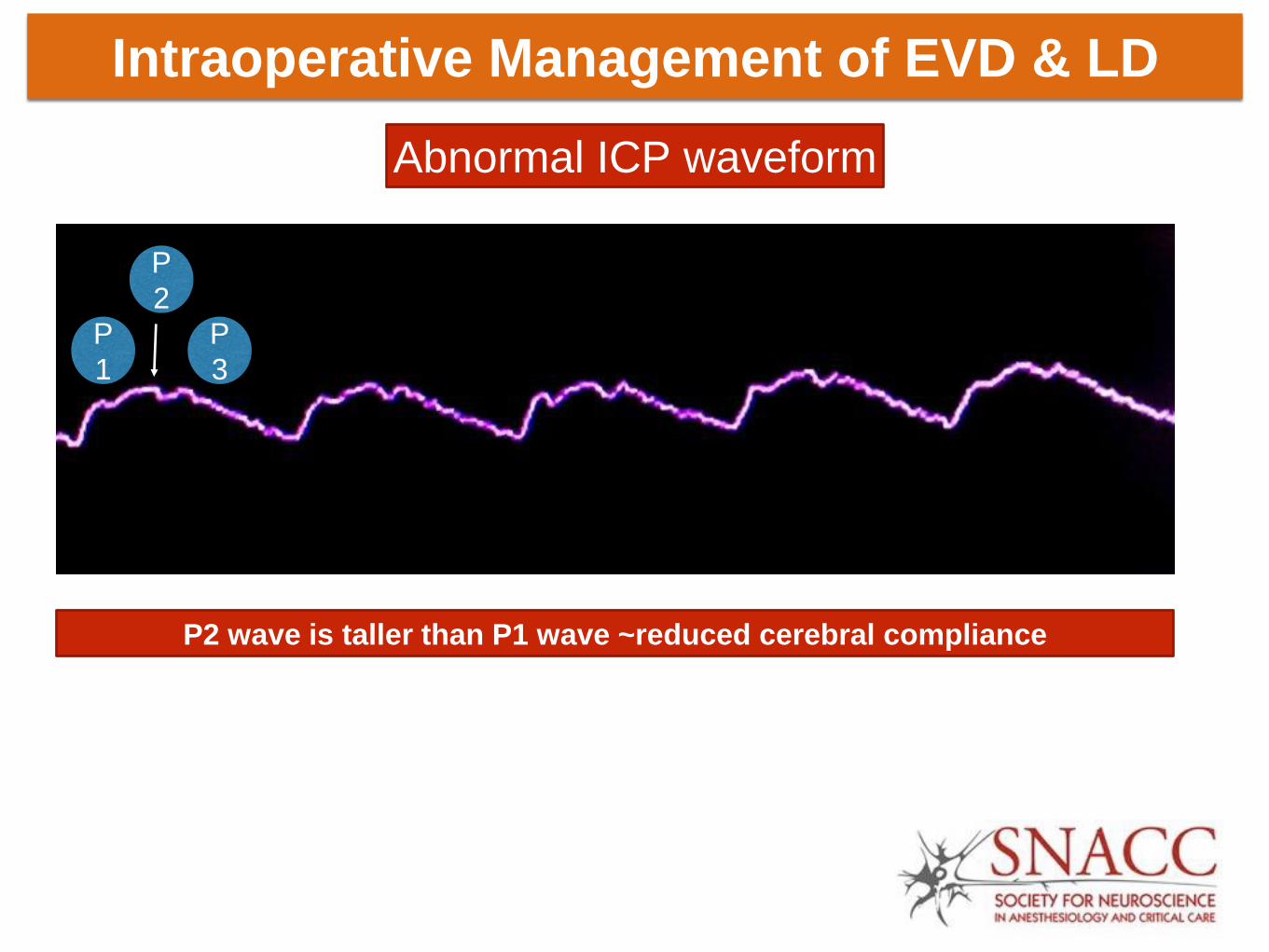
Abnormal ICP waveform
P
1
P
2
P
3
P2 wave is taller than P1 wave ~reduced cerebral compliance
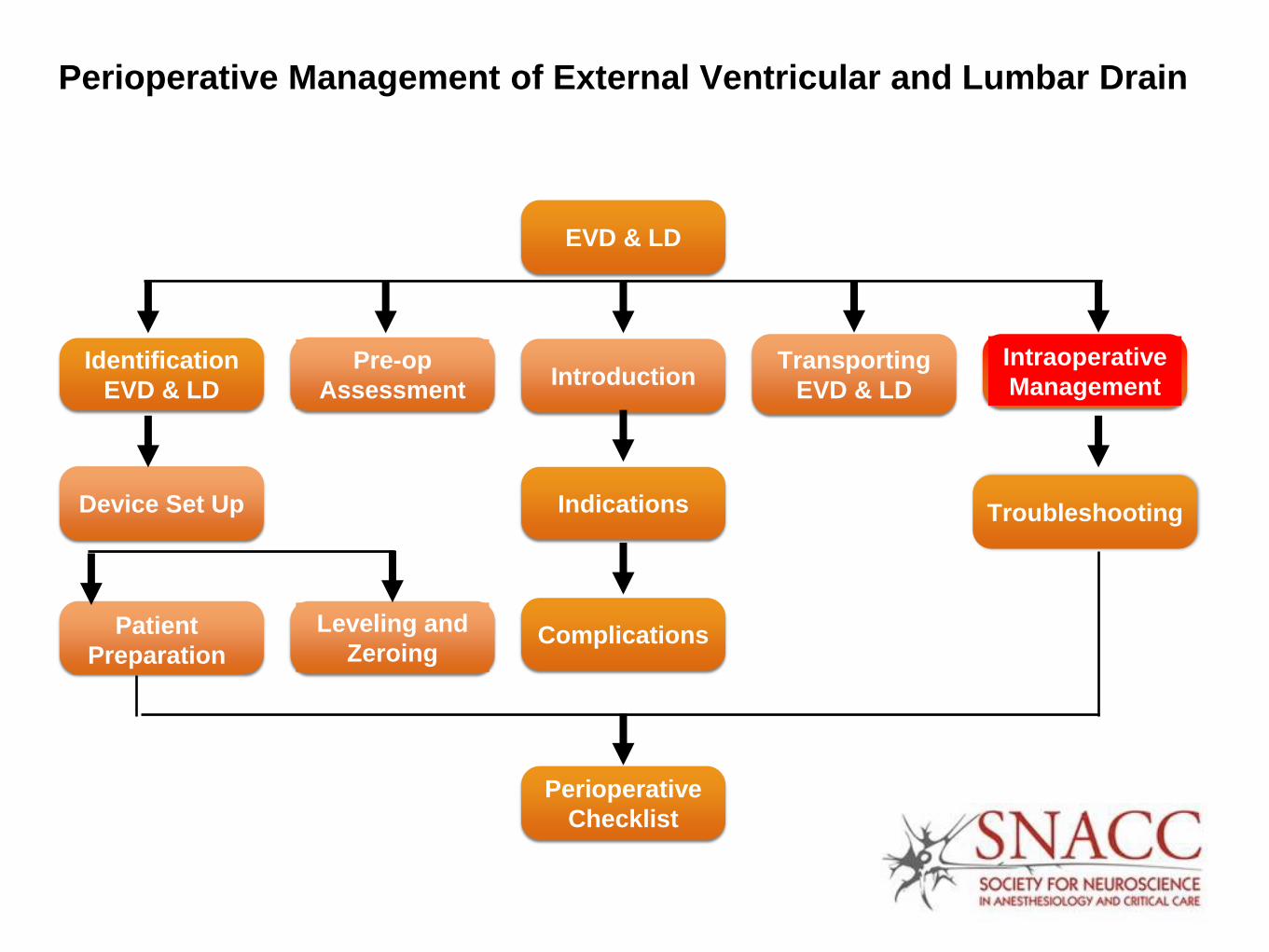
Introduction Identification
EVD & LD
Device Set Up
Pre-op
Assessment
Transporting
EVD & LD
Intraoperative
Management
Patient
Preparation
Leveling and
Zeroing
Indications
Complications
EVD & LD
Perioperative
Checklist
Perioperative Management of External Ventricular and Lumbar Drain
Troubleshooting

Is EVD continuously draining in the neuro ICU or is it
clamped for drainage ?
What is hourly CSF drainage ?
What is CSF output over 24 hours ?
Was an EVD clamp trial conducted in the neuro ICU ?
What are the results of such clamping trial ?
What is the baseline ICP ( < 15 mmHg, 15-19 mmHg,
or > 20 mmHg)
What is the reason for transporting patient to the
anesthesia suite ( Diagnostic vs. therapeutic procedure)
Pre-transport screening questionnaire
Transporting Patients with EVD or LD

Open to drain CSF Closed to drain CSF
CSF flow from patient CSF flow from patient
Transporting Patients with EVD or LD
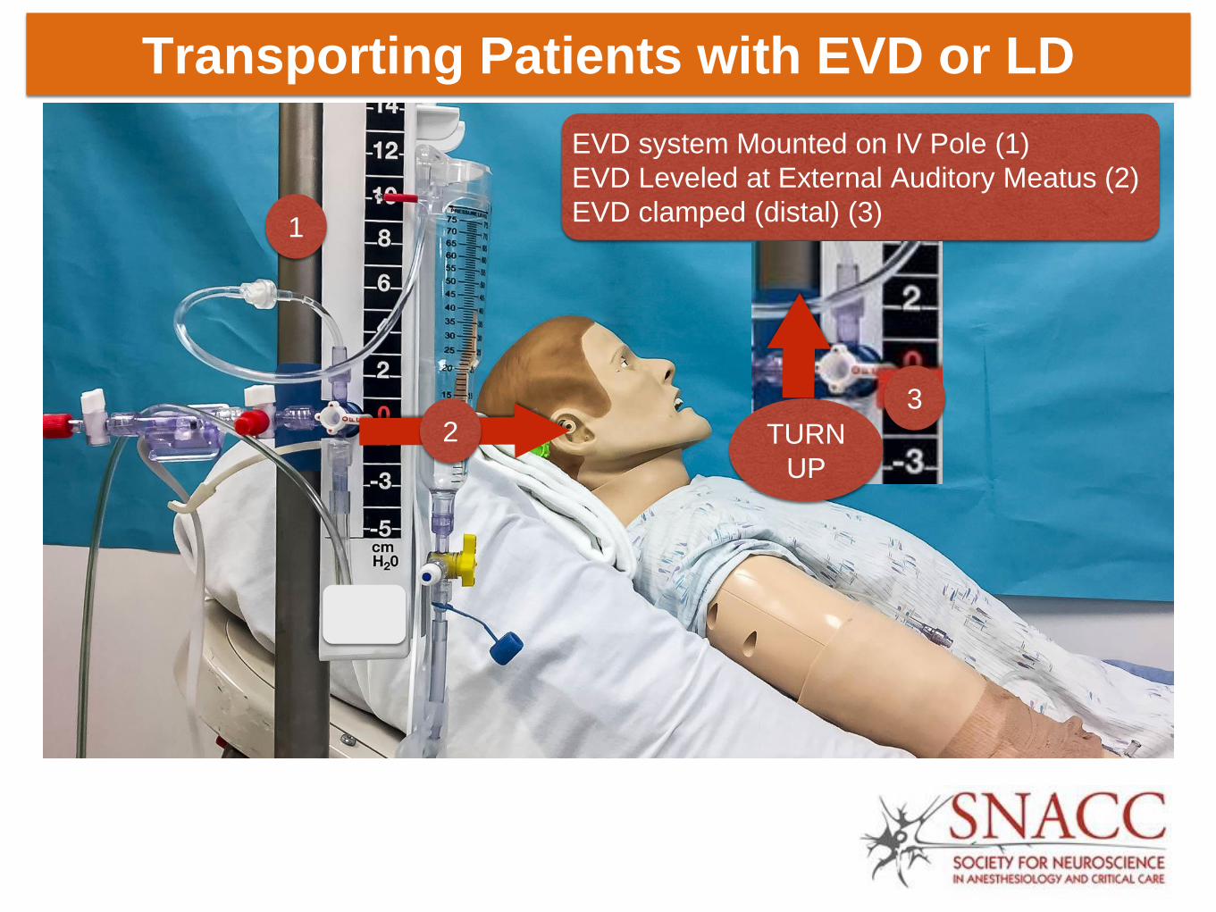
Next Page
EVD system Mounted on IV Pole (1)
EVD Leveled at External Auditory Meatus (2)
EVD clamped (distal) (3) 1
2 3
TURN
UP
Transporting Patients with EVD or LD

Monitoring During Transport
Transporting Patients with EVD or LD
Continue all pre-transport monitoring and documentation
End tidal carbon dioxide
Mean and systolic arterial pressures
Intracranial pressure, brain tissue oxygenation
Cerebral perfusion pressure
Use a dedicated intravenous pole to mount EVD and LD
Transport personnel be prepared to treat intracranial
hypertension during intrahospital transport
Individualize decision to transport with EVD open vs.
closed to CSF drainage
Introduction Identification
EVD & LD
Device Set Up
Pre-op
Assessment
Transporting
EVD & LD
Intraoperative
Management
Patient
Preparation
Leveling and
Zeroing
Indications
Complications
EVD & LD
Perioperative
Checklist
Perioperative Management of External Ventricular and Lumbar Drain
Troubleshooting

Intraoperative Management of EVD & LD
Label EVD and LD

Intraoperative Management of EVD & LD

Intraoperative Management of EVD & LD
Document the following in the anesthetic record at least every
hourly or as situation demands
Pressure = ICP/CPP or intraspinal pressure (ISP)/ spinal cord
perfusion pressure (SCPP),
Amount of CSF drainage (expressed in ml),
Color of CSF and any change in color of CSF observed
during the procedure,
Drain height relative to the reference level, and
EVD / LD status as set by the stopcocks in the device (i.e.
open, clamped)
Incorporate all information pertinent to EVD and LD into a
standardized intraoperative handoff between anesthesia
providers

Introduction Identification
EVD & LD
Device Set Up
Pre-op
Assessment
Transporting
EVD & LD
Intraoperative
Management
Patient
Preparation
Leveling and
Zeroing
Indications
Complications
EVD & LD
Perioperative
Checklist
Perioperative Management of External Ventricular and Lumbar Drain
Troubleshooting

Promptly recognize any accidental intrathecal injection
Lavage of intrathecal space after intrathecal injection is not
recommended
Routine flushing of the EVD or LD should not be performed
EVD or LD tubing that are accidently disconnected should be
clamped immediately
If EVD or LD system are contaminated by disconnection, all
distal parts should be replaced with new sterile tubing
Troubleshooting
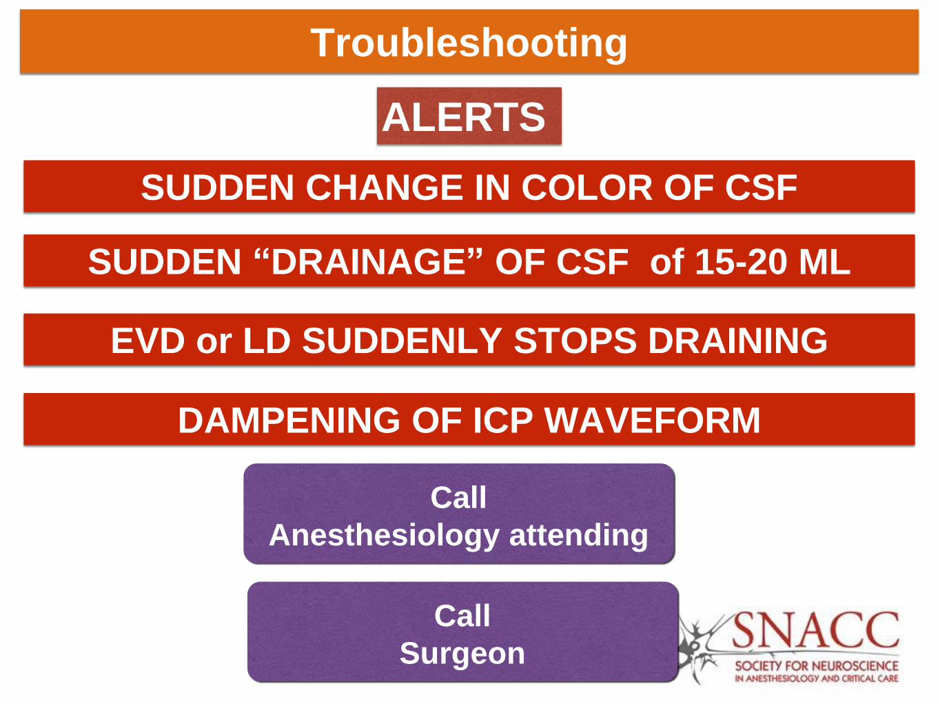
Troubleshooting
SUDDEN CHANGE IN COLOR OF CSF
ALERTS
SUDDEN “DRAINAGE” OF CSF of 15-20 ML
DAMPENING OF ICP WAVEFORM
EVD or LD SUDDENLY STOPS DRAINING
Call
Surgeon
Call
Anesthesiology attending

Introduction Identification
EVD & LD
Device Set Up
Pre-op
Assessment
Transporting
EVD & LD
Intraoperative
Management
Patient
Preparation
Leveling and
Zeroing
Indications
Complications
EVD & LD
Perioperative
Checklist
Perioperative Management of External Ventricular and Lumbar Drain
Troubleshooting

Perioperative Checklist
Next age
Preoperative assessment Obtain baseline neurological examination Review EVD (cmH20) & LD setting (in ml/hr of CSF drained) Review hourly CSF output to obtain baseline Review baseline ICP mmHg, ICP trends, and available multimodal monitoring data Review baseline CSF color and consistency Review clamp trials data if available Review coagulopathy profile Review antibiotic plan if anticipating new EVD /LD insertion in the operating room Provide EVD and LD details during pre-operative handoff between intensive care / ward providers and the anesthesia providers. Transporting patients with EVD and LD Confirm decision to travel with EVD or LD clamp vs. open If travelling with EVD clamp, ensure clamping at both proximal port on EVD and distal port on CSF collecting system Confirm HOB status during transport Confirm availability of dedicated intravenous pole for EVD / LD mount Confirm leveling EVD at external auditory meatus & LD at phlebostatic axis or at lumbar catheter insertion site Enable ICP monitoring during transport Confirm availability of medications needed to treat intracranial hypertension during transport Intraoperative management of indwelling drains Prepare transducer cable Identify EVD/ LD tubing by appropriate unique labeling Confirm HOB status during surgical procedure Confirm leveling of EVD at external auditory meatus & LD at phlebostatic axis Obtain ICP waveform & baseline ICP value Record q 1-hour EVD /LD setting Record at least q 1-hour ICP values (recorded with EVD closed to drain) Record at least q 1-hour EVD /LD drain output (expressed in ml) Provide EVD and LD details during intraoperative handoffs between anesthesia providers Inform surgeon if any one or more of the following Sudden decline in CSF drainage or no drainage from EVD or LD, or occlusion of EVD or LD If drain output is greater than 15-20 ml at any time or in any given hour Sudden change in CSF color Dampening or loss of ICP waveform

1. During pre-operative assessment of patient with indwelling EVD, the anesthesia provider should perform all of the following EXCEPT :
A. Perform EVD clamp trial B. Focused neurological examination C. Inspection of EVD system D. Obtain hourly and 24-hour EVD output data
Click for answer
SNACC EVD & LD QUIZ

• Perform EVD clamp trial
• Answer A
Question 2

2. During transporting a patient to and from the operating room, which of the following is true ?
A. It is ok to place CSF collecting system horizontal in the patients bed
B. It is required to mount CSF collecting system on an intravenous pole
C. It is ok not to monitor ICP during transport
D. It is ok to connect EVD and LD to a flushable pressure transducer system Click for
answer

• It is required to mount CSF collecting system on an intravenous pole
• Answer B
Question 3
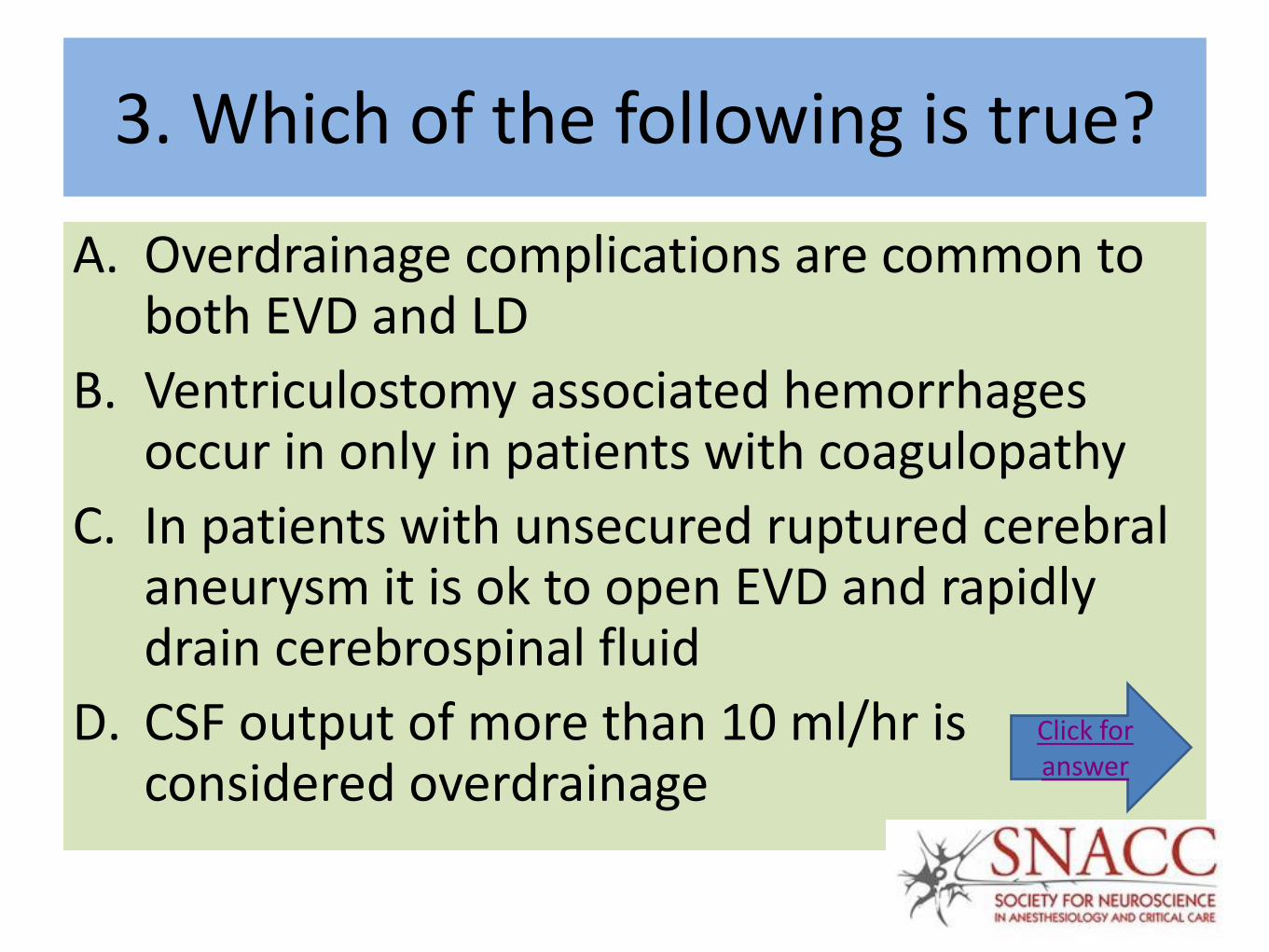
3. Which of the following is true?
A. Overdrainage complications are common to both EVD and LD
B. Ventriculostomy associated hemorrhages occur in only in patients with coagulopathy
C. In patients with unsecured ruptured cerebral aneurysm it is ok to open EVD and rapidly drain cerebrospinal fluid
D. CSF output of more than 10 ml/hr is considered overdrainage
Click for answer

• Overdrainage complications are common to both EVD and LD
• Answer A
Question 4
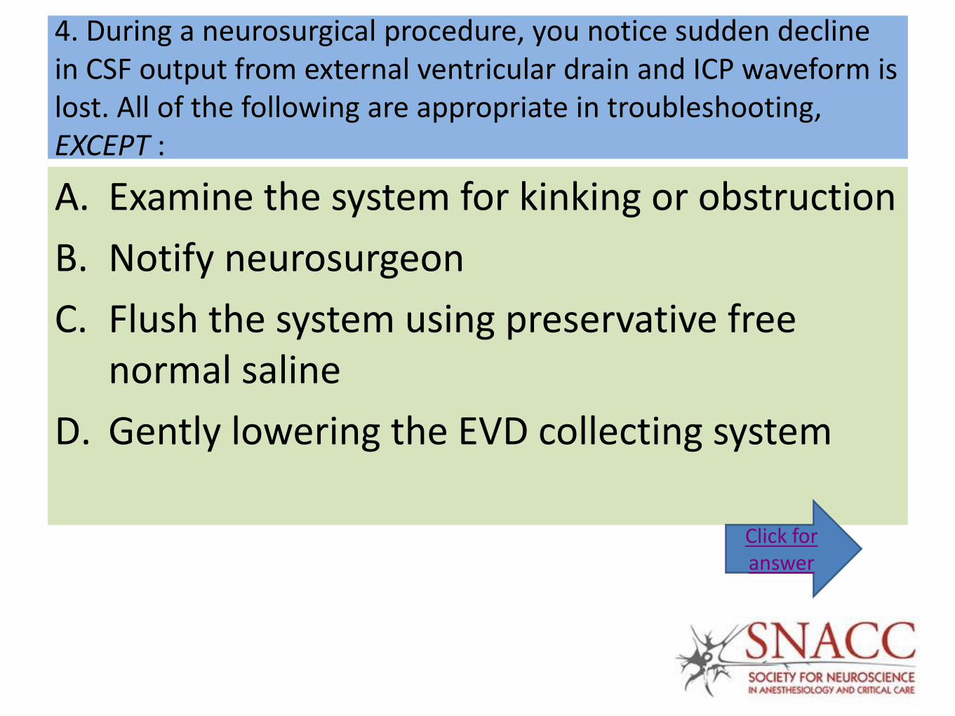
4. During a neurosurgical procedure, you notice sudden decline in CSF output from external ventricular drain and ICP waveform is lost. All of the following are appropriate in troubleshooting, EXCEPT :
A. Examine the system for kinking or obstruction
B. Notify neurosurgeon
C. Flush the system using preservative free normal saline
D. Gently lowering the EVD collecting system
Click for answer
• Flush the system using preservative free normal saline
• Answer C
Question 5

5. For accurately measuring ICP, which of the following stopcock position is appropriate?
a) Stopcock closed to CSF drain b) Stopcock open to drain CSF
Click for answer
• Stopcock closed to CSF drain
• Answer A

SNACC Task Force
Perioperative Management of External Ventricular
and Lumbar Drains
Abhijit Lele M.B.B.S, M.D., M.S.
Amie Hofnagel M.D.
Nina Schloemerkemper M.D., Dr.med, FRCA
David Wyler M.D.
Nophanan Chaikittisilpa M.D.
Monica Vavilala M.D.
Bhiken Naik M.B.B. Ch.
James Williams M.D. Ph.D.
Lashmikumar Venkat Raghavan M.B.B.S., M.D., FRCA, FRCPC
Ines Koerner M.D., Ph.D.