periodontal microbiology (dental plaque) dental plaque … · 3 associated with calculus formation...
TRANSCRIPT
1
Periodontal Microbiology (Dental Plaque)
Dental plaque Dental plaque is soft deposits that form the biofilm adhering to the tooth surface of other hard surface in the oral cavity, including removable and fixed restoration. Biofilm is defined as the relatively undefinable microbial community associated with a tooth surface or any other hard, non-shedding material. Types of Dental Plaque: Based on its relationship to the gingival margin, Plaque is differentiated into two categories: a) Supragingival plaque: is found at or above the gingival margin b) Sub gingival plaque: is found below the gingival margin. Differences between supra gingival and sub gingival plaque Supragingival plaque Subgingival plaqueMatrix 50% Matrix Little or no matrixFlora Mostly gram-positive Mostly gram-negativeMotile bacteria few CommonAnaerobic / Aerobic Aerobic unless thick Highly anaerobic area
presentMetabolism Predominantly
carbohydratesPredominantly Proteins
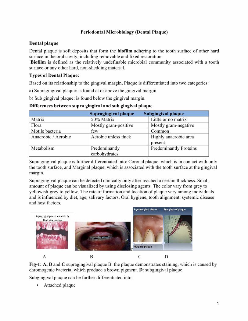
Supragingival plaque is further differentiated into: Coronal plaque, which is in contact with only the tooth surface, and Marginal plaque, which is associated with the tooth surface at the gingival margin. Supragingival plaque can be detected clinically only after reached a certain thickness. Small amount of plaque can be visualized by using disclosing agents. The color vary from grey to yellowish-grey to yellow. The rate of formation and location of plaque vary among individuals and is influenced by diet, age, salivary factors, Oral hygiene, tooth alignment, systemic disease and host factors.
c A B C D Fig-1: A, B and C supragingival plaque B. the plaque demonstrates staining, which is caused by chromogenic bacteria, which produce a brown pigment. D: subgingival plaque Subgingival plaque can be further differentiated into:
• Attached plaque
2
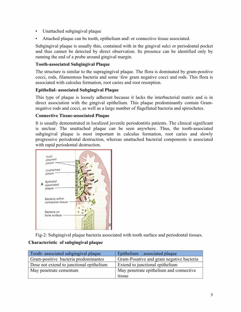
• Unattached subgingival plaque • Attached plaque can be tooth, epithelium and\ or connective tissue associated. Subgingival plaque is usually thin, contained with in the gingival sulci or periodontal pocket and thus cannot be detected by direct observation. Its presence can be identified only by running the end of a probe around gingival margin. Tooth-associated Subgingival Plaque The structure is similar to the supragingival plaque. The flora is dominated by gram-positive cocci, rods, filamentous bacteria and some \few gram negative cocci and rods. This flora is associated with calculus formation, root caries and root resorption. Epithelial- associated Subgingival Plaque This type of plaque is loosely adherent because it lacks the interbacterial matrix and is in direct association with the gingival epithelium. This plaque predominantly contain Gram-negative rods and cocci, as well as a large number of flagellated bacteria and spirochetes. Connective Tissue-associated Plaque It is usually demonstrated in localized juvenile periodontitis patients. The clinical significant is unclear. The unattached plaque can be seen anywhere. Thus, the tooth-associated subgingival plaque is most important in calculus formation, root caries and slowly progressive periodontal destruction, whereas unattached bacterial components is associated with rapid periodontal destruction.
Fig-2: Subgingival plaque bacteria associated with tooth surface and periodontal tissues.
Characteristic of subgingival plaque Tooth- associated subgingival plaque Epithelium �associated plaqueGram-positive bacteria predominantes Gram-Posative and gram negative bacteriaDose not extend to junctional epithelium Extend to junctional epitheliumMay penetrate cementum May penetrate epithelium and connective
tissue
3
Associated with calculus formation and root caries
Associated with gingivitis and periodontitis
Macroscopic structure and composition of dental plaque � Dental plaque can be defined as the soft deposits that form the biofilm adhering to the tooth surface or other hard surfaces in the oral cavity, including removable and fixed restorations. � Materia alba refers to soft accumulations of bacteria and tissue cells that lack the organized structure of dental plaque and are easily displaced with a water spray. � Calculus is a hard deposit that forms by mineralization of dental plaque and is generally covered by a layer of unmineralized plaque. Dental plaque is composed primarily of microorganisms. Cultivation studies, in which bacteria are isolated and characterized in the laboratory, indicate that more than 500 distinct microbial species are found in dental plaque. Nonbacterial microorganisms that are found in plaque include Mycoplasma species, yeasts, protozoa, and viruses. The microorganisms exist within an intercellular matrix that also contains a few host cells such as epithelial cells, macrophages, and leukocytes. The intercellular matrix, estimated to account for 20% to 30% of the plaque mass, consists of organic and inorganic materials derived from saliva, gingival crevicular fluid, and bacterial products. Organic constituents of the matrix include: Glycoproteins from saliva are an important component of the pellicle & incorporated into the developing plaque biofilm. Polysaccharides produced by bacteria, of which dextran is the predominant form, contribute to the organic portion of the matrix, provide not only energy, but also organic skeleton of the plaque. Albumin (proteins), probably from crevicular fluid, has been identified as a component of the plaque matrix. The lipid material consists of debris from the membranes of disrupted bacterial and host cells and possibly food debris. The Inorganic Component of plaque is predominately calcium and phosphorus, with trace amounts of other minerals such as sodium, potassium, and fluoride. The source of inorganic constituents of supragingival plaque is primarily saliva & derived from crevicular fluid, which is a serum transudate. The process of plaque formation can be divided into three phases: a) Formation of Dental Pellicle b) Initial colonization of bacteria c) Secondary colonization & plaque maturation. Formation of the dental pellicle on the tooth surface is the initial phase of plaque development. � All surfaces are coated with a glycoprotein pellicle. This pellicle is derived from components of saliva and crevicular fluid as well as bacterial and host tissue cell products and debris. The hydroxyapatite surface has a predominance of negatively charged phosphate groups that interact
4
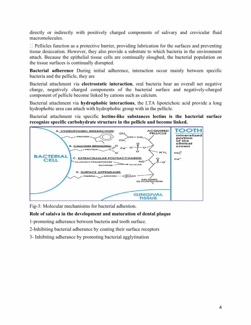
directly or indirectly with positively charged components of salivary and crevicular fluid macromolecules. � Pellicles function as a protective barrier, providing lubrication for the surfaces and preventing tissue desiccation. However, they also provide a substrate to which bacteria in the environment attach. Because the epithelial tissue cells are continually sloughed, the bacterial population on the tissue surfaces is continually disrupted. Bacterial adherence During initial adherence, interaction occur mainly between specific bacteria and the pellicle, they are Bacterial attachment via electrostatic interaction, oral bacteria bear an overall net negative charge, negatively charged components of the bacterial surface and negatively-charged component of pellicle become linked by cations such as calcium. Bacterial attachment via hydrophobic interactions, the LTA lipoteichoic acid provide a long hydrophobic area can attach with hydrophobic group with in the pellicle. Bacterial attachment via specific lectins-like substances lectins is the bacterial surface recognize specific carbohydrate structure in the pellicle and become linked.
Fig-3: Molecular mechanisims for bacterial adhestion. Role of salaiva in the development and maturation of dental plaque 1-promoting adherance between bacteria and tooth surface. 2-Inhibiting bacterial adherance by coating their surface receptors 3- Inhibiting adherance by promoting bacterial agglytination
5
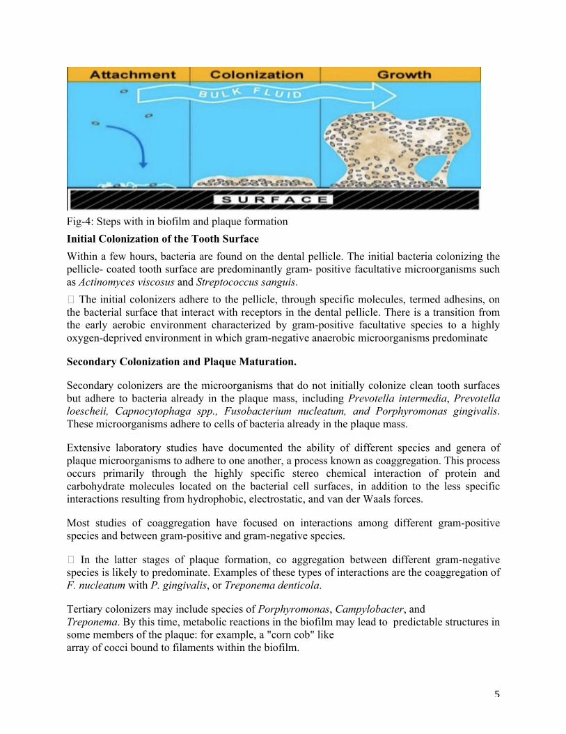
Fig-4: Steps with in biofilm and plaque formation Initial Colonization of the Tooth Surface Within a few hours, bacteria are found on the dental pellicle. The initial bacteria colonizing the pellicle- coated tooth surface are predominantly gram- positive facultative microorganisms such as Actinomyces viscosus and Streptococcus sanguis. � The initial colonizers adhere to the pellicle, through specific molecules, termed adhesins, on the bacterial surface that interact with receptors in the dental pellicle. There is a transition from the early aerobic environment characterized by gram-positive facultative species to a highly oxygen-deprived environment in which gram-negative anaerobic microorganisms predominate
Secondary Colonization and Plaque Maturation.
Secondary colonizers are the microorganisms that do not initially colonize clean tooth surfaces but adhere to bacteria already in the plaque mass, including Prevotella intermedia, Prevotella loescheii, Capnocytophaga spp., Fusobacterium nucleatum, and Porphyromonas gingivalis. These microorganisms adhere to cells of bacteria already in the plaque mass.
Extensive laboratory studies have documented the ability of different species and genera of plaque microorganisms to adhere to one another, a process known as coaggregation. This process occurs primarily through the highly specific stereo chemical interaction of protein and carbohydrate molecules located on the bacterial cell surfaces, in addition to the less specific interactions resulting from hydrophobic, electrostatic, and van der Waals forces.
Most studies of coaggregation have focused on interactions among different gram-positive species and between gram-positive and gram-negative species.
� In the latter stages of plaque formation, co aggregation between different gram-negative species is likely to predominate. Examples of these types of interactions are the coaggregation of F. nucleatum with P. gingivalis, or Treponema denticola.
Tertiary colonizers may include species of Porphyromonas, Campylobacter, and Treponema. By this time, metabolic reactions in the biofilm may lead to predictable structures in some members of the plaque: for example, a "corn cob" like array of cocci bound to filaments within the biofilm.
6
Microscopic Structure and Physiologic Properties of Dental Plaque
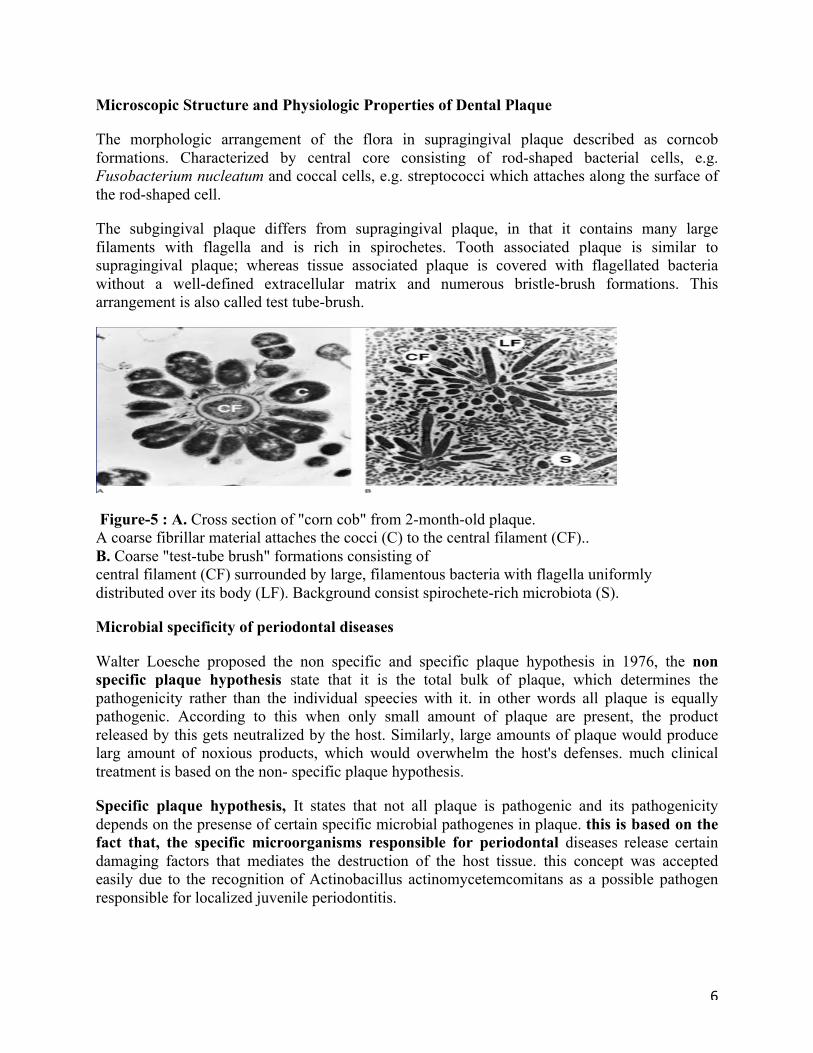
The morphologic arrangement of the flora in supragingival plaque described as corncob formations. Characterized by central core consisting of rod-shaped bacterial cells, e.g. Fusobacterium nucleatum and coccal cells, e.g. streptococci which attaches along the surface of the rod-shaped cell.
The subgingival plaque differs from supragingival plaque, in that it contains many large filaments with flagella and is rich in spirochetes. Tooth associated plaque is similar to supragingival plaque; whereas tissue associated plaque is covered with flagellated bacteria without a well-defined extracellular matrix and numerous bristle-brush formations. This arrangement is also called test tube-brush.
Figure-5 : A. Cross section of "corn cob" from 2-month-old plaque. A coarse fibrillar material attaches the cocci (C) to the central filament (CF).. B. Coarse "test-tube brush" formations consisting of central filament (CF) surrounded by large, filamentous bacteria with flagella uniformly distributed over its body (LF). Background consist spirochete-rich microbiota (S).
Microbial specificity of periodontal diseases
Walter Loesche proposed the non specific and specific plaque hypothesis in 1976, the non specific plaque hypothesis state that it is the total bulk of plaque, which determines the pathogenicity rather than the individual speecies with it. in other words all plaque is equally pathogenic. According to this when only small amount of plaque are present, the product released by this gets neutralized by the host. Similarly, large amounts of plaque would produce larg amount of noxious products, which would overwhelm the host's defenses. much clinical treatment is based on the non- specific plaque hypothesis.
Specific plaque hypothesis, It states that not all plaque is pathogenic and its pathogenicity depends on the presense of certain specific microbial pathogenes in plaque. this is based on the fact that, the specific microorganisms responsible for periodontal diseases release certain damaging factors that mediates the destruction of the host tissue. this concept was accepted easily due to the recognition of Actinobacillus actinomycetemcomitans as a possible pathogen responsible for localized juvenile periodontitis.
7
What make plaque pathogenic? the following are the possible pathogenic mechanisms by which the plaque microorganisims can couse periodontal disease.
1-physical nature of plaque
2-Invation of tissues by bacteria
3-Release of toxic and inflammatory substances
4- Role of bacterial specifity.
Bacterial associated with periodental health and disease
Health
Actinomyses (viscosus and naeslundii); Streptococcus(S. mitis and S. Sangius); Veillonella parvula, small amounts of gram-negative species are also found.
Chronic gingivitis S. sangius; S. mitis; S.oralis, A. viscosus, A. naeslundi, Peptostreptococcus micros. Gram -ve include Fusibacterium nulceatum, Prevotella intermedia, Veillonella parvula, Haemophilus, Capnocytophage and campylobacter species.
Pregnancy associated gingivitis: Prevotella intermedia
Acute necrotizing ulcerative gingivitis: Spirochetes; Prevotella intermedia
Adult periodontitis: Prphyromonas gingivales; Bacteriods forsythus; Prevotella intermedia; Campylobacter rectus; Eikenella corrodens; Fusobacterium nucleatum; Actinobacillus actinomycetemcomitans; Peptostreptococcus micros; Treponema and Eubacterium species. Viruses such as: EBV-1(Ebstein -Barr virus); HCMV (Human cytomegalovirus).
Localized juvenile periodontitis: Actinobacillus actinomycetemcomitans; Porphyromonas gingivalis; Eikenella corrodens; Campylobacter rectus; Fusobacterium nucleatum; Bacteroides capillus; Eubacterium brachy; capnocytophaga; Herpes virus.
Generalized juvenile periodontitis: Actinobacillus actinomycetemcomitans; Porphyromonas gingivalis; Prevotella intermedia, Capnocytophage; Eikenella corrodens; Nisseria .
Refractory periodontitis: Actinobacillus actinomycetemcomitans; Bacteroides forsythus
Porphyromonas gingivalis; Prevotella intermedia; Wolinella recta.
Abscesses of the periodontium: Fusobacterium nucleatum; Prevotella intermedia; Peptostreptococcus micros; Bacteroides forsythus and Porphyromonas gingivalis.

8
Prepared by
Prof. Dr. Raad Ali
A. Prof. Dr. Zainab Almahdi