periodontal ligament
TRANSCRIPT

PERIODONTAL LIGAMENT

CONTENTS
►Introduction►Cells►Fibers►Development►Biochemistry►Functions

PERIODONTAL LIGAMENT
► Functional unit of tissues that surrounds the teeth► It is composed of complex vascular and highly cellular
connective tissue that surrounds the tooth root and connects it to the inner wall of alveolar bone
► PDL space diminished in unerupted teeth and teeth that are not in function, increased in hyper function
► Thickness .15mm-.38mm, thinnest in middle portion.► Hour glass shape► Tissue with high turnover rate


DEVELOPMENT OF PDLDEVELOPMENT OF PDL
► After beginning of root formation, formation of After beginning of root formation, formation of outer dentinal layer of root, PDL is formedouter dentinal layer of root, PDL is formed
► Formation of hertwigs epithelial root sheathFormation of hertwigs epithelial root sheath► The root sheath is stretched and fragments to form The root sheath is stretched and fragments to form
cluster of cells called epithelial cell rests of cluster of cells called epithelial cell rests of malassezmalassez
► The enamel organ and root sheath are surrounded The enamel organ and root sheath are surrounded by dental follicle that is formed by condensed cellsby dental follicle that is formed by condensed cells
► The cells of dental follicle divide and differentiate The cells of dental follicle divide and differentiate into cementoblasts, fibroblasts, osteoblastsinto cementoblasts, fibroblasts, osteoblasts


1. Hertwigs root sheath
2. Epithelial cell rests3. Dental follicle4. Cementoblasts5. Periodontal ligament6. Alveolar cells7. Bone8. odontoblasts
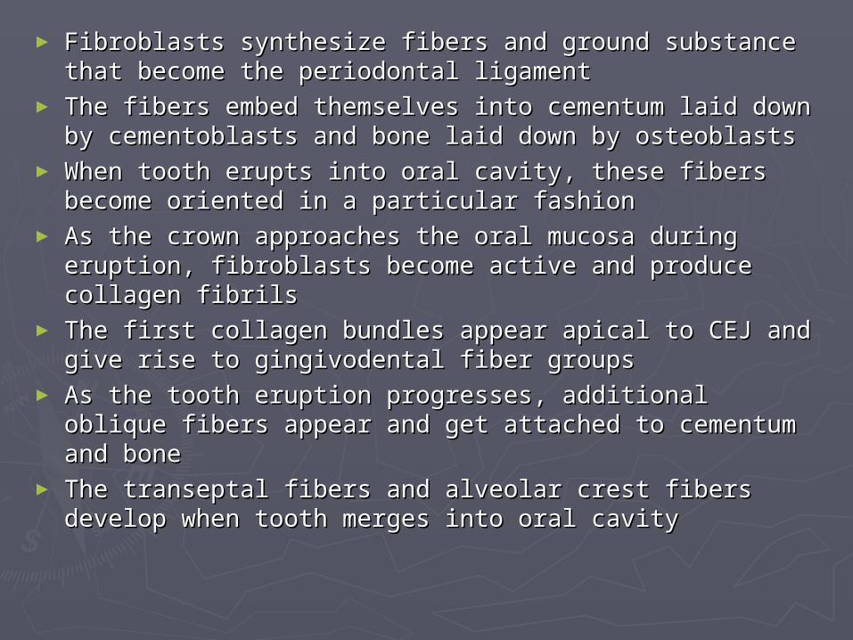
► Fibroblasts synthesize fibers and ground substance Fibroblasts synthesize fibers and ground substance that become the periodontal ligamentthat become the periodontal ligament
► The fibers embed themselves into cementum laid The fibers embed themselves into cementum laid down by cementoblasts and bone laid down by down by cementoblasts and bone laid down by osteoblastsosteoblasts
► When tooth erupts into oral cavity, these fibers When tooth erupts into oral cavity, these fibers become oriented in a particular fashionbecome oriented in a particular fashion
► As the crown approaches the oral mucosa during As the crown approaches the oral mucosa during eruption, fibroblasts become active and produce eruption, fibroblasts become active and produce collagen fibrilscollagen fibrils
► The first collagen bundles appear apical to CEJ and The first collagen bundles appear apical to CEJ and give rise to gingivodental fiber groupsgive rise to gingivodental fiber groups
► As the tooth eruption progresses, additional oblique As the tooth eruption progresses, additional oblique fibers appear and get attached to cementum and bonefibers appear and get attached to cementum and bone
► The transeptal fibers and alveolar crest fibers develop The transeptal fibers and alveolar crest fibers develop when tooth merges into oral cavitywhen tooth merges into oral cavity

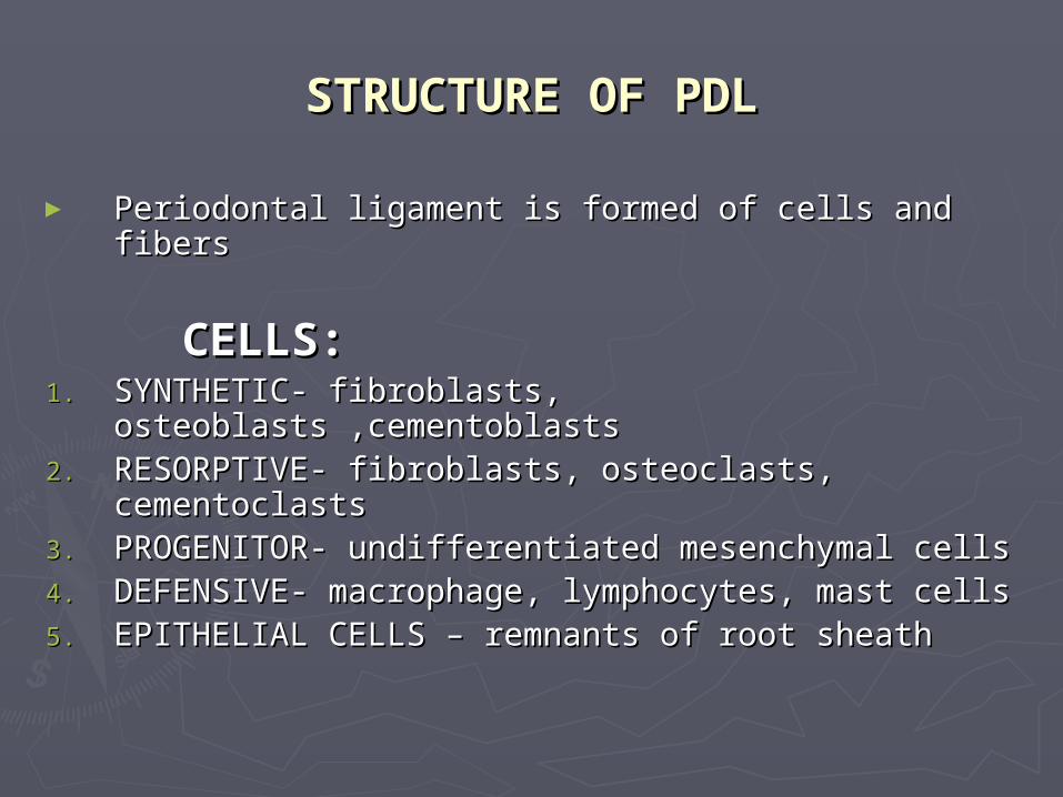
STRUCTURE OF PDLSTRUCTURE OF PDL
► Periodontal ligament is formed of cells and fibersPeriodontal ligament is formed of cells and fibers
CELLS:CELLS:1.1. SYNTHETIC- fibroblasts, osteoblasts ,cementoblastsSYNTHETIC- fibroblasts, osteoblasts ,cementoblasts2.2. RESORPTIVE- fibroblasts, osteoclasts, cementoclastsRESORPTIVE- fibroblasts, osteoclasts, cementoclasts3.3. PROGENITOR- undifferentiated mesenchymal cellsPROGENITOR- undifferentiated mesenchymal cells4.4. DEFENSIVE- macrophage, lymphocytes, mast cellsDEFENSIVE- macrophage, lymphocytes, mast cells5.5. EPITHELIAL CELLS – remnants of root sheathEPITHELIAL CELLS – remnants of root sheath


FIBROBLASTS
► Principal cells, lies between collagen fibers► Flattened irregular disc shaped, prominent nucleus► Contains numerous organelles associated with
protein synthesis and degradation► cells have a number of intercellular contacts► They produce extracellular matrix of PDL► Degradation of collagen occurs intracellularly and
extracellularly► Cells are responsible for connective tissue
morphogenesis, generating force of eruption by contraction.

CEMENTOBLASTSCEMENTOBLASTS
► Ovoid to cubical shape, basophilic cytoplasmOvoid to cubical shape, basophilic cytoplasm► responsible for secretion of organic matrix of cementumresponsible for secretion of organic matrix of cementum► When active, appear as a distinct layer on root surfaceWhen active, appear as a distinct layer on root surface
OSTEOBLASTSOSTEOBLASTS
► Osteoblasts are found on surface on alveolar boneOsteoblasts are found on surface on alveolar bone► When active, form a layer of cuboidal cellsWhen active, form a layer of cuboidal cells► As bone deposition proceeds, osteoblasts become incorporated in matrix as As bone deposition proceeds, osteoblasts become incorporated in matrix as
osteocytesosteocytes

EPITHELIAL CELL RESTSCEMENTOBLASTS
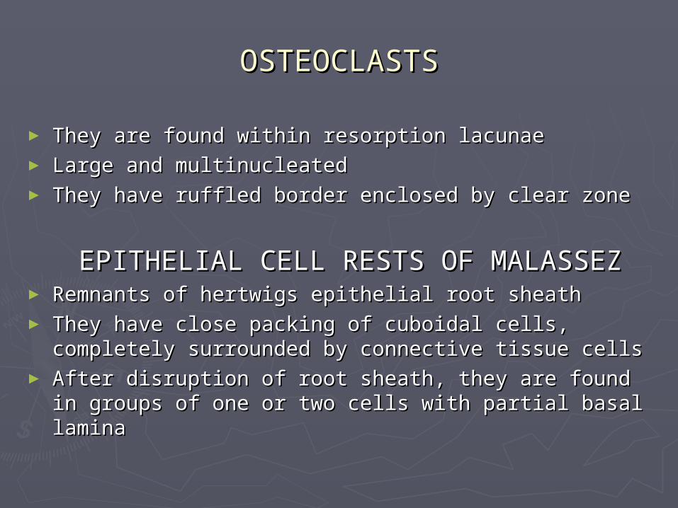
OSTEOCLASTSOSTEOCLASTS
► They are found within resorption lacunaeThey are found within resorption lacunae► Large and multinucleatedLarge and multinucleated► They have ruffled border enclosed by clear zoneThey have ruffled border enclosed by clear zone
EPITHELIAL CELL RESTS OF MALASSEZEPITHELIAL CELL RESTS OF MALASSEZ► Remnants of hertwigs epithelial root sheathRemnants of hertwigs epithelial root sheath► They have close packing of cuboidal cells, They have close packing of cuboidal cells,
completely surrounded by connective tissue cellscompletely surrounded by connective tissue cells► After disruption of root sheath, they are found in After disruption of root sheath, they are found in
groups of one or two cells with partial basal laminagroups of one or two cells with partial basal lamina

► Cell rests appear as separate, duct like cluster of Cell rests appear as separate, duct like cluster of cells or network of interconnecting strandscells or network of interconnecting strands
► Less numerous in older individuals, more in childrenLess numerous in older individuals, more in children► They have high nuclear cytoplasmic ratioThey have high nuclear cytoplasmic ratio► Upto second decade, found in apical region, later it Upto second decade, found in apical region, later it
is seen cervically in the gingivais seen cervically in the gingiva► They have a role in limiting resorption, They have a role in limiting resorption,
maintanence of periodontal spacemaintanence of periodontal space
UNDIFFERENTIATED MESENCHYMAL CELLSUNDIFFERENTIATED MESENCHYMAL CELLS
► Source of new cellsSource of new cells► Apoptosis- selective deletion of ligament cells, Apoptosis- selective deletion of ligament cells,
allows turnover without disruption of tissueallows turnover without disruption of tissue

DEFENSE CELLSDEFENSE CELLS
MACROPHAGE► defensive cell, phagocytic, mobile
LEUCOCYTE► Appear when stressed by disease
MAST CELLS► Defensive against pathogens
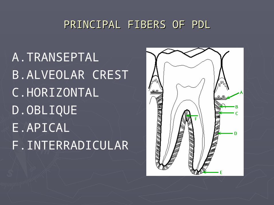
PRINCIPAL FIBERS OF PDLPRINCIPAL FIBERS OF PDL
A.TRANSEPTALB.ALVEOLAR CRESTC.HORIZONTALD.OBLIQUEE.APICALF.INTERRADICULAR

GINGIVAL FIBERS OF PDL
► A.CIRCULARA.CIRCULAR► B.DENTOGINGIVALB.DENTOGINGIVAL► C.DENTOPERIOSTALC.DENTOPERIOSTAL► D.ALVEOLOGINGIVAD.ALVEOLOGINGIVA
LL

COLLAGEN FIBERS OF PDLCOLLAGEN FIBERS OF PDL
► Collagen of PDL is largely type 1, with lesser amounts of Collagen of PDL is largely type 1, with lesser amounts of type 3, 4, 5, 6, 12type 3, 4, 5, 6, 12
► Collagen type 6 is a short chain molecule, it is a Collagen type 6 is a short chain molecule, it is a microfibril forming collagen that ramifies extracellular microfibril forming collagen that ramifies extracellular matrixmatrix
► Collagen type 4 is found in epithelial restsCollagen type 4 is found in epithelial rests► Fibers have structural requirements to withstand Fibers have structural requirements to withstand
intrusive forces from masticationintrusive forces from mastication► Collagen fibril diameters are relatively small, with mean Collagen fibril diameters are relatively small, with mean
diameters of 45- 55nm with unimodal distributiondiameters of 45- 55nm with unimodal distribution► Small diameter of fibrils is due to high rate of collagen Small diameter of fibrils is due to high rate of collagen
turnover and absence of mature collagen fibrilsturnover and absence of mature collagen fibrils

ELASTIC FIBERS OF PDLELASTIC FIBERS OF PDL
► Three fibrous components are oxytalan, elaunin, Three fibrous components are oxytalan, elaunin, elastinelastin
► Oxytalan fibers form a three dimensional Oxytalan fibers form a three dimensional network that connects cementum to blood network that connects cementum to blood vesselsvessels
► It is oriented in apico occlusal planeIt is oriented in apico occlusal plane► Oxytalan and elaunin fibers are precursors of Oxytalan and elaunin fibers are precursors of
elastin fiberelastin fiber► They are composed of microfilaments They are composed of microfilaments
surrounded by amorphous materialsurrounded by amorphous material► Elastin fibers are composed of elastin.Elastin fibers are composed of elastin.► Elastin is composed of microfibrillar glycoprotein Elastin is composed of microfibrillar glycoprotein

A.A. CEMENTUMCEMENTUM
B.B. OXYTALAN FIBEROXYTALAN FIBER
C.C. OXYTALAN TRACTOXYTALAN TRACT
D.D. PERIODONTAL PERIODONTAL VESSELVESSEL
FUNCTIONAL ADAPTATIONS OF COLLAGEN FUNCTIONAL ADAPTATIONS OF COLLAGEN FIBERSFIBERS
► Internal orientation of fibers influence mechanical Internal orientation of fibers influence mechanical properties of tissueproperties of tissue
► The arrangement into horizontal and oblique groups The arrangement into horizontal and oblique groups is to resist axial forcesis to resist axial forces
► The overlapping arrangement is to resist rotational The overlapping arrangement is to resist rotational and intrusive forcesand intrusive forces
► The complex three dimensional arrangement The complex three dimensional arrangement suggests that some bundles will be placed in suggests that some bundles will be placed in tension, this enables local areas of PDL to resist tension, this enables local areas of PDL to resist compressive forcescompressive forces
COLLAGEN CRIMPINGCOLLAGEN CRIMPING► Collagenous tissues exhibit a periodicity of structure Collagenous tissues exhibit a periodicity of structure
ranging from submicroscopic to anatomical, ranging from submicroscopic to anatomical, waveform that describes this periodicity is referred to waveform that describes this periodicity is referred to an crimpan crimp
► It is due to sharp zigzag arrangement of collagen It is due to sharp zigzag arrangement of collagen fibrils and due to microanatomical organisation of fibrils and due to microanatomical organisation of sheets and bundlessheets and bundles
► It enables the ligament to absorb tensile loads It enables the ligament to absorb tensile loads without extending collagen fibrilswithout extending collagen fibrils
SHARPEYS FIBERSSHARPEYS FIBERS► Collagen bundles of PDL are embedded into Collagen bundles of PDL are embedded into
cementum and bone - sharpeys fiberscementum and bone - sharpeys fibers► These fibers are enclosed in a sheath of type 3 These fibers are enclosed in a sheath of type 3
collagen and is concentrated in crestal regioncollagen and is concentrated in crestal region
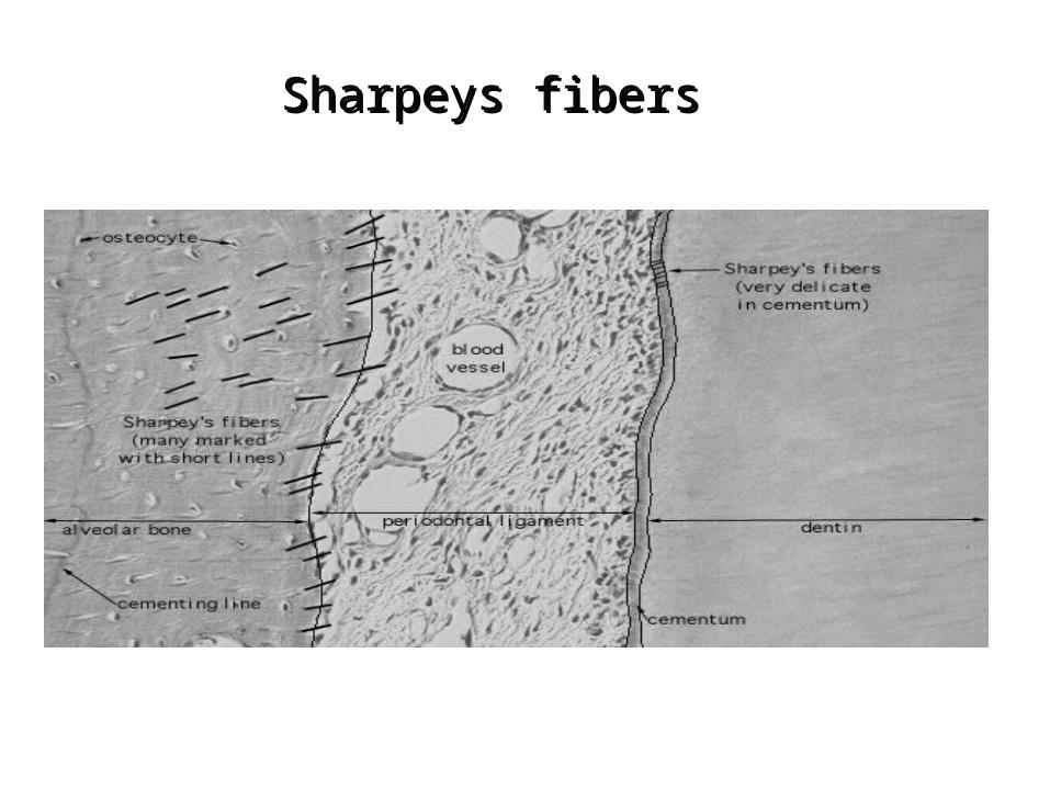
Sharpeys fibersSharpeys fibers
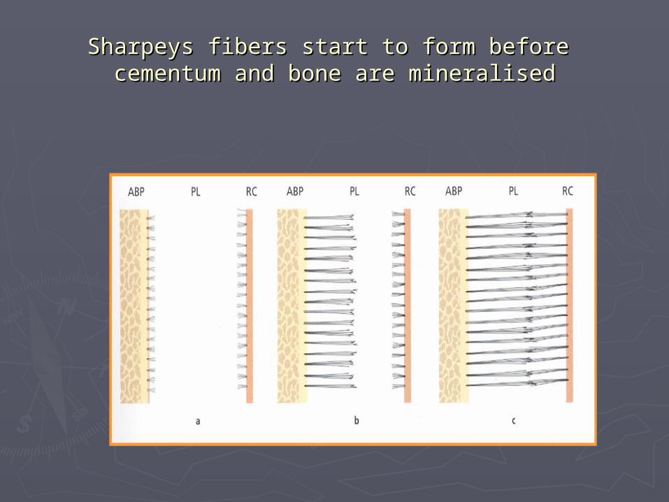
Sharpeys fibers start to form before Sharpeys fibers start to form before cementum and bone are mineralisedcementum and bone are mineralised

FUNCTIONAL ADAPTATIONS OF ELASTIC FUNCTIONAL ADAPTATIONS OF ELASTIC FIBERSFIBERS
► Elastic fibers are more seen in coronal one Elastic fibers are more seen in coronal one thirdthird
► Elastic fibers dampen lateral masticatory Elastic fibers dampen lateral masticatory stresses, low modulus and long range stresses, low modulus and long range reversible extensibility of elastin is more reversible extensibility of elastin is more important than resilience as physical propertyimportant than resilience as physical property
► Oxytalan fibers have a role in stabilizing the Oxytalan fibers have a role in stabilizing the tooth in functional situationstooth in functional situations
► Oxytalan and collagen bundles interweave at Oxytalan and collagen bundles interweave at right angles to each otherright angles to each other
► Oxytalan fibers act as a part of Oxytalan fibers act as a part of mechanoreceptor systemmechanoreceptor system

BIOCHEMISTRY OF FIBERSBIOCHEMISTRY OF FIBERS
► Collagen fibers are unique in their Collagen fibers are unique in their arrangement, rapid assimilation into fibrils arrangement, rapid assimilation into fibrils compared to other connective tissuescompared to other connective tissues
► Minor fibers comprise oxytalan, elaunin but Minor fibers comprise oxytalan, elaunin but no mature elastic fibers indicating a foetal no mature elastic fibers indicating a foetal like connective tissuelike connective tissue
► Collagen is a protein composed of Collagen is a protein composed of aminoacids like glycine , proline, hydroxy aminoacids like glycine , proline, hydroxy lysine, hydroxy prolinelysine, hydroxy proline
► Collagen biosynthesis occurs inside Collagen biosynthesis occurs inside fibroblasts to form tropocollagen moleculesfibroblasts to form tropocollagen molecules

COLLAGEN BIOSYNTHESISCOLLAGEN BIOSYNTHESIS
► They aggregated to form microfibrils that are They aggregated to form microfibrils that are packed together to form fibrils.packed together to form fibrils.
► Fibrils associate to form fiber and bundleFibrils associate to form fiber and bundle► Oxytalan fibers are derived from microfibrillar Oxytalan fibers are derived from microfibrillar
proteinsproteins► The biochemistry of fibers suggest that this is an The biochemistry of fibers suggest that this is an
unusual connective tissue with foetal like unusual connective tissue with foetal like characteristicscharacteristics
► This may be related to tissue functionThis may be related to tissue function

FUNCTIONS OF PDLFUNCTIONS OF PDL
PHYSICAL FUNCTIONSPHYSICAL FUNCTIONS► PDL serves as a shock absorber by providing PDL serves as a shock absorber by providing
resistance to forcesresistance to forces► Transmission of occlusal forces to boneTransmission of occlusal forces to bone► Protection of vessels and nervesProtection of vessels and nerves► Attachment of teeth to boneAttachment of teeth to bone► Maintenance of gingival positionMaintenance of gingival position
THEORIES REGARDING MECHANICALTHEORIES REGARDING MECHANICAL FUNCTIONFUNCTION
► Tensional theoryTensional theory► Viscoelastic theoryViscoelastic theory

TENSIONAL THEORYTENSIONAL THEORY
► Initially there is gradual Initially there is gradual unfolding of fiber bundles unfolding of fiber bundles before taking up the tensionbefore taking up the tension
► Secondly, fiber bundles Secondly, fiber bundles straighten transmitting straighten transmitting forces to alveolar boneforces to alveolar bone
► In the final phase, the In the final phase, the straightened fibers deliver straightened fibers deliver increasing load to the increasing load to the alveolar bone which is then alveolar bone which is then transmitted to the basal transmitted to the basal
bone of the jawsbone of the jaws..
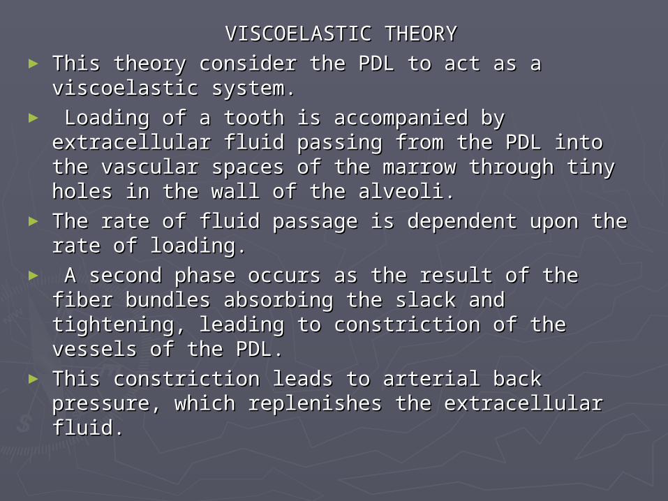
VISCOELASTIC THEORYVISCOELASTIC THEORY► This theory consider the PDL to act as a viscoelastic This theory consider the PDL to act as a viscoelastic
system.system.► Loading of a tooth is accompanied by extracellular Loading of a tooth is accompanied by extracellular
fluid passing from the PDL into the vascular spaces fluid passing from the PDL into the vascular spaces of the marrow through tiny holes in the wall of the of the marrow through tiny holes in the wall of the alveoli. alveoli.
► The rate of fluid passage is dependent upon the The rate of fluid passage is dependent upon the rate of loading.rate of loading.
► A second phase occurs as the result of the fiber A second phase occurs as the result of the fiber bundles absorbing the slack and tightening, leading bundles absorbing the slack and tightening, leading to constriction of the vessels of the PDL. to constriction of the vessels of the PDL.
► This constriction leads to arterial back pressure, This constriction leads to arterial back pressure, which replenishes the extracellular fluid. which replenishes the extracellular fluid.

REMODELLING FUNCTIONREMODELLING FUNCTION► PDL serves by providing cells that are able to form as PDL serves by providing cells that are able to form as
well as resorb tissues that form the attachment well as resorb tissues that form the attachment apparatus apparatus
► Undifferentiated mesenchymal cells differentiate into Undifferentiated mesenchymal cells differentiate into osteoblasts, cementoblasts, fibroblastsosteoblasts, cementoblasts, fibroblasts
► Osteoclasts are derived from macrophagesOsteoclasts are derived from macrophages
NUTRITIVE FUNCTIONNUTRITIVE FUNCTION► It has a nutritive function that maintains the vitality of It has a nutritive function that maintains the vitality of
various cellsvarious cells
SENSORY FUNCTIONSENSORY FUNCTION► Attributed to mechano receptors in PDLAttributed to mechano receptors in PDL

► Mechanoreceptors are organised or ruffini endingsMechanoreceptors are organised or ruffini endings► They have a role in touch thresholds, masticatory They have a role in touch thresholds, masticatory
salivary reflex, reflexes of masticatory musclessalivary reflex, reflexes of masticatory muscles
ROLE IN GENERATION OF ERUPTIVE FORCEROLE IN GENERATION OF ERUPTIVE FORCE► Periodontal collagen contraction hypothesisPeriodontal collagen contraction hypothesis► Periodontal fibroblast traction hypothesisPeriodontal fibroblast traction hypothesis► Vascular and tissue hydrostatic pressure hypothesisVascular and tissue hydrostatic pressure hypothesis
ROLE IN PERIODONTAL WOUND HEALINGROLE IN PERIODONTAL WOUND HEALING► PDL generates new fibers, prevents apical migration PDL generates new fibers, prevents apical migration
of epithelium, contributes cells to restore cementum, of epithelium, contributes cells to restore cementum, acts as a biological sensor to regulate its own widthacts as a biological sensor to regulate its own width
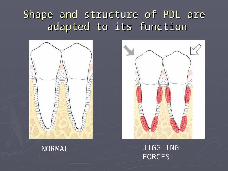
Shape and structure of PDL areShape and structure of PDL are adapted to its function adapted to its function
NORMAL JIGGLING FORCES

WIDENED PDL ADAPTATION
AFTER OCCLUSAL ADJUSTMENT