periodontal disease and the oral microbiota in newonset ... · abstract (250 words) objective. to...
TRANSCRIPT
Periodontal Disease and the Oral Microbiota in New-Onset
Rheumatoid Arthritis
Jose U. Scher,1* Carles Ubeda,2,5* Michele Equinda,2 Raya Khanin,2 Yvonne
Buischi,3 Agnes Viale,2 Lauren Lipuma,2 Mukundan Attur,1 Michael H. Pillinger,1
Gerald Weissmann,4 Dan R. Littman,4 Eric G. Pamer,2 Walter A. Bretz,3 and Steven
B. Abramson1
1Jose U. Scher, MD, Mukundan Attur, PhD, Michael H. Pillinger, MD, Steven B.
Abramson, MD: New York University School of Medicine and NYU Hospital for Joint
Diseases, New York, New York; 2Carles Ubeda, PhD, Michele Equinda, BS, Raya
Khanin, PhD, MSc, Agnes Viale, PhD, Lauren Lipuma, MS, Eric G. Pamer, MD:
Memorial Sloan-Kettering Cancer Center, The Lucille Castori Center for Microbes,
Inflammation and Cancer, New York, New York; 3Yvonne Buischi, DDS, PhD, Walter A.
Bretz, DDS, DrPH: NYU College of Dentistry, New York, New York; 4Gerald Weissmann,
MD, Dan R. Littman, MD, PhD: New York University School of Medicine, New York,
New York; 5Carles Ubeda, PhD, current address: Department of Genomics and Health,
Center for Advanced Research in Public Health, Valencia, Spain.
* Drs. Scher and Ubeda contributed equally to this work.
ClinicalTrials.gov identifier: NCT01198509
Supported by Grant No. RC2 AR058986 to Drs. Abramson and Littman from the
National Institute of Arthritis and Musculoskeletal and Skin Diseases (NIAMS) through
the American Recovery and Reinvestment Act (ARRA) of 2009. and by a KL2 Program
in Translational Research to Dr. Scher, Grant No. 1 UL1 RR029893 from the National
Center for Research Resources, NIH. The Lucille Castori Center for Microbes,
Inflammation, and Cancer, at Memorial Sloan-Kettering Cancer Center, is supported by
the Tow Foundation.
Address correspondence to: Jose U. Scher, M.D. Division of Rheumatology NYU Hospital for Joint Diseases 301 East 17th Street, Room 1611 New York, NY 10003 Ph: 212-598-6513 Email: [email protected]
Word count: 4,180
Running title: Oral Microbiota and Periodontal Status in RA
Full Length Arthritis & RheumatismDOI 10.1002/art.34539
© 2012 American College of RheumatologyReceived: Sep 29, 2011; Revised: Feb 28, 2012; Accepted: May 03, 2012
This article has been accepted for publication and undergone full peer review but has not beenthrough the copyediting, typesetting, pagination and proofreading process which may lead todifferences between this version and the Version of Record. Please cite this article as an ‘Accepted Article’, doi: 10.1002/art.34539

ABSTRACT (250 words)
Objective. To profile the subgingival oral microbiota abundance and diversity in
never-treated, new-onset rheumatoid arthritis (NORA) patients.
Methods. Periodontal disease (PD) status, clinical activity and sociodemographic
factors were determined in patients with NORA, chronic RA (CRA) and healthy subjects.
Massively parallel pyrosequencing was used to compare the composition of subgingival
microbiota and establish correlations between presence/abundance of bacteria and
disease phenotypes. Anti-P. gingivalis antibodies were tested to assess prior exposure.
Results. The more advanced forms of periodontitis are already present at
disease onset in NORA patients. The subgingival microbiota of NORA is distinct from
controls. In most cases, however, these differences can be attributed to PD severity and
are not inherent to RA. The presence and abundance of P. gingivalis is directly
associated with PD severity as well, is not unique to RA, and does not correlate with
anti-citrullinated peptide antibody (ACPA) titers. Overall exposure to P. gingivalis is
similar in RA and controls, observed in 78.4% and 83.3%, respectively. Anaeroglobus
geminatus correlated with ACPA/RF presence. Prevotella and Leptotrichia species are
the only characteristic taxa in the NORA group irrespective of PD status.
Conclusions. NORA patients exhibit a high prevalence of PD at disease onset,
despite their young age and paucity of smoking history. The subgingival microbiota of
NORA patients is similar to CRA and healthy subjects of comparable PD severity.
Although colonization with P. gingivalis correlates with PD severity, overall exposure is
similar among groups. The role of A. geminatus and Prevotella/Leptotrichia species in
this process merits further study.
of 34
John Wiley & Sons
Arthritis & Rheumatism
INTRODUCTION
The term “microbiome” was coined a decade ago (1) and implies the totality of
microbes (commensal and pathogenic), their genomes, and environmental interactions
in a defined biological niche. In this symbiotic relationship, humans provide nutrients
and an adequate environment for microorganisms that, in return, shape the human
immune system, degrade polysaccharides and produce vitamins and other essential
factors we would be otherwise unable to obtain. In 2008, the NIH Human Microbiome
Project (2) embraced the notion that it is impossible to fully understand human health
and disease unless this collective human-microbiome “superorganism” is better studied
and defined.
Rheumatoid arthritis (RA) is a systemic, inflammatory autoimmune disorder. It is
regarded as a complex multifactorial disease, in which multiple genes and
environmental factors act in concert to cause pathological events (3). Despite recent
advances in molecular pathogenesis its etiology is almost completely unknown.
Although genes certainly contribute to RA susceptibility (4), genetic effects appear to
require environmental factors (i.e., smoking, hormones, and infection) in order to explain
differences in incidence of the disease (5).
Among the more intriguing environmental covariates modulating autoimmunity is
the bidirectional crosstalk between the human host and the oral and intestinal
microbiomes. Multiple lines of investigation have suggested a link between oral
microbes, periodontal diseases (PD) and RA (6;7). However, most clinical studies
implicating specific oral microorgansims as triggers for RA have relied only on
serological methods. Data describing the subgingival microbiota in patients with RA is
Page 3 of 34
John Wiley & Sons
Arthritis & Rheumatism

virtually non-existent.
In the present study, we aimed to determine the periodontal status of RA patients
and healthy controls and to directly correlate, for the first time, the subgingival
microbiota with RA status utilizing 16S rRNA pyrosequencing. Because we wanted to
understand whether specific oral microbiota is associated with the initiation of RA, we
focused our attention on patients with new-onset RA (NORA) who were steroid- and
DMARD-naïve at the time of enrollment.
PATIENTS AND METHODS
Study participants. Consecutive patients from the NYU Rheumatology clinics
and offices were screened for the presence of RA. After informed consent was signed,
past medical history (chart review and interview/questionnaire), diet and medications
were determined. A screening musculoskeletal exam and laboratory assessments were
also performed or reviewed; all RA patients who met study criteria were offered
enrollment. Inclusion criteria involved RA patients meeting 2010 ACR/EULAR criteria for
RA, including presence of rheumatoid factor (RF) and/or anti-citrullinated peptide
antibodies (ACPA; Anti-CCP ELISA, EUROIMMUN), and age 18 years or older. New-
onset rheumatoid arthritis (NORA) was defined as disease duration of >6 weeks and
absence of any treatment with disease-modifying anti-rheumatic drug (DMARD) or
steroids (ever). Chronic-established RA (CRA) was defined as any patient meeting
criteria for RA with minimum disease duration of 6 months. Most CRA subjects were
receiving DMARDs (oral and/or biologic agents) and/or corticosteroids at the time of
enrollment. Healthy controls were age-, sex- and ethnicity-matched individuals with no
of 34
John Wiley & Sons
Arthritis & Rheumatism

personal history of inflammatory arthritis. Exclusion criteria for all groups were: recent
(<3 months prior) use of any antibiotic therapy; current extreme diet (parenteral nutrition,
macrobiotic diet, etc.); known inflammatory bowel disease; known history of malignancy;
current consumption of probiotics; any GI tract surgery leaving permanent residua (e.g.,
gastrectomy, bariatric surgery, colectomy); significant liver, renal or peptic ulcer disease.
This study was approved by the Institutional Review Board (IRB) of New York University
School of Medicine.
Classification of periodontal diseases. All periodontal examinations were
performed at the NYU College of Dentistry. Periodontal status was assessed by three
calibrated examiners (blinded for RA status) and defined according to the American
Academy of Periodontology (AAP) (8). The following parameters were recorded:
probing depth (Prob), clinical attachment level (AL), and bleeding on probing (BoP).
Patients and controls were classified as: 1) Healthy, no bleeding upon probing; 2)
Gingivitis with bleeding upon probing; 3) Slight chronic periodontitis (at least one
periodontal site with 1-2 mm AL and ≥4 mm Prob); 4) Moderate chronic periodontitis (at
least two teeth with 3-4 mm AL or at least 2 teeth with ≥4 mm Prob); 5) Severe chronic
periodontitis (at least two teeth with ≥5 mm AL and one tooth with ≥5 mm Prob).
In all, 31 NORA patients, 34 CRA patients, and 18 healthy controls were
available for the analyses presented here.
Sample collection and DNA extraction. Oral samples were obtained by
collection of subgingival biofilm from the six most periodontally diseased sites of all
patients. Oral samples were harvested using a Gracey curette (after removal and
discard of supragingival biofilm to avoid potential salivary contamination). All samples
Page 5 of 34
John Wiley & Sons
Arthritis & Rheumatism
were pooled and directly suspended in MoBio buffer-containing tubes (MoBio). DNA
was extracted within 1 hr of sample collection using a combination of the MoBio Power
Soil kit (MoBio) and a mechanical disruption (bead-beater) method based on a
previously described protocol (9). Samples were stored at –80°C.
V1-V2 16S rRNA region amplification and 454/pyrosequencing. For each
sample, 3 replicate PCRs were performed to amplify the V1 and V2 regions as
previously described by Ubeda et. al. (10). PCR products were sequenced on a 454 GS
FLX Titanium platform (454 Roche). Sequences have been deposited in the NCBI
Sequence Read Archive under the accession number SRA050292.
Sequence analysis. Sequence data were compiled and processed using mothur
(11) and converted to standard FASTA format. Sequences were trimmed and aligned to
the V1-V2 region of the 16S gene, using as template the SILVA reference alignment
(12) Potentially chimeric sequences were removed using ChimeraSlayer (13). To
minimize the effect of pyrosequencing errors in overestimating microbial diversity (14),
low abundance sequences were merged to the high abundant sequence using the
pre.cluster option in mothur. Sequences were grouped into operational taxonomic units
(OTUs) using the average neighbor algorithm. Sequences with distance-based similarity
of ≥97% were assigned to the same OTU. For each sample, OTU-based microbial
diversity was estimated by calculating the Shannon diversity and the Simpson diversity
indexes (15) and richness was estimated using the Chao index. Yue and Clayton
diversity measure and Principal Coordinate of Analysis were performed using Mothur.
Phylogenetic classification was performed using the Bayesian classifier algorithm with
the bootstrap cutoff 60% (16).
of 34
John Wiley & Sons
Arthritis & Rheumatism
Serum ELISA for anti-HtpG P18γγγγ peptide antibodies. P. gingivalis HtpG
peptides were prepared in the laboratories of Drs. Sweier and Shelbourne (University of
Michigan School of Dentistry, Ann Arbor) as previously described (17) and loaded into
the wells of microtiter plates. 25 µl of each serum sample was added, incubated with
goat anti-human IgG (γ-chain specific) and analyzed according to protocol.
Statistical analyses. To determine statistically significant differences between
samples from disease and healthy individuals, bacteria with <5 mean count in both
conditions were removed, and t-test was applied to log2 transformed scaled count-data,
and rescaled using DESeq R package (18). To adjust for multiple hypothesis testing, we
employed the False Discovery Rate (FDR) approach (19), and used fdr.R package. The
final results were filtered for p value < 0.05 and a FDR ≤ 0.1.
For cross-sectional analyses of baseline characteristics, differences were
evaluated using Student’s t test, Mann–Whitney U test or chi-squared tests, when
appropriate. SPSS V.16.0 software (SPSS, Chicago, Illinois, USA) was used for the
analysis, two-tailed significance testing was employed and significance was set as
p<0.05.
RESULTS
New-onset rheumatoid arthritis patients present with advanced periodontal
disease. Of 31 NORA subjects included in this study, 68% were females with a mean
age of 42.2 years (Table 1). Mean disease duration was 3.4 months (median 2 months)
and no patient had ever received steroids, oral DMARDs or biologics. Mean disease
activity score 28 (DAS28) was 5.8. In concordance with inclusion criteria all patients
Page 7 of 34
John Wiley & Sons
Arthritis & Rheumatism
were “seropositive”: 96% of NORA subjects were ACPA seropositive and 92% were RF
positive; more than two thirds had never smoked tobacco (16% of participants were
current smokers) (Table 1). Healthy controls were age-, sex- and ethnicity-matched.
The CRA group had proportionally more female participants; mean age for this group
was 47.7 years (p=NS compared to NORA) and 88% were ACPA positive. Mean
disease duration was 62.9 months (median 34) and the mean DAS28 was 4.4, reflecting
moderate disease presumably altered by DMARD intervention; 70% of CRA subjects
had no history of smoking.
As shown in Table 1, more than 75% of NORA and CRA patients were found to
have moderate to severe forms of PD, a significantly higher proportion when compared
to healthy controls. The prevalence of periodontal disease in our healthy controls was
consistent with the expected prevalence (30-40%) of extensive PD in the general
population (20). An interesting finding was the presence of PD in several of our NORA
subjects younger than 30 years of age without PD risk factors, such as smoking, when
periodontitis is typically absent.
The oral microbiota is equally rich and diverse in NORA, CRA and control
groups. Overall, 83 oral samples were obtained from all participants yielding a total of
206,378 16S RNA high-quality sequences (average 2037 sequences/ sample; range
443-5008 reads; p=NS). Using a distance-based similarity of >97% for species-level
operational taxonomic unit (OTU) assignment, a total of 2136 OTUs were identified.
(supplemental Figure 1A).
We first studied the impact of RA status in microbial diversity by using the
Inverse Simpson and Shannon indexes. Both take into account, when calculating the
of 34
John Wiley & Sons
Arthritis & Rheumatism
diversity of a sample, not only the number of OTUs (~species) present but also the
relative frequency of the different OTUs within that sample. A high index reflects a more
diverse microbiota. Utilizing both calculations, no significant differences in microbial
diversity were observed between RA groups and controls (supplemental Figures 1C and
1D). We then analyzed if RA status had an impact on microbial richness. When applying
the Chao index (which estimates how many OTUs constitute the microbiota of a specific
sample), no significant differences were found among groups (supplemental Figure 1D).
In an attempt to discriminate among study groups, we also performed clustering
analyses at the various taxonomic levels. Although certain significant differences were
found, no particular oral bacterial phylum, class, order or family was able to discriminate
between NORA, CRA or healthy groups. Rather, differences were evident when groups
were combined by periodontal disease severity. As previously described by others
(21;22), our assessment showed that the healthy periodontal microbiota is dominated
by 7 phyla, including Bacteroidetes (21.3%), Firmicutes (10.9%), Actinobacteria (21.8%),
Proteobacteria (16.9%), Fusobacteria (24%), Spirochaetes (2.5%) and TM7 (1.6%)
(Figure 1A). The moderate to severe forms of PD revealed an increase in the relative
abundance of Bacteroidetes, Spirochaetes and TM7, and a concomitant decrease in
Actinobacteria and Proteobacteria (Figure 1B).
To further analyze if NORA microbiota was distinct from that of healthy controls,
we applied the Yue and Clayton diversity measure (which compares the relative
abundance of OTUs present in different samples). We then applied PCoA to the
quantified similarity distances between samples and clustered them along orthogonal
axes of maximal variance. Two principal coordinates (PC1 and PC2) explain most of the
Page 9 of 34
John Wiley & Sons
Arthritis & Rheumatism

variation observed between the samples. No clustering due to RA status could be
observed (Figure 1C). However, as shown in Figure 1D, PC1 did cluster a group of
samples (circle) obtained from patients with severe and moderate periodontitis. This
result suggests that differences at the OTU level characterize the more advanced forms
of PD and do not represent a specific signature for RA oral microbiota.
Prevotella and Leptotrichia species are characteristic of the NORA oral
microbiota. We next sought to identify a bacterium or groups of bacteria responsible for
the clustering that identifies patients with advanced PD. Applying multivariate statistical
analyses taking into consideration patient groups, we also looked for bacterial taxa that
were significantly different in the NORA group (either increased or decreased)
compared to the others. At most taxonomic levels the oral microbiota of NORA patients
is not significantly different from that of other groups (Table 2 and Supplementary Table).
However, the genera Corynebacterium and Streptococcus are underrepresented in RA
subjects, which reflects the lack of a healthy microbiota, and is therefore consistent with
PD, per se. Interestingly, OTU 60 (Prevotella spp.) and OTU 87 (Leptotrichia spp.) are
the only characteristic taxa in the NORA group irrespective of PD status (present in
32.2% and 25.8% of patients, respectively), and are completely absent in the oral
microbiota of controls.
Abundance of periodontopathic bacteria is high in NORA but diminished in
the oral microbiota of CRA. To directly survey the presence of bacteria associated
with the development of PD in patients with RA, we next examined how these
phylotypes differed in the early and late phases of RA. Interestingly, OTU members of
the Red Complex Bacteria (23) (a triad of the most virulent periodontopathic bacteria
0 of 34
John Wiley & Sons
Arthritis & Rheumatism
including Porphyromonas, Tannerella and Treponema) are more prevalent in NORA
microbiota compared to CRA patients (Figure 2).
The genus Porphyromonas and P. gingivalis-related OTUs are significantly
associated with PD severity and are not specific of the RA microbiota. Although
with variability in level of abundance, the genus Porphyromonas was present almost
universally in all participants (Figures 3A and 3C).
We analyzed the 2136 different OTUs among all patient groups (including 59
OTUs within Porphyromonas), and found that OTU 1, with 100% 16S rRNA sequence
homology to P. gingivalis, was significantly more prevalent and abundant in patients
with PD (more than 60-fold increase in the severe forms compared to healthy gums)
and had no direct correlation to RA (Figures 3B and 3D). P. gingivalis was present in
55% of NORA and 47% of CRA patients, while the prevalence in healthy controls was
27% (p=0.18, ANOVA). We further stratified NORA patients into two categories
according to presence or absence of PD and found that P. gingivalis was also more
prevalent (62.5% vs 28.5%; p<0.05) and abundant (mean 6.2% vs 0.78%; p<0.05) in
the advanced PD group (supplemental Figure 2). Many other OTUs known to be
associated to PD showed similar elevations, although less pronounced (data not shown).
Taken together, these data suggest that although colonization of P. gingivalis is twice as
common in RA patients compared to controls, the difference can be explained by the
higher prevalence of PD in the RA population.
Exposure to P. gingivalis is not significantly different among groups.
Because the absence of P. gingivalis in the oral microbiota did not exclude prior
exposure to the organism, we tested a previously validated antibody assay against the
Page 11 of 34
John Wiley & Sons
Arthritis & Rheumatism

highly specific P. gingivalis chaperone HtpG (IgG class anti-P18γ). We found that 63.3%
of NORA patients, 50% of CRA patients and 72.2% of healthy controls tested positive
for anti-P18γ (p=0.45, ANOVA; Figure 4A). We next analyzed overall exposure to P.
gingivalis based upon either P. gingivalis colonization by 16s and/or a positive antibody
response. Interestingly, we found that while the NORA and CRA groups had a rate of
exposure of 84% and 71%, respectively, healthy individuals also revealed an exposure
prevalence of 83% (N.S., ANOVA; Figure 4B), largely due to an increased serological
response.
Presence of circulating RA-related autoantibodies correlates with
Anaeroglobus, an unusual bacterial taxon. Proposed mechanisms through which P.
gingivalis might promote the pathogenesis of RA include its capacity to citrullinate
peptides via the enzyme peptidylarginine deiminase (PAD), theoretically promoting
generation of neoantigens and subsequent production of ACPA (24;25). We therefore
examined the different taxonomic levels to look for phylotypes associated with
circulating autoantibodies. There was no association between RF or ACPA with the
higher taxonomic levels (phylum, class, order or family). In particular, there was no
association between RF or ACPA and the presence of neither the genus
Porphyromonas in the oral microbiota nor OTUs related to P. gingivalis. Indeed, several
patients with ACPA lacked P. gingivalis-related OTUs and the autoantibody titer was not
positively associated with the genera abundance (data not shown). Unexpectedly,
however, the presence of the genus Anaeroglobus and its species-level OTU99 (closely
related to Anaeroglobus geminatus) significantly correlated with both circulating RF and
ACPA (P <0.05). Moreover, OTU99 was associated with PD, and found in 77.5% and
2 of 34
John Wiley & Sons
Arthritis & Rheumatism

50% of NORA and CRA patients, respectively (p=NS), and only 16.7% of healthy
controls (P <0.005 vs. NORA or CRA). OTU130, with 94% similarity to Porphyromonas
catoniae, was also significantly associated with circulating ACPA.
DISCUSSION
An accumulating body of epidemiological data suggests a role of clinical
periodontal diseases in the development of RA. In concordance with our findings,
periodontitis was more common and severe in patients with RA compared to patients
with OA in a cohort of U.S. veterans (26). In another study (27), RA patients had an 8-
fold increased likelihood of periodontitis compared to controls. Multiple recent studies
have also implicated P. gingivalis as a possible triggering factor. Interestingly, however,
none of these reports have directly looked at the presence of oral microorganisms.
Rather, they relied upon serological methods (28;29) or limited, low-throughput PCR-
based techniques (30;31). To our knowledge, no prior report has specifically assessed
the presence of P. gingivalis (or other periodontopathic bacteria) in subgingival biofilms
in RA patients.
Our study is the first utilizing multiplexed-454 pyrosequencing to compare the
bacterial composition of the subgingival microbiota in RA (early and chronic) and
controls. This approach permitted a broad and comprehensive portrayal of the
subgingival microbial communities associated with RA at different stages of the disease.
Several conclusions can be drawn from our data:
First, we corroborated previous observations that early RA patients present with
incident PD (32). Our data are striking in that a high prevalence of moderate to severe
Page 13 of 34
John Wiley & Sons
Arthritis & Rheumatism
periodontitis is observed in a steroid- and DMARD-naïve population, a finding reported
here for the first time in this unique untreated cohort. Moreover, our NORA cohort is
mostly composed of young non-smokers, whereas smoking is otherwise a significant
risk indicator of periodontal diseases (33;34) and has been proposed as a central driver
of gene-environment interaction in seropositive RA (35). These results are consistent
with the notion that PD, present at the time of diagnosis in the majority of NORA
patients, may represent a risk factor for RA development independent from smoking
status. Intriguingly, some periodontopathic OTUs (e.g., Tannerella/OTU13,
Treponema/OTU32) were significantly higher in NORA microbiota and tended to
diminish with established better-controlled disease (CRA). We speculate that this
difference could result from RA therapeutic regimens over time. It is conceivable that a
variety of immunomodulatory regimens -particularly those with proposed antibacterial
properties, such as methotrexate or hydroxychloroquine (36;37)- have an impact on the
ecological adaptation of the oral microbial niche.
Second, we found that the subgingival microbial communities of NORA patients
generally do not have a unique fingerprinting compared to controls. However, two
OTUs, OTU 60/Prevotella spp. and OTU 87/Leptotrichia spp., were detected only in the
NORA population. Although the genus Porphyromonas is present in virtually all subjects,
its relative abundance is directly correlated with PD severity, regardless of RA status.
We corroborated prior findings using low-throughput techniques (23;38;39) that the
advanced forms of PD are overrepresented by genera such as Porphyromonas,
Tannerella and Treponema, all of which have been implicated in PD pathogenesis (23).
Our ability to go beyond the genus taxonomy has also allowed us to investigate the
4 of 34
John Wiley & Sons
Arthritis & Rheumatism

OTUs within the genus Porphyromonas. The most abundant OTU within the genus was
identical to P. gingivalis. Interestingly, none of the other OTUs whose 16S was similar to
P. gingivalis were significantly overrepresented in the subgingival oral microbiota of
NORA patients. Our data demonstrate that while most individuals carry Porphyromonas
in their subgingival domain, a particular OTU (OTU1) is mostly found in advanced PD,
whether in RA patients or otherwise healthy controls. However, and given the low
number of non-RA PD subjects in our study, we could not categorically establish
whether bacterial exposure can be attributed to the presence of subgingival
inflammation alone. It is quite possible that P. gingivalis may serve as a shared causal
pathway in some cases of RA. A large replication cohort should help elucidate this
question in the future.
Third, an unanticipated finding was that OTU99/Anaeroglobus geminatus
significantly correlated with serum titers of RF and ACPA. A. geminatus is the only
described species of the genus belonging to the family Veillonellaceae. A strictly
anaerobic gram-negative cocci, this bacteria was originally isolated from a post-
operative fluid collection (40). There is scarce literature about A. geminatus, although
two studies have described the presence of a closely related species (Megasphaera
spp.) in the setting of PD (41). Even more intriguingly, two other organisms were found
to be prevalent only in NORA, Prevotella/OTU60 and Leptotrichia/OTU87. Using
publically available alignment tools, we found that both OTUs aligned to yet-uncultured
microorganisms. In the case of Leptotrichia, the closest known 16S gene belonged to L.
wadei (91% identity). This species has been previously recovered from patients with
periodontitis (42). OTU60 aligned only to uncultured oral Prevotella species. The role of
Page 15 of 34
John Wiley & Sons
Arthritis & Rheumatism
the genus Prevotella (i.e., P. intermedia) in PD is well established (23). Although not
fully sequenced and poorly understood, our preliminary observations show newly
described species (Prevotella/OTU60, Leptotrichia/OTU87 and A. geminatus/OTU99)
that merit further study as candidate periodontal microbial triggers of RA.
The significance of periodontal inflammation in new-onset RA continues to be an
important yet unanswered question. It is clear from these and prior studies that there is
a high prevalence of PD in new onset disease that cannot be explained by
immunosuppressive treatments. However, it remains undetermined whether local PD
precedes RA development. This question can be addressed in the future by the study of
at-risk cohorts (43;44). There are remarkable similarities in the histopathologies of PD
and RA, and evidence of co-association between the two, including animal models of
RA that develop periodontal inflammation (6;45). It is possible, therefore, that both the
periodontal tissue and the joints are preferential targets of the same autoimmune
process, thus raising an alternative concept, namely that periodontitis may be an extra-
articular feature of RA.
Several other questions remain. First, if certain Porphyromonas are indeed at
least partially responsible triggers for RA (as suggested by many lines of investigation)
(28;29;38), how is it possible to explain disease in patients without P. gingivalis? Based
on our findings, only 55% of new-onset RA patients were colonized with P. gingivalis.
However, when serological testing was also considered, over 80% of NORA patients
exhibited evidence of exposure to P. gingivalis. It is possible that in those patients
without prior exposure to P. gingivalis, other bacterial organisms might serve as disease
initiators. Noticeably, a near identical proportion of healthy subjects showed similar
6 of 34
John Wiley & Sons
Arthritis & Rheumatism

results, albeit mostly due to presence of antibodies. Intriguingly, 72% of subjects in our
healthy control group tested positive for anti-P. gingivalis HtpG antibodies compared to
63.3% of NORA and 50% of CRA patients, respectively. This may either reflect the
proposed protective nature of these particular antibodies (17), the inability of some RA
patients to mount a serological response to the organism or a combination of both.
Prior studies utilized anti-P. gingivalis antibodies against whole cell or bacterial LPS
(28;29). Although they found a similar rate of exposure in RA patients (~60%), healthy
controls had a more limited antibody response. The sensitivity, specificity and biological
properties of all these antibodies (including the one utilized in our study) are yet to be
refined, adding complexity and potential limitations to the use of P. gingivalis serology
as a surrogate for prior exposure.
Our data remain consistent with the prevailing speculation that P. gingivalis may
serve as an environmental trigger for RA. It is reasonable to posit that a particular
Porphyromonas species with defined virulent attributes (i.e., invasion properties, high
PAD enzyme activity) might serve as a triggering factor for RA in susceptible individuals.
We did not find any correlation with HLA-DR1, -DR4 and PTPN22; data not shown. It is
possible that other Porphyromonas strains, in combination with overabundant bacteria
from other genus such as Anaeroglobus or Prevotella (and/or lower abundance of
commensal symbionts, such as Actinomycetales) may also play a role, and in this
regard our data suggest that OTUs 60/Prevotella and 87/Leptotrichia, which are unique
to NORA patients irrespective of PD status, should be further studied as potential
pathogenic triggers.
Page 17 of 34
John Wiley & Sons
Arthritis & Rheumatism

Exposure to bacterial antigens at other body sites, such as the lung or intestine
(46;47), may also contribute as triggering factors for autoimmune arthritis. The intestinal
microbiome is by far the most abundant and diverse. With about 3.3 million protein-
coding genes (100 times more than the human genome), it outnumbers the host cells in
a ratio of 10 to 1. Several studies have looked at the effects of this antigen load in
animal models of RA (48). Most recently, a single commensal bacterium was sufficient
to induce inflammatory arthritis in a RA-like mouse model (49). An assessment of the
role of the intestinal microbiota in human RA utilizing parallel sequencing methods is
currently underway in our laboratories (47).
Our studies represent a new comprehensive approach for the study of the
relationship between the role of bacteria and the initiation of RA. Indeed, this approach
has identified at least three novel organisms (Anaeroglobus, Prevotella and
Leptotrichia), that merit further study. Mechanistic insights into possible causation will
require analyses of microbial virulent factors, isolation of candidate microorganisms,
and in vivo experiments in animal models. A prospective cohort of individuals with
periodontal diseases and other risk factors for the development of RA (e.g., first-degree
relatives, or individuals with autoantibodies and/or genetic predisposition) may help
elucidate some of these questions and continue to narrow the knowledge gap in the
field.
18 of 34
John Wiley & Sons
Arthritis & Rheumatism

ACKNOWLEDGMENTS
This work was supported by Grant No. RC2 AR058986 to Drs. Abramson and
Littman from the National Institute of Arthritis and Musculoskeletal and Skin Diseases
(NIAMS) through the American Recovery and Reinvestment Act (ARRA) of 2009. and
by a KL2 Program in Translational Research to Dr. Scher, Grant No. 1 UL1 RR029893
from the National Center for Research Resources, NIH. The Lucille Castori Center for
Microbes, Inflammation, and Cancer, at Memorial Sloan-Kettering Cancer Center, is
supported by the Tow Foundation.
The authors wish to thank Drs. Pamela Rosenthal, Peter Izmirly, Jonathan
Samuels, Vera Tang and Gary Solomon for patient recruitment and performance of
clinical assessments; Ms. Sonja Rivera and Ms. Rhina Medina for assistance in patient
screening, enrollment, scheduling, and data collection; Ms. Peg Katholi and Ms. Jyoti
Patel for processing, storage and distribution of samples; and Ms. Ann Rupel for
assistance in preparation of the manuscript, tables and figures.
AUTHOR CONTRIBUTIONS
All authors were involved in drafting the article or revising it critically for important
intellectual content, and all authors approved the final version to be published. Dr.
Abramson had full access to all of the data in the study and takes responsibility for the
integrity of the data and the accuracy of the data analysis.
Study conception and design. Scher, Ubeda, Pillinger, Weissmann, Littman, Pamer,
Bretz, Abramson.
Page 19 of 34
John Wiley & Sons
Arthritis & Rheumatism

Acquisition of data. Scher, Ubeda, Equinda, Khanin, Buischi, Viale, Lipuma, Attur,
Pamer, Bretz.
Analysis and interpretation of data. Scher, Ubeda, Khanin, Pillinger, Weissmann,
Littman, Pamer, Bretz, Abramson.
Other critical study activities: Obtained NIH funding. Littman, Abramson.
REFERENCES
1. Lederberg J, McCray AT. 'Ome Sweet 'Omics — A Genealogical Treasury of
Words. The Scientist 2001;15:8.
2. Turnbaugh PJ, Ley RE, Hamady M, Fraser-Liggett CM, Knight R, Gordon JI. The
human microbiome project. Nature 2007;449:804-10.
3. Klareskog L, Catrina AI, Paget S. Rheumatoid arthritis. Lancet 2009;373:659-72.
4. Stahl EA, Raychaudhuri S, Remmers EF, Xie G, Eyre S, Thomson BP, et al.
Genome-wide association study meta-analysis identifies seven new rheumatoid
arthritis risk loci. Nat Genet 2010;42:508-14.
5. Silman AJ. Commentary: Do genes or environment influence development of
rheumatoid arthritis? BMJ 2002;324:264.
6. de Pablo P, Chapple IL, Buckley CD, Dietrich T. Periodontitis in systemic
rheumatic diseases. Nat Rev Rheumatol 2009;5:218-24.
7. Rosenstein ED, Weissmann G, Greenwald RA. Porphyromonas gingivalis,
periodontitis and rheumatoid arthritis. Med Hypotheses 2009;73:457-8.
8. Armitage GC. Development of a classification system for periodontal diseases and
conditions. Ann Periodontol1999;4:1-6.
0 of 34
John Wiley & Sons
Arthritis & Rheumatism
9. Costello EK, Lauber CL, Hamady M, Fierer N, Gordon JI, Knight R. Bacterial
community variation in human body habitats across space and time. Science
2009;326:1694-7.
10. Turnbaugh PJ, Hamady M, Yatsunenko T, Cantarel BL, Duncan A, Ley RE, et al.
A core gut microbiome in obese and lean twins. Nature 2009;457:480-4.
11. Schloss PD, Westcott SL, Ryabin T, Hall JR, Hartmann M, Hollister EB, et al.
Introducing mothur: open-source, platform-independent, community-supported
software for describing and comparing microbial communities. Appl Environ
Microbiol 2009;75:7537-41.
12. Pruesse E, Quast C, Knittel K, Fuchs BM, Ludwig W, Peplies J, et al. SILVA: a
comprehensive online resource for quality checked and aligned ribosomal RNA
sequence data compatible with ARB. Nucleic Acids Res 2007;35:7188-96.
13. Haas BJ, Gevers D, Earl AM, Feldgarden M, Ward DV, Giannoukos G, et al.
Chimeric 16S rRNA sequence formation and detection in Sanger and 454-
pyrosequenced PCR amplicons. Genome Res 2011;21:494-504.
14. Huse SM, Welch DM, Morrison HG, Sogin ML. Ironing out the wrinkles in the rare
biosphere through improved OTU clustering. Environ Microbiol 2010;12:1889-98.
15. Magurran AE. Measuring Biological Diversity. Oxford, UK: Blackwell Publishing;
2004.
16. Wang Q, Garrity GM, Tiedje JM, Cole JR. Naive Bayesian classifier for rapid
assignment of rRNA sequences into the new bacterial taxonomy. Appl Environ
Microbiol 2007;73:5261-7.
Page 21 of 34
John Wiley & Sons
Arthritis & Rheumatism

17. Sweier DG, Shelburne PS, Giannobile WV, Kinney JS, Lopatin DE, Shelburne CE.
Immunoglobulin G (IgG) class, but not IgA or IgM, antibodies to peptides of the
Porphyromonas gingivalis chaperone HtpG predict health in subjects with
periodontitis by a fluorescence enzyme-linked immunosorbent assay. Clin
Vaccine Immunol 2009;16:1766-73.
18. Anders S, Huber W. Differential expression analysis for sequence count data.
Genome Biol 2010;11:R106.
19. Benjamini Y, Hochberg Y. Controlling the False Discovery Rate: A practical and
powerful approach to multiple testing. J Royal Stat Society Series B 1995;57:289-
300.
20. Burt B. Position paper: epidemiology of periodontal diseases. J Periodontol
2005;76:1406-19.
21. Dewhirst FE, Chen T, Izard J, Paster BJ, Tanner AC, Yu WH, et al. The human
oral microbiome. J Bacteriol 2010;192:5002-17.
22. Keijser BJ, Zaura E, Huse SM, van d, V, Schuren FH, Montijn RC, et al.
Pyrosequencing analysis of the oral microflora of healthy adults. J Dent Res
2008;87:1016-20.
23. Socransky SS, Haffajee AD, Cugini MA, Smith C, Kent RL, Jr. Microbial
complexes in subgingival plaque. J Clin Periodontol 1998;25:134-44.
24. Wegner N, Lundberg K, Kinloch A, Fisher B, Malmstrom V, Feldmann M, et al.
Autoimmunity to specific citrullinated proteins gives the first clues to the etiology of
rheumatoid arthritis. Immunol Rev 2010;233:34-54.
2 of 34
John Wiley & Sons
Arthritis & Rheumatism
25. Wegner N, Wait R, Sroka A, Eick S, Nguyen KA, Lundberg K, et al.
Peptidylarginine deiminase from Porphyromonas gingivalis citrullinates human
fibrinogen and alpha-enolase: implications for autoimmunity in rheumatoid arthritis.
Arthritis Rheum 2010;62:2662-72.
26. Dissick A, Redman RS, Jones M, Rangan BV, Reimold A, Griffiths GR, et al.
Association of periodontitis with rheumatoid arthritis: a pilot study. J Periodontol
2010;81:223-30.
27. Pischon N, Pischon T, Kroger J, Gulmez E, Kleber BM, Bernimoulin JP, et al.
Association among rheumatoid arthritis, oral hygiene, and periodontitis. J
Periodontol 2008;79:979-86.
28. Mikuls TR, Payne JB, Reinhardt RA, Thiele GM, Maziarz E, Cannella AC, et al.
Antibody responses to Porphyromonas gingivalis (P. gingivalis) in subjects with
rheumatoid arthritis and periodontitis. Int Immunopharmacol 2009;9:38-42.
29. Hitchon CA, Chandad F, Ferucci ED, Willemze A, Ioan-Facsinay A, van der
Woude D, et al. Antibodies to Porphyromonas gingivalis are associated with
anticitrullinated protein antibodies in patients with rheumatoid arthritis and their
relatives. J Rheumatol 2010;37:1105-12.
30. Ziebolz D, Pabel SO, Lange K, Krohn-Grimberghe B, Hornecker E, Mausberg RF.
Clinical periodontal and microbiologic parameters in patients with rheumatoid
arthritis. J Periodontol 2011; 82:1424-32.
31. Martinez-Martinez RE, Abud-Mendoza C, Patino-Marin N, Rizo-Rodriguez JC,
Little JW, Loyola-Rodriguez JP. Detection of periodontal bacterial DNA in serum
Page 23 of 34
John Wiley & Sons
Arthritis & Rheumatism

and synovial fluid in refractory rheumatoid arthritis patients. J Clin Periodontol
2009;36:1004-10.
32. Bingham CO, III, Giles JT, Jones PE, Bathon JM, Bartlett SJ. High prevalence of
moderate to severe periodontal disease in rheumatoid arthritis patients. [abstract
#1017] Arthritis Rheum 2007; 56(suppl):S420.
33. Pihlstrom BL, Michalowicz BS, Johnson NW. Periodontal diseases. Lancet
2005;366:1809-20.
34. Tomar SL, Asma S. Smoking-attributable periodontitis in the United States:
findings from NHANES III. National Health and Nutrition Examination Survey. J
Periodontol 2000;71:743-51.
35. Padyukov L, Silva C, Stolt P, Alfredsson L, Klareskog L. A gene-environment
interaction between smoking and shared epitope genes in HLA-DR provides a
high risk of seropositive rheumatoid arthritis. Arthritis Rheum 2004;50:3085-92.
36. Greenstein RJ, Su L, Haroutunian V, Shahidi A, Brown ST. On the action of
methotrexate and 6-mercaptopurine on M. avium subspecies paratuberculosis.
PLoS One 2007;2:e161.
37. Rolain JM, Colson P, Raoult D. Recycling of chloroquine and its hydroxyl
analogue to face bacterial, fungal and viral infections in the 21st century. Int J
Antimicrob Agents 2007;30:297-308.
38. Ledder RG, Gilbert P, Huws SA, Aarons L, Ashley MP, Hull PS, et al. Molecular
analysis of the subgingival microbiota in health and disease. Appl Environ
Microbiol 2007;73:516-23.
4 of 34
John Wiley & Sons
Arthritis & Rheumatism

39. Delima SL, McBride RK, Preshaw PM, Heasman PA, Kumar PS. Response of
subgingival bacteria to smoking cessation. J Clin Microbiol 2010;48:2344-9.
40. Carlier JP, Marchandin H, Jumas-Bilak E, Lorin V, Henry C, Carriere C, et al.
Anaeroglobus geminatus gen. nov., sp. nov., a novel member of the family
Veillonellaceae. Int J Syst Evol Microbiol 2002;52(Pt 3):983-6.
41. Kumar PS, Griffen AL, Moeschberger ML, Leys EJ. Identification of candidate
periodontal pathogens and beneficial species by quantitative 16S clonal analysis.
J Clin Microbiol 2005;43:3944-55.
42. Eribe ERK, Paster BJ, Caugant DA, Dewhirst FE, Stromberg VK, Lacy GH, et al.
Genetic diversity of Leptotrichia and description of Leptotrichia goodfellowii sp.
nov., Leptotrichia hofstadii sp. nov., Leptotrichia shahii sp. nov. and Leptotrichia
wadei sp. nov. Int J Syst Evol Microbiol 2004;54(Pt 2):583-92.
43. Kolfenbach JR, Deane KD, Derber LA, O’Donnell C, Weisman MH, Buckner JH, et
al. A prospective approach to investigating the natural history of preclinical
rheumatoid arthritis (RA) using first-degree relatives of probands with RA. Arthritis
Rheum 2009;61:1735-42.
44. van de Sande MG, de Hair MJ, van der Leij C, Klarenbeek PL, Bos WH, Smith MD,
et al. Different stages of rheumatoid arthritis: features of the synovium in the
preclinical phase. Ann Rheum Dis 2011;70:772-7.
45. Queiroz-Junior CM, Madeira MF, Coelho FM, Costa VV, Bessoni RL, Sousa LF, et
al. Experimental arthritis triggers periodontal disease in mice: involvement of TNF-
alpha and the oral Microbiota. J Immunol 2011;187:3821-30.
46. McInnes IB, Schett G. The pathogenesis of rheumatoid arthritis. N Engl J Med
2011;365:2205-19.
Page 25 of 34
John Wiley & Sons
Arthritis & Rheumatism
47. Scher JU, Abramson SB. The microbiome and rheumatoid arthritis. Nat Rev
Rheumatol 2011; 7(10):569-78.
48. Bjork J, Kleinau S, Midtvedt T, Klareskog L, Smedegard G. Role of the bowel flora
for development of immunity to hsp 65 and arthritis in three experimental models.
Scand J Immunol 1994;40:648-52.
49. Wu HJ, Ivanov II, Darce J, Hattori K, Shima T, Umesaki Y, et al. Gut-residing
segmented filamentous bacteria drive autoimmune arthritis via T helper 17 cells.
Immunity 2010;32:815-27.
6 of 34
John Wiley & Sons
Arthritis & Rheumatism
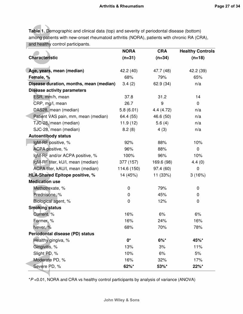
Table 1. Demographic and clinical data (top) and severity of periodontal disease (bottom)
among patients with new-onset rheumatoid arthritis (NORA), patients with chronic RA (CRA),
and healthy control participants.
NORA CRA Healthy Controls
Characteristic (n=31) (n=34) (n=18)
Age, years, mean (median) 42.2 (40) 47.7 (48) 42.2 (39)
Female, % 68% 79% 65%
Disease duration, months, mean (median) 3.4 (2) 62.9 (34) n/a
Disease activity parameters
ESR, mm/h, mean 37.8 31.2 14
CRP, mg/l, mean 26.7 9 0
DAS28, mean (median) 5.8 (6.01) 4.4 (4.72) n/a
Patient VAS pain, mm, mean (median) 64.4 (55) 46.6 (50) n/a
TJC-28, mean (median) 11.9 (12) 5.6 (4) n/a
SJC-28, mean (median) 8.2 (8) 4 (3) n/a
Autoantibody status
IgM-RF positive, % 92% 88% 10%
ACPA positive, % 96% 88% 0
IgM-RF and/or ACPA positive, % 100% 96% 10%
IgM-RF titer, kU/l, mean (median) 377 (157) 169.6 (98) 4.4 (0)
ACPA titer, kAU/l, mean (median) 114.6 (150) 97.4 (60) 0
HLA-Shared Epitope positive, % 14 (45%) 11 (33%) 3 (16%)
Medication use
Methotrexate, % 0 79% 0
Prednisone, % 0 45% 0
Biological agent, % 0 12% 0
Smoking status
Current, % 16% 6% 6%
Former, % 16% 24% 16%
Never, % 68% 70% 78%
Periodontal disease (PD) status
Healthy gingiva, % 0* 6%* 45%*
Gingivitis, % 13% 3% 11%
Slight PD, % 10% 6% 5%
Moderate PD, % 16% 32% 17%
Severe PD, % 62%* 53%* 22%*
*P <0.01, NORA and CRA vs healthy control participants by analysis of variance (ANOVA)
Page 27 of 34
John Wiley & Sons
Arthritis & Rheumatism

Table 2. Oral microbiota differ significantly among patients with new-onset rheumatoid arthritis (NORA), patients with
chronic rheumatoid arthritis (CRA), and healthy control participants, and between individuals with healthy gingiva versus
periodontal diseases (PD), at the level of both genus and species/operational taxonomic unit (OTU).
Taxonomy NORA vs Healthy Controls NORA vs CRA All RA vs Healthy Controls Healthy gingiva vs PD
Genus ↑Anaeroglobus* ↑Uncl. Prevotellaceae* ↑Phocaeiola* ↓Corynebacterium* ↓Mitsuokella* ↓Streptococcus*
↓Uncl. Veillonellaceae** ↓Mitsuokella*
↑Anaeroglobus** ↓Corynebacterium**
↑Anaeroglobus**** ↑Phocaeiola**** ↑Dialister*** ↑Schwartzia*** ↑Uncl_Prevotellaceae*** ↑Prevotella* ↑Tannerella* ↑Trponema* ↑Porphyromonas* ↓Actinomyces**** ↓Corynebacterium*** ↓Neisseria*** ↓Uncl_Flavobacteriaceae*** ↓Uncl_Propionibacteriac.*** ↓Granulicatella** ↓Streptococcus*
Species (OTU)
↑Anaeroglobus_OTU99*** ↑Leptotrichia_OTU87*** ↑Prevotella_OTU60*** ↑Selenomonas_OTU168** ↑Phocaeiola_OTU92* ↑Prevotella_OTU31* ↑Prevotella_OTU134* ↑Neisseria_OTU16* ↑Porphyromonas_OTU1* ↓Leptotrichia_OTU12** ↓Corynebact_OTU4* ↓Uncl.TM7_OTU58*
↑Porphyromonas_OTU57* ↑Selenomonas_OTU231* ↑Prevotella_OTU26* ↑Treponema_OTU32* ↑Tannerella_OTU13* ↓Prevotella_OTU39***
↑Anaeroglobus_OTU99*** ↑Prevotella_OTU134*** ↑Prevotella_OTU60*** ↑Selenomonas_OTU168* ↑Prevotella_OTU31* ↓Leptotrichia_OTU12*** ↓Corynebact_OTU4*** ↓Leptotrichia_OTU86* ↓Leptotrichia_OTU9* ↓Capnocytophaga_OTU74*
↑Anaeroglobus_OTU99**** ↑Prevotella_OTU62**** ↑Prevotella_OTU20**** ↑Treponema_OTU139**** ↑Tannerella_OTU13*** ↑Porphyromonas_OTU1*** ↑Treponema_OTU32*** ↑Selenomonas_OTU168*** ↑Prevotella_OTU39*** ↓Corynebacterium_OTU4*** ↓Corynebacter._OTU77*** ↓Granulicatella_OTU162** ↓Actinomyces_OTU63** ↓Corynebacter._OTU146**
↑ = significant increase in NORA (or PD); ↓ = significant decrease in NORA (or PD). *P <0.05; ** P <0.01; ***P <0.005; ****P <0.0005.
28 of 34
John Wiley & Sons
Arthritis & Rheumatism

FIGURE LEGENDS
Figure 1. Clustering of species-level operational taxonomic units (OTU) among groups
studied. Bar graphs show relative abundance of oral microbiota (phylum level) among
(A) NORA, CRA and healthy controls and (B) participants with No PD vs PD. Clustering
by Yue and Clayton analysis comparing (C) patients with new-onset rheumatoid arthritis
(NORA) vs patients with chronic rheumatoid arthritis (CRA) vs healthy control
participants, and (D) participants with healthy gingiva or gingivitis (No PD) vs those with
slight, moderate or severe periodontal disease (PD).
Figure 2. Abundance of periodontopathic bacteria (OTU-level) in patients with new-
onset rheumatoid arthritis (NORA) and chronic rheumatoid arthritis (CRA). Species
associated with periodontal disease, such as (A) Tannerella forsythia and (B)
Treponema medium, are significantly more abundant in NORA patients compared to
CRA. (C) No significant differences (NS) are observed for Porphyromonas gingivalis.
Figure 3. Prevalence and abundance of Porphyromonas and P. gingivalis (OTU1) in 83
study participants, grouped by rheumatoid arthritis (RA) status [healthy controls (HC),
new-onset RA (NORA), chronic RA (CRA)] and periodontal disease (PD) status [healthy
gingiva or gingivitis (No PD); slight (SLT), moderate (MOD), and severe (SEV) PD].
Although the genus Porphyromonas (A, C) is present almost universally and
irrespective of RA or PD status, P. gingivalis (B, D) is significantly associated with
moderate and severe PD, and not with presence of RA. (A, B) Each square represents
Page 29 of 34
John Wiley & Sons
Arthritis & Rheumatism
a single individual. The darker the intensity of the box, the greater the relative
abundance of Porphyromonas or P. gingivalis. NS = not significant.
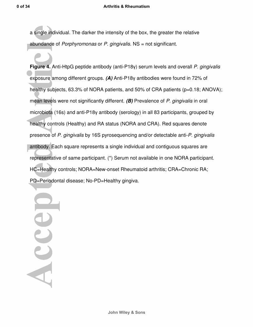
Figure 4. Anti-HtpG peptide antibody (anti-P18γ) serum levels and overall P. gingivalis
exposure among different groups. (A) Anti-P18γ antibodies were found in 72% of
healthy subjects, 63.3% of NORA patients, and 50% of CRA patients (p=0.18; ANOVA);
mean levels were not significantly different. (B) Prevalence of P. gingivalis in oral
microbiota (16s) and anti-P18γ antibody (serology) in all 83 participants, grouped by
healthy controls (Healthy) and RA status (NORA and CRA). Red squares denote
presence of P. gingivalis by 16S pyrosequencing and/or detectable anti-P. gingivalis
antibody. Each square represents a single individual and contiguous squares are
representative of same participant. (*) Serum not available in one NORA participant.
HC=Healthy controls; NORA=New-onset Rheumatoid arthritis; CRA=Chronic RA;
PD=Periodontal disease; No-PD=Healthy gingiva.
0 of 34
John Wiley & Sons
Arthritis & Rheumatism

Figure 1. Clustering of species-level operational taxonomic units (OTU) among groups studied. Clustering by Yue and Clayton analysis comparing (A)
patients with new-onset rheumatoid arthritis (NORA) vs patients with chronic rheumatoid arthritis (CRA) vs healthy control participants, and (B)
participants with healthy gingiva or gingivitis (No PD) vs those with slight, moderate or severe periodontal disease (PD). Bar graphs show relative
abundance of oral microbiome (phylum level) among (C) NORA, CRA and healthy controls and (D) participants with No PD vs PD.
Page 31 of 34
John Wiley & Sons
Arthritis & Rheumatism
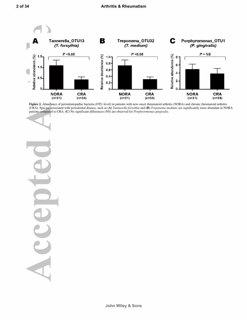
Figure 2. Abundance of periodontopathic bacteria (OTU-level) in patients with new-onset rheumatoid arthritis (NORA) and chronic rheumatoid arthritis
(CRA). Species associated with periodontal disease, such as (A) Tannarella forsythia and (B) Treponema medium, are significantly more abundant in NORA
patients compared to CRA. (C) No significant differences (NS) are observed for Porphyromonas gingivalis.
2 of 34
John Wiley & Sons
Arthritis & Rheumatism

Figure 3. Prevalence and abundance of Porphyromonas and P. gingivalis (OTU1) in 83 study participants, grouped by rheumatoid arthritis (RA) status
[healthy controls (HC), new-onset RA (NORA), chronic RA (CRA)] and periodontal disease (PD) status [healthy gingiva or gingivitis (No PD); slight (SLT),
moderate (MOD), and severe (SEV) PD]. Although the genus Porphyromonas (A, C) is present almost universally and irrespective of RA or PD status, P.
gingivalis (B, D) is significantly associated with moderate and severe PD, and not with presence of RA. (A, B) Each square represents a single individual.
The darker the intensity of the box, the greater the relative abundance of Porphyromonas or P. gingivalis. NS = not significant.
Page 33 of 34
John Wiley & Sons
Arthritis & Rheumatism

Figure 4. Anti-HtpG peptide antibody (anti-P18γ) serum levels and
overall P. gingivalis exposure among different groups. (A) Anti-P18γ
antibodies were found in 72% of healthy subjects, 63.3% of NORA
patients, and 50% of CRA patients (p=0.18; ANOVA); mean levels were
not significantly different. (B) Prevalence of P. gingivalis in oral
microbiota (16s) and anti-P18γ antibody (serology) in all 83 participants,
grouped by healthy controls (Healthy) and RA status (NORA and CRA).
Red squares denote presence of P. gingivalis by 16S pyrosequencing
and/or detectable anti-P. gingivalis antibody. Each square represents a
single individual and contiguous squares are representative of same
participant. (*) Serum not available in one NORA participant.
HC=Healthy controls; NORA=New-onset Rheumatoid arthritis;
CRA=Chronic RA; PD=Periodontal disease; No-PD=Healthy gingiva.
A
B
4 of 34
John Wiley & Sons
Arthritis & Rheumatism