microbiota diversity and gene expression dynamics in human oral
TRANSCRIPT
Benítez-Páez et al. BMC Genomics 2014, 15:311http://www.biomedcentral.com/1471-2164/15/311
RESEARCH ARTICLE Open Access
Microbiota diversity and gene expressiondynamics in human oral biofilmsAlfonso Benítez-Páez1,2*†, Pedro Belda-Ferre1†, Aurea Simón-Soro1 and Alex Mira1*
Abstract
Background: Micro-organisms inhabiting teeth surfaces grow on biofilms where a specific and complex successionof bacteria has been described by co-aggregation tests and DNA-based studies. Although the composition of oralbiofilms is well established, the active portion of the bacterial community and the patterns of gene expressionin vivo have not been studied.
Results: Using RNA-sequencing technologies, we present the first metatranscriptomic study of human dentalplaque, performed by two different approaches: (1) A short-reads, high-coverage approach by Illumina sequencingto characterize the gene activity repertoire of the microbial community during biofilm development; (2) A long-reads,lower-coverage approach by pyrosequencing to determine the taxonomic identity of the active microbiomebefore and after a meal ingestion. The high-coverage approach allowed us to analyze over 398 million reads,revealing that microbial communities are individual-specific and no bacterial species was detected as key player atany time during biofilm formation. We could identify some gene expression patterns characteristic for early andmature oral biofilms. The transcriptomic profile of several adhesion genes was confirmed through qPCR bymeasuring expression of fimbriae-associated genes. In addition to the specific set of gene functions overexpressedin early and mature oral biofilms, as detected through the short-reads dataset, the long-reads approach detectedspecific changes when comparing the metatranscriptome of the same individual before and after a meal, which cannarrow down the list of organisms responsible for acid production and therefore potentially involved in dental caries.
Conclusions: The bacteria changing activity during biofilm formation and after meal ingestion were person-specific.Interestingly, some individuals showed extreme homeostasis with virtually no changes in the active bacterialpopulation after food ingestion, suggesting the presence of a microbial community which could be associated todental health.
Keywords: Dental plaque, Metatranscriptomics, Biofilm formation, Human microbiome, RT-qPCR, RNAseq
BackgroundThe study of microbial communities from environment-and human-derived samples through Next GenerationSequencing (NGS) methods has revealed a vast complexityin those ecological niches where hundreds or thousandsof microbial species co-inhabit and functionally inter-act. One of these complex communities is that found inthe human oral dental plaque (hereinafter, human oral
* Correspondence: [email protected]; [email protected]†Equal contributors1Oral Microbiome Group – Department of Health and Genomics, Center forAdvanced Research in Public Health (CSISP-FISABIO), Avda. Catalunya 21,46020 Valencia, Spain2Bioinformatics Analysis Group – GABi. Centro de Investigación y Desarrolloen Biotecnología (CIDBIO), Bogotá, D.C 111221, Colombia
© 2014 Benítez-Páez et al.; licensee BioMed CeCreative Commons Attribution License (http:/distribution, and reproduction in any mediumDomain Dedication waiver (http://creativecomarticle, unless otherwise stated.
biofilm). Although some studies, using NGS methodsand 16S rRNA-based analysis, estimate that microbialdiversity of the oral cavity is composed by thousands ofspecies [1], more recent data have limited these esti-mates to a few hundreds [2-4]. Contrary to Koch's pos-tulates, dental caries is not considered etiologically theoutcome of a single-agent but is associated to an un-balance of microbial species that synergistically causeenamel demineralization by their acidogenic activity[5,6]. Thus, characterizing the composition of wholebacterial communities that actively engage in biofilmformation and sugar fermentation after the ingestionof food is vital for understanding community dynamicsunder health and disease conditions [7].
ntral Ltd. This is an Open Access article distributed under the terms of the/creativecommons.org/licenses/by/2.0), which permits unrestricted use,, provided the original work is properly credited. The Creative Commons Publicmons.org/publicdomain/zero/1.0/) applies to the data made available in this
Benítez-Páez et al. BMC Genomics 2014, 15:311 Page 2 of 13http://www.biomedcentral.com/1471-2164/15/311
Although the set of species present in the human oralbiofilm is almost fully depicted, new efforts have to beconducted to establish microbial agonistic or antagonis-tic associations, to distinguish actively-growing bacteriafrom inactive or transient species, as well as to outlinethe role of individual species during biofilm formationon tooth surfaces. The co-aggregation detected to occurbetween streptococci and Actinomyces species has beenproposed to be a major promoter of human oral biofilmformation [8]. Like most biofilms, the dental plaque isbuilt in a continued process characterized by successionof different bacterial species, each one with relevantroles in every step of biofilm construction [9]. Formationof the oral biofilm could be dissected in three majorstages, namely: i) attachment; ii) colonization; and iii) bio-film development [10]. However, species participating ofthe entire process are traditionally characterized as “early”and “late” colonizers, where early colonizers would be re-sponsible of the two first stages [9]. Among early colo-nizers the viridans streptococci group is considered as acornerstone of the oral biofilm puzzle given its ability tobind saliva proteins through Antigens I and II. In thismanner, streptococci species become the first colonizersable to bind tooth surfaces and promoting arrival of sec-ondary colonizers by intergeneric coaggregation (reviewedin [9]). Actinomyces naeslundii is one of the secondary col-onizers and a well known coaggregation partner of strepto-cocci [8,11]. Fusobacterium nucleatum is considered a keyplayer given its capability to coaggregate both with earlyand late colonizers of the oral biofilm [12], the latter groupcharacterized by species belonging to Bacteroidetes andSpirochaetes [6,9]. It is noteworthy to highlight that inter-generic coaggregation not only contributes to bacterialgrowth and colonization [13], but it is thought to facilitatethe genetic and metabolic exchange among species, andeven to create the adequate environment for arrival ofsome obligate anaerobic bacteria [10]. Therefore, any dis-ruption in the development of the oral biofilm caused byimpairing of early colonizers tooth attachment or inabilityto recruit other key players during biofilm formation,would affect the entire process avoiding presence of patho-gens responsible for periodontal disease or caries [7].Although few attempts to link specific gene expression
profiles in oral bacteria with the establishment and mat-uration of oral biofilm have been done [14], further stud-ies are needed to understand global gene dynamics andintracellular signalling which are the basis for cell-to-cellcommunication among oral bacteria and to promotebiofilm formation on tooth surfaces. There are import-ant limitations to study gene expression from in vivooral samples, including RNA instability and amounts ofsampling material, but a sequencing approach of totalcDNA from an in vitro oral biofilm model has recentlybeen performed [15].
Because gene transcripts typically occupy a small frac-tion of total bacterial RNA, even after mRNA-enrichingprotocols, a massive coverage is normally required toquantify gene expression by RNAseq technologies. On theother hand, high-coverage sequencing technologies arenormally coupled to short read lengths, which jeopardizeaccurate taxonomic assignment of the sequences. Thelatter can be achieved through the use of longer reads,at the expense of a lower coverage of mRNA transcripts.In the present manuscript we present the first metatran-scriptome analysis of in vivo human oral biofilm sam-ples through two approaches: A short read-length, highcoverage Illumina® approach to study oral biofilm for-mation through time, and a long read-length, lowercoverage pyrosequencing strategy to study changes incommunity composition before and after a meal. Forthe first approach, a total of 16 samples of supragingivalplaque from 4 healthy individuals were collected at fourdifferent time points (6, 12, 24 and 48 hours after a pro-fessional ultrasound cleaning) to disclose the microbiotaand gene expression dynamics during oral biofilm for-mation. For the second experiment, the metatranscrip-tome of dental plaque from five individuals was studied30 minutes before and after a controlled meal, in orderto characterize the potential shifts in the active bacterialcommunity when dietary nutrients are available forgrowth.
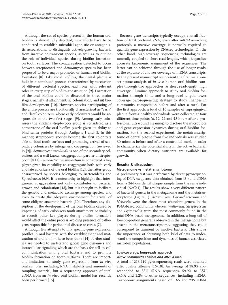
Results & discussionMetagenome vs metatranscriptomeA preliminary test was performed by direct pyrosequenc-ing of DNA (sequence data obtained from [3]) and cDNAfrom a 24-hour dental plaque sample from the same indi-vidual (NoCa1). The results show a very different patternof bacterial genera in the metagenome and the metatran-scriptome (Figure 1). Actinomyces, Corynebacterium andNeisseria were the three most abundant genera in theRNA-based community whereas Veillonella, Streptococcusand Leptotrichia were the most commonly found in thetotal DNA-based metagenome. In addition, a long tail oflow-proportion genera is observed in the metagenome butabsent in the metatranscriptome, suggesting they couldcorrespond to transient or inactive bacteria. This showsthe importance of obtaining both kind of data to under-stand the composition and dynamics of human-associatedmicrobial populations.
Low-coverage, long-reads approachActive communities before and after a mealA total of 213,419 pyrosequencing reads were obtainedafter quality filtering [16-18]. An average of 38.9% cor-responded to SSU rRNA sequences, 59.9% to LSUrRNA and 1.2% to other sequences, including mRNA.Taxonomic assignments based on 16S and 23S rDNA
Actino
myces
Coryn
ebac
terium
Neisse
riaVeil
lonell
a
Cardio
bacte
rium
gene
ra_in
certa
e_se
dis
Strepto
cocc
us
Capno
cytop
haga
Campy
lobac
terPre
votel
la
Fuso
bacte
rium
Porph
yrom
onas
Seleno
monas
Lepto
trich
iaKing
ella
Aggre
gatib
acter
Rothia
Haemop
hilus
Trep
onem
aActi
noba
cillus
Abiotro
phia
Luteo
cocc
usGem
ella
Tann
erell
aBur
khold
eria
Granu
licate
lla
Chrys
eoba
cteriu
mMan
nheim
iaPas
teure
llaCato
nella
Acineto
bacte
rBac
teroid
esBas
fiaClos
tridiu
mEike
nella
Enhyd
roba
cter
Histop
hilus
Morax
ella
Parab
acter
oides
Psych
roba
cter
Rumino
cocc
us
0
5
10
15
20
25
30
Metatranscriptome Metagenome
Rel
ativ
e ab
unda
nce
(%)
Figure 1 Total (DNA-based) and active (RNA-based) microbiota composition in the human oral biofilm from individual NoCa-01.Microbial diversity is inferred from 16S rDNA and 16S cDNA taxonomic assignment, respectively, from reads obtained by direct pyrosequencing ofthe DNA and cDNA of a 24 h dental plaque sample. Relative abundance of bacterial genera from metagenomic data differs from that obtainedfrom metatranscriptomic data. The former would correspond to the total bacterial composition in the sample whereas the latter would representthe “active microbiota” as inferred by their presence in samples’ RNA.
Benítez-Páez et al. BMC Genomics 2014, 15:311 Page 3 of 13http://www.biomedcentral.com/1471-2164/15/311
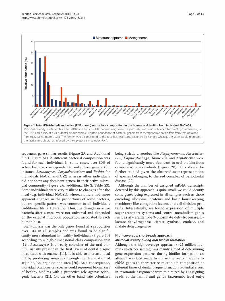
sequences gave similar results (Figure 2A and Additionalfile 1: Figure S1). A different bacterial composition wasfound for each individual. In some cases, over 80% ofactive bacteria corresponded to only three genera (forinstance Actinomyces, Corynebacterium and Rothia forindividuals NoCa1 and Ca2) whereas other individualsdid not show any dominant genera in their active micro-bial community (Figure 2A, Additional file 2: Table S3).Some individuals were very resilient to changes after themeal (e.g. individual NoCa1), whereas others had moreapparent changes in the proportions of some bacteria,but no specific pattern was common to all individuals(Additional file 3: Figure S2). Thus, the changes in activebacteria after a meal were not universal and dependedon the original microbial population associated to eachhuman host.Actinomyces was the only genus found at a proportion
over 10% in all samples and was found to be signifi-cantly more abundant in healthy individuals (Figure 2B)according to a high-dimensional class comparison test[19]. Actinomyces is an early colonizer of the oral bio-film, usually present in the first layers of dental plaquein contact with enamel [11]. It is able to increase localpH by producing ammonia through the degradation ofarginine, lysilarginine and urea [20]. As a consequence,individual Actinomyces species could represent biomarkersof healthy biofilms with a protective role against acido-genic bacteria [21]. On the other hand, late colonizers
being strictly anaerobes like Porphyromonas, Fusobacter-ium, Capnocytophaga, Tannerella and Leptotrichia werefound significantly more abundant in oral biofilm fromcaries-bearing individuals (Figure 2B). This should befurther studied given the observed over-representationof species belonging to the red complex of periodontaldisease [22].Although the number of assigned mRNA transcripts
detected by this approach is quite small, we could identifysome genes being expressed in all samples such as thoseencoding ribosomal proteins and basic housekeepingmachinery like elongation factors and cell division pro-teins. Interestingly, we found expression of multiplesugar transport systems and central metabolism genessuch as glyceraldehyde 3-phosphate dehydrogenase, L-lactate dehydrogenase, citrate synthase, enolase, andmalate dehydrogenase.
High-coverage, short-reads approachMicrobial activity during oral biofilm formationAlthough the high-coverage approach (~25 million Illu-mina reads per sample) was mainly aimed at determininggene expression patterns during biofilm formation, anattempt was first made to utilize the reads mapping torRNA genes to characterize microbiota composition atdifferent times of dental plaque formation. Potential errorsin taxonomic assignment were minimized by 1) assigningreads at the family and genus taxonomic level only;
Rel
ativ
e F
requ
ency
(%
)
A
B
VeillonellaTreponemaTM7_genera_incertae_sedisTessaracoccusTannerellaStreptococcusSelenomonasRothiaPrevotellaPorphyromonasOtherOribacteriumNeisseriaLeptotrichiaKingellaHaemophilusGranulicatellaGemellaFusobacteriumEubacteriumDialisterCorynebacteriumCardiobacteriumCapnocytophagaCampylobacterAggregatibacterActinomycesActinobacillusAbiotrophia
No
Ca1
_Bef
ore
No
Ca1
_Aft
er
No
Ca1
2_B
efo
re
No
Ca1
2_A
fter
Ca1
_01_
Bef
ore
Ca1
_01_
Aft
er
Ca2
_Bef
ore
Ca2
_Aft
er
Ca0
24_B
efo
re
Ca0
24_A
fter
Figure 2 Meal-uptake-dependent active microbiota and association with health and disease. A – Graphical representation of the generadistribution according to 16S rRNA assignation of metatranscriptomic reads (obtained by pyrosequencng of total cDNA) based on RDP classifier.Relative frequency for most predominant active genera is shown in dental plaque samples obtained before and after a carbohydrate-rich meal.B – Health- and disease-associated genera in the above metatranscriptome, as inferred from Linear Discrimination Analysis (LDA) performed fordimensional class comparisons. The data were generated by using LEfSe test, available in the Galaxy Web Server toolkit. NoCa = individuals withno caries; Ca = individuals with caries.
Benítez-Páez et al. BMC Genomics 2014, 15:311 Page 4 of 13http://www.biomedcentral.com/1471-2164/15/311
2) selecting matches against 16S rRNA database of 100%sequence identity; and 3) eliminating hits to conserved re-gions of the 16S rRNA gene, keeping only hypervariable,informative regions.When we tried to compare samples according to mat-
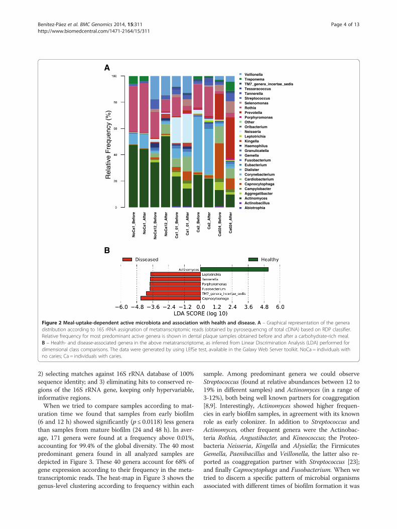
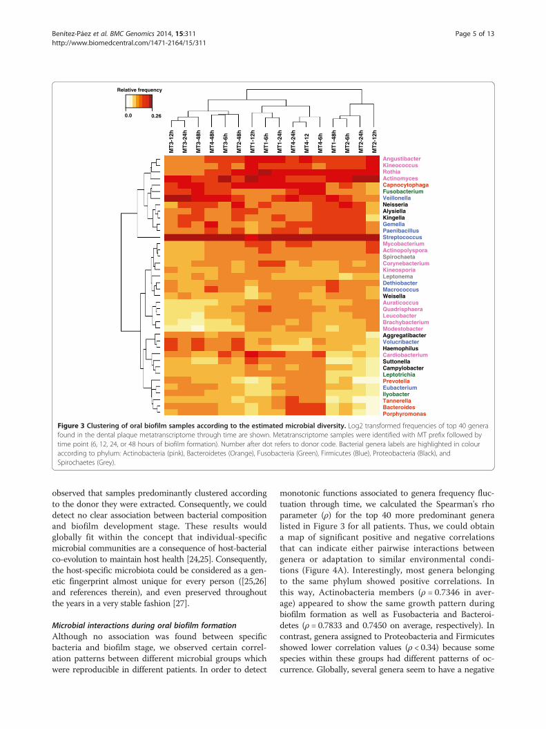
uration time we found that samples from early biofilm(6 and 12 h) showed significantly (p ≤ 0.0118) less generathan samples from mature biofilm (24 and 48 h). In aver-age, 171 genera were found at a frequency above 0.01%,accounting for 99.4% of the global diversity. The 40 mostpredominant genera found in all analyzed samples aredepicted in Figure 3. These 40 genera account for 68% ofgene expression according to their frequency in the meta-transcriptomic reads. The heat-map in Figure 3 shows thegenus-level clustering according to frequency within each
sample. Among predominant genera we could observeStreptococcus (found at relative abundances between 12 to19% in different samples) and Actinomyces (in a range of3-12%), both being well known partners for coaggregation[8,9]. Interestingly, Actinomyces showed higher frequen-cies in early biofilm samples, in agreement with its knownrole as early colonizer. In addition to Streptococcus andActinomyces, other frequent genera were the Actinobac-teria Rothia, Angustibacter, and Kineococcus; the Proteo-bacteria Neisseria, Kingella and Alysiella; the FirmicutesGemella, Paenibacillus and Veillonella, the latter also re-ported as coaggregation partner with Streptococcus [23];and finally Capnocytophaga and Fusobacterium. When wetried to discern a specific pattern of microbial organismsassociated with different times of biofilm formation it was
Relative frequency
0.0 0.26
AngustibacterKineococcusRothiaActinomycesCapnocytophagaFusobacteriumVeillonellaNeisseriaAlysiellaKingellaGemellaPaenibacillusStreptococcusMycobacteriumActinopolysporaSpirochaetaCorynebacteriumKineosporiaLeptonemaDethiobacterMacrococcusWeisellaAuraticoccusQuadrisphaeraLeucobacterBrachybacteriumModestobacterAggregatibacterVolucribacterHaemophilusCardiobacteriumSuttonellaCampylobacterLeptotrichiaPrevotellaEubacteriumIlyobacterTannerellaBacteroidesPorphyromonas
MT
3-12
h
MT
3-24
h
MT
3-48
h
MT
4-48
h
MT
3-6h
MT
2-48
h
MT
1-12
h
MT
1-6h
MT
1-24
h
MT
4-24
h
MT
4-12
MT
4-6h
MT
1-48
h
MT
2-6h
MT
2-24
h
MT
2-12
h
Figure 3 Clustering of oral biofilm samples according to the estimated microbial diversity. Log2 transformed frequencies of top 40 generafound in the dental plaque metatranscriptome through time are shown. Metatranscriptome samples were identified with MT prefix followed bytime point (6, 12, 24, or 48 hours of biofilm formation). Number after dot refers to donor code. Bacterial genera labels are highlighted in colouraccording to phylum: Actinobacteria (pink), Bacteroidetes (Orange), Fusobacteria (Green), Firmicutes (Blue), Proteobacteria (Black), andSpirochaetes (Grey).
Benítez-Páez et al. BMC Genomics 2014, 15:311 Page 5 of 13http://www.biomedcentral.com/1471-2164/15/311
observed that samples predominantly clustered accordingto the donor they were extracted. Consequently, we coulddetect no clear association between bacterial compositionand biofilm development stage. These results wouldglobally fit within the concept that individual-specificmicrobial communities are a consequence of host-bacterialco-evolution to maintain host health [24,25]. Consequently,the host-specific microbiota could be considered as a gen-etic fingerprint almost unique for every person ([25,26]and references therein), and even preserved throughoutthe years in a very stable fashion [27].
Microbial interactions during oral biofilm formationAlthough no association was found between specificbacteria and biofilm stage, we observed certain correl-ation patterns between different microbial groups whichwere reproducible in different patients. In order to detect
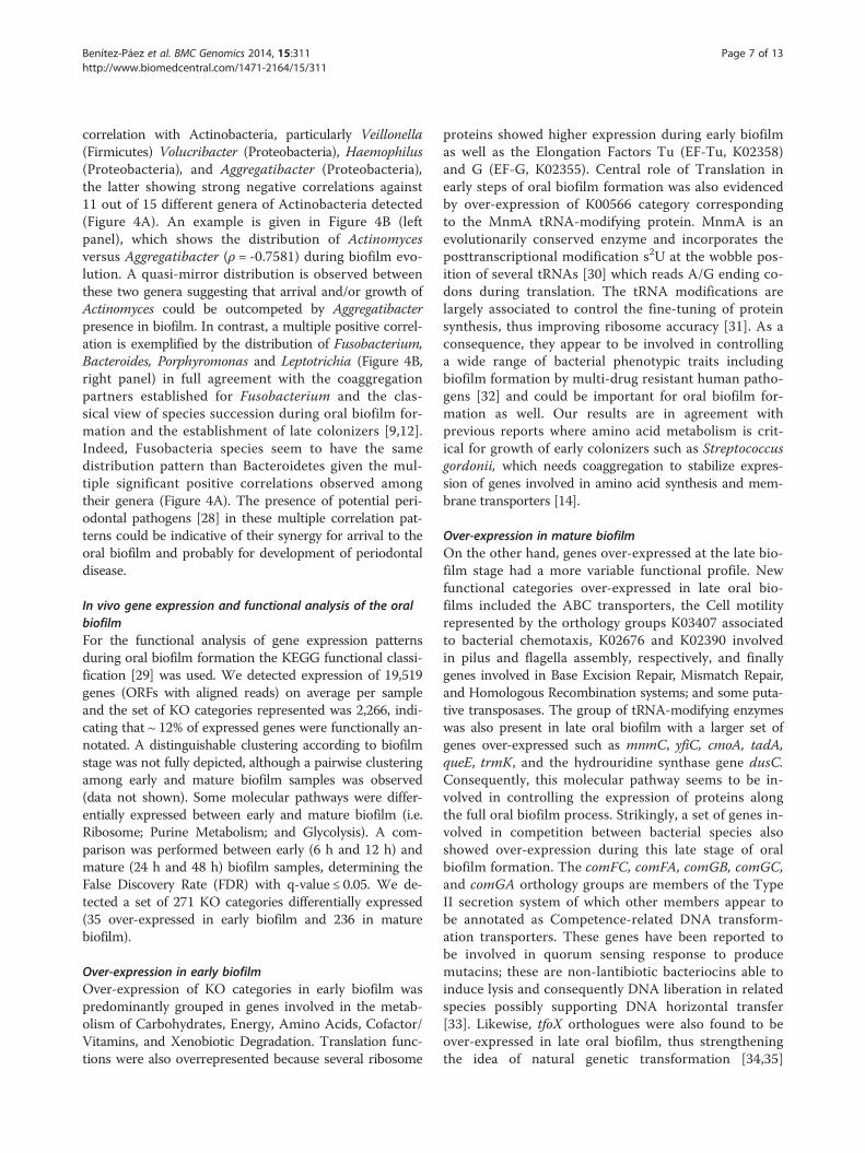
monotonic functions associated to genera frequency fluc-tuation through time, we calculated the Spearman's rhoparameter (ρ) for the top 40 more predominant generalisted in Figure 3 for all patients. Thus, we could obtaina map of significant positive and negative correlationsthat can indicate either pairwise interactions betweengenera or adaptation to similar environmental condi-tions (Figure 4A). Interestingly, most genera belongingto the same phylum showed positive correlations. Inthis way, Actinobacteria members (ρ = 0.7346 in aver-age) appeared to show the same growth pattern duringbiofilm formation as well as Fusobacteria and Bacteroi-detes (ρ = 0.7833 and 0.7450 on average, respectively). Incontrast, genera assigned to Proteobacteria and Firmicutesshowed lower correlation values (ρ < 0.34) because somespecies within these groups had different patterns of oc-currence. Globally, several genera seem to have a negative

6h 12h 24h 48h-5.0
-4.0
-3.0
-2.0
-1.0
0.0
1.0
2.0
Fusobacterium PorphyromonasLeptotrichia Bacteroides
6h 12h 24h 48h-3.0
-2.0
-1.0
0.0
1.0
2.0
3.0
4.0
Actinomyces Aggregatibacter
A
Time (hours)
B
Rel
ativ
eF
req
uen
cy(L
og
2)
LeptotrichiaCampylobacterSuttonellaCardiobacteriumCorynebacteriumLeptonemaPaenibacillusGemellaStreptococcusMacrococcusWeisellaDethiobacterAlysiellaKingellaNeisseriaBacteroidesPorphyromonasEubacteriumIlyobacterFusobacteriumPrevotellaTannerellaCapnocytophagaVeillonellaAggregatibacterVolucribacterHaemophilusMycobacteriumModestobacterRothiaSpirochaetaLeucobacterAngustibacterAuraticoccusActinopolysporaQuadrisphaeraBrachybacteriumKineococcusActinomycesKineosporia
-1 10
Rho value ( )
Lep
totrich
iaC
amp
ylob
acterS
utto
nella
Card
iob
acterium
Co
ryneb
acterium
Lep
ton
ema
Paen
ibacillu
sG
emella
Strep
toco
ccus
Macro
coccu
sW
eisellaD
ethio
bacter
Alysiella
Kin
gella
Neisseria
Bactero
ides
Po
rph
yrom
on
asE
ub
acterium
Ilyob
acterF
uso
bacteriu
mP
revotella
Tan
nerella
Cap
no
cytop
hag
aV
eillon
ellaA
gg
regatib
acterV
olu
cribacter
Haem
op
hilu
sM
ycob
acterium
Mo
desto
bacter
Ro
thia
Sp
iroch
aetaL
euco
bacter
An
gu
stibacter
Au
raticoccu
sA
ctino
po
lyspo
raQ
uad
risph
aeraB
rachyb
acterium
Kin
eoco
ccus
Actin
om
ycesK
ineo
spo
ria
Figure 4 Positive and negative interactions in the oral biofilm active microbiota. A – Relative abundance of bacterial genera (based on 16Sassignment, see Methods) and fluctuation through time were studied in a pairwise manner calculating the Spearman’s rho parameter (see Methods).Genera labels are highlighted in color according to phylum: Actinobacteria (pink), Bacteroidetes (Orange), Fusobacteria (Green), Firmicutes(Blue), Proteobacteria (Black), and Spirochaetes (Grey). B – Detail of the Spearman correlations found in the analysis. The left panel shows astrong negative (ρ ~ - 0.75) correlation between Actinomyces and Aggregatibacter through time. In the right panel, a positive correlations ispresented among late colonizers such as Leptotrichia, Fusobacterium, Porphyromonas, and Bacteroides (ρ ~ 0.80).
Benítez-Páez et al. BMC Genomics 2014, 15:311 Page 6 of 13http://www.biomedcentral.com/1471-2164/15/311
Benítez-Páez et al. BMC Genomics 2014, 15:311 Page 7 of 13http://www.biomedcentral.com/1471-2164/15/311
correlation with Actinobacteria, particularly Veillonella(Firmicutes) Volucribacter (Proteobacteria), Haemophilus(Proteobacteria), and Aggregatibacter (Proteobacteria),the latter showing strong negative correlations against11 out of 15 different genera of Actinobacteria detected(Figure 4A). An example is given in Figure 4B (leftpanel), which shows the distribution of Actinomycesversus Aggregatibacter (ρ = -0.7581) during biofilm evo-lution. A quasi-mirror distribution is observed betweenthese two genera suggesting that arrival and/or growth ofActinomyces could be outcompeted by Aggregatibacterpresence in biofilm. In contrast, a multiple positive correl-ation is exemplified by the distribution of Fusobacterium,Bacteroides, Porphyromonas and Leptotrichia (Figure 4B,right panel) in full agreement with the coaggregationpartners established for Fusobacterium and the clas-sical view of species succession during oral biofilm for-mation and the establishment of late colonizers [9,12].Indeed, Fusobacteria species seem to have the samedistribution pattern than Bacteroidetes given the mul-tiple significant positive correlations observed amongtheir genera (Figure 4A). The presence of potential peri-odontal pathogens [28] in these multiple correlation pat-terns could be indicative of their synergy for arrival to theoral biofilm and probably for development of periodontaldisease.
In vivo gene expression and functional analysis of the oralbiofilmFor the functional analysis of gene expression patternsduring oral biofilm formation the KEGG functional classi-fication [29] was used. We detected expression of 19,519genes (ORFs with aligned reads) on average per sampleand the set of KO categories represented was 2,266, indi-cating that ~ 12% of expressed genes were functionally an-notated. A distinguishable clustering according to biofilmstage was not fully depicted, although a pairwise clusteringamong early and mature biofilm samples was observed(data not shown). Some molecular pathways were differ-entially expressed between early and mature biofilm (i.e.Ribosome; Purine Metabolism; and Glycolysis). A com-parison was performed between early (6 h and 12 h) andmature (24 h and 48 h) biofilm samples, determining theFalse Discovery Rate (FDR) with q-value ≤ 0.05. We de-tected a set of 271 KO categories differentially expressed(35 over-expressed in early biofilm and 236 in maturebiofilm).
Over-expression in early biofilmOver-expression of KO categories in early biofilm waspredominantly grouped in genes involved in the metab-olism of Carbohydrates, Energy, Amino Acids, Cofactor/Vitamins, and Xenobiotic Degradation. Translation func-tions were also overrepresented because several ribosome
proteins showed higher expression during early biofilmas well as the Elongation Factors Tu (EF-Tu, K02358)and G (EF-G, K02355). Central role of Translation inearly steps of oral biofilm formation was also evidencedby over-expression of K00566 category correspondingto the MnmA tRNA-modifying protein. MnmA is anevolutionarily conserved enzyme and incorporates theposttranscriptional modification s2U at the wobble pos-ition of several tRNAs [30] which reads A/G ending co-dons during translation. The tRNA modifications arelargely associated to control the fine-tuning of proteinsynthesis, thus improving ribosome accuracy [31]. As aconsequence, they appear to be involved in controllinga wide range of bacterial phenotypic traits includingbiofilm formation by multi-drug resistant human patho-gens [32] and could be important for oral biofilm for-mation as well. Our results are in agreement withprevious reports where amino acid metabolism is crit-ical for growth of early colonizers such as Streptococcusgordonii, which needs coaggregation to stabilize expres-sion of genes involved in amino acid synthesis and mem-brane transporters [14].
Over-expression in mature biofilmOn the other hand, genes over-expressed at the late bio-film stage had a more variable functional profile. Newfunctional categories over-expressed in late oral bio-films included the ABC transporters, the Cell motilityrepresented by the orthology groups K03407 associatedto bacterial chemotaxis, K02676 and K02390 involvedin pilus and flagella assembly, respectively, and finallygenes involved in Base Excision Repair, Mismatch Repair,and Homologous Recombination systems; and some puta-tive transposases. The group of tRNA-modifying enzymeswas also present in late oral biofilm with a larger set ofgenes over-expressed such as mnmC, yfiC, cmoA, tadA,queE, trmK, and the hydrouridine synthase gene dusC.Consequently, this molecular pathway seems to be in-volved in controlling the expression of proteins alongthe full oral biofilm process. Strikingly, a set of genes in-volved in competition between bacterial species alsoshowed over-expression during this late stage of oralbiofilm formation. The comFC, comFA, comGB, comGC,and comGA orthology groups are members of the TypeII secretion system of which other members appear tobe annotated as Competence-related DNA transform-ation transporters. These genes have been reported tobe involved in quorum sensing response to producemutacins; these are non-lantibiotic bacteriocins able toinduce lysis and consequently DNA liberation in relatedspecies possibly supporting DNA horizontal transfer[33]. Likewise, tfoX orthologues were also found to beover-expressed in late oral biofilm, thus strengtheningthe idea of natural genetic transformation [34,35]
Benítez-Páez et al. BMC Genomics 2014, 15:311 Page 8 of 13http://www.biomedcentral.com/1471-2164/15/311
occurring among close species in the mature oral biofilm.Globally, over-expression of these competence-relatedgenes, permitting DNA transformations in vivo, couldsupport the specific low ratio between functional diver-sity of genes and operational taxonomic units detectedin supragingival plaque, thus indicating high functional re-dundancy and microbial population homogenization [36].Other important functional categories over-expressed inlate oral biofilm included those involved in EnvironmentalInformation Processing and membrane transporters, suchas those belonging to the Phosphotransferase System(PTS) as well as MFS membrane receptors specializedin the importing/exporting of small molecules. Bothmajor families of membrane transporters were found tobe preferentially expressed from Actinomyces speciesindicating a high level of metabolic exchange betweenthis genus and its environment. However, subfamiliessuch a salX-like ABC transporters associated to bacteri-ocin export and defense were detected to be predomin-antly active in streptococci species. Over-expression ofsome KEGG orthology categories belonging to Two-Component family of proteins indicate an active role ofcells in perceiving external signals of nutrient availabil-ity in the environment. An over-expression was foundof the PTS-Ntr-EIIA enzyme and the GlnB protein,both involved in nitrogen regulation, and the sigma fac-tor 54 of the RNA polymerase involved in expression ofgenes for nitrogen metabolism. Therefore, processes re-lated to nitrogen uptake/metabolism appear to be veryrelevant in the mature stage of oral biofilm probably in-dicating that nitrogen is a limiting factor for oral biofilmprogression. In recent studies of cDNA massive sequen-cing from an in vitro five-species oral biofilm microbialcommunity, similar results were obtained in terms of over-represented functions in mature biofilms [15]. Finally, luxShomologue in Neisseria spp. was found to be significantlyover-expressed in early biofilm. The luxS genes are re-sponsible of Autoinducer-2 (AI-2) synthesis, a moleculeconsidered as a major interspecies signal for cell-cellcommunication [9,10]. Evidence for AI-2 role to controlbiofilm formation was previously observed when a luxSnull strain of S. gordonii was unable to form a mixed-species biofilm with P. gingivalis [37].
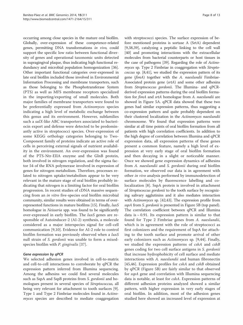
Gene expression by qPCRWe selected adhesion genes involved in cell-to-matrixand cell-to-cell interactions to corroborate by qPCR theexpression pattern inferred from Illumina sequencing.Among the adhesins we could find several moleculessuch as SspA and SspB proteins from S. gordonii and ho-mologues present in several species of Streptococcus, allbeing very relevant for attachment to tooth surfaces [9].Type 1 and Type 2 Fimbriae molecules found in Actino-myces species are described to mediate coaggregation
with streptococci species. The surface expression of be-fore mentioned proteins is sortase A (SrtA) dependent[9,38,39], catalyzing a peptidic linking to the cell wall[40] and promoting interactions with the extracellularmolecules from bacterial counterparts or host tissues inthe case of pathogens [39]. Regarding the role of Actino-myces sp. Type 2 Fimbriae in coaggregation with Strepto-coccus sp. [8,41], we studied the expression pattern of itsgene (fimA) together with the A. naeslundii Fimbriae-Associated protein gene (srtA) and some other adhesinsfrom Streptococcus gordonii. The Illumina- and qPCR-derived expression patterns during the oral biofilm forma-tion for fimA and srtA homologue from A. naeslundii areshowed in Figure 5A. qPCR data showed that these twogenes had similar expression patterns, thus suggesting aco-expression pattern and quite probably dependent ontheir clustered localization in the Actinomyces naeslundiichromosome. We found that expression patterns weresimilar at all time points of oral biofilm formation from allpatients with high correlation coefficients. In addition tothe high degree of correlation between Illumina and qPCRexpression data, all expression patterns of these genespresent a common feature, namely a high level of ex-pression at very early stage of oral biofilm formationand then decaying in a slight or noticeable manner.Once we showed gene expression dynamics of adhesinsfrom A. naeslundii and S. gordonii during oral biofilmformation, we observed our data is in agreement withother in vivo analysis performed by immunodetection ofsurface molecules by fluorescence labelling and co-localization [8]. SspA protein is involved in attachmentof Streptococcus gordonii to the tooth surface by recogniz-ing salivary agglutinins and it also mediates interactionwith Actinomyces sp. [42,43]. The expression profile fromsspA from S. gordonii is presented in Figure 5B (top panel).The correlation coefficient between qPCR and Illuminadata is ~ 0.91. Its expression pattern is similar to thatfound for Type 2 Fimbriae genes from A. naeslundii,which is in agreement with the role of streptococci asfirst colonizers and the requirement of SspA for attach-ing to the tooth surface and promote arrival of otherearly colonizers such as Actinomyces sp. [9,44]. Finally,we studied the expression patterns of cshA and cshBgenes coding for two cell surface antigens in S. gordoniithat increase hydrophobicity of cell surface and mediateinteractions with A. naeslundii and human fibronectin[45,46]. Expression profiles for cshA and cshB obtainedby qPCR (Figure 5B) are fairly similar to that observedfor sspA gene and correlation with Illumina sequencingdata is notable, at least for cshA. Expression patterns ofdifferent adhesion proteins analyzed showed a similarpattern, with higher expression in very early stages oforal biofilm. In addition, most of the adhesion genesstudied here showed an increased level of expression at
6h 12h 24h 48h-4.00
-3.00
-2.00
-1.00
0.00
1.00
2.00
6h 12h 24h 48h-4.00
-3.00
-2.00
-1.00
0.00
-6.00
-4.00
-2.00
0.00
2.00
4.00
A B
6h 12h 24h 48h0.00
1.00
2.00
3.00
4.00
0.00
1.00
2.00
3.00
4.00fimA
fimbriae-associated (srtA)
R = 0.9683
Illumina ExpressionqPCR Expression
sspA
6h 12h 24h 48h0.00
1.00
2.00
3.00
-2.00
-1.00
0.00
1.00
2.00cshA
6h 12h 24h 48h0.00
1.00
2.00
3.00
-1.00
0.00
1.00
2.00cshB
R = 0.9134
R = 0.0658
R = 0.8843RQ
(Lo
g2 )
RQ
(Lo
g2 )
R = 0.9359
Rel
ativ
eE
xpre
ssio
n(L
og
2)
Rel
ativ
eE
xpre
ssio
n(L
og
2)
Figure 5 Gene expression comparison between Illumina sequencing and qPCR during biofilm formation. A - Genes associated to Type 2Fimbriae assembly in Actinomyces naeslundii were analyzed and Pearson correlations calculated from expression values obtained by these twoapproaches. B – Adhesion genes from Streptococcus gordonii were also analyzed and their expression pattern was compared.
Benítez-Páez et al. BMC Genomics 2014, 15:311 Page 9 of 13http://www.biomedcentral.com/1471-2164/15/311
the end point of study (48 h). We hypothesize that suchlevel of expression would reflect the last stage of thebiofilm cycle where biofilm detachment occurs, thus re-leasing bacterial cells to colonize new niches [10,47].
ConclusionsOur study shows for the first time the microbial diversityand gene expression dynamics in the complex oral micro-bial community in vivo. We could follow oral biofilm for-mation and determine proportions of active microbiotathrough time, including before and after a carbohydrate-rich meal, when the process of acid production, respon-sible of enamel demineralization, takes place. We presenta large set of correlations among bacterial groups and gen-era being in agreement with biological and classical inter-actions reported to be central for biofilm installation anddevelopment [8,48,49]. In the functional exploration ofgenes expressed during human oral biofilm formation, wepresent a quantitative analysis, further supported by re-sults obtained by qPCR, demonstrating several functionalcategories of prevalence at different oral biofilm stages.Among them we showed that translation machinery ispredominantly expressed in early biofilm stages whereasmore specialized genes are required in mature biofilm.Some genes involved in competence, and reported to beinvolved in quorum sensing response and functionallyrelated to mutacin production and DNA uptake, wereover-expressed in late biofilm supporting the intricate
level of cell-to-cell interactions in mature biofilm andsuggesting strong competition for colonization. Morethan 70% of the genetic information compiled from thisoral metatranscriptome has no functional assignment;therefore, further efforts must be conducted for classifica-tion and characterization of genes and their involvementin biofilm development and/or cell-to-cell communica-tion. From an applied point of view, the identification ofactive bacterial species after food uptake can be consid-ered a first step to narrow down the list of potentialetiological agents of dental caries from the large set ofmicro-organisms found in the metagenome of dentalplaque and cavities [3]. The striking homeostasis foundin one of the individuals who had never suffered fromdental caries, and where virtually no changes were foundin the active microbiota before and after a meal, couldindicate that the microbiota of some individuals is notaffected by food ingestion, potentially reducing the riskof acidic pH and promoting dental health.
MethodsSample collection and RNA processingThe sampling procedure was approved by the EthicalCommittee for Clinical Research from the DGSP-CSISP(Valencian Health Authority, Spain) and all donorssigned an informed consent. The oral health status ofeach individual was evaluated before sampling and fol-lowing recommendations and nomenclature from the

Benítez-Páez et al. BMC Genomics 2014, 15:311 Page 10 of 13http://www.biomedcentral.com/1471-2164/15/311
WHO. Donors were 20-30 years of age, had all 28 teethpresent (excluding third molars) had not suffered fromany systemic disease and had not taken systemic antimi-crobials in the previous 6 months. Dental plaque sam-ples were taken with autoclaved spoon excavators fromvestibular and lingual surfaces of teeth excluding a 1mm region on the edges.For the biofilm formation experiments, 16 supragingival
dental plaque samples were obtained from 4 caries-freevolunteers (DMFT= 0 [decayed, Missing, Filling Teeth],OHI = 1 [oral Hygiene Index]. GI = 1 [gingival Index]).The volunteers were subjected to professional teeth ultra-sound cleaning. Oral biofilm (supragingival plaque) fromall teeth surfaces was pooled and collected from each vol-unteer at 6, 12, 24, and 48 h of biofilm formation. Afterevery sampling a professional brushing was performed toreset biofilm formation for next sampling. Total RNA wasextracted using the MasterPureTM RNA Purification Kit(Epicentre®). Samples were collected and processed forelimination of 5S rRNA and tRNAs through ion exchangechromatography with KCl gradient in Nucleobond AX 20columns (Macherey-Nagel). Pre- and post-processed RNAwere loaded in RNA chip (Agilent Technologies) and ana-lyzed for integrity using Agilent Bioanalyzer 2100 (AgilentTechnologies). The first strand of cDNA from processedRNA was synthesized using High-Capacity cDNA ReverseTranscription Kit (Applied Biosystems). For this aim, twocDNA reactions were prepared for each RNA sample andmodifying some manufacturer’s instructions to obtain bestperformance during synthesis. Specifically, each cDNAreaction had 100U of Multiscribe Reverse transcriptaseand synthesis was completed at 48°C during 210 minusing 5-10 ug of RNA as template. Doubled strandedcDNA (ds-cDNA) was achieved in 100 uL of reactioncontaining 35U E.coli DNA Polymerase I (New EnglandBiolabs), 5U E. coli DNA Ligase (New England Biolabs),5U RNase H (Epicentre®), 300 uM dNTP's, and two re-actions of first strand cDNA synthesis. The ds-cDNAsynthesis was initiated by incubation during 150 minat 16°C and completed by adding 4.5U of T4 DNAPolymerase (New England Biolabs), and 1X BSA (NewEngland Biolabs) followed by incubation during additional30 min at 16°C. Purified ds-cDNAs were obtained usingHigh Pure PCR Product Purification Kit (Roche®) andsent to GATC Biotech AG (Konstanz, Germany) forparallel single-end sequencing using HiSeq2000 system(Illumina®).The five donors for the before/after meal transcrip-
tome were asked not to brush their teeth for 16 hours.Three of them had active caries at the moment of sam-pling (Decayed Teeth = 3, OHI = 1, GI = 1) and the othertwo had no history of dental caries (DMFT = 0, OHI = 1,GI = 1). None of the donors had periodontal disease.All donors ingested the same meal, whose nutritional
characteristics are indicated in Additional file 4: Table S1.Supragingival dental plaque was obtained from the rightmaxillary and left mandibular quadrant free teeth surfacesfor the sample 30 minutes before eating and from the leftmaxillary and right mandibular quadrant 30 minutes afterfood intake, without touching caries lesions, if they werepresent. Opposite quadrants were sampled because, whenanalysing PCR-amplified 16S rRNA from cDNA, an equiva-lence in terms of taxonomic composition was found whensampling opposite mandibular and maxillary quadrants(Additional file 5: Figure S3). The obtained total ds-cDNA(as described above) was purified and enriched in fragmentslonger than 400 bp using AMPure beads (Agencourt).Those long cDNA fragments were sequenced using 454GS-FLX technology with titanium chemistry (Roche).
Taxonomic assignment and correlationsFiltering and trimming of original data set was assistedby Galaxy Web Server [16-18], filtering by quality usingthe sliding window method (window size 25 with a mini-mum quality in the window of 20), and sequences shorterthan 200 bp were removed. For the high-coverage biofilmsamples, microbial diversity was established by taxonomicassignment using reads matching 16S rRNA sequences.For this aim we constructed a RDP-based (Release 10,Update 29) database containing almost 10,000 referencesequences of 16S rDNA annotated according to NCBItaxonomy [50]. This reference database was processed tofilter out the conserved regions of 16S rDNA genes usingHidden Markov Models [51]. Then, using MegaBlastv2.2.21 algorithm [52] and selecting alignments for 48 ntin length and 100% identity we could assign taxonomy atgenus level using only hypervariable regions of 16S rDNAsequences, thus determining predominant microbiota.Heat maps of taxonomic composition were generatedusing the gplots library of R [53], frequencies were log2transformed and clustered with Euclidean distance. In thecase of samples before/after a meal, microbial diversitywas established using the 16S and 23S rRNA gene. 16Sand 23S sequences were binned using META-RNA 1.0[54]. 16S sequences were assigned using the online RDPassigner [50]. 23S sequences were assigned using theSILVA database and SINA assigner [55,56]. All statisticalanalyses were conducted on R v2.15. Non-parametricSpearman rank correlation was calculated among top 40most frequent genera to associate frequency fluctuationsduring biofilm formation between genera. Then, Spear-man's (ρ) coefficient and t-test significance was calculatedfor pairs of genera from all patients, using a Bonferronicorrection for multiple comparisons.
Functional analysisBased on predominant microbiota present at all states ofbiofilm formation, available complete and WGS genomes
Benítez-Páez et al. BMC Genomics 2014, 15:311 Page 11 of 13http://www.biomedcentral.com/1471-2164/15/311
were retrieved from the RefSeq and the Human OralMicrobiome databases [57,58]. More than 80 genomesof oral related microorganisms were downloaded andused to build a local database with almost 300,000 cod-ing sequences. More than 800 small predicted ORFs(100-400 nt) were removed, being 98-100% identical todifferent regions of 16S or 23S rDNAs [59]. The remainingset of ORFs were then submitted to the KEGG AutomaticAnnotation Server [29] for KEGG Orthology (KO) assign-ment. Using MegaBlast v2.2.21 algorithm [52] with e-valuecutoff 1e-08 and selecting alignments longer than 60%of read with >80% of identity, we assigned KO numbersand PATH categories to the BRITE functional hierarchy[29]. Negative binomial distribution contained in DESeq[60] bioconductor v2.10 package (default parameters)was employed for differential expression analysis. KOover-representation was determined by comparison be-tween early (6-12 h) and late (24-48 h) biofilm sampleswith q values ≤ 0.05. Counting of reads per gene andgenome were normalized against genus frequency andsize dataset and then transformed in log2 for compari-son with qPCR expression data.
Quantitative PCRPrimers for qPCR were designed submitting the respectiveORF sequences from S. gordonii and A. naeslundii to thePrimer3Plus webserver [61] (Additional file 6: Table S2).Gene amplification was performed using LightCycler® 480System (Roche), SYBR Green I Master (Roche), and asmall aliquot from the respective sample sequenced byIllumina. The Cp values were calculated from three repli-cates using the LightCycler® 480 SW software v1.5(Roche). Expression was normalized against 16S rRNA ex-pression from S. gordonii and A. naeslundii, respectively,and referred to expression seen for every gene at 6h for allpatients in average using the ΔΔCt method.
Data accessAll sequence data derived from 454 pyrosequencing ofcDNA from samples after/before meal experiments, mi-crobial diversity associated to dental quadrants, and Illu-mina HiSeq2000 sequencing of cDNA from oral biofilmare stored in the MG-RAST server to be publicly availableby accessing to the “Oral Metatranscriptome” project, id935 (http://metagenomics.anl.gov/linkin.cgi?project=935).Sequence data is also available at the European NucleotideArchive (ENA-EBML) with provisional accession numberERP003984.
Additional files
Additional file 1: Figure S1. Bacterial genera composition according to23S rDNA. The taxonomic assignation was based on SINA analysis againstreference samples from the SILVA database. Bars show the relative
frequency for most predominant genera in metatranscriptomic samplesobtained before and after a carbohydrate-rich meal.
Additional file 2: Table S3. Shannon Diversity Indexes for samplesfrom the low-coverage approach.
Additional file 3: Figure S2. Bacterial relative abundances betweensamples obtained before and after a meal. Positive values (expressed aslog2 ratios) are colored in green and indicate a higher abundance of agiven genus in the sample before the meal; negative values (alsoexpressed as log2 ratios), colored in red, indicate a higher abundance inthe after-meal sample.
Additional file 4: Table S1. Number of reads analyzed for taxonomyassignment from the low-coverage approach.
Additional file 5: Figure S3. Bacterial diversity analysis of the 24 hhuman oral biofilm according to dental quadrants. Bacterial compositionwas estimated by pyrosequencing of the 16S rRNA gene obtained byPCR amplification of cDNA. Diversity at the family taxonomic level(Actinobacteria as Phylum) was determined in biofilm samples comingfrom four dental quadrants of a unique donor. Pie charts for everyquadrant show relative frequency for most predominant bacterialfamilies. Rarefaction curves for each quadrant display a similar diversityfor all samples and bacterial composition piecharts indicate slightdifferences at the frequency of some families like Neisseriaceae being lessfrequent in upper quadrants.
Additional file 6: Table S2. Sequence information for oligonucleotidesused in the qPCR approach.
Competing interestsThe authors declare that they have no competing interests.
Authors’ contributionsAll authors participated in the study design. ASS carried out the sampling.ABP and PBF performed the molecular biology studies and high-throughputdata analysis. ABP, PBF and AM worked in manuscript preparation. All authorsread and approved the final manuscript.
AcknowledgmentsAuthors thank nutritionist Mercedes Mora-Ruiz for advice in designing thecarbohydrate-rich diet for the before/after meal experiment. Authors alsothank Anny Camelo for assistance in qPCR procedures and Bernadent DentalClinic (Valencia, Spain) for facilities during sample collection. This study wasfunded by projects MICROGEN CSD2009-00006 and 2012-40007 from SpanishMECO.
Received: 22 September 2013 Accepted: 10 April 2014Published: 27 April 2014
References1. Keijser BJ, Zaura E, Huse SM, van der Vossen JM, Schuren FH, Montijn RC,
ten Cate JM, Crielaard W: Pyrosequencing analysis of the oral microfloraof healthy adults. J Dent Res 2008, 87(11):1016–1020.
2. Ahn J, Yang L, Paster BJ, Ganly I, Morris L, Pei Z, Hayes RB: Oral microbiomeprofiles: 16S rRNA pyrosequencing and microarray assay comparison.PLoS One 2011, 6(7):e22788.
3. Belda-Ferre P, Alcaraz LD, Cabrera-Rubio R, Romero H, Simon-Soro A,Pignatelli M, Mira A: The oral metagenome in health and disease. ISME J2012, 6(1):46–56.
4. Bik EM, Long CD, Armitage GC, Loomer P, Emerson J, Mongodin EF, NelsonKE, Gill SR, Fraser-Liggett CM, Relman DA: Bacterial diversity in the oralcavity of 10 healthy individuals. ISME J 2010, 4(8):962–974.
5. Marsh PD: Dental plaque as a biofilm and a microbial community -implications for health and disease. BMC Oral Health 2006, 6(Suppl 1):S14.
6. Wilson M: The oral cavity and its indigenous microbiota. In Microbialinhabitants of humans. Edited by Wilson M. New York: Cambridge UniversityPress; 2005:318–374.
7. Jenkinson HF, Lamont RJ: Oral microbial communities in sickness and inhealth. Trends Microbiol 2005, 13(12):589–595.
8. Palmer RJ Jr, Gordon SM, Cisar JO, Kolenbrander PE: Coaggregation-mediated interactions of streptococci and actinomyces detected ininitial human dental plaque. J Bacteriol 2003, 185(11):3400–3409.
Benítez-Páez et al. BMC Genomics 2014, 15:311 Page 12 of 13http://www.biomedcentral.com/1471-2164/15/311
9. Kolenbrander PE, Andersen RN, Blehert DS, Egland PG, Foster JS, Palmer RJJr: Communication among oral bacteria. Microbiol Mol Biol Rev 2002,66(3):486–505.
10. Hojo K, Nagaoka S, Ohshima T, Maeda N: Bacterial interactions in dentalbiofilm development. J Dent Res 2009, 88(11):982–990.
11. Dige I, Raarup MK, Nyengaard JR, Kilian M, Nyvad B: Actinomyces naeslundiiin initial dental biofilm formation. Microbiology 2009, 155(Pt 7):2116–2126.
12. Kolenbrander PE, Andersen RN, Moore LV: Coaggregation ofFusobacterium nucleatum, Selenomonas flueggei, Selenomonas infelix,Selenomonas noxia, and Selenomonas sputigena with strains from11 genera of oral bacteria. Infect Immun 1989, 57(10):3194–3203.
13. Periasamy S, Kolenbrander PE: Aggregatibacter actinomycetemcomitansbuilds mutualistic biofilm communities with Fusobacterium nucleatumand Veillonella species in saliva. Infect Immun 2009, 77(9):3542–3551.
14. Jakubovics NS, Gill SR, Iobst SE, Vickerman MM, Kolenbrander PE: Regulationof gene expression in a mixed-genus community: stabilized argininebiosynthesis in Streptococcus gordonii by coaggregation withActinomyces naeslundii. J Bacteriol 2008, 190(10):3646–3657.
15. Frias-Lopez J, Duran-Pinedo A: Effect of periodontal pathogens on themetatranscriptome of a healthy multispecies biofilm model. J Bacteriol2012, 194(8):2082–2095.
16. Blankenberg D, Von Kuster G, Coraor N, Ananda G, Lazarus R, Mangan M,Nekrutenko A, Taylor J: Galaxy: a web-based genome analysis tool forexperimentalists. Curr Protoc Mol Biol 2010, 89:19.10.1–19.10.21.
17. Giardine B, Riemer C, Hardison RC, Burhans R, Elnitski L, Shah P, Zhang Y,Blankenberg D, Albert I, Taylor J, Miller W, Kent WJ, Nekrutenko A: Galaxy: aplatform for interactive large-scale genome analysis. Genome Res 2005,15(10):1451–1455.
18. Goecks J, Nekrutenko A, Taylor J: Galaxy: a comprehensive approach forsupporting accessible, reproducible, and transparent computationalresearch in the life sciences. Genome Biol 2010, 11(8):R86.
19. Segata N, Izard J, Waldron L, Gevers D, Miropolsky L, Garrett WS,Huttenhower C: Metagenomic biomarker discovery and explanation.Genome Biol 2011, 12(6):R60.
20. Liu Y, Hu T, Zhang J, Zhou X: Characterization of the Actinomycesnaeslundii ureolysis and its role in bacterial aciduricity and capacity tomodulate pH homeostasis. Microbiol Res 2006, 161(4):304–310.
21. Takahashi N, Nyvad B: The role of bacteria in the caries process:ecological perspectives. J Dent Res 2011, 90(3):294–303.
22. Socransky SS, Haffajee AD, Cugini MA, Smith C, Kent RL Jr: Microbialcomplexes in subgingival plaque. J Clin Periodontol 1998, 25(2):134–144.
23. Chalmers NI, Palmer RJ Jr, Kolenbrander PE, Cisar JO: Characterization of aStreptococcus sp.-Veillonella sp. community micromanipulated fromdental plaque. J Bacteriol 2008, 190(24):8145–8154.
24. Chung H, Pamp SJ, Hill JA, Surana NK, Edelman SM, Troy EB, Reading NC,Villablanca EJ, Wang S, Mora JR, Umesaki Y, Mathis D, Benoist C, Relman DA,Kasper DL: Gut immune maturation depends on colonization with ahost-specific microbiota. Cell 2012, 149(7):1578–1593.
25. Dethlefsen L, McFall-Ngai M, Relman DA: An ecological and evolutionaryperspective on human-microbe mutualism and disease. Nature 2007,449(7164):811–818.
26. Filoche S, Wong L, Sissons CH: Oral biofilms: emerging concepts inmicrobial ecology. J Dent Res 2010, 89(1):8–18.
27. Rajilic-Stojanovic M, Heilig HG, Tims S, Zoetendal EG, de Vos WM: Long-termmonitoring of the human intestinal microbiota composition. EnvironMicrobiol 2012, 15:1146–1159.
28. Abiko Y, Sato T, Mayanagi G, Takahashi N: Profiling of subgingival plaquebiofilm microflora from periodontally healthy subjects and from subjectswith periodontitis using quantitative real-time PCR. J Periodontal Res 2010,45(3):389–395.
29. Moriya Y, Itoh M, Okuda S, Yoshizawa AC, Kanehisa M: KAAS: an automaticgenome annotation and pathway reconstruction server. Nucleic Acids Res2007, 35(Web Server issue):W182–W185.
30. Björk GR, Hagervall TG: Transfer RNA modification. In EcoSal—Escherichia coliand Salmonella: cellular and molecular biology. Edited by Böck RCI JB, NeidhardtFC, Nyström T, Rudd KE, Squires CL. Washington, D.C: ASM Press; 2005.
31. Grosjean H: Fine tuning of RNA functions by modification and editing. InTopics in Current Genetics. Edited by Hohmann S. New York: Springer Verlag; 2005.
32. Shin JH, Lee HW, Kim SM, Kim J: Proteomic analysis of Acinetobacterbaumannii in biofilm and planktonic growth mode. J Microbiol 2009,47(6):728–735.
33. Kreth J, Merritt J, Shi W, Qi F: Co-ordinated bacteriocin production andcompetence development: a possible mechanism for taking up DNAfrom neighbouring species. Mol Microbiol 2005, 57(2):392–404.
34. Bhattacharjee MK, Fine DH, Figurski DH: tfoX (sxy)-dependent transformationof Aggregatibacter (Actinobacillus) actinomycetemcomitans. Gene 2007,399(1):53–64.
35. Pollack-Berti A, Wollenberg MS, Ruby EG: Natural transformation of Vibriofischeri requires tfoX and tfoY. Environ Microbiol 2010, 12(8):2302–2311.
36. Human Microbiome Project Consortium: A framework for humanmicrobiome research. Nature 2012, 486(7402):215–221.
37. McNab R, Ford SK, El-Sabaeny A, Barbieri B, Cook GS, Lamont RJ:LuxS-based signaling in Streptococcus gordonii: autoinducer 2 controlscarbohydrate metabolism and biofilm formation with Porphyromonasgingivalis. J Bacteriol 2003, 185(1):274–284.
38. Nobbs AH, Vajna RM, Johnson JR, Zhang Y, Erlandsen SL, Oli MW, Kreth J,Brady LJ, Herzberg MC: Consequences of a sortase A mutation inStreptococcus gordonii. Microbiology 2007, 153(Pt 12):4088–4097.
39. Ton-That H, Marraffini LA, Schneewind O: Protein sorting to the cell wallenvelope of Gram-positive bacteria. Biochim Biophys Acta 2004,1694(1–3):269–278.
40. Ton-That H, Mazmanian SK, Faull KF, Schneewind O: Anchoring of surfaceproteins to the cell wall of Staphylococcus aureus. Sortase catalyzedin vitro transpeptidation reaction using LPXTG peptide and NH(2)-Gly(3)substrates. J Biol Chem 2000, 275(13):9876–9881.
41. Mishra A, Wu C, Yang J, Cisar JO, Das A, Ton-That H: The Actinomyces oristype 2 fimbrial shaft FimA mediates co-aggregation with oralstreptococci, adherence to red blood cells and biofilm development.Mol Microbiol 2010, 77:841–854.
42. Jakubovics NS, Kerrigan SW, Nobbs AH, Stromberg N, van Dolleweerd CJ,Cox DM, Kelly CG, Jenkinson HF: Functions of cell surface-anchoredantigen I/II family and Hsa polypeptides in interactions of Streptococcusgordonii with host receptors. Infect Immun 2005, 73(10):6629–6638.
43. Jakubovics NS, Stromberg N, van Dolleweerd CJ, Kelly CG, Jenkinson HF:Differential binding specificities of oral streptococcal antigen I/II familyadhesins for human or bacterial ligands. Mol Microbiol 2005,55(5):1591–1605.
44. Kolenbrander PE, Palmer RJ Jr, Periasamy S, Jakubovics NS: Oralmultispecies biofilm development and the key role of cell-cell distance.Nat Rev Microbiol 2010, 8(7):471–480.
45. McNab R, Holmes AR, Clarke JM, Tannock GW, Jenkinson HF: Cell surfacepolypeptide CshA mediates binding of Streptococcus gordonii to otheroral bacteria and to immobilized fibronectin. Infect Immun 1996,64(10):4204–4210.
46. McNab R, Jenkinson HF, Loach DM, Tannock GW: Cell-surface-associatedpolypeptides CshA and CshB of high molecular mass are colonizationdeterminants in the oral bacterium Streptococcus gordonii. Mol Microbiol1994, 14(4):743–754.
47. Stratul S, Didilescu A, Hanganu C, Greabu M, Totan A, Spinu T, Onisei D,Rusu D, Jentsch H, Sculean A: On the molecular basis of biofilmformation. Oral biofilms and systemic infections. TMJ 2008, 58:118–123.
48. Loozen G, Ozcelik O, Boon N, De Mol A, Schoen C, Quirynen M, Teughels W:Inter-bacterial correlations in subgingival biofilms: a large-scale survey.J Clin Periodontol 2014, 41(1):1–10.
49. Ammann TW, Belibasakis GN, Thurnheer T: Impact of early colonizers onin vitro subgingival biofilm formation. PLoS One 2013, 8(12):e83090.
50. Cole JR, Wang Q, Cardenas E, Fish J, Chai B, Farris RJ, Kulam-Syed-MohideenAS, McGarrell DM, Marsh T, Garrity GM, Tiedje JM: The Ribosomal DatabaseProject: improved alignments and new tools for rRNA analysis. NucleicAcids Res 2009, 37(Database issue):D141–D145.
51. Hartmann M, Howes CG, Abarenkov K, Mohn WW, Nilsson RH: V-Xtractor:an open-source, high-throughput software tool to identify and extracthypervariable regions of small subunit (16S/18S) ribosomal RNA genesequences. J Microbiol Methods 2010, 83(2):250–253.
52. Altschul SF, Gish W, Miller W, Myers EW, Lipman DJ: Basic local alignmentsearch tool. J Mol Biol 1990, 215(3):403–410.
53. Warnes G, Bolker B, Bonebakker L, Gentleman R, Liaw WHA, Lumley T,Maechler M, Magnusson A, Moeller S, Schwartz M, Venables B: gplots:Various R programming tools for plotting data. In The Comprehensive RArchive Network; 2009.
54. Huang Y, Gilna P, Li W: Identification of ribosomal RNA genes inmetagenomic fragments. Bioinformatics 2009, 25(10):1338–1340.
Benítez-Páez et al. BMC Genomics 2014, 15:311 Page 13 of 13http://www.biomedcentral.com/1471-2164/15/311
55. Pruesse E, Peplies J, Glockner FO: SINA: accurate high-throughput multiplesequence alignment of ribosomal RNA genes. Bioinformatics 2012,28(14):1823–1829.
56. Pruesse E, Quast C, Knittel K, Fuchs BM, Ludwig W, Peplies J, Glockner FO:SILVA: a comprehensive online resource for quality checked and alignedribosomal RNA sequence data compatible with ARB. Nucleic Acids Res2007, 35(21):7188–7196.
57. Chen T, Yu WH, Izard J, Baranova OV, Lakshmanan A, Dewhirst FE: TheHuman Oral Microbiome Database: a web accessible resource forinvestigating oral microbe taxonomic and genomic information.Database (Oxford) 2010, 2010:baq013.
58. Pruitt KD, Tatusova T, Brown GR, Maglott DR: NCBI Reference Sequences(RefSeq): current status, new features and genome annotation policy.Nucleic Acids Res 2012, 40(Database issue):D130–D135.
59. Tripp HJ, Hewson I, Boyarsky S, Stuart JM, Zehr JP: Misannotations of rRNAcan now generate 90% false positive protein matches inmetatranscriptomic studies. Nucleic Acids Res 2011, 39(20):8792–8802.
60. Anders S, Huber W: Differential expression analysis for sequence countdata. Genome Biol 2010, 11(10):R106.
61. Untergasser A, Nijveen H, Rao X, Bisseling T, Geurts R, Leunissen JA:Primer3Plus, an enhanced web interface to Primer3. Nucleic Acids Res2007, 35(Web Server issue):W71–W74.
doi:10.1186/1471-2164-15-311Cite this article as: Benítez-Páez et al.: Microbiota diversity and geneexpression dynamics in human oral biofilms. BMC Genomics 2014 15:311.
Submit your next manuscript to BioMed Centraland take full advantage of:
• Convenient online submission
• Thorough peer review
• No space constraints or color figure charges
• Immediate publication on acceptance
• Inclusion in PubMed, CAS, Scopus and Google Scholar
• Research which is freely available for redistribution
Submit your manuscript at www.biomedcentral.com/submit