percutaneous treatment of vascular access-site
TRANSCRIPT

ORIGINAL ARTICLE Open Access
Percutaneous treatment of vascular access-site complications: a ten years’ experiencein two centresRoberto Minici1* , Sara Paone2, Marisa Talarico3, Lorenzo Zappia1, Karim Abdalla4, Maria Petullà1 andDomenico Laganà1,5
Abstract
Background: The spread of percutaneous arterial catheterization in diagnostic and therapeutic procedures has ledto a parallel increase of vascular access site complications. The incidence of these events is between 0.2–1%. Adetailed analysis of injuries by type of procedure shows a higher incidence of injuries after therapeutic procedures(3%) than those observed for diagnostic ones (1%), due to the greater size of the vascular devices used and thenecessity to frequently administer anticoagulant and antiplatelet therapy during procedures. The iatrogenic arterialinjuries requiring treatment are the pseudoaneurysm, arteriovenous fistula, arterial rupture and dissection. Lessfrequent complications include distal embolization of the limbs, nerve damage, abscess and lymphocele.Moreover, the use of percutaneous vascular closure devices (VCD) has further expanded the types of complications,with an increased risk of stenosis, thrombosis, distal embolism and infection. Our work aims to bring the personal10 years’ experience in the percutaneous treatment of vascular access-site complications.
Results: Ninety-two pseudoaneurysms (PSA), 12 arteriovenous fistulas (AVF), 15 retrograde dissections (RD) and 11retroperitoneal bleedings (RB) have been selected and treated. In 120/130 cases there were no periproceduralcomplications with immediate technical success (92.3%). Nine femoral PSA, treated with percutaneous ultrasound-guided thrombin injection, showed a failure to close the sac and therefore they were treated by PTA ballooninflation with a contralateral approach and cross-over technique. Only one case of brachial dissection, in which theprolonged inflation of the balloon has not led to a full reimbursement of the dissection flap, was then surgicallyrepaired. At the 7 days follow-up, complications were two abscesses in retroperitoneal bleedings, treated bypercutaneous drainage. At 3 months, acute occlusion of 3 covered femoral stents occurred, then treated by loco-regional thrombolysis and PTA. A total of 18 major complications was recorded at 2 years, with a complication rateat 2 years of 13.8%.
Conclusions: The percutaneous treatment of vascular access-site complications is the first-choice treatment. Itrepresents a safe and effective option, validated by a high technical success rate and a low long-term complicationrate, that allows avoiding the surgical approach in most cases.
Keywords: Pseudoaneurysm, Retroperitoneal bleedings, Dissections, Percutaneous treatment, Vascular access-sitecomplications
© The Author(s). 2020 Open Access This article is licensed under a Creative Commons Attribution 4.0 International License,which permits use, sharing, adaptation, distribution and reproduction in any medium or format, as long as you giveappropriate credit to the original author(s) and the source, provide a link to the Creative Commons licence, and indicate ifchanges were made. The images or other third party material in this article are included in the article's Creative Commonslicence, unless indicated otherwise in a credit line to the material. If material is not included in the article's Creative Commonslicence and your intended use is not permitted by statutory regulation or exceeds the permitted use, you will need to obtainpermission directly from the copyright holder. To view a copy of this licence, visit http://creativecommons.org/licenses/by/4.0/.
* Correspondence: [email protected] Division, Department of Experimental and Clinical Medicine,Magna Graecia University of Catanzaro, University Hospital Mater Domini,Viale Europa, 88100 Catanzaro, CZ, ItalyFull list of author information is available at the end of the article
CVIR EndovascularMinici et al. CVIR Endovascular (2020) 3:29 https://doi.org/10.1186/s42155-020-00120-7
BackgroundThe spread of percutaneous arterial catheterization indiagnostic and therapeutic procedures has led to a paral-lel increase of vascular complications at the access site.The incidence of these events is between 0.2–1% (Tsetis2010).A detailed analysis of injuries by type of procedure
(diagnostic versus interventional) shows a higher inci-dence of injury after therapeutic procedures (3%) thanobserved for diagnostic procedures (1%), due to thegreater size of the vascular devices used and the neces-sity to frequently administer anticoagulant and antiplate-let therapy during procedures (Tonnessen 2011; Keelinget al. 2009; Katzenschlager et al. 1995).Concerning puncture site, in literature is described a
prevailing incidence of complications at the femoral ac-cess (Katzenschlager et al. 1995), as the common fem-oral artery (CFA) is by far the most common access sitefor endovascular procedures. The complications of radialand popliteal access sites are less frequent, while brachialand axillary access sites are rarely used in comparison tothe femoral and radial ones (Romaguera et al. 2012;Xiong et al. 2012; Johnson et al. 1994; Reich et al. 2017;Ortiz et al. 2014).The iatrogenic arterial injuries requiring treatment are
pseudoaneurysm, arteriovenous fistula, arterial ruptureand dissection. Less frequent complications include dis-tal embolization of the limbs, nerve damage, abscess andlymphocele (Tsetis 2010; Tonnessen 2011; Keeling et al.2009; Katzenschlager et al. 1995; Romaguera et al. 2012;Xiong et al. 2012; Johnson et al. 1994). Moreover, theuse of percutaneous vascular closure devices (VCD) hasfurther expanded the types of complications, with an in-creased risk of stenosis, thrombosis, distal embolism andinfection (Cianci et al. 2013).The success of surgical repair of vascular complica-
tions is close to 100%, but such treatments are associ-ated with a rate of post-operative morbidity up to 25%,with a mortality rate up to 3.5%, because of the signifi-cant comorbidities (Morgan and Belli 2003; Thalhammeret al. 2000).Percutaneous treatment is a valid alternative to sur-
gery. These procedures are performed under local anaes-thesia and are usually well-tolerated, associated withlower costs and shorter hospitalization compared tosurgery.Our work aims to bring the personal experience of 10
years in the multimodal treatment of the complicationsof the percutaneous vascular access site.
MethodsStudy design and populationThe Institutional Review Board approval and informedwritten consent from each patient have been obtained.
This study is a two-centers (Insubria University of Va-rese and Magna Graecia University of Catanzaro, Italy),retrospective analysis of prospectively collected data ofconsecutive patients undergone percutaneous treatmentof vascular access-site complications, from September2009 to September 2019. Inclusion criteria are: I) majorvascular access site complications: pseudoaneurysms(PSA), arteriovenous fistulas (AVF), retrograde dissec-tions (RD) and retroperitoneal bleedings (RB); II) evalu-ation by a multidisciplinary team of vascular surgeons,interventional radiologists and anaesthetists; III) refusalof surgical approach by patients or being considered un-fit for surgery when surgery has been considered thebetter choice among treatment options by the multidis-ciplinary team. The main exclusion criteria is a glomeru-lar filtration rate (GFR) < 30mL/min in non-dialyzingpatients, requiring i.v. administration of iodine contrastmedia.The clinical diagnosis was confirmed by Doppler
Ultrasound (DUS) (Acuson Sequoia 512; Philips iU22)and/or CT angiography (Light-SpeedPlus®, GE, Milwau-kee, USA; Angio-CT Aquilion 64, Toshiba, Tokyo,Japan).In the case of PSA, the size of the aneurismal sac, the
vessel of origin and the size of the neck (narrow/wide)were evaluated by Doppler ultrasound; PSA were classi-fied as simple (unilocular) and multilocular.In the case of AVF, A-V communication, as well as
the velocimetry gradient through the AVF, were docu-mented by the assessment of peak systolic velocity,which allowed us to classify them into low and high flowAVF. Doppler ultrasound was integrated with CT-angiography, in patients presenting retroperitonealbleeding.The RD of the iliac axis, initially diagnosed with Dop-
pler ultrasound, were also submitted to CT-angiography,to assess accurately the extent of the dissection and thepresence of thrombosis and/or occluding flap.
Description of treatmentsThe type of treatment of femoral PSA was mainly dic-tated by the morphology (uni/multiloculated), and bythe size of the neck. According to the sonographic ap-pearance, the femoral unilocular PSA with small neckwere treated by percutaneous approach with ultrasoundguidance using a 15–7MHz probe (Philips iU22), understerile conditions, with the injection of bovine thrombin(1000 U/ml, D-STAT, Flowable Hemostat, Vascular So-lution, NGC Medical Italy), injected into the aneurismalsac with a 22 gauge needle (Fig. 1). The amount ofthrombin solution was determined by direct vision ofthe sac with doppler ultrasound (average values of lengthof the neck < 1 cm and dimensions from 3 to 6 cm), toassess the formation of a clot until the complete filling
Minici et al. CVIR Endovascular (2020) 3:29 Page 2 of 10

of the lumen and the disappearance of the Color flowDoppler signals. The unilocular PSA with wide neckwere treated by blocking flow with cross-over approach,with contralateral access, placing a catheter for translu-minal angioplasty (PTA) with a balloon dilator of thecalibre of 6–8 mm and a length of 4 cm (Cordis, Warren,NJ, USA); the balloon was dilated near the neck of thePSA, for about 10 to 15min and, once the flow withinPSA was excluded, thrombin was injected under ultra-sound guidance.The remaining multilocular femoral PSA were treated
with Nitinol PTFE-covered stent-graft (Fluency - Bard,Tempe, AZ, USA) (7/8/9 mm of diameter and length of3–4 cm). A femoral angiography from an ipsilateral ob-lique view (e.g., the right anterior oblique [RAO] forright femoral artery) was performed to best displays thebifurcation of the profunda and superficial femoralbranches. Hence, a femoral angiography from an obliqueprojection indicates the suitability of the device inser-tion, allowing to check the presence of an adequatelanding zone in the CFA respect to the origin of the pro-funda femoral artery. The PSA localized in axillary artery
were treated with Nitinol PTFE-covered stent-graft (Flu-ency - Bard, Tempe, AZ, USA) by placing a 60 cm, 5–6Fr long introducer into the subclavian artery to assessthe exact site of the lesion; it was later advanced a stentcoated on a short 7 cm introducer, 9 Fr calibre, via thetransfemoral route (Fig. 2). The remaining PSA locatedin BA were all treated by percutaneous injection ofthrombin under ultrasound guidance.The high-flux symptomatic AVF were unfit for sur-
gery, due to cardiovascular comorbidities, so they weretreated endovascularly by placing microcoils along theAV fistula or with Nitinol PTFE-covered stent-graft (Flu-ency - Bard, Tempe, AZ, USA).The RB were treated by embolization with microcoils
or, in cases of iliac arteries involvement, with NitinolPTFE-covered stent-graft (Fluency - Bard, Tempe, AZ,USA). It should be pointed out that the diagnosis ofrupture of the iliac artery occurred during a cardiologicprocedure and the treatment was carried out by a teamof radiologists in the interventional cardiology room.The RD, involving iliac or brachial artery, were treated,
respectively, by Nitinol coated stent SMART type
Fig. 1 a-c Pseudoaneurysm (PSA) embolized with thrombin. Doppler US view (a) of a common femoral artery PSA with a small neck. Pre-treatment PSA, B-mode examination (b). Thrombosed PSA (c) after treatment with thrombin injection under ultrasound guidance
Fig. 2 a-c Pseudoaneurysm (PSA) treated with a stent-graft. Massive iatrogenic axillary artery PSA: a angiography shows the small tract throughthe arterial wall which feeds the PSA. b Placement of a covered stent along the axillary artery; c control angiogram after procedure showscomplete exclusion of the PSA with regular patency of subscapular artery
Minici et al. CVIR Endovascular (2020) 3:29 Page 3 of 10

(Cordis, Johnson & Johnson, Miami Lakes, FL, USA)with cross-over technique (Fig. 3) and by prolonged bal-loon inflation, to facilitate the intimal reimbursement.There is a scarcity of data in the literature regarding theoptimal inflation time to achieve the best outcome withballoon angioplasty treatment of retrograde arterial dis-sections. However, the optimal inflation time is likelysimilar to that recorded to minimize dissections afterballoon angioplasty treatment of peripheral arterial dis-ease, so a more than 3min of inflation time (Horie et al.2018). Although few, the cases reported in this study an-ecdotally confirm this finding.
Follow-upExcept for RB, which were clinically monitored, all pa-tients who had immediate technical success, were sub-jected to clinical and radiological follow-up untildischarge. It has been considered as a major complica-tion, a condition needing a percutaneous/surgical treat-ment after the first percutaneous treatment had beenperformed. Instrumental checks were performed byDoppler ultrasound at 1,3,6 and 12 months; subse-quently, the 24 covered stents (12 femoral PSA, 3 axil-lary PSA, 7 AVF and 2 retroperitoneal hematomas of theiliac artery) were subjected to annual Doppler ultra-sound exams.
Statistical analysisData were maintained in an Excel spreadsheet (Micro-soft Inc., Redmond, Wash) and the statistical analyseswere performed on an intention-to-treat basis, using
SPSS software (SPSS Inc., Chicago IL). Kolmogorov-Smirnov test and Shapiro-Wilk test were used to verifythe normality assumption of data. Categorical data arepresented as frequency (percentage value). Continuousnormally distributed data are presented as mean ± stand-ard deviation. Continuous not normally distributed dataare presented as median (interquartile range).
ResultsIn the period between September 2009 and September2019, 130 patients (84 males, 46 females), aged between40 and 92 years (66.6 ± 12.8 years), were treated (Table 1).We selected and treated 92 pseudoaneurysms (PSA), 12arteriovenous fistulas (AVF), 15 retrograde dissections(RD) and 11 retroperitoneal bleedings (RB). It should beemphasized that popliteal accesses have never been usedin our experience.PSA were localized mainly at the level of the femoral
artery (84 out of 92): 57 in the common femoral artery(CFA), 22 in the superficial femoral artery (SFA) and theremaining 5 in the deep femoral artery (DFA). In theremaining 8 cases, PSAs were located in the upper limbs,3/8 in the axillary artery (AA), treated by placement ofNitinol PTFE-covered stent-graft, and 5/8 in the brachialartery (BA), treated by ultrasound-guided injection ofbovine thrombin. PSA located at the level of femoral ar-tery presented a unilocular small neck in 64 cases,treated by ultrasound-guided injection of bovine throm-bin, a unilocular wide neck in 8 cases, treated by block-ing flow placing a balloon for transluminal angioplasty
Fig. 3 a-c Dissection. Sagittal and coronal CT reformats (a) show dissection of the whole iliac axis extending up to the common femoral artery.Angiography with crossover approach (b) confirms the dissection with iliac stenosis proximally to the femoral bifurcation. The dissection hasbeen treated with the placement of two stents; angiography after treatment (c) demonstrates restoration of regular patency of iliac and commonfemoral arteries
Minici et al. CVIR Endovascular (2020) 3:29 Page 4 of 10
(PTA), and a multilocular neck in 12 cases, treated byplacement of Nitinol PTFE-covered stent-graft.The AVF, all of them of high-flux type, were located
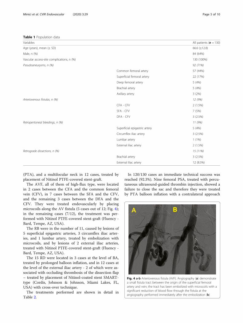
in 2 cases between the CFA and the common femoralvein (CFV), in 7 cases between the SFA and the CFV,and the remaining 3 cases between the DFA and theCFV. They were treated endovascularly by placingmicrocoils along the AV fistula (5 cases out of 12; Fig. 4);in the remaining cases (7/12), the treatment was per-formed with Nitinol PTFE-covered stent-graft (Fluency -Bard, Tempe, AZ, USA).The RB were in the number of 11, caused by lesions of
5 superficial epigastric arteries, 3 circumflex iliac arter-ies, and 1 lumbar artery, treated by embolization withmicrocoils, and by lesions of 2 external iliac arteries,treated with Nitinol PTFE-covered stent-graft (Fluency -Bard, Tempe, AZ, USA).The 15 RD were located in 3 cases at the level of BA,
treated by prolonged balloon inflation, and in 12 cases atthe level of the external iliac artery - 2 of which were as-sociated with occluding thrombosis of the dissection flap– treated by placement of Nitinol-coated stent SMART-type (Cordis, Johnson & Johnson, Miami Lakes, FL,USA) with cross-over technique.The treatments performed are shown in detail in
Table 2.
In 120/130 cases an immediate technical success wasreached (92.3%). Nine femoral PSA, treated with percu-taneous ultrasound-guided thrombin injection, showed afailure to close the sac and therefore they were treatedby PTA balloon inflation with a contralateral approach
Table 1 Population data
Variables All patients (n = 130)
Age (years), mean (± SD) 66.6 (±12.8)
Male, n (%) 84 (64%)
Vascular access-site complications, n (%) 130 (100%)
Pseudoaneurysms, n (%) 92 (71%)
Common femoral artery 57 (44%)
Superficial femoral artery 22 (17%)
Deep femoral artery 5 (4%)
Brachial artery 5 (4%)
Axillary artery 3 (2%)
Arteriovenous fistulas, n (%) 12 (9%)
CFA - CFV 2 (1.5%)
SFA - CFV 7 (5%)
DFA - CFV 3 (2.5%)
Retroperitoneal bleedings, n (%) 11 (9%)
Superficial epigastric artery 5 (4%)
Circumflex iliac artery 3 (2.5%)
Lumbar artery 1 (1%)
External iliac artery 2 (1.5%)
Retrograde dissections, n (%) 15 (11%)
Brachial artery 3 (2.5%)
External iliac artery 12 (8.5%)
Fig. 4 a-b Arteriovenous fistula (AVF). Angiography (a) demonstratea small fistula tract between the origin of the superficial femoralartery and vein; the tract has been embolized with microcoils with asignificant reduction of blood flow through the fistula at theangiography performed immediately after the embolization (b)
Minici et al. CVIR Endovascular (2020) 3:29 Page 5 of 10
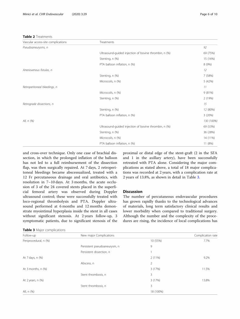
and cross-over technique. Only one case of brachial dis-section, in which the prolonged inflation of the balloonhas not led to a full reimbursement of the dissectionflap, was then surgically repaired. At 7 days, 2 retroperi-toneal bleedings became abscessualized, treated with a12 Fr percutaneous drainage and oral antibiotics, withresolution in 7–10 days. At 3 months, the acute occlu-sion of 3 of the 24 covered stents placed in the superfi-cial femoral artery was observed during Dopplerultrasound control; these were successfully treated withloco-regional thrombolysis and PTA. Doppler ultra-sound performed at 6 months and 12 months demon-strate myointimal hyperplasia inside the stent in all caseswithout significant stenosis. At 2 years follow-up, 3symptomatic patients, due to significant stenosis of the
proximal or distal edge of the stent-graft (2 in the SFAand 1 in the axillary artery), have been successfullyretreated with PTA alone. Considering the major com-plications as stated above, a total of 18 major complica-tions was recorded at 2 years, with a complication rate at2 years of 13.8%, as shown in detail in Table 3.
DiscussionThe number of percutaneous endovascular procedureshas grown rapidly thanks to the technological advancesof materials, long term satisfactory clinical results andlower morbidity when compared to traditional surgery.Although the number and the complexity of the proce-dures are rising, the incidence of local complications has
Table 2 Treatments
Vascular access-site complications Treatments
Pseudoaneurysms, n 92
Ultrasound-guided injection of bovine thrombin, n (%) 69 (75%)
Stenting, n (%) 15 (16%)
PTA balloon inflation, n (%) 8 (9%)
Arteriovenous fistulas, n 12
Stenting, n (%) 7 (58%)
Microcoils, n (%) 5 (42%)
Retroperitoneal bleedings, n 11
Microcoils, n (%) 9 (81%)
Stenting, n (%) 2 (19%)
Retrograde dissections, n 15
Stenting, n (%) 12 (80%)
PTA balloon inflation, n (%) 3 (20%)
All, n (%) 130 (100%)
Ultrasound-guided injection of bovine thrombin, n (%) 69 (53%)
Stenting, n (%) 36 (28%)
Microcoils, n (%) 14 (11%)
PTA balloon inflation, n (%) 11 (8%)
Table 3 Major complications
Follow-up New major Complications Complication rate
Periprocedural, n (%) 10 (55%) 7.7%
Persistent pseudoaneurysm, n 9
Persistent dissection, n 1
At 7 days, n (%) 2 (11%) 9.2%
Abscess, n 2
At 3 months, n (%) 3 (17%) 11.5%
Stent thrombosis, n 3
At 2 years, n (%) 3 (17%) 13.8%
Stent thrombosis, n 3
All, n (%) 18 (100%)
Minici et al. CVIR Endovascular (2020) 3:29 Page 6 of 10

not substantially changed over the years (Tsetis 2010;Kopin et al. 2019).The arteries of the upper limb (radial, brachial, and
rarely axillary) are used as arterial access, especially incardiologic procedures. The trans-radial approach is as-sociated with a lower incidence of major complicationswhen compared to femoral approach (Rigattieri et al.2016); the most common major complication is the oc-clusion of the radial artery, which rarely leads to clinicalmanifestations, due to the dual arterial supply guaran-teed at the hand (Reich et al. 2017). The major compli-cations associated with the brachial approach arethrombosis, PSA and nerve compression; ischemic com-plications (dissection/thrombosis) are more common atthis level than in femoral approach (Romaguera et al.2012; Johnson et al. 1994).At the level of lower limbs, the popliteal access is the
least common (sometimes used in the recanalization ofchronic occlusions of the femoropopliteal arterial axis)(Ortiz et al. 2014); major complications are dissectionand PSA (Chan and Common 2004).The CFA is by far the most used vascular access be-
cause it has several advantages over other sites: the largecalibre, which allows the introduction of larger cathetersand easier cannulation; the femoral head underneathguarantee haemostasis with manual compression (Tsetis2010; Tonnessen 2011; Keeling et al. 2009; Reich et al.2017). Due to the highest number of procedures per-formed via this route, femoral access is subject to morefrequent complications than other arterial access (Ortizet al. 2014).Bleeding is the most frequent complication; hematoma
may present as stable or unstable (uncontrolled bleed-ing), retroperitoneal haemorrhage or PSA. The incidenceof femoral PSA varies from 0.1% to 1.5% after diagnosticangiography and up to 7.7% after interventional proce-dures and increases with the complexity of procedures,patient age and the presence of bleeding disorders(Graham et al. 1992; Carey et al. 2001; Erol et al. 2015).SFA or DFA accesses, compared with CFA accesses,
are more likely to lead to PSA or AVF formation, due tothe smaller size and the lack of bone support againstwhich compress after sheath removal (Morgan and Belli2003).The natural history of iatrogenic femoral PSA is un-
certain (Graham et al. 1992). In a case series of 147 pa-tients, Thalhammer et al. (2000) reported that 86% had aspontaneous resolution (PSA and AVF) after a mean of23 days. Although the rupture of the femoral PSA can bea life-threatening condition (Graham et al. 1992), someauthors believe sufficient Doppler ultrasound observa-tion alone, especially for asymptomatic patients notreceiving anticoagulation therapy with little PSA(diameter ≤ 3 cm) (Johns et al. 1991).
Surgical exploration of a pseudoaneurysm is oftenchallenged by the presence of a haematoma. After theevacuation of the haematoma, another difficulty is theidentification of the bleeding site, that may be more thanone. Finally, surgical treatment may require simple su-ture of the defect or a patch angioplasty (surgical repairusing a patch). Considering the aforementioned limita-tions, surgical repair is indicated in the cases of haemo-dynamically relevant bleeding or shock with a rapidlyexpanding haematoma, risk of skin necrosis due to pres-sure by the haematoma and infectious pseudoaneurysmto ensure debridement of infected tissue (Tisi andCallam 2006; Savolainen et al. 2011). In 1991, theultrasound-guided compression was suggested (Fellmethet al. 1991); the advantages are its simplicity, low costand lack of ionizing radiation. However, this procedurehas some limitations, related to the pain threshold of thepatient and the time necessary to obtain complete clos-ure. Moreover, failure is increased in patients on anti-coagulant therapy (Hajarizadeh et al. 1995).In 1997, Liau et al. (1997) reported the successful use
of percutaneous injection of thrombin with ultrasoundguidance for the closure of 5 cases of PSA of the CFA.In the series of 15 patients reported by Brophy et al.(2000), all the PSA have been successfully treated with500–1000 U of bovine thrombin, regardless of the PSAsize.Later, in a series of 54 PSA (divided into simple, 45,
and complex, 9) reported by Sheiman et al. (2001), thetechnical success was 50/54, with the possibility of a sec-ond approach in case of failure. In some cases (2–4%)you can proceed to a further ultrasound-guided injectionprocedure with an overall high success rate. However, inliterature, the ultrasound-guided injection technique hasa failure rate of 3% -14% (Maleux et al. 2003).The greatest risk from thrombin injection is distal
embolization (which can cause serious complications upto limb loss). Pezzullo et al. (2000) have described distalembolization in one of the 23 patients studied. Theplacement of the tip of the needle at distance from theneck of the pseudoaneurysm under ultrasound guidanceand the slow injection of the drug under ultrasoundcontrol minimizes the risk (Maleux et al. 2003; Pezzulloet al. 2000).Other side effects of thrombin injection include
hypotension and bradycardia, bleeding - because of an‘acquired inhibition of coagulation factor (XI) secondaryto immune cross-reactivity of bovine thrombin - andanaphylactic reactions in patients who have had repeatedexposure to bovine thrombin (Pope and Johnston 2000).Different forms of thrombin are commercially available,the majority of which are of bovine origin and have beenused for many years (Pezzullo et al. 2000; Samal et al.2001). Because of these risks is currently preferred the
Minici et al. CVIR Endovascular (2020) 3:29 Page 7 of 10

human thrombin, which implies a slightly higher cost(Vázquez et al. 2005).The use of human thrombin would not entail the risks
associated with bovine thrombin or any other immuno-logical risk; it must be acknowledged, however, thatthere may be a small risk of infection, although not yetconfirmed (Vázquez et al. 2005).Quarmby et al. (2002) described the embolization of
10 PSA through the use of autologous thrombin withimmediate technical success in 7 cases and the use ofthe new administration of the same in 3 cases.Loose and Haslam described the percutaneous tech-
nique of blocking flow through percutaneous balloonangioplasty (PTA) inflated for about 15 min to preventdistal embolization during injection under ultrasoundguidance in the sac of the PSA. Their method was effect-ive (12/13 cases successfully treated without complica-tions), but is rather expensive and employ ionizingradiation (Loose and Haslam 1998).The use of stent-graft in the treatment of aortoiliac
disease has recently led several authors to employthese devices to exclude aneurysms and peripheralPSA (Xiao et al. 2012; Laganà et al. 2002). In a largerseries of stent-graft used in the treatment of pseudoa-neurysms and AVF, Waigand et al. (1999) andThalhammer et al. (2000) reported a technical successrate of 84–88%. However, there are limits to the indi-cation, as the stent-graft may result in the occlusionof the DFA; theoretically, this complication can pre-vent the use of the site as future access. Hence, afemoral angiography from an oblique projectionshould be performed to evaluate the suitability of thedevice insertion, as it allows to check the presence ofan adequate landing zone in the CFA respect to theorigin of the profunda femoral artery. Besides, hipstress can lead to an increased risk of stent throm-bosis (Thalhammer et al. 2000; Xiao et al. 2012).Retroperitoneal bleedings (RB) are potentially fatal and
not easy to diagnose in clinically haemodynamicallystable patients; RB may occur spontaneously in the pres-ence of coagulopathy, use of combined antiplatelet drugs(eg. dual antiplatelet therapy), oral anticoagulant therapywith heparin or poorly controlled partial thromboplastin.Generally, the target population includes elderly popula-tion. Usually, retroperitoneal hematomas present withabdominal and back pain, less frequently with fatigue,nausea, headache, and dyspnea (Baekgaard et al. 2019;Dolapsakis et al. 2019).Besides, the RB may be iatrogenic; in particular, they
may occur as a complication of arterial or venouscatheterization during endovascular procedures. Theretroperitoneal bleedings may result from the femoralpuncture and are spread either through the Retziusspace or directly on the muscles of the pelvic floor
without the direct involvement of this space (Terotolaet al. 1991).Usually, AVF are between the SFA, the DFA and the
adjacent lateral circumflex femoral vein. The majority ofstudies reported a high probability of spontaneous clos-ure (Toursarkissian et al. 1997), but Kresowik et al.(1991) reported opposite results. Toursarkissian et al.(1997) reported that 86% of the non-symptomatic low-flow AVF resolves spontaneously, requiring only obser-vation and close Doppler ultrasound monitoring every 2weeks until 12 weeks. Until recently, the AVF symptom-atic were treated surgically (Kresowik et al. 1991; Kelmet al. 2002). More recently, percutaneous treatment withstent-graft showed constant technical success, but with asignificant risk of stent occlusion - medium and long-term graft (12% -17%) (Laganà et al. 2002).Dissection is a frequent but often underdiagnosed
complication of arterial catheterization because manycases are asymptomatic (Benjamin et al. 2015). Duringvascular access, coiling of the guidewire under fluoros-copy or resistance to passage may indicate subintimalpassage with resulting retrograde arterial dissection. Thiscomplication is more common in the iliac arteries dueto the presence of plaques by steno-occlusive athero-sclerotic disease and to the tortuosity of these vessels.The operator should immediately recognize the dissec-tion and evaluate the hemodynamic consequences. Gen-erally, the flap of dissection doesn’t cause stenosis andthe simple removal of the catheter and guide usually al-lows a spontaneous resolution, also due to the antero-grade flow of blood. If the flap dissection is not felt bythe sensitivity of the operator and extends for a longerdistance gaining the true lumen, the flap may becomeocclusive - then counter - with acute ischemia, so that itis necessary an immediate repair by angioplasty or stent-ing (Tsetis and Belli 2004).Despite the wide use of vascular closing devices, there
are no studies that prove with certainty their superiorityin terms of safety compared to manual compression.Only the study of Tavris et al. (2004) demonstrated thatvascular complications are lower with the use of VCD.One hundred sixty-six thousand six hundred eightypatients of the National Registry of the AmericanCollege of Cardiology database, undergoing cardiaccatheterization in 2001, have been evaluated; 53,655were treated with VCD for haemostasis, while in 113,025was performed the manual compression. The risk ofvascular complication was 1.1% in the group with VCDand 1.7% in the group with manual compression, withp < 0.001, statistically significant.The evolution of metal alloys with flexible and elastic
materials and the technological innovation on the poros-ity of the cover have now led to the use of stents with ahigher patency rate and a lower rate of acute
Minici et al. CVIR Endovascular (2020) 3:29 Page 8 of 10

thrombosis. All this has led to greater use of devicesclose to the articular femoral site and the possibility ofre-using the access through the stent struts, clearly withcatheters and introducers of small arms. A high tech-nical success rate (92.3%) has been achieved, with a low(13.8%) complication rate at 2 years.Limitations of the study are the lack of long term fol-
low up, except for the cases of stent-graft placement, theretrospectivity of the analysis and the scarcity of data inthe literature, necessary to evaluate the congruence andthe consistency of the data presented.
ConclusionsThe percutaneous treatment of vascular access-site com-plications is the first-choice treatment. It represents asafe and effective option, validated by a high technicalsuccess rate and a low long-term complication rate, thatallows avoiding the surgical approach in most cases.
AbbreviationsCFA: Common femoral artery; VCD: Vascular closure devices;PSA: Pseudoaneurysms; AVF: Arteriovenous fistulas; RD: Retrogradedissections; RB: Retroperitoneal bleedings; SFA: Superficial femoral artery;DFA: Deep femoral artery; AA: Axillary artery; BA: Brachial artery;CFV: Common femoral vein; PTA: Percutaneous transluminal angioplasty
AcknowledgementsNot applicable.
Authors’ contributionsRM and DL conceived the study concepts and design. RM, MT, SP and LZperformed literature research and assisted in data collection. RM, MP and KAwere in charge of manuscript editing. All authors read and approved thefinal manuscript.
FundingNot applicable.
Availability of data and materialsThe datasets used and/or analysed during the current study are availablefrom the corresponding author on reasonable request.
Ethics approval and consent to participateApproval by ethic committee of Magna Graecia University and writteninformed consent by patients were obtained.
Consent for publicationNot applicable.
Competing interestsThe authors declare that they have no competing interests.
Author details1Radiology Division, Department of Experimental and Clinical Medicine,Magna Graecia University of Catanzaro, University Hospital Mater Domini,Viale Europa, 88100 Catanzaro, CZ, Italy. 2IRC - FSH, Department of HealthSciences, Magna Graecia University of Catanzaro, Catanzaro, Italy. 3CardiologyDivision, Department of Biomedical, Metabolic and Neural Sciences,University of Modena and Reggio Emilia, Policlinico di Modena, Modena,Italy. 4Anaesthesia and Intensive Care Unit, Department of Medical andSurgical Sciences, Magna Graecia University of Catanzaro, University HospitalMater Domini, Catanzaro, Italy. 5Radiology Division, University of Insubria,Varese, Italy.
Received: 16 March 2020 Accepted: 30 April 2020
ReferencesBaekgaard JS, Eskesen TG, Lee JM, Yeh DD, Kaafarani HMA, Fagenholz PJ, Avery L,
Saillant N, King DR, Velmahos GC (2019) Spontaneous retroperitoneal andrectus sheath hemorrhage-management, risk factors and outcomes. World JSurg 43(8):1890–1897. https://doi.org/10.1007/s00268-019-04988-y
Benjamin HGE et al (2015) Iatrogenic percutaneous vascular injuries: clinicalpresentation, imaging, and management. Semin Intervent Radiol 32:108–122
Brophy DP, Sheiman RG, Amatulle P, Akbari CM (2000) Iatrogenic femoralpseudoaneurysms: thrombin injection after failed US guided compression.Radiology 214:278–282
Carey D, Martin JR, Moore CA, Valentine MC, Nygaard TW (2001) Complications offemoral artery closure devices. Cathet Cardiovasc Interv 52:3–7
Chan RP, Common AA (2004) Stent-graft repair of femoral pseudoaneurysm/AVfistula using a retrograde popliteal approach. Cardiovasc Intervent Radiol27(5):516–519 Epub 2004 Jun 23
Cianci C, Kowal RC, Feghali G, Hohmann S, Stoler RC, Choi JW (2013) Criticallower limb ischemia from an embolized angio-seal closure device. Proc (BaylUniv Med Cent) 26(4):398–400
Dolapsakis C, Giannopoulou V, Grivakou E (2019) Spontaneous retroperitonealhemorrhage. J Emerg Med 56(6):713–714. https://doi.org/10.1016/j.jemermed.2019.01.037
Erol F, Arslan Ş, Yüksel İÖ, Üreyen ÇM, Serdar S, İnci S, Şenocak H (2015)Determinants of iatrogenic femoral pseudoaneurysm after cardiaccatheterization or percutaneous coronary intervention via the femoral artery.Turk Kardiyol Dern Ars 43(6):513–519. https://doi.org/10.5543/tkda.2015.30356
Fellmeth BD, Roberts AC, Bookstein JJ et al (1991) Post-angiographic femoralartery injuries: nonsurgical repair with US-guided compression. Radiology178:671–675
Graham AN, Wilson CM, Hood JM, Barros D’Sa AA (1992) Risk of rupture of post-angiographic femoral false aneurysm. Br J Surg 79:1022–1025
Hajarizadeh H, LaRosa CR, Cardullo P, Rohrer MJ, Cutler BS (1995) Ultrasound-guided compression of iatrogenic femoral pseudoaneurysm: failure,recurrence, and long-term results. J Vasc Surg 22:425–433
Horie K, Tanaka A, Taguri M, Kato S, Inoue N (2018) Impact of prolonged inflationtimes during plain balloon angioplasty on angiographic dissection inFemoropopliteal lesions. J Endovasc Ther 25(6):683–691. https://doi.org/10.1177/1526602818799733
Johns JP, Pupa LE Jr, Bailey SR (1991) Spontaneous thrombosis of iatrogenicfemoral artery pseudoaneurysms: documentation with color Doppler andtwo-dimensional ultrasonography. J Vasc Surg 14:24–29
Johnson LW, Esente P, Giambartolomei A et al (1994) Peripheral vascularcomplications of coronary angioplasty by the femoral and brachialtechniques. Catheter Cardiovasc Diagn 31:165–172
Katzenschlager R, Ugurluoglu A, Ahmadi A et al (1995) Incidence ofpseudoaneurysm after diagnostic and therapeutic angiography. Radiology195:463–466
Keeling AN, McGrath FP, Lee MJ (2009) Interventional radiology in the diagnosis,management, and follow-up of Pseudoaneurysms. Cardiovasc InterventRadiol 32(1):2–18 Epub 2008 Oct 16
Kelm M, Perings SM, Jax T et al (2002) Incidence and clinical outcome ofiatrogenic femoral arteriovenous fistulas: implications for risk stratificationand treatment. J Am Coll Cardiol 40:291–297
Kopin D, Seth M, Sukul D et al (2019) Primary and secondary vascular access sitecomplications associated with percutaneous coronary intervention: insightsfrom the BMC2 registry. JACC Cardiovasc Interv 12:2247–2256
Kresowik TF, Khoury MD, Miller BV et al (1991) A prospective study of theincidence and natural history of femoral vascular complications afterpercutaneous transluminal coronary angioplasty. J Vasc Surg 13(2):328–333
Laganà D, Mangini M, Marras M et al (2002) Percutaneous treatment of femoro-popliteal aneurysms with covered stents. Radiol Med 104(4):322–331
Liau CS, Ho FM, Chen MF, Lee YT (1997) Treatment of iatrogenic femoralartery pseudoaneurysm with percutaneous thrombin injection. J VascSurg 26:18–23
Loose HW, Haslam PJ (1998) The management of peripheral arterial aneurysmsusing percutaneous injection of fibrin adhesive. Br J Radiol l71:1255–1259
Maleux G, Hendrickx S, Vaninbroukx J et al (2003) Percutaneous injection ofhuman thrombin to treat iatrogenic femoral pseudoaneurysms: short- andmidterm ultrasound follow-up. Eur Radiol 13:209–212
Minici et al. CVIR Endovascular (2020) 3:29 Page 9 of 10
Morgan R, Belli AM (2003) Current treatment methods for post catheterizationpseudoaneurysms. J Vasc Interv Radiol 14(6):697–710
Ortiz D, Jahangir A et al (2014) Access site complications after peripheral vascularinterventions. Circ Cardiovasc Interv 7:821–828
Pezzullo JA, Dupuy DE, Cronan JJ (2000) Percutaneous injection of thrombin forthe treatment of pseudoaneurysms after catheterization: an alternative tosonographically guided compression. Am J Roentgenol 175:1035–1040
Pope M, Johnston KW (2000) Anaphylaxis after thrombin injection of a femoralpseudoaneurysm: recommendations for prevention. J Vasc Surg 32:190–191
Quarmby JW, Christoph E, Chitolie A, Morgan RA, Belli A (2002) Autologousthrombin for treatment of pseudoaneurysms. Lancet 359(9310):946-947
Reich R et al (2017) Vascular access complications in patients undergoingpercutaneous procedures in hemodynamics: a scoping review. Rev GaúchaEnferm 38(4):e68716
Rigattieri S, Sciahbasi A, Ratib K, Alonzo A, Cox N, Chodór P, Berni A, Fedele S,Pugliese FR, Cooper CJ, Louvard Y, Nolan J, Rao SV (2016) Comparisonbetween radial approach and femoral approach with vascular closuredevices on the occurrence of access-site complications and Periproceduralbleeding after percutaneous coronary procedures: a systematic review andmeta-analysis. J Invasive Cardiol 28(12):473–479
Romaguera R, Wakabayashi K, Laynez-Carnicero A et al (2012) Associationbetween bleeding severity and long-term mortality in patients experiencingvascular complications after percutaneous coronary intervention. Am JCardiol 109(1):75–81 Epub 2011 Sep 29
Samal AK, White CJ, Collins TJ, Ramee SR, Jenkins JS (2001) Treatment of femoralartery pseudoaneurysm with percutaneous thrombin injection. CathetCardiovasc Interv 53:259–263
Savolainen H, Baumgartner I, Schmidli J, Heller G, Do DD, Willenberg T (2011)Femoral Pseudoaneurysm Requiring Surgical Treatment. Trauma Mon 16(4):194–197. https://doi.org/10.5812/kowsar.22517464.3186
Sheiman RG, Brophy DP (2001) Treatment of iatrogenic femoralpseudoaneurysms with percutaneous thrombin injection: experience in 54patients. Radiology 219:123–127
Tavris DR, Galluresi BA, Lin B et al (2004) Risk of local adverse events followingcardiac catheterization by haemostasis device use and gender. J InvasiveCardiol 16(9):459–464
Terotola SO, Khulman JE, Fischman EK (1991) CT and anatomic study ofpostcatheterization haematomas. Radiographics 11:247–258
Thalhammer C, Kirchherr AS, Uhlich F, Waigand J, Gross CM (2000) Postcatheterization pseudoaneurysms and arteriovenous fistulas: repair withpercutaneous implantation of endovascular covered stents. Radiology214:127–131
Tisi PV, Callam MJ (2006) Surgery versus non-surgical treatment for femoralpseudoaneurysms. Cochrane Database Sys Rev (1):CD004981.https://doi.org/10.1002/14651858.CD004981.pub2
Tonnessen BH (2011) Iatrogenic injury from vascular access and endovascularprocedures. Perspect Vasc Surg Endovasc Ther 23(2):128–135
Toursarkissian B, Allen BT, Petrinec D et al (1997) Spontaneous closure of selectediatrogenic pseudoaneurysms and arteriovenous fistulae. J Vasc Surg 25:803–808
Tsetis D (2010) Endovascular treatment of complications of femoral arterialaccess. Cardiovasc Intervent Radiol 33(3):457–468 Epub 2010 Feb 17
Tsetis D, Belli AM (2004) Guidelines for stenting in infrainguinal arterial disease.Cardiovasc Intervent Radiol 27(3):198–203
Vázquez V, Reus M, Piñero A et al (2005) Human thrombin for treatment ofpseudoaneurysms: comparison of bovine and human thrombin sonogram-guided injection. Am J Roentgenol 184(5):1665–1671
Waigand J, Uhlich F, Gross CM, Thalhammer C, Dietz R (1999) Percutaneoustreatment of pseudoaneuryms and arteriovenous fistulas after invasivevascular procedures. Cathet Cardiovasc Interv 47:157–164
Xiao L, Shen J, Tong JJ (2012) Emergency stent-graft implantation for iatrogenicperipheral arterial rupture. Radiol Med 118(1):152-157
Xiong J, Liu M, Guo W et al (2012) A retrospective study on endovascularmanagement of iatrogenic vascular injuries. Vascular 20(2):65–71 Epub 2012Apr 4
Publisher’s NoteSpringer Nature remains neutral with regard to jurisdictional claims inpublished maps and institutional affiliations.
Minici et al. CVIR Endovascular (2020) 3:29 Page 10 of 10