pedscases: birthmarks
TRANSCRIPT

PedsCases: Birthmarks
Written by Aryan Riahi, MSI, University of British Columbia and
Dr. Joseph Lam, Pediatric Dermatologist, University of British Columbia

Introduction
• Birth marks are discolorations on the skin that are present at birth or during the first weeks of life.
• Overgrowth of blood vessels, melanocytes, fat, fibroblasts, keratinocytes, or smooth muscle.
• Caused by an imbalance in factors that determine the migration of skin cells
• Divided into pigmented and vascular birthmarks.
• While most are noncancerous, effective identification and monitoring is vital.

Objectives
1. Compare and contrast common paediatric vascular anomalies including vascular tumours and vascular malformations.
2. Compare and contrast common birthmarks including congenital melanocytic nevi, dermal melanocytosis, sebaceous nevus, and aplasia cutis.
3. Determine which birthmarks require diagnostic workup.
4. Discuss complications of congenital melanocytic nevi.
5. Identify key questions on history, physical exam, and management.

Case 1
• History of presenting illness: Sara is a 4 week old girl who presents to the clinic with her parents who are concerned about a bruise-like patch on her left shoulder that is growing.
• Past medical history: Sara weighed less than 6 lbs at birth. She and her twin sister were delivered via an uncomplicated Caesarean section.
• Family history: The mother is a healthy 34 year old woman with no medication exposure during pregnancy.

Case 1

Infantile Hemangioma
• Do you know the two categories of pediatric vascular anomalies? How can we tell them apart?
• When should the parents expect their child’s IH to grow the most rapidly?
• What are some physical exam findings for IH?
• What is the management of IH?

Other Common Vascular Birthmarks: Nevus flammeus
• Colloquially known as “Port-Wine Stain”.
• Congenital malformations in the dermal capillaries.
• Red macules that follow a dermatomal distribution and usually do not cross midline.
• Treatment is laser for cosmesis.

Other Common Vascular Birthmarks: Nevus flammeus

Other Common Vascular Birthmarks: Nevus Simplex
• Colloquially known as “Salmon Patch”.
• Congenital dilation of the dermis’ capillaries.
• Nearly one-third of newborns present with these pink or red irregular patches.
• Present in the glabellar area, nape of the neck, and occiput.
• The majority spontaneously regress.

Other Common Vascular Birthmarks: Nevus Simplex
Case 2
• History of presenting illness: Rosie is a 2 week old girl who presents to the clinic with her parents as they are concerned about a large brown area on the right calf. The parents would like to know if this is malignant.
• Past medical history: Rosie had an uncomplicated birth. She was a full-term and underwent vaginal delivery.
• Family history: The mother is a healthy 34 year old woman with no medication exposure during pregnancy.
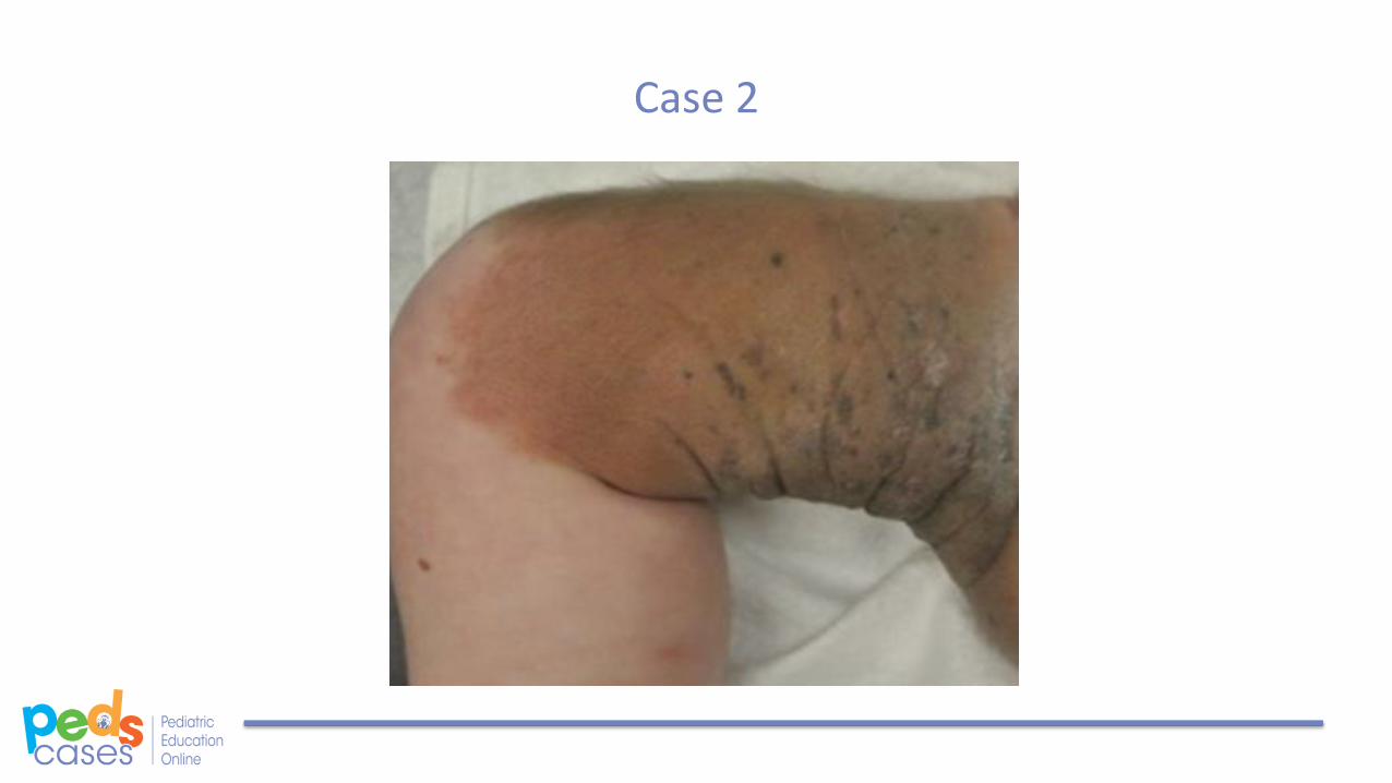
Case 2

Congenital Melanocytic Nevus
• What are some physical exam findings for congenital melanocytic nevi (CMN)?
• What is the management for congenital melanocytic nevi?
• What determines malignancy potential?
• When do we consider excision?

Case 3
• History of presenting illness: Louis is a 2 year old male of Hispanic descent who presents with a large dark gray patch on the lower back and extending to the buttock. This patch has been present since birth. He is otherwise healthy.
• Past medical history: Full term, uncomplicated vaginal delivery.
• Family history: Older sister is 10 years old and was born with a similar patch, but no longer appears to have this pigmentation.

Case 3

Congenital Melanocytosis
• What is the pathogenesis of congenital dermal melanocytosis (CDM)?
• What are the common terms used to refer to CDM?
• What are some physical exam findings for CDM?
• What is the management for CDM?
• Which demographic is more susceptible to developing CDM?
• What is the prognosis for CDM?
Case 4
• History of presenting illness: Eli is a 3 year old male presenting with a hairless lesion on the right side of his forehead proximal to his right eyebrow.
• Past medical history: He had an uncomplicated full-term vaginal delivery. No medications were taken by the mother throughout the course of her pregnancy.

Case 4

Nevus Sebaceous
• What is nevus sebaceous?
• What are some physical exam findings for nevus sebaceous?
• What is the management for nevus sebaceous?
• What are some considerations for malignant potential?
• When should surgical removal be considered?

Case 5
• History of presenting illness: Tricia is a 30-day-old female who presented with an ulcer at birth on the scalp that has now healed but has left behind skin changes.
• Past medical history: Full term uncomplicated vaginal delivery. The mother is a healthy 30 year old woman with no medication use throughout her pregnancy.

Case 5
Aplasia Cutis
• What is aplasia cutis congenita (ACC)?
• What are some physical exam findings for ACC?
• What is a “hair collar sign”?
• What is the management for ACC?
• How is the diagnosis of ACC made?
• When is imaging required?
• When is genetic counseling required?
• What is the prognosis for ACC?

Conclusion
• Infantile hemangioma has a variety of complications depending on its location. Be familiar with association with PHACES.
• Congenital melanocytic nevus is a benign proliferation of melanocytes. Follow large or giant CMN lesions for risk of melanoma.
• Congenital dermal melanocytosis involves entrapment of the skin’s melanocytes in the dermis. Most are benign.
• Nevus sebaceous is a congenital hamartoma of the skin. They are more visually apparent in adolescence.
• Aplasia cutis congenita involves an absence of skin from birth. Be familiar with associations with neural tube defects and “hair collar sign”.

References
Literature references:
1. Del Pozzo-Magana B, Dizon M, et al. “Newborn skin disease, Part 1: Birthmarks.” In: Society for Pediatric Dermatology and the American Academy of Dermatology’s Basic Dermatology Curriculum. Peer review by: Maguiness S. May 2016.
2. Marqueling AL et al. Pediatr Dermatol 2013;30:182–91.
3. Blei F et al. Pediatr Dermatol 2014;31:465–70.
4. Cheng, C. E., & Friedlander, S. F. (2016). Infantile hemangiomas, complications and treatments. Seminars in Cutaneous Medicine and Surgery, 35(3), 108–116. https://doi.org/10.12788/j.sder.2016.050
5. Orlow SJ, Isakoff MS, Blei F. Increased risk of symptomatic hemangiomas of the airway in association with cutaneous hemangiomas in a "beard" distribution J Pediatr. 1997 Oct;131(4):643-6..
6. Leung AK. Mongolian spots in Chinese children. Int J Dermatol. 1988 Mar;27(2):106-8
Images courtesy of www.dermnetnz.com