pediatric rheumatology: a case-based approach to … rheumatology: a case-based approach to the...
TRANSCRIPT

Pediatric Rheumatology:A Case-based Approach
to the Basics
Hilary M. Haftel, MD, MHPEUniversity of Michigan
Department of Pediatrics

Disclosures
I have no relevant financial relationships with the manufacturer(s) of any commercial product(s) and/or provider of commercial services discussed in this CME activity
I am not discussing any off-label uses of medications
Duty hours (if they applied to me) were violated in the making of this presentation

Objectives
Identify signs and symptoms of common rheumatologic disorders
Understand appropriate laboratory testing in each disorder
Understand disease course and therapy for common rheumatologic disorders
The “Five” Pediatric Rheumatology Disorders
Juvenile Arthritis Systemic Lupus Erythematosus Juvenile Dermatomyositis Scleroderma (localized and systemic) Vasculitis

Case One
A two-year-old female is brought to your office by her mother who reports that the patient was running in the front yard and fell and the mother, upon picking her up, noticed that the right knee was swollen and is worried that “something is broken”
Exam reveals a large effusion, widening of the surrounding bones, and you are unable to fully extend the knee on range of motion

Juvenile Idiopathic Arthritis (JIA)(FKA Juvenile Rheumatoid Arthritis)
Most common childhood arthritis** Occurs in 1:1000 children Affects children of all ages, but two
peaks, at age 1-2 and age 8-12**

Basic Questions to Ask
Acute or Chronic? Localized to joints or more systemic
features? Fever or not? Actual arthritis or arthalgias? (ie., evidence
of inflammation?)Complaints proportionate to physical findings
or not?

JIA: Diagnosis**
Evidence of joints inflammation-redness-swelling-limitation of range of motion-pain
Duration of Arthritis > 6 weeksAge at onset < 16 years

JIA: Evidence of Chronicity**
Synovial thickening Bony proliferation Contracture Limb length discrepancy

Classification of JIA Oligoarticular JIA
involvement of <5 joints Polyarticular JIA
involvement of ≥ 5 joints RF positive vs. RF negative
Systemic JIA presence of systemic inflammation
Juvenile Spondyloarthopathies Juvenile ankylosing spondylitis Inflammatory bowel disease-related arthritis Juvenile psoriatic arthritis Reactive Arthritis (FKA Reiter’s syndrome)

Oligoarticular JIA:Clinical Features
Less than 5 joints Typically affects
medium to large joints
Asymmetric
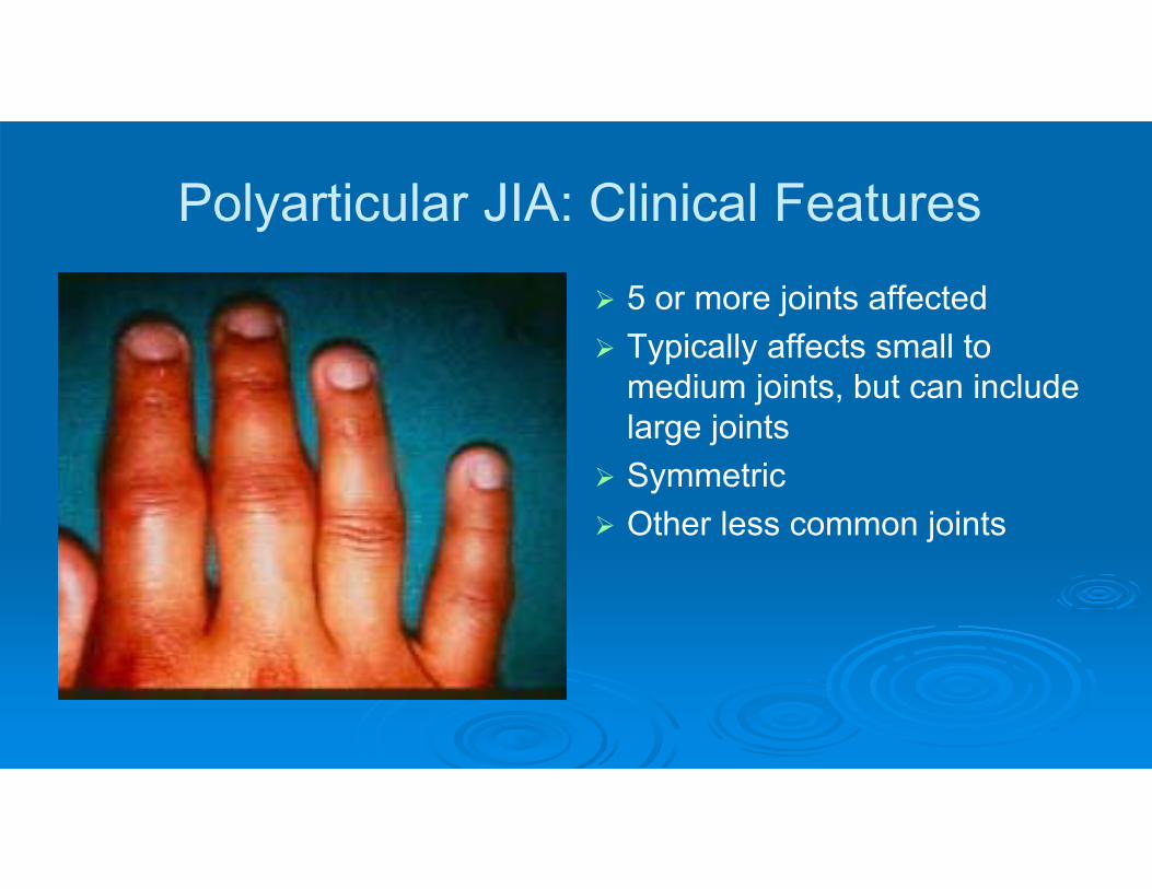
Polyarticular JIA: Clinical Features
5 or more joints affected Typically affects small to
medium joints, but can include large joints
Symmetric Other less common joints

Polyarticular JIA: Other Features
Temporomandibular Joint Cervical Spine Involvement

Systemic JIA (aka Still’s Disease)
Think of it as a systemic inflammation that precedes the onset of arthritis typical fever curve**
• daily or diurnal temperature spike over 39ºC
• returns quickly to below baseline• child feels well between temperature
spikes

Systemic JIA (aka Still’s Disease)
Characteristic rash** • occurs frequently at peak of fever• erythematous macules on trunk and
proximal extremities• migratory and evanescent

Still’s rash

Koebner’s Phenomenon(dermatographia)
Isomorphic reaction in which skin lesions appear at the site of trauma
Systemic JIA (Cont’d) Signs of visceral involvement**
hepatomegaly splenomegaly lymphadenopathy serositis
• pleuritis• pericarditis, including pericardial effusions• peritonitis
No peak age of onsetNo gender predilection


Laboratory Testing in JIA
Evidence of systemic inflammation** white blood cell count red blood cell count platelet count erythrocyte sedimentation rate C reactive protein serum proteins
Antinuclear antibodiesRheumatoid factors
Rheumatoid Factors
Antibodies directed against the Fc portion of another antibody
Rheumatoid factors are negative in children with JIA** Rheumatoid factors are positive in adolescents and
adults with rheumatoid arthritis Most common reason for child to make a RF is
infection.

ANAs in JIA
Frequency varies from 24-66% overallHighest frequency in patients with young (<7yrs), femaleHighest prevalence in patients with oligoarticular JIA and
uveitis** BUT…up to 20% false positive rate, especially with Hep-2
substrate

Laboratory Studies for Critical Exclusions
Joint fluid analysis -if acute presentation, must exclude septic arthritis
Complete blood count try to exclude hematologic malignancy
Serologic testing for Lyme Radiography
may show peri-articular osteopenia
may demonstrate other abnormalities, including osteomyelitis or bone cancer

Goals of therapy for JIA**
Control of pain and inflammation Preservation and improvement of function Prevention of disability and chronic deformity
Pharmacologic Therapy for JIA**
Anti-inflammatory agents-NSAIDs-ASA-[corticosteroids]
Disease-modifying agents-hydroxychloroquine-methotrexate-sulfasalazine-biologic agents (etanercept, infliximab, adalimumab, etc)-anakinra
Complications of JIA: Uveitis**
Inflammation of the iris, ciliary body, and choroid Tends to be asymptomatic in children Screening in required for all children with chronic
arthritis** Number One treatable cause of blindness in children**
Complications of JIA**
Musculoskeletal Deformity Regular stretching program Splinting or bracing Regular physical exercise
Constitutional Growth DelayMacrophage Activation Syndrome (HLH)
Outcome in JIA
Remission rates 70-85% within 2-5 years** oligoarticular >90% polyarticular ~50% systemic <50%
After disease, all that’s left is chronic deformities and visual disturbance previously untreated

Case Two
A 14-year-old male presents to the local ER in September with complaints of headache for one week. He also notes some weight loss, rash on his face, and diffuse joint pain
Exam reveals a tired-appearing male with a pulse of 122, blood pressure 186/96, resp rate 16, temp 36.7
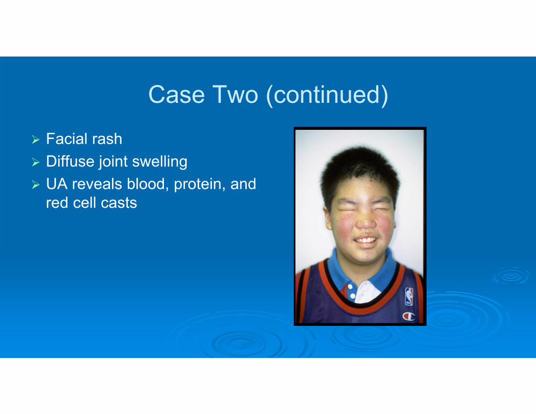
Case Two (continued)
Facial rash Diffuse joint swelling UA reveals blood, protein, and
red cell casts

Systemic Lupus Erythematosus
Incidence/Prevalence 0.53-0.6 per 100,000 incidence approx 5-10,000 cases in US
Approximately 15% present before age 16 rare < 5 years, in freq through adolescence
Gender 0-9 years, 4:3 F:M 10-14 yrs, 4:1 15-19 yrs, 5:1
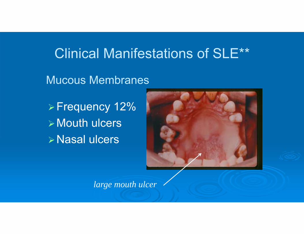
Clinical Manifestations of SLE**
Frequency 12%Mouth ulcersNasal ulcers
large mouth ulcer
Mucous Membranes

Clinical Manifestation of SLE** Musculoskeletal (72%)
arthralgias/arthritis myalgias/myositis
Serositis (30-40%) pericarditis pleuritis peritonitis
Cardiovascular (15%) raynaud’s phenomenon myocarditis/endocarditis
Clinical Manifestations of SLE**
Renal (82%) proteinuria casts
Neurologic (44%) seizures psychosis Depression
Hematologic (50%) Bleeding/bruising Petechiae
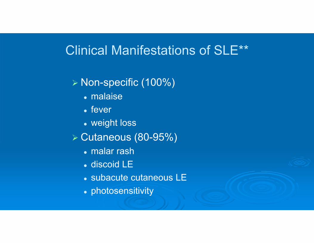
Clinical Manifestations of SLE**
Non-specific (100%) malaise fever weight loss
Cutaneous (80-95%) malar rash discoid LE subacute cutaneous LE photosensitivity
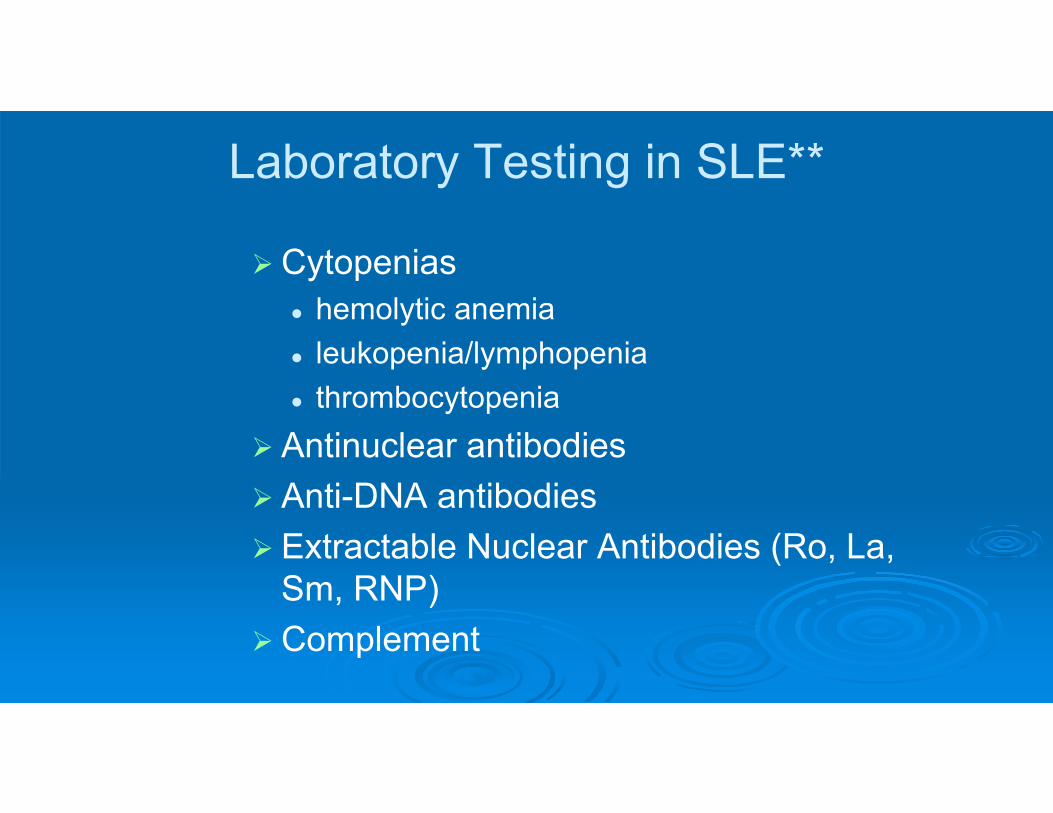
Laboratory Testing in SLE**
Cytopenias hemolytic anemia leukopenia/lymphopenia thrombocytopenia
Antinuclear antibodies Anti-DNA antibodies Extractable Nuclear Antibodies (Ro, La,
Sm, RNP)Complement

Criteria to Diagnose SLE
2012 SLICC Criteria versus 1997 ACR criteria
Require presence of both clinical and immunologic abnormalities
Both require at least 4 criteria** SLICC includes at least 1 clinical and 1
immunologic SLICC includes renal-isolated lupus and
more neuro features

ACR Criteria for SLE 1997: Clinical Malar rash Discoid rash Photosensitivity Oral ulceration Arthritis Serositis Renal Neurologic (seizures or psychosis) Hematologic (hemolytic anemia, leukopenia,
lymphopenia, thrombocytopenia)
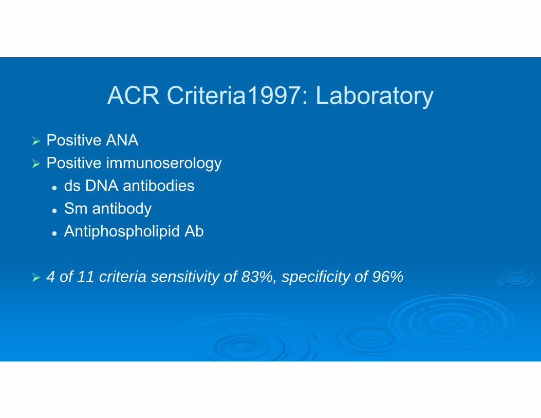
ACR Criteria1997: Laboratory
Positive ANA Positive immunoserology
ds DNA antibodies Sm antibody Antiphospholipid Ab
4 of 11 criteria sensitivity of 83%, specificity of 96%
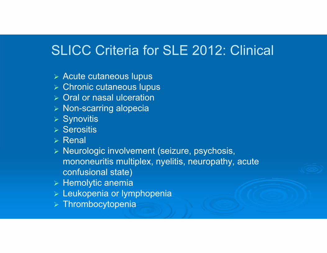
SLICC Criteria for SLE 2012: Clinical
Acute cutaneous lupus Chronic cutaneous lupus Oral or nasal ulceration Non-scarring alopecia Synovitis Serositis Renal Neurologic involvement (seizure, psychosis,
mononeuritis multiplex, nyelitis, neuropathy, acute confusional state)
Hemolytic anemia Leukopenia or lymphopenia Thrombocytopenia

SLICC Criteria 2012: Laboratory Positive ANA Anti-ds DNA antibodies Anti-Sm antibody Antiphospholipid Antibodies (lupus anticoagulant, false-
positive RPR, anticardiolipin antibodies, anti-beta 2-glycoprotein I)
Low complement (low C3, low C4, low CH50) Positive direct Coombs’ test without a hemolytic anemia
4 of 11 criteria sensitivity of 97%, specificity of 94%

SLE: Treatment** Corticosteroids
Oral Intravenous
Hydroxychloroquine (Plaquenil)MethotrexateMycophenolate MofetilCyclophosphamideOther
Calcium Vitamin D
Complications of Treatment** Growth failure Fertility/amenorrheaOsteoporosis Body image
weight gain cushingoid appearance acne

Case Three A five-year-old male is brought to your office by his
parents for complaints of decreased energy and difficulty performing regular tasks. This has been worsening over the last several weeks. He denies any muscle or joint tenderness. On review of systems, his parents report that he has had eczema on his face for the last several months that has been resistant to any medical therapy
Physical exam reveals a cooperative male who is unable to lift his arms above his head or get up from a sitting position. Skin exam reveals a facial rash and red bumps on his fingers.

Juvenile Dermatomyositis (JDM)
Frequency in population 0.5 per 100,000 Bimodal peak of age of onset
10-14 years (approx 16-20%) 45-64 years
Gender predilection: approx 2:1 F:M
Clinical Manifestions of JDM
Cutaneous Manifestations** JDM rash heliotrope rash Gottron’s papules periungual erythema ulcerative disease [acanthosis nigricans]
Proximal muscle weakness** large muscle groups versus small clues on movement and gait

Clinical Manifestations of JDM Evidence of inflammatory myositis**
elevated muscle enzymes eletromyography muscle biopsy magnetic resonance imaging
GastrointestinalRespiratoryCalcinosis Lipoatrophy

Laboratory Testing in JDM
Muscle enzymes** AST/SGOT ALT/SGPT CPK aldolase LDH
Antibodies Antinuclear antibodies (frequency 10-80%) PM1, Jo1, etc (<20% in children)

JDM: Differential Diagnosis
Dermatomyositis versus polymyositisMuscular dystrophyGuillain-Barre diseaseOther inflammatory myopathies

JDM: Treatment**
Immunosuppressive therapy Prednisone Steroid-sparing agents:
-methotrexate-cyclophosphamide -cyclosporine-azathioprine
Intravenous immunoglobulin Stretching to maintain range of motion Continuation of other regular activities
JDM: Complications**
Respiratory impairment
GI vasculitis
Steroid-related side effects
Side effects of steroid-sparing agents
Calcinosis
Insulin resistance/lipoatrophy
JDM: Long Term Outcomes**
70% well and functional 20% long term complications 10% die
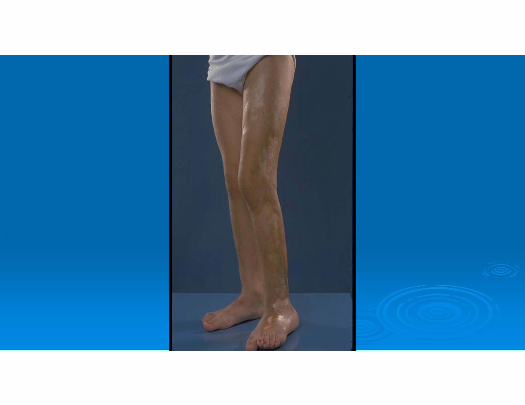
Case Four
A 13-year-old female presents with persistent and increasing rash on her left lower extremity. It is red, nontender, and nonpruritic, but is interfering with her ability to extend her leg
Physical exam reveals a firm, shiny, erythematous strip of skin extending from the left hip, down the lateral side of her leg, across her knee to the dorsal surface of her foot.

Scleroderma and Relatives
Types Localized scleroderma**
• morphea• linear scleroderma
Diffuse scleroderma**• systemic sclerosis• CREST
Mixed Connective Tissue Disease Eosinophilic Fasciitis


Clinical Manifestations of Scleroderma**
Cutaneous (skin thickening) Musculoskeletal
Limitation of range of motion Abnormal limb growth
Raynaud’s phenomenon Gastrointestinal Pulmonary Renal

Laboratory Testing in Scleroderma Antinuclear Antibodies
SCL-70 (DNA-topoisomerase 1) 26% freq anticentromere (kinetochore) 22% freq anti-RNP (100% freq in MCTD)
Rheumatoid Factors Proteinuria/hematuriaHematologic
anemia eosinophilia

Scleroderma: Treatment** Immune modulators/anti-inflammatory therapy
Prednisone Methotrexate Cyclophosphamide
Decrease Collagen cross linking [D-penicillamine]
Prevention of further organ injury:-Ca channel blockers-cold precautions
Preservation of function:-intensive PT/OT
General supportive care

Systemic Vasculitis Very rare in children** Peak age 9-11 yrs, range 3-16 yrs Equal incidence male/femaleMost presentations of systemic vasculitis do
not follow specific patterns Some more common vasculitic syndromes of
childhood present as a constellation of otherwise nonspecific symptoms
Most common vasculitis of childhood is Kawasaki Disease (discussed elsewhere)

Case Five
An eight-year-old boy presents to the emergency room with bruising on his lower extremities and abdominal pain.
Physical exam reveals purpura on both legs, as well as bilateral ankle arthritis. His scrotum is swollen and ecchymotic. Abdominal exam demonstrates normal bowel sounds and no HSM.

Henoch-Schonlein Purpura AKA anaphylactoid purpura Second most common vasculitis of
childhoodMost common in children 5-15 years of
age, rare in adults**Male to female 1.5 : 1 Incidence varies from 0.1 to 13.5 per
100,000 Seasonal variation: peaks during winter
? Relationship to streptococcus
HSP: Clinical Manifestations
Cutaneous Manifestations** Palpable purpura (100%)
• most prominent in dependent areas, legs, buttocks
• range from petechiae to ecchymoses• can be preceded by urticaria or MP
SQ edema of hands and feet, face, scrotum


HSP: Clinical Manifestations
Arthalgias/Arthritis (65-85%)** usually involves large joints (knees, ankles) periarticular swelling and tenderness, but
usually non-erythematous transient, but not migratory lasts few days to a week

HSP: Clinical Manifestations
Gastrointestinal involvement (60-100%)** Gut vasculitis colicky abdominal pain heme (+) stools intestinal perforation within one wk to one month of rash
Renal involvement (20-50%)** acute glomerulonephritis hematuria/proteinuria hypertension renal failure within one to three months of rash

HSP: Laboratory Testing
Pathology: leukocytoclastic vasculitis with IgA deposition
Must not have thrombocytopenia**May have elevated inflammatory
parametersNormochromic normocytic anemia Abnormal urinary sedimentNormal complements

EULAR Consensus Criteria for HSP
classical palpable non-thrombocytopenic purpuric rash and any one of the following: Arthritis or arthralgia Abdominal pain and/or GI bleeding Any biopsy with predominant IgA
deposition

HSP: Treatment**
Supportive careNSAIDs for arthritisCorticosteroids
for severe GI disease Active renal disease 1 mg/kg/d divided bid

HSP: Disease Course
Resolves within one month in 66% At least 50% will have recurrence, usually
of rash or GI symptoms** usually within first 6 wks, but up to 1-2 years
Don’t forget intussussception** Late renal outcome: less than 5% progress
to end stage renal disease