kallick rush, rheumatology 1, :1 heumatology current ... · rheumatology olume ssue 1 11 ss: 16111...
TRANSCRIPT

Volume 4 • Issue 1 • 1000128RheumatologyISSN: 2161-1149 Rheumatology, an open access journal
Rheumatology : Current ResearchKallick Rush, Rheumatology 2014, 4:1
http://dx.doi.org/10.4172/2161-1149.1000128
Review Article Open Access
The Potential Relationship of the Ehrlichia to Immune System Dysfunction: Etiology and PathogenesisCharles A Kallick Rush*
University Medical Center, Department of Medicine, 1825 W, Harrison, Chicago, IL 60612, USA
*Corresponding author: Charles A Kallick Rush, University Medical Center, Department of Medicine, 1825 W, Harrison, Chicago, IL 60612, USA, Tel: 206-783-0986; E-mail: [email protected]
Received November 07, 2013; Accepted December 26, 2013; Published January 25, 2014
Citation: Kallick Rush CA (2014) The Potential Relationship of the Ehrlichia to Immune System Dysfunction: Etiology and Pathogenesis. Rheumatology 4: 128. doi:10.4172/2161-1149.1000128
Copyright: © 2014 Kallick Rush CA. This is an open-access article distributed under the terms of the Creative Commons Attribution License, which permits unrestricted use, distribution, and reproduction in any medium, provided the original author and source are credited.
Keywords: SLE; Autoimmune disorders; Transcription errors; Ehrlichia/Anaplasma; Bone marrow stem cell infection; Specific antibiotic therapy
IntroductionThe mystery of immune disorders, when the immune system turns
against the body’s own tissues, commonly referred to as autoimmune disease (AD), is an unanswered question to a myriad of diseases recognized by immunologists and rheumatologists. These disorders include systemic lupus erythematosus (SLE), rheumatoid arthritis (RA). Multiple sclerosis (MS) and amyotrophic lateral sclerosis (ALS). To date no causes have been identified for any of these diseases. In addition treatment is addressed to symptomatology and empirically discovered therapies, largely based on steroids and immunosuppressive agents. However anecdotal evidence in a molecular study and two reports, points to a bacterial agent as a possible cause of these disorders and explores mechanisms of disease development. Further research in this vein could potentially lead to a breakthrough in identifying the cause of disorders and lead to more specific therapy.
The immune system is mediated by white blood cells and the most important in this discussion are lymphocytes specifically those known as T and B cells. These cells originate from the stem cells in the bone marrow. Their function is to recognize invading antigens including bacteria and eliminate them if they are encountered and recognized as different from self. In autoimmune disorders (AD) this process breaks down if these cells fail to differentiate and may attack normal body tissues, and thus begins to injure cells and organs.
Extensive studies have explored viral and other infectious agents as positive causative agents with limited success. The present hypothesis is based upon published reports, anecdotal cases, research, and the known functioning of the body’s immune system. This information taken as incomplete information suggests that at least one autoimmune disorder SLE, may be caused or associated by the presence of an unusual bacterial agent, well studied in Veterinary Medicine of the genus Ehrlichia (EA). Other diseases classified as rheumatologic disorders may also be caused by these infectious agents. Research into the potential presence of this agent could answer previously unanswered questions about the cause and pathogenesis of these unexplained diseases [1-4].
AbstractIn a study of autoimmune disease (AD), a hemotropic bacterial agent, Ehrlichia/Anaplasma (EA) is suggested as
associated or possibly etiologic. These bacteria are obligate intracellular parasites with demonstrated ability to disrupt transcription in the rapidly dividing bone marrow cell. Two papers have definitively placed this agent in systemic lupus erythematosus (SLE). Bacterial structures resembling endosomes have been shown to be present in bone marrow megakaryocytes when using phase contrast microscopy. Marrow cells construct lymphocytes using fragments of DNA of body tissue; 60% are self-reactive, but are eliminated before release to the circulation. EA have been shown to alter transcription of infected cells and alteration of DNA during transcription can explain many of the factors involved in AD. PCR of a Patient with demonstrated infection of 2% parasitized erythrocytes with EA, revealed a single gene of the major surface protein2 (MSP2), which aligned within 97% of the single gene from an animal EA, Anaplasma phagocytophillium transmitted to man by ticks. EA has limited antibiotic susceptibility, and is sensitive only to rifamycins, tetracyclines, and quinolones. Confirmation of the presence of EA in whole blood examined of patients with AD using the techniques reported here, especially molecular methods to find a bacterium which is very difficult to culture may enlarge the treatment, and research options available to immunologist-rheumatologists.
Hemotropic bacteria and the ehrlichia
The Ehrlichia/Anaplasma (EA) are hemotropic agents, an obscure group of bacteria which primarily infect both white and red blood cells, and their precursors, the cells of immunity, which protect the body against invaders, include bacteria. They EA are well known in veterinary medicine and one very well studied. EA, Ehrlichia canis, causes an aplastic anemia in dogs called tropical canine pancytopenia. Two recently described animal EA, A. phagocytophillum, and E. Chafeensis are agents transmitted to humans from the bite of an infected tick [5].
EA are interesting organisms due to their obligate intracellular parasitism, tropism for cells of the immune system, and difficulty in culture. But more important to identify the potential Link to immune disease is that EA have an unusual ability to alter cellular transcription during cell division. This occurs in bone marrow including the stem cells or any other marrow precursors at a site of intense cell division [6]. Infection of stem or other cells does not cause death of the infected cells, but may cause alterations in subsequent cellular function because of altered DNA. This unique characteristic suggests and may explain the multiple alterations in cell functioning in AD. The EA are bacteria, and they are sensitive only to a few antibiotics: rifamycins (RFM) tetracyclines (TC) and quinolones (QN). Empiric use of Minocycline, a tetracycline antibiotic, is used for rheumatoid arthritis (RA). In RA, RFM when given for diagnosed infectious agents, regularly results in remission of RA, and a quinolone is used for treatment of cutaneous discoid lupus.
Volume 4 • Issue 1 • 1000128RheumatologyISSN: 2161-1149 Rheumatology, an open access journal
Citation: Kallick Rush CA (2014) The Potential Relationship of the Ehrlichia to Immune System Dysfunction: Etiology and Pathogenesis. Rheumatology 4: 128. doi:10.4172/2161-1149.1000128
Page 2 of 4
Mechanisms of immune function and alteration of cellular function
Lymphocytes are formed using fragments of DNA in the bone marrow. Wardemann et al. and other researchers have found that up to 60% of the lymphocytes produced in the marrow are sell reactive (SR) but are eliminated before release into the bloodstream by mechanisms within the marrow [7,8]. However, if the cells have altered DNA, altered because of previous infection with EA and resultant changes of DNA, or are the descendants of cells after previous infection with EA, they may not respond to those normal marrow mechanisms. Such cells may be released into the circulation. The white cells of patients with AD have been shown to have cellular alteration of unknown cause; these alterations may prevent some or all of the programmed function of immune cells such as maturation, function, and cell death. Since the basic pathology of AD, in this construction of pathogenesis is really remote from the site of cellular or antibody injury, it may be difficult to determine the cause of the disease. If precursors of the cells have altered DNA because of previous infection with EA, and therefore do not respond to marrow mechanisms, such cells may find a way into the circulation. The cells of immunity, which came from the stem cells or other infected precursors, may have alterations in DNA because of previous or current infection with EA [9]. The change in DNA may alter their behavior in their programmed mission to defend against invaders.
Cellular and humeral immunity are primarily mediated by T and B cells. These SF functions are presumably associated with changes in either class of cells, and may be very remote from the primary cellular DNA event mediated by EA. This process suggests understanding of the multiple antibodies characteristic of SLE. Other AD disorders could result from either alterations of the T cells or B cells or both. For example, diseases of the nervous system, such as MS, and ALS which demonstrate cellular destruction as a major pathologic event, could have alterations of T cells with resultant tissue destruction. The occurrence of calcification of tissue in the spinal cord in MS, suggests cellular tissue injury by SRT cells and resultant calcification after injury.
Evidence for involvement of EA in various seemingly unrelated disease states
A report published in 2011 in the Journal of Medical Hypotheses presents an in-depth discussion of the concepts described above [10]. The paper sets forth the indirect evidence that supports the presence of EA in the marrow in patients, and with other unpublished data collected by the author suggests that the intracellular inclusions of EA occur in the marrow as observed by the use of phase contrast examination of the spreadout cells of a megakaryocyte, and defined with the special phase contrast microscopic effects. EA can also be found microscopically in erythrocytes. This could help to explain epidemiology and transmission of this organism and subsequent disease. Since red blood cells can be transmitted in semen, transmission may be occurring silently. Red cells are also known to be transmitted in parturition by studies of Rh factor epidemiology. The biology of the EA upon infection suggests the occurrence of almost universal latency in development of disease states.
The paper includes a 90+ amino acid sequences within 97% of the major surface protein MSP2) of the rodent derived EA found in human infection and identified in white cells examined from patients recovering from infection after tick bite. The presence of the published sequences of the MSP2 gene of the presumed EA, if molecular studies confirm these findings, and if Statistical demonstration succeeds, this
could be considered confirmation of the hypothesis that EA infection causes a disruption of DNA, stem and other cells leading to otherwise physiologically eliminated self-reactive cells to be released into the body circulation rather than be eliminated. These compromised cells could cause a variety of autoimmune disorders [9].
Unusual laboratory methods
Our investigations suggest that using accepted primers and probes for EA by PCR would likely fail to find the DNA of EA in suspected human specimens. This is a complicated matter and requires many variations of the mechanism used in molecular identification of bacterial DNA (rRNA). The author discovered this problem in attempting PCR on blood from a patient with suspected infection by the EA, was found in 2% of erythrocytes secondary to previous splenectomy. The spleen normally removes damaged or infected erythrocytes. Published primers for EA did not polymerize the organism. Fortunately, a proprietary primer did polymerize a single gene within 97% alignment of the animal gene of Anaplasma phagocytophillium [11].
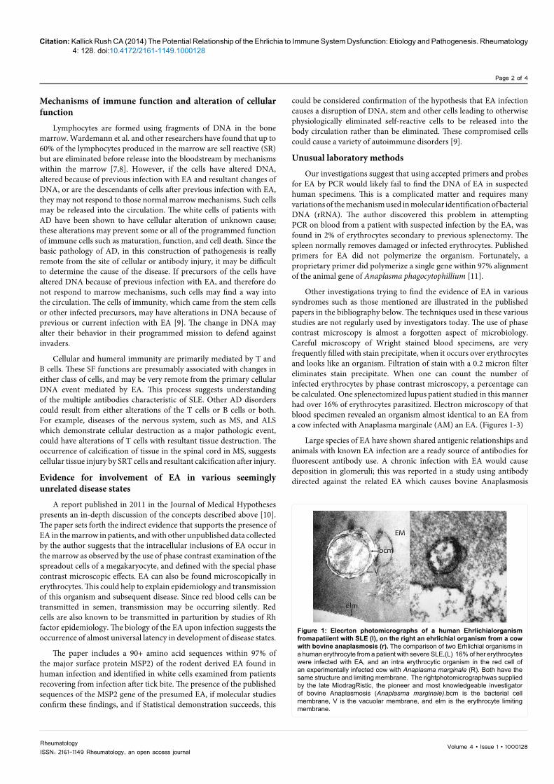
Other investigations trying to find the evidence of EA in various syndromes such as those mentioned are illustrated in the published papers in the bibliography below. The techniques used in these various studies are not regularly used by investigators today. The use of phase contrast microscopy is almost a forgotten aspect of microbiology. Careful microscopy of Wright stained blood specimens, are very frequently filled with stain precipitate, when it occurs over erythrocytes and looks like an organism. Filtration of stain with a 0.2 micron filter eliminates stain precipitate. When one can count the number of infected erythrocytes by phase contrast microscopy, a percentage can be calculated. One splenectomized lupus patient studied in this manner had over 16% of erythrocytes parasitized. Electron microscopy of that blood specimen revealed an organism almost identical to an EA from a cow infected with Anaplasma marginale (AM) an EA. (Figures 1-3)
Large species of EA have shown shared antigenic relationships and animals with known EA infection are a ready source of antibodies for fluorescent antibody use. A chronic infection with EA would cause deposition in glomeruli; this was reported in a study using antibody directed against the related EA which causes bovine Anaplasmosis
Figure 1: Elecrton photomicrographs of a human Ehrlichialorganism fromapatiient with SLE (l), on the right an ehrlichial organism from a cow with bovine anaplasmosis (r). The comparison of two Erhlichial organisms in a human erythrocyte from a patient with severe SLE,(L) 16% of her erythrocytes were infected with EA, and an intra erythrocytic organism in the red cell of an experimentally infected cow with Anaplasma marginale (R). Both have the same structure and limiting membrane. The rightphotomicrographwas supplied by the late MiodragRistic, the pioneer and most knowledgeable investigator of bovine Anaplasmosis (Anaplasma marginale).bcm is the bacterial cell membrane, V is the vacuolar membrane, and elm is the erythrocyte limiting membrane.
Volume 4 • Issue 1 • 1000128RheumatologyISSN: 2161-1149 Rheumatology, an open access journal
Citation: Kallick Rush CA (2014) The Potential Relationship of the Ehrlichia to Immune System Dysfunction: Etiology and Pathogenesis. Rheumatology 4: 128. doi:10.4172/2161-1149.1000128
Page 3 of 4
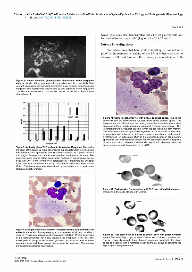
Figure 2: Lupus nephritis glomerluswith flourescent anti-a marginale stain. A biopsied kidney glomerulus from a patient with lupus nephritis.Stain was with conjugated convalescent serum from a cow infected with Anaplasma marginale. This fluorescence was blocked by prior exposure to non-conjugated convalescent bovine serum, but not by normal bovine serum from a non-infected cow [3].
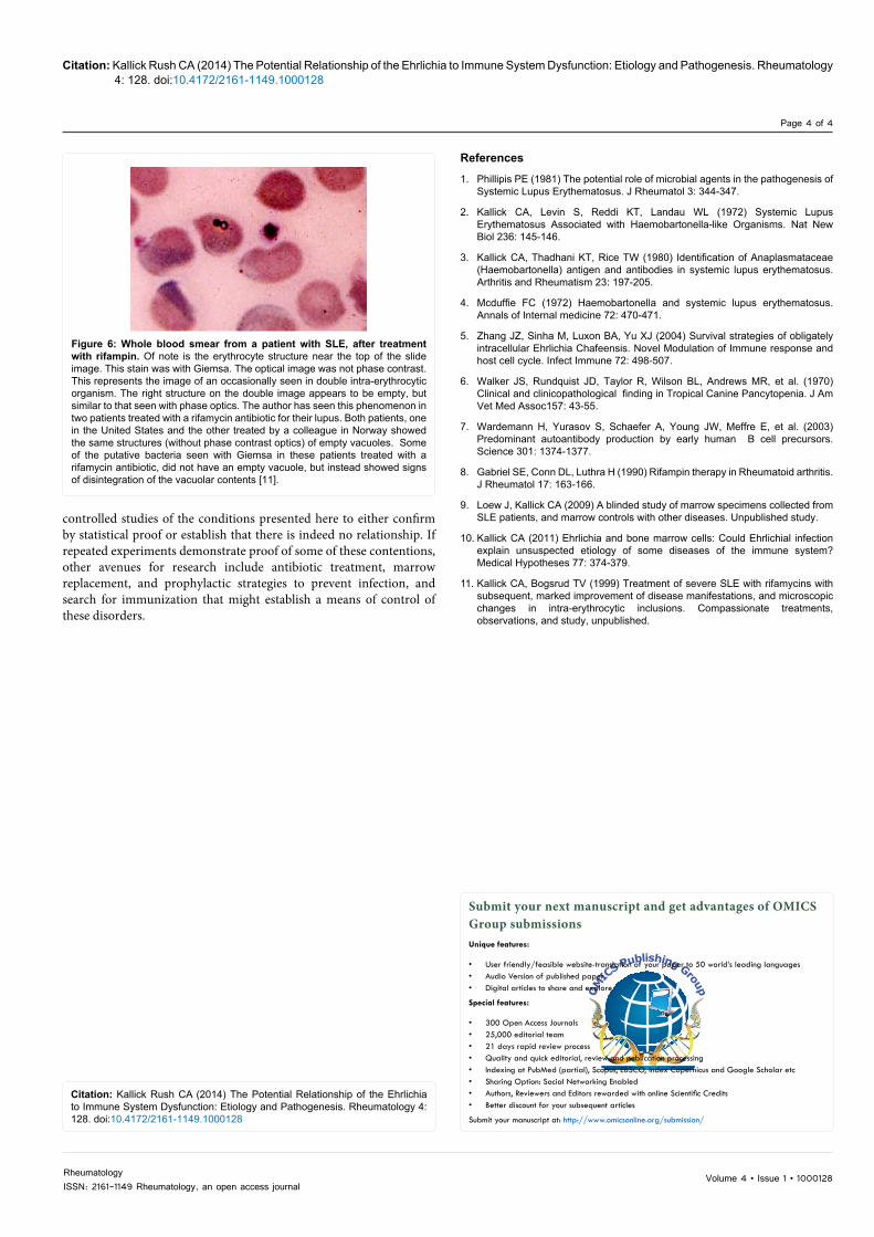
Figure 3: Antibody titer of SLE and controls to anti-a. Marginale. The results of a study of the serum 22 lupus patients and 102 controls which were selected from random blood specimens from ill patients admitted to a public hospital in Chicago. Some of the controls may have had autoimmune disorders. The specimens were blinded before examination and use on specimens of bovine blood with 70% of the erythrocytes parasitized by A marginale an Erhlichial agent. This was an indirect FA study. The human specimens were serially diluted. The florescence was determined by fluorescence from antihuman conjugated goat serum [3].
Figure 4A: Megakaryocyte in marrow from patient with SLE; normal stain and optics. A smear of a megakaryocyte, from a patient with lupus, but without nephritis. This is a megakaryocyte from a patient with SLE. Photomicrographs of the megakaryocytes can easily visualize the cytoplasm of the cell. The arrows point to two granules in thee cytoplasm, and most students of these structures would call these normal marrow granular structures. The staining and optical mechanisms were routine.
Figure 4b:Same Megakaryocyte with phase contrast optics. This is the same cell from the same parent but seen under phase contrast optics. The two granules are different from the other granules because they have a clear area around them, which appears to represent presence in a vacuole. This is consistent with a vacuolar structure within the cell inside the born marrow. The structures show no sign of disintegration, and thus could be described as consistent with a bacterium within a vacuole, suggesting an endosome in a marrow cell. A subsequent study of a large blinded series of bone marrow specimens, with other randomly chosen marrow specimens without a diagnosis of lupus as controls, showed a statistically significant difference batten the lupus specimens and the controls (p= 0.5) [10].
Figure 5A: Erythrocytes from a patient with SLE; two cells with inclusions. Inclusions in two cells, stained with Giemsa.
Figure 5B: The same cells as Figure 5a above, seen with phase contrast optics. The same Erythrocyte as seen in 4A above, in phase contrast optics. The two previously stained intra erythrocytic structures, revealed by the phase optics as a vacuole with a membrane seen on the EM above as similar to the erythrocyte limiting membrane.
(AM). That study also demonstrated that all of 22 patients with SLE had antibodies reacting to AM. (Figures 4A,4B,5A,5B and 6)
Future InvestigationsInformation presented here while compelling, is not definitive
proof of the presence or activity of the EA as either associated or etiologic in AD. To determine if there is really an association, carefully
Volume 4 • Issue 1 • 1000128RheumatologyISSN: 2161-1149 Rheumatology, an open access journal
Citation: Kallick Rush CA (2014) The Potential Relationship of the Ehrlichia to Immune System Dysfunction: Etiology and Pathogenesis. Rheumatology 4: 128. doi:10.4172/2161-1149.1000128
Page 4 of 4
controlled studies of the conditions presented here to either confirm by statistical proof or establish that there is indeed no relationship. If repeated experiments demonstrate proof of some of these contentions, other avenues for research include antibiotic treatment, marrow replacement, and prophylactic strategies to prevent infection, and search for immunization that might establish a means of control of these disorders.
Figure 6: Whole blood smear from a patient with SLE, after treatment with rifampin. Of note is the erythrocyte structure near the top of the slide image. This stain was with Giemsa. The optical image was not phase contrast. This represents the image of an occasionally seen in double intra-erythrocytic organism. The right structure on the double image appears to be empty, but similar to that seen with phase optics. The author has seen this phenomenon in two patients treated with a rifamycin antibiotic for their lupus. Both patients, one in the United States and the other treated by a colleague in Norway showed the same structures (without phase contrast optics) of empty vacuoles. Some of the putative bacteria seen with Giemsa in these patients treated with a rifamycin antibiotic, did not have an empty vacuole, but instead showed signs of disintegration of the vacuolar contents [11].
References
1. Phillipis PE (1981) The potential role of microbial agents in the pathogenesis of Systemic Lupus Erythematosus. J Rheumatol 3: 344-347.
2. Kallick CA, Levin S, Reddi KT, Landau WL (1972) Systemic Lupus Erythematosus Associated with Haemobartonella-like Organisms. Nat New Biol 236: 145-146.
3. Kallick CA, Thadhani KT, Rice TW (1980) Identification of Anaplasmataceae (Haemobartonella) antigen and antibodies in systemic lupus erythematosus. Arthritis and Rheumatism 23: 197-205.
4. Mcduffie FC (1972) Haemobartonella and systemic lupus erythematosus. Annals of Internal medicine 72: 470-471.
5. Zhang JZ, Sinha M, Luxon BA, Yu XJ (2004) Survival strategies of obligately intracellular Ehrlichia Chafeensis. Novel Modulation of Immune response and host cell cycle. Infect Immune 72: 498-507.
6. Walker JS, Rundquist JD, Taylor R, Wilson BL, Andrews MR, et al. (1970) Clinical and clinicopathological finding in Tropical Canine Pancytopenia. J Am Vet Med Assoc157: 43-55.
7. Wardemann H, Yurasov S, Schaefer A, Young JW, Meffre E, et al. (2003) Predominant autoantibody production by early human B cell precursors. Science 301: 1374-1377.
8. Gabriel SE, Conn DL, Luthra H (1990) Rifampin therapy in Rheumatoid arthritis. J Rheumatol 17: 163-166.
9. Loew J, Kallick CA (2009) A blinded study of marrow specimens collected from SLE patients, and marrow controls with other diseases. Unpublished study.
10. Kallick CA (2011) Ehrlichia and bone marrow cells: Could Ehrlichial infection explain unsuspected etiology of some diseases of the immune system? Medical Hypotheses 77: 374-379.
11. Kallick CA, Bogsrud TV (1999) Treatment of severe SLE with rifamycins with subsequent, marked improvement of disease manifestations, and microscopic changes in intra-erythrocytic inclusions. Compassionate treatments, observations, and study, unpublished.
Citation: Kallick Rush CA (2014) The Potential Relationship of the Ehrlichia to Immune System Dysfunction: Etiology and Pathogenesis. Rheumatology 4: 128. doi:10.4172/2161-1149.1000128
Submit your next manuscript and get advantages of OMICS Group submissionsUnique features:
• User friendly/feasible website-translation of your paper to 50 world’s leading languages• Audio Version of published paper• Digital articles to share and explore
Special features:
• 300 Open Access Journals• 25,000 editorial team• 21 days rapid review process• Quality and quick editorial, review and publication processing• Indexing at PubMed (partial), Scopus, EBSCO, Index Copernicus and Google Scholar etc• Sharing Option: Social Networking Enabled• Authors, Reviewers and Editors rewarded with online Scientific Credits• Better discount for your subsequent articles
Submit your manuscript at: http://www.omicsonline.org/submission/