pediatric heart sounds
TRANSCRIPT
Pediatric Heart Sounds

Michael E. McConnellwith contributions by Alan Branigan
Pediatric Heart Sounds
1 3
Michael E. McConnell, MDEmory University and the Sibley Heart CenterCardiology at 52 Executive Park SouthSuite 5200.Atlanta, GA 30329USA
with contributions byAlan Branigan, MA, MEdDirector, Educational SupportEastern Area Health Education Center2000 Venture Tower Dr.Greenville, NC 27835-7224USA
ISBN 978-1-84628-683-4 e-ISBN 978-1-84628-684-1DOI 10.1007/978-1-84628-684-1
British Library Cataloguing in Publication DataA catalogue record for this book is available from the British Library
Library of Congress Control Number: 2008931524
# Springer-Verlag London Limited 2008The software disk accompanying this book and all material contained on it is supplied without anywarranty of any kind. The publisher accepts no liability for personal injury incurred through use ormisuse of the disk.Apart from any fair dealing for the purposes of research or private study, or criticism or review, aspermitted under the Copyright, Designs and Patents Act 1988, this publication may only bereproduced, stored or transmitted, in any form or by any means, with the prior permission inwriting of the publishers, or in the case of reprographic reproduction in accordance with the termsof licences issued by the Copyright Licensing Agency. Enquiries concerning reproduction outsidethose terms should be sent to the publishers.The use of registered names, trademarks, etc. in this publication does not imply, even in theabsence of a specific statement, that such names are exempt from the relevant laws and regulationsand therefore free for general use.The publisher makes no representation, express or implied, with regard to the accuracy of theinformation contained in this book and cannot accept any legal responsibility or liability for anyerrors or omissions that may be made.Product liability: The publisher can give no guarantee for information about drug dosage andapplication thereof contained in this book. In every individual case the respective user must checkits accuracy by consulting other pharmaceutical literature.
Printed on acid-free paper
Springer ScienceþBusiness Mediaspringer.com

Preface
v
Why another book to teach auscultation? Isn’t the use of the stetho-
scope a ‘‘lost art’’, totally unnecessary in the age of echocardiography
and ‘‘hand held’’ imaging ‘‘stethoscopes’’? The answer is that there is
perhaps no other physical examination skill that a physician making
patient care decisions must have that is more important, even now.
If a patient complains of fever and a cough, a chest radiograph
interpreted by someone else will either confirm or rule out the
diagnosis of pneumonia, and a quick look in a textbook can tell
the physician the next course to take. When a patient comes to the
office with no complaints, and on auscultation has a soft systolic
murmur, only good physical examination skills will allow the exam-
iner to reassure the patient that the murmur is non-pathologic.
There is unfortunately ample evidence that auscultatory skills are
very poorly taught to medical students and residents [1]. Research
does show that intensive instruction, followed by reexamining
patients with known lesions, will improve the diagnostic accuracy.
Unfortunately, in busy practices, the ability of the learner to listen,
discuss the findings with the preceptor, and to listen again is often
lacking [2]. The inability to appreciate abnormalities of precordial
activity, to critically listen to the first and second heart sounds, and
to discern the difference between a pathologic murmur and a func-
tional one often leads to unnecessary testing, and potentially leads to
missed diagnoses.
There are ample sources to help improve physical examination
skills, many written by the true great teachers of medicine. Yet, in
spite of these, auscultation skills are poorly learned. With the advent
of new ‘‘multimedia’’ technology, perhaps auscutation can be more
effectively taught. Unfortunately, recent evidence with some multi-
media teaching tools suggests that the learning is still ineffective [1].
The CD-ROM that accompanies this text uses a novel approach to
educate the learner about auscultation skills. It is not meant to be an
exhaustive ‘‘encyclopedia’’, listing every possible abnormal sound
that the heart can make. The goal is to get the learner more
comfortable using the stethoscope in an organized fashion, and
once they have the organized system of auscultation, to improve
their ability to tell pathologic from normal heart sounds. The cardiac
sounds on the CD-ROM were recorded from patients with the
specific cardiac abnormalities listed, and the specific pathology
was confirmed using echocardiography. The programmed nature
Preface vii

of the CD-ROM forces the learner to critically evaluate all aspects of
the cardiac examination. By placing the findings in the spread sheet,
and getting immediate feedback on correct and incorrect responses,
the learner’s ability to listen critically should improve.
References
1. Mahnke CB, Nowalk A, Hofkosh D, Zuberbuhler JR, Law YM
(2004) Comparison of two educational interventions on pediatric
resident auscultation skills. Pediatrics 113(5):1331–1335
2. Favrat B, Pecoud A, Jaussi A (2004) Teaching cardiac ausculta-
tion to trainees in internal medicine and family practice: does it
work? BMC Med Educ 4(1):5
viii Preface

Contents
ix
1 Normal Heart Sounds . . . . . . . . . . . . . . . . . . . . . . . . . . . . . . . . . . 1
The First Heart Sound . . . . . . . . . . . . . . . . . . . . . . . . . . . . . . . . . . . 3
The Second Heart Sound . . . . . . . . . . . . . . . . . . . . . . . . . . . . . . . . . 8
References. . . . . . . . . . . . . . . . . . . . . . . . . . . . . . . . . . . . . . . . . . . . . 12
2 Innocent Heart Murmurs . . . . . . . . . . . . . . . . . . . . . . . . . . . . . . . 13
Introduction . . . . . . . . . . . . . . . . . . . . . . . . . . . . . . . . . . . . . . . . . . . 15
Peripheral Pulmonary Flow Murmurs . . . . . . . . . . . . . . . . . . . . . . . 15
Still’s Murmur . . . . . . . . . . . . . . . . . . . . . . . . . . . . . . . . . . . . . . . . . . 19
The Aortic Outflow Murmur . . . . . . . . . . . . . . . . . . . . . . . . . . . . . . 24
References. . . . . . . . . . . . . . . . . . . . . . . . . . . . . . . . . . . . . . . . . . . . . 25
3 Atrial Septal Defects . . . . . . . . . . . . . . . . . . . . . . . . . . . . . . . . . . . 27
Introduction . . . . . . . . . . . . . . . . . . . . . . . . . . . . . . . . . . . . . . . . . . . 29
Anatomy . . . . . . . . . . . . . . . . . . . . . . . . . . . . . . . . . . . . . . . . . . . . . . 30
Inspection . . . . . . . . . . . . . . . . . . . . . . . . . . . . . . . . . . . . . . . . . . . . . 32
Palpation . . . . . . . . . . . . . . . . . . . . . . . . . . . . . . . . . . . . . . . . . . . . . . 32
Auscultation . . . . . . . . . . . . . . . . . . . . . . . . . . . . . . . . . . . . . . . . . . . 33
Subacute Bacterial Endocarditis Prophylaxis Recommendations
(SBE). . . . . . . . . . . . . . . . . . . . . . . . . . . . . . . . . . . . . . . . . . . . . . . . . 36
References. . . . . . . . . . . . . . . . . . . . . . . . . . . . . . . . . . . . . . . . . . . . . 37
4 Ventricular Septal Defects . . . . . . . . . . . . . . . . . . . . . . . . . . . . . . 39
Incidence. . . . . . . . . . . . . . . . . . . . . . . . . . . . . . . . . . . . . . . . . . . . . . 41
Anatomy . . . . . . . . . . . . . . . . . . . . . . . . . . . . . . . . . . . . . . . . . . . . . . 41
Physiology . . . . . . . . . . . . . . . . . . . . . . . . . . . . . . . . . . . . . . . . . . . . . 43
Natural History . . . . . . . . . . . . . . . . . . . . . . . . . . . . . . . . . . . . . . . . . 45
Medical Management . . . . . . . . . . . . . . . . . . . . . . . . . . . . . . . . . . . . 45
Surgical Options . . . . . . . . . . . . . . . . . . . . . . . . . . . . . . . . . . . . . . 46
Physical Examination . . . . . . . . . . . . . . . . . . . . . . . . . . . . . . . . . . 47
Small Muscular Ventricular Septal Defects . . . . . . . . . . . . . . . . . . . 47
Moderate Ventricular Septal Defects . . . . . . . . . . . . . . . . . . . . . . . . 48
Large Ventricular Septal Defects . . . . . . . . . . . . . . . . . . . . . . . . . . . 50
Subacute Bacterial Endocarditis Prophylaxis Recommendations
(SBE). . . . . . . . . . . . . . . . . . . . . . . . . . . . . . . . . . . . . . . . . . . . . . . . . 53
Summary . . . . . . . . . . . . . . . . . . . . . . . . . . . . . . . . . . . . . . . . . . . . . . 53
References. . . . . . . . . . . . . . . . . . . . . . . . . . . . . . . . . . . . . . . . . . . . . 53
5 Patent Arterial Duct . . . . . . . . . . . . . . . . . . . . . . . . . . . . . . . . . . . . 57
Introduction . . . . . . . . . . . . . . . . . . . . . . . . . . . . . . . . . . . . . . . . . . . 59
Anatomy . . . . . . . . . . . . . . . . . . . . . . . . . . . . . . . . . . . . . . . . . . . . . . 59
Physiology . . . . . . . . . . . . . . . . . . . . . . . . . . . . . . . . . . . . . . . . . . . . . 60
Contents xi
Natural History . . . . . . . . . . . . . . . . . . . . . . . . . . . . . . . . . . . . . . . . . 60
Surgical Options . . . . . . . . . . . . . . . . . . . . . . . . . . . . . . . . . . . . . . . . 61
Auscultation . . . . . . . . . . . . . . . . . . . . . . . . . . . . . . . . . . . . . . . . . 61
Subacute Bacterial Endocarditis Prophylaxis Recommendations
(SBE). . . . . . . . . . . . . . . . . . . . . . . . . . . . . . . . . . . . . . . . . . . . . . . . . 62
References. . . . . . . . . . . . . . . . . . . . . . . . . . . . . . . . . . . . . . . . . . . . . 63
6 Aortic Stenosis . . . . . . . . . . . . . . . . . . . . . . . . . . . . . . . . . . . . . . . . 65
Introduction . . . . . . . . . . . . . . . . . . . . . . . . . . . . . . . . . . . . . . . . . . . 67
Anatomy . . . . . . . . . . . . . . . . . . . . . . . . . . . . . . . . . . . . . . . . . . . . . . 67
Physiology . . . . . . . . . . . . . . . . . . . . . . . . . . . . . . . . . . . . . . . . . . . . . 68
Natural History . . . . . . . . . . . . . . . . . . . . . . . . . . . . . . . . . . . . . . . . . 69
Auscultatory Findings. . . . . . . . . . . . . . . . . . . . . . . . . . . . . . . . . . . . 69
Subacute Bacterial Endocarditis Prophylaxis Recommendations
(SBE). . . . . . . . . . . . . . . . . . . . . . . . . . . . . . . . . . . . . . . . . . . . . . . . . 71
Summary . . . . . . . . . . . . . . . . . . . . . . . . . . . . . . . . . . . . . . . . . . . . . . 71
References. . . . . . . . . . . . . . . . . . . . . . . . . . . . . . . . . . . . . . . . . . . . . 71
7 Pulmonary Stenosis . . . . . . . . . . . . . . . . . . . . . . . . . . . . . . . . . . . . 73
Introduction . . . . . . . . . . . . . . . . . . . . . . . . . . . . . . . . . . . . . . . . . . . 75
Anatomy . . . . . . . . . . . . . . . . . . . . . . . . . . . . . . . . . . . . . . . . . . . . . . 75
Physiology . . . . . . . . . . . . . . . . . . . . . . . . . . . . . . . . . . . . . . . . . . . . . 76
Natural History . . . . . . . . . . . . . . . . . . . . . . . . . . . . . . . . . . . . . . . . . 79
Auscultatory findings . . . . . . . . . . . . . . . . . . . . . . . . . . . . . . . . . . . . 80
Subacute Bacterial Endocarditis Prophylaxis Recommendations
(SBE). . . . . . . . . . . . . . . . . . . . . . . . . . . . . . . . . . . . . . . . . . . . . . . . . 82
References. . . . . . . . . . . . . . . . . . . . . . . . . . . . . . . . . . . . . . . . . . . . . 83
8 Mitral Valve Insufficiency. . . . . . . . . . . . . . . . . . . . . . . . . . . . . . . 85
Introduction . . . . . . . . . . . . . . . . . . . . . . . . . . . . . . . . . . . . . . . . . . . 87
Anatomy . . . . . . . . . . . . . . . . . . . . . . . . . . . . . . . . . . . . . . . . . . . . . . 87
Physiology . . . . . . . . . . . . . . . . . . . . . . . . . . . . . . . . . . . . . . . . . . . . . 89
Natural History . . . . . . . . . . . . . . . . . . . . . . . . . . . . . . . . . . . . . . . . . 89
Surgical Options . . . . . . . . . . . . . . . . . . . . . . . . . . . . . . . . . . . . . . . . 89
Auscultatory Findings. . . . . . . . . . . . . . . . . . . . . . . . . . . . . . . . . . . . 90
Mitral valve regurgitation from a cleft mitral valve . . . . . . . . . . . 90
Mitral valve regurgitation from a prolapsing mitral valve . . . . . . 91
Subacute Bacterial Endocarditis Prophylaxis Recommendations
(SBE). . . . . . . . . . . . . . . . . . . . . . . . . . . . . . . . . . . . . . . . . . . . . . . . . 92
References. . . . . . . . . . . . . . . . . . . . . . . . . . . . . . . . . . . . . . . . . . . . . 93
9 Tetralogy of Fallot . . . . . . . . . . . . . . . . . . . . . . . . . . . . . . . . . . . . . 95
Incidence. . . . . . . . . . . . . . . . . . . . . . . . . . . . . . . . . . . . . . . . . . . . . . 97
Anatomy . . . . . . . . . . . . . . . . . . . . . . . . . . . . . . . . . . . . . . . . . . . . . . 97
xii Contents

Physiology . . . . . . . . . . . . . . . . . . . . . . . . . . . . . . . . . . . . . . . . . . . . . 97
Natural History . . . . . . . . . . . . . . . . . . . . . . . . . . . . . . . . . . . . . . . . . 100
Surgical Options . . . . . . . . . . . . . . . . . . . . . . . . . . . . . . . . . . . . . . . . 101
Auscultatory Findings. . . . . . . . . . . . . . . . . . . . . . . . . . . . . . . . . . . . 101
Subacute Bacterial Endocarditis Prophylaxis Recommendations
(SBE). . . . . . . . . . . . . . . . . . . . . . . . . . . . . . . . . . . . . . . . . . . . . . . . . 103
References. . . . . . . . . . . . . . . . . . . . . . . . . . . . . . . . . . . . . . . . . . . . . 104
Index . . . . . . . . . . . . . . . . . . . . . . . . . . . . . . . . . . . . . . . . . . . . . . . . . . . 105
Contents xiii

Chapter 1
Normal Heart Sounds
M.E. McConnell, Pediatric Heart Sounds,
DOI: 10.1007/978-1-84628-684-1_1, � Springer-Verlag London Limited 2008
1

The First Heart Sound
The normal heart makes many different normal sounds. Proper
cardiac auscultation can only be accomplished if the listener con-
centrates on each cardiac sound individually. The listening areas are
named for the valve that is best heard in that location. It does not
mean that you cannot hear the other valves close, just that the valve
the area is named for is best heard in that location (Fig. 1.1).
Listening to individual sounds and timing is known as listening
with ‘‘dissection.’’ Contraction of the ventricles is termed ventricular
systole and is the time in the cardiac cycle when blood is ejected
from the ventricles into the great vessels. At the beginning of
ventricular systole, the mitral and tricuspid valves close, causing
the first heart sound. Using the high-pitched side of the stethoscope,
the diaphragm, and listening at the lower left sternal border, the first
sound should be easily heard in patients with a normal heart
Fig. 1.1. The listening areas used in cardiac auscultation
The First Heart Sound 3
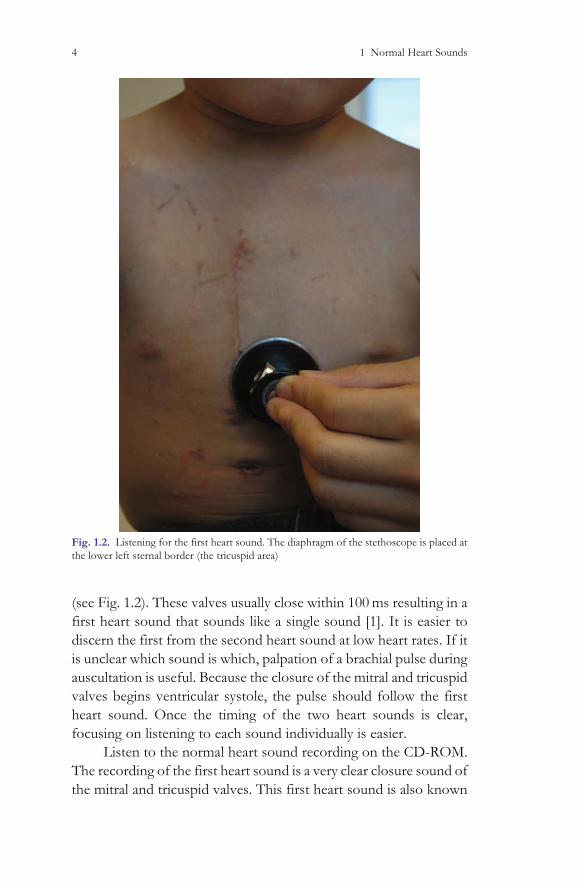
(see Fig. 1.2). These valves usually close within 100 ms resulting in a
first heart sound that sounds like a single sound [1]. It is easier to
discern the first from the second heart sound at low heart rates. If it
is unclear which sound is which, palpation of a brachial pulse during
auscultation is useful. Because the closure of the mitral and tricuspid
valves begins ventricular systole, the pulse should follow the first
heart sound. Once the timing of the two heart sounds is clear,
focusing on listening to each sound individually is easier.
Listen to the normal heart sound recording on the CD-ROM.
The recording of the first heart sound is a very clear closure sound of
the mitral and tricuspid valves. This first heart sound is also known
Fig. 1.2. Listening for the first heart sound. The diaphragm of the stethoscope is placed at
the lower left sternal border (the tricuspid area)
4 1 Normal Heart Sounds

as S1. It is lower pitched than the second heart sound (S2), but is still
well heard at the lower left sternal border using the diaphragm of the
stethoscope (Table 1.1).
If the first heart sound ‘‘slurs’’ or splits, this could be an
indication of cardiac pathology. This slurring of the first heart
sound may be caused by asynchronous closure of the mitral and
tricuspid valves, and could be a normal finding. Other possibilities
for the ‘‘splitting’’ of the first heart sound would include clicks. All
four of the cardiac valves may make clicking sounds. Each of the
clicks has differentiating characteristics that will be discussed below.
A split first heart sound that is heard best at the apex may be
related to an abnormal aortic valve. After the mitral and tricuspid
valves close, the abnormal aortic valve will open, making a clicking
sound. A ‘‘split’’ first heart sound that is heard best at the apex or the
upper right sternal border may be caused by an abnormal aortic
valve. After the mitral and tricuspid valves close, the abnormal aortic
valve will open, making a clicking sound. This type of ‘‘splitting’’ of
the first heart sound is not a true ‘‘split’’ first heart sound. Rather the
first heart sound, caused by the closure of the mitral and tricuspid
valves precedes the aortic valve click [2]. With the stethoscope, the
click sounds like a split first heart sound. For this reason, thinking of
aortic valve clicks as a ‘‘split first heart sound heard best at the apex’’
is a simple way to remember when listening to a patient with an
aortic valve click.
Table 1.1. What you hear on the CD-ROM
Normal
heart
sounds
Upper right
sternal border
Upper left
sternal border
Lower left
sternal border Apex
First heart
sound
Not audible Very soft Low pitched
thud, heard
best at the
lower left
sternal
border
Soft single
sound
Second
heart
sound
Single Splits with
respiration
Slightly louder
than S1,
single
Single sound,
slightly
louder
than S1
Systole No murmur No murmur No murmur No murmur
Diastole No murmur No murmur No murmur No murmur
The First Heart Sound 5

The mitral valve click is best heard at the apex, and often while
the patient is in the standing position. The prolapsing mitral valve
may have more length to the combined length of the papillary
muscle, chordae tendenae, and valve leaflets (see Fig. 1.3). This
allows the mitral valve to prolapse into the left atrium, and may
cause a clicking sound heard at the apex. Maneuvers that decrease
the volume of the left ventricle, such as standing, will allow the
mitral valve to prolapse more. Standing causes intravascular fluid to
move into the lower extremities and the pelvis, leaving less blood to
fill the heart. The mitral valve structures, such as the chordae and the
valve leaflets, have a fixed length that does not change when the
patient is in a standing position. If there is less blood in the heart,
because of the patient standing and gravity taking blood down to the
lower extremities, then the mitral valve will have more length
relative to the size of the ventricle, and will possibly move above
the mitral valve annulus with systole. When this happens, the mitral
valve prolapses. This is why the click of mitral valve prolapse is best
heard while the patient is standing.
Pulmonary valve clicks often sound like split first heart sounds,
and are heard at the upper left sternal border (the pulmonic area) [3].
The clicking pulmonary valve also causes a ‘‘split’’ first heart sound,
Fig. 1.3. Prolapse of the mitral valve caused by decreasing the intracardiac volume. This is
done by having the patient stand while listening with the diaphragm at the apex (the mitral
area)
6 1 Normal Heart Sounds
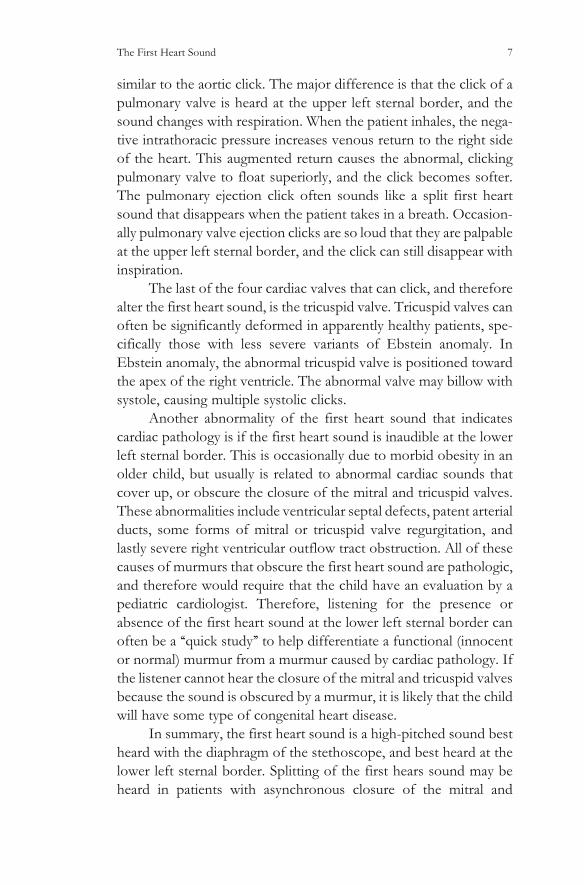
similar to the aortic click. The major difference is that the click of a
pulmonary valve is heard at the upper left sternal border, and the
sound changes with respiration. When the patient inhales, the nega-
tive intrathoracic pressure increases venous return to the right side
of the heart. This augmented return causes the abnormal, clicking
pulmonary valve to float superiorly, and the click becomes softer.
The pulmonary ejection click often sounds like a split first heart
sound that disappears when the patient takes in a breath. Occasion-
ally pulmonary valve ejection clicks are so loud that they are palpable
at the upper left sternal border, and the click can still disappear with
inspiration.
The last of the four cardiac valves that can click, and therefore
alter the first heart sound, is the tricuspid valve. Tricuspid valves can
often be significantly deformed in apparently healthy patients, spe-
cifically those with less severe variants of Ebstein anomaly. In
Ebstein anomaly, the abnormal tricuspid valve is positioned toward
the apex of the right ventricle. The abnormal valve may billow with
systole, causing multiple systolic clicks.
Another abnormality of the first heart sound that indicates
cardiac pathology is if the first heart sound is inaudible at the lower
left sternal border. This is occasionally due to morbid obesity in an
older child, but usually is related to abnormal cardiac sounds that
cover up, or obscure the closure of the mitral and tricuspid valves.
These abnormalities include ventricular septal defects, patent arterial
ducts, some forms of mitral or tricuspid valve regurgitation, and
lastly severe right ventricular outflow tract obstruction. All of these
causes of murmurs that obscure the first heart sound are pathologic,
and therefore would require that the child have an evaluation by a
pediatric cardiologist. Therefore, listening for the presence or
absence of the first heart sound at the lower left sternal border can
often be a ‘‘quick study’’ to help differentiate a functional (innocent
or normal) murmur from a murmur caused by cardiac pathology. If
the listener cannot hear the closure of the mitral and tricuspid valves
because the sound is obscured by a murmur, it is likely that the child
will have some type of congenital heart disease.
In summary, the first heart sound is a high-pitched sound best
heard with the diaphragm of the stethoscope, and best heard at the
lower left sternal border. Splitting of the first hears sound may be
heard in patients with asynchronous closure of the mitral and
The First Heart Sound 7
tricuspid valves, and therefore the splitting of S1 may be a variant of
normal. ‘‘Splitting’’ of the first heart sound may also indicate cardiac
pathology, caused by aortic, mitral, tricuspid, or pulmonary valve
abnormalities. If you cannot hear the closure of the mitral and
tricuspid valves because the first heart sound is obscured by a
murmur, the patient is quite likely to have cardiac pathology
The Second Heart Sound
After listening carefully for the first heart sound at the lower left
sternal border, the listener should then move the diaphragm of the
stethoscope to the upper left sternal border and listen to the second
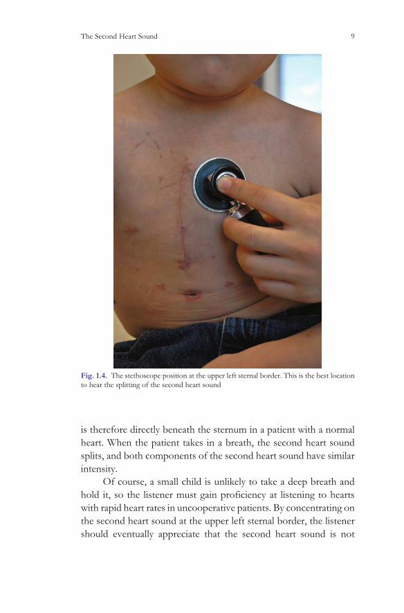
heart sound (S2) (Fig. 1.4). Many cardiologists consider the second
heart sound to be the most important component of cardiac aus-
cultation. It is very important to understand what causes this sound,
and also what causes the changes in the second heart sound during
the respiratory cycle. This is necessary in order to more easily
understand the cardiac auscultatory findings of different cardiac
lesions, as will be discussed later in this book, and heard on the
CD. The upper left sternal border is the best location to hear the
closure sound of the pulmonic valve, as well as the aortic valve.
Together these two closure sounds make up the second heart sound,
or S2. The two semilunar valves close simultaneously during exhala-
tion, and a single sound is audible.
When the patient inhales, the increased venous return to the
right side of the heart brings more blood into the right ventricle. The
ejection of this increased volume of blood takes longer than the left
ventricular ejection, and therefore the pulmonary valve closes after
the aortic valve. The splitting of the second heart sound is related
not only to the increased venous return, but also to the pressures in
the aorta and pulmonary artery. The aortic pressure is significantly
higher than the pulmonary artery pressure. The pressure in the aorta
in diastole is reflected in the blood pressure, and varies with age. The
aortic diastolic pressure is usually within 40 and 80 mmHg. The
pulmonary artery diastolic pressure is much lower than the aortic
pressure, and is generally between 5 and 15 mmHg. Ordinarily this
splitting of the second heart sound is 40 ms or less in normal
children. The pulmonary valve is anterior to the aortic valve, and
8 1 Normal Heart Sounds
is therefore directly beneath the sternum in a patient with a normal
heart. When the patient takes in a breath, the second heart sound
splits, and both components of the second heart sound have similar
intensity.
Of course, a small child is unlikely to take a deep breath and
hold it, so the listener must gain proficiency at listening to hearts
with rapid heart rates in uncooperative patients. By concentrating on
the second heart sound at the upper left sternal border, the listener
should eventually appreciate that the second heart sound is not
Fig. 1.4. The stethoscope position at the upper left sternal border. This is the best location
to hear the splitting of the second heart sound
The Second Heart Sound 9
constant, and that it changes (even if it is slightly). This means that
the pulmonary valve closes after the aortic valve.
If the second heart sound is always split, it means that the
pulmonary valve always closes after the aortic valve. There are two
common explanations for this physical finding. The first is a cardiac
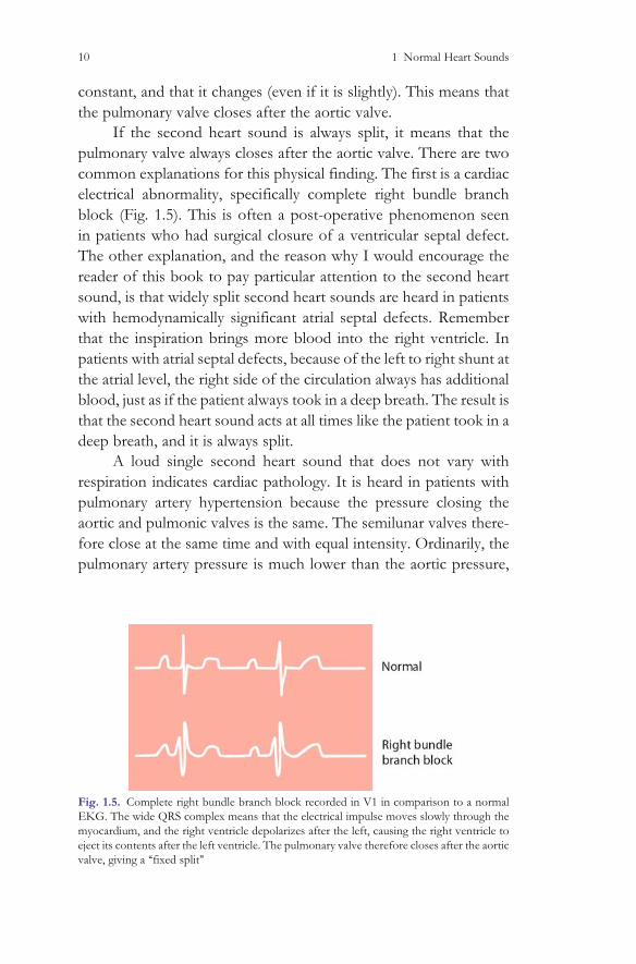
electrical abnormality, specifically complete right bundle branch
block (Fig. 1.5). This is often a post-operative phenomenon seen
in patients who had surgical closure of a ventricular septal defect.
The other explanation, and the reason why I would encourage the
reader of this book to pay particular attention to the second heart
sound, is that widely split second heart sounds are heard in patients
with hemodynamically significant atrial septal defects. Remember
that the inspiration brings more blood into the right ventricle. In
patients with atrial septal defects, because of the left to right shunt at
the atrial level, the right side of the circulation always has additional
blood, just as if the patient always took in a deep breath. The result is
that the second heart sound acts at all times like the patient took in a
deep breath, and it is always split.
A loud single second heart sound that does not vary with
respiration indicates cardiac pathology. It is heard in patients with
pulmonary artery hypertension because the pressure closing the
aortic and pulmonic valves is the same. The semilunar valves there-
fore close at the same time and with equal intensity. Ordinarily, the
pulmonary artery pressure is much lower than the aortic pressure,
Fig. 1.5. Complete right bundle branch block recorded in V1 in comparison to a normal
EKG. The wide QRS complex means that the electrical impulse moves slowly through the
myocardium, and the right ventricle depolarizes after the left, causing the right ventricle to
eject its contents after the left ventricle. The pulmonary valve therefore closes after the aortic
valve, giving a ‘‘fixed split’’
10 1 Normal Heart Sounds
and the pressure closing the pulmonary valve is therefore much
lower. The pulmonary valve is anterior to the aorta, so even if the
sound is lower in intensity, it is well heard. If, for example, the
patient has pulmonary hypertension, the pressure closing the pul-
monary valve causes the anterior semilunar valve to close early,
resulting in a loud single second heart sound. Some forms of con-
genital heart disease have malposition of the great vessels so that the
aorta is anterior to the pulmonary valve. These patients also have
loud single second heart sounds because the closure sound of the
anterior aorta overwhelms the closure of the posterior pulmonary
artery.
Paying particular attention to the second heart sound is possi-
bly the most important part of the cardiac physical examination.
Variable splitting of S2 tells that the patient has two semilunar
valves, that the pressure in the pulmonary artery is lower than the
aortic pressure, and that the contents of the right ventricle vary with
respiration. This is a great deal of information about the cardiovas-
cular system that is gleaned from listening carefully to an isolated
part of the chest!
Now please take time to listen to the CD-ROM, beginning
with the normal heart sounds. Move the virtual stethoscope to the
lower left sternal border, and listen to the first heart sound. When
you are confident you can hear this sound separately from the
second heart sound, move the stethoscope to the upper left sternal
border and concentrate on the second heart sound. Listen carefully,
and convince yourself that the sound is not constant, and that it
changes with respiration. If you cannot convince yourself that the
second heart sound splits or changes with respiration, go to the
ventricular septal defect recording. In this recording, at the upper
left sternal border (the pulmonic area), the second heart sound is
well heard, and it splits every other beat. (This is because the child
that this recording came from was so uncooperative that only two
cardiac cycles of the recording were usable: one with inspiration and
one during exhalation.) Once the listener gets the ‘‘rhythm’’ of the
splitting, it becomes more obvious. Remember, no one was born
with these skills, so with work and attention to details such as the
first and second heart sounds, you can improve your auscultatory
skills.
The Second Heart Sound 11
References
1. Nadas A (1957) Pediatric cardiology. WB Saunders, Philadelphia
2. Leatham A (1951) Phonocardiogram of aortic stenosis. Br Heart
J 13:153.
3. Leatham A, Vogelpool L (1954) The early systolic sound in
dilatation of the pulmonary artery. Br Heart J 16:21
12 1 Normal Heart Sounds

Chapter 2
Innocent Heart Murmurs
M.E. McConnell, Pediatric Heart Sounds,
DOI: 10.1007/978-1-84628-684-1_2, � Springer-Verlag London Limited 2008
13
Innocent Murmurs
Almost every child you listen to with a stethoscope will have a heart
murmur. The incidence of congenital heart disease is approximately
8 per 1,000, so this means that the vast majority of children with
heart murmurs have a normal heart. The aim of this book is to get
you more comfortable not only hearing murmurs but also being able
to tell a ‘‘normal’’ murmur from a pathologic one. The presence or
absence of a murmur cannot be the reason for referral, either to a
cardiologist, or worse yet, to an echo machine. The latter approach
has been shown to be a very cost-ineffective way to evaluate heart
murmurs [1]. The reason is that 80% of normal children have a
murmur and roughly 1% has structural heart disease. You will order
roughly 80,000 dollars worth of echocardiograms (in 2005 US
dollars) for every child who has heart disease, and that is not taking
into account that the etiology of the murmur may be missed on
echocardiography [2]. The hope is that the reader of this book (and
when used in conjunction with the CD-ROM) will be able to
determine if the murmur is likely to be pathologic or functional;
therefore, increasing the likelihood that whether a referral to a
pediatric cardiologist is necessary. Remember, the presence or
absence of a murmur is not the reason a patient should be referred
to a pediatric cardiologist. Normal murmurs are also known as
functional or innocent murmurs. This means that if the heart ejects
its contents, a noise is made. The list of functional murmurs includes
seven possibilities, as reviewed in the excellent article by Pelech et al.
[3]. Because three of these murmurs are by far the most common, in
the interest of simplicity, they will be discussed individually in detail.
The three common innocent or functional murmurs are peripheral
pulmonary ‘‘stenosis,’’ a Still’s murmur, and the adolescent outflow
murmur. All are normal, and the patients have normal heart and do
not require a special medication.
Peripheral Pulmonary Flow Murmurs
Peripheral pulmonary stenosis murmurs commonly present by
6 weeks of age and usually resolve by 1 year of age. They are very
common and are not associated with long-term pathologic
Peripheral Pulmonary Flow Murmurs 15
consequences. In order to understand the peripheral pulmonary
‘‘stenosis’’ murmur, it is important to understand a little about the
fetal circulation.
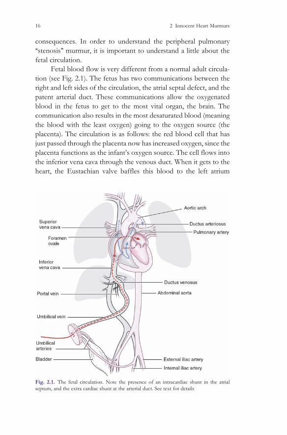
Fetal blood flow is very different from a normal adult circula-
tion (see Fig. 2.1). The fetus has two communications between the
right and left sides of the circulation, the atrial septal defect, and the
patent arterial duct. These communications allow the oxygenated
blood in the fetus to get to the most vital organ, the brain. The
communication also results in the most desaturated blood (meaning
the blood with the least oxygen) going to the oxygen source (the
placenta). The circulation is as follows: the red blood cell that has
just passed through the placenta now has increased oxygen, since the
placenta functions as the infant’s oxygen source. The cell flows into
the inferior vena cava through the venous duct. When it gets to the
heart, the Eustachian valve baffles this blood to the left atrium
Fig. 2.1. The fetal circulation. Note the presence of an intracardiac shunt in the atrial
septum, and the extra cardiac shunt at the arterial duct. See text for details
16 2 Innocent Heart Murmurs
through the hole in the atrial septum called the ostium secundum.
This means that the most oxygenated blood in the fetus is now in the
left atrium where it goes through the mitral valve to the left ventricle,
then to the aorta, and up to the fetus’ developing brain. The brain
extracts oxygen from the blood, and then the blood returns to the
superior vena cava and then to the right atrium. The brain extracts a
great deal of oxygen from the red blood cells, and now the red blood
cell is ‘‘desaturated.’’ The cell then flows from the superior vena cava
to the right atrium, through the tricuspid valve, and into the right
ventricle where it is pumped out to the pulmonary artery. Now the
blood cell has three possible paths. It could go to the right lung via
the right pulmonary artery, to the left lung via the left pulmonary
artery, or to the arterial duct and into the descending aorta. Because
the lungs are not inflated, there is no reason for much blood flow to
pass into either the right or left pulmonary artery. Most of the blood
ejected by the right ventricle goes through the arterial duct and into
the descending aorta. This desaturated blood perfuses either the
fetus’ lower body or the placenta. Because of the limited blood flow
to the lungs, the pulmonary arteries in the newborn are small [4].
The relatively small pulmonary arteries play a part in the peripheral
pulmonary flow murmur.
At birth, multiple changes take place. The infant takes a deep
breath, opening the lungs, and shortly after birth the arterial duct,
the venous duct, and the secundum atrial defect should close. The
pulmonary blood flow then increases from less than 10% of the
fetus’ cardiac output to 100% of the cardiac output.
The other changing event in newborns deals with the hema-
tocrit. The newborn has a very high hematocrit because in the uterus
the oxygen saturation level in the blood stream for the most highly
oxygenated blood is only 80–90%. Therefore, the fetus needs an
increased number of red blood cells to transport oxygen adequately.
After birth, the oxygen saturation should increase to nearly 100%,
and subsequently the hemoglobin in the newborn infant will gradu-
ally decline from the as high as 19 g/dl at birth to 9 g/dl by
7–10 weeks of age (the so-called physiologic ‘‘nadir’’) [5]. So now
there are two factors that cause many normal infants to have a
murmur generated by blood flow into the lungs. First is the pul-
monary arteries that had limited blood flow in the uterus, and are
therefore small, and the second is the increasing cardiac output
Peripheral Pulmonary Flow Murmurs 17
associated with the declining hemoglobin level. The peripheral
pulmonary flow murmur is a common consequence of these two
physiologic events in normal infants. The peripheral pulmonary
stenosis murmur is the consequence of flow turbulence made by
blood flowing from the right ventricle to the pulmonary arteries.
The infant should be asymptomatic, feeding, and growing well. The
precordial palpation is normal, and there should be no thrills or
heaves. Because the sound of a peripheral pulmonary flow murmur
begins after the ventricular contraction and therefore after the AV
valve closes, the first heart sound caused by the closure of the mitral
and tricuspid valve should be easily heard at the lower left sternal
border. The pulmonary artery pressure should be normal in these
infants; therefore, the second heart sound should also be normal.
This means the second heart sound should split with inspiration.
Because of the rapid heartbeat, it may be possible only to note that
the second heart sound does not sound the same at all times. The
murmur of peripheral pulmonary stenosis is heard best in the
pulmonary area, and also radiates along the pulmonary arteries.
What this means is that because the pulmonary arteries go to each
lung, the murmur is often heard in the right and left lateral chest.
There should be no diastolic murmur; therefore, the entire exam
should be as noted in Table 2.1.
I would encourage pediatricians and family physicians caring
for asymptomatic healthy infants with soft systolic ejection mur-
murs at the upper left sternal border as described above to reassure
the parents and follow the patients without referral to a cardiologist.
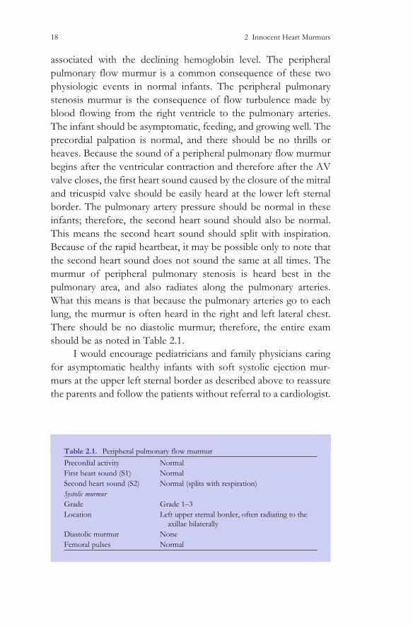
Table 2.1. Peripheral pulmonary flow murmur
Precordial activity Normal
First heart sound (S1) Normal
Second heart sound (S2) Normal (splits with respiration)
Systolic murmur
Grade Grade 1–3
Location Left upper sternal border, often radiating to the
axillae bilaterally
Diastolic murmur None
Femoral pulses Normal
18 2 Innocent Heart Murmurs
These soft systolic murmurs are extremely common and should
resolve by the time the child is about 12 months of age.
Referral of a child with this type of murmur to a pediatric
cardiologist may result in the following scenario. A soft systolic
murmur at the upper left sternal border may be because of the
normal flow across relatively small pulmonary arteries (the periph-
eral pulmonary flow murmur) or could be caused by increased blood
flow across normal pulmonary arteries. The pathologic cause of
increased flow across normal pulmonary arteries would be an atrial
septal defect (the physical examination for this will be discussed at
length in its own section). Because even the most skilled pediatric
cardiologist cannot tell the difference on physical examination
between a peripheral pulmonary flow murmur and an atrial septal
defect in all infants, the infant with a peripheral pulmonary flow
murmur will often get an echocardiogram. Hopefully the echocar-
diogram will be normal, but often a small residual hole in the atrial
septum is seen. This often results in a return appointment to the
cardiologist and the need for a second echocardiogram. The better
scenario is an asymptomatic child with a soft systolic murmur at the
upper left sternal border would be followed by the pediatrician or
family physician at routine child visits and the physician will hope-
fully note that the murmur resolves. If the patient does indeed have
an atrial defect, the physical exam findings should become more
obvious over time. There is no medical urgency to know if an
asymptomatic child has a small defect in the atrial septum that is
likely to close spontaneously. In summary, a peripheral pulmonary
flow murmur is a soft systolic murmur often heard in asymptomatic
infants. The murmur should resolve spontaneously and does not
necessarily warrant referral to a pediatric cardiologist.
Still’s Murmur
The second type of functional or innocent or normal murmur is the
‘‘Still’s murmur.’’ It was first described by Dr. Still in the early
twentieth century, and is commonly heard for the first time in a
child aged 3–6 years. Often parents are quite indignant that they
have never been told their child had a murmur before, but the fact is
that the child may not have had a murmur prior to the 3–6-year
Still’s Murmur 19

examination. The murmur is occasionally heard in infants, and may
be present in adolescents. The exact etiology of a Still’s murmur is
unknown, but hypotheses include vibrations in either the right or
left ventricle, or ‘‘tendons’’ often seen in the left ventricle [6]. Dr. Still
described the murmur as ‘‘twanging,’’ implying that the sound has
musical qualities [7]. The Still’s murmur is frequently lower pitched
than other systolic murmurs, and hence is heard well with the ‘‘low-
frequency’’ side of the stethoscope, the bell. Because the murmur is
not pathologic, the precordial activity is normal, meaning there are
no heaves (which would be caused by increased cardiac output), or
thrills (caused by turbulent rapidly moving blood). The Still’s mur-
mur begins after the mitral and tricuspid valves close, meaning that
S1 at the lower left sternal border is audible and normal. Because
there is neither pulmonary hypertension nor increased pulmonary
blood flow in patients with a Still’s murmur, the second heart sound
should be normal, splitting when the child inhales (Table 2.2).
There are two nuances associated with functional, innocent, or
Still’s murmurs that require understanding. The first is the concept
of a ‘‘venous hum.’’ In the interest of simplicity (remember this is not
an encyclopedia, but a ‘‘how-to’’ manual) all diastolic murmurs
should be considered pathologic. Diastolic murmurs can be caused
by a patent arterial duct, leaking aortic or pulmonic valves, mitral or
tricuspid stenosis, or relative mitral and tricuspid stenosis caused by
increased blood flow across these two valves. But there is one
common sound that is heard in diastole that is not pathologic, and
that sound is a venous hum. The venous hum is probably related to
Table 2.2. Still’s murmur
Precordial activity Normal
First heart sound (S1) Normal
Second heart sound (S2) Normal (splits with respiration)
Systolic murmur
Grade Grade 1–3
Location Left sternal border, often widely throughout the
precordium
Diastolic murmur Venous hum
Femoral pulses Normal
20 2 Innocent Heart Murmurs

blood flow returning from the child’s head and flowing from the
superior vena cava to the right atrium. The venous hum is a con-
tinuous ‘‘whooshing’’ sound that sounds like listening to ‘‘a seashell
at the seashore.’’ This venous hum is commonly heard in children
with Still’s murmurs and is not pathologic. It is usually heard best
with the child in the sitting position while the child is looking
straightforward. Moving the child’s head to either side often stops
the venous hum (but not the Still’s murmur). Venous hums are
softer when the child is lying down and also are altered by light
pressure to the right side of the neck, which temporarily stops blood
flow through the right jugular venous system.
The second nuance with functional, innocent, and Still’s mur-
murs is that the murmur changes with the position of the patient.
Still’s murmurs are loudest when the patient is supine and get softer
when the patient stands. This is a very valuable physical examination
finding, as the soft systolic vibratory murmur that disappears when
the patient stands is very likely to be normal, and unlikely to be
related to congenital heart disease. Remembering this physical exam
trick is also important because the murmur in patients with hyper-
trophic cardiomyopathy, the most common cause of sudden death
in young athletes, behaves just the opposite of a Still’s murmur.
When any patient stands, gravity takes blood to the lower
extremities, and therefore less blood is in the heart. With less blood
filling the ventricle, the walls of the ventricles get closer together, and
in the case of hypertrophic cardiomyopathy, the obstruction within
the cavity of the left ventricle worsens (Fig. 2.2). Remember the very
important distinction that a soft murmur associated with a normal
first and second heart sound that gets softer when the patient’s stand
is very likely to be innocent murmur, but the soft systolic murmur that
gets louder when the patient stands may be related to significant
cardiac pathology.
The innocent murmur on the CD was recorded from an
asymptomatic 4-year-old child with a loud vibratory systolic mur-
mur. Please note that the first and second heart sounds are normal,
and also note the very musical vibratory quality of this murmur.
Click on the box marked standing, and a second recording obtained
in the same patient shows how soft the murmur can get when the
patient with a functional, innocent murmur stands (Table 2.3).
Still’s Murmur 21
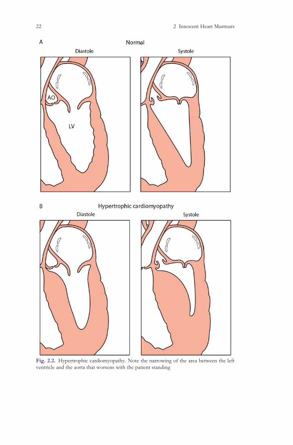
Fig. 2.2. Hypertrophic cardiomyopathy. Note the narrowing of the area between the left
ventricle and the aorta that worsens with the patient standing
22 2 Innocent Heart Murmurs

Ta
ble
2.3
Wh
atyo
uh
ear
on
the
CD
-RO
M
Fu
nct
ion
al
mu
rmu
rSu
pine
Up
per
righ
tst
ern
alb
ord
erU
pp
erle
ftst
ern
alb
ord
erL
ow
erle
ftst
ern
alb
ord
erA
pex
Fir
sth
eart
sou
nd
Sin
gle,
equ
alin
inte
nsi
tyto
S2
Sin
gle,
equ
alin
inte
nsi
tyto
S2
Lo
wp
itch
edth
ud
,sh
ort
ly
bef
ore
the
mu
rmu
rb
egin
s
So
ftsi
ngl
eso
un
d
Sec
on
dh
eart
sou
nd
Sin
gle,
equ
alin
inte
nsi
tyto
S1
Sp
lits
wit
hre
spir
atio
nS
ligh
tly
lou
der
than
S1,
split
s
wit
hre
spir
atio
n,
bu
tn
ot
hea
rdas
wel
las
itis
atth
e
up
per
left
ster
nal
bo
rder
Slig
htl
ylo
ud
erth
anS
1,
split
s
wit
hre
spir
atio
n,
bu
tn
ot
hea
rdas
wel
las
itis
atth
e
up
per
left
ster
nal
bo
rder
Sys
tole
So
ftgr
ade
1/
6ej
ecti
on
mu
rmu
rS
oft
grad
e1/
6ej
ecti
on
mu
rmu
r
Gra
de
2to
3/
6vib
rato
ry
mu
sica
lm
urm
ur
Gra
de
2to
3/
6vib
rato
ry
mu
sica
lm
urm
ur
Dia
sto
leN
om
urm
ur
No
mu
rmu
rN
om
urm
ur
No
mu
rmu
r
Stan
ding
Fir
sth
eart
sou
nd
Sin
gle,
equ
alin
inte
nsi
tyto
S2
Sin
gle,
equ
alin
inte
nsi
tyto
S2
Sin
gle,
equ
alin
inte
nsi
tyto
S2
Sin
gle,
equ
alin
inte
nsi
tyto
S2
Sec
on
dh
eart
sou
nd
Sin
gle,
equ
alin
inte
nsi
tyto
S1
Sp
lits
wit
hre
spir
atio
nS
plit
sw
ith
resp
irat
ion
Sin
gle,
equ
alin
inte
nsi
tyto
S1
Sys
tole
So
ftgr
ade
1/
6ej
ecti
on
mu
rmu
rG
rad
e1/
6m
usi
cal
vib
rato
ry
mu
rmu
r,s
oft
erth
anw
hen
the
pat
ien
tw
asly
ing
do
wn
Gra
de
1to
2/
6m
usi
cal
vib
rato
rym
urm
ur
,so
fter
than
wh
enth
ep
atie
nt
was
lyin
gd
ow
n
Gra
de
1/
6m
usi
cal
vib
rato
ry
mu
rmu
r,s
oft
erth
anw
hen
the
pat
ien
tw
asly
ing
do
wn
Dia
sto
len
om
urm
ur
no
mu
rmu
rn
om
urm
ur
no
mu
rmu
r
Still’s Murmur 23

The Aortic Outflow Murmur
The third and final of the common innocent murmurs is the aortic
outflow murmur of adolescents and young adults. The murmur is
usually grade one or two in intensity, and is associated with normal
first and second heart sounds. The murmur is heard best at the
upper right sternal border (the aortic area) and is therefore felt to be
secondary to blood flow in the left ventricular outflow tract. It is
different from valvar aortic stenosis in that the patients do not have
an ejection click. The murmur is often heard in athletes, who
typically have a low resting heart rate and therefore a large stroke
volume of blood flowing in the left ventricular outflow tract in
systole. The outflow tract murmur of adolescents and young adults
may also be confused with the murmur of hypertrophic cardiomyo-
pathy. Standing a patient with hypertrophic cardiomyopathy should
increase the murmur, whereas standing the patient with the aortic
outflow murmur should result in either a decrease in the murmur, or
no significant change.
It is important for the parents of a child with a functional or
innocent murmur to know that their child is normal and that the
child does not need restriction from any physical activity. In addi-
tion, the child does not need any special medical treatment. This is
particularly confusing when the child goes to a dentist. Because
patients with some cardiac pathology need to take antibiotics prior
to certain dental procedures, the forms in dentist’s office often ask
simply ‘‘Do you have a heart murmur?’’. An affirmative answer to
this question will often result in additional testing and anxiety. In the
case of a child with a functional or innocent murmur, the proper
Table 2.4 Aortic outflow murmur
Precordial activity Normal
First heart sound (S1) Normal
Second heart sound (S2) Normal (splits with respiration)
Systolic murmur
Grade Grades 1–3
Location Upper right sternal border
Diastolic murmur None
Femoral pulses Normal
24 2 Innocent Heart Murmurs
answer to the question on the dental form is that the child does not
have a murmur. This will eliminate anxiety and additional unnecessary
testing (Table 2.4).
References
1. Danford DA (1995) Cost-effectiveness of echocardiography for
evaluation of children with murmurs. Echocardiography
12:153–162
2. Shub C (2003) Echocardiography or auscultation? How to eval-
uate systolic murmurs. Can Family Physician 49:163–167
3. Pelech AN (2004) The physiology of cardiac auscultation. Pediatr
Clin North Am 51(6):1515–1535
4. Gardiner HM (2002) Physiology of the developing human fetal
heart. In: Anderson RH et al. (eds.) Paediatric cardiology Church-
ill Livingstone, London, pp 660–661
5. Walters M, Abelson HT (1996) Interpretation of the complete
blood count. Pediatr Clin North Am 43(3):599–622
6. Rosenthal A (1984) How to distinguish between innocent and
pathologic murmurs in childhood. Pediatr Clin of North Am
31:1229–1240
7. Still GH (1918) Common disorders and diseases of childhood,
3rd edn. Oxford University Press, London, p 495
References 25

Chapter 3
Atrial Septal Defects
M.E. McConnell, Pediatric Heart Sounds,
DOI: 10.1007/978-1-84628-684-1_3, � Springer-Verlag Lodon Limited 2008
27

Introduction
As discussed in Chapter 2, we all had an atrial septal defect at some
point in our lives. Hopefully the defect that allowed oxygenated
blood to flow from the right atrium to the left atrium while in the
uterus closed after birth, but in approximately 0.075% of all chil-
dren, the hole remains [1]. A tiny hole between the two atrial
chambers does not cause excessive pulmonary blood flow and
therefore cannot be diagnosed on physical examination. Rarely, a
tiny hole in the atrial septum is important to diagnose, as some
patients with small atrial septal defects may have passage of the
deoxygenated blood from the right atrium to the left atrium or clots
passing from the right atrium to the left atrium, causing a stroke.
There is also some evidence suggesting that migraine headaches may
improve after closure of a small patent foramen ovale [2]. But this is
a murmur book, and the goal is to get the reader more comfortable
using the stethoscope to diagnose innocent or pathologic murmurs.
In my opinion, the ability to diagnose an atrial septal defect means
that the listener is using his or her eyes, hands, ears, and brain to
diagnose a subtle cardiac abnormality. For example, if a patient has a
Grade 5 systolic murmur of aortic stenosis, it requires very little skill
for the examiner to recognize that the patient has significant heart
Table 3.1. Comparison of the physical examination features of a patient with an
atrial septal defect and a patient with a functional or innocent (normal) murmur
Atrial septal defect Innocent murmur
Precordial activity Increased Normal
First heart sound
(S1)
Normal Normal
Second heart
sound (S2)
Widely split Normal (splits with respiration)
Systolic murmur
Grade Grade 1–3 Grade 1–3
Location Upper left sternal
border
Left sternal border, often widely
throughout the precordium
Quality Crescendo
decrescendo ‘‘flow’’
Ejection crescendo decrescendo
‘‘musical’’
Diastolic murmur ‘‘Rumble’’ at the lower
left sternal border
Venous hum
Femoral pulses Normal Normal
Introduction 29
disease. The reported incidence of all congenital heart diseases varies
between 0.5 and 1%, depending on the study. Patients with atrial
septal defects are roughly 7–11% of all patients with congenital heart
disease, meaning that roughly 1 in a 1,000 children will have a
hemodynamically significant atrial septal defect [3]. Trying to tell
the 1 in 1,000 patient with an atrial septal defect from the approxi-
mately 800 per 1,000 children who have a functional or innocent
murmur requires a great deal of skill. Developing this skill is what
this book and CD-ROM are all about. It would not be cost effective
to refer all 800 children with murmurs to a pediatric cardiologist, but
it certainly would not be optimal to miss the rare patient with an
atrial septal defect (Table 3.1).
Anatomy
The atrial septum is a complex structure that contains a large hole in
the center during fetal life (the foramen ovale, see Fig. 2.1). This hole
allows oxygenated blood in the fetus to travel from the right atrium
to the left atrium so it can go to perfuse the infant’s brain. After
birth, the foramen ovale should close, but in many people, a probe
patent opening between the two atria remains. A persistent large
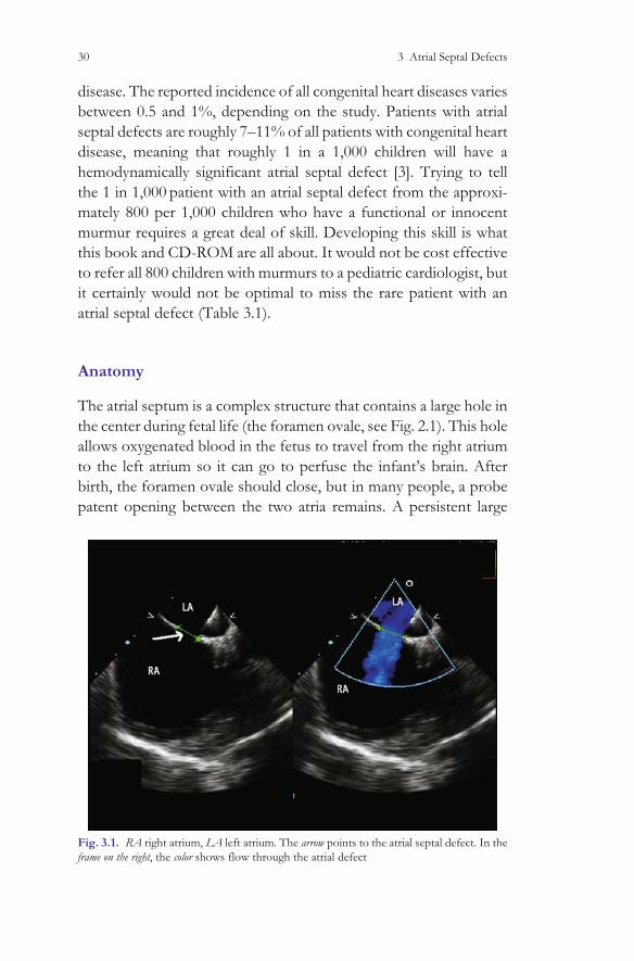
Fig. 3.1. RA right atrium, LA left atrium. The arrow points to the atrial septal defect. In the
frame on the right, the color shows flow through the atrial defect
30 3 Atrial Septal Defects

atrial defect may cause significant long-term health problems, such
as recurrent pneumonia, failure to thrive, and eventually pulmonary
hypertension (Fig. 3.1).
The life expectancy of a patient with a large atrial defect is
significantly shortened, but if the defect is repaired before the
development of pulmonary hypertension, the life expectancy of a
patient with an atrial septal defect should be normal. As shown in
the figure, the atrial septum may also have holes in locations other
than the secundum septum, including the superior and inferior sinus
venosus defects, the coronary sinus defects, and the ostium primum
defects (Fig. 3.2). As this is an auscultation book, and not a pediatric
cardiology text book, the discussion will center on the physical
examination of a child and young adult with a hemodynamically
significant atrial septal defect.
Fig. 3.2. Atrial septal defect locations. Both the superior and inferior sinus venosus ASDs
may be associated with right to left shunting, even in the absence of pulmonary hyperten-
sion. The secundum defect is the most common, and is in the location of the fetal foramen
ovale
Anatomy 31
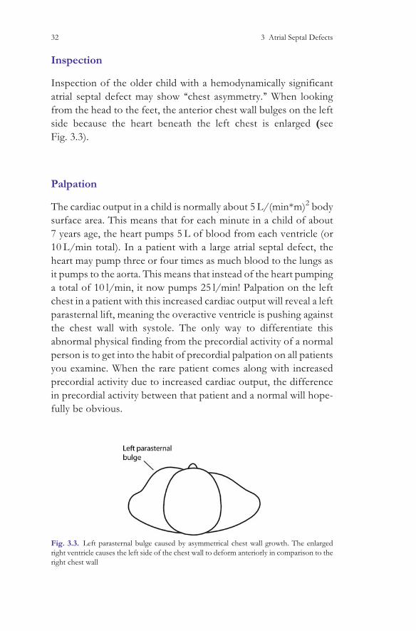
Inspection
Inspection of the older child with a hemodynamically significant
atrial septal defect may show ‘‘chest asymmetry.’’ When looking
from the head to the feet, the anterior chest wall bulges on the left
side because the heart beneath the left chest is enlarged (see
Fig. 3.3).
Palpation
The cardiac output in a child is normally about 5 L/(min*m)2 body
surface area. This means that for each minute in a child of about
7 years age, the heart pumps 5 L of blood from each ventricle (or
10 L/min total). In a patient with a large atrial septal defect, the
heart may pump three or four times as much blood to the lungs as
it pumps to the aorta. This means that instead of the heart pumping
a total of 10 l/min, it now pumps 25 l/min! Palpation on the left
chest in a patient with this increased cardiac output will reveal a left
parasternal lift, meaning the overactive ventricle is pushing against
the chest wall with systole. The only way to differentiate this
abnormal physical finding from the precordial activity of a normal
person is to get into the habit of precordial palpation on all patients
you examine. When the rare patient comes along with increased
precordial activity due to increased cardiac output, the difference
in precordial activity between that patient and a normal will hope-
fully be obvious.
Fig. 3.3. Left parasternal bulge caused by asymmetrical chest wall growth. The enlarged
right ventricle causes the left side of the chest wall to deform anteriorly in comparison to the
right chest wall
32 3 Atrial Septal Defects
Auscultation
After inspecting the chest and palpating for a parasternal lift, use the
stethoscope diaphragm at the lower left sternal border and listen
carefully for the first heart sound. In a patient with an atrial septal
defect, the closure of the mitral and tricuspid valves should be easily
heard. The first sound should be single or narrowly split, and there
are no clicks. Now use the diaphragm and listen at the upper left
sternal border for the second heart sound. In Chapter 1, we learned
that the second heart sound is caused by the closure of the pulmonic
and aortic valves and that the pulmonary components of the second
heart sound ‘‘splits’’ and moves away from the aortic closure sound
when the patient takes a deep breath. This is because the deep breath
brings additional venous return to the right side of the heart, and the
right ventricle takes longer to eject its contents. The pulmonary
valve therefore closes after the aortic valve. In a patient with an
atrial septal defect, the blood flow flowing from the left atrium to the
right atrium through the atrial defect meets with the superior vena
cava blood and the inferior vena cava blood and then flows into the
right ventricle. Therefore, the right ventricle always has more blood
in it than the left ventricle, and the pulmonary valve closure sound
always resembles the second heart sound heard when a patient takes
a deep breath. This means the pulmonic component closes after the
aortic component. This is also known as a ‘‘fixed split’’ second heart
sound. It sounds like thump (the first heart sound) then thump
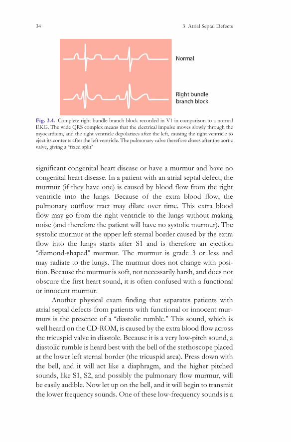
thump. Another cause of a fixed split second heart sound is a
complete right bundle branch block. This may be post surgical or
idiopathic. In complete right bundle branch block, the right ventricle
always depolarizes after the left ventricle and the pulmonary valve
always closes after the aortic valve (Fig. 3.4). In summary, in a patient
with atrial septal defect, the precordial activity is increased, the first
heart sound is normal, and the second heart sound is widely split.
Now we get to the murmur in patients with atrial septal
defects. One of the big problems with diagnosing ASDs and why
auscultation is so important to perform properly is that there are
many people with large atrial septal defects who have no systolic
murmur ! Therefore, the presence or absence of a systolic murmur
is not necessarily the reason a patient should be referred to a
pediatric cardiologist. You may have no murmur and have
Auscultation 33
significant congenital heart disease or have a murmur and have no
congenital heart disease. In a patient with an atrial septal defect, the
murmur (if they have one) is caused by blood flow from the right
ventricle into the lungs. Because of the extra blood flow, the
pulmonary outflow tract may dilate over time. This extra blood
flow may go from the right ventricle to the lungs without making
noise (and therefore the patient will have no systolic murmur). The
systolic murmur at the upper left sternal border caused by the extra
flow into the lungs starts after S1 and is therefore an ejection
‘‘diamond-shaped’’ murmur. The murmur is grade 3 or less and
may radiate to the lungs. The murmur does not change with posi-
tion. Because the murmur is soft, not necessarily harsh, and does not
obscure the first heart sound, it is often confused with a functional
or innocent murmur.
Another physical exam finding that separates patients with
atrial septal defects from patients with functional or innocent mur-
murs is the presence of a ‘‘diastolic rumble.’’ This sound, which is
well heard on the CD-ROM, is caused by the extra blood flow across
the tricuspid valve in diastole. Because it is a very low-pitch sound, a
diastolic rumble is heard best with the bell of the stethoscope placed
at the lower left sternal border (the tricuspid area). Press down with
the bell, and it will act like a diaphragm, and the higher pitched
sounds, like S1, S2, and possibly the pulmonary flow murmur, will
be easily audible. Now let up on the bell, and it will begin to transmit
the lower frequency sounds. One of these low-frequency sounds is a
Fig. 3.4. Complete right bundle branch block recorded in V1 in comparison to a normal
EKG. The wide QRS complex means that the electrical impulse moves slowly through the
myocardium, and the right ventricle depolarizes after the left, causing the right ventricle to
eject its contents after the left ventricle. The pulmonary valve therefore closes after the aortic
valve, giving a ‘‘fixed split’’
34 3 Atrial Septal Defects
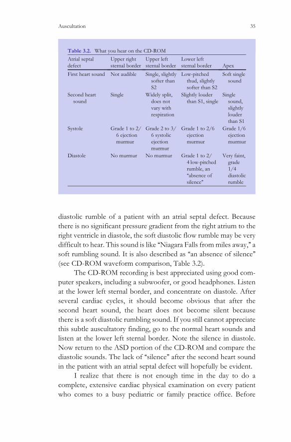
diastolic rumble of a patient with an atrial septal defect. Because
there is no significant pressure gradient from the right atrium to the
right ventricle in diastole, the soft diastolic flow rumble may be very
difficult to hear. This sound is like ‘‘Niagara Falls from miles away,’’ a
soft rumbling sound. It is also described as ‘‘an absence of silence’’
(see CD-ROM waveform comparison, Table 3.2).
The CD-ROM recording is best appreciated using good com-
puter speakers, including a subwoofer, or good headphones. Listen
at the lower left sternal border, and concentrate on diastole. After
several cardiac cycles, it should become obvious that after the
second heart sound, the heart does not become silent because
there is a soft diastolic rumbling sound. If you still cannot appreciate
this subtle auscultatory finding, go to the normal heart sounds and
listen at the lower left sternal border. Note the silence in diastole.
Now return to the ASD portion of the CD-ROM and compare the
diastolic sounds. The lack of ‘‘silence’’ after the second heart sound
in the patient with an atrial septal defect will hopefully be evident.
I realize that there is not enough time in the day to do a
complete, extensive cardiac physical examination on every patient
who comes to a busy pediatric or family practice office. Before
Table 3.2. What you hear on the CD-ROM
Atrial septal
defect
Upper right
sternal border
Upper left
sternal border
Lower left
sternal border Apex
First heart sound Not audible Single, slightly
softer than
S2
Low-pitched
thud, slightly
softer than S2
Soft single
sound
Second heart
sound
Single Widely split,
does not
vary with
respiration
Slightly louder
than S1, single
Single
sound,
slightly
louder
than S1
Systole Grade 1 to 2/
6 ejection
murmur
Grade 2 to 3/
6 systolic
ejection
murmur
Grade 1 to 2/6
ejection
murmur
Grade 1/6
ejection
murmur
Diastole No murmur No murmur Grade 1 to 2/
4 low-pitched
rumble, an
‘‘absence of
silence’’
Very faint,
grade
1/4
diastolic
rumble
Auscultation 35
considering referral to a pediatric cardiologist for evaluation of a
murmur, I would strongly encourage you to go through the com-
plete exam as outlined above and ask yourself, ‘‘Does this patient
have an atrial septal defect?’’. This should remind you to look for a
left parasternal bulge and then to feel the precordial activity, feeling
for increased cardiac activity. It will also remind you to listen
critically for the first and second heart sounds and to listen through-
out the precordium in systole and in diastole with both the bell and
the diaphragm. Have the patient stand and note if the murmur gets
softer. Hopefully by following this routine, you will refer fewer
patients with functional or innocent murmurs and appropriately
refer patients who might have an atrial septal defect.
It is also important to note that although all children who have
large hemodynamically atrial septal defects are born with a defect in
the atrial septum, the hole may grow over time. Therefore, their
physical examination may change over time as the right ventricle
dilates and the amount of pulmonary blood flow increases. It is
unlikely for a baby to have a classic atrial septal defect exam because
it takes time for the right ventricle to dilate enough so that the RV
volume is significantly greater than the LV volume. Until the RV
volume significantly exceeds the LV volume, the precordial activity
will remain normal, and the second heart sound will remain normal
as well. The patient also will not have a diastolic rumble. The point is
that the physical examination is dynamic and will hopefully become
more obvious over time. If you see an asymptomatic child and you
are not certain if the child has a functional murmur or an atrial septal
defect, re-evaluation in the next year may suggest the correct diag-
nosis. There is no urgent reason to know whether an asymptomatic
child has an atrial septal defect or a functional murmur, as patients
with atrial septal defects do not require antibiotic prophylaxis, and
when the patient is asymptomatic, closure of the defect is elective.
Subacute Bacterial Endocarditis Prophylaxis
Recommendations (SBE)
According to the guidelines published by the American Heart Asso-
ciation in 2007, patients with atrial septal defects do not require
antibiotic prophylaxis at times of endocarditis risk [4].
36 3 Atrial Septal Defects

References
1. Samanek M, Slavik Z, Zborilova B, Hrobonova V, Voriskova M,
Skovranek J (1989) Prevalence, treatment, and outcome of heart
disease in live-born children: a prospective analysis of 91,823 live-
born children. Pediatr Cardiol 10(4):205–211
2. Post MC, Thijs V, Herroelen L, Budts WI (2007) Closure of a
patent foramen ovale is associated with a decrease in prevalence
of migraine. Neurology 62(8):1439–1440
3. Beerman LB, Zuberbuhler JR (2002) Arial septal defect. In:
Anderson RH et al (eds.) Paediatric cardiology.. Churchill Living-
stone, London, pp 901–930
4. Wilson W, Taubert K, Gewitz M et al. (2007) Prevention of
infective endocarditis. Guidelines from the American Heart
Association. Rheumatic Fever, Endocarditis, and Kawasaki Dis-
ease Committee, Council on Cardiovascular Disease in the
Young, and the Council on Clinical Cardiology, Council on
Cardiovascular Surgery and Anesthesia, and the Quality of Care
and Outcomes Research Interdisciplinary Working Group 2007.
Circulation April 19
References 37
Chapter 4
Ventricular Septal Defects
M.E. McConnell, Pediatric Heart Sounds,
DOI: 10.1007/978-1-84628-684-1_4, � Springer-Verlag London Limited 2008
39
Incidence
Defects in the interventricular septum are common. A recent study
suggested that the incidence was as high as 2–4% of all newborn
infants [1, 2] Understanding when these defects might present
clinically is important in understanding the physiology of the dis-
order. When infants are born, until the arterial duct and the atrial
septal defect close, the pressures in the right and left ventricles may
be similar. As the pulmonary vascular resistance drops, and the
arterial duct closes, the right and left ventricles will have different
pressures. This drop in pulmonary vascular resistance, and therefore
right ventricular pressure, is usually complete by 6 weeks of age.
The ability to hear a murmur of a ventricular septal defect depends
on the difference in pressure from the left to the right ventricle. If,
for example, the pulmonary vascular resistance drops quickly, the
harsh murmur of a restrictive ventricular septal may be heard in the
first few days of life. On the other hand, if the pulmonary vascular
resistance is high, then the pressure in the right ventricle will remain
high, and there will be little pressure difference between the LV and
the RV. In this case, it may be very difficult to hear the murmur of a
ventricular septal defect.
Anatomy
Ventricular septal defects are named based on their location within
the ventricular septum. The right ventricle wraps around the conical
left ventricle (Fig. 4.1) One way to visualize this relationship is to use
your right hand, with the palm pointing toward your chest, and
the hand slightly cupped. The thumb of your right hand would be in
the tricuspid valve, and the fingers of the right hand would be in the
right ventricular outflow tract. All along this three-dimensional
structure, a defect could join the right ventricle with the left. If the
defect was in the posterior part of the RV, near the tricuspid valve
(the thumb), it would be called an inlet ventricular septal defect.
These defects are usually associated with abnormalities of the tri-
cuspid and mitral valves, and are not likely to close spontaneously.
These inlet ventricular septal defects are common in children with
Down syndrome. When the inlet ventricular septal defect is also
associated with an inferior atrial septal defect (a so-called ostium
Anatomy 41

primum ASD) they are called atrioventricular septal defects or
atrioventricular canal defects.
Defects near the aortic valve on the left side, and the tricuspid
valve on the right side are called membranous or perimembranous
ventricular septal defects (Fig. 4.2). Ventricular septal defects vary in
size, and may close spontaneously. Often they are large enough and
have a large enough volume of blood flowing through the defect
that the children cannot eat and grow well, and surgical intervention
is warranted. These perimembranous defects would be near the
junction of the thumb and the palm, when using the example
above (see Fig. 4.1).
Ventricular septal defects in the palm of the hand, potentially
going into the apex of both ventricles are called muscular ventricular
septal defects. These defects are often small, but may be multiple
(the so-called ‘‘Swiss-cheese’’ septum). Small muscular defects are
common in normal newborns, and have a tendency to close very
quickly. One study suggests that about 75% of muscular defects
close within the first year of life [2].
The last location of ventricular septal defects is in the outlet
septum. If the defect is beneath the right aortic cusp and the
Fig. 4.1. The normal relationship between the right and the left ventricle. The right
ventricle wraps around the conical left ventricle. See text
42 4 Ventricular Septal Defects
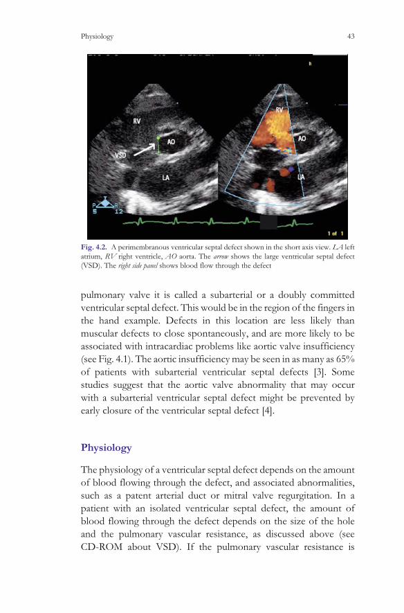
pulmonary valve it is called a subarterial or a doubly committed
ventricular septal defect. This would be in the region of the fingers in
the hand example. Defects in this location are less likely than
muscular defects to close spontaneously, and are more likely to be
associated with intracardiac problems like aortic valve insufficiency
(see Fig. 4.1). The aortic insufficiency may be seen in as many as 65%
of patients with subarterial ventricular septal defects [3]. Some
studies suggest that the aortic valve abnormality that may occur
with a subarterial ventricular septal defect might be prevented by
early closure of the ventricular septal defect [4].
Physiology
The physiology of a ventricular septal defect depends on the amount
of blood flowing through the defect, and associated abnormalities,
such as a patent arterial duct or mitral valve regurgitation. In a
patient with an isolated ventricular septal defect, the amount of
blood flowing through the defect depends on the size of the hole
and the pulmonary vascular resistance, as discussed above (see
CD-ROM about VSD). If the pulmonary vascular resistance is
Fig. 4.2. A perimembranous ventricular septal defect shown in the short axis view. LA left
atrium, RV right ventricle, AO aorta. The arrow shows the large ventricular septal defect
(VSD). The right side panel shows blood flow through the defect
Physiology 43
low, and the hole is moderate in size, there might be a great deal of
blood flowing from the left ventricle through the ventricular septal
defect, and into the right ventricle. This blood will subsequently flow
from the right ventricle into the pulmonary artery. Patients with a
large left-to-right shunt often have increased work of breathing,
because their lungs are overfilled with blood. The increased cardiac
output causes an increased metabolic rate, and the increased work of
breathing often means that the infants with large left-to-right shunts
cannot eat as well as a child who breathes normally. The total energy
expenditure in infants with ventricular septal defects and congestive
heart failure symptoms can be 50% higher than the energy expen-
diture of age matched control infants [5]. The amount of blood flow
through the defect is also a variable phenomenon, and it can increase
as the pulmonary vascular resistance decreases. It is important to
note that the left-to-right shunt of a ventricular septal defect sends
oxygenated blood from the left ventricle back into the right ven-
tricle. The patients are therefore not cyanotic (blue) unless there is
pulmonary hypertension. In patients with large ventricular septal
defects, there may not be a pressure difference between the left and
the right ventricle and there may not be a classic ventricular septal
defect murmur. There are rare children with large ventricular septal
defects who never drop their pulmonary vascular resistance, and
therefore who never have signs of a large left-to-right shunt. These
patients can be very difficult to diagnose, unless the observer does a
careful cardiac examination. Children with Down syndrome have a
50% incidence of significant congenital heart disease. Rarely, a child
with Down syndrome will have high pulmonary vascular resistance
at birth, and therefore will not have a large left-to-right shunt. They
may remain asymptomatic for many years, until the pulmonary
vasculature becomes so damaged that the patient begins to shunt
blue blood from the right ventricle to the left side of the circulation.
This condition is known as Eisenmengers syndrome (see CD-ROM
large VSD with pulmonary hypertension). At this point, the children
are not operable, as an operation to close either the atrial or ven-
tricular defect is likely to cause worsening symptoms or death.One
way to avoid this scenario is to refer all children with Down
syndrome for at least one cardiac evaluation. Because the incidence
of disease is so high (50% compared to a general population of less
than 1%)the chances of finding an abnormality are very high.
44 4 Ventricular Septal Defects
Natural History
The natural history of a patient with a ventricular septal defect is quite
variable. Many of the small defects close spontaneously, especially the
muscular ventricular septal defects. One study showed that muscular
ventricular septal defects had a 69% chance of spontaneous closure
within the first 6 years of life. This same study showed that muscular
ventricular septal defects were also unlikely to require surgical inter-
vention, with only 1 in 39 patients requiring surgery [2].Perimembra-
nous ventricular septal defects have a higher likelihood of causing
significant symptoms, and therefore are more likely to require surgical
intervention. Spontaneous closure occurred in 28% of patients with a
perimembranous ventricular septal defect followed for 6 years, but
surgical intervention to close the defect was needed in 41% over the
same time period. Even if a membranous ventricular septal defect is
felt to be ‘‘asymptomatic’’ and not large enough to warrant surgical
intervention, long-term complications are common. The incidence of
endocarditis is estimated at 14.5/10,000 patient years [6]. Other com-
plications of even small ventricular septal defects include subaortic
obstruction, aortic valve insufficiency, muscular right ventricular out-
flow tract obstruction, and congestive heart failure. Aortic valve
insufficiency can be seen in up to 7.5% of adults with perimembra-
nous or subarterial ventricular septal defects, and is often progressive
[7]. A recent study suggested that the 8-year event-free survival for
adults with small ventricular septal defects was over 95% [8]. Events
in their study were defined as death, endocarditis, or need for surgical
closure of the ventricular septal defect. Other authors suggest that the
risk of the above-listed complications in adults with small ventricular
septal defects is as high as 25% [9].
Medical Management
Because most ventricular septal defects close spontaneously, or at
least get smaller over time, if the child is able to eat reasonably well
and grow, surgery should be avoided. In some infants, the large left-
to-right shunt through the ventricular septal defect causes increased
pulmonary circulation, resulting in increased work of breathing. This
is coupled with decreased systemic blood flow, as the blood leaving
Medical Management 45

the left ventricle can more easily flow through the VSD and into the
pulmonary circulation than flow through the aortic valve and into
the systemic circulation. Decreased systemic blood flow results in
the activation of the renin–angiotensin–aldosterone system and the
sympathetic nervous system. The infants are often irritable, feed
poorly, are pale, and have increased work of breathing [10]. Tradi-
tionally these infants are treated with digoxin and diuretics, although
the evidence that these medications are effective for this hemody-
namic derangement is lacking [11]. In adult patients with congestive
heart failure, treatment with medications to block the elevated
neurohormones, such as angiotensin converting enzyme inhibitors
and beta blockers have been shown to improve survival signifi-
cantly. Although the mechanism of congestive heart failure in
infants with ventricular septal defects is very different from the
mechanism in most adults with congestive heart failure, the neuro-
hormonal elevation is similar, suggesting that possibly the medica-
tions that are effective for adults with congestive heart failure might
be useful for symptomatic patients with large left-to-right shunts
[12, 13].
Surgical Options
If the infant is not thriving, surgical closure of the ventricular septal
defect may be the safest course. In most major centers, closure of a
membranous ventricular septal defect, even in a small child, carries a
mortality rate of less than 1%. Because chronic congestive heart
failure is not benign, with infants at risk for RSV infections and
possibly neurologic abnormalities, early intervention to close ven-
tricular septal defects in symptomatic infants should be
considered [14].
For some patients, closure of the ventricular septal defect
surgically carries increased risk. This might include patients with
large muscular ventricular septal defects that are not easily closed
surgically. For some of these patients, closure of the defect in the
catheterization laboratory may be the safest procedure. In 2004,
Knauth published a 13-year experience involving 170 patients with
ventricular septal defects ‘‘selected for device closure if they had 1 or
more VSDs that were ascertained to result in sufficient
46 4 Ventricular Septal Defects
hemodynamic derangement to warrant intervention and either a
type of VSD that was technically difficult to close surgically or an
overall medical condition with associated surgical risks sufficient to
justify the known and potential unknown risks of the device.’’ A total
of 69% of the patients had muscular ventricular septal defects, and
the remainder had either intentionally fenestrated VSD patches or
postoperative residual defects along the margin of patches used to
close VSDs in the membranous or outflow portions of the sep-
tum.A total of 296 devices were implanted in the 170 patients.There
were 332 adverse events reported in 153 of the 170 patients (90%),
including hemorrhage, arrhythmia, new onset regurgitation of the
aortic or tricuspid valve, death, and need for device explantation.
The authors felt that only one patient died as a result of the device
implantation, and that the majority had a significant decrease in the
size of the shunt through the ventricular septal defect. As new
devices become available, catheter closure of ventricular septal
defects may become the preferred method [15].
Physical Examination
The physical examination in a patient with a ventricular septal defect
varies greatly depending on the size of the defect, the pulmonary
vascular resistance, and the amount of blood flowing through the
defect.
Small Muscular Ventricular Septal Defects
The tiny muscular ventricular septal defects discussed earlier allow
very little blood to flow from the left to the right ventricle, and are
also associated with normal pulmonary vascular resistance. Patients
with small muscular ventricular septal defects would have normal
precordial activity, because the heart is pumping a normal amount of
blood. It would also be unusual to palpate a thrill in a patient with a
small muscular ventricular septal defect. The shunt through the
ventricular septum starts as the AV valves are closing (the ‘‘isovo-
lumic contraction phase’’) and the murmur therefore ‘‘obscures’’ the
first heart sound. The murmur is harsh, not musical like a functional
or innocent murmur. In many small muscular ventricular septal
Small Muscular Ventricular Septal Defects 47

defect patients, the murmur stops before the second heart sound.
This is because the muscular ventricular septal defect is so small that
it actually closes during systole, cutting off the blood flow from the
left to the right ventricle. In patients with a small muscular ventri-
cular septal defect, the pulmonary artery pressure should be normal,
and therefore the second heart sound should split and move nor-
mally with respiration. The pulses will be normal (Table 4.1).
Moderate Ventricular Septal Defects
In patients with moderate ventricular septal defects, the physical
examination again depends a great deal on the size of the defect, the
pulmonary vascular resistance, and the amount of flow through
the defect. The location of the moderate defects is not critical to
the physical examination, but rarely there can be differentiating
factors that point to the location of the defect within the interven-
tricular septum.
Inspection of the chest might show a left parasternal bulge,
caused by chest deformation secondary to the large heart (see
Fig. 3.3). Also on inspection the child who is in significant conges-
tive heart failure might have ‘‘Harrison’s Groove’’, a longitudinal
depression at the site of the insertion of the diaphragm. If the patient
has a large left-to-right shunt through the ventricular septal defect,
there might be three or four times as much blood flowing into the
pulmonary artery as is flowing into the aorta. The heart may then be
pumping two or three times as much blood as a normal child, and
this difference should be easily palpable by placing your hand on the
Table 4.1. Small muscular VSD
Precordial activity Normal
First heart sound (S1) Obscured
Second heart sound (S2) Normal (splits with respiration)
Systolic murmur
Grade Grade 1–3
Location lower left sternal border, may stop in mid-systole
Diastolic murmur None
Femoral pulses Normal
48 4 Ventricular Septal Defects
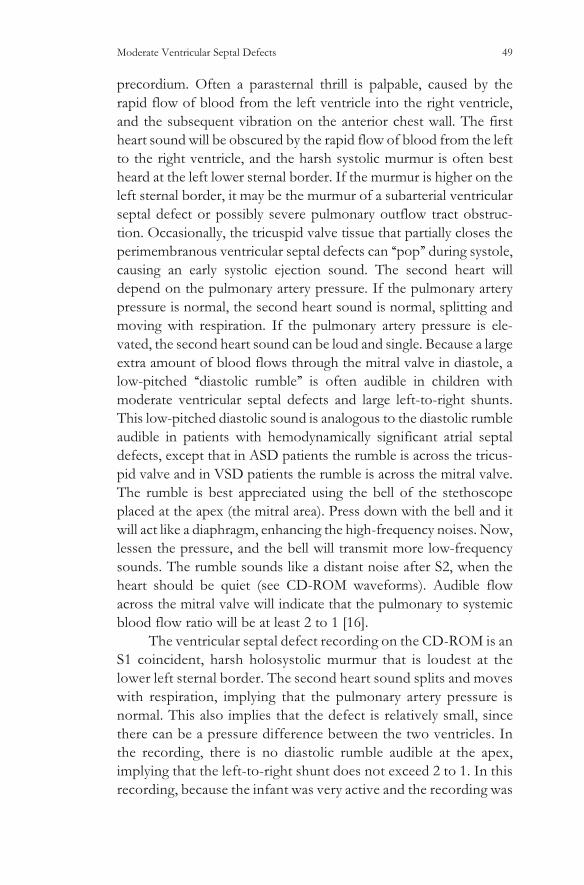
precordium. Often a parasternal thrill is palpable, caused by the
rapid flow of blood from the left ventricle into the right ventricle,
and the subsequent vibration on the anterior chest wall. The first
heart sound will be obscured by the rapid flow of blood from the left
to the right ventricle, and the harsh systolic murmur is often best
heard at the left lower sternal border. If the murmur is higher on the
left sternal border, it may be the murmur of a subarterial ventricular
septal defect or possibly severe pulmonary outflow tract obstruc-
tion. Occasionally, the tricuspid valve tissue that partially closes the
perimembranous ventricular septal defects can ‘‘pop’’ during systole,
causing an early systolic ejection sound. The second heart will
depend on the pulmonary artery pressure. If the pulmonary artery
pressure is normal, the second heart sound is normal, splitting and
moving with respiration. If the pulmonary artery pressure is ele-
vated, the second heart sound can be loud and single. Because a large
extra amount of blood flows through the mitral valve in diastole, a
low-pitched ‘‘diastolic rumble’’ is often audible in children with
moderate ventricular septal defects and large left-to-right shunts.
This low-pitched diastolic sound is analogous to the diastolic rumble
audible in patients with hemodynamically significant atrial septal
defects, except that in ASD patients the rumble is across the tricus-
pid valve and in VSD patients the rumble is across the mitral valve.
The rumble is best appreciated using the bell of the stethoscope
placed at the apex (the mitral area). Press down with the bell and it
will act like a diaphragm, enhancing the high-frequency noises. Now,
lessen the pressure, and the bell will transmit more low-frequency
sounds. The rumble sounds like a distant noise after S2, when the
heart should be quiet (see CD-ROM waveforms). Audible flow
across the mitral valve will indicate that the pulmonary to systemic
blood flow ratio will be at least 2 to 1 [16].
The ventricular septal defect recording on the CD-ROM is an
S1 coincident, harsh holosystolic murmur that is loudest at the
lower left sternal border. The second heart sound splits and moves
with respiration, implying that the pulmonary artery pressure is
normal. This also implies that the defect is relatively small, since
there can be a pressure difference between the two ventricles. In
the recording, there is no diastolic rumble audible at the apex,
implying that the left-to-right shunt does not exceed 2 to 1. In this
recording, because the infant was very active and the recording was
Moderate Ventricular Septal Defects 49
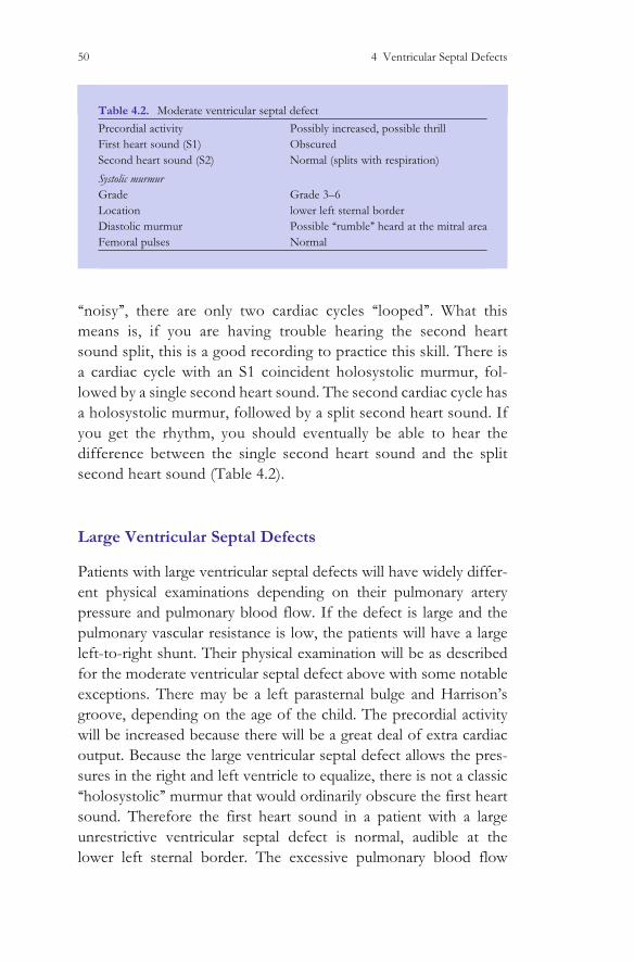
‘‘noisy’’, there are only two cardiac cycles ‘‘looped’’. What this
means is, if you are having trouble hearing the second heart
sound split, this is a good recording to practice this skill. There is
a cardiac cycle with an S1 coincident holosystolic murmur, fol-
lowed by a single second heart sound. The second cardiac cycle has
a holosystolic murmur, followed by a split second heart sound. If
you get the rhythm, you should eventually be able to hear the
difference between the single second heart sound and the split
second heart sound (Table 4.2).
Large Ventricular Septal Defects
Patients with large ventricular septal defects will have widely differ-
ent physical examinations depending on their pulmonary artery
pressure and pulmonary blood flow. If the defect is large and the
pulmonary vascular resistance is low, the patients will have a large
left-to-right shunt. Their physical examination will be as described
for the moderate ventricular septal defect above with some notable
exceptions. There may be a left parasternal bulge and Harrison’s
groove, depending on the age of the child. The precordial activity
will be increased because there will be a great deal of extra cardiac
output. Because the large ventricular septal defect allows the pres-
sures in the right and left ventricle to equalize, there is not a classic
‘‘holosystolic’’ murmur that would ordinarily obscure the first heart
sound. Therefore the first heart sound in a patient with a large
unrestrictive ventricular septal defect is normal, audible at the
lower left sternal border. The excessive pulmonary blood flow
Table 4.2. Moderate ventricular septal defect
Precordial activity Possibly increased, possible thrill
First heart sound (S1) Obscured
Second heart sound (S2) Normal (splits with respiration)
Systolic murmur
Grade Grade 3–6
Location lower left sternal border
Diastolic murmur Possible ‘‘rumble’’ heard at the mitral area
Femoral pulses Normal
50 4 Ventricular Septal Defects
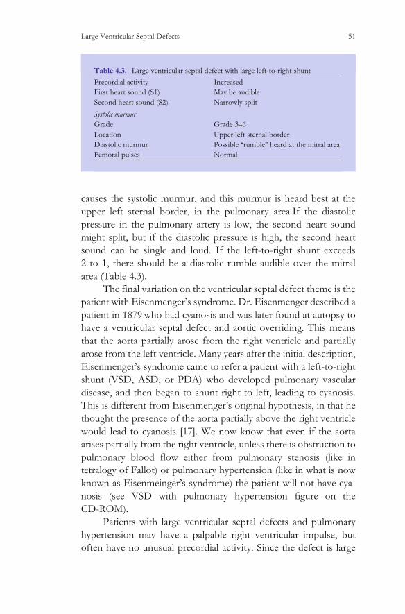
causes the systolic murmur, and this murmur is heard best at the
upper left sternal border, in the pulmonary area.If the diastolic
pressure in the pulmonary artery is low, the second heart sound
might split, but if the diastolic pressure is high, the second heart
sound can be single and loud. If the left-to-right shunt exceeds
2 to 1, there should be a diastolic rumble audible over the mitral
area (Table 4.3).
The final variation on the ventricular septal defect theme is the
patient with Eisenmenger’s syndrome. Dr. Eisenmenger described a
patient in 1879 who had cyanosis and was later found at autopsy to
have a ventricular septal defect and aortic overriding. This means
that the aorta partially arose from the right ventricle and partially
arose from the left ventricle. Many years after the initial description,
Eisenmenger’s syndrome came to refer a patient with a left-to-right
shunt (VSD, ASD, or PDA) who developed pulmonary vascular
disease, and then began to shunt right to left, leading to cyanosis.
This is different from Eisenmenger’s original hypothesis, in that he
thought the presence of the aorta partially above the right ventricle
would lead to cyanosis [17]. We now know that even if the aorta
arises partially from the right ventricle, unless there is obstruction to
pulmonary blood flow either from pulmonary stenosis (like in
tetralogy of Fallot) or pulmonary hypertension (like in what is now
known as Eisenmeinger’s syndrome) the patient will not have cya-
nosis (see VSD with pulmonary hypertension figure on the
CD-ROM).
Patients with large ventricular septal defects and pulmonary
hypertension may have a palpable right ventricular impulse, but
often have no unusual precordial activity. Since the defect is large
Table 4.3. Large ventricular septal defect with large left-to-right shunt
Precordial activity Increased
First heart sound (S1) May be audible
Second heart sound (S2) Narrowly split
Systolic murmur
Grade Grade 3–6
Location Upper left sternal border
Diastolic murmur Possible ‘‘rumble’’ heard at the mitral area
Femoral pulses Normal
Large Ventricular Septal Defects 51
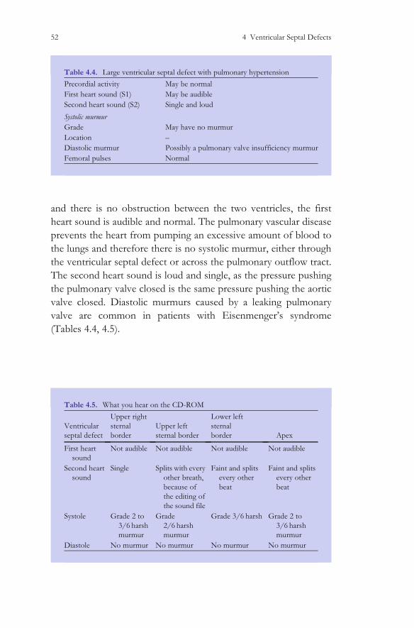
and there is no obstruction between the two ventricles, the first
heart sound is audible and normal. The pulmonary vascular disease
prevents the heart from pumping an excessive amount of blood to
the lungs and therefore there is no systolic murmur, either through
the ventricular septal defect or across the pulmonary outflow tract.
The second heart sound is loud and single, as the pressure pushing
the pulmonary valve closed is the same pressure pushing the aortic
valve closed. Diastolic murmurs caused by a leaking pulmonary
valve are common in patients with Eisenmenger’s syndrome
(Tables 4.4, 4.5).
Table 4.5. What you hear on the CD-ROM
Ventricular
septal defect
Upper right
sternal
border
Upper left
sternal border
Lower left
sternal
border Apex
First heart
sound
Not audible Not audible Not audible Not audible
Second heart
sound
Single Splits with every
other breath,
because of
the editing of
the sound file
Faint and splits
every other
beat
Faint and splits
every other
beat
Systole Grade 2 to
3/6 harsh
murmur
Grade
2/6 harsh
murmur
Grade 3/6 harsh Grade 2 to
3/6 harsh
murmur
Diastole No murmur No murmur No murmur No murmur
Table 4.4. Large ventricular septal defect with pulmonary hypertension
Precordial activity May be normal
First heart sound (S1) May be audible
Second heart sound (S2) Single and loud
Systolic murmur
Grade May have no murmur
Location –
Diastolic murmur Possibly a pulmonary valve insufficiency murmur
Femoral pulses Normal
52 4 Ventricular Septal Defects
Subacute Bacterial Endocarditis Prophylaxis
Recommendations (SBE)
According to the guidelines published by the American Heart Asso-
ciation in 2007, patients with ventricular septal defects do not
require antibiotic prophylaxis at times of endocarditis risk, unless
they have had a prior case of endocarditis. Rarely, some postopera-
tive patients who have residual ventricular septal defects will need
SBE prophylaxis [18].
Summary
Patients with ventricular septal defects are common. The physical
examination can vary in many different ways, depending on the size
of the defect and the amount of blood flowing through the defect.
In a patient with a moderate-sized ventricular septal defect, there
may be a palpable thrill, increased precordial activity, an obscured
first heart sound, a normal second heart sound, and a harsh systolic
murmur at the mid-left sternal border.
References
1. Sands A, Lynch C, Casey F, Craig B, Dornan J, Mulholland C
(1998). Ventricular septal defects: the relationship of social class
and area of residence to occurrence rate Fetal Diagn Ther
13(Suppl I):148
2. Hiraishi S, Agata Y, Nowatari M et al. (1992) Incidence and
natural course of trabecular ventricular septal defect: two dimen-
sional echocardiography and color Doppler flow imaging study.J
Pediatr 120:409–415.
3. Komai H. Naito Y. Fujiwara K. Noguchi Y. Nishimura Y.
Uemura S (1997) Surgical strategy for doubly committed sub-
arterial ventricular septal defect with aortic cusp prolapse. Ann
Thoracic Surg 64(4):1146–1149
References 53
4. Kobayashi J, Koike K, Senzaki H, Kobayashi T, Tsunemoto M,
Ishizawa A, Ohta Y, Shimada M, Omoto R (1999) Correlation
of anatomic and hemodynamic features with aortic valve leaflet
deformity in doubly committed subarterial ventricular septal
defect. Heart Vessels 14(5):240-245
5. Farrell AG, Schamberger MS, Olson IL, Leitch CA (2001)
Large left-to-right shunts and congestive heart failure increase
total energy expenditure in infants with ventricular septal
defect. Am J Cardiol 87(9):1128–1131, A10
6. Gersony WM, Hayes CJ, Driscoll DJ, et al. (1993) Bacterial
endocarditis in patients with aortic stenosis, pulmonary stenosis
or ventricular septal defects. Circulation 87(Suppl I):1121–1126
7. Eroglu AG, Oztunc F, Saltik L, Dedeoglu S, Bakari S, Ahunbay G
(2003) Aortic valve prolapse and aortic regurgitation in patients
with ventricular septal defect. Pediatr Cardiol 24(1):36–39
8. Gabriel HM, Heger M, Innerhofer P, Zehetgruber M, Mundigler G,
Wimmer M, Maurer G, Baumgartner H (2002) Long-term outcome
of patients with ventricular septal defect considered not to require
surgical closure during childhood. J Am College Cardiol
39(6):1066–1071
9. Neumayer U, Stone S, Somerville J (1998) Small ventricular
septal defects in adults. Eur Heart J 19(10):1573–1582
10. Kay JD, Colan SD, Graham TP Jr (2001) Congestive heart
failure in pediatric patients. AHJ 142(5)
11. Kimball TR, Daniels SR, Meyer RA, Hannon DW, Tian J,
Shukla R, Schwartz DC (1991) Effect of digoxin on contractility
and symptoms in infants with a large ventricular septal defect.
Am J Cardiol 68(13):1377–1382
12. Ross RD, Daniels SR, Schwartz DC, Hannon DW, Kaplan S
(1987) Return of plasma norepinephrine to normal after
54 4 Ventricular Septal Defects
resolution of congestive heart failure in congenital heart disease.
Am J Cardiol 60(16):1411–1413
13. Buchhorn R, Hulpke-Wette M, Ruschewski W, Ross RD, Fielitz J,
Pregla R, Hetzer R, Regitz-Zagrosek V (2003) Effects of thera-
peutic beta blockade on myocardial function and cardiac remo-
delling in congenital cardiac disease. Cardiol Young 13(1):36–43
14. Limperopoulos C, Majnemer A, Shevell MI, et al. (2000) Neu-
rodevelopmental status of newborns and infants with congeni-
tal heart defects before and after open heart surgery. J Pediatr
137:638–645
15. Knauth AL, Lock JE, Perry SB, McElhinney DB, Gauvreau K,
Landzberg MJ, Rome JJ, Hellenbrand WE, Ruiz CE, Jenkins KJ
(2004) Transcatheter device closure of congenital and post-
operative residual ventricular septal defects. Circulation
110(5):501–507
16. Tynan M, Anderson RH (2002) Ventricular septal defect.
In:Anderson RH et al. (eds.) Paediatric cardiology. Churchill
Livingstone, London, pp 983–1014
17. Wilkinson JL (2000). In: Moller JH and Hoffman JIE (eds.)
Ventricular septal defect in pediatric cardiovascular medicine.
Churchill Livingstone, New York, pp 289–309
18. Wilson W, Taubert K, Gewitz M et al. (2007) Prevention of
infective endocarditis. Guidelines from the American Heart
Association. A Guideline From the American Heart Associa-
tion Rheumatic Fever, Endocarditis, and Kawasaki Disease
Committee, Council on Cardiovascular Disease in the Young,
and the Council on Clinical Cardiology, Council on Cardiovas-
cular Surgery and Anesthesia, and the Quality of Care and
Outcomes Research Interdisciplinary Working Group 2007.
Circulation April 19
References 55

Chapter 5
Patent Arterial Duct
M.E. McConnell, Pediatric Heart Sounds,
DOI: 10.1007/978-1-84628-684-1_5, � Springer-Verlag London Limited 2008
57

Introduction
The arterial duct, also known as a patent ductus arteriosus (PDA) is a
relatively uncommon congenital malformation. Although all new-
borns have a patent arterial duct, it should close shortly after birth.
Because the duct closes spontaneously in the vast majority of
infants, persistence into later life is relatively rare. The estimated
incidence of a PDA in children by 4 years of age is about 3/10,000
children.
Anatomy
The arterial duct is an intrauterine structure that connects the aorta
and the pulmonary artery. In utero, the ductus carries relatively
deoxygenated blood from the fetal right ventricle into the descend-
ing aorta, so that the deoxygenated blood perfuses the placenta.
There the blood is oxygenated and returns to the fetal heart, cour-
sing through the umbilical vein through the venous duct and into the
right atrium (see Fig. 2.1). After birth, many biochemical signals,
including the increase in oxygen level in the newborn circulation, act
on the ductus to cause closure. The ductus is encircled with mus-
cular tissue that constricts, so that within the first 24 h of post-natal
life, the ductus is functionally closed in most newborns. If these
signals do not cause the ductus to close, the patient will have a patent
arterial duct, or PDA (Fig. 5.1).
Fig. 5.1. The patent arterial duct. Note the connection between the aorta and the pulmonary
artery, allowing the oxygenated (red) blood to flow from the aorta into the pulmonary artery
Anatomy 59

Physiology
While the fetus is in the uterus, the arterial duct is necessary to allow
blood to leave the right ventricle, flow into the main pulmonary
artery, and then down into the aorta. After the infant is born, the
pulmonary artery pressure drops significantly, and therefore the
aortic pressure is higher than the pulmonary artery pressure. If the
duct remains patent, the aortic high-pressure blood will flow into the
low-pressure pulmonary artery in both systole and diastole. The
amount of this blood flow depends on the size of the ductus, as
well as the difference in pressure and vascular resistance between the
aorta and the pulmonary artery. In cases where there is a very large
left-to-right shunt at the ductal level, the large amount of pulmonary
venous return will cause left atrial and left ventricular enlargement.
Natural History
The natural history of a patent arterial duct depends greatly on the
size of the duct, the pulmonary vascular resistance, and the amount
of flow from the aorta into the pulmonary artery. If the duct is quite
small, there may be no murmur, and the diagnosis is only made
‘‘accidentally’’ on an echocardiogram. Whether or not those patients
are at risk for endocarditis is controversial. Because the true inci-
dence of a ‘‘silent ductus’’ is unknown, and the risk of endocarditis is
very low, many pediatric cardiologists do not believe that the silent
ductus requires therapy.
If the ductus is larger, the flow from the aorta into the pul-
monary artery hits against the anterior wall of the pulmonary
artery [1]. This rapid blood flow can cause a murmur in systole
and diastole, often called a ‘‘machinery’’ murmur. Because this rapid
flow can damage the endothelium of the pulmonary artery (the
lining) this area is susceptible to bacterial deposition and subsequent
growth, meaning endocarditis. For this reason, patients with an
audible ductus should have the ductus closed.
Rarely, the ductus is large enough that the pressure transmitted
into the pulmonary artery, along with the extra blood flow into the
pulmonary artery results in pulmonary vascular disease. When the
pulmonary arterial resistance reaches that of the systemic vascular
60 5 Patent Arterial Duct
resistance, the ductus can now shunt from the pulmonary artery into
the aorta, otherwise known as right to left. In this situation the
patient has ‘‘Eisenmeinger’s syndrome’’. This is most frequently seen
in patients with a predisposition for pulmonary vascular disease,
such as patients with Down syndrome.
Surgical Options
Because of the risk of endocarditis, patients with an audible patent
arterial duct should have closure of the ductus. This can be accom-
plished either in the catheterization laboratory, or in the operating
room, depending upon the size of the duct and the local expertise.
Either technique should provide a safe and effective way to com-
pletely close the duct, so that the patient is no longer at increased risk
of bacterial endocarditis [2].
Auscultation
As with the physiology, the auscultation in a patient with a patent
arterial duct depends on the size of the duct and the amount of flow
from the aorta into the pulmonary artery. In patients with a very
small duct, the physical examination should be normal, with normal
precordial activity, a normal first and second heart sound, and no
murmur. If the duct is moderate in size, the loud machinery systolic
murmur may obscure both the first and the second heart sounds, as
the flow from the aorta into the pulmonary artery occurs both in
systole and diastole. Because of the large blood return to the left
atrium, a diastolic rumble across the mitral valve is occasionally
heard (see CD-ROM waveform). The peripheral pulses should be
bounding, as the systolic pressure is normal or slightly elevated, and
the diastolic pressure is low. If the patient has a large duct and
pulmonary vascular disease (Eisenmeinger’s syndrome) the precor-
dial activity is normal, the first heart sound normal, and the second
heart sound is loud and single, as the pressure in the pulmonary
artery and aorta is equal. The pulses are normal, but depending on
the amount of the right-to-left shunt, the patient may have cyanotic
extremities (Tables 5.1, 5.2).
Surgical Options 61

Subacute Bacterial Endocarditis Prophylaxis
Recommendations (SBE)
According to the guidelines published by the American Heart Asso-
ciation in 2007, patients with a patent arterial defect do not require
antibiotic prophylaxis at times of endocarditis risk [3].
In summary, persistent patency of the arterial duct is relatively
rare. It causes a loud continuous murmur heard at the upper left
sternal border, heard in both systole and diastole.
Table 5.1. Patent arterial duct
Precordial activity Increased if there is a large amount of flow
First heart sound (S1) May be obscured
Second heart sound (S2) May be obscured
Systolic murmur Harsh ‘‘machinery’’
Grade Grade 1–6
Location Upper left sternal border
Diastolic murmur Upper left sternal border
femoral pulses Bounding
Table 5.2. What you hear on the CD-ROM
Patent
ductus
arteriosus
Upper right
sternal
border
Upper left
sternal border
Lower left
sternal border Apex
First heart
sound
Not audible Not audible Not audible Not audible
Second
heart
sound
Single Splits
normally
Faint and
single
Soft single
sound
Systole Grade
2/6 harsh
murmur
Grade
3/6 harsh
murmur
Grade
2/6 harsh
Grade
2/6 harsh
murmur
Diastole Grade
1/4 high
pitched
Grade
3/4 high
pitched
Grade
2/4 high
pitched
Grade
2/4 high
pitched
62 5 Patent Arterial Duct
References
1. Bennhagen RG, Benson LN (2003) Silent and audible persistent
ductus arteriosus: an angiographic study. Pediatr Cardiol
24(1):27–30
2. Ewert P (2005) Challenges encountered during closure of patent
ductus arteriosus. Pediatr Cardiol 26(3):224–229
3. Wilson W, Taubert K, Gewitz M et al. (2007) Prevention of
infective endocarditis. Guidelines from the American Heart
Association. A Guideline From the American Heart Association
Rheumatic Fever, Endocarditis, and Kawasaki Disease Commit-
tee, Council on Cardiovascular Disease in the Young, and the
Council on Clinical Cardiology, Council on Cardiovascular Sur-
gery and Anesthesia, and the Quality of Care and Outcomes
Research Interdisciplinary Working Group 2007. Circulation
April 19
References 63

Chapter 6
Aortic Stenosis
M.E. McConnell, Pediatric Heart Sounds,
DOI: 10.1007/978-1-84628-684-1_6, � Springer-Verlag London Limited 2008
65
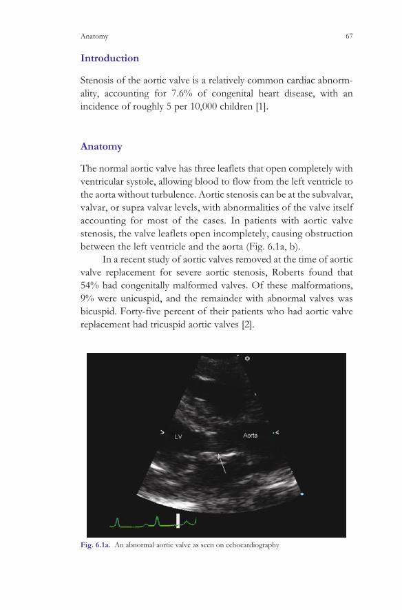
Introduction
Stenosis of the aortic valve is a relatively common cardiac abnorm-
ality, accounting for 7.6% of congenital heart disease, with an
incidence of roughly 5 per 10,000 children [1].
Anatomy
The normal aortic valve has three leaflets that open completely with
ventricular systole, allowing blood to flow from the left ventricle to
the aorta without turbulence. Aortic stenosis can be at the subvalvar,
valvar, or supra valvar levels, with abnormalities of the valve itself
accounting for most of the cases. In patients with aortic valve
stenosis, the valve leaflets open incompletely, causing obstruction
between the left ventricle and the aorta (Fig. 6.1a, b).
In a recent study of aortic valves removed at the time of aortic
valve replacement for severe aortic stenosis, Roberts found that
54% had congenitally malformed valves. Of these malformations,
9% were unicuspid, and the remainder with abnormal valves was
bicuspid. Forty-five percent of their patients who had aortic valve
replacement had tricuspid aortic valves [2].
Fig. 6.1a. An abnormal aortic valve as seen on echocardiography
Anatomy 67
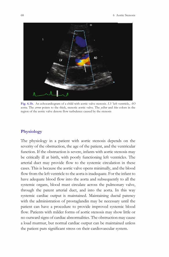
Physiology
The physiology in a patient with aortic stenosis depends on the
severity of the obstruction, the age of the patient, and the ventricular
function. If the obstruction is severe, infants with aortic stenosis may
be critically ill at birth, with poorly functioning left ventricles. The
arterial duct may provide flow to the systemic circulation in these
cases. This is because the aortic valve opens minimally, and the blood
flow from the left ventricle to the aorta is inadequate. For the infant to
have adequate blood flow into the aorta and subsequently to all the
systemic organs, blood must circulate across the pulmonary valve,
through the patent arterial duct, and into the aorta. In this way
systemic cardiac output is maintained. Maintaining ductal patency
with the administration of prostaglandin may be necessary until the
patient can have a procedure to provide improved systemic blood
flow. Patients with milder forms of aortic stenosis may show little or
no outward signs of cardiac abnormalities. The obstruction may cause
a loud murmur, but normal cardiac output can be maintained unless
the patient puts significant stress on their cardiovascular system.
Fig. 6.1b. An echocardiogram of a child with aortic valve stenosis. LV left ventricle, AO
aorta. The arrow points to the thick, stenotic aortic valve. The yellow and blue colors in the
region of the aortic valve denote flow turbulence caused by the stenosis
68 6 Aortic Stenosis
Natural History
The natural history of aortic valve stenosis depends on the severity of
the left ventricular outflow tract obstruction. Unlike pulmonary valve
stenosis, that is felt to be slowly progressive, if at all, patients with aortic
valve stenosis often have worsening of the outflow tract obstruction [3].
This progressive obstruction may lead to symptoms of exercise intol-
erance, syncope, angina, and progressive dyspnea [4] Patients with
bicuspid aortic valves are also at risk of developing dilatation of the
ascending aorta. In the past, this dilatation was felt to be ‘‘post stenotic’’,
meaning it was a consequence of the obstruction between the left
ventricle and the aorta, and the subsequent rapid blood flow from the
ventricle into the ascending aorta. Findings of dilated aortic roots in
patients with bicuspid aortic valves and no significant obstruction bring
this theory into question. The important point is that patients with an
abnormal aortic valve can have an abnormal aorta as well, and these
patients deserve lifelong cardiology follow up. Severe enlargement of the
ascending aorta may result in aortic dissection or rupture, and interven-
tion to replace a significantly dilated ascending aorta is often warranted.
Auscultatory Findings
The auscultatory findings in a patient with valvar aortic stenosis depend
on the severity of the obstruction. With mildly abnormal aortic valves,
the precordial activity would be normal, and there would not be a thrill
palpable in the suprasternal notch. At the lower left sternal border, the
first heart sound may sound like it has two parts to it, or that it splits.
Actually this ‘‘splitting’’ is the clicking open of the abnormal aortic
valve, and it is usually heard best at the apex. The first component of the
split first heart sound is caused by the mitral and the tricuspid valves
closing, and this is followed closely by the clicking open of the abnor-
mal aortic valve. In contrast to the pulmonary valve ejection click, the
aortic click does not vary with respiration. Following the aortic ejection
click, a systolic murmur caused by blood coursing over the abnormal
aortic valve should be heard at the upper sternal border, usually best on
the right side. The murmur can range from grade 1 to 3/6 in the case of
mild aortic stenosis (see CD-ROM waveform). The second heart
sound should be normal. If the abnormal aortic valve leaks, the patient
will have a diastolic murmur, heard best at the lower left sternal border.
Auscultatory Findings 69
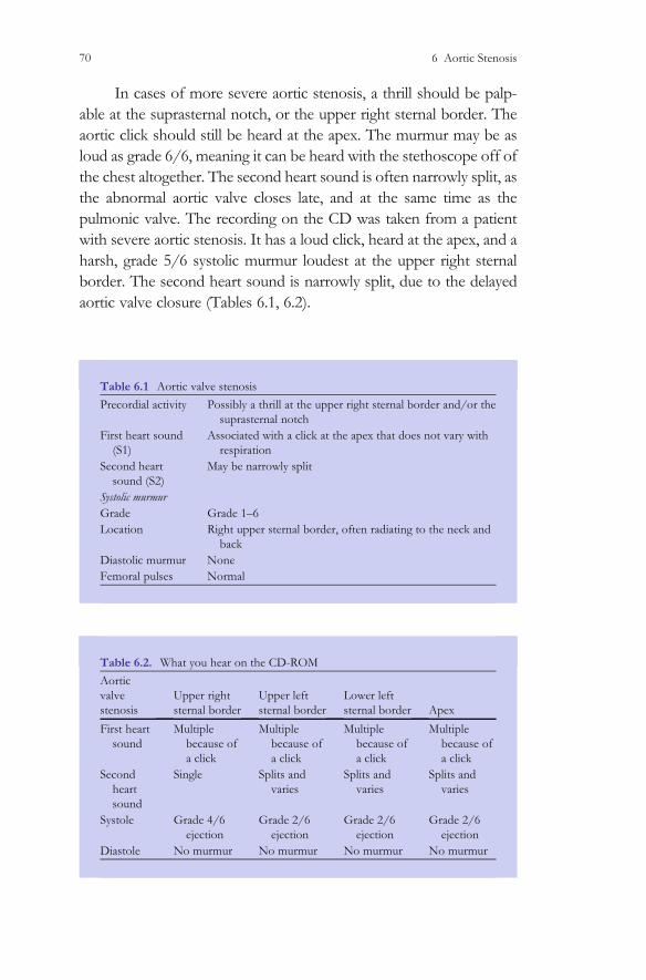
In cases of more severe aortic stenosis, a thrill should be palp-
able at the suprasternal notch, or the upper right sternal border. The
aortic click should still be heard at the apex. The murmur may be as
loud as grade 6/6, meaning it can be heard with the stethoscope off of
the chest altogether. The second heart sound is often narrowly split, as
the abnormal aortic valve closes late, and at the same time as the
pulmonic valve. The recording on the CD was taken from a patient
with severe aortic stenosis. It has a loud click, heard at the apex, and a
harsh, grade 5/6 systolic murmur loudest at the upper right sternal
border. The second heart sound is narrowly split, due to the delayed
aortic valve closure (Tables 6.1, 6.2).
Table 6.1 Aortic valve stenosis
Precordial activity Possibly a thrill at the upper right sternal border and/or the
suprasternal notch
First heart sound
(S1)
Associated with a click at the apex that does not vary with
respiration
Second heart
sound (S2)
May be narrowly split
Systolic murmur
Grade Grade 1–6
Location Right upper sternal border, often radiating to the neck and
back
Diastolic murmur None
Femoral pulses Normal
Table 6.2. What you hear on the CD-ROM
Aortic
valve
stenosis
Upper right
sternal border
Upper left
sternal border
Lower left
sternal border Apex
First heart
sound
Multiple
because of
a click
Multiple
because of
a click
Multiple
because of
a click
Multiple
because of
a click
Second
heart
sound
Single Splits and
varies
Splits and
varies
Splits and
varies
Systole Grade 4/6
ejection
Grade 2/6
ejection
Grade 2/6
ejection
Grade 2/6
ejection
Diastole No murmur No murmur No murmur No murmur
70 6 Aortic Stenosis
Subacute Bacterial Endocarditis Prophylaxis
Recommendations (SBE)
According to the guidelines published by the American Heart Asso-
ciation in 2007, patients with aortic stenosis do not require antibiotic
prophylaxis at times of endocarditis risk, unless they have had a prior
case of endocarditis [5].
Summary
Aortic valve disease is relatively common and can progress over the
patients’ lifetime. It is often associated with a click at the apex, which
sounds like a ‘‘split first heart sound’’. Because of long-term com-
plications of aortic valve disease, such as endocarditis, progressive
obstruction to left ventricular outflow, and aortic root dilatation,
diagnosis and lifelong cardiology follow up is important.
References
1. Samanek M, Slavik Z, Zborilova B, Hrobonova V, Voriskova M,
Skovranek J (1989) Prevalence, treatment, and outcome of heart
disease in live-born children: a prospective analysis of
91,823 live-born children. Pediatr Cardiol 10(4):205–211
2. Roberts WC, Ko JM (2005) Frequency by decades of unicuspid,
bicuspid, and tricuspid aortic valves in adults having isolated
aortic valve replacement for aortic stenosis, with or without
associated aortic regurgitation. Circulation 111(7):920–925
3. Friedman WF, Modlinger J, Morgan J (1971) Serial hemody-
namic observations in asymptomatic children with valvar aortic
stenosis. Circulation 43:91
4. Hunter S (2002) Congenital anomalies of the aortic valve and left
ventricular outflow tract. In: Anderson RH, Backer EJ et al (eds.)
Paediatric cardiology. Churchill Livingstone, London, second
edn pp 1481–1503
References 71

5. Wilson W, Taubert K, Gewitz M et al. (2007) Prevention of
Infective Endocarditis. Guidelines From the American Heart
Association. A Guideline From the American Heart Association
Rheumatic Fever, Endocarditis, and Kawasaki Disease Commit-
tee, Council on Cardiovascular Disease in the Young, and the
Council on Clinical Cardiology, Council on Cardiovascular Sur-
gery and Anesthesia, and the Quality of Care and Outcomes
Research Interdisciplinary Working Group 2007. Circulation
April 19
72 6 Aortic Stenosis

Chapter 7
Pulmonary Stenosis
M.E. McConnell, Pediatric Heart Sounds,
DOI: 10.1007/978-1-84628-684-1_7, � Springer-Verlag London Limited 2008
73
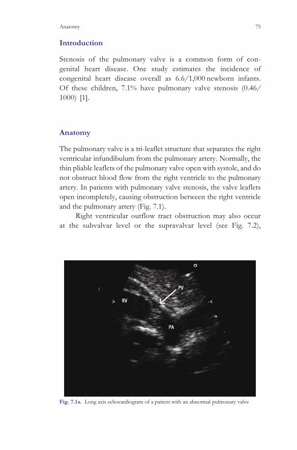
Introduction
Stenosis of the pulmonary valve is a common form of con-
genital heart disease. One study estimates the incidence of
congenital heart disease overall as 6.6/1,000 newborn infants.
Of these children, 7.1% have pulmonary valve stenosis (0.46/
1000) [1].
Anatomy
The pulmonary valve is a tri-leaflet structure that separates the right
ventricular infundibulum from the pulmonary artery. Normally, the
thin pliable leaflets of the pulmonary valve open with systole, and do
not obstruct blood flow from the right ventricle to the pulmonary
artery. In patients with pulmonary valve stenosis, the valve leaflets
open incompletely, causing obstruction between the right ventricle
and the pulmonary artery (Fig. 7.1).
Right ventricular outflow tract obstruction may also occur
at the subvalvar level or the supravalvar level (see Fig. 7.2),
Fig. 7.1a. Long axis echocardiogram of a patient with an abnormal pulmonary valve
Anatomy 75

but these lesions are much less common than valvar pulmonic
stenosis [2].
Physiology
The physiology of the patient with pulmonary valve stenosis
depends on the amount of stenosis, the pulmonary vascular
resistance, and any associated defects in the heart. For a patient
with mild stenosis, normal pulmonary vascular resistance, and
no other cardiac abnormalities, there should be no significant
physiologic consequences from the mildly stenotic pulmonary
valve. The patient should not have a right to left or left to right
shunt, should have normal cardiac output, and should have no
symptoms related to the abnormal pulmonary valve. A newborn
with severe pulmonary stenosis will have a great variability in
physiology. For example, if the valve is severely stenotic, and
very little blood can flow from the right ventricle to the
pulmonary artery, the patient may require the presence of the
Fig. 7.1b. Color flow mapping across the same stenotic pulmonary valve. RV right
ventricle, PA pulmonary artery, PV pulmonary valve. The arrow points to the thick, stenotic
pulmonary valve leaflets, and the multiple colors are caused by the turbulent flow across the
stenotic valve
76 7 Pulmonary Stenosis
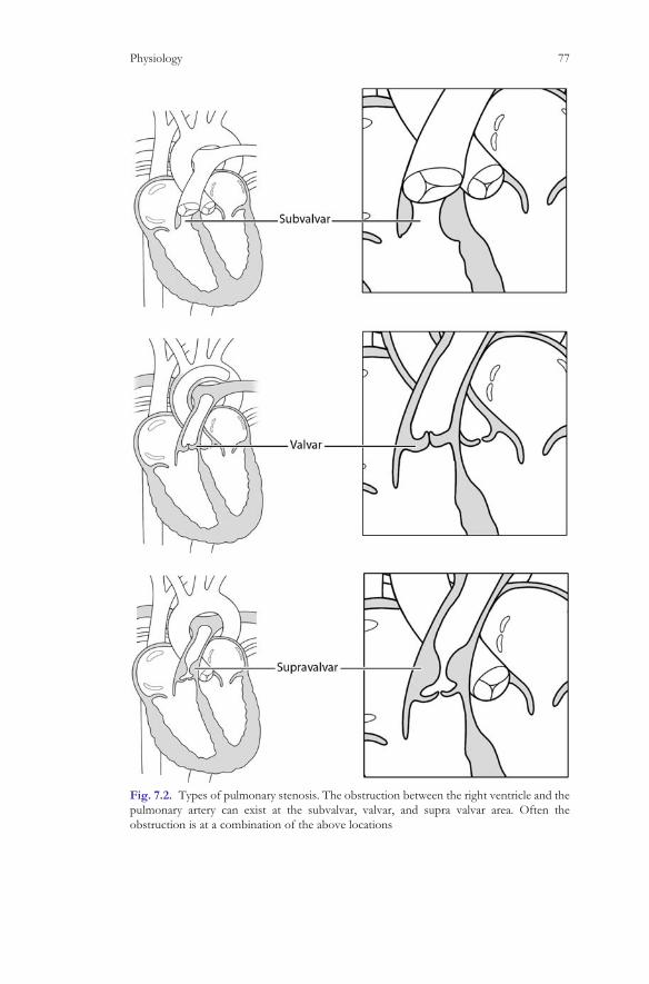
Fig. 7.2. Types of pulmonary stenosis. The obstruction between the right ventricle and the
pulmonary artery can exist at the subvalvar, valvar, and supra valvar area. Often the
obstruction is at a combination of the above locations
Physiology 77
arterial duct and a patent atrial septum in order to have ade-
quate cardiac output and adequate pulmonary blood flow.
The severely stenotic pulmonary valve would not allow ade-
quate amounts of blood to leave the RV and go into the pulmonary
artery. Therefore, the right atrial pressure will rise, and the deox-
ygenated blood will flow from the right atrium to the left atrium.
This blood will then mix with the oxygenated blood returning
from the pulmonary veins, and then flow through the mitral
valve and into the left ventricle, where it will be pumped out of
the aorta. If the arterial duct is open, some of the aortic blood will
pass through the ductus and into the pulmonary artery. The new-
born with severe pulmonary valve stenosis may therefore be
cyanotic, because of the right to left flow of deoxygenated blood
at the atrial level. The child may also be ‘‘ductal dependent’’ mean-
ing that the blood through the arterial duct is necessary to provide
adequate pulmonary blood flow to sustain life. In this situation,
institution of medications that will maintain duct patency may be
life saving.
Another example of the variable physiology in pulmonary valve
stenosis is the infant with moderate-to-severe pulmonary valve steno-
sis and increased pulmonary vascular resistance shortly after birth. The
infant may be asymptomatic, with a soft systolic murmur at the upper
left sternal border, caused by flow across the stenotic pulmonary valve.
Over the next few weeks, as the pulmonary vascular resistance drops,
the obstruction between the RV and the pulmonary artery becomes
more noticeable. This is because the murmur intensity is dependent on
the change in pressure between the right ventricle and the pulmonary
artery. In the newborn, the pulmonary artery pressure may be high,
and therefore the difference in pressure between the right ventricle
and the pulmonary artery will be low. This small difference between
the right ventricular pressure and the pulmonary artery pressure at
birth means that the murmur caused by flow across the stenotic
pulmonary valve will be soft. As the pulmonary vascular resistance
drops, the stenosis does not change, but the pressure difference
between the right ventricle and the pulmonary artery may change
significantly. Thus, a child thought to have mild pulmonary valve
stenosis in the newborn period may have severe obstruction between
the RV and PA by 2–3 months of life (see Fig. 7.3)
78 7 Pulmonary Stenosis

Natural History
The natural history of a patient with valvar pulmonic stenosis depends
on the severity of the lesion. In patients with mild stenosis, exercise
tolerance and life expectancy are near normal, and therefore no
intervention should be contemplated for these patients. For patients
with severe pulmonary valve stenosis, intervention to open the pul-
monary valve, either with a balloon catheter or in the operating room
may be life saving. In the first Natural History Study of Congenital
Heart Defects (NHS), 592 patients with pulmonary stenosis were
enrolled after catheterization, between 1958 and 1969. If the patient
had mild pulmonary stenosis, defined as a gradient in the catheteriza-
tion laboratory of <50 mmHg, the patients were managed medically.
This group of patients had excellent long-term survival, and a less
than 20% chance of requiring intervention on the right ventricular
outflow tract. If the gradient was >50 mmHg (measured directly in
the catheterization laboratory), most patients required intervention to
enlarge the right ventricular outflow tract. Morbid events were
uncommon in either the operated or unoperated patients with pul-
monary stenosis, and the long-term life expectancy was identical to
people without congenital heart disease. Patients who entered the
Fig. 7.3. The difference in measured obstruction or ‘‘pressure gradient’’ between the right
ventricle and the pulmonary artery depending on the pulmonary artery pressure. In the left
panel, the pulmonary artery pressure is low, and the ‘‘pressure gradient’’ (the difference between
the systolic pressure in the right ventricle and the systolic pressure in the pulmonary artery) is
50 mmHg (70 mmHg – 20 mmHg = 50 mmHg). In the right panel, the pulmonary artery pressure
is high, and therefore the gradient between the RV and PA is only 10 mmHg (60 – 50 mmHg).
This can occur in newborn infants with severe right ventricular outflow tract obstruction who still
have high pulmonary artery pressure
Natural History 79
study without cardiomegaly on CXR had no mortality during the
study period, whereas patients with cardiomegaly who entered the
study at more than 12 years of age had a 25-year survival rate of
<80% [3]
In 1982 Kan et al. reported the first case of dilation of a stenotic
pulmonary valve using a balloon catheter [4]. Since then, percuta-
neous dilatation of a stenotic pulmonary valve has become the
treatment of choice for infants, children, and adults with right ven-
tricular outflow tract obstruction at the level of the pulmonary valve.
The long-term survival of patients treated with balloon dilatation is
excellent. Although reintervention after an operation to open the
pulmonary valve was rare, it is possible that the long-term effect of a
leaking pulmonary valve will result in right ventricular dilatation and
the need for pulmonary valve replacement in some patients [5].
Because of the benign natural history of mild pulmonary valve
stenosis, and the fact that balloon dilatation of the pulmonary valve
will often result in pulmonary valve insufficiency, some patients with
mild pulmonary valve stenosis may not benefit in the long term from
balloon dilatation of the mildly stenotic pulmonary valve, and should
therefore be left alone. Endocarditis was very rare in patients with
pulmonary valve stenosis. In fact there were no cases of endocarditis
reported in the NHS study. The American Heart Association no
longer recommends antibiotic prophylaxis at times of endocarditis
risk for patients with pulmonic stenosis [6].
Auscultatory Findings
The physical examination of a patient with pulmonary valve stenosis
depends greatly on the severity of the obstruction between the two
ventricles, and the pulmonary artery pressure, as discussed above. For
mild pulmonary stenosis, the precordial activity should be normal,
and there will be no palpable thrill at the upper left sternal border (the
pulmonary area). In patients with severe pulmonary stenosis, palpa-
tion of the precordium may reveal a left parasternal heave consistent
with high right ventricular pressure. The flow across the stenotic
pulmonary valve causes such turbulence that a thrill is felt in the left
upper sternal border, over the pulmonic area. The thrill may also be
palpable over the left side of the neck [7].
80 7 Pulmonary Stenosis
The abnormal sounds will be high pitched, and therefore heard
best with the diaphragm of the stethoscope. Because the obstruction to
blood flow is at the pulmonary valve level, the murmur will begin only
after the pulmonary valve opens. The first heart sound is normal, but
there is often a pulmonary ejection click caused by the opening of the
abnormal pulmonary valve. The click is thought to be caused by the
abnormal pulmonary valve clicking open, and therefore follows the first
heart sound. The click of pulmonary valve stenosis sounds like ‘‘splitting’’
of the first heart sound. The mitral and tricuspid valves close, making the
first sound. This is followed very rapidly (approximately 40 msec) [8] by
the clicking open of the pulmonary valve. This splitting is best heard at
the upper left sternal border, in the pulmonary area. Because the pul-
monary valve leaflets move with inspiration, the valve leaflets ‘‘float’’
superiorly when the patient takes in a breath. This movement of the
pulmonary valve makes the valve open from a different location, and
therefore the pulmonary ejection sound often disappears with inspira-
tion. Thus the click sounds like a split first heart sound that varies with
respiration. The click can be so loud it is palpable, and the palpable click
can disappear with the patient taking in a deep breath. On the CD-ROM
recording, the pulmonary valve ejection sound is a loud ‘‘pop’’ heard
intermittently at either the lower left sternal border, or the upper left
sternal border. Listen carefully to the first heart sound until it is clear
there is an ‘‘extra’’ sound that is not constant. This extra sound is the
clicking open of the pulmonary valve. The second heart sound is caused
by the closure of the abnormal pulmonary valve. In mild-to-moderate
pulmonary valve stenosis, the abnormal pulmonic valve closes late,
resulting in a split second heart sound. In patients with severe pulmonary
valve stenosis, the pulmonary valve closure sound may be inaudible. The
systolic murmur is caused by the flow of blood across the stenotic
pulmonary valve, and causes a crescendo/decrescendo systolic murmur
heard best at the upper left sternal border. The murmur does not change
with respiration or position . In the case of severe obstruction between
the right ventricle and the pulmonary artery, the murmur caused by the
obstruction can obscure both the first and second heart sounds. In a
young child, where the difference in the upper left sternal border and the
lower left sternal border is small, a patient with severe pulmonary valve
stenosis may be mistaken for a patient with a ventricular septal defect.
Unless there is leakage of the pulmonary valve, there is no diastolic
murmur (see Tables 7.1, 7.2)
Auscultatory Findings 81
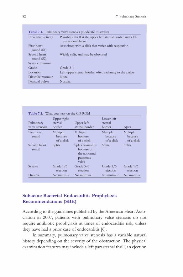
Subacute Bacterial Endocarditis Prophylaxis
Recommendations (SBE)
According to the guidelines published by the American Heart Asso-
ciation in 2007, patients with pulmonary valce stenosis do not
require antibiotic prophylaxis at times of endocarditis risk, unless
they have had a prior case of endocarditis [6].
In summary, pulmonary valve stenosis has a variable natural
history depending on the severity of the obstruction. The physical
examination features may include a left parasternal thrill, an ejection
Table 7.2. What you hear on the CD-ROM
Pulmonary
valve stenosis
Upper right
sternal
border
Upper left
sternal border
Lower left
sternal
border Apex
First heart
sound
Multiple
because
of a click
Multiple
because
of a click
Multiple
because
of a click
Multiple
because
of a click
Second heart
sound
Splits Splits constantly
because of
the abnormal
pulmonic
valve
Splits Splits
Systole Grade 1/6
ejection
Grade 3/6
ejection
Grade 1/6
ejection
Grade 1/6
ejection
Diastole No murmur No murmur No murmur No murmur
Table 7.1. Pulmonary valve stenosis (moderate to severe)
Precordial activity Possibly a thrill at the upper left sternal border and a left
parasternal heave
First heart
sound (S1)
Associated with a click that varies with respiration
Second heart
sound (S2)
Widely split, and may be obscured
Systolic murmur
Grade Grade 3–6
Location Left upper sternal border, often radiating to the axillae
Diastolic murmur None
Femoral pulses Normal
82 7 Pulmonary Stenosis

click that varies with respiration, a systolic murmur at the upper left
sternal border, and a widely split second heart sound.
References
1. Samanek M, Slavik Z, Zborilova B, Hrobonova V, Voriskova M,
Skovranek J (1989) Prevalence, treatment, and outcome of heart
disease in live-born children: a prospective analysis of
91,823 liveborn children. Pediatr Cardiol 10:205–211
2. Kjellberg SR., Mannheimer E., Rudhe U, Jonsson B (1955)
Diagnosis of congenital heart disease. Year Book Publishers,
Chicago
3. Hayes CJ, Gersony WM, Driscoll DJ, Keane JF, Kidd L, O’Fallon
WM, Pieroni DR, Wolfe RR, Weidman WH (1993) Second natural
history study of congenital heart defects. Results of treatment of
patients with pulmonary valvar stenosis. Circulation 87(2
Suppl):I28–I37
4. Kan JS, White RI Jr, Mitchell SE, Gardner TJ (1982) Percuta-
neous balloon valvuloplasty: a new method for treating congeni-
tal pulmonary-valve stenosis. [Case Reports. Journal article] New
Engl J Med 307(9):540–542
5. Poon LK, Menahem S (2003) Pulmonary regurgitation after
percutaneous balloon valvoplasty for isolated pulmonary valvar
stenosis in childhood. Cardiol Young 13(5):444–450
6. Wilson W, Taubert K, Gewitz M et al. (2007) Prevention of
Infective Endocarditis. Guidelines from the American Heart
Association. A Guideline From the American Heart Association
Rheumatic Fever, Endocarditis, and Kawasaki Disease Commit-
tee, Council on Cardiovascular Disease in the Young, and the
Council on Clinical Cardiology, Council on Cardiovascular Sur-
gery and Anesthesia, and the Quality of Care and Outcomes
Research Interdisciplinary Working Group 2007. Circulation
April 19
References 83

7. Levine SA, Harvey WP (1959) Clinical auscultation of the heart.
W.B. Saunders, New York, pp 453–463
8. Waider W, Craige E (1975) First heart sound and ejection sounds.
Echocardiographic and phonocardiographic correlation with
valvular events. Am J Cardiol 35(3):346–356
84 7 Pulmonary Stenosis

Chapter 8
Mitral Valve Insufficiency
M.E. McConnell, Pediatric Heart Sounds,
DOI: 10.1007/978-1-84628-684-1_8, � Springer-Verlag London Limited 2008
85

Introduction
Isolated congenital abnormalities of the mitral valve are relatively
rare, estimated at less than 0.1 per 1,000 live births [1]. The mitral
valve may be damaged by rheumatic fever, or also may degenerate
over time, resulting in mitral valve regurgitation.
Anatomy
The anatomy of a leaking mitral valve is variable, but for the sake of
simplicity can be divided into two main abnormalities. The first,
where the two leaflets of the mitral valve cannot completely close
because there is a deformity in the valve, such as a cleft (see Fig. 8.1).
Fig. 8.1. A cleft mitral valve. a A normal mitral valve with a complete coaptation of the two
leaflets. b An abnormal ‘‘cleft’’ mitral valve, with a wedge of tissue missing in the anterior
leaflet of the mitral valve
Anatomy 87
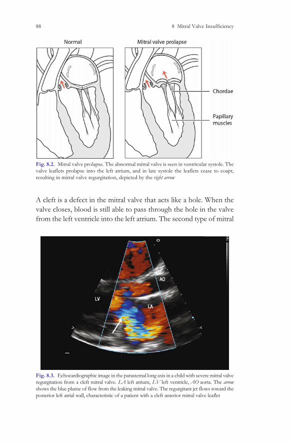
A cleft is a defect in the mitral valve that acts like a hole. When the
valve closes, blood is still able to pass through the hole in the valve
from the left ventricle into the left atrium. The second type of mitral
Fig. 8.2. Mitral valve prolapse. The abnormal mitral valve is seen in ventricular systole. The
valve leaflets prolapse into the left atrium, and in late systole the leaflets cease to coapt,
resulting in mitral valve regurgitation, depicted by the right arrow
Fig. 8.3. Echocardiographic image in the parasternal long axis in a child with severe mitral valve
regurgitation from a cleft mitral valve. LA left atrium, LV left ventricle, AO aorta. The arrow
shows the blue plume of flow from the leaking mitral valve. The regurgitant jet flows toward the
posterior left atrial wall, characteristic of a patient with a cleft anterior mitral valve leaflet
88 8 Mitral Valve Insufficiency
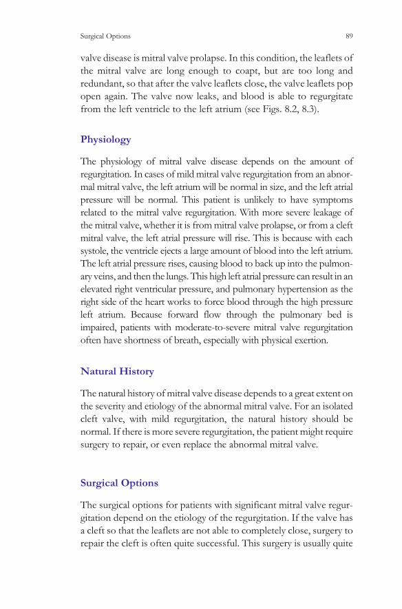
valve disease is mitral valve prolapse. In this condition, the leaflets of
the mitral valve are long enough to coapt, but are too long and
redundant, so that after the valve leaflets close, the valve leaflets pop
open again. The valve now leaks, and blood is able to regurgitate
from the left ventricle to the left atrium (see Figs. 8.2, 8.3).
Physiology
The physiology of mitral valve disease depends on the amount of
regurgitation. In cases of mild mitral valve regurgitation from an abnor-
mal mitral valve, the left atrium will be normal in size, and the left atrial
pressure will be normal. This patient is unlikely to have symptoms
related to the mitral valve regurgitation. With more severe leakage of
the mitral valve, whether it is from mitral valve prolapse, or from a cleft
mitral valve, the left atrial pressure will rise. This is because with each
systole, the ventricle ejects a large amount of blood into the left atrium.
The left atrial pressure rises, causing blood to back up into the pulmon-
ary veins, and then the lungs. This high left atrial pressure can result in an
elevated right ventricular pressure, and pulmonary hypertension as the
right side of the heart works to force blood through the high pressure
left atrium. Because forward flow through the pulmonary bed is
impaired, patients with moderate-to-severe mitral valve regurgitation
often have shortness of breath, especially with physical exertion.
Natural History
The natural history of mitral valve disease depends to a great extent on
the severity and etiology of the abnormal mitral valve. For an isolated
cleft valve, with mild regurgitation, the natural history should be
normal. If there is more severe regurgitation, the patient might require
surgery to repair, or even replace the abnormal mitral valve.
Surgical Options
The surgical options for patients with significant mitral valve regur-
gitation depend on the etiology of the regurgitation. If the valve has
a cleft so that the leaflets are not able to completely close, surgery to
repair the cleft is often quite successful. This surgery is usually quite
Surgical Options 89
safe, with an operative survival in good centers approaching 100%.
If the valve is floppy and redundant, like in some patients with the
Marfan syndrome, surgical repair of the valve is more difficult. In
situations where the repaired valve is likely to degenerate in a short
period of time, the valve should probably be replaced, not repaired.
Auscultatory Findings
The auscultatory findings in a patient with mitral valve regurgita-
tion depend on the severity of the regurgitation, and also on
whether the valve leaks as the leaflet closes (like in a patient with
a cleft mitral valve) or if the valve leaks only after the prolapsing
mitral valve pops open.
Mitral valve regurgitation from a cleft mitral valve
If the patient has a cleft in the mitral valve that leaks as the mitral
valve closes, the patient will have a murmur that begins as the
mitral valve closes, therefore obscuring the first heart sound. If the
regurgitation is mild, the left atrial pressure is normal, and the
pulmonary artery pressure is likewise normal. The second heart
sound will therefore be normal, and will split with respiration. If
the valve leaks severely, the left atrial pressure will be high, and
therefore the pulmonary artery pressure will also be high. This high
pulmonary artery pressure will cause a loud second heart sound, as
the pulmonary valve and the aortic valve are forced closed by
similar high pressures. The systolic murmur caused by the leaking
mitral valve is a harsh sound, heard best at the apex. If the
regurgitation is severe, there will be a diastolic murmur caused by
the large volume of blood flowing from the left atrium into the left
ventricle across the mitral valve. This diastolic rumbling sound is
low pitched, and often difficult to appreciate. It has been described
as an ‘‘absence of silence’’, and can be heard best on the CD in the
patient with an ASD. The mitral valve regurgitation recording is
not of a large enough volume to cause a diastolic murmur. If it was
present, the diastolic rumble would be heard best with the bell of
the stethoscope, placed at the apex of the left ventricle, and pressed
down so that the skin beneath the mitral valve is tight, and the bell
90 8 Mitral Valve Insufficiency
acts like a diaphragm. The high-frequency sounds are then best
heard. Then you should lighten up on the bell, and let it act more
like a bell, where it will transmit the low-frequency sounds. Then,
in cases where there is a large volume of blood flowing from the
LA to the LV in diastole, you should hear a rumbling sound in
diastole. This implies that the mitral valve regurgitation is severe
(Table 8.1).
Mitral valve regurgitation from a prolapsing mitral valve
Mitral valve regurgitation from a prolapsing mitral valve causes a
physical examination that may be quite different from the examina-
tion in someone with a cleft mitral valve. If there is minimal mitral
valve regurgitation the precordial activity will be normal. In classic
mitral valve prolapse, the mitral valve closure causes a normal first
heart sound. After the mitral valve closes, the valve pops superiorly
into the left atrium, causing a click. After the valve clicks, the leaflets
may open, causing mitral valve regurgitation. The murmur can
occasionally have a strange, ‘‘honking’’ type quality. If the mitral
valve leakage is mild, there will be normal left atrial pressure, and
therefore a normal second heart sound. In the case of minimal mitral
valve regurgitation, there will be no diastolic rumble. If the mitral
valve prolapse results in severe mitral valve regurgitation, the patient
may have a loud second heart sound, as well as a diastolic rumble
across the mitral valve [2] (Tables 8.2, 8.3).
Table 8.1. Mitral valve regurgitation from a cleft mitral valve
Mitral valve regurgitation Cleft mitral valve
Precordial activity Increased if there is a large amount of regurgitation
First heart sound (S1) Obscured
Second heart sound (S2) May be loud if there is a large amount of regurgitation
Systolic murmur
Grade Grade 1–6
Location Apex (mitral area)
Diastolic murmur Possible diastolic rumble across the mitral valve
femoral pulses Normal
Auscultatory Findings 91
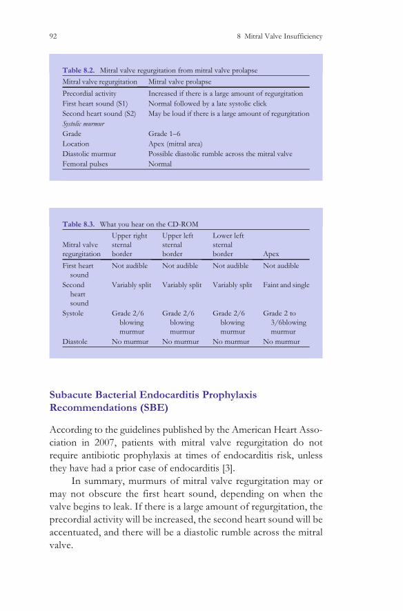
Subacute Bacterial Endocarditis Prophylaxis
Recommendations (SBE)
According to the guidelines published by the American Heart Asso-
ciation in 2007, patients with mitral valve regurgitation do not
require antibiotic prophylaxis at times of endocarditis risk, unless
they have had a prior case of endocarditis [3].
In summary, murmurs of mitral valve regurgitation may or
may not obscure the first heart sound, depending on when the
valve begins to leak. If there is a large amount of regurgitation, the
precordial activity will be increased, the second heart sound will be
accentuated, and there will be a diastolic rumble across the mitral
valve.
Table 8.2. Mitral valve regurgitation from mitral valve prolapse
Mitral valve regurgitation Mitral valve prolapse
Precordial activity Increased if there is a large amount of regurgitation
First heart sound (S1) Normal followed by a late systolic click
Second heart sound (S2) May be loud if there is a large amount of regurgitation
Systolic murmur
Grade Grade 1–6
Location Apex (mitral area)
Diastolic murmur Possible diastolic rumble across the mitral valve
Femoral pulses Normal
Table 8.3. What you hear on the CD-ROM
Mitral valve
regurgitation
Upper right
sternal
border
Upper left
sternal
border
Lower left
sternal
border Apex
First heart
sound
Not audible Not audible Not audible Not audible
Second
heart
sound
Variably split Variably split Variably split Faint and single
Systole Grade 2/6
blowing
murmur
Grade 2/6
blowing
murmur
Grade 2/6
blowing
murmur
Grade 2 to
3/6blowing
murmur
Diastole No murmur No murmur No murmur No murmur
92 8 Mitral Valve Insufficiency
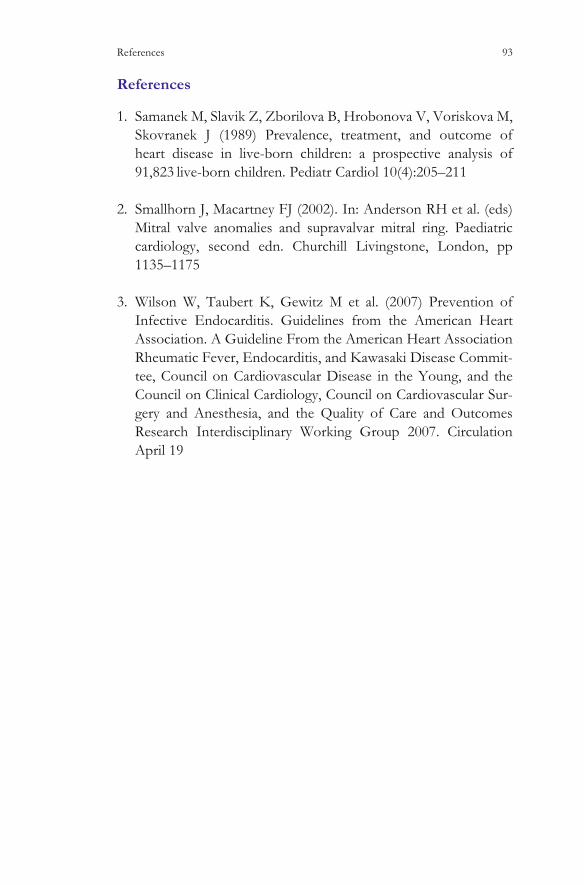
References
1. Samanek M, Slavik Z, Zborilova B, Hrobonova V, Voriskova M,
Skovranek J (1989) Prevalence, treatment, and outcome of
heart disease in live-born children: a prospective analysis of
91,823 live-born children. Pediatr Cardiol 10(4):205–211
2. Smallhorn J, Macartney FJ (2002). In: Anderson RH et al. (eds)
Mitral valve anomalies and supravalvar mitral ring. Paediatric
cardiology, second edn. Churchill Livingstone, London, pp
1135–1175
3. Wilson W, Taubert K, Gewitz M et al. (2007) Prevention of
Infective Endocarditis. Guidelines from the American Heart
Association. A Guideline From the American Heart Association
Rheumatic Fever, Endocarditis, and Kawasaki Disease Commit-
tee, Council on Cardiovascular Disease in the Young, and the
Council on Clinical Cardiology, Council on Cardiovascular Sur-
gery and Anesthesia, and the Quality of Care and Outcomes
Research Interdisciplinary Working Group 2007. Circulation
April 19
References 93

Chapter 9
Tetralogy of Fallot
M.E. McConnell, Pediatric Heart Sounds,
DOI: 10.1007/978-1-84628-684-1_9, � Springer-Verlag London Limited 2008
95
Incidence
Tetralogy of Fallot is a relatively common form of congenital heart
disease, and is seen in roughly 2–3 per 10,000 infants [1]. Because
therapy for this disorder is usually successful, there are many adults
with tetralogy of Fallot, and in some countries, there are more adults
with the condition than children.
Anatomy
In 1888, Dr. Fallot described the cardiac anatomy of a patient with
cyanosis who had a ventricular septal defect, an aorta that originated
from both the right and left ventricles, obstruction between the right
ventricle and the pulmonary artery, and a hypertrophied right ventricle
(Fig. 9.1)
Although this anatomic combination had been described as
early as the seventeenth century, it was Fallot’s description that gave
the eponym to this common form of cyanotic congenital heart
disease [2]. The obstruction to blood flow from the right ventricle
to the pulmonary artery may be at the pulmonary valve annulus, the
sub-pulmonary area or the supra valvar area. Often, the obstruction
from the right ventricle to the pulmonary artery is at all three levels.
Physiology
The physiology of a patient with tetralogy of Fallot is quite variable,
depending on the amount of pulmonary blood flow. This is true for
many forms of cyanotic congenital heart disease, and this is the
reason that tetralogy of Fallot is used on the CD-ROM as an
example of cyanotic heart disease. For example, in the situation
where there is minimal obstruction between the right ventricle and
the pulmonary artery, the ventricular septal defect will be the pre-
dominant pathologic feature, and the pulmonary blood flow will be
increased. In this situation, the patient will not have cyanosis, and if
there is too much pulmonary blood flow the patient may even
exhibit signs of congestive heart failure, just like a patient with a
large isolated ventricular septal defect. These patients may still have
an overriding aorta, obstruction between the right ventricle and the
pulmonary artery (albeit mild) and a hypertrophied right ventricle; so
Physiology 97

technically they have tetralogy of Fallot. But their physiology is very
different from the classic cases, and often these children will require
early surgical intervention because of excessive pulmonary blood
flow. These patients with tetralogy of Fallot and large left-to-right
shunts are often referred to as ‘‘pink tets’’. In a more typical case of
tetralogy of Fallot, the obstruction between the right ventricle and
the pulmonary artery is moderate to severe. In this situation some of
the blood leaving the right ventricle may flow into the aorta. Because
this right ventricular blood is non-oxygenated blood, the blue blood
within the aorta will cause cyanosis. It is important to note that the
obstruction between the right ventricle and the pulmonary artery
Fig. 9.1. Schematic representation of a patient with tetralogy of Fallot. Note the large
ventricular septal defect, the aorta that can get blood flow from either the left or the right
ventricle, the obstruction between the right ventricle and the pulmonary artery, and the thick
right ventricle
98 9 Tetralogy of Fallot
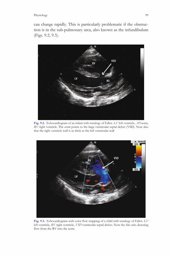
can change rapidly. This is particularly problematic if the obstruc-
tion is in the sub-pulmonary area, also known as the infundibulum
(Figs. 9.2, 9.3).
Fig. 9.2. Echocardiogram of an infant with tetralogy of Fallot. LV left ventricle, AO aorta,
RV right ventricle. The arrow points to the large ventricular septal defect (VSD). Note also
that the right ventricle wall is as thick as the left ventricular wall
Fig. 9.3. Echocardiogram with color flow mapping of a child with tetralogy of Fallot. LV
left ventricle, RV right ventricle, VSD ventricular septal defect. Note the blue color, denoting
flow from the RV into the aorta
Physiology 99
At times, especially if the child is dehydrated, the distance
between the muscular walls of the infundibulum gets closer together,
causing increased obstruction between the right ventricle and the
pulmonary artery. When this happens, the pulmonary blood flow
decreases, and the cyanosis worsens. This is also known as a tetral-
ogy or ‘‘tet’’ spell. During a ‘‘tet’’ spell, calming the infant is impor-
tant. In addition, during a tet spell, pushing the infant’s knees into
their chest will increase the systemic vascular resistance, and force
more blood into the pulmonary circulation. This is why unoperated
children with tetralogy of Fallot may ‘‘squat’’. This position effect-
ively cuts off the femoral arterial blood flow, and increases the
pulmonary blood flow. Patients with a ventricular septal defect
and pulmonary stenosis have their physiology governed by the
amount of pulmonary blood flow. This physiology is important in
other forms of congenital heart disease as well. Examples include
tricuspid atresia and single ventricles in which the amount of pul-
monary blood flow regulates the amount and type of symptoms, just
as it does in patients with tetralogy of Fallot.
Natural History
Because of operations to palliate patients with tetralogy of Fallot
were first performed in the 1940s, and operations to correct the
anatomical abnormalities were performed in the 1950s, in many
countries, the ‘‘natural history’’ of tetralogy of Fallot is no longer
applicable. The most recent estimate, however, suggests that with-
out surgical intervention, at least 90% of patients with tetralogy of
Fallot will die by 25 years of age [3]. Fortunately, with modern
surgical techniques, most patients with uncomplicated tetralogy of
Fallot can do very well. Long-term survival depends to a great
extent on the quality of the pulmonary arteries preoperatively. if
the pulmonary arteries are normal in size, in the current era,
survival should be near that of the normal population. If the
pulmonary arteries are small, or have multiple obstructions within
them, the outlook may be much worse than the normal population.
Twenty-five-year survival rates for patients with tetralogy of Fallot
exceed 90% [4].
100 9 Tetralogy of Fallot
Surgical Options
Surgery for patients with tetralogy of Fallot involves closing the
ventricular septal defect and opening the obstruction between the
right ventricle and the pulmonary artery. The timing of this interven-
tion varies from center to center, but should be safely performed at
almost any age in the current era. In situations where the pulmonary
arteries are small, or if the child is so small that complete correction is
felt to be dangerous, a shunt is sometimes performed. The shunt
operation connects the systemic arterial circulation to the pulmonary
circulation, usually via a prosthetic tube. Post-operatively, some of the
blood leaving the heart into the aorta is able to flow through the
shunt, and therefore into the pulmonary artery, bypassing the obstruc-
tion between the right ventricle and the pulmonary artery. This shunt
is named after the surgeon and pediatric cardiologist who participated
in the initial surgical work, Drs. Blalock and Taussig. Additional credit
is also due to Dr. Vivien Thomas, who was integral in the research
leading to the first successful palliation of cyanotic congenital heart
disease [5]. During the complete repair of tetralogy of Fallot, the
surgeon attempts to open the right ventricle to pulmonary artery
communication by removing the muscular tissue beneath the pul-
monary valve. Often, the pulmonary valve annulus is significantly
smaller than the aortic valve annulus. In order to make the outflow
area from each ventricle similar in size, the surgeons often place an
incision across the pulmonary valve annulus, and then sew a patch in
the incision to widen the area (Fig. 9.4).
The significance of this operative technique is that the patient
leaves the operating room without a functional pulmonary valve,
and therefore will eventually have free pulmonary valve insuffi-
ciency. This situation will require replacement of a pulmonary
valve in the distant future, often 15–30 years after the initial surgery.
Auscultatory Findings
The physical examination findings in a patient with tetralogy of
Fallot vary widely, just like the physiology. When there is a moderate
amount of obstruction from the right ventricle to the pulmonary
artery, the patient should have normal precordial activity. Depend-
ing on the amount of blood flowing from the right ventricle into the
Auscultatory Findings 101

aorta (right-to-left shunting), the patient will have a variable amount
of cyanosis. The first heart sound may be obscured, but often a
pulmonary ejection click is audible at the upper left sternal border.
The second heart sound is often single, because the closure sound of
the smaller pulmonary valve is inaudible. The murmur is caused by
blood flowing from the high-pressure right ventricle into the low-
pressure pulmonary artery, and can vary from a grade 2 to 3/6
ejection murmur, to a grade 6/6 harsh systolic murmur that is so
loud it obscures all other heart sounds. When the obstruction
between the right ventricle and the pulmonary artery is in the
muscular tissue between the right ventricle and the pulmonary artery
(the infundibulum) the physical examination can vary from minute to
Fig. 9.4. Surgical repair of tetralogy of Fallot. Note the closure of the ventricular septal
defect, as well as the widening of the right ventricular outflow tract. This widening results in a
dysfunctional pulmonary valve. The pulmonary valve will eventually need to be replaced in
almost all patients with repaired tetralogy of Fallot
102 9 Tetralogy of Fallot
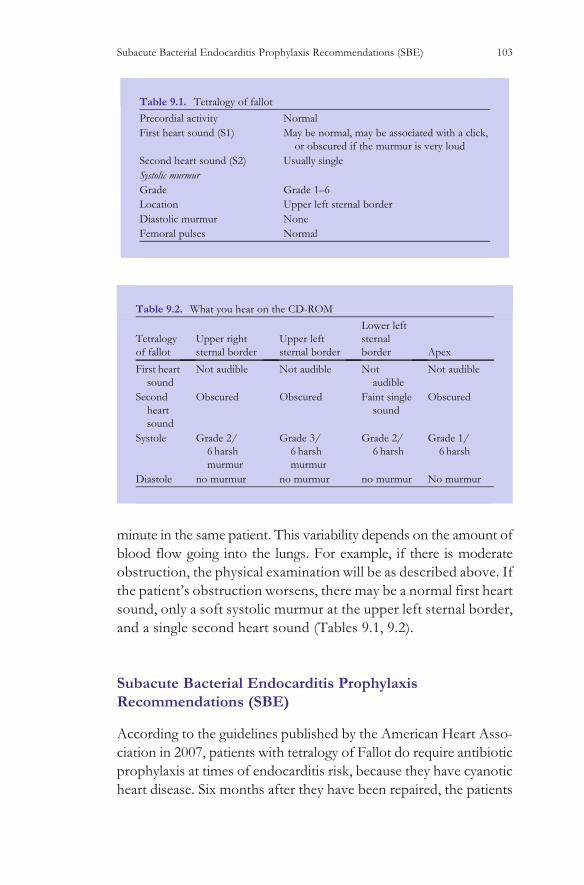
minute in the same patient. This variability depends on the amount of
blood flow going into the lungs. For example, if there is moderate
obstruction, the physical examination will be as described above. If
the patient’s obstruction worsens, there may be a normal first heart
sound, only a soft systolic murmur at the upper left sternal border,
and a single second heart sound (Tables 9.1, 9.2).
Subacute Bacterial Endocarditis Prophylaxis
Recommendations (SBE)
According to the guidelines published by the American Heart Asso-
ciation in 2007, patients with tetralogy of Fallot do require antibiotic
prophylaxis at times of endocarditis risk, because they have cyanotic
heart disease. Six months after they have been repaired, the patients
Table 9.1. Tetralogy of fallot
Precordial activity Normal
First heart sound (S1) May be normal, may be associated with a click,
or obscured if the murmur is very loud
Second heart sound (S2) Usually single
Systolic murmur
Grade Grade 1–6
Location Upper left sternal border
Diastolic murmur None
Femoral pulses Normal
Table 9.2. What you hear on the CD-ROM
Tetralogy
of fallot
Upper right
sternal border
Upper left
sternal border
Lower left
sternal
border Apex
First heart
sound
Not audible Not audible Not
audible
Not audible
Second
heart
sound
Obscured Obscured Faint single
sound
Obscured
Systole Grade 2/
6 harsh
murmur
Grade 3/
6 harsh
murmur
Grade 2/
6 harsh
Grade 1/
6 harsh
Diastole no murmur no murmur no murmur No murmur
Subacute Bacterial Endocarditis Prophylaxis Recommendations (SBE) 103

no longer require antibiotic prophylaxis at times of endocarditis risk,
unless they have a residual ventricular septal defect that jets against
prosthetic patch material, or if they have a prosthetic valve. They will
also require endocarditis prophylaxis if they have had a prior case of
endocarditis [6].
In summary, tetralogy of Fallot is relatively common, and is an
excellent example to help understand cyanotic congenital heart
disease. Aucsultatory findings, as well as the amount of cyanosis
vary widely, and depend on the severity of the obstruction between
the right ventricle and the pulmonary artery.
References
1. Samanek M, Slavik Z, Zborilova B, Hrobonova V, Voriskova M,
Skovranek J (1989) Prevalence, treatment, and outcome of heart
disease in live-born children: a prospective analysis of
91,823 live-born children. Pediatr Cardiol 10(4):205–211
2. Fallot A (1888) Contribution a l’anatomie pathologique de la
maladie bleue (cyanose cardiaque). Marseille, Barlatier-Feissat
3. Rygg IH, Olesen K, Boesen I (1971) The life history of tetralogy
of Fallot. Dan Med Bull 18(Suppl 2):25–30
4. Pokorski RJ (2000)Long-term survival after repair of tetralogy of
Fallot.J Insurance Med 32(2):89–92
5. Brogan TV, Alfieris GM (2003) Has the time come to rename the
Blalock-Taussig shunt? Pediatr Crit Care Med 4(4):450–453
6. Wilson W, Taubert K, Gewitz M et al. (2007) Prevention of
Infective Endocarditis. Guidelines from the American Heart
Association. A Guideline From the American Heart Association
Rheumatic Fever, Endocarditis, and Kawasaki Disease Commit-
tee, Council on Cardiovascular Disease in the Young, and the
Council on Clinical Cardiology, Council on Cardiovascular Surgery
and Anesthesia, and the Quality of Care and Outcomes Research
Interdisciplinary Working Group 2007. Circulation April 19
104 9 Tetralogy of Fallot

Index
105

A
Abnormal aortic valve, 67–68
Abnormal sounds, 81
‘‘An absence of silence’’, 35, 90
Angiotensin converting enzyme
inhibitors, 46
Antibiotic prophylaxis, 62
Aortic diastolic pressure, 8
Aortic outflow murmur, 24–25
Aortic stenosis, 67
anatomy, 67–68
auscultatory findings, 69–71
infants with, 68
natural history, 69
physiology, 68–69
SBE Prophylaxis recommendations, 71
Aortic valve, 67
annulus, 101
diseases, 71
insufficiency, 45
Arterial duct, 59
Atrial septal defects, 16, 29–30
anatomy, 30–31
auscultation, 33–36
inspection, 32
locations, 31
palpation, 32
SBE Prophylaxis recommendations, 36
Atrial septum, hole in, 29
Atrioventricular canal/septal defects, 41–42
Auscultation, 61–62
B
Beta blockers, 46
C
Cardiac auscultation, listening areas in, 3–4
Cardiac electrical abnormality, 10
Chest asymmetry, 32
Cleft, 88
Cleft mitral valve, 87
Cyanosis, 98, 101–102
Cyanotic congenital heart disease, 97
D
Defects in interventricular septum
incidence, 41
large ventricular septal defects, 50–52
medical management, 45–46
physical examination, 47
surgical options, 46–47
moderate ventricular septal defects,
48–50
natural history, 45
physiology, 43–45
SBE Prophylaxis recommendations, 53
small muscular ventricular septal defects,
47–48
Diamond-shaped murmur, 34
Diastolic murmurs, 5, 8, 20, 23, 35, 36, 49,
52, 60, 61, 62, 70, 82, 91, 92, 103
Diastolic rumbling sound, 90
Digoxin, 46
Diuretics, 46
Doubly committed ventricular septal defect,
42–43
Down syndrome, 41, 44, 61
E
Ebstein anomaly, 7
Eisenmenger’s syndrome, 44, 51, 52, 61
F
Failure to thrive, 31
Fallot’s tetralogy, see Tetralogy of Fallot
Fetal blood flow, 16
First heart sound (S1), 3–8, 18, 33, 35, 47, 48,
50, 51, 52, 61, 62, 69, 70, 81, 90,
91, 92, 102, 103
Fixed split second heart sound, 33
G
Grade 5 systolic murmur of aortic
stenosis, 29
H
Harrison’s Groove, 48
Harsh murmur of restrictive ventricular
septum, 41
I
Implantation, 47
Infundibulum, 99–100
Inlet ventricular septal defects, 41
Interventricular septum, defects in,
see Defects in interventricular
septum
Isovolumic contraction phase, 47
K
Kan, J. S., 83
Knauth, A. L., 46
L
Large ventricular septal defects, 50–52
Listening areas in cardiac auscultation, 3–4
Listening with ‘‘dissection’’, 3
Index 107

Loud continuous murmur, 62
Low-pitched diastolic sound, 49
M
Machinery murmur, 60
Marfan syndrome, 90
Migraine headaches, 29
Mitral valve click, 6
Mitral valve insufficiency, 87
anatomy, 87–89
auscultatory findings, 90
mitral valve regurgitation from cleft
mitral valve, 90–91
mitral valve regurgitation from
prolapsing mitral valve, 91–92
natural history, 89
physiology, 89
SBE Prophylaxis recommendations, 92
surgical options, 89–90
Mitral valve prolapse, 88, 92
Mitral valve regurgitation
and cleft mitral valve, 91
from cleft mitral valve, 90–91
and mitral valve prolapse, 92
from prolapsing mitral valve, 91–92
recording, 90
Moderate ventricular septal defects, 48–50
Murmur, 81, 102
grade 5 systolic murmur of aortic
stenosis, 29
harsh murmur of restrictive ventricular
septum, 41
loud continuous, 62
machinery, 60
normal, 15
aortic outflow murmur, 24–25
peripheral pulmonary flow murmurs,
15–19
Still’s murmur, 19–23
of peripheral pulmonary stenosis, 18
See also Diastolic murmurs; First heart
sound (S1); Second heart sound
(S2); Systolic murmur
N
Neurohormones, 46
Normal heart sounds
first heart sound (S1), 3–8, 18, 33, 35, 47,
48, 50, 51, 52, 61, 62, 69, 70, 81,
90, 91, 92, 102, 103
second heart sound (S2), 8–11, 20,
21, 23, 24, 33, 35, 36, 48, 49,
50, 51, 52, 61, 62, 70, 81, 90,
91, 92, 102
Normal murmurs, 15
aortic outflow murmur, 24–25
peripheral pulmonary flow murmurs,
15–19
Still’s murmur, 19–23
O
Obstruction, 68–69
Ostium primum ASD, 41–42
Ostium secundum, 16–17
Outflow tract murmur, 24
P
Palpation of precordium, 80
Patent arterial duct, 16, 59
anatomy, 59
antibiotic prophylaxis, 62
natural history, 60–61
physiology, 60
SBE Prophylaxis recommendations, 62
surgical options, 61
auscultation, 61–62
Patent ductus arteriosus (PDA), 59
anatomy, 59
antibiotic prophylaxis, 62
natural history, 60–61
physiology, 60
SBE Prophylaxis recommendations, 62
surgical options, 61
auscultation, 61–62
PDA, see Patent ductus arteriosus (PDA)
Pelech, A. N., 15
Perimembranous ventricular septal
defects, 45
Peripheral pulmonary flow murmurs, 15–19
Peripheral pulmonary stenosis,
murmur of, 18
Physiologic ‘‘nadir’’, 17
Pink tets, 98
Pulmonary artery pressure, 10, 49
Pulmonary ejection click, 81
Pulmonary hypertension, 11, 31
Pulmonary valve, 8–9
annulus, 97, 101
ejection sound, 81
Pulmonary valve stenosis, 75
anatomy, 75–76
auscultatory findings, 80–82
moderate to severe, 82
natural history, 79–80
physiology, 76–79
SBE, Prophylaxis recommendations,
82–83
types of, 77–78
108 Index

Pulmonary vascular disease, see
Eisenmenger’s syndrome
Pulmonary vascular resistance, 41
R
Recurrent pneumonia, 31
Renin–angiotensin–aldosterone system, 46
Roberts, W. C., 71
S
S1, see First heart sound (S1)
S2, see Second heart sound (S2)
SBE, see Subacute Bacterial Endocarditis
(SBE)
Second heart sound (S2), 8–11, 20, 21, 23,
24, 33, 35, 36, 48, 49, 50, 51, 52,
61, 62, 70, 81, 90, 91, 92, 102
Septal defects, large ventricular, 50–52
Shunt operation, 101
Silent ductus, 60
Single ventricles, 100
Small muscular ventricular septal defects,
47–48
Stenosis of aortic valve
anatomy, 67–68
auscultatory findings, 69–71
infants with, 68
natural history, 69
physiology, 68–69
SBE, Prophylaxis recommendations, 71
Stenosis of pulmonary valve, 75
anatomy, 75–76
auscultatory findings, 80–82
moderate to severe, 82
natural history, 79–80
physiology, 76–79
SBE Prophylaxis recommendations,
82–83
types of, 77–78
Still’s murmur, 19–23
Subacute Bacterial Endocarditis Prophylaxis
recommendations (SBE)
aortic stenosis, 71
atrial septal defects, 36
mitral valve insufficiency, 92
patent ductus arteriosus (PDA), 62
pulmonary stenosis, 82–83
Tetralogy of Fallot, 103–104
ventricular septal defects (VSD), 53
Subacute Bacterial Endocarditis (SBE), 37,
53, 62, 71, 82, 92, 103
Subarterial ventricular septal defect,
42–43
Supra valvar area, 97
Swiss-cheese septum, 42
Sympathetic nervous system, 46
Systolic murmur, 5, 6, 23, 35, 49, 52, 62, 67,
69, 70, 89, 90, 103
T
Tetralogy, 100
Tetralogy of Fallot
anatomy, 97
auscultatory findings, 101–103
incidence, 97
natural history, 100
physiology, 97–100
SBE Prophylaxis recommendations,
103–104
surgical options, 101
Tet spell, 100
Tricuspid atresia, 100
Tricuspid valves, 7
Tricuspid valve tissue, 49
V
Venous hum, 20–21
Ventricular septal defects (VSD)
anatomy, 41–43
incidence, 41
large ventricular septal defects, 50–52
medical management, 45–46
physical examination, 47
surgical options, 46–47
moderate ventricular septal defects,
48–50
natural history, 45
physiology, 43–45
SBE Prophylaxis recommendations, 53
small muscular ventricular septal defects,
47–48
Ventricular systole, 3, 4
Index 109