auscultation of the heart: normal heart sounds and their
TRANSCRIPT
Auscultation of the heart: normal heart sounds and their changes, heart murmurs and their diagnostic significance
Professor T.V. ASHCHEULOVA
head of Propedeutics to Internal Medicine N1, Basis of Bioethics
and Biosafety
Kharkiv National Medical University
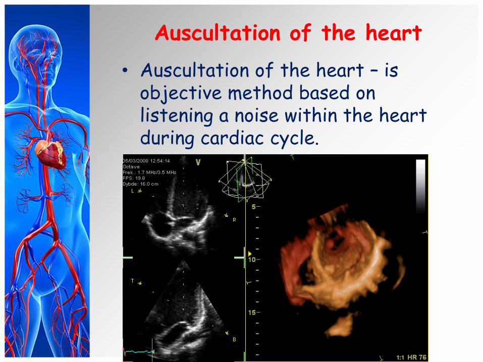
Auscultation of the heart
• Auscultation of the heart – is objective method based on listening a noise within the heart during cardiac cycle.
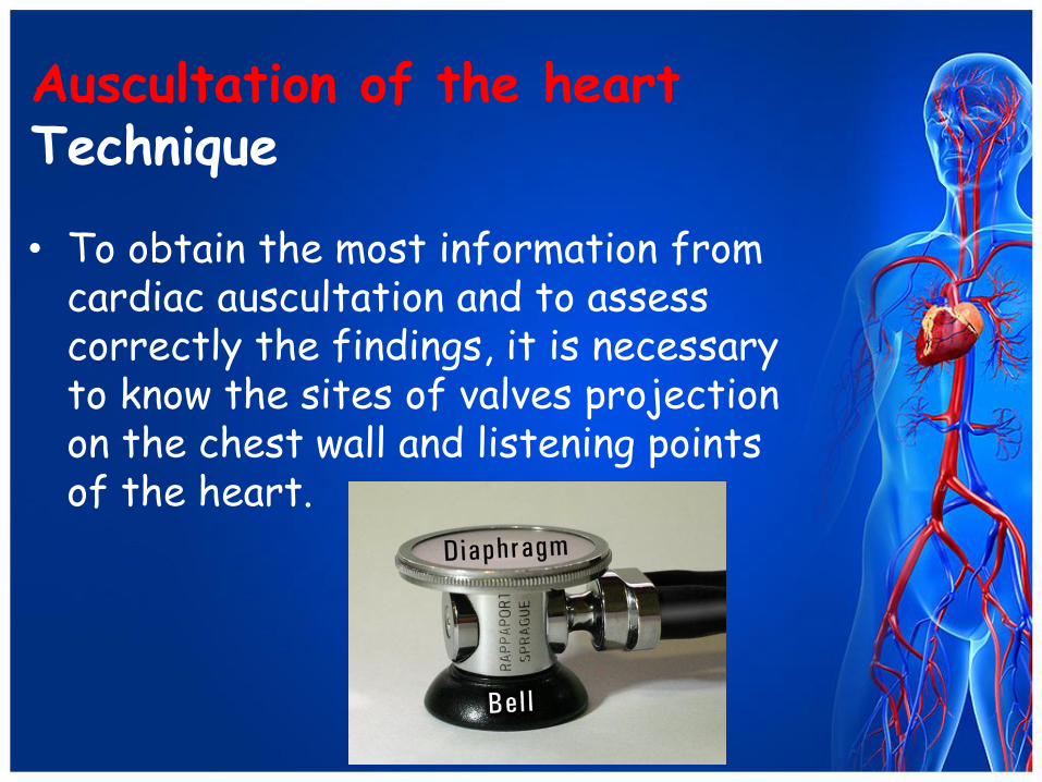
Auscultation of the heart Technique
• To obtain the most information from cardiac auscultation and to assess correctly the findings, it is necessary to know the sites of valves projection on the chest wall and listening points of the heart.
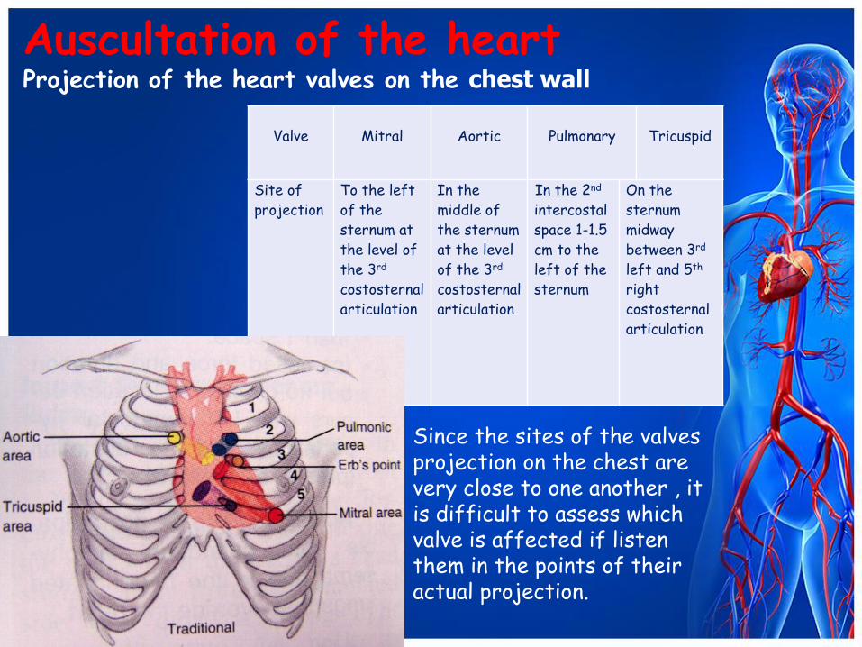
Auscultation of the heart Projection of the heart valves on the chest wall
Valve
Mitral
Aortic
Pulmonary
Tricuspid
Site of
projection
To the left
of the
sternum at
the level of
the 3rd
costosternal
articulation
In the
middle of
the sternum
at the level
of the 3rd
costosternal
articulation
In the 2nd
intercostal
space 1-1.5
cm to the
left of the
sternum
On the
sternum
midway
between 3rd
left and 5th
right
costosternal
articulation
Since the sites of the valves projection on the chest are very close to one another , it is difficult to assess which valve is affected if listen them in the points of their actual projection.
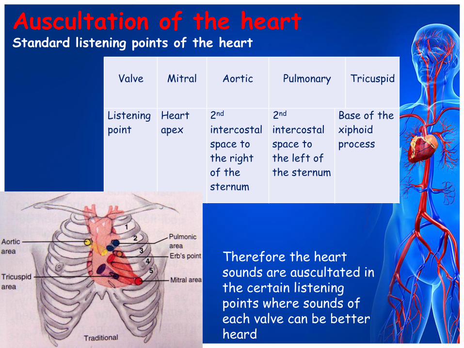
Auscultation of the heart Standard listening points of the heart
Valve
Mitral
Aortic
Pulmonary
Tricuspid
Listening
point
Heart
apex
2nd
intercostal
space to
the right
of the
sternum
2nd
intercostal
space to
the left of
the sternum
Base of the
xiphoid
process
Therefore the heart sounds are auscultated in the certain listening points where sounds of each valve can be better heard
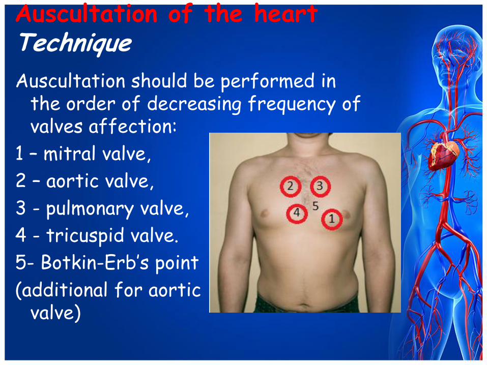
Auscultation of the heart Technique Auscultation should be performed in
the order of decreasing frequency of valves affection:
1 – mitral valve,
2 – aortic valve,
3 - pulmonary valve,
4 - tricuspid valve.
5- Botkin-Erb’s point
(additional for aortic valve)
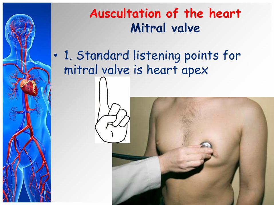
Auscultation of the heart Mitral valve
• 1. Standard listening points for
mitral valve is heart apex

Auscultation of the heart Aortic Valve
• 2. Standard listening points for aortic valve is 2nd interspace to the right of the sternum
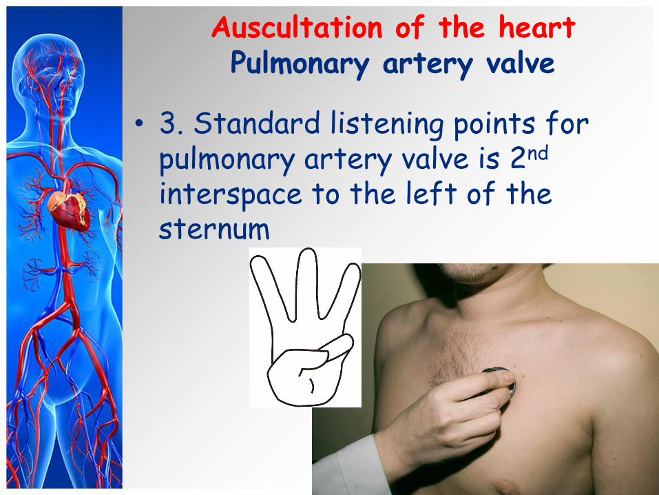
Auscultation of the heart Pulmonary artery valve
• 3. Standard listening points for pulmonary artery valve is 2nd interspace to the left of the sternum

Auscultation of the heart Tricuspid valve
• 4. Standard listening points for tricuspid is base of the xiphoid process
Auscultation of the heart Botkin-Erb’s point
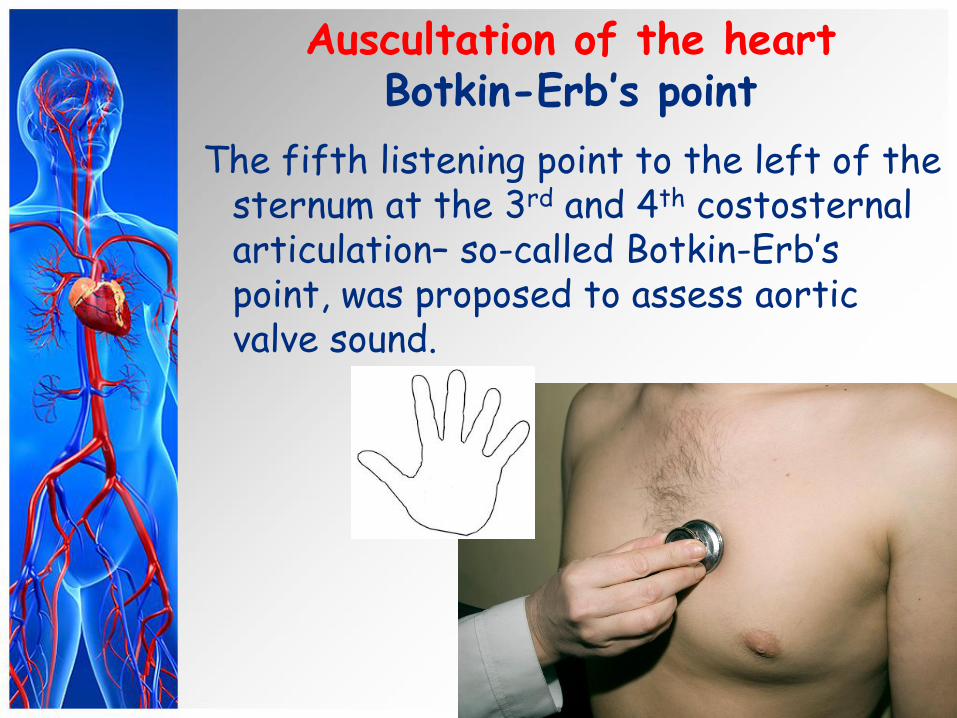
The fifth listening point to the left of the sternum at the 3rd and 4th costosternal articulation– so-called Botkin-Erb’s point, was proposed to assess aortic valve sound.
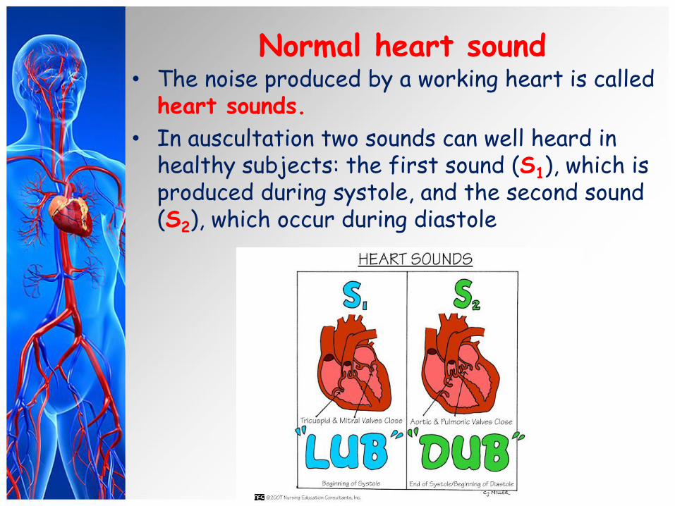
Normal heart sound • The noise produced by a working heart is called
heart sounds.
• In auscultation two sounds can well heard in healthy subjects: the first sound (S1), which is produced during systole, and the second sound (S2), which occur during diastole
1 2
3 4
5 6
7 8
1 2
3 4 A B C
S 1 S 2
Systole Diastole
S 1 S 2
Normal heart sounds
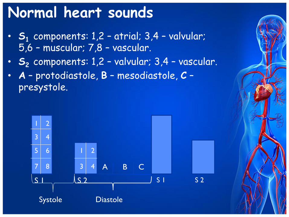
• S1 components: 1,2 – atrial; 3,4 – valvular; 5,6 – muscular; 7,8 – vascular.
• S2 components: 1,2 – valvular; 3,4 – vascular.
• A – protodiastole, B – mesodiastole, C – presystole.
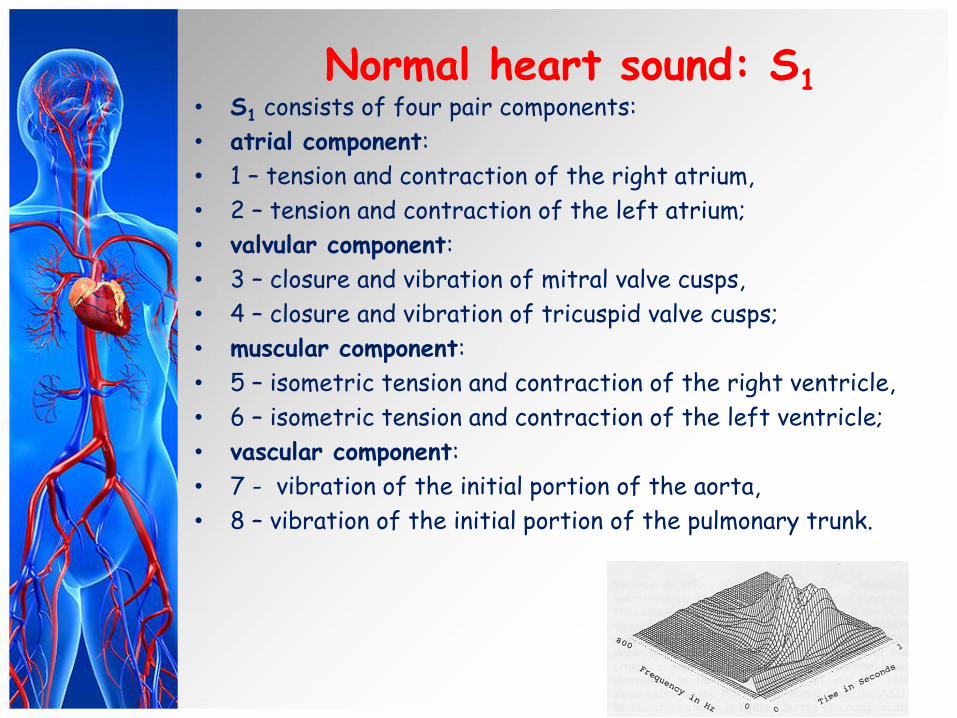
Normal heart sound: S1 • S1 consists of four pair components:
• atrial component:
• 1 – tension and contraction of the right atrium,
• 2 – tension and contraction of the left atrium;
• valvular component:
• 3 – closure and vibration of mitral valve cusps,
• 4 – closure and vibration of tricuspid valve cusps;
• muscular component:
• 5 – isometric tension and contraction of the right ventricle,
• 6 – isometric tension and contraction of the left ventricle;
• vascular component:
• 7 - vibration of the initial portion of the aorta,
• 8 – vibration of the initial portion of the pulmonary trunk.

Normal heart sound: S2
• S2 consists of two pair components:
• valvular component:
• 1 – closure and vibration of the aortic valve cusps,
• 2 – closure and vibration of the pulmonary valve cusps;
• vascular component:
• 3 – vibration of the aortic walls,
• 4 – vibration of pulmonary trunk walls.
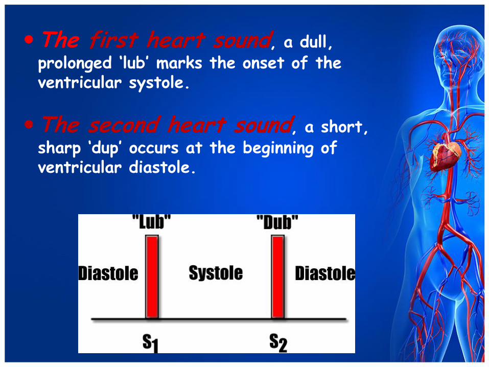
The first heart sound, a dull, prolonged ‘lub’ marks the onset of the ventricular systole.
The second heart sound, a short, sharp ‘dup’ occurs at the beginning of ventricular diastole.
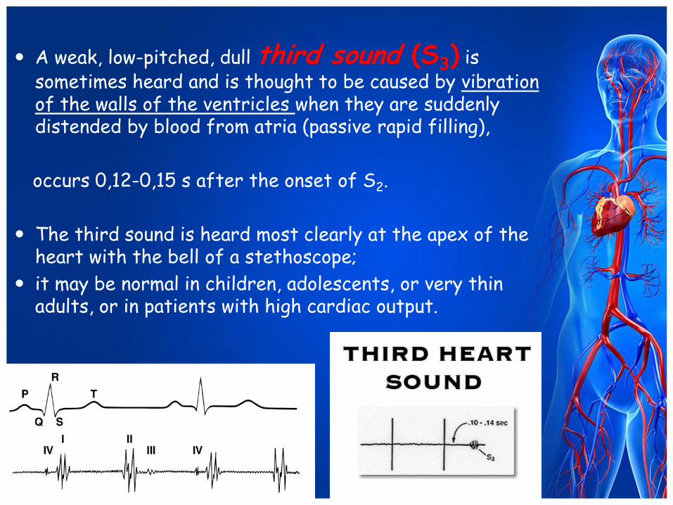
A weak, low-pitched, dull third sound (S3) is sometimes heard and is thought to be caused by vibration of the walls of the ventricles when they are suddenly distended by blood from atria (passive rapid filling),
occurs 0,12-0,15 s after the onset of S2.
The third sound is heard most clearly at the apex of the heart with the bell of a stethoscope;
it may be normal in children, adolescents, or very thin adults, or in patients with high cardiac output.
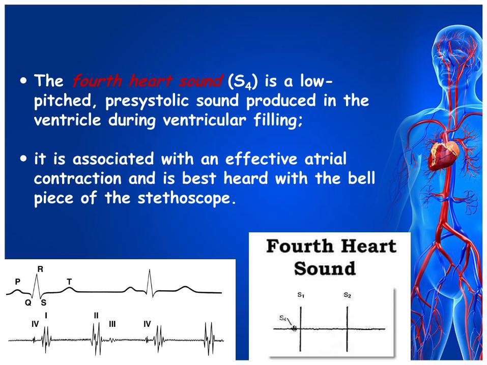
The fourth heart sound (S4) is a low-pitched, presystolic sound produced in the ventricle during ventricular filling;
it is associated with an effective atrial contraction and is best heard with the bell piece of the stethoscope.
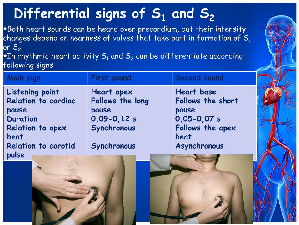
Main sign First sound Second sound
Listening point Relation to cardiac pause Duration Relation to apex beat Relation to carotid pulse
Heart apex Follows the long pause 0,09-0,12 s Synchronous Synchronous
Heart base Follows the short pause 0,05-0,07 s Follows the apex beat Asynchronous
Differential signs of S1 and S2 Both heart sounds can be heard over precordium, but their intensity changes depend on nearness of valves that take part in formation of S1 or S2. In rhythmic heart activity S1 and S2 can be differentiate according following signs
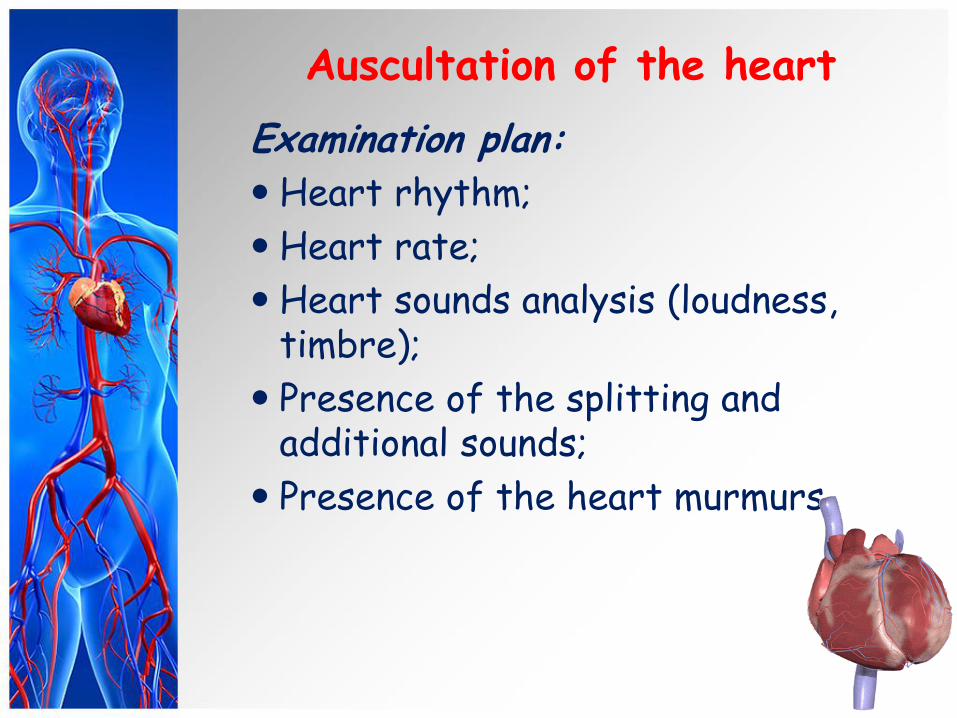
Auscultation of the heart
Examination plan:
Heart rhythm;
Heart rate;
Heart sounds analysis (loudness, timbre);
Presence of the splitting and additional sounds;
Presence of the heart murmurs.
Cardiac rhythm In healthy subjects S1 and S2, S2
and S1 follow one another at regular intervals:
the heart activity is said to be rhythmic or regular.
When the cardiac activity is arrhythmic, the heart sounds follow at irregular intervals.
Heart rate (HR)
Heart rate (HR) in normal conditions is 60-80 beats per minute.
Acceleration of the heart rate to
more than 90 beats per minute is called tachycardia.
A heart rate less than 60 beat per
minute is called bradycardia.
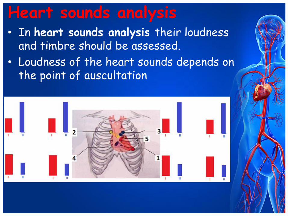
Heart sounds analysis • In heart sounds analysis their loudness
and timbre should be assessed.
• Loudness of the heart sounds depends on the point of auscultation
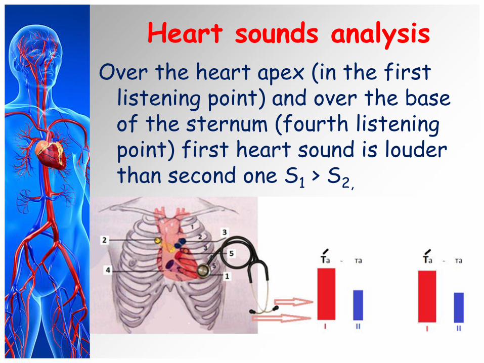
Heart sounds analysis
Over the heart apex (in the first listening point) and over the base of the sternum (fourth listening point) first heart sound is louder than second one S1 > S2,
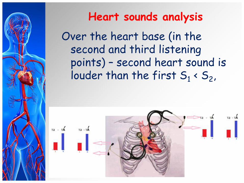
Heart sounds analysis
Over the heart base (in the second and third listening points) – second heart sound is louder than the first S1 < S2,
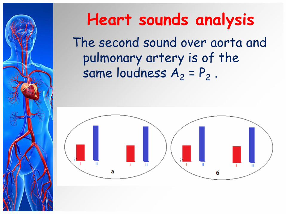
Heart sounds analysis
The second sound over aorta and pulmonary artery is of the same loudness A2 = P2 .
Heart sounds analysis
The loudness of the heart sounds can be changed in several physiological and pathological conditions.
Loudness of one or both heart sounds may increase or decrease.
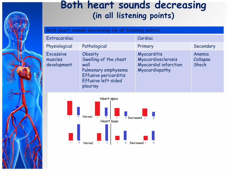
Both heart sounds decreasing (in all listening points)
Both heart sounds decreasing (in all listening points).
Extracardiac Cardiac
Physiological Pathological Primary Secondary
Excessive muscles development
Obesity Swelling of the chest wall Pulmonary emphysema Effusive pericarditis Effusive left-sided pleurisy
Myocarditis Myocardiosclerosis Myocardial infarction Myocardiopathy
Anemia Collapse Shock
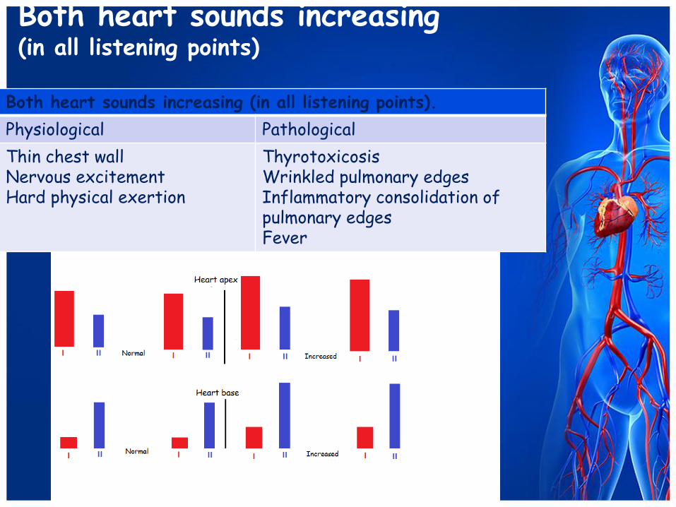
Both heart sounds increasing (in all listening points)
Both heart sounds increasing (in all listening points).
Physiological Pathological
Thin chest wall Nervous excitement Hard physical exertion
Thyrotoxicosis Wrinkled pulmonary edges Inflammatory consolidation of pulmonary edges Fever
Heart sounds analysis
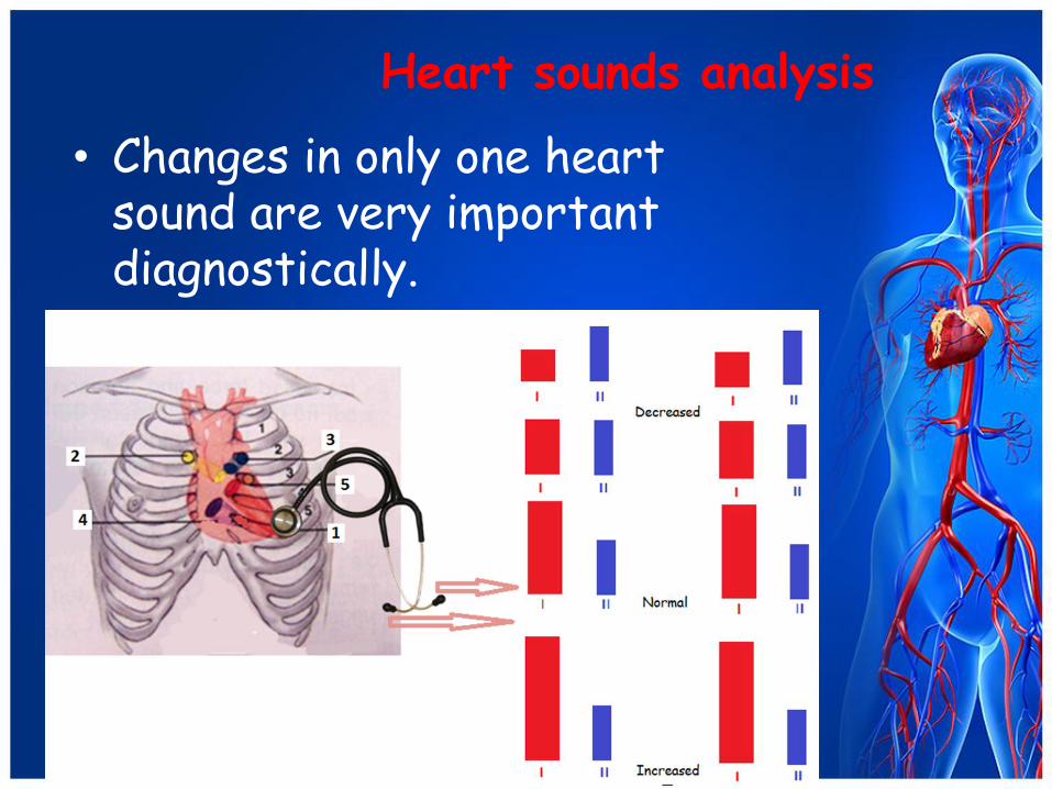
• Changes in only one heart sound are very important diagnostically.
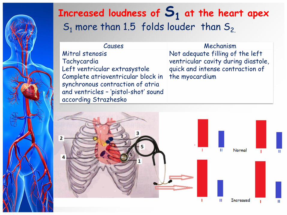
Increased loudness of S1 at the heart apex
S1 more than 1.5 folds louder than S2.
Causes Mechanism Mitral stenosis Tachycardia Left ventricular extrasystole Complete atrioventricular block in synchronous contraction of atria and ventricles – ‘pistol-shot’ sound according Strazhesko
Not adequate filling of the left ventricular cavity during diastole, quick and intense contraction of the myocardium
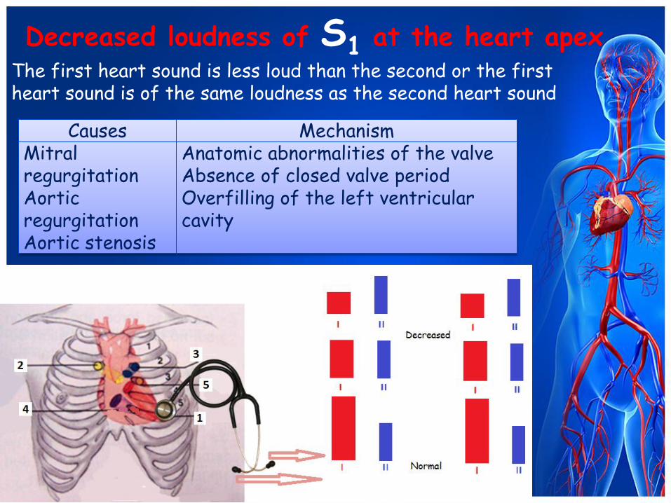
Decreased loudness of S1 at the heart apex
The first heart sound is less loud than the second or the first heart sound is of the same loudness as the second heart sound
Causes Mechanism Mitral regurgitation Aortic regurgitation Aortic stenosis
Anatomic abnormalities of the valve Absence of closed valve period Overfilling of the left ventricular cavity

Causes
Mechanism
Complete heat block Atrial fibrillation Extrasystolic arrhythmia Ventricular flutter
Different ventricular filling in each cardiac cycle
Different loudness of S1 at the heart apex
• The first heart sound is not of the same intensity in the different cycles

Accentuated S2 over aorta
The second sound over aorta is louder than over pulmonary artery.
Causes Mechanism Physiological Pathological
Emotional exertion Physical exertion
Essential hypertension Symptomatic hypertension Aortic atherosclerosis Syphilitic mesoaortitis
Pressure elevation in the systemic circulation, decreased elasticity of the aorta
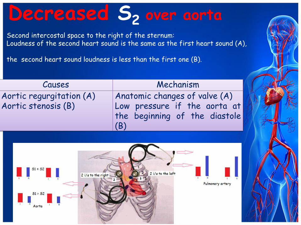
Decreased S2 over aorta
Causes Mechanism Aortic regurgitation (A) Aortic stenosis (B)
Anatomic changes of valve (A) Low pressure if the aorta at the beginning of the diastole (B)
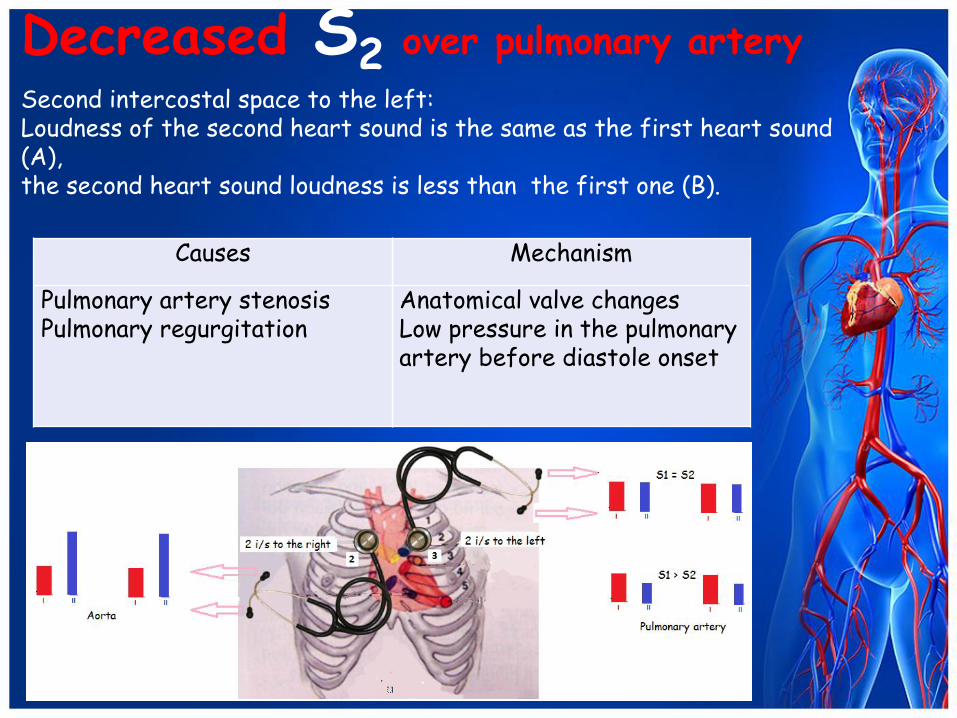
Second intercostal space to the right of the sternum: Loudness of the second heart sound is the same as the first heart sound (A), the second heart sound loudness is less than the first one (B).
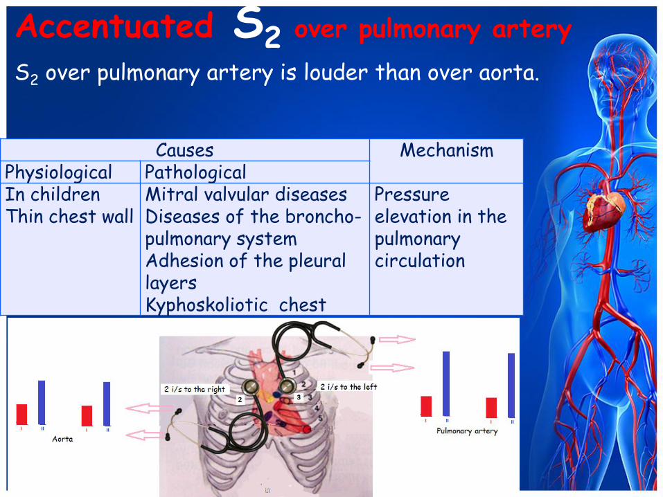
Accentuated S2 over pulmonary artery
S2 over pulmonary artery is louder than over aorta.
Causes Mechanism Physiological Pathological In children Thin chest wall
Mitral valvular diseases Diseases of the broncho-pulmonary system Adhesion of the pleural layers Kyphoskoliotic chest
Pressure elevation in the pulmonary circulation
Decreased S2 over pulmonary artery
Second intercostal space to the left: Loudness of the second heart sound is the same as the first heart sound (A), the second heart sound loudness is less than the first one (B).
Causes Mechanism
Pulmonary artery stenosis Pulmonary regurgitation
Anatomical valve changes Low pressure in the pulmonary artery before diastole onset

Decreased loudness of S1 at the base of the sternum
Causes Mechanism Tricuspid regurgitation
Anatomic changes of the valve Absence of closed valves period Overfilling of the right ventricular cavity
• The first heart sound is less loud than the second or the first heart sound is of the same loudness as the second heart sound
Reduplication and splitting of the heart sounds
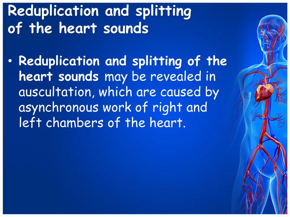
• Reduplication and splitting of the heart sounds may be revealed in auscultation, which are caused by asynchronous work of right and left chambers of the heart.

Reduplication and splitting of the heart sounds
• Reduplication – two short
sounds follow one another are heard instead S1 or S2.
Reduplication and splitting of the heart sounds
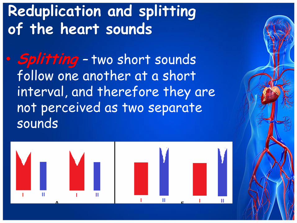
• Splitting – two short sounds
follow one another at a short interval, and therefore they are not perceived as two separate sounds

Reduplication and splitting of the heart sounds
• Splitting of the two high-pitched
components of S1 by 10-30 ms is a normal phenomenon, which is recorded by phonocardiography. The third component of S1 is attributed to mitral valve closure, and the fourth to tricuspid valve closure. Widening of the interval between these two components is heard as S1 splitting or reduplication at the heart apex or at the base of the xiphoid process.
Reduplication and splitting of the heart sounds
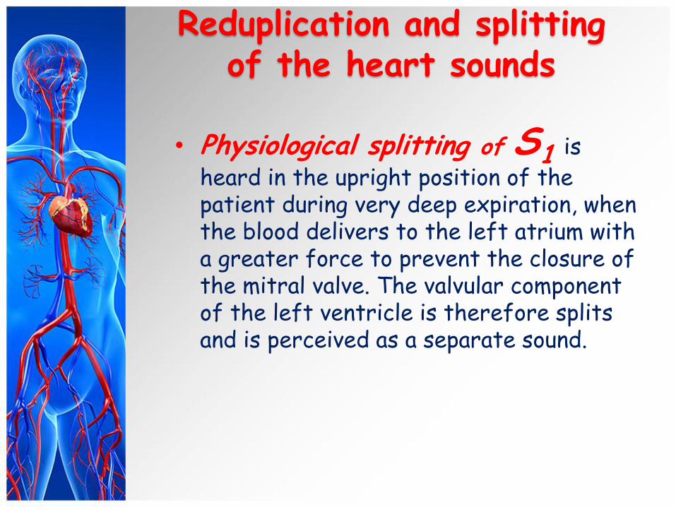
• Physiological splitting of S1 is
heard in the upright position of the patient during very deep expiration, when the blood delivers to the left atrium with a greater force to prevent the closure of the mitral valve. The valvular component of the left ventricle is therefore splits and is perceived as a separate sound.
Reduplication and splitting of the heart sounds
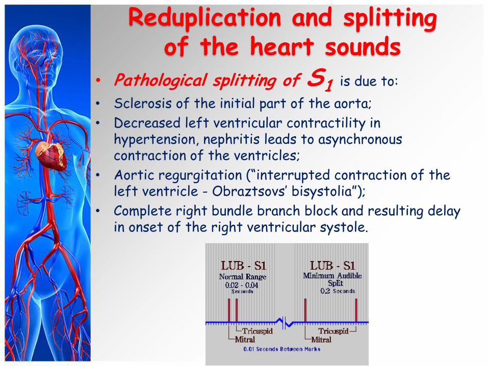
• Pathological splitting of S1 is due to:
• Sclerosis of the initial part of the aorta;
• Decreased left ventricular contractility in hypertension, nephritis leads to asynchronous contraction of the ventricles;
• Aortic regurgitation (“interrupted contraction of the left ventricle - Obraztsovs’ bisystolia”);
• Complete right bundle branch block and resulting delay in onset of the right ventricular systole.
Reduplication and splitting of the heart sounds
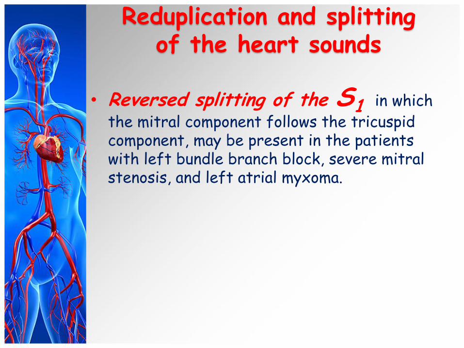
• Reversed splitting of the S1 in which
the mitral component follows the tricuspid component, may be present in the patients with left bundle branch block, severe mitral stenosis, and left atrial myxoma.
Reduplication and splitting of the heart sounds
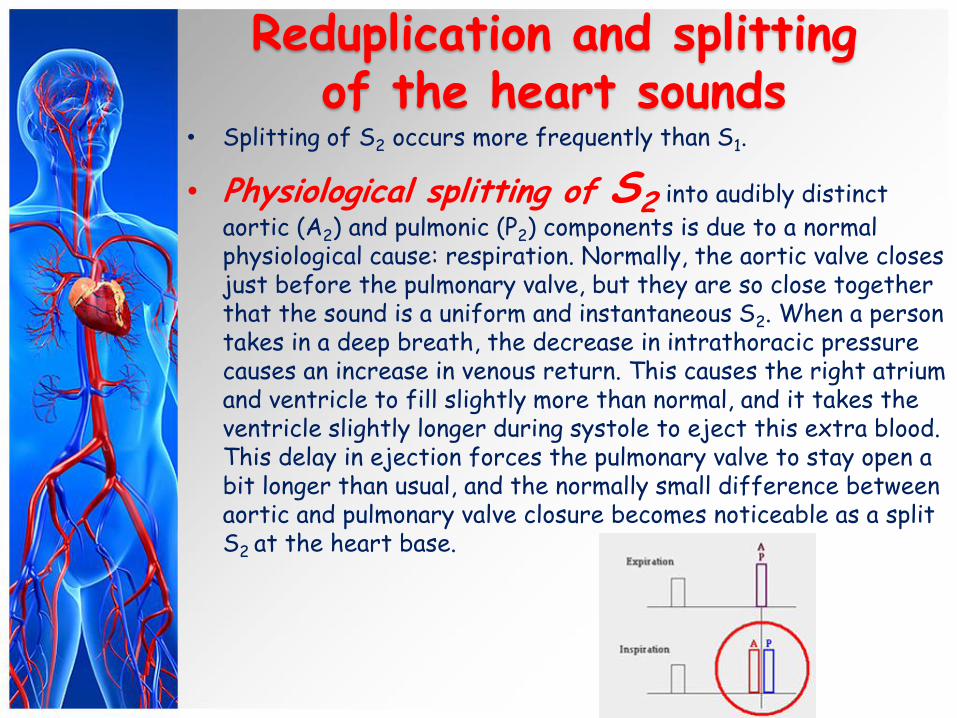
• Splitting of S2 occurs more frequently than S1.
• Physiological splitting of S2 into audibly distinct
aortic (A2) and pulmonic (P2) components is due to a normal physiological cause: respiration. Normally, the aortic valve closes just before the pulmonary valve, but they are so close together that the sound is a uniform and instantaneous S2. When a person takes in a deep breath, the decrease in intrathoracic pressure causes an increase in venous return. This causes the right atrium and ventricle to fill slightly more than normal, and it takes the ventricle slightly longer during systole to eject this extra blood. This delay in ejection forces the pulmonary valve to stay open a bit longer than usual, and the normally small difference between aortic and pulmonary valve closure becomes noticeable as a split S2 at the heart base.
Reduplication and splitting of the heart sounds
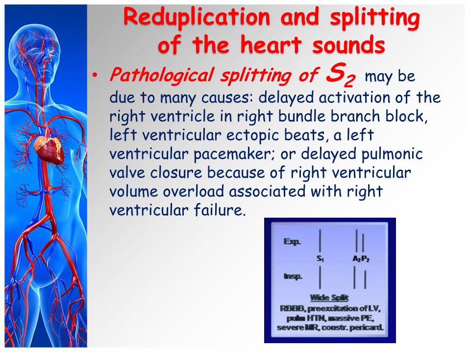
• Pathological splitting of S2 may be
due to many causes: delayed activation of the right ventricle in right bundle branch block, left ventricular ectopic beats, a left ventricular pacemaker; or delayed pulmonic valve closure because of right ventricular volume overload associated with right ventricular failure.
Reduplication and splitting of the heart sounds
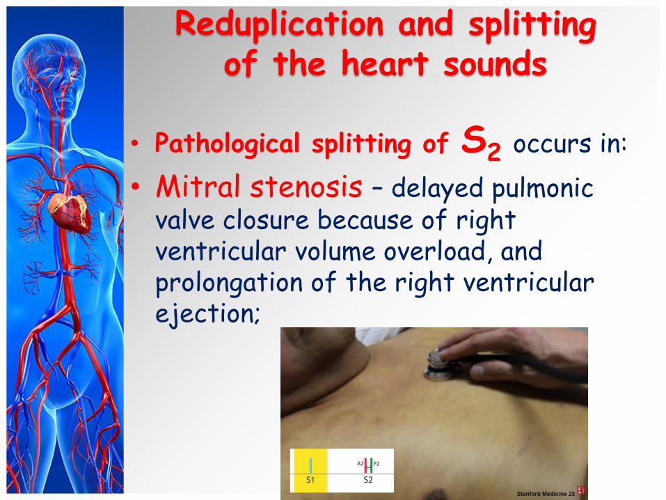
• Pathological splitting of S2 occurs in:
• Mitral stenosis – delayed pulmonic valve closure because of right ventricular volume overload, and prolongation of the right ventricular ejection;
Reduplication and splitting of the heart sounds
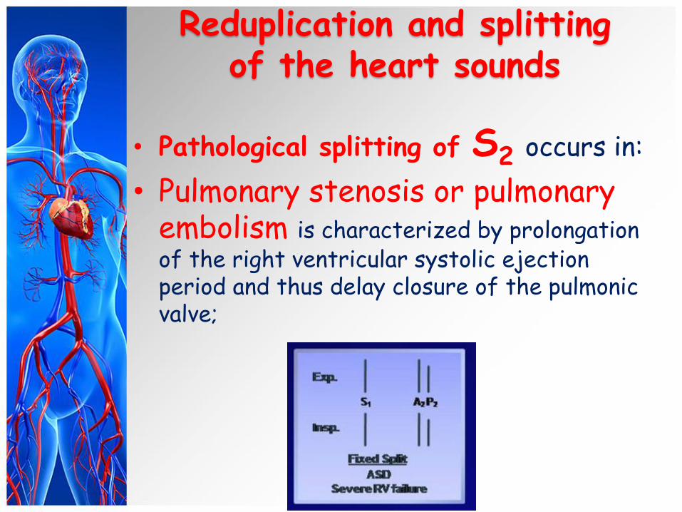
• Pathological splitting of S2 occurs in:
• Pulmonary stenosis or pulmonary embolism is characterized by prolongation of the right ventricular systolic ejection period and thus delay closure of the pulmonic valve;
Reduplication and splitting of the heart sounds
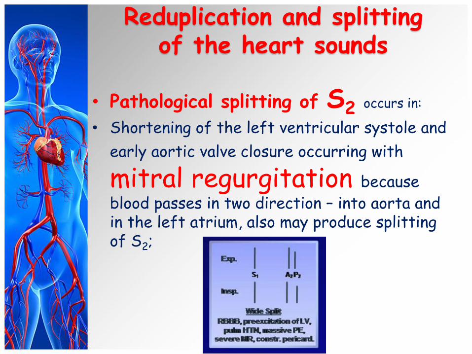
• Pathological splitting of S2 occurs in:
• Shortening of the left ventricular systole and
early aortic valve closure occurring with
mitral regurgitation because
blood passes in two direction – into aorta and in the left atrium, also may produce splitting of S2;
Reduplication and splitting of the heart sounds
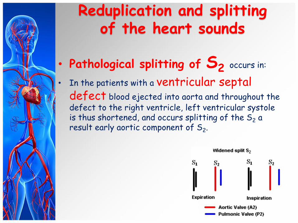
• Pathological splitting of S2 occurs in:
• In the patients with a ventricular septal defect blood ejected into aorta and throughout the defect to the right ventricle, left ventricular systole is thus shortened, and occurs splitting of the S2 a result early aortic component of S2.

Reduplication and splitting of the heart sounds
• Pathological splitting of S2 occurs in:
• An atrial septal defect leads to increased diastolic filling of the right ventricle and early aortic valve closure.
Reduplication and splitting of the heart sounds
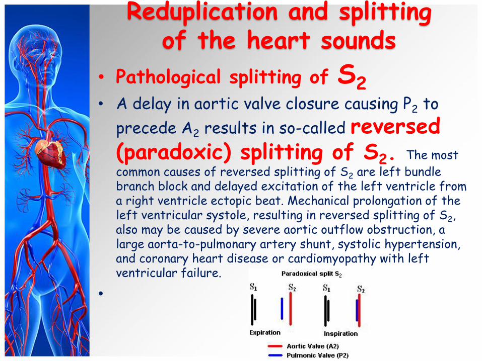
• Pathological splitting of S2
• A delay in aortic valve closure causing P2 to
precede A2 results in so-called reversed (paradoxic) splitting of S2. The most
common causes of reversed splitting of S2 are left bundle branch block and delayed excitation of the left ventricle from a right ventricle ectopic beat. Mechanical prolongation of the left ventricular systole, resulting in reversed splitting of S2, also may be caused by severe aortic outflow obstruction, a large aorta-to-pulmonary artery shunt, systolic hypertension, and coronary heart disease or cardiomyopathy with left ventricular failure.
•
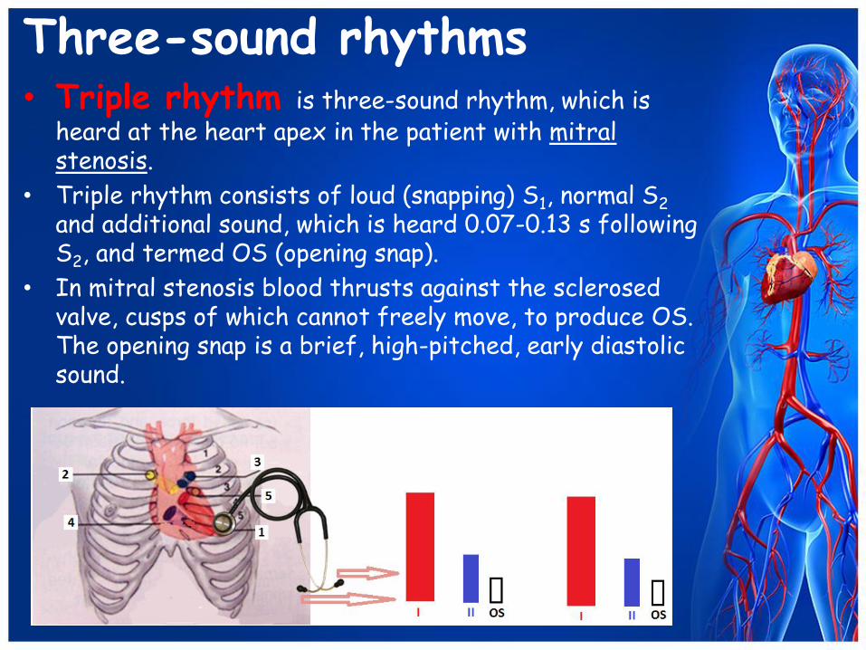
Three-sound rhythms • Triple rhythm is three-sound rhythm, which is
heard at the heart apex in the patient with mitral stenosis.
• Triple rhythm consists of loud (snapping) S1, normal S2 and additional sound, which is heard 0.07-0.13 s following S2, and termed OS (opening snap).
• In mitral stenosis blood thrusts against the sclerosed valve, cusps of which cannot freely move, to produce OS. The opening snap is a brief, high-pitched, early diastolic sound.

Gallop rhythm Three-sound rhythm of a peculiar acoustic
character, termed gallop rhythm, is also of considerable diagnostic value. The sounds of gallop rhythm are usually soft and low, resemble the galloping of a horse, and are best heard in direct auscultation. Gallop rhythm is heard as three separate audibly distinct sounds in approximately equal intervals.
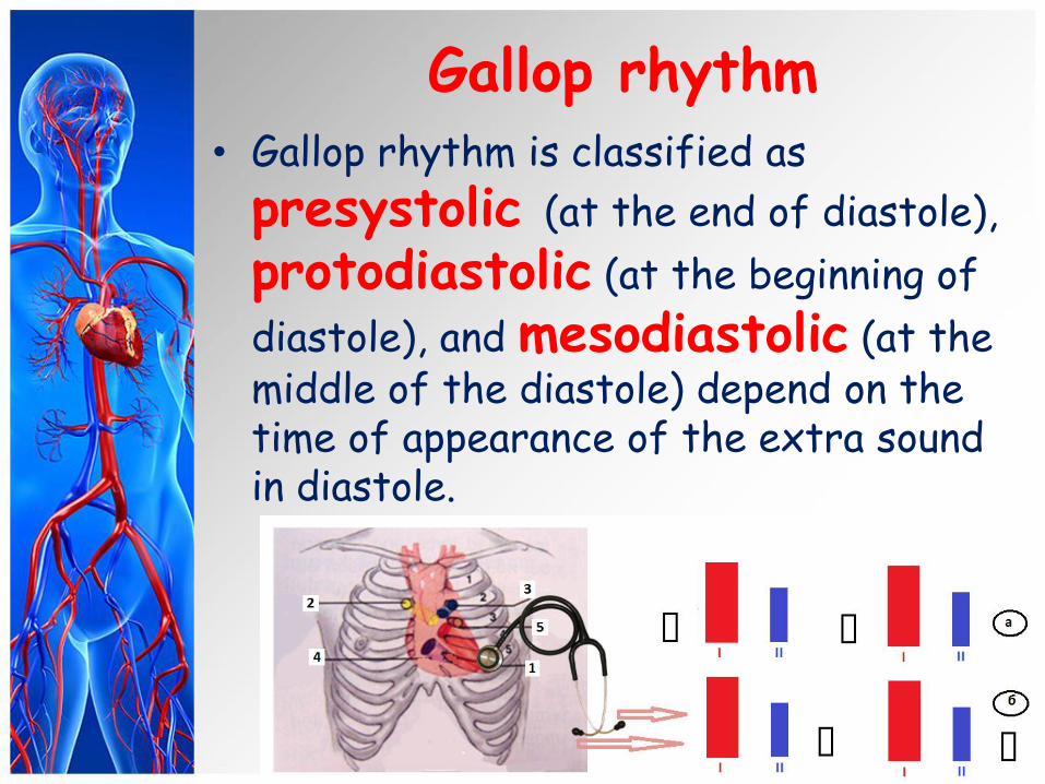
Gallop rhythm • Gallop rhythm is classified as
presystolic (at the end of diastole),
protodiastolic (at the beginning of
diastole), and mesodiastolic (at the middle of the diastole) depend on the time of appearance of the extra sound in diastole.
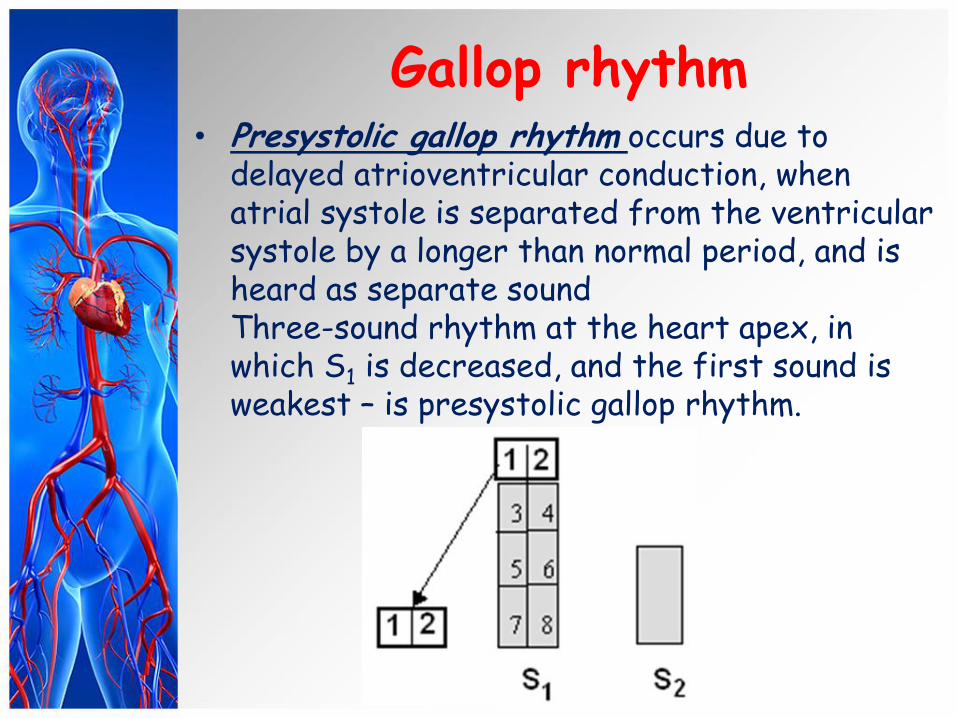
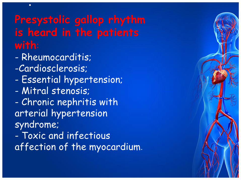
Gallop rhythm • Presystolic gallop rhythm occurs due to
delayed atrioventricular conduction, when atrial systole is separated from the ventricular systole by a longer than normal period, and is heard as separate sound Three-sound rhythm at the heart apex, in which S1 is decreased, and the first sound is weakest – is presystolic gallop rhythm.
•
Presystolic gallop rhythm is heard in the patients with: - Rheumocarditis; -Cardiosclerosis; - Essential hypertension; - Mitral stenosis; - Chronic nephritis with arterial hypertension syndrome; - Toxic and infectious affection of the myocardium.
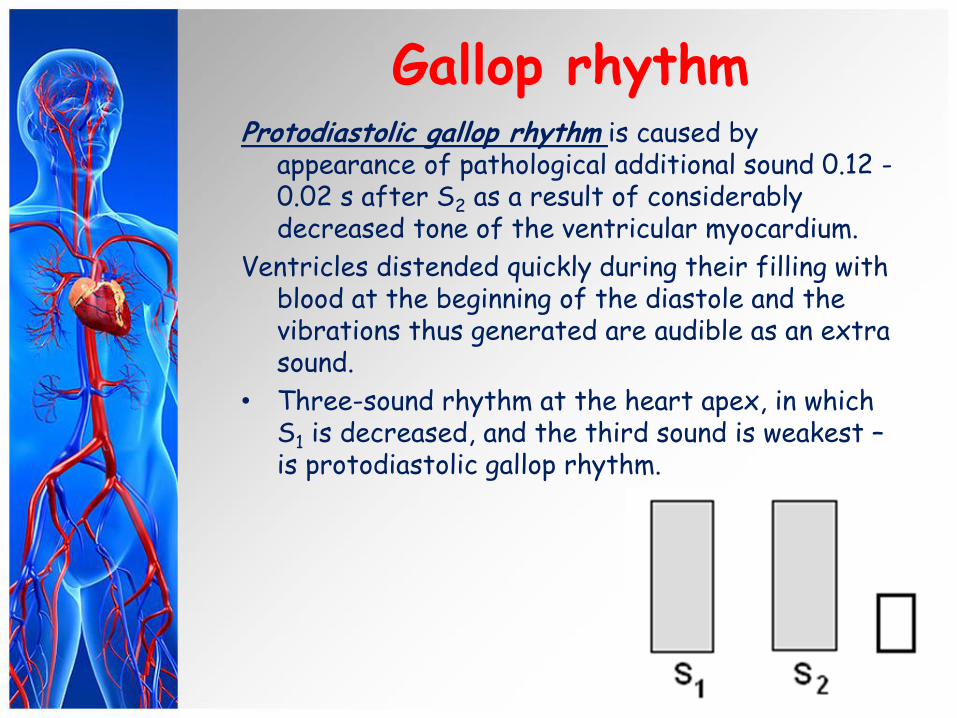
Gallop rhythm Protodiastolic gallop rhythm is caused by
appearance of pathological additional sound 0.12 - 0.02 s after S2 as a result of considerably decreased tone of the ventricular myocardium.
Ventricles distended quickly during their filling with blood at the beginning of the diastole and the vibrations thus generated are audible as an extra sound.
• Three-sound rhythm at the heart apex, in which S1 is decreased, and the third sound is weakest – is protodiastolic gallop rhythm.

This auscultation phenomenon is observed in the patients with: Acute and chronic myocarditis;
• Myocardiosclerosis; Heart failure;
• Toxicosis; Thyrotoxicosis;
• Anaemias .
Protodiastolic gallop rhythm
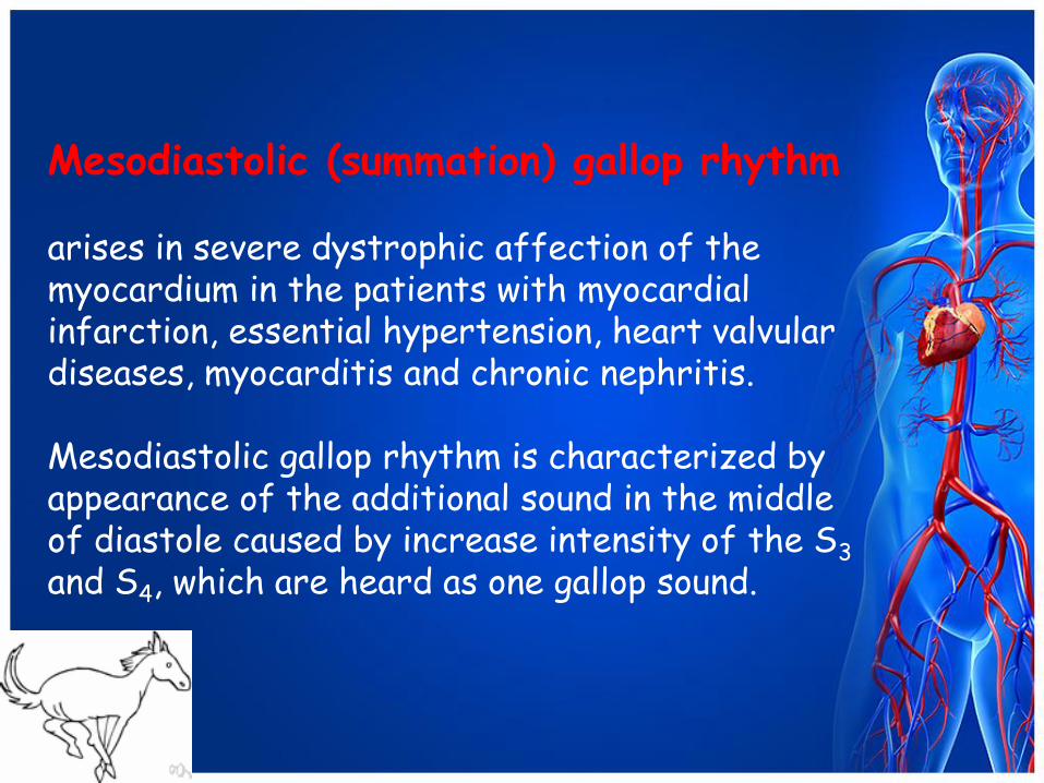
Mesodiastolic (summation) gallop rhythm arises in severe dystrophic affection of the myocardium in the patients with myocardial infarction, essential hypertension, heart valvular diseases, myocarditis and chronic nephritis. Mesodiastolic gallop rhythm is characterized by appearance of the additional sound in the middle of diastole caused by increase intensity of the S3 and S4, which are heard as one gallop sound.
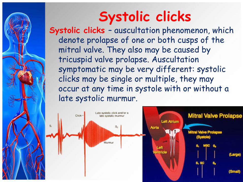
Systolic clicks Systolic clicks – auscultation phenomenon, which
denote prolapse of one or both cusps of the mitral valve. They also may be caused by tricuspid valve prolapse. Auscultation symptomatic may be very different: systolic clicks may be single or multiple, they may occur at any time in systole with or without a late systolic murmur.
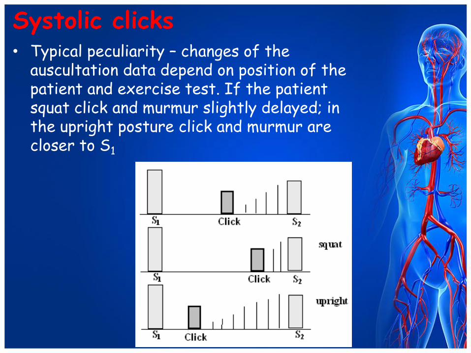
Systolic clicks • Typical peculiarity – changes of the
auscultation data depend on position of the patient and exercise test. If the patient squat click and murmur slightly delayed; in the upright posture click and murmur are closer to S1
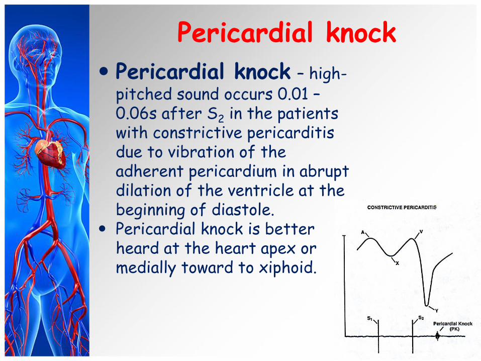
Pericardial knock
Pericardial knock – high-pitched sound occurs 0.01 – 0.06s after S2 in the patients with constrictive pericarditis due to vibration of the adherent pericardium in abrupt dilation of the ventricle at the beginning of diastole.
Pericardial knock is better heard at the heart apex or medially toward to xiphoid.
Embryocardial or pendulum rhythm
Embryocardial or pendulum rhythm occurs in severe heart failure, attacks of paroxysmal tachycardia, high fever, etc.
Tachycardia makes diastolic pause almost as short as the systolic one.
A peculiar auscultative picture, in which heart sounds are similar in intensity, resembles foetal rhythm is termed embryocardia.
Cardiac murmurs • In addition to the normal heart
sounds, abnormal sounds known as murmurs may be heard in auscultation. Cardiac murmurs may both endocardiac and exocardiac.
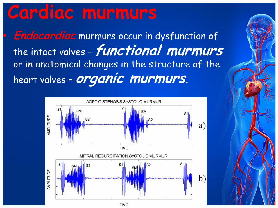
Cardiac murmurs • Endocardiac murmurs occur in dysfunction of
the intact valves – functional murmurs or in anatomical changes in the structure of the
heart valves – organic murmurs.
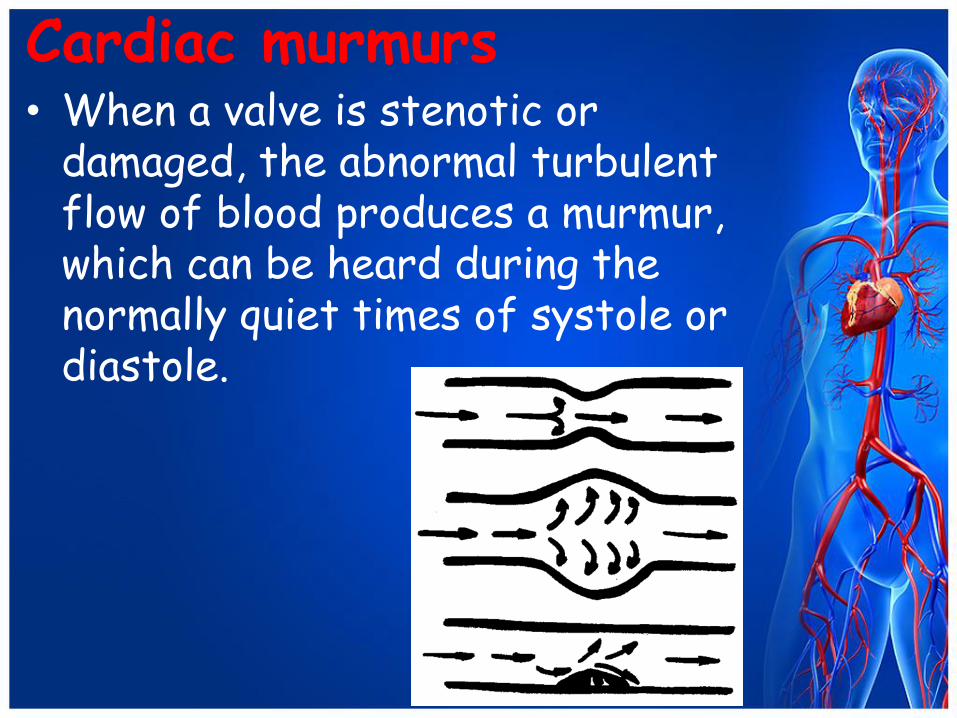
Cardiac murmurs • When a valve is stenotic or
damaged, the abnormal turbulent flow of blood produces a murmur, which can be heard during the normally quiet times of systole or diastole.
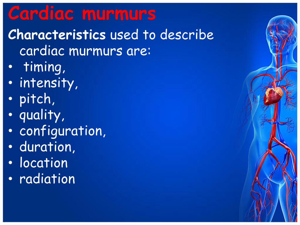
Cardiac murmurs Characteristics used to describe
cardiac murmurs are: • timing, • intensity, • pitch, • quality, • configuration, • duration, • location • radiation
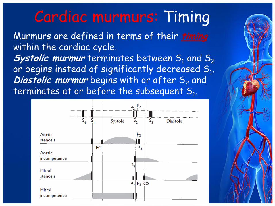
Murmurs are defined in terms of their timing within the cardiac cycle. Systolic murmur terminates between S1 and S2 or begins instead of significantly decreased S1. Diastolic murmur begins with or after S2 and terminates at or before the subsequent S1.
Cardiac murmurs: Timing
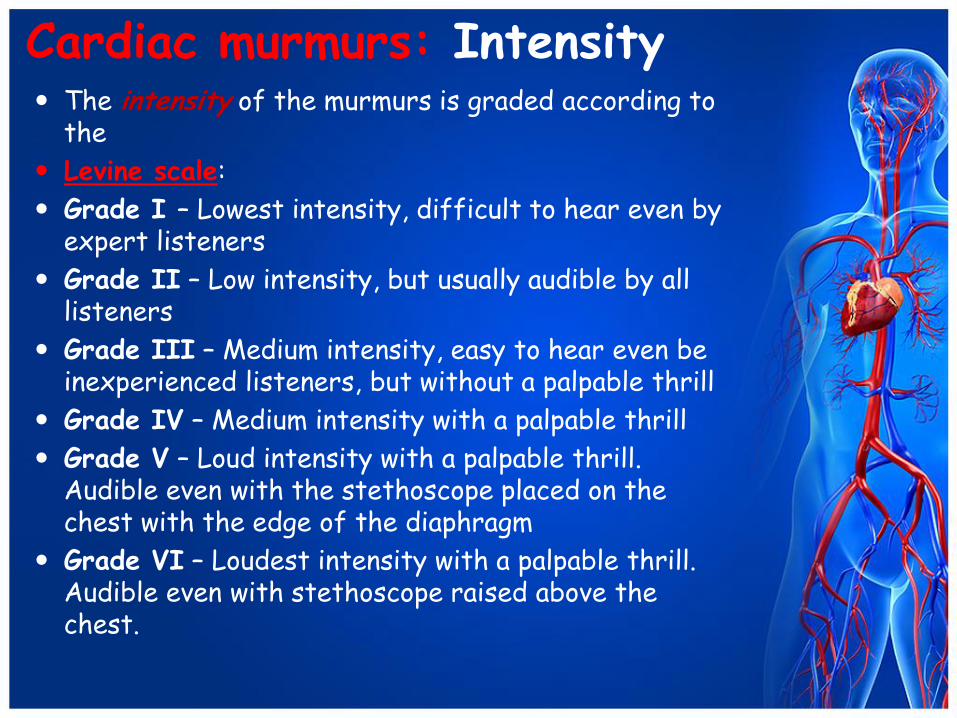
Cardiac murmurs: Intensity The intensity of the murmurs is graded according to
the
Levine scale:
Grade I – Lowest intensity, difficult to hear even by expert listeners
Grade II – Low intensity, but usually audible by all listeners
Grade III – Medium intensity, easy to hear even be inexperienced listeners, but without a palpable thrill
Grade IV – Medium intensity with a palpable thrill
Grade V – Loud intensity with a palpable thrill. Audible even with the stethoscope placed on the chest with the edge of the diaphragm
Grade VI – Loudest intensity with a palpable thrill. Audible even with stethoscope raised above the chest.
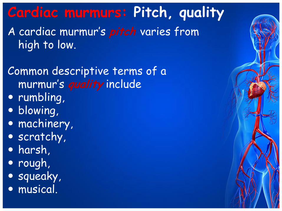
Cardiac murmurs: Pitch, quality A cardiac murmur’s pitch varies from
high to low.
Common descriptive terms of a murmur’s quality include
rumbling, blowing, machinery, scratchy, harsh, rough, squeaky, musical.
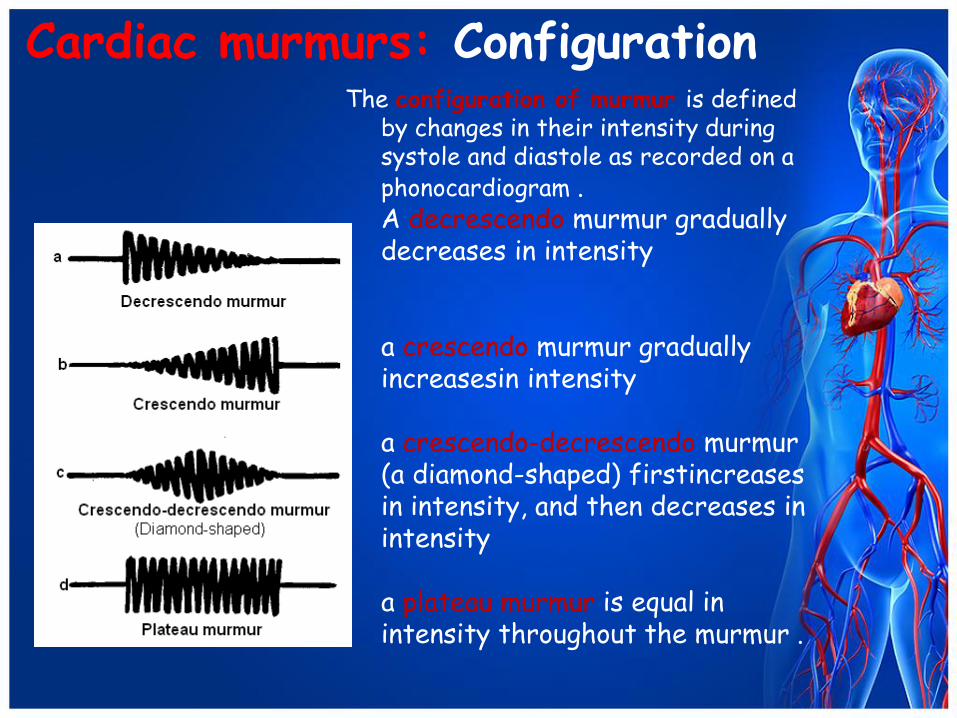
Cardiac murmurs: Configuration The configuration of murmur is defined
by changes in their intensity during systole and diastole as recorded on a phonocardiogram . A decrescendo murmur gradually decreases in intensity a crescendo murmur gradually increasesin intensity a crescendo-decrescendo murmur (a diamond-shaped) firstincreases in intensity, and then decreases in intensity a plateau murmur is equal in intensity throughout the murmur .

Cardiac murmurs: Duration
• A murmur’s duration can be of different length
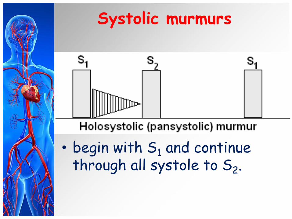
Systolic murmurs
• begin with S1 and continue through all systole to S2.
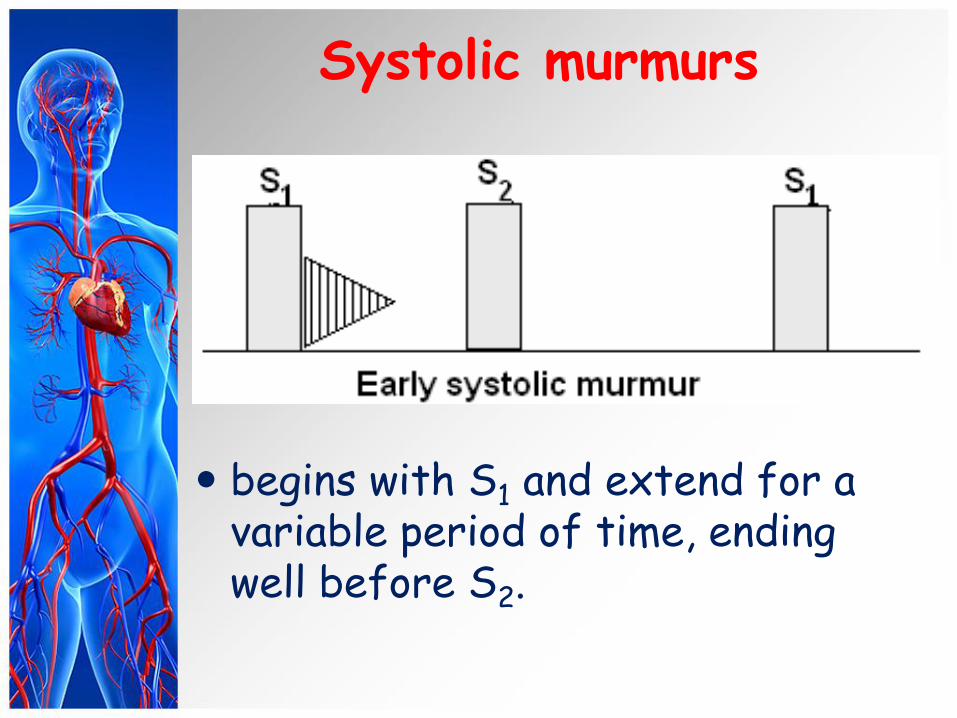
Systolic murmurs
begins with S1 and extend for a variable period of time, ending well before S2.
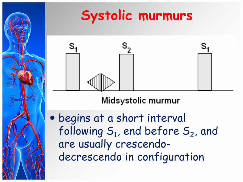
Systolic murmurs
begins at a short interval following S1, end before S2, and are usually crescendo-decrescendo in configuration

Systolic murmurs
• begins well after the onset of ejection that is at the end of systole.
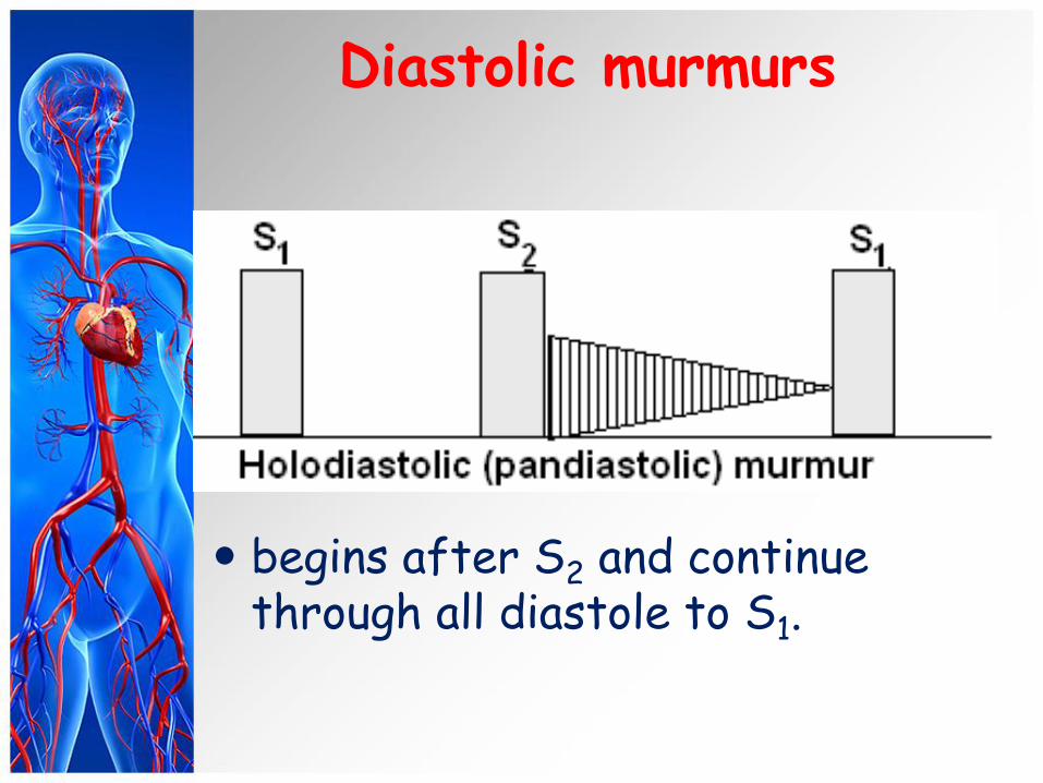
Diastolic murmurs
begins after S2 and continue through all diastole to S1.
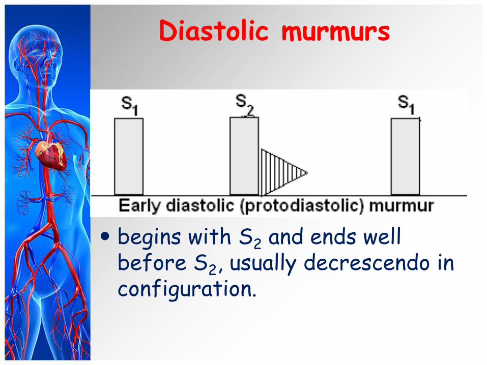
Diastolic murmurs
begins with S2 and ends well before S2, usually decrescendo in configuration.
Diastolic murmurs
• begins at a short interval following S2, end before S2
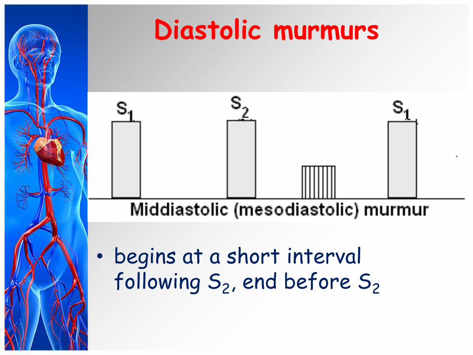
Diastolic murmurs
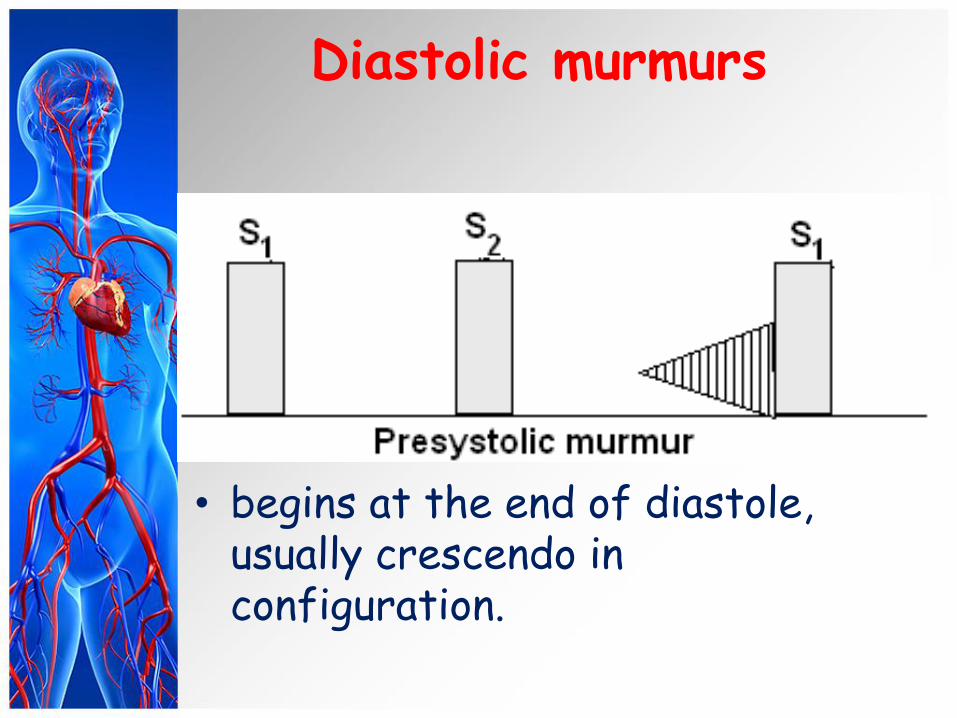
• begins at the end of diastole, usually crescendo in configuration.

Cardiac murmurs: Location
Cardiac murmurs may not be audible over all areas of the chest, and it is important to note where it is heard best and where it radiate to.
The location on the chest wall where the murmur is best heard and the areas to which it radiates can be helpful in identifying the cardiac structure from which the murmur originates.
Best auscultatory areas of a cardiac
murmurs. Topographic classification of murmurs.
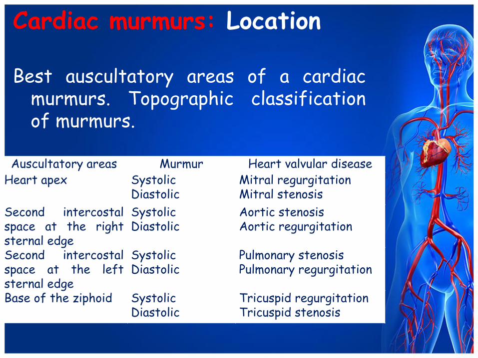
Cardiac murmurs: Location
Best auscultatory areas of a cardiac murmurs. Topographic classification of murmurs.
Auscultatory areas Murmur Heart valvular disease
Heart apex Systolic Diastolic
Mitral regurgitation Mitral stenosis
Second intercostal space at the right sternal edge
Systolic Diastolic
Aortic stenosis Aortic regurgitation
Second intercostal space at the left sternal edge
Systolic Diastolic
Pulmonary stenosis Pulmonary regurgitation
Base of the ziphoid Systolic Diastolic
Tricuspid regurgitation Tricuspid stenosis
Cardiac murmurs: Radiation
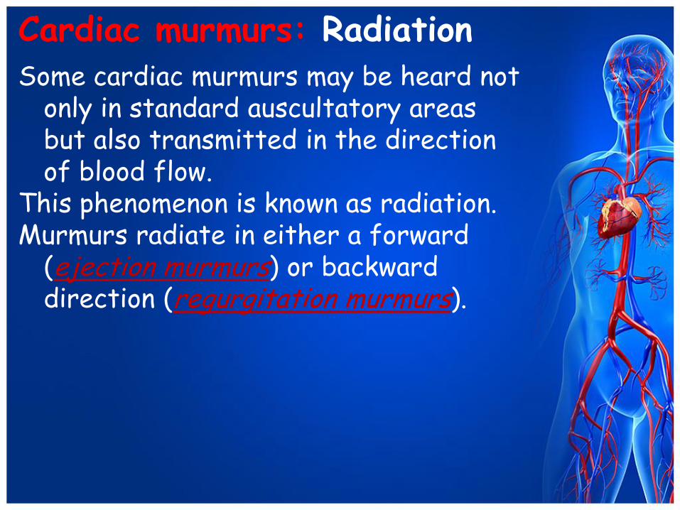
Some cardiac murmurs may be heard not only in standard auscultatory areas but also transmitted in the direction of blood flow.
This phenomenon is known as radiation. Murmurs radiate in either a forward
(ejection murmurs) or backward direction (regurgitation murmurs).
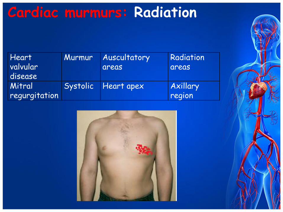
Cardiac murmurs: Radiation
Heart valvular disease
Murmur Auscultatory areas
Radiation areas
Mitral regurgitation
Systolic Heart apex Axillary region
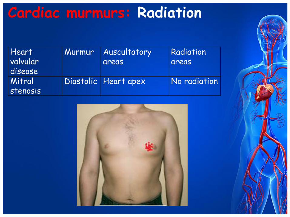
Cardiac murmurs: Radiation
Heart valvular disease
Murmur Auscultatory areas
Radiation areas
Mitral stenosis
Diastolic Heart apex No radiation
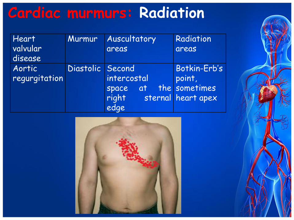
Cardiac murmurs: Radiation
Heart valvular disease
Murmur Auscultatory areas
Radiation areas
Aortic regurgitation
Diastolic Second intercostal space at the right sternal edge
Botkin-Erb’s point, sometimes heart apex
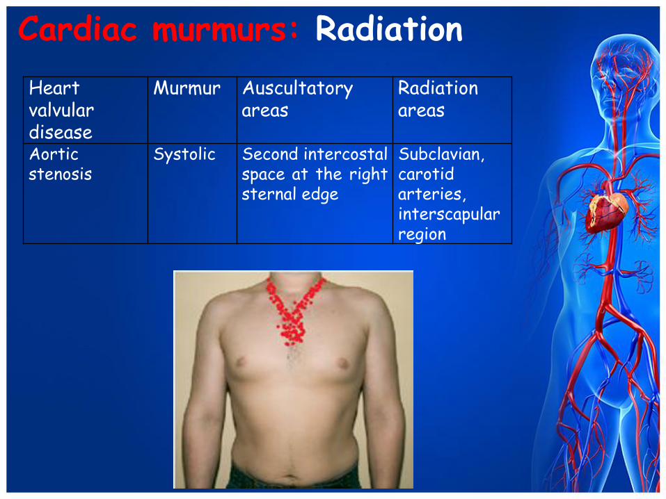
Cardiac murmurs: Radiation
Heart valvular disease
Murmur Auscultatory areas
Radiation areas
Aortic stenosis
Systolic Second intercostal space at the right sternal edge
Subclavian, carotid arteries, interscapular region

Aortic stenosis One of the most frequent pathologic systolic murmurs
is due to aortic stenosis. The murmur of aortic stenosis heard best over
“aortic area”, second intercostal space along right sternal border, with radiation into the neck, along carotid arteries, into the interscapular region (ejection murmur).
SYSTOLIC MURMURS
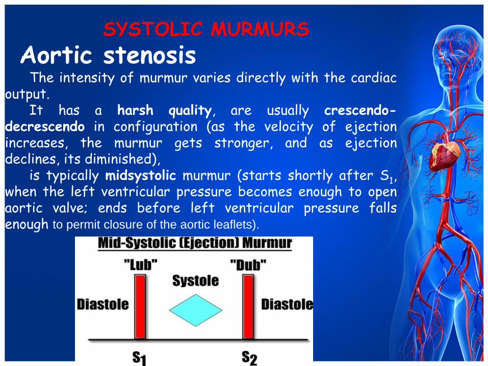
Aortic stenosis The intensity of murmur varies directly with the cardiac
output. It has a harsh quality, are usually crescendo-
decrescendo in configuration (as the velocity of ejection increases, the murmur gets stronger, and as ejection declines, its diminished),
is typically midsystolic murmur (starts shortly after S1, when the left ventricular pressure becomes enough to open aortic valve; ends before left ventricular pressure falls enough to permit closure of the aortic leaflets).
SYSTOLIC MURMURS
SYSTOLIC MURMURS
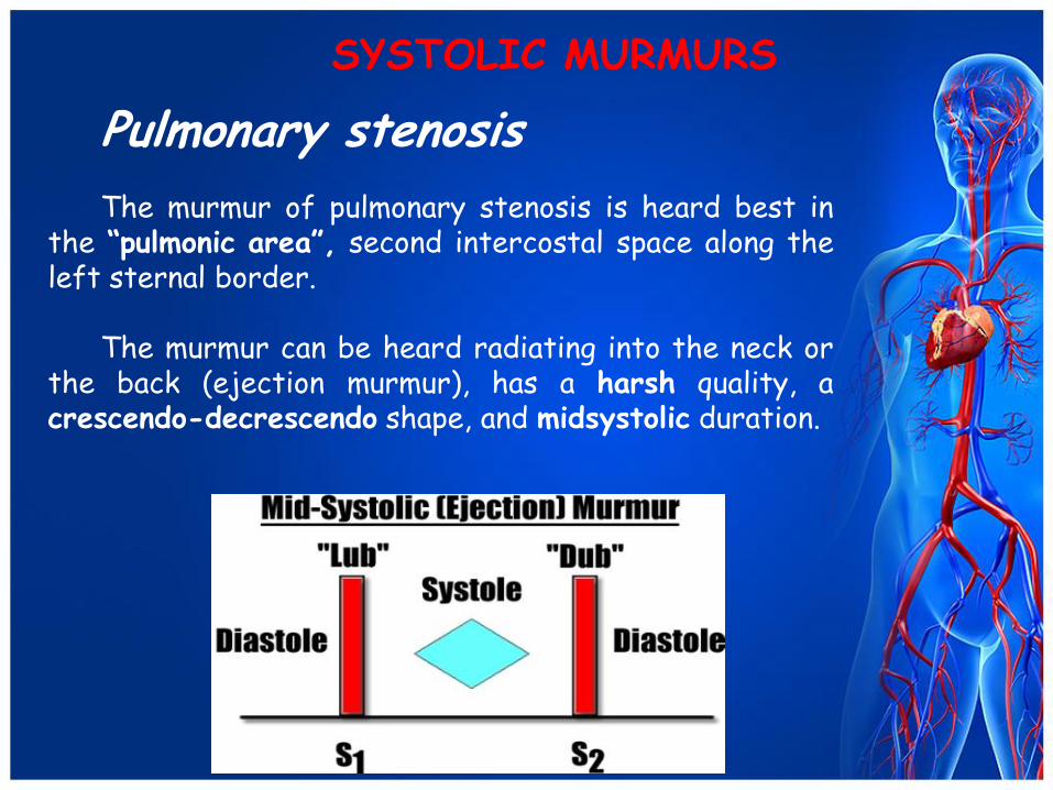
Pulmonary stenosis The murmur of pulmonary stenosis is heard best in
the “pulmonic area”, second intercostal space along the left sternal border.
The murmur can be heard radiating into the neck or
the back (ejection murmur), has a harsh quality, a crescendo-decrescendo shape, and midsystolic duration.
SYSTOLIC MURMURS
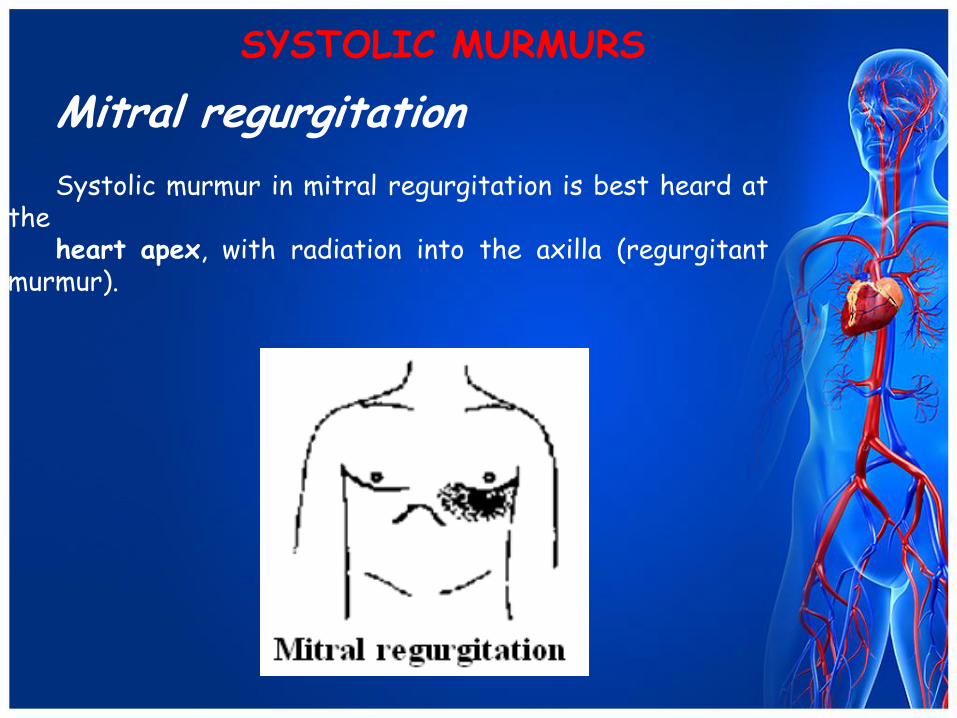
Mitral regurgitation Systolic murmur in mitral regurgitation is best heard at
the heart apex, with radiation into the axilla (regurgitant
murmur).

SYSTOLIC MURMURS
Mitral regurgitation The quality of murmur is usually described as
blowing, frequency – as high-pitched, the configuration of murmur may vary considerably, and its duration is holosystolic.
SYSTOLIC MURMURS
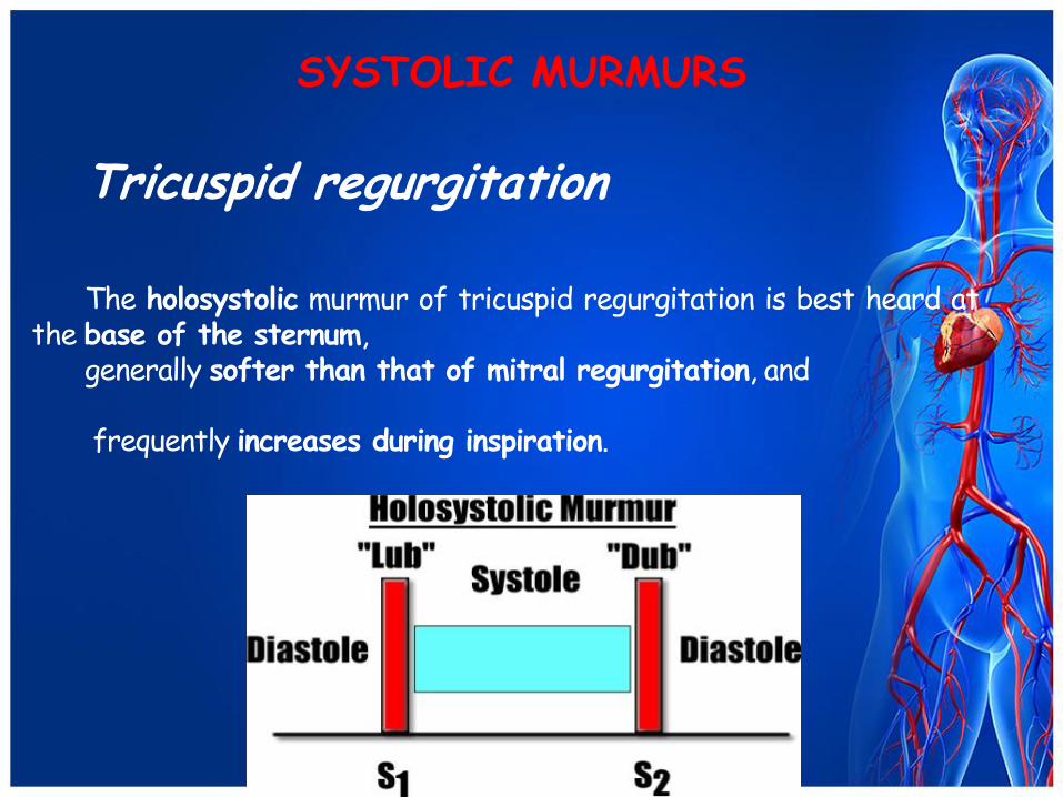
Tricuspid regurgitation The holosystolic murmur of tricuspid regurgitation is best heard at
the base of the sternum, generally softer than that of mitral regurgitation, and frequently increases during inspiration.
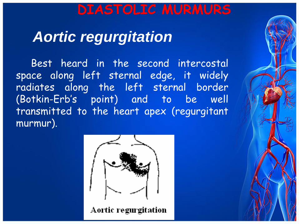
Aortic regurgitation Best heard in the second intercostal
space along left sternal edge, it widely radiates along the left sternal border (Botkin-Erb’s point) and to be well transmitted to the heart apex (regurgitant murmur).
DIASTOLIC MURMURS
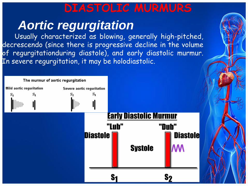
Aortic regurgitation Usually characterized as blowing, generally high-pitched,
decrescendo (since there is progressive decline in the volume of regurgitationduring diastole), and early diastolic murmur. In severe regurgitation, it may be holodiastolic.
DIASTOLIC MURMURS
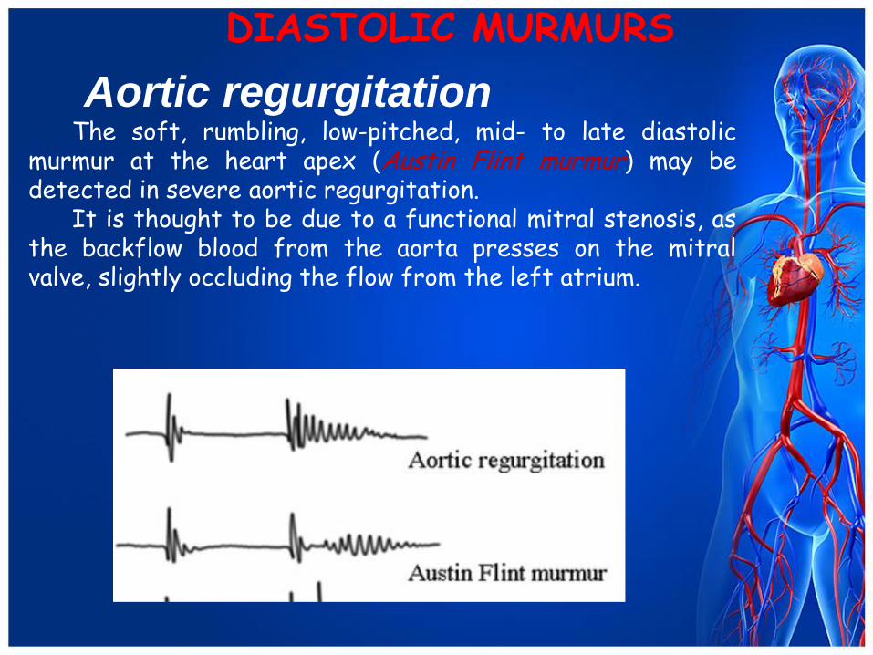
Aortic regurgitation The soft, rumbling, low-pitched, mid- to late diastolic
murmur at the heart apex (Austin Flint murmur) may be detected in severe aortic regurgitation.
It is thought to be due to a functional mitral stenosis, as the backflow blood from the aorta presses on the mitral valve, slightly occluding the flow from the left atrium.
DIASTOLIC MURMURS
DIASTOLIC MURMURS
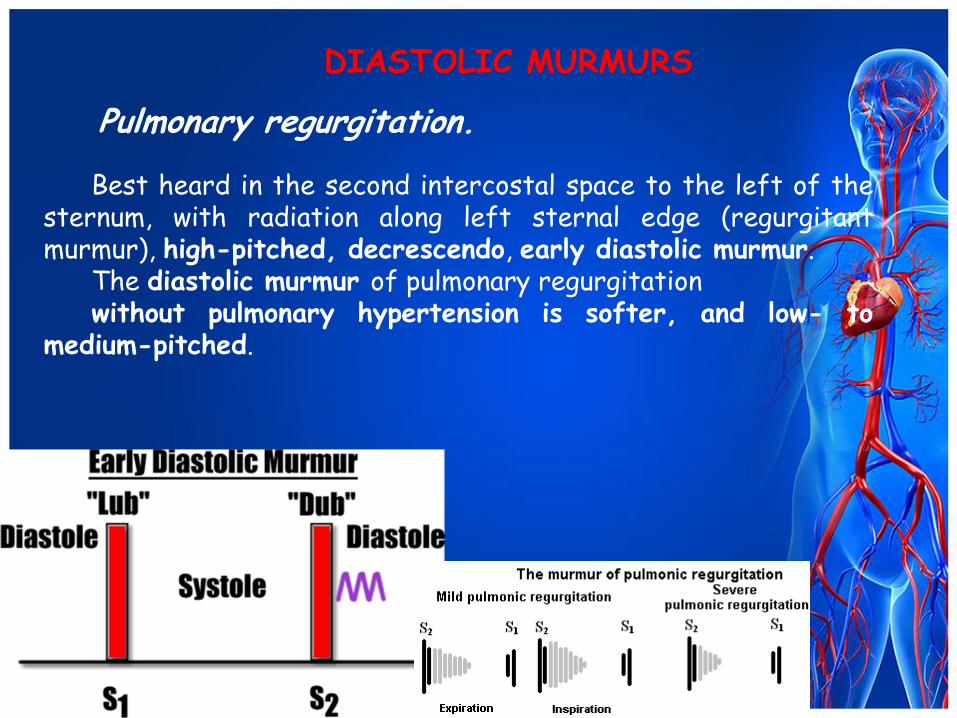
Pulmonary regurgitation. Best heard in the second intercostal space to the left of the
sternum, with radiation along left sternal edge (regurgitant murmur), high-pitched, decrescendo, early diastolic murmur.
The diastolic murmur of pulmonary regurgitation without pulmonary hypertension is softer, and low- to
medium-pitched.
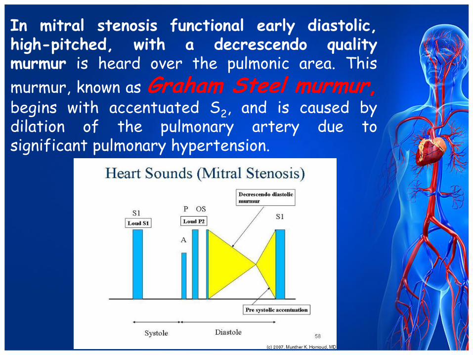
In mitral stenosis functional early diastolic, high-pitched, with a decrescendo quality murmur is heard over the pulmonic area. This
murmur, known as Graham Steel murmur, begins with accentuated S2, and is caused by dilation of the pulmonary artery due to significant pulmonary hypertension.
DIASTOLIC MURMURS
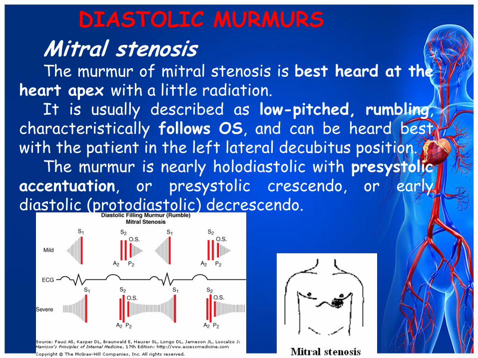
Mitral stenosis The murmur of mitral stenosis is best heard at the
heart apex with a little radiation. It is usually described as low-pitched, rumbling,
characteristically follows OS, and can be heard best with the patient in the left lateral decubitus position.
The murmur is nearly holodiastolic with presystolic accentuation, or presystolic crescendo, or early diastolic (protodiastolic) decrescendo.
DIASTOLIC MURMURS
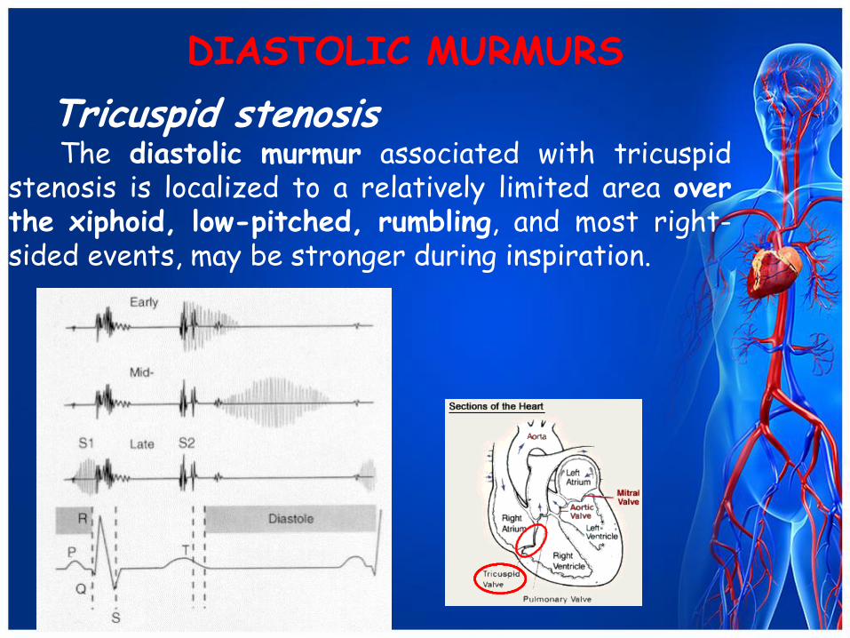
Tricuspid stenosis The diastolic murmur associated with tricuspid
stenosis is localized to a relatively limited area over the xiphoid, low-pitched, rumbling, and most right-sided events, may be stronger during inspiration.