chronic copper poisoning in sheep / i.b. boughton...
TRANSCRIPT

LIBRARY,
A 8: M COLLEGE.
CAMPUS.
E-109-8M-L180
TEXAS AITRICULTURAL EXPERIMENT STATION
A. B. CONNER, DIRECTOR
commzcrs STATION: BRAZOS COUNTY. TEXAS
BULLETIN NO. 499 " DECEMBER, 1934
DIVISION OF VETERINARY SCIENCE
CHRONIC COPPER POISONING
IN SHEEP
. '- 4 B R A R Y
Agrlcullural & Mechanical College o! Texas
AGRICULTURAL AND MECHANICAL COLLEGE OF TEXAS
T. O. WALTON, President
Genera
ted f
or
mem
ber
(Texas
A&
am
p;M
Univ
ers
ity)
on 2
01
5-0
8-0
7 2
1:5
8 G
MT /
htt
p:/
/hdl.handle
.net/
20
27
/txa.t
arb
00
42
78
Public
Dom
ain
/
htt
p:/
/ww
w.h
ath
itru
st.o
rg/a
ccess
_use
#p
d

Administration : -< - Veterinary Science:
A. B. Conner, M. S., Director ‘M. Francis, D. V. M., Chief
R. E. Karper, M. S., Vice Director H. Schmidt, D. V. M., Veterinarian
Clarice Mixson, B. A., Secretary "F. P. Mathews, D. V. M., M. S., Veterin "
M. P. Holleman, Chief Clerk J. B. Mims, D. V. M., Asst. Veterinarian ,
J. K. Francklow, Asst. Chief Clerk Plant Pathology and Physiology: r"
Chester Higgs, Executive Assistant J. J. Taubenhaus, Ph. D., Chief .._"
Howard Berry, B. S., Technical Asst. W. N. Ezekiel, Ph. D., Plant Pathologist -.
Chemistry: L. B, Loring, M. S., Asst. Plant Patholo i.
G. S. Fraps, Ph. D., Chief; State Chemist G. E. Altstatt, M. S., Asst. Plant Patholo’
S. E. Asbury, M. S., Chemist "Glenn Boyd, B. S., Asst. Plant Pathologist f
J. F. Fudge, Ph. D., Chemist Farm and Ranch Economics: ,:
E. C. Carlyle, M. S., Asst. Chemist L. P. Gabbard, M. S., Chief i
T. L. Ogier, B. S., Asst. Chemist W. E. Paulson, Ph. D., Marketing
A. J. Sterges, M. S., Asst. Chemist C. A. Bonnen, M. S., Farm Management §
Ray Treichler, M. S., Asst. Chemist 1**W. R. Nisbet, B. S., Ranch Management
W. H. Walker, Asst. Chemist "A. C. Magee, M. S., Ranch Management
Velma Graham, Asst. Chemist Rural Home Research:
Jeanne F. DeMottier, Asst. Chemist Jessie Whitacre, Ph. D., Chief
W, E. Merrill, M. S., Asst. Chemist Mary Anna. Grimes, M. S., Textiles
W. H. Garman, M. S., Asst Chemist Sylvia Cover, Ph. D., Foods '
A. R. Kemmerer, Ph. D., Asst. Chemist Soil Survey:
A. W. Walde, Ph. D., Asst. Chemist "W. T. Carter, B. S., Chief
Horticulture: E. H. Templin, B. S., Soil Surveyor
S- H- Yflrflell. $0- D» Chief J. W. Huckabee, B. S., Soil Surveyor
Range Animal Husbandry: I. C. Mowery, B. S., Soil Surveyor
J. M. Jones, A. M. Chief Botany:
B. L. Warwick, Ph. D., Breeding Investiga. V. L. Cory, M. S., Acting Chief
S. P Davis, Wool and Mohair Swine Husbandry:
J. H. Jones, B. S., Animal Husbandman Fred Hale, M. S., Chief
Entomology: Dairy Husbandry:
F. L. Thomas, Ph. D., Chief; State O. C. Copeland, M. S., Dairy Husbandman a
Entomologist Poultry Husbandry:
H. J. Reinhard, B. S., Entomologist R. M. Sherwood, M. S., Chief
R. K. Fletcher, Ph. D., Entomologist J. R. Couch, M. S., Assoc. Poultry Husb.
W. L. Owen, Jr., M. S., Entomologist Paul D. Sturkie, B. S., Asst. Poultry Hush.
J. N. Roney, M. S., Entomologist Agricultural Engineering:
J. C. Gaines, Jr., M. S., Entomologist H, P, Smith, M, 3,, Chief
S. E. Jones, M. S., Entomologist Main Station Farm:
F- F- Bibby, B- S“ EIItOIHOIOSZiSt G .T. McNess, Superintendent
"R. W. Moreland, B. S., Asst.-Entomologist Apiculture (San Antonio):
C. E. Heard, B. S., Chief Inspector H. B. Parks, B. S., Chief
C. J. Burgin, B. S., Foulbrood Inspector A. H. Alex, B. S., Queen Breeder
Agronomy: Feed Control Service:
E. B. Reynolds, Ph. D., Chief F. D. Fuller, M. S., Chief
R. E. Karper, M. S., Agronomist James Sullivan, Asst. Chief.
P. C. Mangelsdorf, Sc. D., Agronomist S. D. Pearce, Secretary
D. T. Killough, M. S., Agronomist J. H. Rogers, Feed Inspector
J. T. Vantine, Jr., M. S., Asst. Agronomist K. L. Kirkland, B. S., Feed Inspector
J- 0- Beasley, M- 3-, ABSL Agronomist S. D. Reynolds, Jr., Feed Inspector
Publications: P. A. Moore, Feed Inspector
A. D. Jackson, Chief E. J. Wilson, B. S., Feed Inspector
H. G. Wickes, D. V. M., Feed Inspector
SUBSTATIO ,_ ~,‘;,-.;,~
No. 1, Beeville, Bee County: ' m‘! 9, 'Balmorhea, Reeves County:
R. A. Hall, B. S., Superintendent J. J. Bayles, B. S., Superintendent
No. 2, Tyler, Smith County: No. 10, College Station, Brazos County;
P. R. Johnson, M. S., Superintendent R. M. Sherwood, M. S., In Charge
"B. H. Hendrickson, B. S., Sci. in Soil Erosion L. J. McCall, Farm Superintendent
"R. W. Baird, M. S., Assoc. Agr. Engineer No. ll, Nacogdoches, Nacogdoches County:
No. 3, Angleton, Brazoria County: H. F. Morris, M. S. Superintendent
R. H. Stansel, M. S., Superintendent "No. 12, Chillicothe, Hardeman County:
H. M. Reed, B. S., Horticulturist **J. R. Quinby, M. S. Superintendent
No. 4, Jefferson County: "J. C. Stephens, M. A., Asst. Agronomist
R. H. Wyche, B. S., Superintendent No. l4, Sonora, Sutton-Edwards Counties:
"H. M. Beachell, B. S., Junior Agronomist W. H. Dameron, B. S., Superintendent
No. 5, Temple, Bell County: I. B. Boughton, D. V. M., Veterinarian
Henry Dunlavy, M. S., Superintendent W. T. Hardy, D. V. M., Veterinarian
C. H. Rogers, Ph. D., Plant Pathologist O. L. Carpenter, Shepherd
H. E. Rea, B. S., Agronomist "O. -G. Babcock, B. S., Asst. Entomologist
"E. B. Deeter, B. S., Soil Erosion No. 15, Weslaco, Hidalgo County:
"P. L. HODRiIIS. B. S., Junior Civil Engineer W. H. Friend, B. S., Superintendent
No. 6 Denton, Denton County: S. W. Clark, B. S., Entomologist
P. B- Dlmkle. M. S., Superintendent W. J. Bach, M. S., Plant Pathologist
"I. M. Atkins, B. S., Junior Agronomist J. F. Wood, B. S., Horticulturist
No. 7. Spur, Dickens County: No. l6, Iowa Park, Wichita County:
R. E. Dickson, B. S., Superintendent C. H. McDowell, B. S., Superintendent
B. C. Langley, M. S., Agronomist L. E. Brooks, B. S., Horticulturist
N0. 8, Lubbock, Lubbock County: No. 19, Winterhaven, Dimmit County:
D. L. Jones, Superintendent . E. Mortensen, B. S., Superintendent
Frank Gaines, Irrig. and Forest Nurs. "L. R. Hawthorn, M. S., Horticulturist
Members of Teaching Staff Carrying Cooperative Projects on the Station:
G. W. Adriance, Ph. D., Horticulture W. R. Horlacher, Ph. D., Genetics
S. W. Bilsing, Ph. D., Entomology J. H. Knox, M. S., Animal Husbandry
D. Scoates, A. E., Agricultural Engineering A. L. Darnell, M. A., Dairy Husbandry
A. K. Mackey, M. S., Animal Husbandry R. 0. Berry, B. S., Biology
R. G. Reeves, Ph. D., Biology ’ R. T. Stewart, Ph.D., Agronomy
J. S. Mogford, M. S., Agronomy V. A. Little, M. S., Entomology
F. R. Brison, M. S., Horticulture
‘Dean, School of Veterinary Medicine. TAs of December, 1934
"In cooperation with U. S. Department of Agriculture.
IIn cooperation with Texas Extension Service.
“In woperatiou with State Department of Agriculture.
Genera
ted f
or
mem
ber
(Texas
A&
am
p;M
Univ
ers
ity)
on 2
01
5-0
8-0
7 2
1:5
8 G
MT /
htt
p:/
/hdl.handle
.net/
20
27
/txa.t
arb
00
42
78
Public
Dom
ain
/
htt
p:/
/ww
w.h
ath
itru
st.o
rg/a
ccess
_use
#p
d

The long-continued ingestion of salt mixtures containing rela-
tively small percentages of copper sulphate caused chronic copper
poisoning among flocks of range sheep on several West Texas
ranches during the past year. The salt licks were placed before
the sheep as a means of preventing or controlling stomach worm
infestation despite a‘ lack of experimental or field evidence that
they were of any practical value in this regard. Investigations
on seven ranches where the disease occurred revealed practically
the same history in each case; the animals had been licking a
medicated salt for periods varying from 7 months to several years.
Sheep of all ages were affected and the losses were exceedingly
heavy on all but one ranch.
Chronic copper poisoning in sheep is characterized by a yellow
discoloration of the tissues and brown to black urine, loss of ap-
petite, and weakness. Once the symptoms appear, the disease
runs a rapidcourse and terminates fatally in the great majority
of cases. Probably many sheep suffer from a subclinical type of
the disease which is manifested principally by loss of condition.
The disease, which has been called icterohemoglobinuria during
the past few years, was reproduced experimentally in 12 healthy
sheep by feeding the commercial salt mixtures concerned. In
some experiments measured amounts of the mixture were admin-
istered daily in gelatine capsule, while in others a definite amount
was mixed with a handful of cottonseed meal every day and placed
in the feed box. (In addition the disease was experimentally re-
produced in 14 healthy sheep by feeding other salt mixtures con-
taining copper sulphate). Analysis of the livers of both field and
experimental cases of the disease showed definitely toxic amounts
of copper to be present in these animals.‘
An experiment showed that there is no danger of producing
t-he chronic copper poisoning by the routine drenching of sheep
to control stomach worm infestation.
Predisposing factors were dry, short range, overstocking, lack
of condition in many sheep, and frequent “working” of the
animals. There seems to be a definite seasonal variation in the
condition, the most severe outbreaks occurring during the spring
and fall months.
There is no treatment for the disease. It can be prevented by
removing the cause. Losses may continue for at least five months
after feeding of the salt mixture is discontinued.
\
Genera
ted f
or
mem
ber
(Texas
A&
am
p;M
Univ
ers
ity)
on 2
01
5-0
8-0
7 2
1:5
8 G
MT /
htt
p:/
/hdl.handle
.net/
20
27
/txa.t
arb
00
42
78
Public
Dom
ain
/
htt
p:/
/ww
w.h
ath
itru
st.o
rg/a
ccess
_use
#p
d

CONTENTS
Introduction
Review of Literature
Field Observations of Chronic Copper Poisoning
Experimental Investigations
Transmission Tests
Copper Sulphate Feeding Tests--_--
- Administered in Gelatine Capsules-
Administered in Feed
Fed Free Choice in Troughs
Administered as a Drench
Symptoms and Pathology
Treatment
Discussion
Summary and Conclusions
Literature Cited
Genera
ted f
or
mem
ber
(Texas
A&
am
p;M
Univ
ers
ity)
on 2
01
5-0
8-0
7 2
1:5
8 G
MT /
htt
p:/
/hdl.handle
.net/
20
27
/txa.t
arb
00
42
78
Public
Dom
ain
/
htt
p:/
/ww
w.h
ath
itru
st.o
rg/a
ccess
_use
#p
d

BULLETIN NO. 499 DECEMBER, 1934‘
CHRONIC COPPER POISONING IN SHEEP
I. B. BOUGHTON AND W. T. HARDY
A trouble occurring among sheep on numerous ranches in the Edwards
plateau region of Texas characterized clinically by generalized icterus,
hemoglobinuria and hematuria, inappetence, and extreme Weakness, was
found to be chronic copper poisoning. There is an extremely rapid, irregu-
lar pulse, accompanied by shallow, hurried respirations in practically all
cases but the temperature seldom, if ever, rises above normal. The
disease ordinarily terminates fatally within 24 to 48 hours after the first
"appearance of the characteristic symptoms but occasionally affected
animals linger for considerably longer periods of time. Spontaneous re-
covery is comparatively rare but does occur. The notable lesions at
autopsy are a yellowish, friable liver; enlarged, very dark brown to black
kidneys; a swollen, “blackberry jam” spleen; generalized icterus; poorly-
collapsed, doughy lungs, and a pale flaccid heart.
As a matter of fact the condition is really a cumulative poisoning
since the affected animals have ingested small doses of the material
containing copper sulphate for weeks or months before there are any
clinical manifestations of the disease. Once symptoms appear, the course
of the disease usually becomes acute.
During the past several months we have investigated seven different
outbreaks of chronic copper poisoning among range sheep caused by the
long-continued ingestion of salt mixtures containing varying amounts of
copper sulphate in addition to tobacco dust and sodium chloride.
Such salt licks are sold in many parts of the West Texas sheep country
as a preventive of stomach worm infestation despite the lack ‘of ex-
perimental and field evidence that such mixtures are of any practical
value in this regard. Analysis of two of these commercial mixtures fed
on ranches where the disease occurred showed powdered copper sulphate
(CuSOhSHQO) in amounts varying from 5.3% to 9.992? There is little
question that some of the commercial mixtures contain even larger per-
centages of this copper salt.
REVIEW OF THE LITERATURE
Ellenberger & Hofmeister (1) and Baum & Seeliger (2), in the latter
part of the past century, proved definitely that chronic copper poisoning,
following the continued ingestion of small amounts of various copper
salts, is a distinct clinical entity in domestic animals. Ellenberger &
Hofmeister (1) describe the typical symptoms and lesions. Their work
included studies of the elimination of the ingested copper from the
body. This metal disappeared from the urine a few days after feeding
of the copper salt was discontinued but small amounts of copper oxide
continued to be excreted daily with the feces for weeks or months. They
*Al1 copper analyses reported in this paper were made in the laboratory of the Division
of Chemistry, Texas Agricultural Experiment Station.
Genera
ted f
or
mem
ber
(Texas
A&
am
p;M
Univ
ers
ity)
on 2
01
5-0
8-0
7 2
1:5
8 G
MT /
htt
p:/
/hdl.handle
.net/
20
27
/txa.t
arb
00
42
78
Public
Dom
ain
/
htt
p:/
/ww
w.h
ath
itru
st.o
rg/a
ccess
_use
#p
d

6 BULLETIN NO. 499, TEXAS AGRICULTURAL EXPERIMENT STATION
advanced the theory that part of the fecal copper was reabsorbed in
the lower part of the intestines, such prolonged reabsorption having a
deleterious effect on the liver and eventuating in chronic copper poisoning.
Our observations confirm this idea since We have noted in the out-
breaks studied that deaths from this condition occur for as long as five
months after the feeding of the salt mixtures containing copper sulphate
was discontinued. The authors mentioned above found copper oxide in
all the tissues of the poisoned animals, the largest amount (.175%) in
the liver and the smallest (.0058%) in the muscles. It should be noted
that the largest amount of copper oxide found in the liver, namely, 0.175%
was found in a sheep that developed acute copper poisoning five and
one-half weeks after the feeding of copper sulphate was discontinued.
Baum & Seeliger (2) in their experiments fed smaller amounts of
copper salts than Ellenberger and Hofmeister (1) daily to 15 sheep for
periods of time varying from about 50 days to several months. Analyses
of the livers of these animals showed a copper content varying from
0.0044% to 0.0525%. According to these investigators the oleate is the
most poisonous copper salt with the sulphate following very closely. In
their experiments cats, sheep, goats, and dogs are the most susceptible
to poisoning in the order named. These authors found icterus and hemo-
globinuria to be constantly present in sheep but failed to observe either
in their work with goats.
In 1927 Schaper and Luetje (3) observed a fatal disease among sheep
characterized by depression, generalized icterus, hematuria, and hemo-
globinuria, stormy pulse, increased respirations, edema and eczema of the
ears. These animals were grazing in orchards where the trees had been
sprayed with Bordeaux mixture two months previously. Autopsies on
nine affected animals revealed lesions similar to those mentioned by the
present writers except that the enlarged spleens were rose-red in color
and firm in consistency. The losses stopped before the cause had been
determined. '
During the following year (1928) Schaper and Luetje (3) investigated’
a similar outbreak among sheep grazing in the same orchards which pad
been sprayed three months previously with Bordeaux mixture. During
this investigation some 267 affected sheep were autopsied and the diag-
nosis of chronic copper poisoning was made. Traces of lead and arsenic
were found during analyses of the livers of some of the affected animals
but such quantities were always within physiological limits. Cattle graz-
ing in these orchards were not observed to be sick although several owners
stated that they had lost cattle manifesting symptoms similar to those
shown by affected sheep. Occasional attacks of diarrhea among horses
and colts were observed but no fatalities occurred.
The symptoms and lesions observed by Schaper and Luetje during this
_ last investigation agreed very well with those found the previous year.
In some of the autopsied animals retinal hemorrhages were seen, but this
lesion was never noted in the living animal.
Beijers (4) described chronic copper poisoning in a few sheep which
had also been grazing in orchards where the trees had been sprayed with
1 ufilQim~flJL.-sm'.....u. . ‘L\‘(
Genera
ted f
or
mem
ber
(Texas
A&
am
p;M
Univ
ers
ity)
on 2
01
5-0
8-0
7 2
1:5
8 G
MT /
htt
p:/
/hdl.handle
.net/
20
27
/txa.t
arb
00
42
78
Public
Dom
ain
/
htt
p:/
/ww
w.h
ath
itru
st.o
rg/a
ccess
_use
#p
d

CHRONIC COPPER POISONING IN SHEEP 7
Bordeaux mixture. He noted the rapid, fatal course of the disease once
clinical symptoms appeared, stating that the condition appeared only after
the sheep had been grazing in the orchards for several months. Symptoms
and lesions similar to those described by previous investigators were noted.
In addition, Beijers counted 3,190,000 red blood cells and 26,100 white
cells per cubic millimeter in a typical case. These cell counts agree very
closely with the results obtained by the present writers in both field and
experimental cases.
During parts of the years 1930-1932 a similar condition appeared among
sheep on this Station. Schmidt (5) and Schmidt and Hardy (6) investigated
this condition, describing the clinical symptoms and the post mortem lesions
very thoroughly but did not establish the cause. They were unsuccessful
in transmitting the disease to healthy sheep either by contact or through
injections of spleen tissue emulsions or blood from typical cases. No blood
parasites were found. Schmidt (5) noted the extreme red-cell destruction
and remarked upon the difficulty of satisfactory examination of blood
smears because of the agglomerations of debris resulting from the cell
destruction. Schmidt (5) named the condition icterohemoglobinuria inas-
much as the outstanding symptoms were the generalized icterus and the
pronounced hemoglobinuria. It later became known to Schmidt and Hardy
(6) that all of these sheep had had free access to a salt mixture consisting
of copper sulphate 15%, tobacco dust 25%, sodium chloride 60%, from
May, 1929, until September, 1931.
Olafson (7) under the title of icterohemoglobinuria, described a con-
dition occurring in two bands of sheep in New York State, which, from the
symptoms, lesions, and blood changes found, seems to be analogous to the
subject under discussion. He was unsuccessful in transmitting the disease
and did not establish the etiology. In a personal communication, Olafson
states that he noted the scarcity of gastro-intestinal parasites in his
autopsies. When he remarked on this the owner stated that it was due
to the fact that these animals had had access to a salt mixture con-
taining copper sulphate.
The condition in South Africa which DeKock (8) studied in 1928 in one
or two bands of sheep brought from the Karroo district to Onderstepoort
for blue tongue vaccine production and which he called enzootic icterus,
is exceedingly similar to the subject under discussion. DeKock noted the
remarkable distribution of the disease and the fact that! it did not spread
to any of the healthy sheep even after prolonged contact. The symptoms
a.nd lesions described by this author agree very well with those we observed.
He noted the presence of many “ghost” erythrocytes in blood smears
and described a “pigment” cell which he found in the various tissues of
typical cases. Olafson ('7) mentions this same type of cell. DeKock, in
his summary, suggests the dietetic origin of the condition. The present
writers know nothing about the diet of the sheep which DeKock observed,
but the extreme similarity of the symptoms and lesions suggest the pos-
sibility that this author was dealing with chronic copper poisoning.
Newsom and Cross (9) in 1921 reported several outbreaks of ictero-
hematuria among bands of sheep in Colorado. The similarity of the
Genera
ted f
or
mem
ber
(Texas
A&
am
p;M
Univ
ers
ity)
on 2
01
5-0
8-0
7 2
1:5
8 G
MT /
htt
p:/
/hdl.handle
.net/
20
27
/txa.t
arb
00
42
78
Public
Dom
ain
/
htt
p:/
/ww
w.h
ath
itru
st.o
rg/a
ccess
_use
#p
d
8 BULLETIN NO. 499, TEXAS AGRICULTURAL EXPERIMENT STATION
symptoms and lesions described together with the fact that these authors
Were unable either to find any blood cell parasites or to transmit the
disease to healthy sheep suggests the possibility that they may have been
dealing with chronic copper poisoning. In 1931 the same authors (10) gave
a general survey bf the disease as it occurred in their state and remarked
that newly-introduced sheep seemed to be most susceptible.
a personal communication, states that they found no gastro-intestinal
parasites among autopsied animals.
Williams (11) and Johnson (12) (13) in Montana, reported an icterohema-
turia in sheep, both authors believing‘ that the condition was a true piro-
plasmosis (babesiellosis) which had been previously described from Roumania
by Babes (14). Neither of these authors was successful in transmitting
the disease to healthy sheep by injection of blood or tissue emulsions from
affected animals. They reported the presence of small blue-staining bodies
in the red cells of typical cases but it is worthy of note that, according
to Olafson (7), “Nuttall states the recorded presence of ovine piroplasmosis
in North America is due to errors of observation.” The symptoms and
lesions described by both William and Johnson coupled with the regional
nature of the outbreaks described is suggestive of chronic copper poisoning.
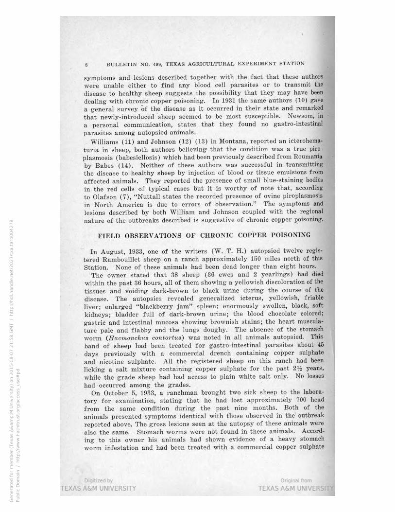
FIELD OBSERVATIONS OF CHRONIC COPPER POISONING
In August, 1933, one of the writers (W. T. H.) autopsied twelve regis-
tered Rambouillet sheep on a ranch approximately 150 miles north of this
Station. None of these animals had been dead longer than eight hours.
The owner stated that 38 sheep (36 ewes and 2 yearlings) had died
within the past 36 hours, all of them showing a yellowish discoloration of the
tissues and voiding dark-brown to black urine during the course of the
disease. The autopsies revealed generalized icterus, yellowish, friable
liver; enlarged “blackberry jam” spleen; enormously swollen, black, soft
kidneys; bladder full of dark-brown urine; the blood chocolate colored;
gastric and intestinal mucosa showing brownish stains; the heart muscula-
ture pale and flabby and the lungs doughy. The absence of the stomach
Worm (Haemonchus contortus) was noted in all animals autopsied. This
band of sheep had been treated for gastro-intestinal parasites about 45
days previously with a commercial drench containing copper sulphate
and nicotine sulphate. All the registered sheep on. this ranch had been
licking a salt mixture containing copper sulphate for the past 2% years,
while the grade sheep had had access to plain white salt only. N0 losses
had occurred among the grades.
On October 5, 1933, a ranchman brought two sick sheep to the labora-
tory for examination, stating that he had lost approximately 700 head
from the same condition during the past nine months. Both of the
animals presented symptoms identical with those observed in the outbreak
reported above. The gross lesions seen at the autopsy of these animals were
also the same. Stomach worms were not found in these animals. Accord-
ing to this owner his animals had shown evidence of a heavy stomach
worm infestation and had been treated with a commercial copper sulphate
..m;.....-i1 Missal‘ l;
Newsom, in -
Genera
ted f
or
mem
ber
(Texas
A&
am
p;M
Univ
ers
ity)
on 2
01
5-0
8-0
7 2
1:5
8 G
MT /
htt
p:/
/hdl.handle
.net/
20
27
/txa.t
arb
00
42
78
Public
Dom
ain
/
htt
p:/
/ww
w.h
ath
itru
st.o
rg/a
ccess
_use
#p
d

CHRONIC COPPER POISONING IN SHEEP 9
and Black Leaf 40 drench several times during the past year. This flock had
been licking a medicated salt mixture (the same type of mixture as that
mentioned in the first outbreak) for the past twelve months.
This man was accompanied by a neighbor from an adjoining ranch who
stated that he too had lost and was still losing sheep from the same
condition. The history of his flock, both as regards drenching and the
feeding of a similar medicated salt mixture, was about the same. His losses
started about the same time and he estimated that so far 500 sheep had
died from the same condition. In both of these outbreaks, by far the
largest loss had been among adult sheep, only a relatively few lambs
(6 months or more of age) having been affected.
The sick animal which was left at the laboratory showed extreme
paleness of visible mucous membranes, weakness, inappetence, and hemo-
globinuria. The temperature was normal (102.4° F.) but an erratic pulse
of 120 and hurried shallow respirations were noted. A count showed
only 4,384,000 red blood cells and 16,200 white blood cells per cubic milli-
meter. A differential count gave: neutrophiles 66.5%, lymphocytes 27.5%,
monocytes (large lymphocytes) 5.5%, and basophiles 0.5%. Examination of
stained blood slides showed marked anisocytosis, numerous “ghost” red
cells, punctate basophilia, and polychromasia. Much red cell debris on
every slide made detailed observation difficult. Thorough search of many
slides failed to reveal the presence of organisms either in or between the
cells. This animal died on the third day, presenting the same lesions as
those observed in the first outbreak reported above. Six stomach worms
(Haemonchus contortus) were found in the abomasum. A detailed search
for intestinal parasites was not made.
Chemical analyses of the livers from two typical field cases of the
disease in sheep occurring on the ranch from which the case just described
originated showed 14.7 milligrams (0.0147%) and 53 milligrams (0.053%)
of copper respectively in 100 grams of tissue (Sheep No. 771 and Sheep
No. 801 in Table 8).
Personal investigation on these two ranches revealed practically the
same range conditions as those obtaining on the first ranch visited
in August. The grass and weeds were very short and dry as a resultlof
the prolonged drouth. The sheep did little grazing, remaining around the
water tank much of the time; the salt troughs were all placed next to this
tank. It was observed in several instances that the sheep, after licking
the salt went immediately to the tank and drank. This observation had
also been made on the first ranch where chronic copper poisoning was
found. The ranchmen concerned had noted that their sheep stayed around
the Watering places much more than they usually did during seasonable
years. There is little doubt that the sheep were licking more salt than
they would during times of good range.
About ten days later, at the time of our second visit to the two ranches
mentioned above, a neighbor from an adjoining ranch said that he had
lost approximately 25 head of grown ewes and a good many lambs (about
7 months of age), all of which showed the same symptoms as those
described above. Autopsy of a typical case on this ranch revealed the
Genera
ted f
or
mem
ber
(Texas
A&
am
p;M
Univ
ers
ity)
on 2
01
5-0
8-0
7 2
1:5
8 G
MT /
htt
p:/
/hdl.handle
.net/
20
27
/txa.t
arb
00
42
78
Public
Dom
ain
/
htt
p:/
/ww
w.h
ath
itru
st.o
rg/a
ccess
_use
#p
d

10 BULLETIN NO. 499, TEXAS AGRICULTURAL EXPERIMENT STATION
characteristic lesions observed in the other outbreaks. Medicated salt
(a similar mixture to that used on the two adjoining ranches) had been
before his sheep for five months when his losses started.
Later, chemical analysis of the liver of a ewe from this ranch dead
from the typical condition showed 28.3 milligrams (0028392) of copper
in 100 grams of tissue (Sheep 802 in Table 7).
While the greatest loss Was among pregnant and suckling ewes many
fatal cases occurred in lambs (6-7 months old) and rams. The majority
of the animals died within 36-48 hours after symptoms were noted by the
ranchmen. Spontaneous recovery had been noted in a few cases.
Thorough search of several animals revealed the spinose ear tick,
Ornithodoros megvzi/n-i, as the only external parasite present.
Tests maturing on December 12, 1933, in which experimental feeding of
a salt mixture containing copper sulphate to healthy sheep produced symp-
toms and lesions indistinguishable from the field cases described in the
foregoing pages, definitely proved that the condition we were dealing
with was a chronic copper poisoning. Previous to this time we had called
the condition icterohemoglobinuria. With this information we investigated
on December 28, 1933, losses among sheep on another ranch located some
50 miles northwest of this Station and found precisely the same condi-
tion as that observed on the ranches previously mentioned. At the time
of our visit the owner stated that some 45 head of sheep had died within
the past two weeks.
Clinical symptoms and autopsy lesions in two typical cases were the same
as those observed in previous outbreaks. The absence of stomach worms
was again noted. Later, examination of stained blood slides showed
changes similar to those reported above. Analysis of the liver of a
typically-affected lamb (approximately 8 months old) showed 48.8 milli-
grams (0.0488%) of copper in 100 grams of tissue (Sheep No. 807 in
Table 7).
A commercial medicated salt mixture containing copper sulphate had
been constantly before the sheep on this ranch since the previous April
(8 months). On another ranch this man had been feeding plain white salt
only and had not lost any sheep from this condition. The condition of the
range on both ranches was very poor with the grass short, dry, and dead.
On January 4, 1934, the same condition was found on a ranch 25 miles
north of this Station, four ewes having died during the preceding months.
Autopsy of a clinically-sick ewe revealed the characteristic lesions and the
absence of gastric parasites. This ranchman had kept the medicated salt
mixture before his sheep almost continuously since 1927.
On February 7, 1934, another outbreak of the same condition was in-
vestigated on a ranch some 80 miles west of this Station. The history,
as regards the range condition, feeding of a commercial medicated salt
mixture, and animals lost were typical. Medicated salt had been in the
troughs for the past 2% years. The owner estimated a loss of 250 sheep
(mostly ewes) during the past six months. While we did not see any
affected sheep the symptoms and lesions described by the owner left no
doubt as to the true condition.
Genera
ted f
or
mem
ber
(Texas
A&
am
p;M
Univ
ers
ity)
on 2
01
5-0
8-0
7 2
1:5
8 G
MT /
htt
p:/
/hdl.handle
.net/
20
27
/txa.t
arb
00
42
78
Public
Dom
ain
/
htt
p:/
/ww
w.h
ath
itru
st.o
rg/a
ccess
_use
#p
d

CHRONIC COPPER POISONING IN SHEEP 11
The last outbreak of the condition investigated by us 0n February 8,
1934, occurred 0n a small ranch about 125 miles north of this Station.
Sixty head of registered ewes had died during the past four months.
Autopsies of two typically-affected sheep showed the characteristic lesions;
stomach Worms were not found in either of these animals. Analysis of the
liver taken from a typically-affected grown sheep showed 24.8 milligrams
(0024870) of copper in 100 grams of tissue (Sheep 115 in Table 8).
A mIxture containing 4% of copper sulphate in addition to tobacco
dust, bone meal, and salt had been in the troughs almost continuously
for the past twelve years. This man usually sold many of his sheep
(weaned lambs and ewes) as breeding stock every year. Apparently
none of the animals sold had developed the condition since none of the
buyers had ever complained of any unusual death loss.
EXPERIMENTAL INVESTIGATIONS
Transmission Tests
Although no organisms had been found either in the blood or organs
of affected animals, several attempts to transmit the disease were made
before the true cause of the condition was determined.
A healthy mutton was given intravenously 5 cc. of whole blood taken
from the jugular vein of a typical field case of the disease. This animal
remained healthy and was released on the 41st day.
Three healthy sheep were given intravenously 2 cc. each of a heavy
emulsion of spleen tissue from a typical field case of the disease. These
animals likewise remained healthy and were released on the 35th day.
In the outbreaks studied the only external parasite found on any of
the sheep was the common spinose ear tick, Ornithodoros megnini. There
was nothing to indicate that thettick played any part in the causation or
transmission of the disease. Despite this fact we injected two healthy
sheep subcutaneously with a saline emulsion of the mashed bodies of ear
ticks collected from field cases of the disease with negative results.
Seven live first-stage nymphs of the common spinose ear tick, O. vitegvzini,
were collected from the ears of a typical field case and placed in one ear
of a healthy ewe, where they attached. This animal was held under daily
observation for 32 days and then released, having remained healthy. Seven
live third stage nymphs of O. megnini, were placed in one ear of another
ewe. This animal also remained healthy and was released on the 32nd
day.
Previous to the studies reported here Schmidt and Hardy (6) tried to
transmit the disease by placing the common sheep tick, Melophagzrs ovinus,
collected from a typically-affected animal, on a healthy mutton sheep.
The ticks attached and fed on this animal, which was released as healthy
after the observation period of 100 days.
Copper Sulphate Feeding Tests
The history of the flock and the limitation of the disease to the regis-
tered sheep, on the first ranch where the condition occurred led us to
Genera
ted f
or
mem
ber
(Texas
A&
am
p;M
Univ
ers
ity)
on 2
01
5-0
8-0
7 2
1:5
8 G
MT /
htt
p:/
/hdl.handle
.net/
20
27
/txa.t
arb
00
42
78
Public
Dom
ain
/
htt
p:/
/ww
w.h
ath
itru
st.o
rg/a
ccess
_use
#p
d

12 BULLETIN NO. 499, TEXAS AGRICULTURAL EXPERIMENT STATION
suspect the commercial salt mixture containing copper sulphate as being
the cause of the trouble. The histories in the subsequent outbreaks coin-
cided largely with the first one.
In a preliminary test three pregnant ewes were given daily, in gelatine
capsules, a measured amount of a mixture composed of:
Copper sulphate . . . . , , _ _ T _ _ w 25 "/0
Sodium chloride _____________________________________________________________ _. 60%
Tobacco dust 15%
The average daily dose was 14.8 grams of this mixture. All three of
these animals succumbed within less than 40 days.
The symptoms were very much the same as those observed in range
sheep except that the animals voided greenish-black soft feces during
the course of the disease. The autopsy lesions were practically identical;
a severe gastritis and a severe enteritis noted in these animals were
evidently due to the local irritant action of the mixture, which contained
a much larger percentage of copper sulphate than any of the commercial
mixtures.
In another test a pregnant ewe which ate 15 grams daily of a similar
mixture (to which was added a handful of cottonseed meal to mask the
salt mixture) succumbed after 86 days, exhibiting symptoms and lesions
practically identical with those observed in the field cases.
In view of the results of these preliminary trials with mixtures con-
taining a large percentage of copper sulphate, feeding tests of the com-
mercial salt mixtures which had been used on the ranches where the con-
dition occurred were made. For this purpose two different mixtures,
containing 9.9% and 5.3% of copper sulphate respectively, were secured
from the owners.
Administered in Gelatine Capsules: Five healthy sheep were used in a
feeding test with the mixture containing 9.9% of copper sulphate; in this
experiment the mixture was administered daily in hard gelatine capsules.
These animals were placed in individual pens and fed a maintenance
ration consisting of chopped stalks and heads of hegari and cottonseed
cake. _
Sheep 786 consumed 410 grams of the mixture or 41 grams of copper
sulphate in 28 days and died on the 29th day. The first symptoms, icterus
and brownish urine, were noted on the 25th day, and a red cell count
of 5,008,000 per cubic millimeter was obtained on the 27th day. At this
time stained blood slides showed anisocytosis, “ghost” erythrocytes, and
punctate basophilia. The animal exhibited marked weakness accompanied
by a rapid stormy pulse and hurried shallow respirations. The tempera-
ture remained normal, never exceeding 103.1° F.
The autopsy revealed: generalized icterus, a yellowish, friable, slightly
enlarged liver; the spleen swollen, soft with the parenchyma about the
color and consistency of blackberry jam; the kidneys greatly swollen, almost
black in color and very friable; the heart musculature pale and flabby with
an icteric tinge; lungs doughy, poorly collapsed; spots of brownish stain
Genera
ted f
or
mem
ber
(Texas
A&
am
p;M
Univ
ers
ity)
on 2
01
5-0
8-0
7 2
1:5
9 G
MT /
htt
p:/
/hdl.handle
.net/
20
27
/txa.t
arb
00
42
78
Public
Dom
ain
/
htt
p:/
/ww
w.h
ath
itru
st.o
rg/a
ccess
_use
#p
d

CHRONIC COPPER POISONING IN SHEEP 1b
scattered over the abomasal and intestinal mucosa; chocolate-colored blood;
dark-brown urine in the bladder. The serum separated form the blood
cells by centrifuging was a port-wine color.
The symptoms and lesions observed in this animal demonstrate clearly
that it died from chronic copper poisoning. Further, the condition produced
Table 1. Feeding commercial salt mixture containing 9.9% coDDer sulphate
(administered in gelatine capsules)
s:
2 g 2 >. l? v E3 5 I F?» 2E
‘E £1 55-; a w} Q > ‘l-l
4 z "=92: 23w 53 5451:? .. safes“ 3t
w >< ° 34> =1 e T’ E x1: ‘it. we >= w o =1 b“ n?
w w b0 -.-. ginmu mi awe» E52 an) Ill-Q: 3R5 Q:
i V1 <1 Q $8.1.‘ agg _.E“" an; Q“ 5QQW-Eg 0;;
3 5,3138 331;; 3-... ‘REA? 31g Q33 o“
g. o --< @ ..-
i. 5.2 °' e ° A 3 gs 3 s a
l -l l l l l l l l l
Yrs. Lbs. l Grams l Grams l Grams llGramsl l Days l Days l] Per Cent
l
786 M 3 17 14.12 1.39 l 41o l 41 2s l 2s l 29 lNO analysis
l l l l l
784 M 3 81 | 14.21 1.40 l‘ 412 l] 41 29 27 l] 3O do
785 M I 3 I 100 ll 13.44 l] 1.33 ll 49s l 49 l] 37 l‘ 37 l! 3s l .0452
l
794 ll F* lagedll 101 ll 16.85 ll 1.66 ll 590 II 58 35 l! 35 36 l .0812
793 l l] 15.82 ll 1.56 ll 649 l 64 l] 41 l 42 l 42 l .0195
F* I do l|126
*Pregnant at time of experiment.
in this animal by the feeding of a commercial salt mixture containing copper
sulphate was indistinguishable from the disease observed in the various
field outbreaks reported above.
Sheep 784 was given 412 grams of the mixture, or 41 grams of copper
sulphate in 29 consecutive days. Icterus and hemoglobinuria appeared
on the 27th day, persisting until death from the typical disease on the 30th
day. The temperature the day before death was 102.1° F., the pulse 50,
weak, irregular, and the respirations 36. Autopsy lesions were character-
istic; no stomach worms were found and the microscopic blood picture was
typical.
Sheep 785 received 498 grams of the mixture, or 49 grams of copper
sulphate, in 37 consecutive days. Slight icterus was noted on the 36th
day. The next day the animal carried a temperature of 102.4° F., respira-
tions 116, very fast and shallow, pulse 140, weak, irregular. Pronounced
hemoglobinuria and hematuria were noted while the characteristic muco-
sanguineous nasal discharge almost occluded the nostrils. A count of
2,024,000 red blood cells per cubic millimeter was made. Blood slides re-
vealed many nucleated red cells, marked anisocytosis, stippling, a few
“ghost” erythrocytes, and slight polychromasia. The animal died during
the morning of the 38th day of the experiment, showing typical lesions
of chronic copper poisoning at autopsy. A thorough search failed to
reveal any stomach worms. Analysis of this animal’s liver showed 45.2 mil-
ligrams (0.0452%) of copper in 100 grams of tissue (Sheep 785 in Table 2).
Genera
ted f
or
mem
ber
(Texas
A&
am
p;M
Univ
ers
ity)
on 2
01
5-0
8-0
7 2
1:5
9 G
MT /
htt
p:/
/hdl.handle
.net/
20
27
/txa.t
arb
00
42
78
Public
Dom
ain
/
htt
p:/
/ww
w.h
ath
itru
st.o
rg/a
ccess
_use
#p
d

14 BULLETIN NO. 499,'TEXAS AGRICULTURAL EXPERIMENT STATION
Sheep 794 consumed 590 grams of the mixture, or 58 grams of copp
sulphate in 35 consecutive days. The appetite was capricious from the 3211
day until death four days later. Slight conjunctival icterus first appear
on the 35th day; no other symptoms were noted. The next morning (36t
day) this animal was found dead. Autopsy revealed characteristic lesio
of chronic copper poisoning and no stomach worms were noted. A norm
fetus, approximatel
three months old, w
found in the uterus;
Analysis of the liv
revealed 81.2 mil
grams (0.0812%) o
copper in 100 gram
of tissue (Sheep '-
in Table 8).
Sheep 793 recei
649 grams of sal
mixture or 64 gra,
grams of copper sul‘
phate in 41 consec
tive days. Anemia, p
evidenced by excee
Fig. 1. (Sheep 793 in Table 1) Experimental chronic copper ing paleness 0f thfi
, I€gIISI§JIIrIIIaI1IgiS2i gigklullés tzfgegi; Iltge first symptoms appeared. skin and visible_ mu
_ cous membranes, -a
peared on the 37th day. A greenish-black diarrhea persisting until deat a
occurred on the 39th day. 0n the 42nd day the pulse was 116, very weak
and irregular, respirations 100, shallow and labored, and temperature
103.4° F. A red cell count showed 3,708,000 per cubic millimeter an _
examination of blood
cell count showed 3,- i a
708,000 per cubic mil-
limeter and examina-
tion of blood slides re-
vealed marked aniso-
cytotis and many stip-
pled cells. Clinical
symptoms were typi-
cal. Death occurred on
the 42nd day. Autopsy
lesions were charac-
teristic of chronic
c o p p e r poisoning;
stomach worms ‘were
not found. Analysis of
100 ms. of liver fro
g m Fig. 2. Same animal as above, photographed three hours later,
this _ animal Showed moribund. Note nasal discharge.
19.5 milligrams (0.0195%) of copper. (Sheep 793 in Table 1).
Genera
ted f
or
mem
ber
(Texas
A&
am
p;M
Univ
ers
ity)
on 2
01
5-0
8-0
7 2
1:5
9 G
MT /
htt
p:/
/hdl.handle
.net/
20
27
/txa.t
arb
00
42
78
Public
Dom
ain
/
htt
p:/
/ww
w.h
ath
itru
st.o
rg/a
ccess
_use
#p
d

CHRONIC COPPER POISONING IN SHEEP 15
On January 5, 1934, a feeding experiment with the commercial mixture
containing 5.3% of copper sulphate was started. Two healthy sheep were
penned individually, given a maintenance ration consisting of chopped
heads and stalks of hegari and cottonseed cake, and started on a salt
mixture which was fed in hard gelatine capsules.
Table 2. Feeding commercial medicated salt mixture containing 5.3% copper sulphate
(administered in gelatine capsules)
c:
3’ g2» w £8.55 3 ‘E51
v- 49 ' -
p gm» sis “w: ‘figs g: 2.2a p54
i‘ >< g, "i .95 s? gee 5&3 as 5.x: age as
= #3 <1 '6 2m 2..= 0:2 5.2- ‘H’: 42,83 w“ g~==
m 3 he" Q-qq) [-1 x g Q 02w“ Ocubo a
@135 as!" E-rw ~H>< y $97G 0'"
> w" a =°°~'._ "E a“
4 gin: E 39.5 Eng .20
o“ Hg‘ *" Q”
O
I l I l
Yrs. Lbs. I! Grams I Grams Grams Gramsl lDays Days Per Cent
3 M 2 67 i 12.18 .645 500 26 40 20 41 .0712
4 F aged‘ 85 1 12.18 .645 I 500 26 41 22 41* No analysis
*Pneumonia and chronic copper poisoning.
Sheep 3 received 500 grams of the mixture, or 26 grams of copper sul-
phate, in 40 consecutive days. A very slight icterus and a capricious
appetite were noted on the 20th day; these symptoms persisted until death
from chronic copper poisoning on the 41st day. This animal lost weight
gradually from the 20th day forward, becoming increasingly weaker.
A count on the 22nd day showed 3,952,000 red blood cells per cubic milli-
meter, while blood slides revealed many nucleated red cells, punctate
basophilia, and slight anisocytosis. On the 34th day a hemoglobin estimate
(Talquist) of 50% and a red cell count of 6,002,000 per cubic millimeter
were obtained. On the 40th day we noted pulse 100, respiration 100,
temperature 102.8° F. The chocolate color of the blood made a color-
metric hemoglobin estimate impossible. There were 1,732,000 red blood
cells per cubic millimeter and slides showed fragmented and “ghost” red
cells, marked anisocytosis, and polychromasia. The animal died on the
next day presenting typical lesions at autopsy; no stomach worms were
found. In 100 grams of liver from this animal analysis showed 71.2
milligrams (0.0712%) of copper (Sheep 3 in Table 8).
The other animal in this experiment, Sheep 4, received 500 grams of
the salt mixture, or 26 grams of copper sulphate, in 41 consecutive days.
On the 22nd day 7,728,000 red cells per cubic millimeter were counted;
the hemoglobin (Talquist) amounted to 55%. Slight icterus, remaining
static until death, developed on the 22nd day. A capricious appetite per-
sisted during the last 16 days, while hemoglobinuria and hematuria ap-
peared on the 39th day. The animal gradually became weak, lying down
most of the time during the last ten days. Symptoms of pneumonia
developed on the 34th day. On this date We found a count of 6,080,000
red blood cells per cubic millimeter and estimated the hemoglobin at 60%
Genera
ted f
or
mem
ber
(Texas
A&
am
p;M
Univ
ers
ity)
on 2
01
5-0
8-0
7 2
1:5
9 G
MT /
htt
p:/
/hdl.handle
.net/
20
27
/txa.t
arb
00
42
78
Public
Dom
ain
/
htt
p:/
/ww
w.h
ath
itru
st.o
rg/a
ccess
_use
#p
d

16 BULLETIN NO. 499, TEXAS AGRICULTURAL EXPERIMENT STATION
(Talquist). The animal died on the 41st day; autopsy revealed the lesions
of chronic copper poisoning, complicated by lobar pneumonia. Again there
was a complete absence of stomach worms.
Administered in Feed: Feeding experiments, in which the 15 grams of the
commercial salt mixtures were masked with a handful of cottonseed meal
and fed daily to healthy sheep, were carried out concomitantly with the cap- ii.»
sule feeding tests. The animals were penned individually and allowed a main- f
tenance ration of cottonseed cake and chopped hegari heads and stalks.
In no cases were the animals given additional salt mixture until the
previous day’s dose had been consumed. I
Three healthy sheep were fed 15 grams of the commercial mixture
containing 9.9% copper sulphate.
Table 3. Feeding commercial medicated salt mixture containing 9.9% copper
sulphate with ground feed
s:
Q) Q-(n-l o
3' ‘a? a >. w: O35 I '8 '55
.. 45-33 iii, ‘Se: 5g: ‘=2 eés M.
a ~ s = aw aw w" as Eran-s a‘:
u El M ..-. g9» mE 05H 8'52 03¢ Wipe; °9~w a“:
4 V1 <1 ‘l’ amfi Ho“ °o3 cw: Q“ *-‘°"J§ F“ °
U) n ° 0H v E" >< 4a m m o Q b” U l:
B QQB p.03» S... "*3 I: @951 "1
> a m d g} a S g..- h E q; 98"‘
- <1: 0 =1 w ° o. E 3 "' 8
0"‘ [-4 8 Q
l l l l l
I Yrs. Lbs. I Grams I Grams I Grams IGrams | Days I Days Per Cent
790 M 3 88 15 1.485 645 64 49* 5O 50 .0259
789 M 3 88 15 1.485 1665 165 113** 84-891‘ 115 N0 analysis
792 M 3 84 15 1.485 1290 128 85 85-101 i do
*No salt mixture for 7 days.
**No salt mixture for 2 days.
TRecovered; symptoms reappeared on 113th day.
iRecovered.
Sheep 790 consumed 645 grams of the mixture or 64 grams of copper
sulphate in 49 consecutive days and died of typical chronic copper poison-
ing on the 50th day. The animal remained healthy, so far as clinical
signs were concerned until the 49th day, when the skin and visible mucous
membranes were noticed to be extremely pale; the hemoglobin (Talquist)
was 70%, the red cell count 9,888,000 per cubic millimeter, and a stained
slide showed a few nucleated red cells and slight anisocytosis. The next
day (50th) this sheep was off feed, showed the typical mucosanguineous
nasal discharge, pronounced icterus, black urine, arched back, extreme
weakness, pulse 120, respiration 100, and temperature 99.4° F. Death
occurred later in the day, the autopsy showing the characteristic lesions
of chronic copper poisoning and no stomach worms were found. Analysis
showed 25.9 milligram (0.0259%) of copper in 100 grams of liver tissue
from this animal (Sheep 790 in Table 8).
Sheep 789 ate 1665 grams of the mixture, or 165 grams of copper sulphate,
in 113 consecutive days, dying of typical chronic copper poisoning on the
115th day. Hemoglobin (Talquist) was 70% on the 3rd day of the test; on
Genera
ted f
or
mem
ber
(Texas
A&
am
p;M
Univ
ers
ity)
on 2
01
5-0
8-0
7 2
1:5
9 G
MT /
htt
p:/
/hdl.handle
.net/
20
27
/txa.t
arb
00
42
78
Public
Dom
ain
/
htt
p:/
/ww
w.h
ath
itru
st.o
rg/a
ccess
_use
#p
d

CHRONIC COPPER POISONING IN SHEEP 17
the 49th day it was 65%, and 9,184,000 red blood cells per cubic milli-
meter were counted. The 64th day showed hemoglobin (Talquist) 75%
and red cells 12,828,000 per cubic millimeter. On the 84th day hemo-
globin (Talquist) had increased to 80%; the red cells had fallen to
8,768,000 per cubic millimeter, while the white cells numbered 7,320 per
cubic millimeter. At this time a faint icteric tinge, lasting for six days,
was noted. On the 113th day this animal showed a slight icterus, deeply
yellow urine, and normal pulse, respiration, and temperature. The next
day the typical symptoms were exhibited; hemoglobin (Talquist) was
55%, red cells numbered 5,652,000, and white cells were 17,600 per
cubic millimeter. Slight anisocytosis and much cellular debris were noted on
several slides, while neutrophiles constituted 85% of the white cells.
Death occurred during the night and autopsy revealed typical lesions of
chronic copper poisoning; no stomach worms were found.
Sheep 792 consumed 1290 grams of the mixture, or 128 grams of copper
sulphate, in 85 days, showed typical symptoms of chronic copper poison-
ing from the 85th to the 101st day, refused further salt mixture, and
gradually recovered, being released as healthy on the 137th day. Hemo-
globin (Talquist) was 65% on the 2nd day and 60% on the 49th day
with a red cell count of 8,048,000 per cubic millimeter. The 84th day,
hemoglobin (Talquist) showed 70%, red cells 7,608,000 per cubic milli-
meter, white cells 6,900. The animal was off feed, showed a faint icteric
tinge in the conjunctiva, faintly colored urine, and a pulse of 108, but
the respirations and temperature were normal. Two days later we counted
8,704,000 red blood cells and 7,250 white blood cells per cubic millimeter.
Twenty-four hours later, pronounced hemoglobinuria and icterus appeared;
red cells were 8,768,000 per cubic millimeter, white cells 9,850, pulse 120,
weak and irregular, respirations 40, and temperature 104.4° F. A few
stippled cells and slight anisocytosis were noted in slides. For the ensuing
fourteen days this animal exhibited hemoglobinuria, pronounced icterus,
inappetence, irregular pulse, hurried respirations, and a normal tempera-
ture. On the 98th day the blood showed 4,360,000 red cells and 11,410
white cells per cubic millimeter, and 60% hemoglobin (Talquist). Blood
slides showed polychromasia, some stippling, slight anisocytosis, and in-
creased neutrophiles. By the 101st day the symptoms were abating rapidly.
A cell count on the 114th day showed 6,704,000 red blood cells and 4,000
white blood cells per cubic‘ millimeter; the hemoglobin was estimated at
65% (Talquist), while blood slides revealed only normal cells. A differential
white cell count showed normal percentages. Recovery was uneventful.
When released on the 136th day, red blood cells amounted to 7,432,000
and white cells to 6,650 per cubic millimeter, while the hemoglobin had
mounted to 80% (Talquish).
The history of this animal demonstrated that spontaneous recovery
from chronic copper poisoning does occur under experimental condi-
tions but such recoveries are relatively rare on the range.
The feeding test of the commercial mixture containing 5.3% copper
sulphate was carried out with two healthy muttons. These animals re-
ceived 15 grams per day of the preparation mixed with a handful of
Genera
ted f
or
mem
ber
(Texas
A&
am
p;M
Univ
ers
ity)
on 2
01
5-0
8-0
7 2
1:5
9 G
MT /
htt
p:/
/hdl.handle
.net/
20
27
/txa.t
arb
00
42
78
Public
Dom
ain
/
htt
p:/
/ww
w.h
ath
itru
st.o
rg/a
ccess
_use
#p
d

18 BULLETIN NO. 499, TEXAS AGRICULTURAL EXPERIMENT STATION
cottonseed meal; in addition they were fed a maintenance ration 0
chopped hegari (heads and stalks) and cottonseed meal.
Table 4. Feeding commercial medicated salt mixture containing 5.3% copper
sulphate with ground feed
$3
Q q) q-"Fi o
2i E22» we 335 s? '5 5 S-a
4-’ £133 _ QHQE °=H figs 3n 852w an
9' q, £1 "Ma-g -u.» Q Q o m w a ,5; Q*= a: a:
8 i? a .2" gWB 3g 3s» °~w W3 $33 on“; e3:
.2 w <1 <1» “m8 $44.)“ °°= 55H “m nos-I w» 2;"
@158 gas-n -~I-|"-‘ pm gg Um
> g "1 Q a 5 s? ".5 g; E '8 a.“
<1 <> s °’ *6 é: a 2 r °
K o
I I I .
Yrs. Lbs. Grams Grams Grams Grams Days Days l Per Cent
I
1 M 2 68 15 .795 1530 81 102 102 103 ||N0 analysis
2 M 2 61 15 ' .795 1170 62 82* 79 82 l] do
*No salt mixture for 2 days.
Sheep 2 consumed 1170 grams of the salt mixture, or 62 grams of coppe
sulphate, in 82 consecutive days. The 32nd day we counted 5,824,000 red‘
blood cells per cubic millimeter and estimated the hemoglobin at 55%‘:
(Talquist); the 47th day red cells were 7,752,000 per cubic millimeter with
. hemoglobin at 70'?
(Talquist); the 68
day red cells numbe
ed 6,600,000 per cubi
millimeter, white cell
10,800, and hemogIO-é;
bin 65% (Talquist
Slight conjunctival 1c
terus appeared
the 79th day accom-
panied by a pulse o
144, respirations 20
and temperatur
104.4° F. On the 81st‘
day the pulse was 140
respirations 30, tem
perature normal
Typical symptoms ofl
chronic copper poison
Fig. 3. (Sheep 2 in Table 4) Experimental chronic copper ing‘ were n1anifest
gaoliiioning resulting from ingestion of a medicated on this date. A con
showed 2,472,000 re
blood cells and 18,400 white blood cells per cubic millimeter, with hemo“
globin approximately 40% (Talquist). Slides showed anisocytosis, mark '
polychromasia, “ghost” and many fragmented red cells, and stipplin
(average of 17 stippled cells per field).
Genera
ted f
or
mem
ber
(Texas
A&
am
p;M
Univ
ers
ity)
on 2
01
5-0
8-0
7 2
1:5
9 G
MT /
htt
p:/
/hdl.handle
.net/
20
27
/txa.t
arb
00
42
78
Public
Dom
ain
/
htt
p:/
/ww
w.h
ath
itru
st.o
rg/a
ccess
_use
#p
d

CHRONIC COPPER POISONING IN SHEEP 19
Approximately 10% of the red cells were young, nucleated normablasts.
Urine sediment showed whole and fragmented red cells and free hemo-
globin. Death occurred on the 82nd day and at autopsy typical lesions
of chronic copper poisoning were found. Stomach Worms were absent.
Sheep 1 received 1530 grams of the mixture or 81 grams of copper
sulphate in 102 consecutive days, dying on the 103 day of typical chronic
copper poisoning. On the 32nd day the blood showed 7,776,000 red blood
cells per cubic millimeter and hemoglobin 50% (Talquist). On the 68th
day the blood showed 7,624,000 red cells and 8,500 white cells per cubic
millimeter with hemoglobin at 65%.
This animal was off feed and evidently sick on the 102nd day and 24
hours later typical symptoms were manifest. Pulse 149, respiration 58,
and temperature 103.7° F. The blood changes in smear slides were typical.
Autopsy lesions after death were characteristic of chronic copper poisoning.
N0 stomach worms could be found.
Fed Free Choice in Trough: In a field experiment started on November 28,
1933, nineteen healthy, grown sheep were placed in a 118-acre pasture
Table 5. Feeding medicated salt mixture containing 13.2% cooper sulphate
1n trough in pastures, free choice
Status of the health of the animal on
Age
Sheep in 5-29-34 6-20-34 6-25-34 7-15-34
years clinical Hemoglobin clinical clinical
examination (Dare) examination examination
Per Cent
703 3 Healthy 78 Healthy Healthy
708 aged do 81 Dead ; chronic -
~ copper poisoning
711 do do* 58 Healthy Healthy
712 do do 94 Dead ; chronic -
copper poisoning
713 2 do 95 Healthy Healthy
715 2 do 80 _ do do
716 3 do —~ do do
717 2 do 93 do do
719 2 do 63 do do
720 2 do 96 do do
721 2 do 120 do Dead; chronic
copper poisoning
723 aged do 100 do Healthy
724 2 do 60 do do
725 2 do 84 do do
726 2 do Dead; chronic —~ -
copper
poisoning
727 1 % do“ 88 Healthy Healthy
728 2 do 75 do do
729 2 do 80 do do
731 2 do 87 do do
*Red blood corpuscles 8,299,000 per cmm., hemoglobin (Talquist) 70 "/0.
“Red blood corpuscles 9,144,000 per cmm., hemoglobin (Talquist) 75%.
and allowed free access to a mixture containing 13.2% copper sulphate
in addition to tobacco dust and plain salt. The salt mixture was placed
in the salt trough near the watering tanks, where the animals had free
Genera
ted f
or
mem
ber
(Texas
A&
am
p;M
Univ
ers
ity)
on 2
01
5-0
8-0
7 2
1:5
9 G
MT /
htt
p:/
/hdl.handle
.net/
20
27
/txa.t
arb
00
42
78
Public
Dom
ain
/
htt
p:/
/ww
w.h
ath
itru
st.o
rg/a
ccess
_use
#p
d

20 BULLETIN NO. 499, TEXAS AGRICULTURAL EXPERIMENT STATION
access to it at all times. The range in this pasture was in fair condition
as regards grazing.
From the beginning of the experiment the animals were rounded up at
intervals of two weeks and examined clinically for signs of disease.
Six months after the start of the experiment, all animals were clinically
healthy. Two animals were chosen at random: one showed 8,299,000 red
blood cells per cubic millimeter, hemoglobin (Talquist) 70%; the other
showed 9,144,000 red blood cells per cubic millimeter, hemoglobin 75%
(Talquist) .
One month later these animals were rounded up and examined. They
were held in the pen without food or water for 24 hours. All of the animals
but one were in good condition so far as clinical examination showed.
Hemoglobin (Dare) estimates at this time showed only normal percentages.
Sheep 726 was noticed to be showing the typical symptoms when found in
the pasture and died in the pen from chronic copper poisoning. The lesions
found at autopsy were typical of the disease and no stomach worms were
found. i
Five days later two sheep, Nos. 708 and 712, succumbed to chronic
copper poisoning after presenting the typical symptoms for 48 hours
before death. Autopsy lesions were characteristic and we could not find
stomach worms in either of the two animals.
Fifteen days later the remaining animals were penned over night without
food or water and released in the morning. The afternoon of the same day
they were rounded up and examined. One sheep, No. 721, was showing
the clinical symptoms of chronic copper poisoning. It was held in the pen
and died the next morning; autopsy lesions were typical and no stomach
worms were found. The rest of the animals were in apparently good con-
dition but it was noted that all of them showed a distinct paleness of the
visible musous membranes.
The animals were held without food or water on the two occasions
mentioned because it had been observed during the outbreaks on this
Station and on the various ranches that cases of the disease usually
appeared shortly after the animals were rounded up and starved for
12 to 24 hours.
Administered As a Drench: There is no reference in any of the available
literature to chronic copper poisoning following the routine use of aqueous
solutions of copper sulphate as a vermicidal drench. Wright and Bozicevich
(15) drenched two sheep with 100 cc. of a 1% aqueous solution of copper
sulphate at weekly intervals for 42 and 52 weeks respectively. Analysis
of the livers of these two animals showed more than twice the amount
of copper than was found in the liver of a control animal, yet neither of
the treated sheep showed any indications of chronic copper poisoning.
Hardy and Schmidt (16) reported the regular monthly drenching of one
flock of sheep over a period of four years without causing chronic copper
poisoning. In this work a 1%% aqueous solution of copper sulphate in
doses of’ 100 cc. for adult sheep and 50 cc. for lambs was used.
Genera
ted f
or
mem
ber
(Texas
A&
am
p;M
Univ
ers
ity)
on 2
01
5-0
8-0
7 2
1:5
9 G
MT /
htt
p:/
/hdl.handle
.net/
20
27
/txa.t
arb
00
42
78
Public
Dom
ain
/
htt
p:/
/ww
w.h
ath
itru
st.o
rg/a
ccess
_use
#p
d
CHRONIC COPPER POISONING IN SHEEP 21
Chronic copper poisoning depends upon the amount of some copper
salt ingested and the rate at which this metal is excreted. Small amounts
of copper, normally derived from the feed, are found in the animal
body, principally in the liver, in all healthy animals. When the intake of
copper is increased there is an increase in the amount of the metal
deposited in the tissues of the body, especially in the liver, such increase
in deposition depending upon the rate of excretion and the time elapsing
between repeated ingestions of the copper salt. Since in routine drenching
of sheep with copper sulphate in the control of stomach Worms the animal
is forced to ingest an unusual amount of this salt, usually at definite
intervals, the question arose as to whether sufficient time was allowed to
elapse between drenchings to permit the excretion of enough copper to
prevent the occurrence of chronic copper poisoning.
In order to determine the rate at which copper is excreted following
the usual doses of this metal used in routine drenching Schmidt (personal
communication) administered orally 100 cc. of an aqueous solution contain-
ing 1% % copper sulphate and 0.8% Black Leaf 40 to each of two healthy
sheep. Both these animals, Sheep 3 in Figure 4 and Sheep 4 in Figure 5
were placed in separate metabolism crates for one week prior to treatment.
At the end of this time they were drenched as stated and all feces and urine
excreted were collected separately daily for 15 and 27 days respectively.
The rate of copper execretion, as determined by analysis of these materials,
is shown in the following graphs (Figures 4 and 5).
The sharp increase in copper excretion in the urine of both animals
on the second day after drenching is followed the next day by a sharp
decline to a low daily level of excretion, which continued throughout the
test periods. The same is true as regards the fecal excretion of copper
except that the amount was much greater and the rate of excretion began
to increase on the third day and reached its maximum on the fourth and
sixth day subsequent to drenching. These graphs show definitely that the
largest part of an unusual dose of copper is excreted in the urine and
feces within a few days after administration of such a dose. In other
words, the ‘amount of copper retained in the body subsequent to routine
administration of the usual drench containing copper sulphate in the
control of stomach worms is very small and cannot reach toxic proportions.
Two healthy sheep were drenched by the writers with 100 cc. of a 1%
aqueous solution of copper sulphate daily until they succumbed to chronic
copper poisoning. These animals were housed in individual pens and fed
a maintenance ration of cottonseed cake and alfalfa hay during the
experiment.
Sheep 746 received a total of 3,200 cc. (32 grams of copper sulphate)
of the 1% solution at the rate of 100 cc. daily in 36 consecutive days. This
animal first showed typical symptoms on the 34th day and died two days’
later. Autopsy lesions were typical of chronic copper poisoning. No
stomach worms were found.
Sheep 747 received 3,000 cc. (30 grams of copper sulphate) of the 1%
solution in 34 consecutive days. This animal succumbed to typical chronic
copper poisoning during the afternoon of the 34th day, 24 hours after the
Genera
ted f
or
mem
ber
(Texas
A&
am
p;M
Univ
ers
ity)
on 2
01
5-0
8-0
7 2
1:5
9 G
MT /
htt
p:/
/hdl.handle
.net/
20
27
/txa.t
arb
00
42
78
Public
Dom
ain
/
htt
p:/
/ww
w.h
ath
itru
st.o
rg/a
ccess
_use
#p
d

BULLETIN NO. 499, TEXAS AGRICULTURAL EXPERIMENT STATION
22
/0
W
¥zcm_-'
Fig. 4. Analysis of urine and feces for copper
aqueous solution containing 1% per cent copper sulphate and .8 per cent Black-
leaf 40).
(Sheep 3 drenched with 100 cc. of an
first appearance of typical symptoms. The autopsy of this animal revealed
generalized icterus, characteristic kidney and spleen changes but the liver
changes were grossly those encountered in pregnancy diseases. A diagnosis
of chronic copper poisoning complicated with pregnancy disease seemed to
Genera
ted f
or
mem
ber
(Texas
A&
am
p;M
Univ
ers
ity)
on 2
01
5-0
8-0
7 2
1:5
9 G
MT /
htt
p:/
/hdl.handle
.net/
20
27
/txa.t
arb
00
42
78
Public
Dom
ain
/
htt
p:/
/ww
w.h
ath
itru
st.o
rg/a
ccess
_use
#p
d

CHRONIC COPPER POISONING IN SHEEP 23
w? x4
“Wk
. I
§1§
161/020
9w» --
Fig. 5. Analysis of urine and feces for copper (Sheephl drenched with 100 cc. of aqueous
solution containing 1% per cent copper sulphate and .8 per cent Blackleaf 49)-
be warranted. In 100 grams of liver from this animal, analysis showed
45.6 milligrams (00456 %) of copper (Sheep 747 in Table 8).
The continuous daily drenching of these animals far exceeds the drench-
ing treatment to which sheep are routinely subjected in stomach worm
control.
Genera
ted f
or
mem
ber
(Texas
A&
am
p;M
Univ
ers
ity)
on 2
01
5-0
8-0
7 2
1:5
9 G
MT /
htt
p:/
/hdl.handle
.net/
20
27
/txa.t
arb
00
42
78
Public
Dom
ain
/
htt
p:/
/ww
w.h
ath
itru
st.o
rg/a
ccess
_use
#p
d

24 BULLETIN NO. 499, TEXAS AGRICULTURAL EXPERIMENT STATION
The fact that neither of these animals manifested any signs of chronicl
copper poisoning earlier than the 30th day of consecutivedrenching indi- f.)
cates that the danger of chronic copper poisoning from routine drenching
is negligible, to say the least. Obviously the possibility of producing the
Table 6. Daily drenching of sheep with 100 cc. of a 1% copper sulphate solution
Number First Die‘; from Total Copper
Sheep Sex Age Weight of days symptoms chrotllc 099991‘ amount in
drenched on poisoning of drench liver
after given
Lbs. i Days Per cent
746 M 8 mo. 70 36 34th* day 36 3200 cc, No analysis‘
747 F 3 yrs. 80 34 33rd* day 34*“ 3000 cc. .0456
*No drench given on four days.
“Complicated with pregnancy disease.
condition in sheep which are drenched only three or four times a year
is non-existent since copper is eliminated in the urine and in the feces daily.
The results of daily analyses of urine and feces for copper of the two
sheep shown in Figures -4 and 5 illustrate definitely the truth of the fore-
going statement.
There was no field evidence that suckling lambs were affected by the
milk from ewes that had developed chronic copper poisoning. In the
literature, the excretion of copper in the milk seems to be a controversial
subject, Hess, Supplee and Bellis (17) finding it in the milk of mothers
and cows. Froehner (18) states that copper taken by way of _the mouth
Table 7. Medicated salt mixture containing 15% copper sulphate fed to lactating
ewe in gelatine capsu s
_____.,___-___._. __..,¢
‘ Daily First Milk Copper
Animal Sex Age Weight dose of Days symptoms flow Results in
mixture Fed on stopped liver
| r
Yrs l Lbs. Grams
Sheep l -
114 F 3 i 66 12.46 56 48th day 28th day Died on 56th day; N0 y
i chronic copper analysis ~'
l poisoning ’
Suckling l
lamb »
114A M ‘3 weeksl 16% — —< —— none —— Remained healthy
l‘ Autopsied; normal
._- ______. __1___..
is either not excreted in the milk or is so excreted only in traces and that
such milk is not dangerous for the suckling young.
In an experiment designed to ascertain whether a lamb would develop
copper poisoning while nursing a mother suffering from this condition,
a salt mixture containing 15% copper sulphate was fed to a lactating ewe
with a lamb at her side. In this test the ewe was fed measured amounts
of the mixture daily 1n gelatine capsules and she and her lamb were
Genera
ted f
or
mem
ber
(Texas
A&
am
p;M
Univ
ers
ity)
on 2
01
5-0
8-0
7 2
1:5
9 G
MT /
htt
p:/
/hdl.handle
.net/
20
27
/txa.t
arb
00
42
78
Public
Dom
ain
/
htt
p:/
/ww
w.h
ath
itru
st.o
rg/a
ccess
_use
#p
d

CHRONIC COPPER POISONING IN SHEEP 25
housed in a small pen in the barn. The maintenance ration fed this ewe
consisted of chopped heads and stalks of hegari and cottonseed cake.
It will be noted in Table 7 that the mother (Sheep 114) received
a total of 698 grams of the mixture (105 grams of copper sulphate) in
56 consecutive days. She developed typical symptoms of chronic copper
poisning on the 48th day and died eight days later. The lamb (114A)
nursed its mother regularly until the milk flow ceased on the 28th day and
remained healthy throughout the test. It was killed and autopsied after
the death of the ewe and no lesions of copper poisoning were found.
Analysis of the liver showed a physiological amount of copper.
Very minute quantities of copper are normally present in the animal
body and seem to be essential to the building of hemoglobin according to
the recent Work of several investigators. But a search of available litera-
ture failed to reveal any definite quantity of this metal as a normal
standard. Obviously, a rather wide variation in the amount of copper
present in the body is to be expected inasmuch as it is found in-practically
all the forages which constitute the ration of sheep.
The analyses of the livers of both field and experimental cases of chronic
copper poisoning together with those of four normal lambs are tabulated
in Table 8. The average amount of copper found in experimental cases,
48.1 milligrams per 100 grams of liver, is appreciably greater than the
average amount of 33.92 milligrams per 100 grams of liver, found in the
Table 8. Copper content of livers from sheep fed medicated salt and from normal sheep
Copper sulphate Amount of Copper sulphate Copper in Copper
Sheep Origin content of salt salt mixture in salt mixture 100 grams in
mixture eaten eaten of liver liver
Per cent Grams Grams Milligrams Per cent
771 Field case 9.9 -— - 14.7 .0147
801 do 9.9 ~— —~ 53.0 .053
802 do 9.9 I ~— -~ 28.3 .0283
807 do 5.3 ~— »- 48.8 .0488
115 do 4.0 —— 24.8 .0248
793 Experimental 9.9 649 64 19.5 .0195
785 do 9.9 497 49 45.2 .0452
794 do 9.9 590 48 81.2 .0812
3 do 5.3 500 31 71.2 .0712
790 do 9.9 645 64 25.9 .0259
747 do 1 % solution 3000 cc. 3O 45.6 .0456
as drench
A Normal — —— -—- .48 .00048
B do ~~ —~ ~ .8 .0008
C do - —~ —- 1.36 .00136
D do - ~ —— -—- 4.19 00419
field cases. This is to be expected since the experimental animals received
probably larger daily amounts of the salt mixtures over a shorter period of
time than the sheep would lick under range conditions. It is interesting
to note the very small amounts of copper found in the normal lambs as
compared to the amounts found in both the field and experimental cases
of copper poisoning. The quantities found in the field and experimental
cases are definitely of toxic proportions exceeding as they do by many times
the amounts found in the normal lambs.
Genera
ted f
or
mem
ber
(Texas
A&
am
p;M
Univ
ers
ity)
on 2
01
5-0
8-0
7 2
1:5
9 G
MT /
htt
p:/
/hdl.handle
.net/
20
27
/txa.t
arb
00
42
78
Public
Dom
ain
/
htt
p:/
/ww
w.h
ath
itru
st.o
rg/a
ccess
_use
#p
d

26 BULLETIN NO. 499, TEXAS AGRICULTURAL EXPERIMENT STATION
The variation in the amounts of copper found in the normal lambs is to
be expected since the conditions under which sheep are kept, character of
forage and grazing (feed lot, farm, or range) undoubtedly play a very’
important part in the amount of copper in the tissues and organs of the
body. The two lambs which showed 1.36 and 4.19 milligrams, respectively,
per 100 grams of liver were ordinary range yearlings. They had never
had access to any mixture containing copper sulphate and had not been
treated with a copper sulphate drench for about 8 months.
SYMPTOMS AND PATHOLOGY
Once the symptoms become apparent the course of the condition is rapid,
the affected animal usually succumbing within 24 to 48 hours. On the
range the animal is noted to be listless and dull, lagging behind the
band. Closer examination shows the conjunctival membranes stained a
dirty yellow. The skin carries the same yellow color with a peculiar
greenish cast. Hemoglobinuria, manifesting itself in a urine port-wine
to black in color and rather copious in quantity, is a characteristic symptom
in sheep and often the first one noted by the owner. A fairly profuse
mucosanguineous nasal discharge which strings from the nostrils appears
shortly after the onset of the last stage of the disease and continues until
death. This discharge is characteristic but may not always be present.
The pulse is fast, 120 to 160, weak and irregular, and the heart beat is
tumultuous. The respirations are hurried, 40 to 60, shallow and may be
labored. The temperature remains approximately normal throughout the
disease, 102 to 104.4° F. We have never found any temperatures which
could be classified as more than high normal in any of our cases.
Ordinarily the animal remains standing during; the greater part of the
disease, becoming recumbent a short time before death. Typically-affected
sheep show a peculiar “humped” position of the lumbar region, probably
resulting from the pain caused by inflamed kidneys. Trembling and ex-
treme weakness are often seen during the last few hours of life. As a
general rule there is no evidence of either diarrhea or constipation, the
feces being normal both in appearance and quantity.
Autopsies of a few affected sheep showed a pronounced dryness of the
fecal material in the anterior portion of the colon, evidencing the existence
of a mild constipation. The greenish-black soft feces characteristic of
acute copper poisoningiwere observed in a few experimental sheep which
were capsule-fed approximately 15 grams daily of a mixture containing
25% copper sulphate, 15% tobacco dust, and 60% sodium chloride. But
this type of feces was never seen in either field or experimental cases where
the commercial salt mixtures were fed.
Distinct paleness of visible mucous membranes and skin is seen in
animals when such lack of color is not masked by the characteristic dirty-
yellow discoloration. This paleness of the mucous membranes resembles
to some extent the “porcelain” membranes of sheep heavily infested with
the common stomach worm (H. contortus) and is always noted in ex-
perimental cases a few days before the appearance of icterus.
Genera
ted f
or
mem
ber
(Texas
A&
am
p;M
Univ
ers
ity)
on 2
01
5-0
8-0
7 2
1:5
9 G
MT /
htt
p:/
/hdl.handle
.net/
20
27
/txa.t
arb
00
42
78
Public
Dom
ain
/
htt
p:/
/ww
w.h
ath
itru
st.o
rg/a
ccess
_use
#p
d

CHRONIC COPPER POISONING IN SHEEP 27
The appetite, capricious at the onset of symptoms, is usually com-
pletely absent for several hours before death. On the range the sick
animal grazes spasmodically, if at all.
In many cases the wool assumes a harsh, dry appearance. Loosening
of the wool, as evidenced by the ease with which it can be pulled out, and
spontaneous slipping have been observed in many cases. The ranchmen,
where the condition occurred, stated that their wool clip was considerably
less than normal and that much dead wool dragged from the sheep by_
brush and weeds, could be found in the pastures.
In none of our field cases have we noted the edema of the head and
ears (followed by eczema) described by Schaper and Luetje (3) and
Beijers (4).
At autopsy the generalized icterus, involving all the tissues, presents
a striking picture. The characteristic dirty-yellow color often becomes
brighter upon exposure to the air.
The liver is slightly enlarged, with rounded edges, very friable, and
has a yellowish (tawny) color. On cross section the parenchyma has
a decidedly greenish cast. The gall bladder is usually distended with a
thick, often flocculent bile of a greenish-brown color.
The kidney changes are practically constant. They are enlarged, usually
enormously, and show a black, marbled surface when the capsule is re-
moved. On cross section it is often difficult to distinguish between the
cortex and the medulla. The tissue is friable and tears easily if pressed
with the point of the finger. Invariably the hilus shows the character-
istic icteric discoloration.
As a rule the bladder is distended with a brown to black urine and the
mucous membrane may show yellowish to brown spots of discoloration.
Microscopic examination of the centrifuged urine sediment shows much free
hemoglobin and many red cells, fragmented and whole.
The spleen is always enlarged, sometimes twice its normal size, with the
parenchyma about the color and consistence of blackberry jam. In cases
where the disease runs a course of several days the parenchyma may be
fairly firm but shows the characteristic deep brown to black color.
The peritoneal surfaces of the stomach usually have an icteric tinge
while the mucosa of the abomasum very often shows brownish stains spotted
over the surface. This same brownish stain is noted throughout the
intestinal tract. A slight swelling of the mucosa of the cecum was noted
in one experimental case.
As a rule the contents of the digestive tract are normal in quantity and
consistency although the fecal matter in the colon is sometimes noted to be
dryer than normal. The mesenteric and gastric lymph glands have a
watery appearance and may show an icteric tinge.
The lining of the abdominal cavity presents the characteristic yellow
color. The pannicular part of the diaphragm shows a decided icteric tinge
which may extend into the muscular portion. '
The lungs are very poorly collapsed and doughy in consistency; the
edges are rounded and the entire organ may show a dirty-yellowish to
brownish color.
Genera
ted f
or
mem
ber
(Texas
A&
am
p;M
Univ
ers
ity)
on 2
01
5-0
8-0
7 2
1:5
9 G
MT /
htt
p:/
/hdl.handle
.net/
20
27
/txa.t
arb
00
42
78
Public
Dom
ain
/
htt
p:/
/ww
w.h
ath
itru
st.o
rg/a
ccess
_use
#p
d

28 BULLETIN NO. 499, TEXAS AGRICULTURAL EXPERIMENT STATION
The heart musculature is pale and flabby with an icteric tinge. The
pericardial fluid is increased in quantity and is characteristically a deep-
yellow color with a reddish tinge.
The blood characteristically has a chocolate color and a greasy feel.
When drawn from the jugular vein in the final stage of the disease,
it coagulates very slowly or not at all in the tube when left over-night in
the ice-box. During autopsy it is observed that the blood stains the hand.
After veinous puncture it is noted that blood continues to drip from
the small wound for hours.
The blood serum is always tinted a light-red to brown color due to the
presence of hemoglobin liberated through the destruction of red blood
cells. Whether the serum be allowed to separate naturally from the
cells in the drawn blood or whether it be separated by centrifugalization,
it always shows the presence of hemoglobin.
Blood cell changes are characteristic. If the cell count is made after the
appearance of symptoms, it may show red cells decreased to 2,000,000 per
cubic millimeter and the white cells increased to 26,000 per cubic milli-
meter. The hemoglobin content of the blood remains relatively high,
figuring the great decrease in the number of red cells.
Stained blood slides show enormous red-cell destruction. In many
cases these slides show so much red-cell debris that satisfactory examina-
tion is rendered difficult. Marked anisocytosis, polychromasia, and punctate
basophilia (stippling) are present in the red cells, although we have
examined blood slides from typical, fatal cases where neither polychromasia
nor stippling was noted. Many “ghost” erythrocytes are noted; sometimes
the cell wall is intact enclosing an empty space, while in other cells only
a portion of the wall remains to indicate the presence of a red cell devoid
of hemoglobin.
Slides obtained in the later stage of the disease usually show many
nucleated red blood cells, principally young normablasts and a few mega-
loblasts. In one case normablasts constituted approximately 10% of the
intact red cells.
In most of the blood slides we have examined from typical cases, the
ratio of white cells remains approximately normal. In some cases an
increase of granular neutrophiles is noted.
Microscopically, many liver cells show vacuolation near the central vein;
in some functional cells the nucleus is gone and the cytoplasm is much
shrunken. Many of the cells show atrophy, which begins at the center
of the lobule. A few large mononuclear granular cells staining a reddish-
pink with hematoxylin-eosin were seen in some of the liver sections. Small
yellow-brown deposits of what is evidently copper are sometimes seen in
liver tissue.
The kidneys show degenerative changes. Red cells (whole and frag-
mented) and hemoglobin are seen blocking the tubules and in many cases
the tubular epithelial cell nucleus is very hard to see. Bowman’s capsule
is often distended with a homogenous material which has a brownish-
pink color when stained with hematoxylin-eosin. The spleen tissue shows
Genera
ted f
or
mem
ber
(Texas
A&
am
p;M
Univ
ers
ity)
on 2
01
5-0
8-0
7 2
1:5
9 G
MT /
htt
p:/
/hdl.handle
.net/
20
27
/txa.t
arb
00
42
78
Public
Dom
ain
/
htt
p:/
/ww
w.h
ath
itru
st.o
rg/a
ccess
_use
#p
d
CHRONIC COPPER POISONING IN SHEEP 29
degenerative changes. The organ is crowded with whole and fragmented
red cells, which probably explains the enlargement of this organ.
The results of experimental feeding of a copper-sulphate-containing
salt mixture to goats agrees with the results obtained by other investiga-
tors. In two goats, fed the mixture in gelatine capsules, a fatal chronic
copper poisoning was induced but neither of these animals exhibited the
characteristic icterus and hemoglobinuria. A slight icterus was noted at
autopsy but the urine, both that voided before death and that found at
autopsy, remained clear and of normal color.
Schmidt, in a personal communication, reports the appearance of marked
generalized icterus and hemoglobinuria, prior to death from chronic copper
poisoning, in two goats fed experimentally one with 10 grams daily of a
commercial medicated salt mixture containing 5.3% copper sulphate and the
other With 11/; grams copper sulphate mixed with 10 grams of stock salt,
both mixtures being administered in hard gelatine capsules. The goat re-
ceiving 10 grams of the commercial salt mixture daily, died after 10 months
while the other animal, receiving 11/2 grams copper sulphate daily, died after
51/2 months. Both animals showed typical lesions at autopsy.
TREATMENT
Naturally there is nothing in the way of specific treatment of the
condition. Removal of the salt mixture and thorough cleaning of the
troughs should be carried out immediately. Losses from chronic copper
poisoning persisted on all the ranches for at least five months after the
salt mixtures were removed. The appearance of the flocks in general
five months after the salt mixture had been removed was very noticeably
improved; the animals were in better flesh and decidedly more active. Two
of the ranchmen consulted at this time stated that their wool clip, per
sheep, was much increased over the individual clip last year during the
occurrence of the disease. There is a probability that many of the animals
suffer from a subclinical type of chronic copper poisoning which is
manifested principally in the lack of condition and the character of the
wool.
DISCUSSION
The foregoing experiments in which commercial medicated salt mixtures
containing copper sulphate were fed to healthy sheep on maintenance
rations show conclusively that such mixtures are capable of inducing
a fatal copper poisoning if fed over sufficiently long periods of time.
Experimentally there was no indication that males or lambs are more
resistant to chronic copper poisoning than are pregnant or suckling
ewes. There was some evidence to show that the strain of parturition
is sometimes sufficient to precipitate a fatal poisoning which might
otherwise have appeared only at a later date.
Few suckling lambs manifest symptoms of the disease in the range out-
breaks. In the experiment shown in Table 7 there is certainly no indica-
tion that the lamb derived any ill effects from the milk it received from
Genera
ted f
or
mem
ber
(Texas
A&
am
p;M
Univ
ers
ity)
on 2
01
5-0
8-0
7 2
1:5
9 G
MT /
htt
p:/
/hdl.handle
.net/
20
27
/txa.t
arb
00
42
78
Public
Dom
ain
/
htt
p:/
/ww
w.h
ath
itru
st.o
rg/a
ccess
_use
#p
d

30 BULLETIN NO. 499, TEXAS AGRICULTURAL EXPERIMENT STATION
its mother which died of chronic copper poisoning. There is little question
but that the appearance of chronic copper poisoning in suckling lambs
results from the continued ingestion, other than through the milk, of
small amounts of some salt of this metal.
In the experiments in which the salt mixture was added to a small
amount of cottonseed meal the blood of some of the sheep showed a 3
decided increase in both hemoglobin content and number of red blood cells
about three weeks after the feeding started. How much of such increase
was due to the maintenance ration received and how much was due to the
blood-stimulating effects of the ingested copper sulphate is a debatable
question. It has been the experience of the writers that the red cell
content of blood in range sheep is greatly influenced by the presence or
absence of internal and external parasites and by the ration. We con-
sider 7,000,000 to 12,000,000 red blood cells per cubic millimeter a normal
count.
As stated in the foregoing pages we found the common sheep stomach
worm, Halemonchzts contortus, in only one animal which was suffering from
chronic copper poisoning. In controlled field experiments conducted at
this Station with salt mixtures containing copper sulphate, careful search
of the stomach and intestinal tract of sheep which had access to the
mixtures for several months showed no appreciable difference between
the number of stomach worms in these animals and the number found
in the control sheep which had free access to plain white salt only. The
explanation of the scarcity or total absence of stomach worms in the
animals dead from copper poisoning probably lies in the fact that the
concentration of the copper in the animals becomes so strong that most or
all of the worms are killed. But the fact that some of these parasites
were found in one fatal case of poisoning shows that the stomach worm
can survive even in such cases. And the field observations of the writers
and many other people interested in the sheep industry have been that
clinical stomach worm infestation is common among sheep which have
been licking salt mixtures containing copper sulphate for weeks or months.
In none of our autopsies did we make a detailed search for the small
intestinal parasites by washing the contents of the gut into containers.
Gross examination of the lightly-scraped intestinal mucosa at the time of
autopsy failed to reveal any parasites adhering thereto in any of the cases
‘cited above. Schmidt, in a personal communication, states that he
found many of the small intestinal nematodes (Trichostrongylus sp.)
in a goat dead from chronic copper poisoning.
In all probability the exceedingly dry condition of the range during the
past year favored the occurrence of chronic copper poisoning on the various
ranches. The grazing was short, most of the pastures overstocked, and
many of the animals were not in top condition. There is little question
that the animals licked more of the salt mixture than they would during
a time of good range.
Observations, both on this Station and on the ranches where medicated
salt mixtures containing copper sulphate were fed and where outbreaks
occurred, indicate that “working” the sheep usually is followed within a
Genera
ted f
or
mem
ber
(Texas
A&
am
p;M
Univ
ers
ity)
on 2
01
5-0
8-0
7 2
1:5
9 G
MT /
htt
p:/
/hdl.handle
.net/
20
27
/txa.t
arb
00
42
78
Public
Dom
ain
/
htt
p:/
/ww
w.h
ath
itru
st.o
rg/a
ccess
_use
#p
d

CHRONIC COPPER POISONING IN SHEEP ' 31
short time by the appearance of several cases of the disease, especially
when the animals are held away from feed and water for 12 to 24 hours.
This was particularly evident on one of the ranches where the sheep
had been pennedat three-week intervals during a period of about three and
one-half months. The owner was treating the sheep at these intervals
with a copper sulphate drench for stomach worms. Many of the animals
died in the pen before they were treated and many others died shortly
after they were turned into the pasture. In the latter case the appearance
of the disease was probably due to the combination of an additional
dose of copper sulphate and the lowered resistance of the animal as a
result of crowding in the pen and the lack of food and water.
During the outbreaks there seems to be a seasonal occurrence of the
disease; it occurs mainly in the spring and fall months, checking to a
large extent during the summer and winter. The appearance of many
more cases in the spring and fall months can be explained to some extent
by the fact that the animals, under range conditions, are “worked” more
during these months for the purpose of marking, shearing, weaning, etc.
There is no evidence to lead one to believe that the tobacco dust in
the salt mixture plays any part in the causation of chronic copper poison-
ing. That this material has any favorable effect on the sheep, either as
regards prevention of tapeworm or increase in wool production, is highly
questionable. Mixtures of sodium chloride and tobacco dust, in varying pro- v
portions, have been placed before sheep in many localities and for many
years but no deleterious effects have ever been reported. We conducted
one experiment in which a healthy two-year-old mutton received 14
grams daily in hard gelatinc capsules of a mixture containing sodium
chloride 85% and tobacco dust 15% for 42 consecutive days. Altogether
this animal received 575 grams of the mixture, or 86.25 grams of tobacco
dust. During this time this sheep was penned and fed a maintenance
ration of chopped heads and stalks of hegari and cottonseed cake. Both
the hemoglobin content and the number of red cells remained at a normal
level. The animal had gained two pounds at the time of its release on
the 42nd day of the test. This animal was under daily observation dur-
ing the next 30 days. It remained healthy.
SUMMARY AND CONCLUSIONS
So-called icterohemoglobinuria, as it has occurred in West Texas in the
past few years, is in reality chronic copper poisoning.
This condition resulted from the long-continued ingestion of commercial
salt mixtures which contained relatively small percentages of copper sul-
phate in addition to sodium chloride and tobacco dust.
Typical cases of the condition, as it occurred on the range, were produced
experimentally by the feeding of two such commercial mixtures to healthy
sheep.
There is no treatment for the condition since the animal dies within a
day or two after it manifests the characteristic symptoms. Because
copper is eliminated very slowly from the body many animals die from
Genera
ted f
or
mem
ber
(Texas
A&
am
p;M
Univ
ers
ity)
on 2
01
5-0
8-0
7 2
1:5
9 G
MT /
htt
p:/
/hdl.handle
.net/
20
27
/txa.t
arb
00
42
78
Public
Dom
ain
/
htt
p:/
/ww
w.h
ath
itru
st.o
rg/a
ccess
_use
#p
d

32 BULLETIN NO. 4.9.9, TEXAS A GRICUL TURAL EXPERIMENT STA TI ON
the poisoning for weeks or a few months after the source of the copper
has been removed.
Experiments with a weak aqueous solution of copper sulphate as a drench
show that danger of chronic copper poisoning resulting from the average
routine use of this agent in controlling stomach worms is negligible,
if not impossible. '
LITERATURE CITED
1. Ellenberger and Hofmeister. 1883. Die physiologischen Wirkungen
des Kupfers auf dem Organismus der wiederkauenden Tiere. Archiv
f. Wissensch. und prakt. Tierheilk. 9:325.
2. Baum and Seeliger. 1898. Die chronische Kupfervergiftung. Arch.
f. wissensch. und praktvTier-heilk. 24:80.
3. Schaper and Luetje. 1931. Copper Poisoning Among Sheep and Cattle
Following the Spraying of Fruit Trees with Bordeaux Mixture in
1928. (Translated title). Berl. Tierarztl. Wochenschrift. 3:36.
4. Beijers, J. A. 1932. Kopervergiftiging bij schapen. Tijdschr. Dierge-
neesk. 59:1317.
5. Schmidt, H. 1931. Icterohemoglobinuria in Sheep. Sheep and Goat
Raisers’ Magazine. Vol. 11, No. 7.
6. Schmidt, H., and Hardy, W. T. 1930. Forty-third Annual Report. Texas
Agri. Expt. Station, p. 12. l .
’ 7. Olafson, P. 1931. Icterohemoglobinuria in Sheep. Cornell Veterinarian.
20:232.
8. DeKock, G. 1928. A Study of the Reticulo-endothelial System of the
Sheep. 13th and 14th Reports of Director of Vet. Education and
Research, Dept. of Agric., Union of South Africa. Part I. 659.
/9. Newsom, I. E. and Cross, F. 1921. Icterohematuria in Sheep. North
American Veterinarian. 2:205.
10. Cross, F., and Newsom, I. E. 1931. A Survey of Icterohematuria in
Sheep in Colorado. Veterinary Medicine. 26:75.
11. Williams, W. L. 1895. Montana Agr. Expt. Sta., Bull. 8, 212.
12. Johnson, H. P. Montana State Vet. Dept., Bull. S, No. 11.
13. Johnson, H. P. 1903. Proc. Am. Vet. Med. Assoc. 30,1.
14. Lestoquard, F. 1926. Les Piroplasmoses du Mouton et de la Chevre.
Bulletin Institut Pasteur d’Algerie. p. 7.
\/ 15. Wright, W. H. and Bozicevich, J. 1931. Control of Gastro-intestinal
- Parasites of Sheep by Weekly Treatments with Various Anthelmintics.
Jr. of Agr. Research. 43:1053.
16. Hardy, W. T., and Schmidt, H. 1932. Stomach Worms in Sheep and
Goats. Bulletin 448, Tex. Agr. Expt. Station. p. 13.
/ 17. Hess, Supplee and Bellis. 1923. Legal Medicine and Toxicology. Jr.
Biol. Chemistry. 57:725.
18. Froehner. 1927. Lehrbuch der Toxikologie fuer Tieraerzte. Ferdi-
nand Enke, Stuttgart.
Genera
ted f
or
mem
ber
(Texas
A&
am
p;M
Univ
ers
ity)
on 2
01
5-0
8-0
7 2
1:5
9 G
MT /
htt
p:/
/hdl.handle
.net/
20
27
/txa.t
arb
00
42
78
Public
Dom
ain
/
htt
p:/
/ww
w.h
ath
itru
st.o
rg/a
ccess
_use
#p
d

Genera
ted f
or
mem
ber
(Texas
A&
am
p;M
Univ
ers
ity)
on 2
01
5-0
8-0
7 2
1:5
8 G
MT /
htt
p:/
/hdl.handle
.net/
20
27
/txa.t
arb
00
42
78
Public
Dom
ain
/
htt
p:/
/ww
w.h
ath
itru
st.o
rg/a
ccess
_use
#p
d
Chronic copper poisoning in sheep / I.B. Boughton and W.T. Hardy.Boughton, I. B. (Ivan Bertrand), 1893-College Station, Tex. : Agricultural and Mechanical College of Texas, 1934.
http://hdl.handle.net/2027/txa.tarb004278
Public Domainhttp://www.hathitrust.org/access_use#pd
We have determined this work to be in the public domain,meaning that it is not subject to copyright. Users arefree to copy, use, and redistribute the work in part orin whole. It is possible that current copyright holders,heirs or the estate of the authors of individual portionsof the work, such as illustrations or photographs, assertcopyrights over these portions. Depending on the natureof subsequent use that is made, additional rights mayneed to be obtained independently of anything we canaddress.