pd catheter placement and management · pdf filepd catheter placement and management rajnish...
TRANSCRIPT

PD Catheter Placement and Management
Rajnish Mehrotra1 and John Crabtree2
1Harbor-UCLA Medical Center, Torrance, CA and2Kaiser Permanente, Bellflower, CA
1

Importance of PD AccessStatus of PD Patients At One Year
All 55,587 incident PD patients in US, 1996-2003
Mehrotra et al, J Am Soc Nephrol 2007; 18: 2781-8
A larger proportion of peritoneal dialysis patients transfer tohemodialysis every year, than the converse. In the first year, 12% of patients who start treatment with peritoneal dialysis transfer to hemodialysis. Many of the underlying causes of transfer to hemodialysis are preventable. Hence, while infectious complications still remain the most common reason for transfer of peritoneal dialysis patients to hemodialysis, catheter-related problems are the second most common cause. Care taken at the time of placement of the catheter for peritoneal dialysis can minimize transfers to hemodialysis. Thus, it is critical for the nephrology team to engage with the process to ensure appropriate placement of peritoneal dialysis catheter.
2
Outline of Presentation
• Selection of PD catheter:– Design Issues
• Key Placement Issues:– Who should place it?– Key technical issues
• Management issues – avoid temporary HD:– Planned start of PD– Emergent start of PD
3

Why Is Design Or Surgical Technique Important?
• Reduce risk for catheter-related complications
• Reduce risk for transfer to HD
Mechanical Complications
Inadequate Hydraulic FunctionOmental Entrapment
Leaks
Infectious Complications
Exit-Site Tunnel
Peritonitis
4

Catheter Material
• Catheters are made either of polyurethane or silicone rubber
• Exit-site, antibiotic prophylaxis – either mupirocin or gentamicin - may damage polyurethane catheters
• Manifestations of Damage:– Opacification of catheter– Leaks – leading to peritonitis– Rarely – rupture of catheter
• Know what the catheter that is used at your center is made of; make sure to completely avoid polyurethane catheters *
* Cruz polyurethane catheters were withdrawn from the market August 2010
5

Catheter Design Issues
Intra-Peritoneal Segment
Tunneled Segment
External Segment
Crabtree J. Kidney Int Suppl 2006; 70: S27-37
A peritoneal dialysis catheter can be considered to have three segments:
• The external segment – the part that is outside the body and visible to us• The tunneled segment – the part of the catheter that is tunneled through the subcutaneous tissue and the rectus muscle and•The intra-peritoneal segment – the part of the catheter inside the peritoneal cavity
We will briefly review if design variations in each of the three segments have any effect on outcomes.
6

Catheter Design And OutcomesTunneled Segment
Cuffs (Dacron)Superficial and/or deepMechanical anchors
not microbiologicbarriers
Inter-Cuff SegmentStraight
Swan-Neck
There are two variations to the design of the part of the catheter that is tunneled – the number of cuffs, and whether it has a pre-formed bend or not (swan-neck or straight respectively).
There are no controlled data to recommend one variation in the design of the tunneled catheter over other. Two-cuff catheters provide anchorage at two different points the tunnel and are generally preferable over single-cuff catheters.
Whether one uses a straight or swan-neck catheter depends upon where you desire to have an exit site (see next slide for explanation)
7

Catheter Design And OutcomesExtra-Peritoneal Segment – Extended
Catheters
Crabtree J. Kidney Int Suppl 2006; 70: S27-37
Pre-Sternal Catheters
Upper Abdominal Extended Catheters
As can be evident from the figure above, if the exit site is to be placed in the lower abdomen, a swan-neck catheter is preferable (to avoid migration of the intraperitoneal tip for the catheter to resume its default position). On the other hand, if the exit is to be placed pointing laterally, a straight catheter is preferable over a swan-neck catheter.
In selected patients, exit site may be placed at either the upper abdomen or in the pre-sternal area.
8

Catheter Design And OutcomesIntra-Peritoneal Segment
The four common variations in the intra-peritoneal segment are depicted in this slide. Each of these catheters has multiple small openings along the length of the catheter tip – this minimizes the “jet effect” at the time of instillation of dialysate. Most of the catheters that are used today have a coiled tip.
There are no consistent data to support the superiority of any intraperitoneal catheter design over other – there is a single randomized controlled trial from Australia in which patients with a straight catheter had a higher rate of transfer to hemodialysis secondary to inadequate solute clearances than those with a coiled tip catheter (Johnson et al, Am J Kidney Dis, 2006). However, the biologic plausibility of these findings is uncertain and is insufficient to recommend any one intra-peritoneal catheter design over another.
9

Selection of PD CatheterSummary
• Numerous design innovations; no conclusive proof that one is superior to other
• Probably best to avoid polyurethane catheters entirely
• Two-cuff catheters with coiled intra-peritoneal segment most commonly used:
– Two cuff catheters may lower risk of Staph aureus peritonitis
• Selection of swan-neck or straight catheters may be determined by:• Belt line and• Placement of exit site
• Use of extended catheters or pre-sternal catheters is often dictated by body habitus
10

Key Technical Issues
• Pre-operative antibiotic prophylaxis
• Location of the catheter tip
• Placement of the deep cuff
• Placement of the exit site:– Location on the abdominal wall– Direction – downward, lateral, or upward pointing?– Location of superficial cuff relative to exit site
It is important for the nephrology team to be involved in pre-operative management, including marking the abdomen prior to the placement of the PD catheter. It is best to mark the abdomenwith the patient standing upright and taking into account the patient’s belt-line. The abdomen should be marked for:
Location of deep cuffLocation of superficial cuffLocation of exit site
11
Location of Catheter Tip
Crabtree J. Kidney Int Suppl 2006; 70: S27-37
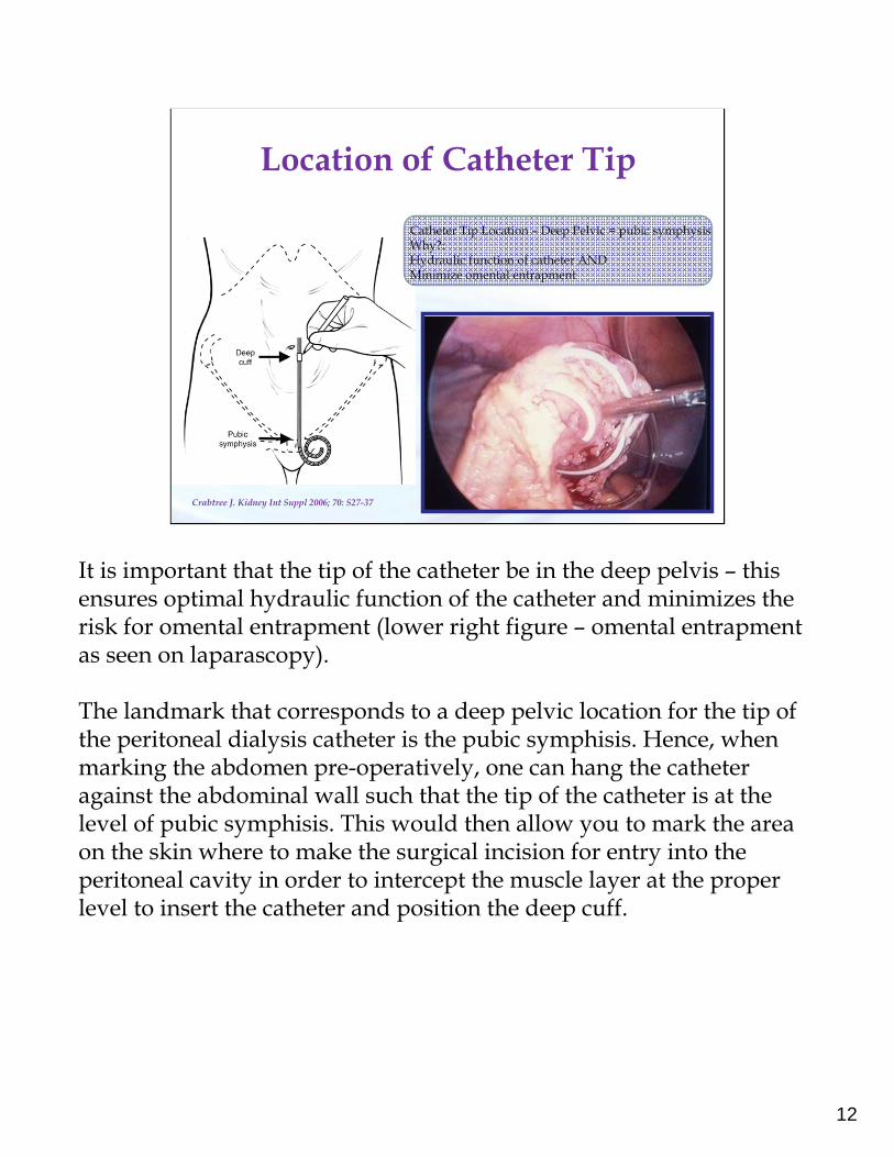
Catheter Tip Location – Deep Pelvic = pubic symphysisWhy?:Hydraulic function of catheter ANDMinimize omental entrapment
It is important that the tip of the catheter be in the deep pelvis – this ensures optimal hydraulic function of the catheter and minimizes the risk for omental entrapment (lower right figure – omental entrapment as seen on laparascopy).
The landmark that corresponds to a deep pelvic location for the tip of the peritoneal dialysis catheter is the pubic symphisis. Hence, when marking the abdomen pre-operatively, one can hang the catheter against the abdominal wall such that the tip of the catheter is at the level of pubic symphisis. This would then allow you to mark the area on the skin where to make the surgical incision for entry into the peritoneal cavity in order to intercept the muscle layer at the proper level to insert the catheter and position the deep cuff.
12

Location of Deep Cuff
Rectus Muscle
Paramedian position -away from the linea alba
The catheter enters the peritoneal cavity just distal to the deep cuff. As said earlier, deep cuff should be located at a place that allows the tip of the catheter to be in the deep pelvis. Furthermore, the entry point into the peritoneal cavity (and hence, the location of the deep cuff)) should be paramedian – this ensures that the catheter is not going through but linea alba (collagenous tissue in the midline that does not adequately close around the PD catheter and increases risk for leaks).
13

Principles in Fashioning Exit Site
• Should be away from belt-lines, skin creases, and folds
• Should be clearly visible to the patient to perform daily exit site care
• Inserted through the abdominal wall with least amount of tubing stress
• About one inch from the superficial cuff
• Generally achieved when planned with patient upright, rather than supine
Once the location of the catheter tip and deep cuff is identified, the next steps would be to mark the abdomen for the location of the superficial cuff and the exit site. The principles outlined in the slide above allow one to identify the correct placement of the exit site, which in turn would allow identification of the appropriate site for the superficial cuff.
14

Where Is It Relative to the Belt-Line?Need to Determine Before Patient Sedated
Above the Umblicus:Exit site below the umbilicus
Below the Umblicus:Exit site above the umbilicus
It is imperative to consider the belt-line when marking the abdomen for the location of the exit-site. In patients whose belt-line is above the umbilicus, the exit site should be located below the umbilicus. It is best achieved by using a swan-neck catheter with the exit site pointed downwards. In contrast, in patients whose belt-line is below the umbilicus, the exit site is best located in the upper abdomen. This is best achieved with a straight catheter with the exit-site pointed laterally.
15

Is the Exit Site Visible?Particularly Important for the Obese
Should be visible for a patient to perform daily exit=site care
Pictures Courtsey Dr. John Crabtree – Kaiser Bellflower
Proper placement of the exit site is particularly important for obese individuals – it should be visible to the patient to perform daily exit site care and should not be buried under the pannus to prevent recurrent exit site infections. The best way to identify the correct site for the placement of the exit site is pre-operatively, with the patient upright. When the patient is lying supine in the operating room, the pannus falls onto the side, making it difficult to identify the correct location for the exit site.
The two images on the left demonstrate an inappropriately placed exit site since it is buried under the pannus and not visible to the patient to perform daily exit-site care.
There are two alternative approaches that can be considered for obese patients – upper-abdominal exit site ( demonstrated in the middle panel; may require the use of extended catheters) or pre-sternal catheters (right panel).
16
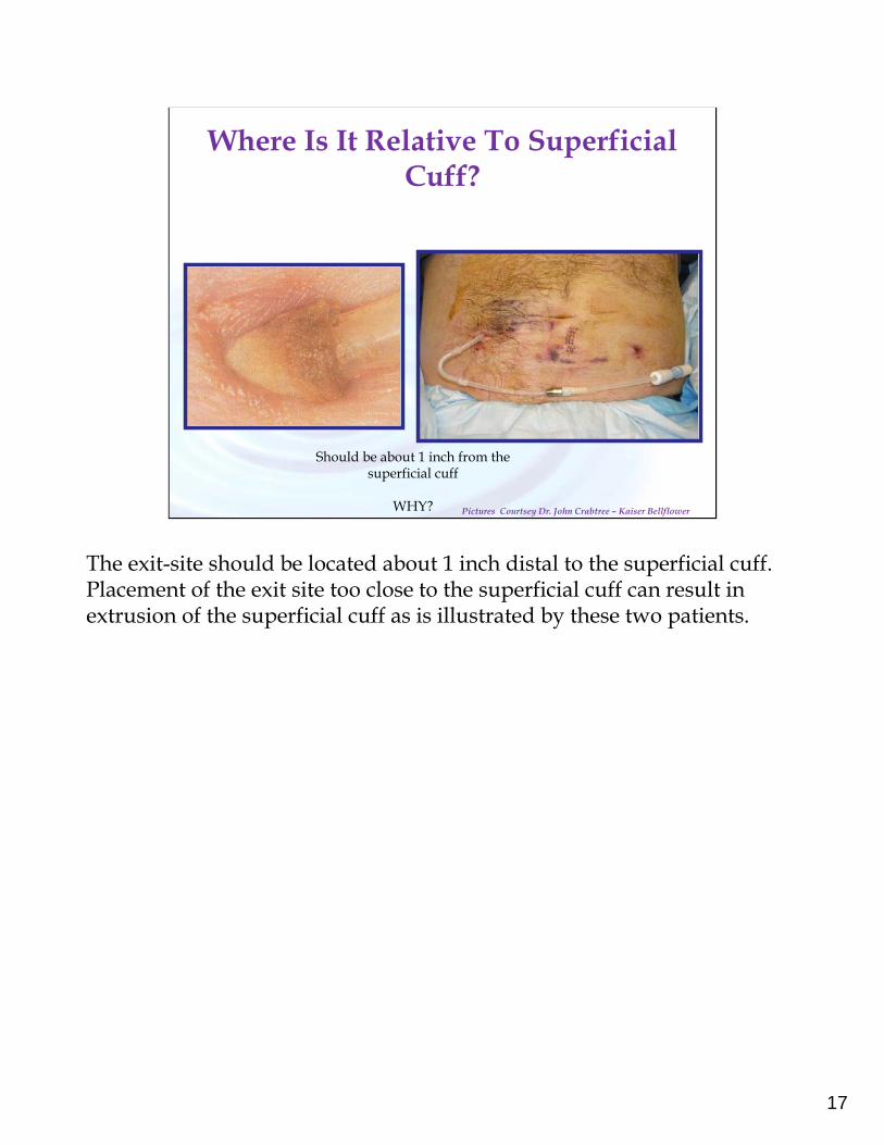
Where Is It Relative To Superficial Cuff?
Should be about 1 inch from the superficial cuff
WHY? Pictures Courtsey Dr. John Crabtree – Kaiser Bellflower
The exit-site should be located about 1 inch distal to the superficial cuff. Placement of the exit site too close to the superficial cuff can result in extrusion of the superficial cuff as is illustrated by these two patients.
17

Use Of Pre-Operative Marking May Obviate Exit Site Problems
Crabtree J. Kidney Int Suppl 2006; 70: S27-37
To summarize, the abdomen should be marked pre-operatively with the patient upright to minimize long-term problems. Several manufacturers include a stencil with each PD catheter placement kit and they can be used. Alternatively, stencils can be created by dialysis programs using the catheters that are being used by the surgeons to whom the patients are referred.
To recap, the following principles should be used when marking the abdomen:- The abdomen should be marked with the patient recumbent but themarking should be checked with the patient upright.-The location of the skin incision for entry into the peritoneal cavity should be first identified and marked – this should be a paramedian location, at a point that allows the tip of the catheter to be at the pubic symphisis-- The exit site should be marked such that:
-It would be visible to the patient for appropriate exit site care-It is away from the belt-line and-It is 1 inch from the superficial cuff
18

No Sutures or Staples At Exit Site!
Pictures Courtsey Dr. John Crabtree – Kaiser Bellflower
The exit-site should be fashioned such that it is not patulous. This is best achieved by creating it by pushing the trocar, with the catheter threaded over at the distal end, through the skin, rather than using a blade to make an incision. This would also obviate the need for stitches/sutures at the exit site. Indeed, stitches/sutures at the exit-site should be assiduously avoided as they prevent complete healing of the exit-site and increase risk for infections.
19

How Should PD Catheters Be Placed?
• Methods of placement of PD catheters:– Percutaneous, blind (with/without fluroscopy)– Direct visualization:
• Peritoneoscopic (Y-Tec®)• Open, surgical dissection • Laparoscopic (local anesthesia, using nitrous oxide or helium
insuffulation)
• Who should place PD catheters?– Whoever places them well in your area; depends on local
expertise:• Surgeons• Nephrologists, including interventional nephrologists in stand-
alone access centers• Interventional radiologists
Many different specialists have published their successful experience in placing PD catheters. It is best to place the PD catheter under direct vision and there are many advantages of advanced laparaoscopy (discussed subsequently).
There are no controlled clinical trials that have compared advanced laparoscopy with open surgical placement of PD catheters. The few clinical trials that have compared basic laparoscopy to laprotomy have shown similar results with the two techniques. Furthermore, excellent results have been reported for catheters placed by nephrologists or interventional radiologists. Hence, if local expertise for advanced laparoscopy is not available locally, alternative approaches may be considered and are acceptable.
20

What Is Advanced Laparoscopy
Open Dissection (n=63)
Basic Laparascopy (n=78)
Adva1nced Lapara0scopy
(n=200)
Post-op FU (mo) 49.5 ± 13.7 55.8 ± 13.1 54.4 ± 104.3
Previous laparotomy 30% 55.1% 53%
Implanted as outpt 78% 68% 93%
Simultaneous hernia repair 5% 9% 7%
Procedural Comp:Cath flow obstPostop peri-cannular leakPeri-cannular herniaSup cuff extrusionVisceral Perf
1111-1
101030
14020
Advanced laparascopy –Rectus sheath tunneling, selective prophylactic omentopexy, and/or adhesiolysis
Crabtree and Fishman, Am Surg 2005; 71: 135-143
There are three components of advanced laparoscopy:
Rectus Sheath tunneling implies that the catheter traverses the thickness of the rectus muscle obliquely, rather than perpendicularly. This ensures a longer track through the muscle and a better anchorage of the catheter;
Selective omentopexy is performed in patients with redundant omentum. The omentum is tagged with a stitch to the parietal peritoneum and this keeps the omentum away from the tip of the PD catheter and minimizes chances for omental entrapment;
Selective adhesiolysis in patients with intra-peritoneal adhesions.
As is evident from the table, use of advanced laparoscopy can be performed as an outpatient and is associated with a very low rate of complications and a very high long-term success rate.
One additional point to be noted is that hernias, if identified pre-operatively, can be fixed at the same time as the placement of the PD catheter.
21

Timing of Placement of PD Access
• General principle:– Wait for two weeks from the time of placement of PD
catheter before starting PD (“break-in” period)– Initial, and periodic flushing during break-in– Break-in period may be longer if wound healing impaired:
• Post-transplant failure OR• Patient on immunosuppressives
• Implant catheter too early:– prolonged need for catheter care before dialysis started
• Implant catheter too late:– Need early “break-in”
22

Embedded CathetersFashion the Exit-Site At A Different Time
Crabtree J. Kidney Int Suppl 2006; 70: S27-37
First described with Moncrief-Popovich catheterBut can be used with virtually any catheter
One way to take the “guessing” out of the decision as to when to place a PD catheter, is to place the catheter shortly after the patient decides on his/her dialysis modality. However, rather than exteriorizing the external limb of the PD catheter at the time that it is placed, it is placed in the subcutaneous tissue. Prior to embedding the catheter, it is flushed with heparinized saline to minimize the risk for the formation of fibrin plugs. Furthermore, the tip of the catheter is capped – this can be done using a cover from the tip of a syringe that is secured with a suture. Alternatively, the catheter tip can be plugged using a titanium catheter plug approved by the FDA for this purpose (Medigroup, Oswego, IL) or an off-label use of the Vaxcel tunneler. The catheter is then left in place till the patient needs to begin dialysis. The catheter can be exteriorized in the office setting – at the time of exteriorization, the tunnel is completely healed. Hence, full-dose PD can begin on the day that the catheter is exteriorized, without concern for leaks
23
Burying the External Limb of PD Catheter
• Initial rationale:– All catheters rapidly form biofilm– Exteriorizing the external limb only after tunnel is completely
healed would prevent the formation of biofilm– Reduce risk for exit-site infection and/or peritonitis
• Rationale for which it is being increasingly used:– PD catheter placed shortly after patient decides on the modality– Obviates guessing about timing of catheter placement relative to
need to start RRT and– Catheter can be used on the day of the exteriorization of the
external limb
24

How Long Can Catheters Be Embedded Before Exteriorization?
Author, year No Mean/ median
Range Placement Futile, %
Initial Drainage
Problem, %
Catheter Never
Worked, %
Prischl, ’97 26 11.4 wks 4- 96 wks 19% 29% 0%
Danielsson, ‘02 30 7.1 wks 1-170 wks 0 3% 0%
McCormick, ’06 266 13.1 wks 5-42 wks 11% 15% 7%
Junejo, ’08 20 15.7 wks 5-127 wks 10% 5% -
Prischl et al, Nephrol Dial Transplant 1997; 12: 1661-7
Danielsson et al, Perit Dial Int 2002; 22: 211-9
McCormick et al, Kidney Int suppl 2006; 70: S38-S43
Junejo et al, Perit Dial Int 2008; 28: 305-8
Causes of Futility:Pre-emptive transplantation
Patient DeathStarted HD instead of PD
This slide summarizes the results with embedded catheters from different parts of the world. A few important points need to be made:
• Catheters can be successfully used after having been embedded for over two years•One should expect a finite futility rate – catheters that are placed but never used (causes listed in slide)•Ten percent of more catheters have initial problems with drainage. However, they are generally easily resolved with flushing the catheters and primary failure of the catheter is rare.
25

How Long Should Catheters Be Embedded?
Tertile One
Tertile Two
Tertile Three
P-value
Days 11-47 48-133 134-2041
Primary Failure Rate 6.9% 1.7% 9.4% 0.04
Required Intervention for mechanical complications
7.8% 6.0% 15.4% 0.04
Brown et al, Nephrol Dial Transplant 2008; 23: 2299-2303
Based upon this one study, the optimal length for embedding the catheter is between 6 weeks to 6 months. Hence, if you anticipate that the patient will need PD within six weeks, the catheter should be placed using the conventional approach without embedding the external limb of the catheter.
26

Embedded Catheters: Final Word
Allows elective start of PD (“fistula-concept” applied to PD) No conclusive evidence that embedding itself reduces
peritonitis risk
Should be attempted only if there is at least 4-6 weeks from the time of surgery to anticipated need of catheter
Anticipate a finite futility rate
Initial non-function often related to fibrin thrombi: Amenable to interventions that often don’t require placement
of a new catheter
27

Early Break-In
Start PD (“break-in”) within 24 hours of placement of PD catheter
Risks: Peri-catheter leak
Solution: Low-volume, supine dialysis (1 L dwell volume x 6-10 exchanges)
Challenges: Inadequate dialysisPlace where dialysis is performed –
home, hospital, or dialysis clinic
Generally, we wait for about two weeks from the time of placement of the PD catheter before first using it for peritoneal dialysis. This is done to allow the catheter tunnel to heal and minimize the risk of leak. However, if necessary, PD can be commenced on the same day as the catheter is placed and in appropriate cases, will minimize the need for temporary HD.
There are some important considerations for “early break-in”. During this period, it is important to perform put only small volumes of the fluid at a time with the patient supine (“low-volume, supine PD”). This will minimize the increase in intra-peritoneal pressure and reduce the risk for leaks. This is best done with the use of a cycler.
It should be recognized that a patient in whom an “early break-in” still needs to be trained – training can happen while the patient is connected to the cycler in an outpatient dialysis unit. Alternatively, the patient can come in to a dialysis unit two to three times a week for intermittent PD while training is scheduled for a later time. While inadequate, this would be enough to patient to maintain biochemical and volume control till full-dose PD can be started.
28

Conclusions
• Placement technique and skill of operator most important determinant of catheter outcomes
• Advanced laparoscopy has advantages and desirable, if available
• Careful planning of exit site placement very important
• If enough time available, embedding external limb is a good option
• Early break-in possible, if needed
• All above would minimize/eliminate need for temporary HD
29

Question #1
• Which ONE of the following describes the BEST DEMONSTRATED PRACTICES for fashioning the exit site?
– A. Pre-operative marking with the patient recumbent; confirm with the patient upright
– B Intra-operatively after the deep cuff has been placed; confirm post-operatively
– C. Pre-operative marking with the patient upright; confirm with the patient recumbent
– D. Intra-operatively using the stencil provided by the manufacturer; confirm after placement of deep cuff

Question #1: Correct answer is A

Question #2
• Which ONE of the following BEST describes the desirable interval between placement of PD catheter and start of peritoneal dialysis therapy?
– A. PD therapy is contra-indicated during the first two weeks after placement of PD catheter
– B. It is imperative that the external limb of the PD catheter be buried subcutaneously for four weeks for adequate wound healing
– C. PD catheters can be used within 24 hours of placement
– D. PD therapy is contra-indicated during the first seven days after placement of PD catheter.

Question #2: Correct answer is C