pathway in cardiac hypertrophy and fibrosis the role of ... · the role of the grb2–p38 mapk...
TRANSCRIPT

The role of the Grb2–p38 MAPK signalingpathway in cardiac hypertrophy and fibrosis
Shaosong Zhang, … , Yibin Wang, Anthony J. Muslin
J Clin Invest. 2003;111(6):833-841. https://doi.org/10.1172/JCI16290.
Cardiac hypertrophy is a common response to pressure overload and is associated withincreased mortality. Mechanical stress in the heart can result in the integrin-mediatedactivation of focal adhesion kinase and the subsequent recruitment of the Grb2 adaptermolecule. Grb2, in turn, can activate MAPK cascades via an interaction with the Rasguanine nucleotide exchange factor SOS and with other signaling intermediates. Weanalyzed the role of the Grb2 adapter protein and p38 MAPK in cardiac hypertrophy. Micewith haploinsufficiency of the Grb2 gene (Grb2+/– mice) appear normal at birth but havedefective T cell signaling. In response to pressure overload, cardiac p38 MAPK and JNKactivation was inhibited and cardiac hypertrophy and fibrosis was blocked in Grb2+/– mice.Next, transgenic mice with cardiac-specific expression of dominant negative forms of p38a(DN-p38a) and p38b (DN-p38b) MAPK were examined. DN-p38a and DN-p38b micedeveloped cardiac hypertrophy but were resistant to cardiac fibrosis in response to pressureoverload. These results establish that Grb2 action is essential for cardiac hypertrophy andfibrosis in response to pressure overload, and that different signaling pathways downstreamof Grb2 regulate fibrosis, fetal gene induction, and cardiomyocyte growth.
Article Cardiology
Find the latest version:
http://jci.me/16290-pdf

IntroductionHuman cardiac hypertrophy is a common conditionthat often develops as a by-product of hypertension orvalvular heart disease. Adult cardiomyocytes are unableto divide, but respond to stress and growth stimuli byenlarging (1). Cardiac hypertrophy is associated withan increased risk of cardiac arrhythmias, diastolic dys-function, congestive heart failure, and death (2, 3).
Pressure overload activates a wide variety of signalingpathways in cardiac tissue. For example, increased wallstress leads to the local release of ligands such asangiotensin II and endothelin-1 (1, 4). The preciserequirement for these ligands in the hypertrophic
process is not completely understood at this time,although angiotensin II receptor blockade inhibits thedevelopment of aortic banding–induced murine car-diac hypertrophy in a fashion that is independent of itsability to lower blood pressure (5).
In addition to causing the local release of ligands,pressure overload may also activate intrinsic “stretch”receptors in cardiomyocytes. Leading candidates forcardiac stretch receptors include the integrin family oftransmembrane proteins (6–8). Outside-in integrin sig-naling is mediated by the binding of extracellularmatrix proteins to an αβ heterodimeric integrin pair(9–12). Binding of a matrix protein to an integrin het-erodimer typically results in the activation of the non-receptor tyrosine kinase, focal adhesion kinase (FAK)(6). Activated FAK, in turn, recruits the non-receptortyrosine kinase c-Src, the multifunctional adapter mol-ecule Grb2, and other signaling intermediates (6, 8, 13).
Recent studies demonstrated that integrin activation,in concert with G protein activation, might be essentialfor the growth of cardiomyocytes. For example, bal-loon-mediated ventricular stretch of isolated adult ratheart results in the rapid activation of FAK, promotesthe association of Grb2 with FAK, and causes the acti-vation of extracellular signal-regulated kinase 1/2(ERK) (14). In addition, stretch of cultured rat neona-tal cardiomyocytes results in the rapid activation ofFAK and c-Src, the formation of a Grb2-FAK complex,and the activation of p38 MAPK (8). Furthermore,
The Journal of Clinical Investigation | March 2003 | Volume 111 | Number 6 833
The role of the Grb2–p38 MAPK signaling pathway in cardiac hypertrophy and fibrosis
Shaosong Zhang,1,2 Carla Weinheimer,1,2 Michael Courtois,1,2 Attila Kovacs,1,2
Cindy E. Zhang,1,2 Alec M. Cheng,2,3 Yibin Wang,4 and Anthony J. Muslin1,2,5
1Center for Cardiovascular Research, 2Department of Medicine, and3Department of Pathology, Washington University School of Medicine, St. Louis, Missouri, USA4Department of Pharmacology, University of Maryland School of Medicine, Baltimore, Maryland, USA5Department of Cell Biology & Physiology, Washington University School of Medicine, St. Louis, Missouri, USA
Cardiac hypertrophy is a common response to pressure overload and is associated with increasedmortality. Mechanical stress in the heart can result in the integrin-mediated activation of focaladhesion kinase and the subsequent recruitment of the Grb2 adapter molecule. Grb2, in turn, canactivate MAPK cascades via an interaction with the Ras guanine nucleotide exchange factor SOSand with other signaling intermediates. We analyzed the role of the Grb2 adapter protein and p38MAPK in cardiac hypertrophy. Mice with haploinsufficiency of the Grb2 gene (Grb2+/– mice) appearnormal at birth but have defective T cell signaling. In response to pressure overload, cardiac p38MAPK and JNK activation was inhibited and cardiac hypertrophy and fibrosis was blocked inGrb2+/– mice. Next, transgenic mice with cardiac-specific expression of dominant negative formsof p38α (DN-p38α) and p38β (DN-p38β) MAPK were examined. DN-p38α and DN-p38β micedeveloped cardiac hypertrophy but were resistant to cardiac fibrosis in response to pressure over-load. These results establish that Grb2 action is essential for cardiac hypertrophy and fibrosis inresponse to pressure overload, and that different signaling pathways downstream of Grb2 regulatefibrosis, fetal gene induction, and cardiomyocyte growth.
J. Clin. Invest. 111:833–841 (2003). doi:10.1172/JCI200316290.
Received for publication June 26, 2002, and accepted in revised formDecember 17, 2002.
Address correspondence to: Anthony J. Muslin, Washington University School of Medicine, 660 South EuclidAvenue, Campus Box 8086, St. Louis, Missouri 63110, USA.Phone: (314) 747-3525; Fax: (314) 362-0186; E-mail: [email protected] of interest: The authors have declared that no conflict ofinterest exists.Nonstandard abbreviations used: focal adhesion kinase (FAK);extracellular signal-regulated kinase 1/2 (ERK); atrial natriureticfactor (ANF); Src-homology type 3 (SH3); α-myosin heavy chain(α-MHC); transverse aortic constriction (TAC); left ventricular(LV); sarcoplasmic-endoplasmic reticulum calcium ATPase(SERCA); left ventricular weight to body weight ratio (LVW/BW);left ventricular mass to body weight ratio (LVM/BW); MAPKkinase kinase (MAPKKK); Grb2-associated binder 1 (Gab1).

expression of an integrin β1D inhibitor in cultured ratneonatal cardiomyocytes blocks phenylephrine-induced hypertrophy and atrial natriuretic factor(ANF) expression (7). Finally, expression of a dominantnegative form of FAK, or expression of a mutant formof FAK that is unable to bind to Grb2, in cultured car-diomyocytes blocks phenylephrine-induced hypertro-phy and ERK activation (6, 7).
Cardiac pressure overload results in the activation ofintegrin/FAK-mediated responses, but the importanceof downstream components of this signaling pathwayin the hypertrophic process remains to be determined.Recently, mice were developed with targeted disruptionof the Grb2 gene (15). Grb2 is a scaffolding protein thatcontains two Src-homology type 3 (SH3) domains thatflank a single SH2 domain (16, 17). SH3 domains bindto polyproline peptide motifs, and SH2 domains bindto phosphotyrosine-containing peptide motifs (18, 19).The SH3 domain of Grb2 binds to a polyproline motifon the Ras guanine nucleotide exchange factor SOS,and the SH2 domain of Grb2 binds to phosphotyro-sine motifs present on activated FAK, Shc, and receptortyrosine kinases. Grb2–/– mice do not survive embryon-ic development due to defective endoderm differentia-tion and because they are unable to form the epiblast.However, Grb2+/– mice survive embryogenesis, appearnormal at birth, and are fertile. Grb2+/– mice have a40–50% reduction in Grb2 protein in all tissues testedto date and have a defect in T cell signaling (20).Although ERK activation is normal in Grb2+/– T cells,p38 MAPK and JNK activation is markedly reduced.Based on these defects in signal transduction, wethought Grb2+/– mice would be an excellent model sys-tem in which to study cardiac hypertrophy.
MethodsGrb2+/– mice. Grb2+/– mice in the 129/SvJ strain weregenerated as previously described (15). Grb2–/– mice donot survive embryonic development. Grb2+/– mice werecompared with Grb2+/+ 129/SvJ littermates in all exper-iments described here.
All research involving the use of mice was performedin strict accordance with protocols approved by theAnimal Studies Committee of Washington UniversitySchool of Medicine.
Transgenic mice with cardiac-specific expression of dominantnegative forms of p38α or p38β MAPK. The coding regionof the cDNA’s of human DN-p38α MAPK or DN-p38βMAPK (21, 22) were subcloned into a vector containingthe α-myosin heavy chain (α-MHC) promoter and anSV40 polyadenylation site as previously described (23).Linearized DNA was injected into the pronuclei of one-cell Black Swiss embryos. Progeny were analyzed byPCR to detect transgene integration (24). Multiple linesin the Black Swiss genetic background were obtainedfor the construct, and integration of the transgene wasanalyzed by dot blot analysis. The highest-expressinglines, with integration of ten copies of the DN-p38αtransgene and 15 copies of the DN-p38β transgene,
were used in this study. Transgenic DN-p38α or DN-p38β mice were compared with congenic non-transgenic Black Swiss mice in every experiment.
Transverse aortic constriction. Transverse aortic con-striction (TAC) was performed as previouslydescribed (25, 26). The surgeon was blinded in allcases to the transgenic status of the mice. After 7days, surviving animals were subjected to transtho-racic echocardiography and cardiac catheterizationto determine cardiac function and proximal aorticpressure. Animals were then killed and the heartswere dissected out and weighed (24).
Transthoracic echocardiography. Transthoracic echocar-diography was performed in awake mice by use of anAcuson Sequoia 256 Echocardiography Systemequipped with a 15-MHz transducer (model 15L8) asdescribed previously (Siemens Medical Solutions,Mountain View, California, USA) (24, 27). Theechocardiographer was blinded in all cases to thetransgenic status of the mice.
Cardiac catheterization. Closed-chest cardiac catheter-ization was performed as previously described (24).Continuous aortic pressure and left ventricular (LV)systolic and diastolic pressures were recorded on aGould chart recorder (Gould Instruments, ValleyView, Ohio, USA).
Histological analysis. Seven days after TAC, Grb2+/– andGrb2+/+ 129/SvJ, DN-p38α, DN-p38β, and nontrans-genic Swiss Black mice were sacrificed and LV tissuewas obtained. Tissue was fixed in 10% formalin,embedded in paraffin, and sectioned with a micro-tome. Tissue sections were stained with H&E or withMasson trichrome (24).
Quantitative real-time RT-PCR. RNA was purified fromquick-frozen cardiac tissue by use of the RNeasy pro-tocol (QIAGEN Inc., Valencia, California, USA). TheTaqMan Gold RT-PCR kit was used according to themanufacturer’s instructions (Applied Biosystems, Fos-ter City, California, USA). Quantitative PCR was per-formed by use of real-time detection technology andanalyzed on a model 7700 Sequence Detector (AppliedBiosystems) with specific primers and fluorescentprobes for sarcoplasmic-endoplasmic reticulum calci-um ATPase (SERCA), medium chain acyl-CoA dehy-drogenase, ANF, and β-myosin heavy chain (β-MHC).Levels of mRNA were compared at various timepointsafter correction by use of concurrent GAPDH messageamplification, with GAPDH probe and primers used asan internal standard.
Protein analysis. Cytosolic extracts of ventricular tissuewere separated by SDS-PAGE and proteins were elec-trophoretically transferred to nitrocellulose filters (26).Filters were blocked in Tris-buffered saline containing1% Tween 20 and 2% nonfat dried milk. Filters werewashed and incubated with primary antibody. Primaryantibodies used included: rabbit polyclonal anti-FAKantibody, murine monoclonal anti-Grb2 antibody,murine monoclonal anti-ERK antibody, rabbit poly-clonal anti–p38α MAPK antibody, goat polyclonal
834 The Journal of Clinical Investigation | March 2003 | Volume 111 | Number 6

anti–p38β MAPK antibody (Santa Cruz BiotechnologyInc., Santa Cruz, California, USA), rabbit polyclonalanti-phospho p38 MAPK antibody, rabbit polyclonalanti–phospho-JNK antibody, rabbit polyclonalanti–p38 MAPK antibody, and rabbit polyclonal anti-JNK antibody (Cell Signaling Technology, Beverly,Massachusetts, USA). Filters were extensively washedin Tris-buffered saline containing 1% Tween 20 andthen incubated with horseradish peroxidase–conjugat-ed anti-rabbit, anti-goat, or anti-mouse secondary anti-body (Amersham Pharmacia Biotech Inc., Piscataway,New Jersey, USA). Bands were visualized by use of theECL system (Amersham Pharmacia Biotech Inc.) (26).
In vitro protein kinase assays. In vitro ERK activityassays were performed using a kit from Cell SignalingTechnology in accordance with the manufacturer’sinstructions. In brief, anti–phospho-ERK immuno-precipitates were derived from ventricular cytosoliclysates obtained 7 days after TAC. 2 µg of thespecific substrate protein Elk-1, 200 µM ofATP, and 50 µl of 1X kinase reaction bufferwere added to the immunoprecipitates. Kinasereactions were terminated after a 30-minuteincubation period, and proteins were separatedby SDS-PAGE and then analyzed byimmunoblotting with a specific antibodyagainst phospho–Elk-1.
Statistical analysis. All data are reported as mean± SEM. Statistical analysis was performed bytwo-tailed Student t test, χ2 analysis, andANOVA where applicable. Multiple group com-parison was carried out by ANOVA with theFisher post-hoc comparison. A value of P < 0.05was considered to be statistically significant.
ResultsCardiac pressure overload promotes Grb2 associationwith FAK. Previous work demonstrated thatGrb2 is expressed in heart tissue and isolatedcardiomyocytes. In addition, Grb2 is recruitedby activated FAK in response to mechanicalstress in cultured rat neonatal cardiomyocytesor in intact isolated rat hearts (8, 14). We inves-tigated the ability of pressure overload to pro-mote the association of FAK with Grb2 inmurine cardiac tissue. TAC was performed on12-week-old wild-type Grb2+/+ mice, and ven-tricular tissue was isolated 7 days after the sur-gical procedure. Anti-FAK immunoprecipitatesderived from ventricular protein lysates wereanalyzed by anti-Grb2 immunoblotting; thisrevealed that Grb2 formed a complex with FAKin Grb2+/+ ventricular tissue after TAC, but notafter a sham operation (Figure 1a).
Reduced cardiac Grb2 protein levels in haploinsuf-ficient mice. To investigate the role of Grb2 inthe cardiac hypertrophic growth program,Grb2+/– mice were analyzed. Grb2+/– mice in the129/SvJ strain appear normal at birth and are
fertile, and have normal cardiac structure and func-tion at 12 weeks of age (20). Echocardiographic analy-sis of 12-week-old Grb2+/+ and Grb2+/– 129/SvJ micerevealed that Grb2 haploinsufficient mice have nor-mal cardiac structure and intact ventricular systolicfunction, with a baseline LV fractional shortening of51% ± 7%. There was no evidence of cardiac hypertro-phy or of reduced cardiac wall thickness in Grb2+/–
mice in the absence of pressure overload.To determine whether disruption of one allele of
the Grb2 gene resulted in reduced cardiac proteincontent, a series of immunoblot experiments wereperformed with cardiac cytosolic lysates. Anti-Grb2immunoblotting of ventricular protein lysatesdemonstrated an approximately 40% reduction inGrb2 protein levels in Grb2+/– mice compared withGrb2+/+ mice (Figure 1b). This decrease in Grb2 pro-tein levels is consistent with previously published
The Journal of Clinical Investigation | March 2003 | Volume 111 | Number 6 835
Figure 1Biochemical characterization of Grb2 and MAPK in murine cardiac tissue 7days after TAC or sham operation. (a) Load-induced formation of a Grb2-FAK complex. Anti-FAK immunoprecipitates (IP) derived from ventric-ular lysates were separated by SDS-PAGE and analyzed by immunoblottingwith an anti-Grb2 antibody (lower panel). Anti-FAK immunoprecipitateswere also analyzed in parallel by immunoblotting with an anti-FAK antibody(upper panel). (b) Reduced Grb2 protein content in Grb2+/– cardiac tissue.Upper panel, ventricular lysates were analyzed by immunoblotting with ananti-Grb2 antibody. Lower panel, quantification of Grb2 protein levels bydensitometric analysis of immunoreactive bands. (c) Analysis of p38 MAPKactivation in Grb2+/– cardiac tissue. Ventricular lysates were analyzed byimmunoblotting with an anti-phospho–p38 MAPK antibody (upper panel).Lysates were also analyzed in parallel by immunoblotting with an anti–p38MAPK (lower panel) antibody to control for protein content. (d) Analysis ofJNK activation in Grb2+/– cardiac tissue. Lysates were analyzed byimmunoblotting with an anti–phospho-JNK antibody (upper panel). Lysateswere also analyzed in parallel by immunoblotting with an anti-JNK (lowerpanel) antibody to control for protein content. (e) Analysis of ERK activityin Grb2+/– cardiac tissue. Anti-ERK immunoprecipitates derived from ven-tricular lysates were analyzed by in vitro kinase assay by use of Elk-1 proteinas a substrate. Anti-phospho–Elk-1 antibody immunoblotting was per-formed to assess ERK activity (upper panel). Lysates were also analyzed inparallel by immunoblotting with an anti-ERK (lower panel) antibody to con-trol for protein content. Sham, sham operation.

observations in T cells (20). In addition to reducedcardiac Grb2 protein, pressure overload–inducedGrb2/FAK complex formation was markedlydecreased in cardiac tissue obtained from Grb2+/–
mice after TAC (Figure 1a).Analysis of MAPK cascade activation in Grb2+/– mice after
TAC. In wild-type mice, multiple signaling cascadesare activated in response to pressure overloadinduced by TAC. Grb2+/– mice were analyzed 7 daysafter TAC to determine whether there were defects incardiac signal transduction. Grb2+/– mice toleratedTAC and had a survival rate of 92% after 7 days (12 of13 animals). Ventricular cytosolic lysates obtainedfrom animals 7 days afterTAC were analyzed byimmunoblotting using anti-bodies against both phos-pho-JNK and phospho–p38MAPK. In both cases, paral-lel samples were analyzed byanti-JNK and anti-p38 MAPKimmunoblotting to controlfor protein loading. Activa-tion of p38 MAPK and JNKwas markedly decreased afterTAC in Grb2+/– mice com-pared with Grb2+/+ 129/SvJmice (Figure 1, c and d). ERKactivity in ventricular tissuesamples was analyzed by invitro kinase assay withrecombinant Elk-1 protein
used as a substrate. ERK activity increased in bothGrb2+/+ and Grb2+/– mice after TAC. In some experi-ments, the fold increase in ERK activity was mildlyreduced in Grb2+/– ventricular tissue (Figure 1e).
Hypertrophic response of Grb2+/– mice to TAC. In wild-type mice, prominent LV hypertrophy develops 7 daysafter TAC. Typically, there is a 25–30% increase in theratio of LV weight to body weight (LVW/BW) inresponse to pressure overload. In Grb2+/+ 129/SvJmice, LVW/BW increased by 31% 7 days after TAC,from 3.5 ± 0.5 mg/g to 4.6 ± 0.7 mg/g. In contrast,Grb2+/– mice were almost completely resistant to car-diac hypertrophy, and LVW/BW increased by only5.7%, from 3.5 ± 0.3 to 3.7 ± 0.3 mg/g (Figure 2, a andb). Indeed, the difference in LVW/BW between Grb2+/–
and Grb2+/+ mice after TAC was statistically signifi-cant by Student t test (P < 0.01). Echocardiographicanalysis also demonstrated that Grb2+/– mice wereresistant to TAC-induced cardiac hypertrophy.Echocardiographically determined LV mass to bodyweight ratio (LVM/BW) in Grb2+/+ mice increased by39% 7 days after TAC, but increased by only 5.4% inGrb2+/– mice (Table 1). In addition, ventricular con-tractility was enhanced in Grb2+/– mice 7 days afterTAC with a fractional shortening of 57%, comparedwith a fractional shortening of 48% in Grb2+/+ mice(Table 1). The failure of Grb2+/– mice to exhibit cardiachypertrophy in response to TAC was not a result ofless stringent aortic constriction. Proximal systolicaortic pressures increased from 142 ± 20 mmHg to196 ± 6 mmHg in Grb2+/– mice, and increased from132 ± 5 mmHg to 181 ± 12 mmHg in Grb2+/+ mice.
Histological analysis of LV tissue of Grb2+/– andGrb2+/+ mice was performed 7 days after TAC. Grb2+/+
animals exhibited typical myocyte enlargement,myofibrillar disarray, and fibrosis after TAC, whereasGrb2+/– animals had little or no fibrosis and relativelypreserved myofibrillar architecture (Figure 3).
Gene expression in Grb2+/– mice after TAC. Pressureoverload leads to a variety of alterations in cardiac
836 The Journal of Clinical Investigation | March 2003 | Volume 111 | Number 6
Figure 2Reduced hypertrophic response to pressure overload in Grb2+/– mice.(a) Gross morphology of hearts from Grb2+/+ (left) and Grb2+/–
(right) mice 7 days after TAC. (b) Morphometric analysis of cardiachypertrophy in Grb2+/+ and Grb2+/– mice. Seven days after TAC orsham operation, cardiac chambers were dissected and weighed todetermine LVW/BW (*P < 0.01 vs. sham-operated control).
Table 1In vivo echocardiographic assessment of Grb-2+/– mice
Sham-operated TACGrb-2+/+ (n = 6) Grb-2+/– (n = 6) Grb-2+/+ (n = 6) Grb-2+/– (n = 6)
HR (beats/min) 628 ± 69 618 ± 58 555 ± 87 558 ± 107LVIDd (mm) 3.06 ± 0.58 3.11 ± 0.38 3.19 ± 0.48 2.70 ± 0.49LVIDs (mm) 1.59 ± 0.56 1.54 ± 0.29 1.71 ± 0.63 1.18 ± 0.37PWd (mm) 0.72 ± 0.07 0.74 ± 0.05 0.87 ± 0.18 0.83 ± 0.07IVSd (mm) 0.73 ± 0.08 0.68 ± 0.10 0.81 ± 0.18 0.79 ± 0.18LVM/BW (mg/g) 2.55 ± 0.41 2.59 ± 0.16 3.54 ± 0.92A 2.73 ± 0.33FS (%) 49.3 ± 9.2 50.5 ± 6.28 47.7 ± 12.7 57.1 ± 8.7LVW/BW (mg/g) 3.5 ± 0.5 3.5 ± 0.3 4.6 ± 0.7A 3.7 ± 0.3
Echocardiographic measurements obtained from transthoracic M-mode tracings of nontransgenic Grb-2+/+
and Grb-2+/– mice 7 days after TAC or sham operation. HR indicates heart rate; LVIDd and LVIDs, end-dias-tolic and end-systolic LV internal dimensions, respectively; PWd and IVSd, end-diastolic posterior wall andintraventricular septal thickness; LVM, M-mode echocardiogram-derived LV mass; FS, fractional shortening;LV/BW morphometrically-determined left ventricular weight-to-body ration. AP < 0.05 versus sham-operat-ed control (Student’s t test).

gene expression. In particular, β-MHC and ANF geneexpression is typically increased in cardiac tissue inresponse to pressure overload. We used real-timequantitative RT-PCR to assess the expression ofhypertrophic marker genes in mice after TAC. In wild-type 129/SvJ but not Grb2+/– mice, β-MHC and ANFgene expression was markedly induced 7 days afterTAC. Indeed, β-MHC gene expression increased by 23-fold and ANF gene expression increased by 18-fold inwild-type mice (Figure 4, a and b).
Transgenic mice with cardiac-specific expression of dominantnegative p38α or p38β MAPK. Grb2+/– mice are resistantto the development of cardiac hypertrophy and fibro-sis after TAC and have markedly attenuated p38 MAPKand JNK signaling. Previous work demonstrated thatp38α MAPK is activated in cultured cardiomyocytes in
response to hypertrophic agonist stimulation (28, 29).In one study, p38 MAPK, but not JNK, was found to berequired for phenylephrine-induced cardiomyocytehypertrophy (28). In addition, pressure overload inrodents is known to activate cardiac p38α MAPK activ-ity (30). In contrast to work with isolated cultured car-diomyocytes, previous whole-animal work does notsupport the hypothesis that p38 MAPK activity pro-motes cardiomyocyte growth. Liao et al. recently gen-erated transgenic mice with cardiac-specific expressionof activated forms of MKK3bE or MKK6bE, kinasesthat act immediately upstream of p38α and p38βMAPK (31). MKK3bE and MKK6bE transgenic micedeveloped cardiac fibrosis, systolic and diastolic dys-function, and marked fetal gene induction, but no car-diac hypertrophy (31).
To test whether Grb2-facilitated p38 MAPK activa-tion is an essential regulator of cardiac hypertrophy,we analyzed transgenic mice with cardiac-specificexpression of dominant negative mutants of p38αand p38β MAPK. In these mutants, the threonine-X-tyrosine (TXY) activation loop is altered as previous-ly described (21, 22, 26). The α-MHC promoter waslinked to cDNA’s encoding DN-p38α or DN-p38β,
The Journal of Clinical Investigation | March 2003 | Volume 111 | Number 6 837
Figure 3Histological analysis of Grb2+/+ and Grb2+/– LV tissue 7 days after TACor sham operation. Ventricular tissue sections from (a) Grb2+/+
mouse, stained with H&E after sham operation; (b) Grb2+/+ mouse,stained with H&E after TAC; (c) Grb2+/– mouse, stained with H&Eafter sham operation; (d) Grb2+/– mouse, stained with H&E afterTAC; (e) Grb2+/+ mouse, stained with Masson trichrome after shamoperation; (f) Grb2+/+ mouse, stained with trichrome after TAC. Notethe increased extracellular matrix content (blue color), cardiomy-ocyte enlargement, and disarray. (g) Ventricular tissue section fromGrb2+/– mouse, stained with trichrome after sham operation. (h)Ventricular tissue section from Grb2+/– mouse, stained with trichromeafter TAC. The original magnification was ×400 in all sections.
Figure 4Inhibition of β-MHC and ANF gene expression in Grb2+/– cardiac tis-sue after pressure overload. Ventricular tissue was obtained 7 daysafter TAC or sham operation from Grb2+/+ and Grb2+/– mice, andRNA was purified from the tissue samples. Gene expression was ana-lyzed by use of quantitative real-time RT-PCR with specific primersand probes. GAPDH was used as an internal control in all cases. (a)β-MHC gene expression in Grb2+/+ and Grb2+/– ventricular tissue. (b)ANF gene expression in Grb2+/+ and Grb2+/– ventricular tissue. AU,arbitrary units.

and transgenic mice in the Swiss Black strain weregenerated. Multiple lines were generated for eachconstruct, and the highest-expressing lines were ana-lyzed in this work. DN-p38α mice with integration often copies of the transgene and DN-p38β mice withintegration of 15 copies of the transgene were viableand fertile. Expression of the dominant negativeforms of p38 was robust in transgenic mice as deter-mined by immunoblotting of ventricular cytosoliclysates with isoform-specific anti–p38 MAPK pri-mary antibodies (Figure 5a).
The ability of the dominant negative mutant formsof p38 MAPK to specifically inhibit native p38α orp38β kinase activity was analyzed. Cardiac p38α andp38β MAPK activity was analyzed by in vitro kinaseassay following immunoprecipitation of phos-pho–p38 MAPK protein from ventricular lysates.DN-p38α transgenic mice exhibited reduced p38αMAPK activity and DN-p38β transgenic mice exhib-ited reduced p38β MAPK activity in ventricularcytosolic lysates (Figure 5, b and c).
Response of DN-p38α or DN-p38β transgenic mice toTAC. In response to TAC, 15 of 19 DN-p38α mice(79%), and 11 of 17 DN-p38β mice (65%) survived,
and these rates were similar to those observed in non-transgenic littermates. Seven days after TAC, heartswere isolated and LVW/BW was calculated as a meas-ure of cardiac hypertrophy. As expected, LVW/BWincreased by 34% in nontransgenic Swiss Black mice,from 3.5 ± 0.2 to 4.7 ± 0.1 mg/g (P < 0.01). DN-p38αtransgenic mice also developed cardiac hypertrophyafter TAC (Figure 6). LVW/BW increased by 32% inDN-p38α mice, from 3.4 ± 0.1 to 4.5 ± 0.6 mg/g (P < 0.05). DN-p38β mice developed cardiac hypertro-phy 7 days after TAC and LVW/BW increased by 51%,from 3.3 ± 0.5 to 5.0 ± 0.8 mg/g (P < 0.0001) (Figure 6).Echocardiographic analysis confirmed that DN-p38αand DN-p38β mice developed cardiac hypertrophy inresponse to pressure overload. Echocardiographicallyderived LVM/BW increased by 48% in nontransgenic
838 The Journal of Clinical Investigation | March 2003 | Volume 111 | Number 6
Figure 5Biochemical characterization of DN-p38α and DN-p38β transgenicmice. (a) Analysis of DN-p38α and DN-p38β MAPK protein levelsin cardiac tissue from transgenic mice. Ventricular protein lysatesobtained from DN-p38α mice, DN-p38β mice, and nontransgenicSwiss Black mice were separated by SDS-PAGE and examined byimmunoblotting. Upper panel, isoform-specific anti–p38α MAPKimmunoblot. Middle panel, isoform-specific anti–p38β MAPKimmunoblot. Lower panel, anti-ERK immunoblot to control forprotein loading. (b) Reduced p38α MAPK activity in DN-p38αtransgenic mice. Ventricular protein lysates were generated 7 daysafter TAC or sham operation in DN-p38α transgenic mice or non-transgenic Swiss Black mice (NTG). Upper panel, anti-phospho–p38 MAPK immunoprecipitates were analyzed by iso-form-specific anti–p38α MAPK immunoblotting. Middle panel,ventricular lysates were analyzed by anti-ERK immunoblotting tocontrol for protein content. Lower panel, quantification of p38αMAPK protein levels in ventricular lysates by densitometric analysisof immunoreactive bands. Data are from three experiments. (c)Reduced p38β MAPK activity in DN-p38β transgenic mice. Upperpanel, anti-phospho–p38 MAPK immunoprecipitates were ana-lyzed by isoform-specific anti–p38β MAPK immunoblotting. Mid-dle panel, ventricular lysates were analyzed by anti-ERKimmunoblotting to control for protein content. Lower panel, quan-tification of p38β MAPK protein levels by densitometric analysis ofimmunoreactive bands. Data are from three experiments.
Figure 6Preserved hypertrophic response to pressure overload in DN-p38αand DN-p38β transgenic mice. Morphometric analysis of cardiachypertrophy in DN-p38α mice, DN-p38β mice, and nontransgenicSwiss Black mice. Seven days after TAC or sham operation, cardiacchambers were dissected and weighed to determine LVW/BW.
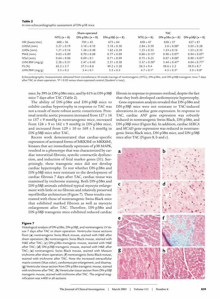
mice, by 39% in DN-p38αmice, and by 61% in DN-p38βmice 7 days after TAC (Table 2).
The ability of DN-p38α and DN-p38β mice toexhibit cardiac hypertrophy in response to TAC wasnot a result of more robust aortic constriction. Prox-imal systolic aortic pressures increased from 127 ± 16to 157 ± 9 mmHg in nontransgenic mice, increasedfrom 126 ± 9 to 162 ± 9 mmHg in DN-p38α mice,and increased from 129 ± 10 to 169 ± 3 mmHg inDN-p38β mice after TAC.
Recent work demonstrated that cardiac-specificexpression of activated forms of MKK3bE or MKK6bE,kinases that act immediately upstream of p38 MAPK,resulted in a phenotype that was characterized by car-diac interstitial fibrosis, systolic contractile dysfunc-tion, and induction of fetal marker genes (31). Sur-prisingly, these transgenic mice did not developcardiac hypertrophy. To test whether DN-p38α andDN-p38β mice were resistant to the development ofcardiac fibrosis 7 days after TAC, cardiac tissue wasexamined by trichrome staining. Both DN-p38α andDN-p38β animals exhibited typical myocyte enlarge-ment with little or no fibrosis and relatively preservedmyofibrillar architecture (Figure 7). These results con-trasted with those of nontransgenic Swiss Black micethat exhibited marked fibrosis as well as myocyteenlargement after TAC. Therefore, DN-p38α and DN-p38β transgenic mice exhibited reduced cardiac
fibrosis in response to pressure overload, despite the factthat they both developed cardiomyocyte hypertrophy.
Gene expression analysis revealed that DN-p38α andDN-p38β mice were not resistant to TAC-inducedalterations in cardiac gene expression. In response toTAC, cardiac ANF gene expression was robustlyinduced in nontransgenic Swiss Black, DN-p38α, andDN-p38β mice (Figure 8a). In addition, cardiac SERCAand MCAD gene expression was reduced in nontrans-genic Swiss Black mice, DN-p38α mice, and DN-p38βmice after TAC (Figure 8, b and c).
The Journal of Clinical Investigation | March 2003 | Volume 111 | Number 6 839
Table 2In vivo echocardiographic assessment of DN-p38 mice
Sham-operated TACNTG (n = 8) DN-p38α (n = 8) DN-p38β (n = 8) NTG (n = 8) DN-p38α (n = 8) DN-p38β (n = 8)
HR (beats/min) 688 ± 36 705 ± 43 673 ± 64 658 ± 47 658 ± 37 627 ± 81LVIDd (mm) 3.27 ± 0.19 3.18 ± 0.10 3.18 ± 0.30 2.84 ± 0.39 3.0 ± 0.08A 3.05 ± 0.28LVIDs (mm) 1.21 ± 0.16 1.36 ± 0.48 1.62 ± 0.29 1.25 ± 0.35 1.24 ± 0.16 1.53 ± 0.35PWd (mm) 0.63 ± 0.09 0.70 ± 0.08 0.77 ± 0.09 0.80 ± 0.13A 0.90 ± 0.07A 0.94 ± 0.07A
IVSd (mm) 0.64 ± 0.06 0.69 ± 0.1 0.71 ± 0.09 0.75 ± 0.25 0.87 ± 0.08A 0.90 ± 0.07A
LVM/BW (mg/g) 2.28 ± 0.31 2.47 ± 0.43 2.51 ± 0.38 3.37 ± 0.30A 3.44 ± 0.47A 4.04 ± 0.77A
FS (%) 63.2 ± 2.7 57.3 ± 8.6 49.2 ± 5.28 56.3 ± 9.4 58.6 ± 2.2 50.0 ± 8.7LVW/BW (mg/g) 3.5 ± 0.2 3.4 ± 0.1 3.3 ± 0.5 4.7 ± 0.1A 4.5 ± 0.5A 5.0 ± 0.8A
Echocardiographic measurements obtained from transthoracic M-mode tracings of nontransgenic (NTG), DN-p38α, and DN-p38β transgenic mice 7 daysafter TAC or sham operation. AP < 0.05 versus sham-operated control (Student’s t test).
Figure 7Histological analysis of DN-p38α, DN-p38β, and nontransgenic LV tis-sue 7 days after TAC or sham operation. Ventricular tissue sectionsfrom (a) nontransgenic Swiss Black mouse, stained with H&E aftersham operation; (b) nontransgenic Swiss Black mouse, stained withH&E after TAC; (c) DN-p38α transgenic mouse, stained with H&Eafter TAC; (d) DN-p38β transgenic mouse, stained with H&E afterTAC; (e) nontransgenic Swiss Black mouse, stained with Massontrichrome after sham operation; (f) nontransgenic Swiss Black mouse,stained with trichrome after TAC. Note the increased extracellularmatrix content (blue color), cardiomyocyte enlargement, and disarray.(g) Ventricular tissue section from DN-p38α transgenic mouse, stainedwith trichrome after TAC. (h) Ventricular tissue section from DN-p38βtransgenic mouse, stained with trichrome after TAC. The original mag-nification was ×400 in all sections.

DiscussionPrevious work suggested that cardiac pressure over-load results in the activation of an integrin-mediatedsignaling pathway that includes the non-receptor tyro-sine kinases FAK and c-Src, the scaffolding proteinGrb2, the small GTPase Ras, and MAPK family mem-bers. In this work, we analyzed the roles of Grb2, p38αMAPK, and p38β MAPK in the development ofmechanical stress–induced cardiac hypertrophy by useof genetically modified mice. First, we analyzed micethat were deficient in one allele of the Grb2 gene,because mice that are deficient in both alleles die earlyin embryonic development.
Unlike Grb2–/– mice, Grb2+/– mice survive embryon-ic development and are fertile (15, 20). In T cells andcardiac tissue, Grb2 protein levels in Grb2+/– mice are
reduced by approximately 40%. In vivo T cell stimu-lation revealed defects in lymphocyte signal trans-duction in Grb2+/– mice that included attenuated p38MAPK and JNK activation, but not ERK activation(20). Similar defects in cardiac signal transduction inGrb2+/– mice were observed in this work in responseto pressure overload. Indeed, we observed a markeddefect in p38 MAPK and JNK activation, but intactERK activation, in cardiac tissue 1 week after TAC. Inaddition, previous work with cultured Tpr-Met–trans-formed fibroblasts revealed that introduction of adominant negative form of Grb2 blocked JNK butnot ERK activation (32).
The defect in p38 MAPK and JNK activation observedin Grb2+/– mice indicates that there may be differingdose-response thresholds of various MAPK cascades toRas activation. In this model, less Ras activity isrequired to activate ERK than to activate p38 or JNK.This may be due to the fact that Raf-1, the MAPKkinase kinase (MAPKKK) binds directly to Ras, andRaf-1 activation may require a relatively small amountof Ras-GTP loading. In contrast, Ask1 and other p38and JNK MAPKKKs do not bind directly to Ras, andtheir activation may depend on the function of severalintermediary proteins.
Cardiac hypertrophy and fibrosis did not developin response to pressure overload in Grb2+/– mice. Tofurther evaluate the relative role of MAPK pathwaysdownstream of Grb2, we analyzed mice with trans-genic expression of dominant negative forms of p38MAPK in the heart. DN-p38α and DN-p38β trans-genic mice developed cardiac hypertrophy after TACdespite having markedly reduced p38 MAPK activi-ties. Despite their ability to develop cardiac hyper-trophy, DN-p38α and DN-p38β transgenic mice didnot develop cardiac fibrosis in response to pressureoverload. These results demonstrate that cardiacfibrosis is not always associated with cardiac hyper-trophy. These results support those of Liao et al. (31),who found that cardiac-specific expression of acti-vated forms of MKK3bE or MKK6bE, directupstream activators of p38 MAPK, resulted in cardiacfibrosis and systolic and diastolic dysfunction, butnot cardiac hypertrophy.
Taken together, these results demonstrate that car-diac hypertrophy is dependent upon a signal transduc-tion pathway that includes Grb2 but not p38 MAPK.One question that remains is which signaling mole-cules downstream of Grb2 are responsible for thegrowth of cardiomyocytes. In addition to the Ras acti-vator SOS, Grb2 binds to several other important sig-naling molecules. For example, Grb2 binds to theGrb2-associated binder 1 (Gab1) and Grb2-associatedbinder 2 (Gab2) proteins, c-Abl, and dynamin (33–35).Gab2 is highly expressed in heart tissue (35). Gab1 pro-tein is known to associate with PI3K and may beinvolved in activation of the Akt pathway (36). Onehypothesis is that Grb2 promotes cardiac hypertrophyvia a Gab1-PI3K-Akt pathway.
840 The Journal of Clinical Investigation | March 2003 | Volume 111 | Number 6
Figure 8Pressure overload–stimulated gene expression in DN-p38α andDN-p38β transgenic cardiac tissue and in nontransgenic SwissBlack cardiac tissue. Ventricular tissue was obtained 7 days afterTAC or sham operation, and RNA was purified from the tissue sam-ples. Gene expression was analyzed by use of quantitative real-timeRT-PCR with specific primers and probes. GAPDH was used as aninternal control in all cases. (a) ANF gene expression. (b) SERCAgene expression. (c) MCAD gene expression.

AcknowledgmentsThis work was supported by a grant from the Pharma-cia/Washington University Biomedical Research Pro-gram (to A.J. Muslin). A.J. Muslin is an EstablishedInvestigator of the American Heart Association and arecipient of the Burroughs Wellcome Fund Clinical Sci-entist Award in Translational Research. S. Zhang is arecipient of an American Heart Association HeartlandAffiliate Beginning Grant-in-Aid.
1. Sadoshima, J., and Izumo, S. 1997. The cellular and molecular responseof cardiac myocytes to mechanical stress. Annu. Rev. Physiol. 59:551–571.
2. Hennersdorf, M.G., and Strauer, B.E. 2001. Arterial hypertension andcardiac arrhythmias. J. Hypertens. 19:167–177.
3. Vakili, B.A., Okin, P.M., and Devereux, R.B. 2001. Prognostic implica-tions of left ventricular hypertrophy. Am. Heart J. 141:334–341.
4. Sadoshima, J., Xu, Y., Slayter, H.S., and Izumo, S. 1993. Autocrine releaseof angiotensin II mediates stretch-induced hypertrophy of cardiacmyocytes in vitro. Cell. 75:977–984.
5. Rockman, H.A., Wachhorst, S.P., Mao, L., and Ross, J., Jr. 1994. ANG IIreceptor blockade prevents ventricular hypertrophy and ANF geneexpression with pressure overload in mice. Am. J. Physiol. Heart Circ.Physiol. 266:H2468–H2475.
6. Taylor, J.M., Rovin, J.D., and Parsons, J.T. 2000. A role for focal adhesionkinase in phenylephrine-induced hypertrophy of rat ventricularmyocytes. J. Biol. Chem. 275:19250–19257.
7. Pham, C.G., et al. 2000. Striated muscle-specific β1D-integrin and FAKare involved in cardiac myocyte hypertrophic response pathway. Am. J.Physiol. Heart Circ. Physiol. 279:H2916–H2926.
8. Aikawa, R., et al. 2002. Integrins play a critical role in mechanical stress-induced p38 MAPK activation. Hypertension. 39:233–238.
9. Damsky, C.H., and Werb, Z. 1992. Signal transduction by integrin recep-tors for extracellular matrix: cooperative processing of extracellularinformation. Curr. Opin. Cell Biol. 4:772–781.
10. Hynes, R.O. 1992. Integrins: versatility, modulation, and signaling in celladhesion. Cell. 69:11–25.
11. Clark, E.A., and Brugge, J.S. 1995. Integrins and signal transductionpathways: the road taken. Science. 268:233–239.
12. Kuppuswamy, D., et al. 1997. Association of tyrosine-phosphorylated c-Src with the cytoskeleton of hypertrophying myocardium. J. Biol. Chem.272:4500–4508.
13. Schlaepfer, D.D., Hanks, S., Hunter, T., and van der Geer, P. 1994. Inte-grin-mediated signal transduction linked to Ras pathway by GRB2 bind-ing to focal adhesion kinase. Nature. 372:786–791.
14. Domingos, P.P., Fonseca, P.M., Nadruz, W., Jr., and Franchini, K.G. 2002.Load-induced focal adhesion kinase activation in the myocardium: roleof stretch and contractile activity. Am. J. Physiol. Heart Circ. Physiol.282:H556–H564.
15. Cheng, A.M., et al. 1998. Mammalian Grb2 regulates multiple steps inembryonic development and malignant transformation. Cell. 95:793–803.
16. Lowenstein, E.J., et al. 1992. The SH2 and SH3 domain-containing pro-tein GRB2 links receptor tyrosine kinases to ras signaling. Cell.70:431–442.
17. Downward, J. 1994. The GRB2/Sem-5 adaptor protein. FEBS Lett.338:113–117.
18. Pawson, T. 1994. Tyrosine kinase signaling pathways. Princess Takamat-su Symp. 24:303–322.
19. Schlessinger, J. 1994. SH2/SH3 signaling proteins. Curr. Opin. Genet. Dev.4:25–30.
20. Gong, Q., et al. 2001. Disruption of T cell signaling networks and devel-opment by Grb2 haploid insufficiency. Nat. Immunol. 2:29–36.
21. Rincon, M., et al. 1998. Interferon-gamma expression by Th1 effector Tcells mediated by the p38 MAP kinase signaling pathway. EMBO J.17:2817–2829.
22. Wang, Y., et al. 1998. Cardiac muscle cell hypertrophy and apoptosisinduced by distinct members of the p38 mitogen-activated proteinkinase family. J. Biol. Chem. 273:2161–2168.
23. Subramaniam, A. 1991. Tissue-specific regulation of the alpha-myosinheavy chain gene promoter in transgenic mice. J. Biol. Chem.266:24613–24620.
24. Rogers, J.H., et al. 1999. RGS4 causes increased mortality and reducedcardiac hypertrophy in response to pressure overload. J. Clin. Invest.104:567–576.
25. Rockman, H.A., et al. 1991. Segregation of atrial-specific and inducibleexpression of an atrial natriuretic factor transgene in an in vivo murinemodel of cardiac hypertrophy. Proc. Natl. Acad. Sci. U. S. A. 88:8277–8281.
26. Xing, H., Zhang, S., Weinheimer, C., Kovacs, A., and Muslin, A.J. 2000.14-3-3 proteins inhibit apoptosis and differentially regulate MAPKcascades. EMBO J. 19:349–358.
27. Rogers, J.H., et al. 2001. RGS4 restores cardiac contractility and nor-malizes hypertrophic gene induction in Gαq overexpressing mice. J. Mol.Cell. Card. 33:209–218.
28. Nemoto, S., Sheng, Z., and Lin, A. 1998. Opposing effects of Jun kinaseand p38 mitogen-activated protein kinases on cardiomyocyte hypertro-phy. Mol. Cell. Biol. 18:3518–3526.
29. Yue, T.L., et al. 2000. Extracellular signal-regulated kinase plays an essen-tial role in hypertrophic agonists, endothelin-1 and phenylephrine-induced cardiomyocyte hypertrophy. J. Biol. Chem. 275:37895–37901.
30. Esposito, G., et al. 2001. Cardiac overexpression of a G(q) inhibitorblocks induction of extracellular signal-regulated kinase and c-JunNH(2)-terminal kinase activity in in vivo pressure overload. Circulation.103:1453–1458.
31. Liao, P., et al. 2001. The in vivo role of p38 MAP kinases in cardiacremodeling and restrictive cardiomyopathy. Proc. Natl. Acad. Sci. U. S. A.98:12283–12288.
32. Rodrigues, G.A., Park, M., and Schlessinger, J. 1997. Activation of theJNK pathway is essential for transformation by the Met oncogene.EMBO J. 16:2634–2645.
33. Holgado-Madruga, M., et al. 1996. A Grb2-associated docking proteinin EGF- and insulin-receptor signaling. Nature. 379:560–564.
34. Weidner, K.M., et al. 1996. Interaction between Gab1 and the c-Metreceptor tyrosine kinase is responsible for epithelial morphogenesis.Nature. 384:173–176.
35. Zhao, C., Yu, D.H., Shen, R., and Feng, G.S. 1999. Gab2, a new pleckstrinhomology domain-containing adapter protein, acts to uncouple signal-ing from ERK kinase to Elk-1. J. Biol. Chem. 274:19649–19654.
36. Holgado-Madruga, M., Moscatello, D.K., Emlet, D.R., Dieterich, R., andWong, A.J. 1997. Grb2-associated binder-1 mediates phosphatidylinosi-tol 3-kinase activation and the promotion of cell survival by nervegrowth factor. Proc. Natl. Acad. Sci. U. S. A. 94:12419–12424.
The Journal of Clinical Investigation | March 2003 | Volume 111 | Number 6 841