pathogenesis of spinally mediated hyperalgesia in...
TRANSCRIPT

Pathogenesis of spinally mediated hyperalgesia in diabetes
Received for publication 8 September 2006 and accepted in revised form 27 January 2007.
Running title: Spinal COX-2, aldose reductase and pain
Khara M. Ramos1, Yun Jiang2, Camilla I. Svensson3, Nigel A. Calcutt2
1 Department of Neurosciences, University of California, San Diego, CA, USA
2 Department of Pathology, University of California, San Diego, CA, USA 3 Department of Anesthesiology, University of California, San Diego, CA, USA
Corresponding author:
Khara M. Ramos Department of Pathology
University of California, San Diego La Jolla, CA 92093-0612 E-mail: [email protected]
Diabetes In Press, published online April 6, 2007
Copyright American Diabetes Association, Inc., 2007
ABSTRACT Hyperalgesia to noxious stimuli is accompanied by increased spinal cyclooxygenase-2 (COX-2) protein in diabetic rats. The present studies were initiated to establish causality between increased spinal COX-2 activity and hyperalgesia during diabetes, and to assess the potential involvement of polyol pathway activity in the pathogenesis of spinally-mediated hyperalgesia. Rats with one, two or four weeks of streptozotocin-induced diabetes exhibited significantly increased levels of spinal COX-2 protein and activity, along with exaggerated paw flinching in response to 0.5% paw formalin injection. Increased flinching of diabetic rats was attenuated by intrathecal pre-treatment with a selective COX-2 inhibitor immediately prior to formalin injection, confirming the involvement of COX-2 activity in diabetic hyperalgesia. Chronic treatment with insulin or ICI222155, an aldose reductase inhibitor (ARI) previously shown to prevent spinal polyol accumulation and formalin-evoked hyperalgesia in diabetic rats, prevented elevated spinal COX-2 protein and activity in diabetic rats. In contrast, the ARI IDD676 had no effect on spinal polyol accumulation, elevated spinal COX-2, or hyperalgesia to paw formalin injection. In the spinal cord, aldose reductase immunoreactivity was present solely in oligodendrocytes, which also contained COX-2 immunoreactivity. Polyol pathway flux in spinal oligodendrocytes provides a pathogenic mechanism linking hyperglycemia to hyperalgesia in diabetic rats.

A proportion of diabetic patients experience chronic pain that severely degrades quality of life. The pathogenesis of painful diabetic neuropathy is unclear and treatment options are currently limited to palliatives that do not target a pathogenic mechanism specific to painful diabetic neuropathy and provide limited efficacy before side effects become intolerable (1). Experimental models of diabetic neuropathy can spur development of novel therapies by providing an understanding of how diabetes alters sensory processing. Diabetic rats display abnormal pain-associated behaviors, measured as exaggerated responses to painful stimuli (hyperalgesia) or nocifensive responses to normally innocuous stimuli (allodynia). Hyperalgesia to paw formalin injection (2,3) and allodynia to tactile stimulation of the paw (4) develop within weeks of induction of hyperglycemia, and as overt structural pathology in peripheral nerves is minimal, attention has focused on biochemical disorders that could exaggerate sensory processing. The formalin test is used to investigate spinal sensitization in animals (5) and allows investigation of sensory processing beyond peripheral nociceptive pathways. Diabetic rats exhibit increased nocifensive behavior during periods of the formalin test associated with spinal sensitization (6,7) in concert with paradoxically decreased spinal release of excitatory neurotransmitters (8,9). This suggests that hyperalgesia is not caused by increased primary afferent input and, together with the observation that direct delivery of substance P to the spinal cord induces a protracted thermal hyperalgesia in diabetic rats (10), has prompted us to investigate the spinal cord as a site of amplified sensory processing in diabetes
that may be pertinent to allodynia, hyperalgesia and spontaneous pain states. We have previously shown that diabetic rats exhibit increased spinal cyclooxygenase-2 (COX-2) protein and a prolonged spinal release of prostaglandin E2 (PGE2) in response to paw formalin injection (11). Further, hyperalgesia to paw formalin injection was attenuated by intrathecal pre-treatment with a PGE2 receptor antagonist or a non-selective COX inhibitor (11). These observations suggest a role for PGE2–mediated spinal sensitization in the hyperalgesia observed in diabetic rats that may be related to increased local COX-2 protein. The present studies were designed to extend these findings by investigating whether increased spinal activity of the COX-2 isozyme contributes to hyperalgesia in diabetic rats and to determine the primary pathogenic mechanism linking hyperglycemia with hyperalgesia. RESEARCH DESIGN AND METHODS Animals. These studies were approved by the University of California, San Diego, Institutional Animal Care and Use Committee and used adult female Sprague-Dawley rats (Harlan, San Diego, CA). Following an overnight fast, rats were made diabetic with one intraperitoneal injection of streptozotocin (50 mg/kg freshly dissolved in sterile saline; Sigma, St. Louis, MO). Hyperglycemia was confirmed two days later and at the conclusion of each study using blood taken by tail-prick and a strip-operated reflectance meter (LifeScan, Milpitas, CA). Only rats with a blood glucose concentration ≥15 mmol/l at the beginning and end of a study were included in diabetic groups. Rats were monitored daily, weighed weekly, and any

with a body weight below 200 g was treated with sufficient insulin to increase body weight without affecting hyperglycemia (12). At the conclusion of each study, any rats showing lethargy and/or general poor health were excluded. Rats were maintained 2-3 per cage with a 12:12 h light/dark cycle and free access to food and water. After catheter implantation, rats were housed individually to prevent cage-mates from chewing implants. Paw thermal response latency. This test was used to measure small sensory fiber function, and was performed as described in detail elsewhere (10). The mobile heat source was calibrated to heat at 1°C per second and this was confirmed on each day of testing. Sciatic nerve conduction velocity. Rats were anesthetized with 3% isoflurane and measurements of peripheral motor and sensory large fiber function were made as described in detail elsewhere (10). Formalin test. 50 µl of 0.5% formalin solution was injected into the dorsum of the hind paw. Defined flinches of the injected paw were counted per minute at 5 min intervals for 60 min, with phases defined as the following time bins: phase I, 1-2 min and 5-6 min post-injection; quiescent phase,10-11 min and 15-16 min post-injection; and phase II, 20-21 min, 25-26 min, 30-31 min, 35-36 min, 40-41 min, 45-46 min, 50-51 min, and 55-56 min post-injection. Comparisons of behavior during each phase were made by summing the flinches recorded at measurement points within the phase. Intrathecal catheterization. Lumbar intrathecal PE-10 polyethylene catheters were implanted under 3% isoflurane anesthesia as described in detail elsewhere (13). Rats were allowed to
recover for 3-5 days before use. Any rats exhibiting neurological dysfunction were removed from the study. Intrathecal drug delivery. The non-steroidal anti-inflammatory drug (NSAID) indomethacin (Cayman Chemical, Ann Arbor, MI), a non-selective COX-1 and COX-2 inhibitor, was dissolved in 20 mmol/l NaOH and 280 mmol/l D-glucose in distilled water. The selective COX-2 inhibitor SC-58125 (Cayman Chemical) was dissolved in 10% dimethylformamide and 5% Tween-80 in saline. Drugs or vehicles were administered intrathecally in a volume of 10 µl followed by 10 µl of saline to flush the catheter. Intrathecal injections were given 10 min before paw formalin injection. Chronic treatments. A group of diabetic rats was treated from the onset of hyperglycemia with the aldose reductase inhibitor (ARI) ICI222155 (4-amino-2,6-dimethylphenyl-sulphonyl nitromethane, Zeneca Pharmaceuticals, Macclesfield, UK), suspended in water and Tween-20, once daily by oral gavage at 20 mg/kg. ICI222155, and its dose and treatment regimen, were chosen because of published observations that it penetrates the spinal cord and prevents the development of formalin hyperalgesia in diabetic rats (6). A second group of diabetic rats was treated from the onset of hyperglycemia with the ARI IDD676 (3-[(4,5,7-trifluorobenzothiazol-2-yl)methyl]indole-N-acetic acid, Institute for Diabetes Discovery, Branford, CT), suspended in water and Tween-20, once daily by oral gavage at 10 mg/kg. IDD676, and its dose and treatment regimen, were chosen based on prior studies showing efficacy in blocking accumulation of polyol pathway metabolites in peripheral nerve (14). A

third group of diabetic rats was treated with insulin from the onset of hyperglycemia using slow-dissolving insulin pellets that deliver approximately 2-4 U insulin per day (Linshin, Scarborough, Ontario, Canada). The primary purpose of this group was to confirm that defects noted in diabetic rats were not due to direct streptozotocin-induced toxicity but rather to insulin deficiency and metabolic consequences thereof. Pellets were implanted subcutaneously and blood glucose was checked regularly to confirm efficacy. Cyclooxygenase immunoblots and activity assay. Spinal cords were obtained from rats by hydraulic extrusion, and levels of COX-2 protein were measured in lumbar spinal cord by Western blotting as previously described (11). Because studies involved substantial numbers of rats, multiple gels were required for each study, with samples from control rats repeated on each gel. Density per unit protein was calculated for each sample by dividing the measured intensity of the COX-2 band by the measured intensity of the β-actin band. To plot samples from multiple gels on one graph, the mean of the control samples on each gel was calculated, and ratios of these means were used to scale all samples to the mean of controls on one randomly chosen gel.
To study COX-2 activity, freshly harvested lumbar spinal cord was rinsed with 1 mmol/l Tris buffer containing 0.16 mg/ml heparin, and homogenized in 0.1 mol/l Tris buffer containing 1 mmol/l EDTA. COX-2 enzymatic activity was measured using a COX activity assay (Cayman Chemical) according to manufacturer instructions. Multiple microplates were used with samples from control rats repeated on each microplate. To reduce inter-assay variability, the
mean of the control samples on each microplate was calculated and set to 100%. This mean was then used to calculate S.E.M.s for the control samples, and to normalize activity levels in all other samples to % control expression. Gas Chromatography. Twenty-four hours after the last ARI treatment, portions of spinal cord and sciatic nerve were removed and stored at -20°C until determination of sugar and polyol content exactly as described elsewhere (6). Immunohistochemistry. Rats were anesthetized via intraperitoneal injection of a cocktail (2 ml/kg) containing pentobarbital (12.5 mg/ml) and diazepam (1.25 mg/ml) in sterile saline and then perfused with saline, followed by 4% paraformaldehyde in 0.1 M phosphase-buffered saline (PBS). The lumbar spinal cord was dissected and post-fixed in 4% paraformaldehyde at 4°C for 2 hours, cryoprotected by immersion in 0.1 M PBS containing 30% sucrose for 48 hours at 4°C, embedded in OCT medium, and stored at -20°C. Tissue was cut into 14 µm-thick sections for double immunofluorescence staining. After blocking, sections were incubated with mouse antibodies against NeuN (Chemicon, MAB377, Temecula, CA), GFAP (Chemicon, MAB360), CD11b (Bioscource, ARS1122, Camarillo, CA), or APC-Ab7 (Calbiochem, OP80, San Diego, CA), followed by goat anti-mouse Alexa-594 (Molecular Probes, Eugene, OR). Afterwards, sections were incubated with rabbit antibodies against COX-2 (Cayman Chemical, 160126) or aldose reductase, followed by goat anti-rabbit Alexa-488 (Molecular Probes). Coverslips were mounted with Prolong anti-fade mounting medium (Molecular Probes), and sections were evaluated

using a Zeiss LSM 510 Axiovert 100 confocal microscope. Data presentation and statistical analyses. All data are presented as group mean ± S.E.M except blood glucose measurements, which are presented as group medians because many diabetic rats had blood glucose levels exceeding the 33.3 mmol/l upper detection limit of the reflectance meter (any such rat assigned a value of 33.3 mmol/l for determining group median). Where appropriate, between-group comparisons were made by unpaired t-test or one-way ANOVA followed by Dunnett’s or Student-Neuman-Keuls’ post-hoc test. RESULTS Time course of changes in spinal COX-2 protein and enzymatic activity and in formalin-evoked behavior. COX-2 protein levels are elevated in the spinal cord of rats with four weeks of diabetes (11). We therefore initially measured spinal COX-2 protein levels at shorter durations of diabetes to establish the duration of hyperglycemia necessary to upregulate COX-2. We confirmed that spinal COX-2 protein levels were doubled after four weeks of diabetes, as compared to controls (P < 0.05, Fig. 1A and B). Rats with one or two weeks of diabetes also exhibited a two-fold increase of spinal COX-2 protein (P < 0.05 and P < 0.01 vs. controls, respectively, Fig. 1B). Because COX-2 is an enzyme that undergoes use inactivation (15), we next measured spinal COX-2 activity. Compared with controls, rats with one week of diabetes exhibited a significant increase in spinal COX-2 activity (P < 0.05) that was maintained through four weeks of diabetes (Fig. 1C). To determine if elevated spinal COX-2 and hyperalgesia to paw formalin injection are co-incident,
we measured behavioral responses to 0.5% formalin injection in a subset of these rats. Formalin-evoked hyperalgesia was evident after one week of diabetes (P < 0.05 vs. controls), and the magnitude of this hyperalgesia increased with duration of diabetes (Fig. 2). All diabetic rats exhibited blood glucose levels above 20 mmol/l (median of 33.3 mmol/l), while the median blood glucose of control rats was 5.7 mmol/l. Significant weight loss was observed only in rats with four weeks of diabetes (235 ± 8 g vs. 258 ± 4 g for controls; P < 0.05). Formalin-evoked behavior and spinal cyclooxygenase inhibition. Having demonstrated that diabetes-induced hyperalgesia to paw formalin injection and elevated spinal COX-2 are co-incident, we next investigated whether intrathecal treatment with the selective COX-2 inhibitor SC-58125 would attenuate hyperalgesia to paw formalin injection after four weeks of diabetes. Diabetic rats exhibited weight loss (211 ± 3 g vs. 240 ± 8 g for controls; P < 0.0001) and hyperglycemia (median blood glucose 32.3 mmol/l vs. 5.6 mmol/l for controls). There was no significant difference in body weight or blood glucose levels between the various sub-groups of diabetic rats (data not shown). Untreated diabetic rats exhibited significant hyperalgesia to paw formalin injection during phase II of the formalin test (P < 0.001 vs. controls, Fig. 3A). Intrathecal treatment with the non-selective COX inhibitor indomethacin or the selective COX-2 inhibitor SC-58125, delivered 10 min before paw formalin injection, attenuated hyperalgesia of diabetic rats during phase II of the formalin test (P < 0.05 for each inhibitor vs. respective vehicle-treated rats, Fig. 3B and C). There was no statistically significant difference in the number of flinches

counted during phase I or the quiescent phase between any of the groups, indicating that diabetes selectively potentiated the spinally-mediated phase II and that none of the treatments affected acute pain behavior. Effect of ICI222155 on spinal cyclooxygenase-2. Previous work has shown that treating diabetic rats systemically with the ARI ICI222155 prevented hyperalgesia to paw formalin injection and blocked accumulation of polyol pathway metabolites in both the sciatic nerve and spinal cord (6). These findings, along with our work associating elevated spinal COX-2 with hyperalgesia to paw formalin injection, prompted us to test whether treating diabetic rats systemically with ICI222155 would prevent the induction of elevated spinal COX-2 protein and activity. A group of streptozotocin-injected rats treated with insulin was incorporated to exclude the possibility that increased spinal COX-2 expression was the result of direct streptozotocin toxicity.
After four weeks of hyperglycemia, untreated and ICI222155-treated diabetic rats exhibited weight loss, whereas insulin-treated streptozotocin-injected rats had normal body weights and tended to have blood glucose levels lower than controls (Table 1). Efficacy of ICI222155 treatment was verified by measuring sciatic nerve sugar and polyol levels. Untreated diabetic rats accumulated glucose, sorbitol, and fructose in the sciatic nerve and this was associated with myo-inositol depletion (Table 1). ICI222155 treatment prevented accumulation of sorbitol and fructose in the sciatic nerve, without significantly affecting glucose levels, and also prevented myo-inositol depletion (Table 1). Insulin-treated streptozotocin-injected rats had normal levels of glucose, sorbitol,
fructose, and myo-inositol in the sciatic nerve (Table 1).
Untreated diabetic rats showed significantly elevated levels of spinal COX-2 protein and activity (P < 0.001 and P < 0.05, respectively, vs. controls, Fig. 4). Treatment with insulin or ICI222155 prevented the increase in COX-2 protein (P < 0.001 and P < 0.01, respectively, vs. untreated diabetic rats, Fig. 4A) and COX-2 activity (P < 0.05 for both, vs. untreated diabetic rats, Fig. 4B). Effect of IDD676 treatment on hyperalgesia and spinal cyclooxygenase-2. Given that diabetic rats exhibit hyperalgesia during the phase of the formalin test that is associated with spinal sensitization, we speculated that only ARIs that cross the blood brain barrier (BBB) in amounts that impede spinal polyol pathway flux would be effective in preventing formalin-evoked hyperalgesia and the increase in spinal COX-2 observed in diabetic rats. The ARI IDD676 did not prevent formalin-evoked hyperalgesia in diabetic rats (14), so we further speculated that IDD676 at the same dose (10 mg/kg) would not block exaggerated polyol pathway flux or elevated COX-2 in the spinal cord of diabetic rats. These hypotheses were tested by assessing the efficacy of systemic IDD676 treatment against accumulation of polyol pathway metabolites in the spinal cord and sciatic nerve, and by determining whether IDD676 treatment would affect a range of diabetes-induced disorders in the PNS and spinal cord.
Four weeks after streptozotocin injection, untreated and IDD676-treated diabetic rats exhibited weight loss and hyperglycemia (Table 2). IDD676 prevented accumulation of polyol pathway products in the sciatic nerve but
not the spinal cord (Table 2). These data indicate that this dose of IDD676 was effective in the PNS, but did not cross the BBB sufficiently to impede polyol pathway flux in the spinal cord. IDD676 treatment also alleviated nerve myo-inositol depletion, nerve conduction velocity slowing, and thermal hyperalgesia in diabetic rats (Table 2), consistent with efficacy against exaggerated polyol pathway flux in the PNS. However, IDD676 treatment had no effect on elevated spinal COX-2 protein (Fig. 5A) and did not prevent hyperalgesia to 0.5% formalin (Fig. 5B). Spinal localization of cyclooxygenase-2 and aldose reductase. Having identified the importance of spinal COX-2 and aldose reductase (AR) in formalin-evoked hyperalgesia of diabetic rats, we examined the spinal localization of these proteins. Spinal cord tissue from control rats was also analyzed to determine if diabetes alters the normal distribution of spinal COX-2 and AR protein. Immunohistochemistry performed on lumbar spinal cord showed that diabetes does not induce COX-2 or AR expression in cell types that do not express these proteins under normal conditions. In spinal cord tissue from control and diabetic rats, we confirmed that COX-2 is expressed in dorsal horn neurons and motor neurons (16; data not shown) and observed COX-2-immunoreactivity (COX-2-IR) in oligodendrocytes, as indicated by the colocalization of COX-2-IR with APC-IR (Fig. 6A). COX-2-IR was not detected in astrocytes or microglia, as indicated by the lack of colocalization of COX-2-IR with GFAP-IR or CD11b-IR, respectively (data not shown). AR-IR was seen in oligodendrocytes, as indicated by the co-localization of AR-IR with APC-IR (Fig. 6B), whereas AR-IR did not co-localize
with NeuN-IR (neurons), GFAP-IR (astrocytes), or CD11b-IR (microglia) (data not shown). In many fields, fine processes also showed AR immunoreactivity in the absence of co-localization with any of the cell markers used (see Fig. 6B). Three-dimensional reconstruction revealed these processes to belong to oligodendrocytes and it became apparent that the oligodendrocyte marker APC was visible only in the cytoplasm of cell bodies and large processes projecting from the cell bodies, but not in the fine distal projections of these processes (data not shown). DISCUSSION One week of diabetes induced a two-fold increase in spinal COX-2 protein and activity levels, and both remained elevated through at least four weeks of diabetes. These findings are important as they demonstrate that elevated COX-2 protein measured in the spinal cord of diabetic rats is enzymatically active and is not residual use-inactivated protein. Measurement of behavioral responses to paw formalin injection revealed that rats with one week of diabetes exhibited exaggerated flinching, and this hyperalgesia increased with duration of diabetes. These data indicate that the onset of formalin hyperalgesia and of up-regulated spinal COX-2 activity and protein in diabetic rats occur in parallel, and prompted us to investigate a possible causal link between elevated spinal COX-2 and formalin hyperalgesia. To do this we pre-treated the spinal cord of diabetic rats with indomethacin or SC-58125, using intrathecal drug doses shown to alleviate hyperalgesia in rats (11,17). Both drugs attenuated exaggerated paw flinching during phase II of the test, indicating that hyperalgesia to paw formalin injection in diabetic rats is

spinally mediated and at least partly due to activity of the COX-2 isozyme. The incomplete inhibition of the exaggerated flinching could be due to the dose of drugs being sub-optimal for complete COX inhibition in diabetic rats, or reflect a contribution of other, as yet unidentified, mechanisms.
We next tested the hypothesis that diabetes-induced increased spinal COX-2 was not due to direct streptozotocin toxicity but resulted from exaggerated polyol pathway flux. The efficacy of insulin treatment in preventing elevated spinal COX-2 levels excluded any direct role for streptozotocin toxicity. The tendency to hypoglycemia noted at the conclusion of the study in insulin treated streptozotocin-injected rats was likely to be transient, as measurements made at intermediate stages of the study showed only normoglycemia (data not shown) and nerve glucose levels at the end of the study were close to those of control rats. Our interest in the potential involvement of the polyol pathway derived from previous work, which indicated that systemic treatment with the ARI ICI222155 prevented formalin hyperalgesia in diabetic rats, and also blocked the accumulation of fructose and sorbitol in both the sciatic nerve and spinal cord (6). The present experiments showed that ICI222155 also prevented the increase of spinal COX-2 protein and activity in diabetes. In contrast, systemic treatment with the ARI IDD676 blocked accumulation of polyol pathway metabolites in the sciatic nerve but not in the spinal cord, and while it protected peripheral nerve function in diabetic rats, it did not affect formalin hyperalgesia or elevated spinal COX-2 levels. These data indicate that protecting the PNS alone from polyol product accumulation does not impact spinal disorders and suggest
that exaggerated polyol pathway flux elsewhere in the body must underlie increased spinal COX-2 and formalin hyperalgesia.
Given that one distinction between the ARIs ICI222155 and IDD676 was that only the former blocked polyol accumulation and increased COX-2 expression in the spinal cord at the doses used, we then examined the localization of AR and COX-2 in the spinal cord. Expression of COX-2 protein in naïve rat spinal cord has been profiled by others, with agreement that it is expressed in dorsal and ventral horn neurons (16), and some debate regarding any presence in radial glia (18), astrocytes (19), or endothelial cells (20). In our experiments, we saw that spinal cords from control and diabetic rats showed COX-2-immunoreactivity only in dorsal horn neurons, motor neurons, and oligodendrocytes, indicating that the increased COX-2 protein and activity observed in diabetes does not involve induction of COX-2 expression in novel cell types. When studying AR expression, we found that spinal cord tissue from both control and diabetic rats showed AR-immunoreactivity only in oligodendrocytes, and an extensive survey of the distribution of AR also confirmed this highly specific localization of AR in the spinal cord (21). This raises the possibility that exaggerated polyol pathway flux in spinal oligodendrocytes of diabetic rats could be an initiating event that triggers increased spinal COX-2 and formalin hyperalgesia. How a pathological process in spinal oligodendrocytes could affect pain processing and perception remains unclear. Hyperglycemia-induced flux through the polyol pathway has many stressful consequences for cellular metabolism (22) and it is plausible that

this could induce local secretion of hyperalgesia-inducing substances from oligodendrocytes. Indeed, oligodendrocytes in diabetic mouse brain show enhanced expression of αB-crystallin, a heat shock protein upregulated under various pathological conditions (23), and oligodendrocytes in vitro secrete inflammatory lipid products, including PGE2, in response to sub-lethal injury with complement complexes (24). In diabetes, exaggerated polyol pathway flux in oligodendrocytes may induce COX-2 activity, resulting in local secretion of PGE2 that could diffuse to dorsal horn neurons and sensitize them to nociceptive input from primary afferents (25). Alternatively, exaggerated polyol pathway flux in spinal oligodendrocytes may disrupt myelination and induce cross-excitation between abnormally myelinated axons (26, 27). Interestingly, a significant portion of patients with the demyelinating disease multiple sclerosis experience pain that has been hypothesized to result from spinal cord lesions (28).
The stress to oligodendrocytes caused by exaggerated polyol pathway flux may alternatively activate other glia, such as astrocytes. Throughout the CNS, gap junctions couple astrocytes to oligodendrocyte cell bodies (29), providing an explanation of how a primary injury to oligodendrocytes could be communicated to astrocytes. We have observed a significant 40% increase in the expression of GFAP protein in spinal cords from diabetic rats (K.M. Ramos, unpublished observations), which is suggestive of astrogliosis. It is therefore possible that diabetes disturbs glial
function, and the role of activated glial cells in various pain states is becoming increasingly appreciated (for review, see 30).
In summary, we have demonstrated that acute spinal inhibition of COX-2 significantly alleviated formalin hyperalgesia in diabetic rats, highlighting the significance of the spinal cord as a site of aberrant nociceptive processing in diabetes. Diabetes-induced upregulation of spinal COX-2 protein and activity was prevented by treatment with an ARI that crosses the BBB effectively, while an ARI that does not impede spinal polyol pathway flux had no effect. In the spinal cord, AR was expressed solely in oligodendrocytes, suggesting an important role for oligodendrocyte-localized exaggerated polyol pathway flux in the upregulation of COX-2 and hyperalgesia observed in diabetes. The relatively ineffective treatment of painful diabetic neuropathy by orally delivered NSAIDs or ARIs may be due to insufficient quantity of drug crossing the BBB to gain access to spinal sites of action, and spinal targeting of such agents may therefore benefit patients with painful diabetic neuropathy.
ACKNOWLEDGMENTS The authors thank Leah Varney for technical assistance and Dr. Andrew Mizisin for advice on confocal microscopy. The AR antibody was a gift of Dr. R. Sorenson. ICI222155 was provided by Dr. D. Mirrlees and IDD676 by Dr. J. Sredy. Supported by NIH award DK057629 (N.A.C.) and an NSF Graduate Research Fellowship (K.M.R.).
REFERENCES 1. Gilron I, Flatters SJ: Gabapentin and pregabalin for the treatment of neuropathic
pain: A review of laboratory and clinical evidence. Pain Res Manag 11 (Suppl A): 16-29, 2006
2. Courteix C, Eschalier A, Lavarenne J: Streptozocin-induced diabetic rats:
behavioural evidence for a model of chronic pain. Pain 53: 81-8, 1993 3. Malmberg AB, Yaksh TL, Calcutt NA: Anti-nociceptive effects of the GM1
ganglioside derivative AGF 44 on the formalin test in normal and streptozotocin-diabetic rats. Neurosci Lett 161: 45-8, 1993
4. Calcutt NA, Jorge MC, Yaksh TL, Chaplan SR: Tactile allodynia and formalin
hyperalgesia in streptozotocin-diabetic rats: effects of insulin, aldose reductase inhibition and lidocaine. Pain 68: 293-9, 1996
5. Dubuisson D, Dennis SG: The formalin test: a quantitative study of the analgesic
effects of morphine, meperidine, and brain stem stimulation in rats and cats. Pain 4: 161-74, 1977
6. Calcutt NA, Li L, Yaksh TL, Malmberg AB: Different effects of two aldose
reductase inhibitors on nociception and prostaglandin E. Eur J Pharmacol 285: 189-97, 1995
7. Malmberg AB, Yaksh TL: Cyclooxygenase inhibition and the spinal release of
prostaglandin E2 and amino acids evoked by paw formalin injection: a microdialysis study in unanesthetized rats. J Neurosci 15: 2768-76, 1995
8. Calcutt NA, Stiller C, Gustafsson H, Malmberg AB: Elevated substance-P-like
immunoreactivity levels in spinal dialysates during the formalin test in normal and diabetic rats. Brain Res 856: 20-27, 2000
9. Malmberg AB, O’Connor WT, Glennon JC, Cesena R, Calcutt NA: Impaired
formalin-evoked changes of spinal amino acid levels in diabetic rats. Brain Res 1115: 48-53, 2006
10. Calcutt NA, Freshwater JD, O'Brien JS: Protection of sensory function and
antihyperalgesic properties of a prosaposin-derived peptide in diabetic rats. Anesthesiology 93: 1271-8, 2000
11. Freshwater JD, Svensson CI, Malmberg AB, Calcutt NA: Elevated spinal
cyclooxygenase and prostaglandin release during hyperalgesia in diabetic rats. Diabetes 51: 2249-55, 2002
12. Calcutt NA: Modeling diabetic sensory neuropathy in rats. Methods Mol Med 99:
55-65, 2004

13. Yaksh TL, Rudy TA: An improved method for chronic catheterization of the rat
spinal subarachnoid space. Physiol Behav 17: 1031-36, 1976 14. Freshwater JD, Calcutt NA: Low doses of formalin reveal allodynia in diabetic
rats. Journal of Neuropathic Pain and Symptom Palliation 1: 39-46, 2005 15. Hemler ME, Lands WE: Evidence for a peroxide-initiated free radical mechanism
of prostaglandin biosynthesis. J Biol Chem 255: 6253-61, 1980 16. Willingale HL, Gardiner NJ, McLymont N, Giblett S, Grubb BD: Prostanoids
synthesized by cyclo-oxygenase isoforms in rat spinal cord and their contribution to the development of neuronal hyperexcitability. Br J Pharmacol 122: 1593-604, 1997
17. Yaksh TL, Dirig DM, Conway CM, Svensson C, Luo ZD, Isakson PC: The acute
antihyperalgesic action of nonsteroidal, anti-inflammatory drugs and release of spinal prostaglandin E2 is mediated by the inhibition of constitutive spinal cyclooxygenase-2 (COX-2) but not COX-1. J Neurosci 21: 5847-53, 2001
18. Ghilardi JR, Svensson CI, Rogers SD, Yaksh TL, Mantyh PW: Constitutive spinal
cyclooxygenase-2 participates in the initiation of tissue injury-induced hyperalgesia. J Neurosci 24: 2727-32, 2004
19. Beiche F, Klein T, Nusing R, Neuhuber W, Goppelt-Struebe M: Localization of
cyclooxygenase-2 and prostaglandin E2 receptor EP3 in the rat lumbar spinal cord. J Neuroimmunol 89: 26-34, 1998
20. Resnick DK, Graham SH, Dixon CE, Marion DW: Role of cyclooxygenase 2 in
acute spinal cord injury. J Neurotrauma 15: 1005-13, 1998 21. Jiang Y, Calcutt NA, Ramos KM, Mizisin AP: Novel sites of aldose reductase
immunolocalization in normal and streptozotocin-diabetic rats. J Peripher Nerv Syst 11: 274-85, 2006
22. Obrosova IG: How does glucose generate oxidative stress in peripheral nerve? Int
Rev Neurobiol 50: 3-35, 2002
23. Yaguchi M, Nagashima K, Izumi T, Okamoto K: Neuropathological study of C57BL/6Akita mouse, type 2 diabetic model: enhanced expression of alphaB-crystallin in oligodendrocytes. Neuropathology 23: 44-50, 2004
24. Shirazi Y, Imagawa DK, Shin ML: Release of leukotriene B4 from sublethally
injured oligodendrocytes by terminal complement complexes. J Neurochem 48: 271-8, 1987

25. Pitchford S, Levine JD: Prostaglandins sensitize nociceptors in cell culture. Neurosci Lett 132: 105-8, 1991
26. Lisney SJ, Devor M: Afterdischarge and interactions among fibers in damaged
peripheral nerve in the rat. Brain Res 415: 122-36, 1987 27. Devor M, Wall PD: Cross-excitation in dorsal root ganglia of nerve-injured and
intact rats. J Neurophysiol 64: 1733-46, 1990 28. Osterberg A, Boivie J, Thuomas KA: Central pain in multiple sclerosis--
prevalence and clinical characteristics. Eur J Pain 9: 531-42, 2005
29. Massa PT, Mugnaini E: Cell junctions and intramembrane particles of astrocytes and oligodendrocytes: a freeze-fracture study. Neuroscience 7: 523-38, 1982
30. Miller G: Neuroscience. The dark side of glia. Science 308: 778-81, 2005
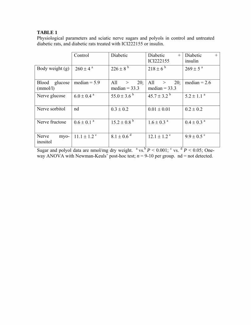
TABLE 1 Physiological parameters and sciatic nerve sugars and polyols in control and untreated diabetic rats, and diabetic rats treated with ICI222155 or insulin. Control Diabetic Diabetic +
ICI222155 Diabetic + insulin
Body weight (g) 260 ± 4 a 226 ± 8 b 218 ± 6 b 269 ± 5 a
Blood glucose (mmol/l)
median = 5.9 All > 20; median = 33.3
All > 20; median = 33.3
median = 2.6
Nerve glucose 6.0 ± 0.4 a 55.0 ± 3.6 b 45.7 ± 3.2 b 5.2 ± 1.1 a
Nerve sorbitol nd 0.3 ± 0.2 0.01 ± 0.01 0.2 ± 0.2
Nerve fructose 0.6 ± 0.1 a 15.2 ± 0.8 b 1.6 ± 0.3 a 0.4 ± 0.3 a
Nerve myo-inositol
11.1 ± 1.2 c 8.1 ± 0.6 d 12.1 ± 1.2 c 9.9 ± 0.5 c
Sugar and polyol data are nmol/mg dry weight. a vs.b P < 0.001; c vs. d P < 0.05; One-way ANOVA with Newman-Keuls’ post-hoc test; n = 9-10 per group. nd = not detected.

TABLE 2 Physiological parameters, indices of peripheral nerve function, and sugar and polyol levels measured in sciatic nerve and spinal cord, in control rats, untreated diabetic rats, and diabetic rats treated with IDD676. Control Diabetic Diabetic + IDD676
Body weight (g) 261 ± 4 a 223 ± 8 c 234 ± 11 b
Blood glucose (mmol/l)
median = 5.5 All > 20; median = 33.3
All > 20; median = 32.6
Paw withdrawal latency (sec)
8.6 ± 0.3 b 7.1 ± 0.4 a 9.2 ± 0.5 c
Motor nerve conduction velocity (m/s)
52.4 ± 1.3 a 46.9 ± 1.0 b 49.4 ± 1.6
Sensory nerve conduction velocity (m/s)
50.4 ± 1.2 a 46.2 ± 1.2 b 48.2 ± 1.0
Nerve glucose 4.2 ± 0.7 d 35.4 ± 2.8 a 47.1 ± 2.6 c
Nerve sorbitol nd 3.3 ± 1.3 1.1 ± 0.6
Nerve fructose 0.4 ± 0.1 d 14.5 ± 1.2 a 1.1 ± 0.5 d
Nerve myo-inositol 13.6 ± 1.2 9.4 ± 0.8 14.0 ± 2.3
Spinal cord glucose 1.7 ± 0.3 d 12.5 ± 1.4 a 12.1 ± 1.3 a
Spinal cord sorbitol nd nd nd
Spinal cord fructose 1.3 ± 0.2 d 3.1 ± 0.3 a 2.4 ± 0.2 a
Spinal cord myo-inositol
28.8 ± 0.7 27.6 ± 1.0 29.4 ± 0.5
Sugar and polyol levels are presented as nmol/mg dry weight. a vs.b P < 0.05; a vs.c P < 0.01; d vs.a,c P < 0.001; One-way ANOVA with Newman-Keuls’ post-hoc test; n = 8 per group. nd = not detected.

FIGURE LEGENDS FIG. 1. A: Western blots of lumbar spinal cord from control rats and rats with four weeks of diabetes, immunostained for COX-2 and β-actin. B: Densitometric quantification of COX-2-immunostained Western blots of lumbar spinal cord and C: COX-2 activity levels measured in lumbar spinal cord from control and diabetic rats. One-way ANOVA with Dunnett’s post-hoc test, n=5-6 per group. Control (C); rats with one week (1WK D), two weeks (2WK D), or four weeks (4WK D) of diabetes.

FIG. 2. A: Time course of formalin-evoked flinching in control and diabetic rats. B: Total flinches counted in 60 min after paw formalin injection in control and diabetic rats. One-way ANOVA with Dunnett’s post-hoc test, n=5-6 per group. Control (C); rats with one week (1WK D), two weeks (2WK D), or four weeks (4WK D) of diabetes.

FIG. 3. Flinches counted during phase I, the quiescent (Q) phase, and phase II of the formalin test. A: Untreated control rats and untreated diabetic rats. B: Diabetic rats treated intrathecally with 10 µl indomethacin vehicle (D+INDO V) or 10 µl vehicle containing 65 µg indomethacin (D+INDO). C: Diabetic rats treated intrathecally with 10 µl SC-58125 vehicle (D+SC-58125 V) or 10 µl vehicle containing 20 µg SC-58125 (D+SC-58125). For each panel, an unpaired t-test was used for each phase to compare matched groups, n=5-6 per group.

FIG. 4. A: Densitometric quantification of COX-2-immunostained Western blots of lumbar spinal cord and B: COX-2 activity levels measured in lumbar spinal cord from control rats (C), untreated diabetic rats (D), and diabetic rats treated with ICI222155 (D+ICI) or insulin (D+I). One-way ANOVA with Student-Newman-Keuls’ post-hoc test, n=9-10 per group.

FIG. 5. A: Densitometric quantification of COX-2-immunostained Western blots of lumbar spinal cord from control rats (C), untreated diabetic rats (D), and diabetic rats treated with IDD676 (D+IDD). B: Total flinches counted in 60 min after paw formalin injection in control rats (C), untreated diabetic rats (D), and diabetic rats treated with IDD676 (D+IDD). One-way ANOVA with Dunnett’s post-hoc test, n=8 per group.
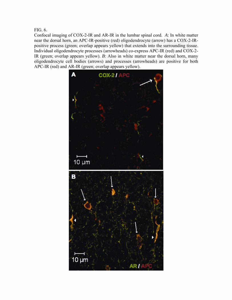
FIG. 6. Confocal imaging of COX-2-IR and AR-IR in the lumbar spinal cord. A: In white matter near the dorsal horn, an APC-IR-positive (red) oligodendrocyte (arrow) has a COX-2-IR-positive process (green; overlap appears yellow) that extends into the surrounding tissue. Individual oligodendrocyte processes (arrowheads) co-express APC-IR (red) and COX-2-IR (green; overlap appears yellow). B: Also in white matter near the dorsal horn, many oligodendrocyte cell bodies (arrows) and processes (arrowheads) are positive for both APC-IR (red) and AR-IR (green; overlap appears yellow).