parental dietary fat intake alters offspring … master mix of proteins and micronutrients before...
TRANSCRIPT

of July 6, 2018.This information is current as
Microbiome and ImmunityParental Dietary Fat Intake Alters Offspring
Paul J. Vithayathil, Julia A. Segre and Sandip K. DattaIan A. Myles, Natalia M. Fontecilla, Brian M. Janelsins,
http://www.jimmunol.org/content/191/6/3200doi: 10.4049/jimmunol.1301057August 2013;
2013; 191:3200-3209; Prepublished online 9J Immunol
MaterialSupplementary
7.DC1http://www.jimmunol.org/content/suppl/2013/08/12/jimmunol.130105
Referenceshttp://www.jimmunol.org/content/191/6/3200.full#ref-list-1
, 10 of which you can access for free at: cites 58 articlesThis article
average*
4 weeks from acceptance to publicationFast Publication! •
Every submission reviewed by practicing scientistsNo Triage! •
from submission to initial decisionRapid Reviews! 30 days* •
Submit online. ?The JIWhy
Subscriptionhttp://jimmunol.org/subscription
is online at: The Journal of ImmunologyInformation about subscribing to
Permissionshttp://www.aai.org/About/Publications/JI/copyright.htmlSubmit copyright permission requests at:
Email Alertshttp://jimmunol.org/alertsReceive free email-alerts when new articles cite this article. Sign up at:
Print ISSN: 0022-1767 Online ISSN: 1550-6606. All rights reserved.1451 Rockville Pike, Suite 650, Rockville, MD 20852The American Association of Immunologists, Inc.,
is published twice each month byThe Journal of Immunology
by guest on July 6, 2018http://w
ww
.jimm
unol.org/D
ownloaded from
by guest on July 6, 2018
http://ww
w.jim
munol.org/
Dow
nloaded from

The Journal of Immunology
Parental Dietary Fat Intake Alters Offspring Microbiome andImmunity
Ian A. Myles,* Natalia M. Fontecilla,*,1 Brian M. Janelsins,*,1 Paul J. Vithayathil,*
Julia A. Segre,† and Sandip K. Datta*
Mechanisms underlying modern increases in prevalence of human inflammatory diseases remain unclear. The hygiene hypothesis
postulates that decreased microbial exposure has, in part, driven this immune dysregulation. However, dietary fatty acids also
influence immunity, partially through modulation of responses to microbes. Prior reports have described the direct effects of
high-fat diets on the gut microbiome and inflammation, and some have additionally shown metabolic consequences for offspring.
Our study sought to expand on these previous observations to identify the effects of parental diet on offspring immunity usingmouse
models to provide insights into challenging aspects of human health. To test the hypothesis that parental dietary fat consumption
during gestation and lactation influences offspring immunity, we compared pups of mice fed either a Western diet (WD) fatty acid
profile or a standard low-fat diet. All pups were weaned onto the control diet to specifically test the effects of early developmental fat
exposure on immune development. Pups from WD breeders were not obese or diabetic, but still had worse outcomes in models of
infection, autoimmunity, and allergic sensitization. They had heightened colonic inflammatory responses, with increased circulating
bacterial LPS and muted systemic LPS responsiveness. These deleterious impacts of the WD were associated with alterations of the
offspring gut microbiome. These results indicate that parental fat consumption can leave a “lard legacy” impacting offspring
immunity and suggest inheritable microbiota may contribute to the modern patterns of human health and disease. The Journal
of Immunology, 2013, 191: 3200–3209.
The prevalence of multiple immune-mediated diseases con-tinues to increase in Western societies (1, 2). The hygienehypothesis, originally proposed as a reason for the inverse
correlation between family size and atopy (3), has expanded tobecome a potential explanation for the increasing prevalence ofinflammatory disorders. It postulates that modern decreases inmicrobial exposure affect immune development and promotedysregulated immunity (1, 4, 5). Animal studies of bacterial LPS,the prototypical surrogate of infectious exposure, and its mam-
malian receptor, TLR4, have led to modern iterations of the hy-giene hypothesis that propose environmental exposure to LPS earlyin life protects against the development of immune dysfunction (4).This is further supported by epidemiological studies that correlatea decreased risk for asthma and allergy in children from homes withhigh LPS levels (6) and a higher risk for multiple sclerosis amongaffluent populations (1).The modern increase in caloric intake and dietary fat in the
“Western diet” (WD) has also been correlated with both metabolicand immune-mediated diseases (7). Fatty acids have been shownto promote inflammatory responses through multiple mechanismsthat include direct action on immune cells, conversion into in-flammatory lipid mediators, and alteration of cell membranecharacteristics (8). Dietary fats can increase colonic permeabilityto gut microbial products such as LPS (9), triggering colonic andsystemic inflammation that may proceed to immune dysregulation.An additional potential link between dietary fats and the hygienehypothesis is the ability of free fatty acids to stimulate TLR4, mim-icking the saturated fatty acids that compose the bioactive lipid Amoiety of LPS (10–12). Multiple studies have correlated fatty acidintake with changes in both LPS response and the rates of“Western” diseases (13, 14) based primarily on epidemiologicaldata or on short-term responses to high-fat meals. Because therates of immune-mediated diseases have dramatically increased inthe population born since the late 1980s (15, 16), whose parentswere among the first with robustly excessive dietary saturated fatcalories (15), we hypothesized that alteration in dietary fat ex-posure during gestation and early perinatal development couldimpact the immune response later in life.
Materials and MethodsDietary exposure
We placed breeding mice on customized specialty diets with fatty acidcontent derived from natural oils (Table I). The diets were derived from
*Bacterial Pathogenesis Unit, Laboratory of Clinical Infectious Diseases, NationalInstitute of Allergy and Infectious Diseases, National Institutes of Health, Bethesda,MD 20892; and †Epithelial Biology Section, National Human Genome Research Insti-tute, National Institutes of Health, Bethesda, MD 20892
1N.M.F. and B.M.J. contributed equally to this work.
Received for publication April 19, 2013. Accepted for publication July 15, 2013.
This work was supported by the Office of Dietary Supplements and the NationalInstitutes of Health Intramural Research Program at National Institute of Allergy andInfectious Diseases and National Human Genome Research Institute.
I.A.M. designed, conducted, and analyzed the experiments, and wrote the manu-script; N.M.F. conducted or assisted on all experiments; B.M.J. performed all experi-ments involving colonic tissue and contributed to writing the manuscript; P.J.V.assisted with chromatin immunoprecipitation; J.A.S. conducted microbiome sequenc-ing and analysis; and S.K.D. oversaw design and analysis of the experiments, wrotethe manuscript, and had primary responsibility for the final content. All authorscritically read the manuscript.
The sequences presented in this article have been submitted to the National Center forBiotechnology Information Sequence Read Archive database (http://www.ncbi.nlm.nih.gov/Traces/sra/sra.cgi?study=SRP026657) under accession number SRP026657.
Address correspondence and reprint requests to Dr. Sandip K. Datta, Bacterial Patho-genesis Unit, Laboratory of Clinical Infectious Disease, National Institute of Al-lergy and Infectious Disease, National Institutes of Health, 9000 Rockville Pike,Building 33, Room 2W10A, Bethesda, MD 20892. E-mail address: [email protected]
The online version of this article contains supplemental material.
Abbreviations used in this article: EAE, experimental autoimmune encephalitis; LBP,LPS binding protein; LF, low fat; MRSA, methicillin-resistant Staphylococcus au-reus; NIAID, National Institute of Allergy and Infectious Diseases; Treg, regulatoryT cell; WD, Western diet.
www.jimmunol.org/cgi/doi/10.4049/jimmunol.1301057
by guest on July 6, 2018http://w
ww
.jimm
unol.org/D
ownloaded from

a master mix of proteins and micronutrients before the carbohydrates anddietary fats were added to ensure differences between diets were primarilyin fatty acid content and the fat/carbohydrate ratio. All diet pellets werepurchased from Research Diets (New Brunswick, NJ) with independentmass spectrometry content verification (Covance, Princeton, NJ). Massspectrometry of samples from two areas of each chow bag upon arrival and6 mo after storage at 280˚C confirmed the reported fatty acid content. Allsamples were within 5% of the reported content and had the expectedratios of fatty acids. There was ,7% breakdown of the fatty acids duringstorage, with maintenance of the fatty acid ratios.
The breeders used in the study were all littermates and were placed on thespecial diets 1 d before being placed in the breeding cages. Their pups werethus exposed to these diets in utero until birth and for an additional 3 wk viabreast milk. At 3 wk of age, the pups were weaned to new cages and all pupswere placed on our low-fat (LF) control diet. After 2–3 wk on the LF diet,the mice were placed into the challenge models described later and weremaintained on the LF diet for the duration of each experiment. Therefore,at time of challenge, the only difference between the mice tested was thedietary fat and carbohydrate consumed by their parents during gestationand nursing. For investigation of the effects of actively being on the WD,we placed mice on WD chow for 2 wk after weaning from breeders ona standard diet.
Mice
BALB/c and C57BL/6 mice were purchased from Jackson Laboratories(Bar Harbor, ME) to set up breeders. Littermates were used as the breedersthat were exposed to the experimental diets. Two to three breeder pairs perdietary group were maintained active at all times for approximately 5 moeach. The breeders were renewed as a unit on two separate occasions duringthe study; all breeder cages were stopped and replaced with new breedingpairs. For cohousing experiments, mice from at least two breeders were usedso that a cage with two pups from a given diet group would come from twodifferent breeding pairs. Mice were given autoclaved, acidified water (pH2.7–3.1). The bedding provided was Maple Sani Chip (Harlan Laborato-ries, Indianapolis, IN). All animal experiments were done in compliancewith the guidelines of the National Institute of Allergy and InfectiousDiseases (NIAID) Institutional Animal Care and Use Committee in spe-cific pathogen-free National Institutes of Health animal care facilities thatwere documented to be free of Norovirus and Helicobacter.
Pulmonary fat content
Lungs from mice were harvested 1 wk after weaning and stored in PBS at280˚C. Mass spectrometry was performed at Covance (Madison, WI).
Escherichia coli sepsis
Mice were infected i.p. with 104 CFUs E. coli K1018 (gift from M. Lu,NIAID) and followed for 2 wk for evidence of moribundity.
Staphylococcus aureus infections
A total of 107 CFU USA300 strain of methicillin-resistant Staphylococcusaureus (MRSA; gift from F. DeLeo, Rocky Mountain Laboratories, NIAID)withCytodexbeads (Sigma, St. Louis,MO)was injected intradermally (100ml)into the shaved back of eachmouse. Resultant abscess size, bacterial burden,and skin cytokine analysis were done as previously described (17). TaqManprobes for TLR2 (Mm00439614_m1*), IL-17A (Mm00439619_m1*), DefB4(Mm00731768_m1*), IL-1b (Mm01336189_m1*), IL-10 (Mm00442346_m1*),vitamin D receptor (Mm00437297_m1*), and CYP27b1 (Mm01165918_g1)were purchased from Life Technologies. Comparison of signal was performedusing the DD–cycle threshold method.
Experimental autoimmune encephalitis
Mice were injected with 200 mg MOG protein (AnaSpec, Fremont, CA)with 300 mg CFA (Difco, Franklin Lakes, NJ) s.c. on day 0. They were alsoinjected with 500 ng pertussis toxin (List Biological Laboratories, Campbell,CA) i.p. on days 0 and 2. They were monitored and scored daily based onthe following scale: 1, limp tail; 2, paralysis of one hind leg; 3, paralysis ofboth legs; 4, paralysis or clumsiness in either front leg; and 5, death.They were scored by animal care facility technicians who were blinded andindependent of our study.
Anaphylaxis
For 4 wk, mice were sensitized by weekly gavage with 1 mg peanut protein(Protein Plus, Fitzgerald, GA) and 20 mg cholera toxin (List BiologicalLaboratories, Campbell, CA) in 200 mcl PBS (Cellgro, Manassas VA). Thirtyminutes before each sensitization, mice were gavaged with 150 mcl bicar-
bonate (Mallinckrodt, Phillipsburg, NJ). On the last day of sensitization, pe-ripheral temperature transponders were injected s.c. (Bio Medic Data Systems,Seaford, DE). One day later, blood was taken to measure peanut-specificIgG and IgE. Then mice were challenged with 5 mg peanut protein i.p.Temperature was measured every 5–15 min for 60–90 min. Anaphylaxisscores were taken at time of temperature measurement and based on the fol-lowing scale: 1, face scratching or swelling around the eyes; 2, heavy breathingor raised hair; 3, no spontaneous movement, other than breathing; 4, nomovement when prodded or seizures; and 5, death. A similar protocol wasfollowed to test anaphylactic response to OVA instead of peanut, using 1 mgfor sensitization and 5 mg for challenge.
Colon stimulation
Whole colons were excised, washed, weighed, and placed in 1 ml DMEM(Cellgro) with 10% FBS (Thermo, Dubuque, IA) for 24–72 h in the presenceof 100 ng/ml LPS (List Biological Laboratories, Campbell, CA). Cytokinelevels of the supernatants were measured with the Bio-Plex suspension arraysystem on a Bio-Plex 200 (Bio-Rad, Hercules, CA).
Splenocyte stimulation
Single-cell suspensions of live splenocytes (2e6/ml) were stimulated with100 ng/ml LPS (List Biological Laboratories) or 1 mg/ml Con A (Sigma) at37˚C under 5% CO2 in MEM (Mediatech, Manassas, VA) supplementedwith 10% newborn calf serum (HyClone, from Thermo Scientific, Dubuque,IA), and 100 U/ml penicillin and 100 g/ml streptomycin (MEM/newborn calfserum) for 24–72 h. Cytokines were measured with the Bio-Plex suspensionarray system on a Bio-Plex 200 (Bio-Rad).
Regulatory T cell detection
Colons were removed and flushed with cold 10% FBS/HBSS solution toclear fecal material. Next, colons were opened via a longitudinal incision,then cut laterally into three pieces. Samples were then incubated in a 10%FBS/HBSS solution supplemented with 0.05M EDTA for 15 min at 37˚Cwith 125 rpm shaking. After incubation, the epithelial layer was gently scrapedoff and colons were minced, exposing the lamina propria. Processed sampleswere then incubated in a 20% FBS/HBSS solution supplemented with 2%collagenase, 2% Dispase, and 0.1 mg DNAse for 1 h at 37˚C with 185 rpmshaking. Digested tissue and solution were then passed through 100- and 40-mm filters to obtain single-cell suspensions. After thorough washing withcold 13 PBS, colonic lamina propria cells were labeled with LIVE/DEADfixable violet dead stain (Invitrogen, Grand Island, NY), anti-mouse CD45(FITC), anti-mouse CD25 (allophycocyanin or PE-CY7), anti-mouse CD4(PE-CY7 or allophycocyanin), and anti-mouse Foxp3 (PE), using Foxp3staining buffer reagents (eBioscience, San Diego, CA). Flow cytometric datawere acquired on an LSRFortessa (BD Bioscience, San Jose, CA) and an-alyzed with FlowJo software (Tree Star, Ashland, OR). Splenic regulatoryT cells (Tregs) were detected in a similar manner after obtaining single-cellsuspensions (2e6/ml).
Macrophage recruitment and processing
Mice were injected with 1 ml sterile Brewer thioglycolate broth (Difco Lab-oratories, Detroit, MI), and peritoneal cells were harvested 5 d later as pre-viously described (18). Cells from 3–5 mice were pooled. RNAwas extractedusing the RNeasy Kit (Qiagen) per the manufacturer’s instructions andevaluated by RT-PCR identically to skin mRNA processing. TaqManprobes for TLR4 (Mm00445273_m1*) and LPS binding protein (LBP;Mm00493139_m1*) were from Life Technologies.
Liver LPS content
Livers were harvested from mice and homogenized using TissueLyser(Qiagen, San Diego, CA) in a 2-ml tube (Eppendorf, Hauppauge, NY) witha steel ball bearing (Qiagen). The homogenized liquid was assayed for LPScontent using a commercial kit (Hycult, Plymouth Meeting, PA).
Chromatin immunoprecipitation
Immunoprecipitation of sheared chromatin from splenocytes was doneaccording to theMilliporeMagna-Chromatin Immunoprecipitation protocolas previously described (19), using Protein G beads (Millipore, Temecula, CA)and anti-H3K9Me3 or isotype control IgG (Millipore). QIAquick PCR puri-fication kit (QIAGEN) was then used to purify DNA. Quantitative PCRwas performed using SYBR green–labeled primers (Applied Biosystems,Foster City, CA). Primers (forward; reverse) were as follows: Gapdh (59-GT-CATCATCTCCGCCCCTTCTGC-39; 59-GATGCCTGCTTCACCACCTTCT-TG-39), LBP (59-GGACAGCAACTCCCTAACTTACCC-39; 59-GCAGAA-ATGGAACCCAGGCTAC-39), CD14 (59-CAGAGAACACCACCGCTGTA-
The Journal of Immunology 3201
by guest on July 6, 2018http://w
ww
.jimm
unol.org/D
ownloaded from

AAG-39; 59-AGATTCCTCAGGTTGGCTCCAG-39), and TLR4 (59-TTCA-GGGCTTTCTGTGGGAAC-39; 59-AGATTTTCATCAGGCTTGGCAG-39).
Microbiome analysis
DNAwas extracted from sterilely excised cecal stool pellets using QIAampDNA stool mini kit (Qiagen). Female BALB/c mice were used. Quantitativeanalysis of 16S rDNAwas performed as previously described on 1000–3000sequences/sample using the established primer sequences (20). All micro-biome sequencing data were uploaded to the National Center for Biotech-nology Information Sequence Read Archive database under accession numberSRP026657; this can be accessed at http://www.ncbi.nlm.gov/Traces/sra/sra.cgi?study=SRP026657.
Ab measurements
Serum was drawn from orally sensitized mice 1 d before anaphylaxis chal-lenge. Total IgE was detected by ELISA using commercial kits per man-ufacturer’s instructions (Bethyl Laboratories, Montgomery, TX). For peanut-specific Abs, serum was incubated in immunoplates (Thermo Scientific,Dubuque, IA) coated with whole peanut protein (Protein Plus, Fitzgerald,GA). After 1 h nonspecific protein blockade with 10% FBS (Thermo Sci-entific), the plate was washed with PBS and 0.05% Tween (Acros, Pitts-burgh, PA). ELISA assay for IgG was performed using commercial kits(Bethyl Laboratories). For peanut-specific IgE, serum was added to ELISAplates coated with anti-IgE (Bethyl Laboratories). Whole peanut protein,biotinylated with a commercial kit (Anaspec, Fremont, CA), was added.Colorimetric detection using streptavidin-HRP and TMB reagents (Sigma)was done on a Beckman Coulter DTX880.
Vitamin D assay
Vitamin D3 levels were evaluated by ELISA per the manufacturer’sinstructions (ALPCO, Windam, NH).
Weight and glucose monitoring
Pups from each litter were weighed weekly after weaning. For fasting bloodglucose measurements, food was removed at 3 pm. At 9 am the followingmorning, a small incision was made in the tail vein and glucose was measuredon the second drop of blood using the Freestyle Lite (Abott Diabetes Care,Alameda, CA).
Statistics
Means were compared using either two-tailed unpaired t test or ANOVAwith Bonferroni’s posttest correction for multiple-group comparison withPrism software (GraphPad, San Diego, CA). The p values are designated asfollows: *p, 0.05, **p, 0.01, ***p, 0.001, or ****p, 0.0001. NS, Notsignificant.
Declaration of approval for animal studies
All animal experiments were done in compliance with the guidelines of theNIAID Institutional Animal Care and Use Committee.
ResultsDietary exposure
The modern WD departs from the recommended diet in 3 majorways: high percentage of calories from fat (40% rather than 30%)(21), increased ratio of v6/v3 fatty acids (8–15:1 rather than 2:1)(21), and twice the recommended simple carbohydrates (22). Toisolate the effects of dietary fatty acids, we formulated experimentaldiets that did not contain excess sugars. Our WD formulation re-flected the average American diet in both fat percentage and source(saturated fat predominantly from palm oil and v6 from soy) (22)but did not provide excess simple carbohydrates. The LF controldiet was essentially identical to standard mouse chow. All diets hadequivalent micronutrient composition and caloric density, with thedifferences in fatty acid content being accounted for by changes incarbohydrate content (Table I). Breeders were placed on the diets1 d before placement in breeding cages and remained on the dietsthroughout the study. Pups were exposed to these diets in utero andduring nursing. All pups from all breeders were weaned to newcages and placed on the LF control diet for 2–3 wk before immu-nologic challenge. Thus, the primary difference in dietary exposure
between the mice tested was the fatty acid composition and the fat/carbohydrate ratio consumed by their parents during gestation andlactation. Fig. 1 provides an overview of the study design. Lung fattyacid composition has been shown to reflect dietary fat intake (23),and analysis of lung tissue by mass spectrometry confirmed differ-ential saturated fat exposure in the pups from WD and LF breeders(Supplemental Fig. 1A, 1B). Although the WD and LF chow haddifferent v3 and v6 fatty acid composition, there was no differ-ence in the v3 or v6 content in the lung (Supplemental Fig. 1B).This may be because the smaller differences, compared with satu-rated fat exposure, were below the assay detection capability orbecause migration of unsaturated fatty acids across the placentamay be regulated differently than saturated fats. Importantly, therewere no differences between WD and LF pups in fasting bloodglucose or weight (Supplemental Fig. 1C–E), indicating any ob-served differences between these mice would not be confoundedby diabetes or obesity. Of note, both groups ate identical LF chowpostweaning, and the lack of weight differences correlated withour finding that the amount of food consumed per mouse per weekdid not differ between the LF or WD pups (data not shown).
Pups from WD breeders had altered disease susceptibility
Because direct fatty acid exposure has been reported to affect LPSresponses (9–12), and immune responses to Gram-negative bac-teria directly involve LPS-triggered innate immunity, we firstevaluated the impact of parental fatty acid intake on pup suscep-tibility to a model of Gram-negative bacterial sepsis. Pups fromWD breeders injected i.p. with the clinical isolate E. coli K1018had significantly greater mortality than pups from LF breeders(Fig. 2A). To test whether the effects of WD exposure extended toinfectious agents that do not contain LPS and thus do not bind toTLR4, we next used a skin infection model with the Gram-positivebacterium MRSA. Suggesting that WD exposure resulted in im-mune modulation beyond direct effects on TLR4, WD pups de-veloped larger abscesses with greater bacterial burdens comparedwith LF pups (Fig. 2B, 2C). Saturated fats have also been reportedto influence signaling by TLR2 (10), the receptor for microbiallipoteichoic acid and peptidoglycan, both of which are producedby S. aureus, induce an inflammatory cascade, and have beenimplicated in the hygiene hypothesis (4). In the abscess tissue ofWD pups, we found a significant reduction in transcript levels ofcentral mediators of the cutaneous anti-MRSA response: TLR2,IL-1b, IL-17A, and b-defensin 4 (24), as well as the regulatorycytokine IL-10 (Fig. 2D). WD pups also showed reduced ex-pression of the vitamin D receptor and the vitamin D activatingenzyme, CYP27b1 (Fig. 2D), both of which are induced by TLR2
Table I. Fatty acid content and source for the diets studied
Dietary Components Human RD v6 Western LF
Protein (% kCal) 20 20 20 20Carb (% kCal) 50 40 40 70Fat (% kCal) ,30 40 40 10% Fat saturated 10 10 40 10% Fat PUFA 77 21 72% Fat MUFA 14 39 18Saturated fat source — Palm oil —v6 source Safflower Soy Soy/Safflowerv3 source – Menhaden Flaxseedv6/v3 ratio 2:1 63:1 8:1 2:1
Breakdown of dietary components in the diets studied are shown, includingprotein, carbohydrates (carb), fat, and % of fat that was saturated, polyunsaturatedfatty acids (PUFA), or monounsaturated fatty acids (MUFA). The dietary source foreach fatty acid is shown. All diets were made from natural oils. Human recommendeddiet (RD) reflects the guidelines of the U.S. Department of Agriculture.
3202 THE WESTERN DIET ALTERS OFFSPRING IMMUNITY
by guest on July 6, 2018http://w
ww
.jimm
unol.org/D
ownloaded from

stimulation and mediate antistaphylococcal immune activity (25).There were no differences in serum vitamin D levels (data notshown), suggesting the observed effects on vitamin D metabolismwere not systemic but occurred within the context of the anti-MRSA immune response.LPS responsiveness has also been implicated in autoimmune and
allergic disease (26, 27). To test the effect of fat exposure on thesedisease states, we examined these pups in established models ofexperimental autoimmune encephalitis (EAE) and oral peanut sen-sitization. In BALB/c mice, which are relatively resistant to EAE(28), WD pups were more likely to develop signs of EAE (Fig. 2E),but the severity of disease was similar to the LF pups (data notshown). In the more susceptible C57BL/6 mouse strain (28), WDpups showed both a higher incidence and more severe manifes-tations of EAE compared with LF pups (Fig. 2F). WD pups sen-sitized orally to peanut protein had more pronounced temperaturedeclines in response to challenge, the most sensitive measure ofmurine anaphylaxis (29) (Fig. 2G). Clinical anaphylaxis scores mir-rored the temperature findings, but differences did not reach statisticalsignificance (Fig. 2H). There was no difference between groups inthe induced levels of total IgE, peanut-specific IgE, or peanut-specificIgG (Supplemental Fig. 2A–C). Mice were sensitized to OVA in anidentical manner to test the effect of dietary fat on anaphylactic re-sponse against another Ag. There were no differences between WDand LF groups in temperature change during OVA challenge (Sup-plemental Fig. 2D). Peanut sensitization of the less susceptibleC57BL/6 mice (30) also revealed that exposure to WD had noeffect on anaphylaxis compared with LF controls (SupplementalFig. 2D). Given the lack of effect in the OVA model and the re-quirement for genetic predisposition, it seems that the WD had astronger effect on sepsis and autoimmunity than on allergic sen-sitization. Taken together, these data suggest that WD fat exposureduring gestation and early life increased susceptibility to a rangeof infectious and immune-mediated diseases.
WD pups had hyperinflammatory colonic responses butdecreased systemic responses to LPS
To explore the immunological basis for the effect of WD exposureon disease susceptibility, we first compared colonic immune responses
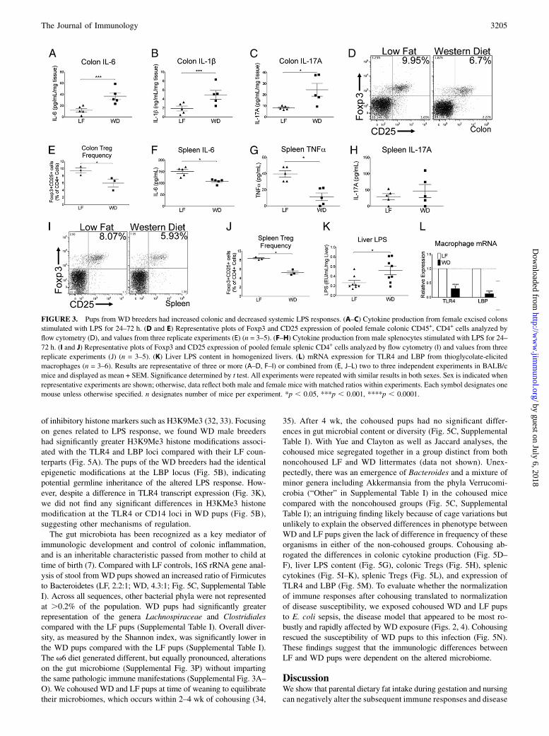
in WD and LF pups. Other studies have shown that a high-fat dietcan produce a low-grade inflammatory response in the colon (9).We postulated that this might drive altered systemic immunitybecause the gut is a major site for immunological education. Inresponse to ex vivo LPS stimulation, WD pup colons producedenhanced levels of IL-6, IL-1b, and IL-17A (Fig. 3A–C), sug-gesting a hyperinflammatory milieu. WD pups had reducedfrequency of colonic Tregs (Fig. 3D, 3E), further indicatingdysregulated gut immunity. In contrast, splenic LPS responses inWD pups suggested a muted systemic LPS response, with reducedproduction of TNF-a and IL-6, but no differences in IL-17A (Fig.3F–H) or IL-1b (data not shown). Similar to the colonic Treg find-ings, WD pups had reduced frequency of splenic Tregs (Fig. 3I, 3J).WD and LF pups did not significantly differ in splenocyte produc-tion of IL-4, IL-5, IL-13, IL-17A, or IFN-g in response to Con A(data not shown), suggesting no baseline skewing of effector T cellpolarization in WD pups. The colonic inflammatory response toa high-fat diet has been shown to increase LPS leakage from thecolon into the portal circulation (9). Consistent with this finding, wefound the LPS content in liver tissue was higher in WD pups (Fig.3K). Macrophage TLR4 and LBP mRNA expression was sup-pressed in WD offspring (Fig. 3L), suggesting a downregulatedcapacity for LPS signaling after increased LPS exposure.These effects of the WD could stem from the increased saturated
fats, the skewed v6/v3 ratio, or both. To isolate the contribution ofhigh dietary v6, we evaluated offspring from breeders fed a high-fat diet with an overrepresented v6/v3 ratio but low saturated fatcontent (Fig. 1A, Table I). Compared with pups from LF breeders,pups from the v6 diet breeders had mild increases in only a subsetof colonic inflammatory markers (Supplemental Fig. 3A–C). Theyshowed trends toward enhanced susceptibility to infection andEAE (Supplemental Fig. 3D–G), but these did not achieve thestatistically significant differences seen in the WD pups. In furthercontrast with WD pups, v6 pups were protected against allergicsensitization (Supplemental Fig. 3H–L). Taken together, these datasuggest that the high saturated fat content of the WD was required toinduce colonic inflammation, resulting in increased systemic LPSexposure and reduced LPS responsiveness that may have contributedto immune dysregulation and disease susceptibility in WD pups.
FIGURE 1. Diagrammatic presentation of study
design. (A) For experiments evaluating the effects
of parental diet, littermate mice were placed on
either LF or WD formulations 1 d before being
placed in breeding cages. Breeder mice were main-
tained on the different diets throughout gestation
and nursing. When the pups were 3 wk postpartum,
they were weaned to new cages. All pups were
weaned onto the LF control diet. Two to 4 wk after
weaning, the mice were evaluated in the described
models. (B) For evaluation of the effects of active
diet consumption, the converse experiment was
performed. Pups from breeders on the LF control
diet were weaned into new cages and placed on
either the WD or LF control diet. (C) For experi-
ments involving cohousing of mice, pups from both
LF and WD breeders were weaned into the same
cage and both placed on the LF control diet.
The Journal of Immunology 3203
by guest on July 6, 2018http://w
ww
.jimm
unol.org/D
ownloaded from

Active consumption of WD did not fully recapitulate thephenotype of mice exposed during early development
The immune phenotype of WD pups conceivably representeda direct and residual effect of saturated fat consumption during the
3 wk of gut exposure through breast milk. In addition, newborn mice
may sample the food eaten by their parents, indicating a window
wherein direct consumption could be the cause of our observed
phenotypes. To test whether such direct exposure could account
for the observed immune modulation, we performed the converse
of the previous experiments, placing female mice on WD chow
after weaning from breeders fed a standard diet (Fig. 1B). Consistent
with previous reports (31), active WD consumption decreased the
survival rate from sepsis (Fig. 4A). However, active consumption
did not affect MRSA-induced skin lesion size (Fig. 4B) or tran-
script levels of anti-MRSA cytokines (Fig. 4C). Active intake of the
WD also did not impact susceptibility to EAE (Fig. 4D) or peanut
anaphylaxis (Fig. 4E, 4F). Active ingestion increased colonic IL-6
production in response to LPS (Fig. 4G), but did not affect other
tested cytokines or colonic Treg frequency (Fig. 4H–J). Similar to
mice exposed only early in development, active WD consumption
increased liver LPS concentrations and reduced splenocyte IL-6 andTNF-a responses (Fig. 4L–N). However, splenic Treg frequencywas not altered (Fig. 4O). Thus, exposure to the WD after weaningappeared to partially alter responses directly related to inflam-matory effects on the colon but could not fully recapitulate theimmune dysregulation seen in mice exposed during prenatal andperinatal development. It is possible that prolonged adult exposureto the WD would further modulate immune responses, but theseresults suggest that the observed phenotype of WD pups requiredeither prenatal or perinatal parental WD exposure and could not beexplained solely by the direct postnatal WD exposure during nursing.
Inheritance of the WD immune phenotype was dependent onaltered gut microbiota
Beyond direct exposure, potential explanations for the alteredimmune phenotype in WD pups include paternal germline epi-genetic changes and/or an altered maternal microbiome, both ofwhich could be transmitted and influence pup immune responses.Previous studies have linked high-fat diets to epigenetic alterations
FIGURE 2. Pups from WD breeders
had altered disease susceptibility. (A) Sur-
vival postinfection with E. coli K1018 in
BALB/c mice (n = 10–19). (B–D) Staph-
ylococcus aureus (MRSA USA300) skin
infection in male BALB/c mice. Lesion
sizes (B), day 6 bacterial counts from
homogenized skin (C), and mRNA ex-
pression in skin abscess tissue normalized
against LF controls (D, dotted line) (n =
5–6). (E) Disease-free survival after in-
duction of EAE in female BALB/c mice
(n = 6). (F) EAE scores in C57BL/6 mice
(n = 7–12). (G and H) Weaned male
BALB/c pups were gavaged with peanut
protein and cholera toxin weekly for 4–8
wk before challenge. Temperature decline
(G) and symptom scores (H) after chal-
lenge (n = 5). Results are representative
of three or more (B, E–H) or combined
from two to four (A, C, D) independent
experiments and displayed as mean +
SEM. Significance determined by t test
(A–C, E–H) or ANOVA with Bonferroni’s
correction (D). All experiments were re-
peated with similar results in both sexes.
Sex is indicated when representative ex-
periments are shown; otherwise, data
reflect both male and female mice with
matched ratios within experiments. Each
symbol designates one mouse unless
otherwise specified. n designates mouse
number per group. *p , 0.05, **p ,0.01, ***p , 0.001, ****p , 0.0001.
3204 THE WESTERN DIET ALTERS OFFSPRING IMMUNITY
by guest on July 6, 2018http://w
ww
.jimm
unol.org/D
ownloaded from
of inhibitory histone markers such as H3K9Me3 (32, 33). Focusingon genes related to LPS response, we found WD male breedershad significantly greater H3K9Me3 histone modifications associ-ated with the TLR4 and LBP loci compared with their LF coun-terparts (Fig. 5A). The pups of the WD breeders had the identicalepigenetic modifications at the LBP locus (Fig. 5B), indicatingpotential germline inheritance of the altered LPS response. How-ever, despite a difference in TLR4 transcript expression (Fig. 3K),we did not find any significant differences in H3KMe3 histonemodification at the TLR4 or CD14 loci in WD pups (Fig. 5B),suggesting other mechanisms of regulation.The gut microbiota has been recognized as a key mediator of
immunologic development and control of colonic inflammation,and is an inheritable characteristic passed from mother to child attime of birth (7). Compared with LF controls, 16S rRNA gene anal-ysis of stool fromWD pups showed an increased ratio of Firmicutesto Bacteroidetes (LF, 2.2:1; WD, 4.3:1; Fig. 5C, Supplemental TableI). Across all sequences, other bacterial phyla were not representedat .0.2% of the population. WD pups had significantly greaterrepresentation of the genera Lachnospiraceae and Clostridialescompared with the LF pups (Supplemental Table I). Overall diver-sity, as measured by the Shannon index, was significantly lower inthe WD pups compared with the LF pups (Supplemental Table I).The v6 diet generated different, but equally pronounced, alterationson the gut microbiome (Supplemental Fig. 3P) without impartingthe same pathologic immune manifestations (Supplemental Fig. 3A–O). We cohoused WD and LF pups at time of weaning to equilibratetheir microbiomes, which occurs within 2–4 wk of cohousing (34,
35). After 4 wk, the cohoused pups had no significant differ-ences in gut microbial content or diversity (Fig. 5C, SupplementalTable I). With Yue and Clayton as well as Jaccard analyses, thecohoused mice segregated together in a group distinct from bothnoncohoused LF and WD littermates (data not shown). Unex-pectedly, there was an emergence of Bacteroides and a mixture ofminor genera including Akkermansia from the phyla Verrucomi-crobia (“Other” in Supplemental Table I) in the cohoused micecompared with the noncohoused groups (Fig. 5C, SupplementalTable I); an intriguing finding likely because of cage variations butunlikely to explain the observed differences in phenotype betweenWD and LF pups given the lack of difference in frequency of theseorganisms in either of the non-cohoused groups. Cohousing ab-rogated the differences in colonic cytokine production (Fig. 5D–F), liver LPS content (Fig. 5G), colonic Tregs (Fig. 5H), spleniccytokines (Fig. 5I–K), splenic Tregs (Fig. 5L), and expression ofTLR4 and LBP (Fig. 5M). To evaluate whether the normalizationof immune responses after cohousing translated to normalizationof disease susceptibility, we exposed cohoused WD and LF pupsto E. coli sepsis, the disease model that appeared to be most ro-bustly and rapidly affected by WD exposure (Figs. 2, 4). Cohousingrescued the susceptibility of WD pups to this infection (Fig. 5N).These findings suggest that the immunologic differences betweenLF and WD pups were dependent on the altered microbiome.
DiscussionWe show that parental dietary fat intake during gestation and nursingcan negatively alter the subsequent immune responses and disease
FIGURE 3. Pups from WD breeders had increased colonic and decreased systemic LPS responses. (A–C) Cytokine production from female excised colons
stimulated with LPS for 24–72 h. (D and E) Representative plots of Foxp3 and CD25 expression of pooled female colonic CD45+, CD4+ cells analyzed by
flow cytometry (D), and values from three replicate experiments (E) (n = 3–5). (F–H) Cytokine production from male splenocytes stimulated with LPS for 24–
72 h. (I and J) Representative plots of Foxp3 and CD25 expression of pooled female splenic CD4+ cells analyzed by flow cytometry (I) and values from three
replicate experiments (J) (n = 3–5). (K) Liver LPS content in homogenized livers. (L) mRNA expression for TLR4 and LBP from thioglycolate-elicited
macrophages (n = 3–6). Results are representative of three or more (A–D, F–I) or combined from (E, J–L) two to three independent experiments in BALB/c
mice and displayed as mean + SEM. Significance determined by t test. All experiments were repeated with similar results in both sexes. Sex is indicated when
representative experiments are shown; otherwise, data reflect both male and female mice with matched ratios within experiments. Each symbol designates one
mouse unless otherwise specified. n designates number of mice per experiment. *p , 0.05, ***p , 0.001, ****p , 0.0001.
The Journal of Immunology 3205
by guest on July 6, 2018http://w
ww
.jimm
unol.org/D
ownloaded from

susceptibility of offspring mice. The inheritance of this immunephenotype is associated with an altered gut microbiota. Prior reportshave described the direct effects of high-fat diets on the gut micro-biome and inflammation, and some have additionally shown meta-bolic consequences for offspring (31–39). Our study has expanded onthese previous observations to identify the effects of parental diet onoffspring immunity. Seeding of microbiota occurs from the motherduring parturition and further diversifies during early life (40). Fattyacid exposure causes rapid changes in the microbiotic composition(41), implying that diet-induced changes in the maternal microbiotawere passed on to offspring in our studies. Studies on the durabilityof these inherited alterations in microbiota would provide additionalinformation on disease susceptibility as would direct comparison ofmaternal versus offspring microbiota. Because of the coprophagic(stool-consuming) habits of mice, cohousing has been shown to bean effective means of transferring microbiota between mice, gen-erating similar microbial shifts and immunological effects com-pared with direct fecal or microbial transfer (34, 42, 43). The resultsfrom our cohousing experiments thus implicate altered microbiotaas the most likely driver of the observed immunological phenotypes.We also found limited inheritance of paternal epigenetic changesconsistent with prior observations of epigenetic influences on met-abolic, developmental, and cardiovascular dysregulations (44–46).However, microbiome alteration by cohousing superseded potential
contributions of these epigenetic changes in our studies. Futureidentification and targeting of species-level changes in the micro-biota promises the possibility of reversing or preventing harmful di-etary effects through isolation and transfer of specific gut organisms.High-fat diet effects have been characterized by increased Gram-
negative bacteria and an increased Firmicutes/Bacteroidetes ratioin the gut microbiome (47), increased colonic inflammation andpermeability (9), and decreased Treg frequency (48). We find thatsimilar effects are inherited by progeny of mice fed a WD and arereversed by subsequent microbiota alteration, suggesting that thechanges in microbiota are a primary effector of the diet-inducedimmune effects. Multiple mechanisms have been proposed to drivethe influences of the microbiota on host immunity, including gutnutrient utilization, microbial metabolic products such as short-chain fatty acids, and differential triggering of gut immune responses(47). Similarly, dietary fats likely alter gut microbiota compositionthrough multiple mechanisms, including altered microbial nutrientavailability and host inflammatory effects. The reported ability ofsaturated fats to directly trigger inflammatory TLR4 signaling (9–12), and the increased levels of LPS in the circulation after dietaryfat exposure in our study and others (9), raises the intriguing pos-sibility that modern diets alter our exposure to TLR4 signaling,potentially resulting in a systemic hyporesponsiveness to LPS thatparadoxically mimics the low LPS exposure postulated by the
FIGURE 4. Postweaning exposure
to WD did not recapitulate the phe-
notype of mice exposed during de-
velopment. Pups from breeders on
a standard diet were placed at 3 wk
of age on LF or WD for 2 wk before
challenge. (A) Survival after injec-
tion with E. coli K1018. (B) Lesion
size in male mice and (C) mRNA ex-
pression in skin abscess tissue nor-
malized against LF controls (dotted
line) after injection with MRSA. (D)
Disease-free survival in female mice
after induction of EAE. (E and F)
Temperature change and symptom
scores for orally sensitized male mice
after challenge with peanut protein.
(G–I) Colonic cytokine induction by
LPS in female mice. (J) Represen-
tative plots of Foxp3 and CD25 ex-
pression of pooled female colonic
CD45+, CD4+ cells analyzed by flow
cytometry (n = 5). (K) Liver LPS
content. (L–N) Splenic cytokine in-
duction by LPS in male mice. (O)
Representative plots of Foxp3 and
CD25 expression of female splenic
CD4+ cells analyzed by flow cytom-
etry (n = 5). Significance determined
by t test. Results are representative
of 2–3 independent experiments, 5–
10 sex- and age-matched BALB/c
mice/group unless designated as in-
dividual symbol, and displayed as
mean + SEM.*p, 0.05, **p, 0.01,
****p , 0.0001.
3206 THE WESTERN DIET ALTERS OFFSPRING IMMUNITY
by guest on July 6, 2018http://w
ww
.jimm
unol.org/D
ownloaded from

hygiene hypothesis to partially drive immune dysregulation. Al-though previous work has established TLR4-dependent effects ofdietary fat on gut inflammation (49), the altered intrinsic suscep-tibility of TLR4-deficient mice to the tested models of infection,autoimmunity, and allergy may complicate future evaluation of therole of TLR4 in dietary fat-induced development of disease. Fur-thermore, the immune dysregulation we observed extended beyonddirect effects on TLR4 signaling, as evidenced by alteration of au-toimmunity, vitamin D regulation, and TLR2 expression. In ad-dition, dysregulation of Treg cells, which are protective againstsepsis (50), S. aureus skin disease (51), autoimmunity (52), andallergic sensitization (53), appeared to correlate with the pathol-ogy generated by the altered microbiome in our studies. Trendingincreases in the incidence of sepsis may be explained by the aging
population and invasive medical procedures, but it is interesting tonote that, similar to the other diseases, sepsis is characterized byimmune dysregulation that may contribute to disease suscepti-bility (2). It appears that broad immune dysregulation inducedby altered microbiota contributed to the range of disease suscep-tibility observed in our studies. Direct changes in LPS respon-siveness by dietary fat likely dominated the susceptibility to sepsisseen in both adult and offspring mice, whereas indirect or devel-opmental effects on Tregs and other immune compartments maybe more important for allergy and autoimmunity models thatmanifested in the offspring. Although direct exposure postwean-ing did not mimic the effects of early life saturated fat exposure,our studies do not absolutely distinguish between effects of intra-uterine and breastfeeding exposure in the offspring. Cross-fostering
FIGURE 5. Cohabitation rescued WD pups from immune alterations. (A and B) Quantitative PCR of selected genes after anti-H3K9Me3 immuno-
precipitation of splenic DNA from breeders and both male and female offspring, displayed as the ratio of input DNA and normalized to isotype Ab control.
(C) 16S rRNA genes in cecal stool samples of female mice. Pups from indicated breeder diets were all weaned to LF diet. Female pups were placed in cages
with their littermates or cohoused for 4 wk with pups from breeders fed opposing diet. Each bar represents one mouse; further breakdown of composition
within each phylum can be found in Supplemental Table I. (D–F) Colonic cytokine induction by LPS in cohoused mice. (G) LPS content in homogenized
liver tissue from cohoused mice. (H) Representative plots of Foxp3 and CD25 expression of pooled colonic CD45+, CD4+ cells from cohoused mice (n = 3–
5) analyzed by flow cytometry. (I–K) LPS induction of cytokines from splenocytes in cohoused mice. (L) Representative plots of Foxp3 and CD25 ex-
pression of splenic CD4+ cells from cohoused mice analyzed by flow cytometry (n = 3–5). (M) mRNA levels of TLR4 and LBP from thioglycolate-elicited
macrophages in cohoused mice relative to LF control (n = 3–4). (N) Survival postinfection with E. coli K1018 (n = 10). Significance determined by t test
(D–N) or ANOVAwith Bonferroni’s correction (A, B). Results are representative of two to three independent experiments in BALB/c mice and displayed as
mean + SEM. Each symbol designates one mouse. n designates number of mice per experiment. *p , 0.05, **p , 0.01.
The Journal of Immunology 3207
by guest on July 6, 2018http://w
ww
.jimm
unol.org/D
ownloaded from
studies that place WD pups with adopted mothers immediatelyafter birth may further discriminate between these windows for del-eterious exposure, although the reported inability to introduce newmicrobiota into mice preweaning (54) may complicate such ex-periments. Regardless, the relevant translational implications andpublic health strategies to decrease early-life exposure would besimilar for both scenarios.Previous reports have documented intermittent prenatal LPS
exposure as a negative risk factor for the development of allergicdisease (55). This apparent contradiction with our results may bepartially explained by the chronic nature of LPS signaling afterdietary fat exposure, as well as additional inflammatory effects ofdietary fat. In addition, we did not find pronounced effects of highv6 intake on the tested disease models despite reports that dietaryv6 inhibits TLR4 activation (11) and has proinflammatory prop-erties (56). A plausible explanation for our findings is that the v6diet generated changes in the microbiome that were different fromthe WD, perhaps because of differences in the nature of the in-flammatory response triggered by these fatty acids and the inter-action of these fatty acids with TLR4 (11). Our study did notdelineate whether immune dysfunction in the WD pups was solelydue to increased saturated fats or whether the addition of the skewedv6/v3 ratio was required. However, a skewed ratio in the absenceof high saturated fat intake did not fully recapitulate the immu-nologic changes. Importantly, because of their lack of obesity orhyperglycemia, our WD pups provide a model to study the im-mune and microbiome effects of dietary fat exposure without con-founding by the metabolic dysfunction seen in most other studiesusing a directly fed WD with excessive sugars.Human gut microbiomes are more dynamic than mice raised in
controlled, specific pathogen-free cages (36). Similar to mice, ourmicrobiota can be influenced by dietary exposure (41). Humanstudies have associated altered microbiota with inflammatory boweldisease, enteric infections, liver inflammation, and gastrointestinalcancers (57). Moreover, gut bacteria alter the energy-absorbingpotential of the mucosa (36), indicating influence over metabo-lism that could confer additional immune impacts. Modern in-creases in fat consumption have been accompanied by alteredinfectious exposures, reduced nutrient intake, and an ever-changingarray of chemical and environmental exposures, all of which mayhave their own impacts on immunity. In fact, dietary intake of re-fined sugars enhances inflammatory microbiota (58), and a high-saltdiet may enhance autoimmunity (59, 60). Considering the WD isenriched for sugar, salt, as well as fat, it may be the perfect recipefor driving multiple pathways of immunological dysfunction. Ourresults identify the potential impact of a proinflammatory diet on im-mune development and the possible contribution of inheritablemicrobiota to the modern patterns of health and disease.
AcknowledgmentsWe thank NIAID buildings 33 and 14BS animal care and breeder techni-
cians for their assistance; Jennifer Thompson and Sean Conlan (National
Human Genome Research Institute) for assistance with microbiome eval-
uation; Sean Conlan, again, along with Vijayaraj Nagarajan and Mariam
Quinones (NIAID) for assistance depositing the microbiome sequencing
data; Matthew Ricci (Research Diets) for help formulating the mouse diets;
The Topolinos (NIAID) for their cooperation and sacrifice during the course
of this project; and Cindy Davis (Office of Dietary Supplements) for critical
reading of the manuscript. We also thank Robert Munford, Mingfang Lu, and
Terry Kho (NIAID) for discussion and assistance.
DisclosuresThe authors have no financial conflicts of interest.
References1. Bach, J. F. 2002. The effect of infections on susceptibility to autoimmune and
allergic diseases. N. Engl. J. Med. 347: 911–920.2. Martin, G. S., D. M. Mannino, S. Eaton, and M. Moss. 2003. The epidemiology
of sepsis in the United States from 1979 through 2000. N. Engl. J. Med. 348:1546–1554.
3. Strachan, D. P. 1989. Hay fever, hygiene, and household size. BMJ 299: 1259–1260.
4. Liu, A. H., and D. Y. Leung. 2006. Renaissance of the hygiene hypothesis. J.Allergy Clin. Immunol. 117: 1063–1066.
5. Rook, G. A. 2012. Hygiene hypothesis and autoimmune diseases. Clin. Rev.Allergy Immunol. 42: 5–15.
6. Gereda, J. E., D. Y. Leung, A. Thatayatikom, J. E. Streib, M. R. Price,M. D. Klinnert, and A. H. Liu. 2000. Relation between house-dust endotoxinexposure, type 1 T-cell development, and allergen sensitisation in infants at highrisk of asthma. Lancet 355: 1680–1683.
7. Kau, A. L., P. P. Ahern, N. W. Griffin, A. L. Goodman, and J. I. Gordon. 2011.Human nutrition, the gut microbiome and the immune system. Nature 474: 327–336.
8. Calder, P. C. 2011. Fatty acids and inflammation: the cutting edge between foodand pharma. Eur. J. Pharmacol. 668(Suppl. 1): S50–S58.
9. Gabele, E., K. Dostert, C. Hofmann, R. Wiest, J. Scholmerich, C. Hellerbrand,and F. Obermeier. 2011. DSS induced colitis increases portal LPS levels andenhances hepatic inflammation and fibrogenesis in experimental NASH. J.Hepatol. 55: 1391–1399.
10. Huang, S., J. M. Rutkowsky, R. G. Snodgrass, K. D. Ono-Moore,D. A. Schneider, J. W. Newman, S. H. Adams, and D. H. Hwang. 2012. Saturatedfatty acids activate TLR-mediated proinflammatory signaling pathways. J. LipidRes. 53: 2002–2013.
11. Lee, J. Y., K. H. Sohn, S. H. Rhee, and D. Hwang. 2001. Saturated fatty acids,but not unsaturated fatty acids, induce the expression of cyclooxygenase-2mediated through Toll-like receptor 4. J. Biol. Chem. 276: 16683–16689.
12. Nguyen, M. T., S. Favelyukis, A. K. Nguyen, D. Reichart, P. A. Scott, A. Jenn,R. Liu-Bryan, C. K. Glass, J. G. Neels, and J. M. Olefsky. 2007. A subpopulationof macrophages infiltrates hypertrophic adipose tissue and is activated by freefatty acids via Toll-like receptors 2 and 4 and JNK-dependent pathways. J. Biol.Chem. 282: 35279–35292.
13. Galli, C., and P. C. Calder. 2009. Effects of fat and fatty acid intake on in-flammatory and immune responses: a critical review. Ann. Nutr. Metab. 55: 123–139.
14. Hoppu, U., M. Kalliomaki, and E. Isolauri. 2000. Maternal diet rich in saturatedfat during breastfeeding is associated with atopic sensitization of the infant. Eur.J. Clin. Nutr. 54: 702–705.
15. Centers for Disease Control and Prevention (CDC). 2004. Trends in intake ofenergy and macronutrients–United States, 1971-2000. MMWR Morb. Mortal.Wkly. Rep. 53: 80–82.
16. Devereux, G. 2006. The increase in the prevalence of asthma and allergy: foodfor thought. Nat. Rev. Immunol. 6: 869–874.
17. Gaidamakova, E. K., I. A. Myles, D. P. McDaniel, C. J. Fowler, P. A. Valdez,S. Naik, M. Gayen, P. Gupta, A. Sharma, P. J. Glass, et al. 2012. Preservingimmunogenicity of lethally irradiated viral and bacterial vaccine epitopes usinga radio-protective Mn2+-Peptide complex from Deinococcus. Cell Host Microbe12: 117–124.
18. Munford, R. S., and C. L. Hall. 1985. Uptake and deacylation of bacterial lipo-polysaccharides by macrophages from normal and endotoxin-hyporesponsivemice. Infect. Immun. 48: 464–473.
19. Valdez, P. A., P. J. Vithayathil, B. M. Janelsins, A. L. Shaffer, P. R. Williamson,and S. K. Datta. 2012. Prostaglandin E2 suppresses antifungal immunity byinhibiting interferon regulatory factor 4 function and interleukin-17 expressionin T cells. Immunity 36: 668–679.
20. Naik, S., N. Bouladoux, C. Wilhelm, M. J. Molloy, R. Salcedo, W. Kastenmuller,C. Deming, M. Quinones, L. Koo, S. Conlan, et al. 2012. Compartmentalizedcontrol of skin immunity by resident commensals. Science 337: 1115–1119.
21. Hibbeln, J. R., L. R. Nieminen, T. L. Blasbalg, J. A. Riggs, and W. E. Lands.2006. Healthy intakes of n-3 and n-6 fatty acids: estimations consideringworldwide diversity. Am. J. Clin. Nutr. 83(Suppl. 6): 1483S–1493S.
22. United States Department of Agriculture (USDA). 2002. Profiling Food Con-sumption in America. In Agricultural Fact Book. USDA, Washington, DC, p.13–21.
23. Yin, H., W. Liu, K. Goleniewska, N. A. Porter, J. D. Morrow, and R. S. Peebles,Jr. 2009. Dietary supplementation of omega-3 fatty acid-containing fish oilsuppresses F2-isoprostanes but enhances inflammatory cytokine response ina mouse model of ovalbumin-induced allergic lung inflammation. Free Radic.Biol. Med. 47: 622–628.
24. Myles, I. A., and S. K. Datta. 2012. Staphylococcus aureus: an introduction.Semin. Immunopathol. 34: 181–184.
25. Gallo, R. L., M. Murakami, T. Ohtake, and M. Zaiou. 2002. Biology and clinicalrelevance of naturally occurring antimicrobial peptides. J. Allergy Clin. Immu-nol. 110: 823–831.
26. Ngoi, S. M., F. A. Sylvester, and A. T. Vella. 2011. The role of microbialbyproducts in protection against immunological disorders and the hygiene hy-pothesis. Discov. Med. 12: 405–412.
27. Liu, A. H. 2002. Endotoxin exposure in allergy and asthma: reconciling a para-dox. J. Allergy Clin. Immunol. 109: 379–392.
28. Abromson-Leeman, S., J. Alexander, R. Bronson, J. Carroll, S. Southwood, andM. Dorf. 1995. Experimental autoimmune encephalomyelitis-resistant mice havehighly encephalitogenic myelin basic protein (MBP)-specific T cell clones that
3208 THE WESTERN DIET ALTERS OFFSPRING IMMUNITY
by guest on July 6, 2018http://w
ww
.jimm
unol.org/D
ownloaded from

recognize a MBP peptide with high affinity for MHC class II. J. Immunol. 154:388–398.
29. Finkelman, F. D. 2007. Anaphylaxis: lessons from mouse models. J. AllergyClin. Immunol. 120: 506–515, quiz 516–517.
30. Wang, C. C., and G. A. Rook. 1998. Inhibition of an established allergic responseto ovalbumin in BALB/c mice by killed Mycobacterium vaccae. Immunology 93:307–313.
31. Rivera, C. A., L. Gaskin, G. Singer, J. Houghton, and M. Allman. 2010. Westerndiet enhances hepatic inflammation in mice exposed to cecal ligation andpuncture. BMC Physiol. 10: 20.
32. Strakovsky, R. S., X. Zhang, D. Zhou, and Y. X. Pan. 2011. Gestational high fatdiet programs hepatic phosphoenolpyruvate carboxykinase gene expression andhistone modification in neonatal offspring rats. J. Physiol. 589: 2707–2717.
33. Yang, K. F., W. Cai, J. L. Xu, and W. Shi. 2012. Maternal high-fat diet programsWnt genes through histone modification in the liver of neonatal rats. J. Mol.Endocrinol. 49: 107–114.
34. Ivanov, I. I., K. Atarashi, N. Manel, E. L. Brodie, T. Shima, U. Karaoz, D. Wei,K. C. Goldfarb, C. A. Santee, S. V. Lynch, et al. 2009. Induction of intestinalTh17 cells by segmented filamentous bacteria. Cell 139: 485–498.
35. Henao-Mejia, J., E. Elinav, C. Jin, L. Hao, W. Z. Mehal, T. Strowig, C. A. Thaiss,A. L. Kau, S. C. Eisenbarth, M. J. Jurczak, et al. 2012. Inflammasome-mediateddysbiosis regulates progression of NAFLD and obesity. Nature 482: 179–185.
36. Turnbaugh, P.J., V.K. Ridaura, J.J. Faith, F.E. Rey, R. Knight, and J.I. Gordon.2009. The effect of diet on the human gut microbiome: a metagenomic analysisin humanized gnotobiotic mice. Sci. Transl. Med. 1: 6ra14.
37. Mozes, S., D. Bujnakova, Z. Sefcıkova, and V. Kmet. 2008. Intestinal microfloraand obesity in rats. Folia Microbiol. (Praha) 53: 225–228.
38. Du, Y., M. Yang, S. Lee, C. L. Behrendt, L. V. Hooper, A. Saghatelian, andY. Wan. 2012. Maternal western diet causes inflammatory milk and TLR2/4-dependent neonatal toxicity. Genes Dev. 26: 1306–1311.
39. Devkota, S., Y. Wang, M. W. Musch, V. Leone, H. Fehlner-Peach, A. Nadimpalli,D. A. Antonopoulos, B. Jabri, and E. B. Chang. 2012. Dietary-fat-inducedtaurocholic acid promotes pathobiont expansion and colitis in Il10-/- mice. Na-ture 487: 104–108.
40. Cho, I., and M. J. Blaser. 2012. The human microbiome: at the interface of healthand disease. Nat. Rev. Genet. 13: 260–270.
41. Tremaroli, V., and F. Backhed. 2012. Functional interactions between the gutmicrobiota and host metabolism. Nature 489: 242–249.
42. Damman, C. J., S. I. Miller, C. M. Surawicz, and T. L. Zisman. 2012. Themicrobiome and inflammatory bowel disease: is there a therapeutic role for fecalmicrobiota transplantation? Am. J. Gastroenterol. 107: 1452–1459.
43. Ivanov, I. I., Rde. L. Frutos, N. Manel, K. Yoshinaga, D. B. Rifkin, R. B. Sartor,B. B. Finlay, and D. R. Littman. 2008. Specific microbiota direct the differen-tiation of IL-17-producing T-helper cells in the mucosa of the small intestine.Cell Host Microbe 4: 337–349.
44. Curley, J. P., R. Mashoodh, and F. A. Champagne. 2011. Epigenetics and theorigins of paternal effects. Horm. Behav. 59: 306–314.
45. Khan, I. Y., V. Dekou, G. Douglas, R. Jensen, M. A. Hanson, L. Poston, andP. D. Taylor. 2005. A high-fat diet during rat pregnancy or suckling induces
cardiovascular dysfunction in adult offspring. Am. J. Physiol. Regul. Integr.Comp. Physiol. 288: R127–R133.
46. Valtonen, T. M., K. Kangassalo, M. Polkki, and M. J. Rantala. 2012. Transgenera-tional effects of parental larval diet on offspring development time, adult body sizeand pathogen resistance in Drosophila melanogaster. PLoS ONE 7: e31611.
47. Gareau, M. G., P. M. Sherman, and W. A. Walker. 2010. Probiotics and the gutmicrobiota in intestinal health and disease. Nat. Rev. Gastroenterol. Hepatol. 7:503–514.
48. Issazadeh-Navikas, S., R. Teimer, and R. Bockermann. 2012. Influence of die-tary components on regulatory T cells. Mol. Med. 18: 95–110.
49. Kim, K. A., W. Gu, I. A. Lee, E. H. Joh, and D. H. Kim. 2012. High fat diet-induced gut microbiota exacerbates inflammation and obesity in mice via theTLR4 signaling pathway. PLoS ONE 7: e47713.
50. Heuer, J. G., T. Zhang, J. Zhao, C. Ding, M. Cramer, K. L. Justen,S. L. Vonderfecht, and S. Na. 2005. Adoptive transfer of in vitro-stimulated CD4+CD25+ regulatory T cells increases bacterial clearance and improves survivalin polymicrobial sepsis. J. Immunol. 174: 7141–7146.
51. Halabi-Tawil, M., F. M. Ruemmele, S. Fraitag, F. Rieux-Laucat, B. Neven,N. Brousse, Y. De Prost, A. Fischer, O. Goulet, and C. Bodemer. 2009. Cuta-neous manifestations of immune dysregulation, polyendocrinopathy, enteropa-thy, X-linked (IPEX) syndrome. Br. J. Dermatol. 160: 645–651.
52. Wright, G. P., M. R. Ehrenstein, and H. J. Stauss. 2011. Regulatory T-celladoptive immunotherapy: potential for treatment of autoimmunity. Expert Rev.Clin. Immunol. 7: 213–225.
53. Saurer, L., and C. Mueller. 2009. T cell-mediated immunoregulation in thegastrointestinal tract. Allergy 64: 505–519.
54. Hansen, C. H., D. S. Nielsen, M. Kverka, Z. Zakostelska, K. Klimesova,T. Hudcovic, H. Tlaskalova-Hogenova, and A. K. Hansen. 2012. Patterns of earlygut colonization shape future immune responses of the host. PLoS ONE 7:e34043.
55. Matsushita, H., S. Ohta, H. Shiraishi, S. Suzuki, K. Arima, S. Toda, H. Tanaka,H. Nagai, M. Kimoto, A. Inokuchi, and K. Izuhara. 2010. Endotoxin toleranceattenuates airway allergic inflammation in model mice by suppression of the T-cell stimulatory effect of dendritic cells. Int. Immunol. 22: 739–747.
56. Patterson, E., R. Wall, G. F. Fitzgerald, R. P. Ross, and C. Stanton. 2012. Healthimplications of high dietary omega-6 polyunsaturated Fatty acids. J. Nutr. Metab.2012: 539426.
57. Sekirov, I., S. L. Russell, L. C. Antunes, and B. B. Finlay. 2010. Gut microbiotain health and disease. Physiol. Rev. 90: 859–904.
58. Spreadbury, I. 2012. Comparison with ancestral diets suggests dense acellularcarbohydrates promote an inflammatory microbiota, and may be the primarydietary cause of leptin resistance and obesity. Diabetes Metab. Syndr. Obes. 5:175–189.
59. Kleinewietfeld, M., A. Manzel, J. Titze, H. Kvakan, N. Yosef, R. A. Linker,D. N. Muller, and D. A. Hafler. 2013. Sodium chloride drives autoimmunedisease by the induction of pathogenic TH17 cells. Nature 496: 518–522.
60. Wu, C., N. Yosef, T. Thalhamer, C. Zhu, S. Xiao, Y. Kishi, A. Regev, andV. K. Kuchroo. 2013. Induction of pathogenic TH17 cells by inducible salt-sensing kinase SGK1. Nature 496: 513–517.
The Journal of Immunology 3209
by guest on July 6, 2018http://w
ww
.jimm
unol.org/D
ownloaded from