parasitism between co‐infecting bacteriophages - yale university
TRANSCRIPT
Bacterial Disease and Treatment And
Genetic Manipulation Increases Rates of DNA Exchange in the Bacterium Acinetobacter baumannii
Justin R. Smith and Jennifer T. Thomas, Ph.D.
Bacteria are single-celled, prokaryotic microorganisms that are known to cause a plethora of diseases. The reasons that bacteria cause disease and the strategies that we have developed to fight these diseases and their perpetrators are fundamental to understanding the problems that treatments are now encountering. If we are to have any hope of renewing our fight against bacterial infections, we must appreciate their role in a larger worldview. Specifically, Acinetobacter baumannii is an opportunistic pathogen that causes severe infections in intensive care unit patients, including sepsis and ventilator-associated pneumonia. Additionally, A. baumannii has developed resistance to nearly all antibiotics, highlighting the need for novel approaches. We hypothesized that exchange of DNA with the environment and other bacteria has resulted in the acquisition of genes responsible for antibiotic resistance. We sought to test for a difference in DNA exchange between wild-type A. baumannii and strains that had been genetically manipulated. DNA exchange was measured by comparing rates of transformation and DNA secretion. Our results show that A. baumannii that had been genetically manipulated had higher rates of DNA uptake and secretion. These data show that previous genetic manipulation increases the propensity for DNA exchange in A. baumannii, which may contribute to acquisition of antibiotic resistant determinants.
Introduction
Bacteria and Disease For as long as human beings have populated the planet our species has participated in a complex network of interconnected organisms, conditions, and environments. One such group of organisms is bacteria. While we now understand a great deal about this Domain, much remains to be discovered. Bacteria are unicellular and prokaryotic, meaning that they lack a true nucleus; their genetic material is contained in a single, circular stretch of DNA that is connected to the inner membrane. Other cellular characteristics that make bacteria unique are their ribosomes and cell wall. Bacteria are capable of inhabiting an enormous diversity of environments, from deep-sea vents to the surface of the human body. Although the overwhelming majority of bacteria do not cause disease in humans, many of the most well-known bacterial species are those that are pathogenic. Bacterial infections in humans can cause disease, and the consequences of this disease are as varied as the bacteria. Bacteria existed long before human beings; most estimates posit that bacteria have existed for approximately 3.5 billion years (National Institute of Allery and Infectious Disease). Clearly they have evolved and existed long before they had the opportunity
to infect human beings. Bacterial pathogens have also been an important factor in the evolutionary history of our species (Karlsson, et al., 2014). What, then, is the logic that has resulted in evolutionary selective pressure favoring infection and colonization of the human body? The body is an enormously complex system that has a wide variety of nutritional and chemical needs. Many of these needs are also shared by bacteria. For example, in order to sustain life, all organisms are required to undergo some form of energy production, and many organisms use simple carbohydrates as the starting material for these processes. The human body presents a rich and diverse source of the chemical requirements for life and successful reproduction to these bacteria. The human body is also a protective environment for many bacteria. Instead of being required to compete and survive environmental stresses outside the body, the bacteria are often shielded from such difficulties when inside the human body. In addition, many of the processes that bacteria undertake when they infect tissue, such as destroying some of the surrounding tissue, is aimed at freeing greater amounts of vital nutrients. Although these reasons are not an exhaustive list of all the factors, they serve to illustrate some of the history behind and reasons why bacteria infect human hosts. If the bacteria are able to receive so much benefit from habitation of the human body, would it not make more sense to leave the organism alive and healthy instead of causing sickness and death? In fact, many bacterial species do live in a mutualistic relationship with human beings. Staphylococcus epidermidis is an example of such an organism that is a skin colonizer that rarely, if ever, causes disease in human (Otto, 2009). The human gut is a well-known host to a wide variety of different microbial species living in a complex network of inter-relatedness to the human body and with other microbes. These microbes seem to be so important to maintaining health that medical treatment aimed at ridding the body of pathogenic microbes often destroys part of this network as a side-effect; this then puts the patient at risk for invasion by other deadly pathogens, such as Clostridium difficile (“Clostridium difficile infection,” 2013). Unfortunately, many bacteria do in fact cause disease in humans and they tend to cause disease in such a way and in locations that maximize chances for successful reproduction. Some of the most common sites of infection in humans are the respiratory tract, the gastrointestinal tract, the genitourinary tract, the skin and soft tissues, mucosal membranes, and infections of the blood, known as septicemia or sepsis. Disease due to bacteria is a state of the human body when an infection begins to produce abnormal and negative effects on the body. The signs and symptoms that people usually experience as a result of a bacterial infection are typically of two causes: the immune system mounting a response to that infection via the innate immune system and the bacteria affecting the tissue around it and throughout the body in different ways. The symptoms caused by the immune system such as swelling, redness, heat, and fever tend to negatively affect the bacteria that have infected the body. These are the direct result of the innate immune cells, such as neutrophils and phagocytes, causing the release of immune molecules that stimulate these effects. These effects are desirable because they increase the body’s ability to fight off the infection in a variety of ways. However, effects that are created by the bacteria tend to be beneficial to the propagation of the bacteria to another host. A bacterium that typically infects the lungs, such as Mycobacterium tuberculosis, will often cause a cough in the person infected (“TB,” 2014). This enhances the likelihood that the afflicted individual will cough up some bacteria that will then be able to go on to another host, thus
furthering the spread and accomplishing the function of reproduction. The consequences on the human host produced by these bacteria and infection often also cause death of the patient, however, and a dead host is not able to spread the bacteria or continue to provide sustenance. Therefore, death of the host appears to not be the desired result of an infection for most bacterial species, but instead a side-effect of the infection. Bacteria, like all organisms, are constantly undergoing change and adapting to new environments. The biological and medical sciences and have made enormous progress in treating and understanding these bacterial infections in the past one hundred years. However, much work remains to be done, especially as new aspects and threats due to bacterial infections become more obvious. In addition, bacteria are continuously modifying the systems that we are targeting and attempting to understand. The nature of bacteria makes it such that if our society wishes to continue to fight off bacterial infections, we must constantly keep pace and explore these important organisms. Acinetobacter baumannii and Disease Acinetobacter baumannii is a species of non-motile, coccobacillus, Gram-negative bacteria which are normally found in soil and water (Peleg, et al. 2008). An infection of these bacteria can cause wound infections, sepsis, and pneumonia in humans where fluid builds up in the alveoli of the lungs (Munoz-Price and Weinstein, 2008). These patients typically develop a fever and present with low blood-oxygen saturation (“Pneumonia”). Pneumonia caused by A. baumannii is particularly malicious because of its penchant to be acquired by patients already suffering with another illness or an injury, making A. baumannii an opportunistic pathogen. Infection with these bacteria is most common in individuals who are mechanically ventilated on a breathing machine. A. baumannii accounts for up to ten percent of all hospital acquired pneumonias (Gaynes and Edwards, 2005), and as many as twenty percent of all ICU infections worldwide (Vincent, et al. 2009). These infections, at least in the United States, are most commonly nosocomial and are rarely acquired outside of a hospital stay (“Acinetobacter in Healthcare Settings,” 2010). A variety of different at-risk groups are pre-disposed to A. baumannii infections. A. baumannii was first recognized as a serious threat to combat soldiers during Vietnam, and returned to clinical importance with the development of a high incidence of antibiotic resistant A. baumannii infections affecting wounded soldiers in Iraq (“Acinetobacter baumannii infections,” 2004). In addition to causing pneumonia in soldiers, these bacteria are capable of causing traumatic wound infections, which are particularly relevant in combat scenarios (O’Shea, 2012). Additionally, intensive care unit stay, mechanical ventilation, tracheostomy, and recent surgery are all associated risk factors for acquiring an A. baumannii infection during a hospital stay (as reviewed by Munoz-Price and Weinstein, 2008). Many of these risk factors are present in critically ill patients, and infection with A. baumannii is associated with longer time of hospitalization and poorer clinical outcomes for such patients (Garnacho-Montero, et al 2005). Asia, Africa, and Eastern Europe all have especially high rates of A. baumannii infections in hospital settings (Vincent, et al. 2009). Nosocomial infection with A. baumannii was also associated with higher likelihood of death in all of the areas studied (Vincent, et al. 2009). This phenomenon is exacerbated in certain regions and populations by the fact that people living in
poverty are disproportionately disadvantaged by the rise of antibiotic resistance, as the drugs that are still effective are often more expensive and harder to acquire (Planta, 2007). Development of the First Antibiotic Compounds The development of the first antibiotic compounds was one of the most profound and important events in the history of medicine. Before the advent of the “antibiotic age” infectious disease was the leading cause of death worldwide. Now, a century later, in many parts of the world these communicable diseases make up only a fraction of all deaths (Jones, et al., 2012). Physicians would often sit and watch helplessly as conditions such as pneumonia, strep throat, battlefield wound infections, and tuberculosis quickly carried patients to their deaths (Crichton, 2013). In 1910, the German scientist Paul Ehrlich shocked the world when he announced that he had, in conjunction with fellow scientists, Alfred Bertheim and Sahachiro Hata, developed a drug that was capable of curing syphilis (Yarnell, 2005). They chose to name this drug Salvarsan. Salvarsan introduced a paradigm shift in the treatment of infectious disease; instead of sitting back and watching as disease destroyed the bodies of their patients, physicians now had an actual treatment to offer their patients. However, Salvarsan, a drug that was the result of endless tweaking of industrial chemicals, did not offer the widespread therapy that would be needed for infectious disease to truly be overcome. Successive modifications of the chemical structure of the drug yielded compounds that were more efficient and less toxic, but they failed to increase the applicability of the treatment (Yarnell, 2005). Even though this drug was not the “Magic Bullet” that Ehrlich had dreamed of in medical school, the drug was very effective as an antisyphilitic agent and Ehrlich shared the 1908 Nobel Prize in medicine for his enormous contributions to the field of medicine and immunology (“The Nobel Prize in Physiology or Medicine 1908”). Motivated by the early success to develop antibiotic compounds, a team of researchers operating at Bayer Pharmaceuticals in the 1920’s began to search for compounds that would have broader effects. Gerhard Domagk was the head scientist of the team that succeeded in this endeavor. Domagk and his team developed the drug sulfonamide and originally marketed this drug as Prontosil (Otten, 1986). This drug was a synthetic compound that was developed from industrial dyes. The drug’s origin as an industrial dye could be seen in its distinctive red color (Hager, 2006). The widespread effectiveness of this drug against a wide-range of Gram-positive bacteria inspired the creation of various other sulfonamides. However, due to the geopolitical developments surrounding World War II and the Nazi regime in Germany, much of the success with this drug was limited to the country in which it had been developed (Hager 2006). Nonetheless, the drug saw widespread use in other countries, specifically Great Britain, where it played a vitally important role early in World War II (Davenport, 2012). Although the development of later drugs was critically important, the value of these early drugs cannot be understated (Davenport, 2012). As the world political situation made it increasingly less likely that Western Europe and the United States would have access to the newly developed drugs being produced in Germany, these governments set out on a collaboration aimed at the development of their own alternatives. Alexander Fleming’s vacation in the second half of 1928 is undoubtedly responsible for the development of one of the most important compounds the world has ever known. Upon
examining some Staphylococci petri dishes that he had set up before his vacation, he noticed that one of the plates contained a contaminating mold; this mold was identified as a specific strain of the fungus Penicillium notatum. What Fleming noted was that the mold exhibited an area of inhibition in which the Staphylococci colonies were not able to grow. Fleming assigned members of his laboratory to attempt to isolate the compound from the fungus that was inhibiting the bacteria, however they were only able to produce poorly isolated batches (American Chemical Society, 1995). Fleming published these results in the British Journal of Experimental Pathology in 1929, but the results were largely forgotten until some ten years later (Fleming, 1929; Crichton, 2013). As World War II began in earnest, the British government was increasingly looking to the scientific community for developments that might assist the war effort. One such avenue was the production of medications for the Allied cause that would help reduce the number of casualties due to battlefield wound infections. During the First World War, more soldiers had died due to wound infections than due to the wounds themselves (Murray, et al., 2008). Therefore, any development that could reduce or eliminate battlefield deaths due to wound infections would have helped the war effort immensely. In 1940, a group of scientists at Cambridge University led by Howard Florey and Ernest Chain published a paper describing the clinical uses of the compound penicillin (Chain, et al., 1940). In that paper, they described successful in vivo effects of penicillin against Staphylococcus and Streptococcus with essentially zero reported toxicity at therapeutic levels. With these successes, it was clear that penicillin production would need to be increased in order to conduct clinical trials and eventually administer the compound as a treatment for infections. Florey and Chain next travelled to the United States in order to find a pharmaceutical company that might begin producing penicillin on a large scale (American Chemical Society, 1995). After meeting with representatives from Merck, Squibb, and Lilly, the team’s hopes seemed initially dashed. However, after meeting with representatives from the U.S. Department of Agriculture and the Office of Scientific Research and Development, it was clear that large-scale production of penicillin would become a government priority. In the years following the war it has become known that the large-scale production of penicillin ranked as the second most important scientific endeavor in the United States behind the Manhattan Project (Crichton, 2013). Penicillin was hailed as one of the primary reasons for success of the D-Day invasion and, ultimately, success in the war. After the war, Penicillin became available for public consumption in the United States in March 1945, and in the United Kingdom it was available in 1946 (American Chemical Society, 1995). The enormous success of penicillin not only revolutionized treatment of infectious disease, but it also inspired researchers to continue to look for other possible sources of antibiotics hidden in the soil. In 1944, a team under the guidance of Selman Waksman announced the discovery of the compound streptomycin (Schatz, et al. 1944). They demonstrated that streptomycin inhibited both Gram-negative and Gram-positive bacteria, but most importantly, it was the first compound ever demonstrated to have an effect against Mycobacterium tuberculosis (American Chemical Society, 2005). Waksman was able to discover streptomycin through exploring the roles of actinomycetes, a type of bacteria that grows filaments similar to fungi, in complex soil ecosystems (American Chemical Society, 2005). Through exploring this group of bacteria and
adopting these techniques, Waksman’s team and others were able to discover many other major classes of natural antibiotics: aminoglycosides, tetracyclines, macrolides, chloramphenicols, glycopeptides, oxazolidinones, ansamycins, quinolones, and streptogramins over the next twenty years (Lewis, 2012). In this same time period, there was only a single broad spectrum synthetic antibiotic developed, fluoroquinolones (Lewis, 2012). However, after nearly twenty years of wild success, the mining of soil bacteria for naturally derived antibiotics came to a halt (Lewis, 2013). This failure was not, however, due to an absence of further naturally derived antibiotics, but in reality a failure to successfully culture the vast majority of soil microorganisms. Most estimates state that current culture techniques are successful in only about one-percent of all soil microorganisms (Ling, et al., 2015). With no breakthrough on the horizon in advancing the soil mining process, researchers began to look at other avenues for new drugs. The failure of the natural drug discovery platform placed increasing importance on the development of novel antibiotics via synthetic or semi-synthetic approaches, with preference placed on semi-synthetic models (Fair & Tor, 2014). However, synthetic drug discovery systems have largely performed more poorly than expected. Of the four classes of synthetic antibiotics that have been developed, all but one were developed outside of traditional drug development systems (Fischbach &Walsh, 2009). Synthetic drug development also tends to take longer and cost more for less effective outcomes (Fair & Tor, 2014). Nonetheless, a great deal of recent technological advances in synthetic antibiotic development as well as a renewed focus on logical screening protocols shows significant promise (Fair & Tor, 2014). The revolutionary changes that occurred in public health and medicine due to the implementation of the first antibiotic drugs were only possible because of the tireless work of many researchers over the past century. However, recent wide-spread emergence of antibiotic resistance in many bacterial pathogens presents the very real possibility of reverting to a state that more closely resembles the period before the use antibiotics. Much work has already been done, and continues to be done to develop new treatment options. However, in order to have the type of holistic and systemic response to antibiotic resistance that the problem demands, we must more fully appreciate the genetic, environmental, and institutional causes that have facilitated the widespread development of antibiotic resistance. Antibiotic Resistance and Its Causes Antibiotic resistance is both a population and individual acquired trait in which a strain of bacteria or an individual bacterium begins to either be less affected or unaffected by an antibiotic agent. The potential to develop resistance to antibiotics was noted very early on in the development of these drugs, including by members of the team that discovered penicillin (Abraham & Chain, 1940). The acquisition of antibiotic resistance is a logical outcome of natural selection and widespread use of antibiotics (Mouton, 2013). However, recent evidence suggests that genes conferring antibiotic resistance have been present in nature far before humans ever began using antibiotics (Aminov & Mackie, 2007). Additionally, bacteria exhibit the ability to transfer antibiotic resistance horizontally, from bacterium to bacterium, without replication. Two of these processes are called bacterial transformation and conjugation. When organisms die, especially bacteria, they can lyse and leave their genetic material behind. This genetic material can then be broken down or picked up by another organism. Transformation occurs when
bacteria pick up DNA from the environment that has presumably been left there by another bacterium or other organisms, and begin to express it. Conjugation is similar except the bacterium acquires the DNA from another bacterium through cell-to-cell contact. Although antibiotic resistance is likely to continue to develop in a world of perfectly responsible antibiotic use, a variety of societal and systemic issues in healthcare and agriculture have rapidly accelerated the development of resistance. One of the factors that has contributed to this is inappropriate administration of antibiotics by physicians and other healthcare professionals. The Centers for Disease Control and Prevention (CDC) estimates that nearly 50% of all antibiotics are prescribed unnecessarily (Centers for Disease Control and Prevention, 2011). This is influenced in part by widespread public misunderstanding of the purpose of antibiotics. A recent survey showed that 41% of adults believe that antibiotics can be used to cure viral infections (YouGov, 2015). The use of these drugs incorrectly creates selective pressure for bacteria to adapt to the presence of the compound in a situation that provides no clinical benefit for the patient. Furthermore, the tendency of patients to not complete the full cycle of antibiotics as prescribed also contributes to the development of resistance ("Mission Critical: Preventing Antibiotic Resistance,” 2014). This trend appears to be reversing as a variety of public education campaigns and antibiotic stewardship programs in hospitals are frequently becoming the norm (“European Antibiotic Awareness Day”; “Get Smart About Antibiotics Week,” 2014; Fishman, 2006). While healthcare is a system that is directly implicated in the problems of antibiotic, another system that is only indirectly related also has an equally important impact. In 1950, a team of researchers discovered the fact that including antibiotics in animal feed can cause the livestock to be both healthier and larger (Ogle, 2013). The popularization of those results caused a shift in agricultural practices such that it became an industry standard to include antibiotics in livestock feed. While there were initial individuals and laboratories that warned about the potential problems of such a practice, market demands for larger quantities of meat for cheaper prices as well as political and social influences cemented the practice. This misuse of antibiotics by the agricultural industry has significantly affected the spread of antibiotic resistant human pathogens. To exacerbate this fact, there has been relatively little governmental oversight surrounding this issue. For example, individuals have been able to acquire these drugs without a prescription for use in livestock (Harris, 2012). The Food and Drug Administration only made it a requirement that individuals requesting antibiotics for agricultural purposes must receive a prescription from a veterinarian in the year 2012 (“FDA takes steps to protect public health,” 2013). The recent trends for sales of antibiotics for agricultural purposes show that sales of important classes of antibiotics to veterinary buyers are increasing (European Medicines Agency, 2011). In a world where agricultural misuse of antibiotics has played an important and noteworthy role in the development of resistance in a variety of important human pathogens (Fair & Tor, 2014), the roles and importance of gaining a measure of control over this facet of antibiotic use is becoming increasingly more critical. New Developments and Options As antibiotic resistance becomes more clearly understood as a clinical and public health epidemic, researchers are emphasizing development of new compounds, protocols, and systems to combat resistant infections. Recently, scientists announced the discovery of a new naturally
derived antibiotic, teixobactin, the first of its kind in over thirty years (Ling, et al., 2015). Not only is this drug likely to provide a new treatment option for patients afflicted with traditional and also antibiotic resistant infections, but the novel way in which the drug was discovered is likely to prompt further discoveries. This team of researchers has developed a new piece of technology, the iChip, for culturing soil microorganisms. Whereas previous standard culturing techniques were capable of successfully growing only about one-percent of all soil microorganisms, it is estimated that this new technology could increase that proportion to as much as half of all soil microorganisms (Ling, et al., 2015). The potential to revive the Waksman model of drug discovery via mining soil microorganisms is ultimately not an answer to the problem of antibiotic resistance, but should help to give researchers more time to continue working towards new solutions. Alongside continued synthetic and natural drug development, the potential to use existing antibiotics in interesting and unique ways has also been proposed as a powerful clinical tool. An enormous amount of work has been undertaken to examine the effects that traditional antibiotics can have when used against bacteria in “non-traditional” settings (Cassir, et al., 2014). This primarily involves prescribing drugs that are not considered first or second line treatments for an infection. Moreover, researchers have been exploring the possibility of combination therapies in which multiple antibiotics are prescribed at the same time, with the idea that the drugs will synergistically increase the total effect (Mehta, et al., 2014). This strategy is especially effective when a rational system of drug selection is conducted based on known information about the genetic and resistance profile of the infection (Mehta, et al., 2014). However, both of these strategies continue to leave the potential for resistance to develop, particularly in an environment where the risk factors for resistance development are not closely monitored. While the development and reimagining of traditional pharmaceutical-based therapies are critically important, some of the more interesting developments do not involve drug therapies. One of the problems of traditional drug therapy is that antibiotics tend to target one of three conserved pathways: cell wall synthesis, the bacterial ribosome, and DNA gyrase or DNA topoisomerase (Lewis, 2013). Emerging therapies tend to be based on secondary targets or these traditional targets in conjunction with new secondary targets. Additionally, one of the problems with traditional protein targets is the potential for the organism to experience a mutation or modification to the target protein so that the drug is less or no longer effective. The ability to select non-protein targets and more generalized secondary targets, and thus reduce the likelihood that a single mutation will make the treatment ineffective, is also a hallmark of many emerging therapies. In fact, some new treatment options are based on using the bacterial proteins as a weapon against the bacteria, such as the creation of vaccinations specific to bacterial proteins. Immunization is the process of preparing a host’s immune system for invasion of a microbe by exposing the system to a small amount of an antigen from that pathogen. After exposure to this foreign antigen the host’s immune system will develop antibodies specific to this antigen. Although this process was first developed for and demonstrated success with viral infections, immunizations against bacterial infections have been making incredible strides. Fundamentally, the process of immunization is not significantly different between bacteria and viruses, as both create antibodies that are specific to a protein or group of proteins on the microorganisms (“How Vaccines Work,” 2013). Success in creating immunizations against bacteria have been based on
immunizing against the toxins that the bacteria produce, conjugating the bacterial polysaccharides to protein adjuvants, as well as attenuated and recombinant versions of live bacteria (“Types of Vaccines,” 2013). Not only does the mechanism for vaccinations differ from antibiotic drug therapy, the two processes may be able to support the deficiencies in the other. A recent study demonstrated that a vaccination was able to offer significant protection to mice against extreme drug resistant forms of Acinetobacter baumannii (Luo, et al., 2012). The use of these immunizations in scenarios where risk factors are understood and potential for infections to develop are high could offer a powerful new tool in the arsenal against bacterial infections. Vaccines represent a continuation on the theme of modifying traditional therapies in novel ways, whereas many new developments present entirely new treatment paradigms. Traditional therapies for treating bacterial infections are based on the notion of developing chemical compounds that kill or prevent the growth of the bacteria. There are a variety of novel developments that may work in new and interesting ways to accomplish the same goals. Many of these new pursuits either do not use chemical means or are intended on affecting the bacteria in ways that differ significantly from the mechanism of action of traditional therapies. Exploring the possibilities of phototherapy, treatments based around light, is a current research pursuit of a variety of different branches of medicine, especially in regards to cancer (Lane, 2003). Many of these therapies are based on the use of a molecule, typically a porphyrin, which will react when exposed to various wavelengths of light. The reaction of these molecules with light induces the creation of reactive oxygen species such as superoxide radical and hydroxyl radical. These are highly reactive molecules which tend to react with the first substance that they come in contact. By creating photo-reactive molecules which electrostatically interacted with bacterial membranes, a team recently demonstrated that these molecules were able to induce bacterial death at concentrations and light intensities which did not affect human keratinocytes (Maisch, et al., 2014).The logistics of either creating an internal light source or a light source which will penetrate to a reasonable depth within human tissue offer important barriers to the potential of this therapy internally. However, the early success could likely be an exciting development in the treatment of bacterial skin infections. Another therapy that shares many similarities with anti-cancer research is the understanding of the role various subtypes of RNA in bacteria. RNA interference (RNAi) is the process whereby small RNA’s prevent the successful translation of messenger RNA (mRNA). Small RNA’s are typically made up of 50-500 nucleotides and work by inhibiting translation of mRNA (Sharma, et al., 2014). The end result of this therapy, a lack of properly functional protein, is similar to the result induced by some classes of antibiotics. There are some important cellular mechanistic differences between bacteria and eukaryotic cells that make this treatment somewhat more difficult to implement in microorganisms. In eukaryotic cells, transcription and translation are functionally and physically separated from each other; transcription occurs in the nucleus of the cell while translation occurs in the cytoplasm (Ralston, 2008). Bacterial genetic material in located in the same cellular compartment, the cytoplasm, as the ribosomes and transcription and translation tend to be functionally coupled (Ralston, 2008). Nonetheless, the existence and widespread roles of small RNA molecules in a variety of bacteria is being broadly studied, especially in regards to their possible function in virulence and antibiotic resistance (Iyer, et al., 2012). A recent study demonstrated a variety of novel small RNA’s in Acinetobacter baumannii
and proposed a possible involvement in antibiotic resistance (Sharma, et al., 2014). RNA based therapies are an example of looking at known substances and attempting to elucidate ways that they could be used therapeutically. While RNA’s are internal to the cell, it is possible that factors that are foreign and external to the cell could also have similar effects. Exploration of metals and their ions is another such reinvestigation of well-known substances. A number of common metals, copper being the principal example, exhibit well-known antibacterial properties (Yasuyuki, et al., 2010). The possibilities of using these metals directly in treatments seems unlikely due to toxicities experienced by the human body, especially when these metals are consumed (“Copper Poisoning,” 2015). However, these metals could be utilized in medical instruments and hospital surfaces to reduce the reservoir of infectious pathogens present in hospital settings (Humphreys, 2014). By reducing the number of pathogens present in a hospital, it is very likely that rates of nosocomial infections would decrease significantly; to date, the use of these strategies in clinical trials remains limited (Humphreys, 2014). Not only is this an attractive prospect, but the mechanism by which metal ions kill bacteria appears to be highly nonspecific, meaning that the likelihood for resistance development is correspondingly low. Although a great deal of time and research has been put into searching for new and inventive methods for treating bacterial infections, the pursuit of new treatment options for bacterial infections is not likely to be finished at any point in the near future, perhaps never. The advent of the “antibiotic era” was accompanied by an attitude of victory over bacterial infections; unfortunately, pathogens are not opponents that simply give up when researchers have produced an effective treatment. Researchers must be constantly aware of and working towards new developments in antibacterial treatments. Physicians and healthcare administrators must be constantly questioning how our systems can be constructed so that the risk of resistance development is minimized. Our entire society should work towards an understanding that infectious disease is not likely to be defeated once and for all, but is an integral part of the human condition. As certain pathogens continue to adapt to their environment they become more concerning from the perspective of global health. The continually changing nature of medicine and the biological sciences makes it difficult to exactly predict which specific pathogens are going to emerge in new ways in the coming decades. For this reason, it is critical that researchers commit time and resources to study the pathogens that have demonstrated the possibility to cause serious damage to individuals and to healthcare systems across the world. This particularly applies to pathogens that are mutating rapidly to develop dangerous new virulence strategies, those that continue to develop increased antibiotic resistance, and pathogens that favor populations of people whom are already considered at-risk due to other factors. One such pathogen is Acinetobacter baumannii. Focus on Acinetobacter baumannii A. baumannii has entered on to the world stage as an emerging health concern of particular note within the last twenty years (Peleg, et. al, 2008). The reason for this is the extremely fast acquisition of antibiotic resistance within this species. In certain circumstances, there is only a single antibacterial agent, colistin, which is effective against infection with these bacteria.
However, this drug is used sparingly due to significant side effects (Boucher, et al. 2009). Additionally, there are strains which now exhibit multi and pan-drug resistance. Antibiotic resistance in these bacteria is not limited to specific families of antibiotics, further limiting clinical options (Peleg, et. al. 2008). The mechanisms that have helped to establish the rapidity of resistance acquisition are currently unclear. A. baumannii’s acquisition of antibiotic resistance highlights the importance of searching for non-antibiotic treatment options for affected persons. In order to develop new treatments, it is necessary to come to a more complete understanding of how these bacteria have developed resistance so rapidly. Eric Skaar’s lab at Vanderbilt University School of Medicine has recently joined the community of researchers that are attempting to understand A. baumannii. Their studies have uncovered factors that affect susceptibility to antibiotic resistance, interactions between the host and pathogen, and metal nutritional requirements (Hood, Becker, et al., 2013; Hood, Uzhachneko, et al., 2013; Mortensen, et al., 2014). As part of an independent line of questioning, a previous researcher in the Skaar noted that genetic manipulation of these bacteria leads to several interesting phenomena. Genetic manipulation is a broad term that refers to altering the genetic information or expression within an organism in some way. Previous research in the Skaar lab had shown that when A. baumannii was genetically manipulated through the inactivation of a variety of different genes, there were significant morphological changes as well as additional genetic changes separate from the original manipulation. This genetic manipulation produces several results that appear to be independent of both the location of the manipulation and the nature of the gene being manipulated. These results include a significant up regulation of four of the genes, measured by messenger RNA (mRNA), that are predicted to produce a multi-protein surface complex called a type 4 secretion system (T4SS), as well as presentation of surface appendages on the manipulated strains that are visible using electron microscopy, but are absent in wild type A. baumannii. Based on the visible appendages and the gene up-regulation, we have hypothesized that the visible surface appendages are protein subunits of a T4SS, although this remains to be proven. Type 4 secretion systems, which are protein and DNA exchange systems, are found in a wide variety of Gram-negative bacteria. T4SSs are multiple protein complexes that span the membrane and are responsible for bacterial exchange of proteins and DNA with the environment and with host cells (Christie, et al. 2014). These T4SSs have been shown to have a role in bacterial conjugation mechanisms that are involved in horizontal gene transfer in some species (Christie, et al. 2014). Another known function of these systems is the transportation of bacterial protein and DNA into host cells. The best documented example of this is Agrobacterium tumefaciens, which causes Crown Gall Disease in plants (Christie, et al. 2014). These systems have also been shown in some species to function in the role of DNA uptake from the environment (Christie, et al. 2014). These systems exhibit a great deal of plasticity in form and function across species, and the specific purpose and functions of these systems within A. baumannii are currently unclear. It is possible that the up-regulation of T4SS genes in response to genetic manipulation, such as acquiring a resistance cassette, has a role in the rapid development of antibacterial resistance.
We hypothesized that the genetic manipulation caused an up-regulation of the putative T4SS genes, which then caused the presentation of the surface appendages as part of a T4SS. If this is the case, then genetically manipulated strains of A. baumannii should exhibit a greater degree of DNA exchange with the environment, which is what we choose to measure. We suggest that this system may have a role in the process by which these bacteria have developed antibacterial resistance so rapidly. Concurrently, by creating knockouts of genes that are potentially involved in the T4SS of genetically manipulated strains of A. baumannii, we hope to understand the role that these genes have in the production of the surface pili and the T4SS.
Materials and Methods
Bacterial Strains and Plasmids Acinetobacter baumannii ATCC 17978 was purchased through the American Type Culture Collection (ATCC). A. baumannii genetically manipulated strain 5A7 contains a kanamycin resistance cassette inactivating lpsB within the chromosome, a lipopoly-saccharide (LPS) biosynthetic gene. A. baumannii genetically manipulated strain 20A11 contains a kanamycin resistance cassette inactivating a gene encoding a putative membrane transporter within the chromosome not related to LPS biosynthesis. Both strains were generously donated by Paul Dunman. Plasmid pWH1266 replicated in A. baumannii and encodes for tetracycline resistance (Hunger, et al. 1990). A. baumannii were grown on either lysogeny broth agar (LBA) or in lysogeny broth (LB) (BD, Sparks, Maryland) at 37 ⁰C. Tetracycline (Alfa Aesar, Ward Hill, Massachusetts) was added to a final concentration of 20 μg/ml. Plasmid Preparation Overnight growth of A. baumannii 17978/pWH1266 was diluted 1:1000 into 250 ml fresh LB containing tetracycline and incubated at 37°C with shaking overnight. Following incubation, bacteria were pelleted, lysed, and plasmid was purified using the Qiagen Plasmid MAXIPrep kit (Qiagen, Venl, Netherlands) according to the manufacturer’s suggested protocol for Gram-negative bacteria. Optical density at 260 nm was used to determine the DNA concentration from plasmid preparations. Transformation Acinetobacter baumannii strains 17978, 5A7, and 20A11 were grown overnight at 37⁰C with shaking and then diluted 1:1000 into 1 mL of fresh LB in a 15 mL conical tube in duplicate. A microgram of pWH1266 was added to one tube and the other tube served as a negative control. Cultures were incubated at 37 ⁰C for 6 hours with shaking. Following incubation, the cultures were transferred to a 1.5 mL micro-centrifuge tube, the bacteria were then pelleted by centrifuging at 13.2k rpm for one minute, resuspended in 100 μL of LB, and then plated onto LB agar with 20 μg/mL of tetracycline. Plates were then incubated for 24-48 hours at 37⁰C. Following incubation, colonies were enumerated.
To confirm transformation, each colony was isolated and then streaked on to LB with 20 μg/mL of tetracycline to confirm resistance. Additionally, plasmid was prepared from a subset of colonies using the Thermo Scientific GeneJET Plasmid Miniprep kit made according to the manufacturer’s suggested protocol for Gram-negative bacteria (Thermo Scientific, Vilinius, Lithuania). To confirm that isolated plasmids were pWH1266, plasmid DNA was digested with site specific restriction enzymes (New England Biolabs) for 1h at 37°C and visualized on ethidium bromide (Sigma-Aldrich, St Louis, Missouri) stained gels made with 0.7% agarose Fischer Scientific, Fair Lawn, New Jersey) in TAE. DNA Secretion Acinetobacter baumannii strains 17978, 5A7, and 20A11 were grown in LB overnight at 37°C with shaking. Following incubation, cultures were then diluted 1:1000 into 1 mL of fresh LB in a 15 mL conical tube and incubated at 37 ⁰C for 6 hours. Following incubation, the cultures were transferred to a 1.5 mL micro-centrifuge tube, bacteria were then pelleted by centrifuging at 13.2k RPM for one minute, and the supernatant was transferred to a fresh micro centrifuge tube. A 250 μL volume of room temperature isopropanol was added to each tube and the sample was mixed by inverting. DNA was pelleted by centrifugation for ten minutes at 13.2k rpm. The supernatant was carefully removed to not disturb the pellet, the pellet was washed with 500 μL of room temperature 70% ethanol, and centrifuged again for ten minutes at 13.2k rpm. The supernatant was carefully removed and the DNA pellet was allowed to air dry. The samples were then resuspended in 50 μL of deionized water and then incubated in a 60 ⁰C water bath for an hour and then transferred to a 4 ⁰C refrigerator until cool. DNA concentration was determined by measuring the optical density at 260nm using a Biotek Synergy 2 microplate reader (Biotek, Winooski, Vermont).
Results
Determination of Selective Concentration of Antibiotic We have predicted that the genetically manipulated strains 5A7 and 20A11 should exchange DNA with the environment at higher rates than wild-type strain 17978. In order to compare and measure transformation in Acinetobacter baumannii it was important to establish the ability of the bacteria to take up a plasmid containing an antibiotic resistance cassette and to then express the plasmid. As an initial test, all the strains: wild-type 17978, genetically manipulated 5A7, and genetically manipulated 20A11, which do not contain an antibiotic resistance plasmid, were streaked on plates containing 5 μg/mL of tetracycline and then incubated overnight. These results indicated that 5A7 was capable of growing on this concentration of antibiotic, even in the absence of a plasmid containing an antibiotic resistance cassette. The same experiment was then performed using LBA plates containing 10 μg/mL of tetracycline. Although there was no initial growth at this concentration, secondary colony growth was observed at this concentration after 24 hours of incubation. The ability for these strains to grow at these concentrations of antibiotic is likely due to the high degree of background resistance present in A. baumannii. The
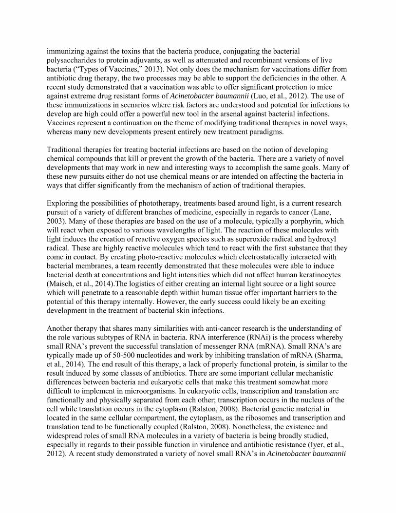
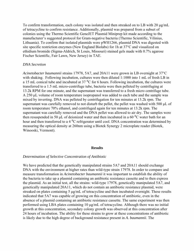
experiments were again performed using LBA plates containing 20 μg/mL of tetracycline, and no initial or secondary colony growth was observed at this concentration (Figure 1).
Figure 1. 17978, 5A7, and 20A11 on LB agar plus tetracycline. All three of the strains were streaked on LBA plates containing variable amounts of the antibiotic tetracycline. A) The genetically manipulated strain 5A7 was capable of growing on 5 μg/mL tetracycline. B) None of the strains were capable of growing on 10 μg/mL tetracycline, but breakout growth (not shown) was observed at this concentration after 24 hours of incubation. C) There is no initial or breakout growth at 20 μg/mL tetracycline. Experiments were performed using 20 μg/mL tetracycline.
Electrocompetent samples were prepared and then copies of the plasmid pWH1266, which contains a tetracycline resistance cassette and is known to replicate in Acinetobacter, were electroporated into the cells. After the rest period, these cells were then plated on to LB agar plus tetracycline; the plating was performed on plates with 20 μg/mL of tetracycline. There was heavy growth, as would be expected in this scenario because of the presence of the plasmid containing an antibiotic resistance cassette for tetracycline (data not shown). In conclusion, 5 μg/mL of tetracycline was not selective, 10 μg/mL of tetracycline was selective initially but demonstrated breakout growth, and 20 μg/mL of tetracycline was selective for growth based on the presence of the plasmid pWH1266. Establishing a Timecourse for Transformation
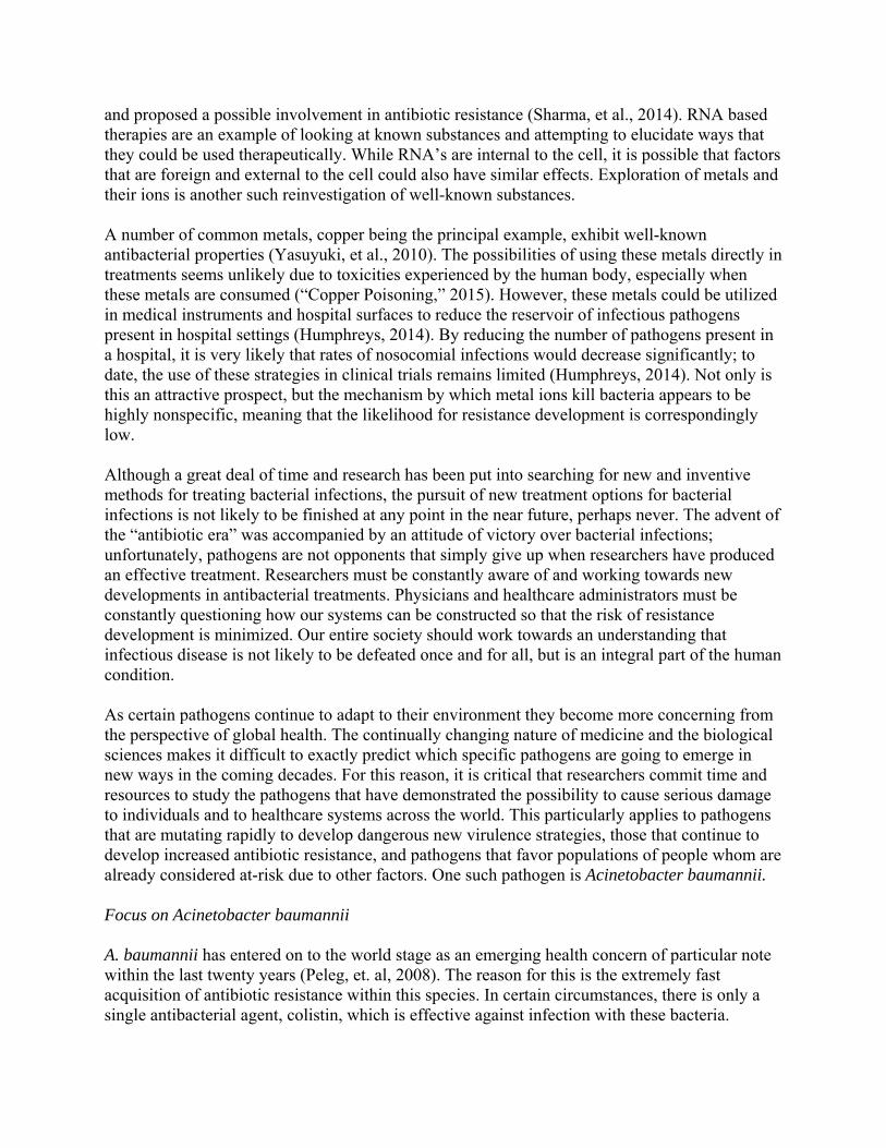
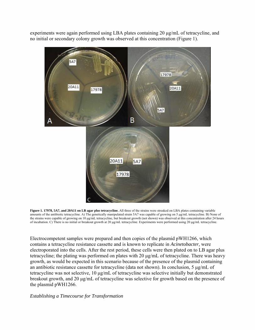
We were then curious about the amount of time that A. baumannii would need to be grown in the presence of the plasmid in order for transformation to occur. This would help us elucidate two things: how frequently transformation events occur and in what phase of growth the bacteria are most likely to undergo transformation. We saw that transformation was most likely to occur after the bacteria had been cultured with the plasmid for 6 hours (Figure 2). This time of transformation roughly correlates to the literature values for late logarithmic phase growth or early stationary phase growth (Dorsey, et al 2002; Aranda et al., 2011).
Figure 2. Transformation timecourse in A. baumannii strains incubated with plasmid DNA. The wild-type strain 17978 and the genetically manipulated strains 5A7 and 20A11 were all grown in LB broth with the plasmid pWH1266. Samples were removed and plated on LBA with 20 μg/mL tetracycline at every hour for each strain. Results are reported as number of transformant colonies that grew after a sample at that time point had been plated and incubated for 24 hours. Transformation begins to occur around the 5 hour time point and then is occurring in all three strains by the six hour time point.
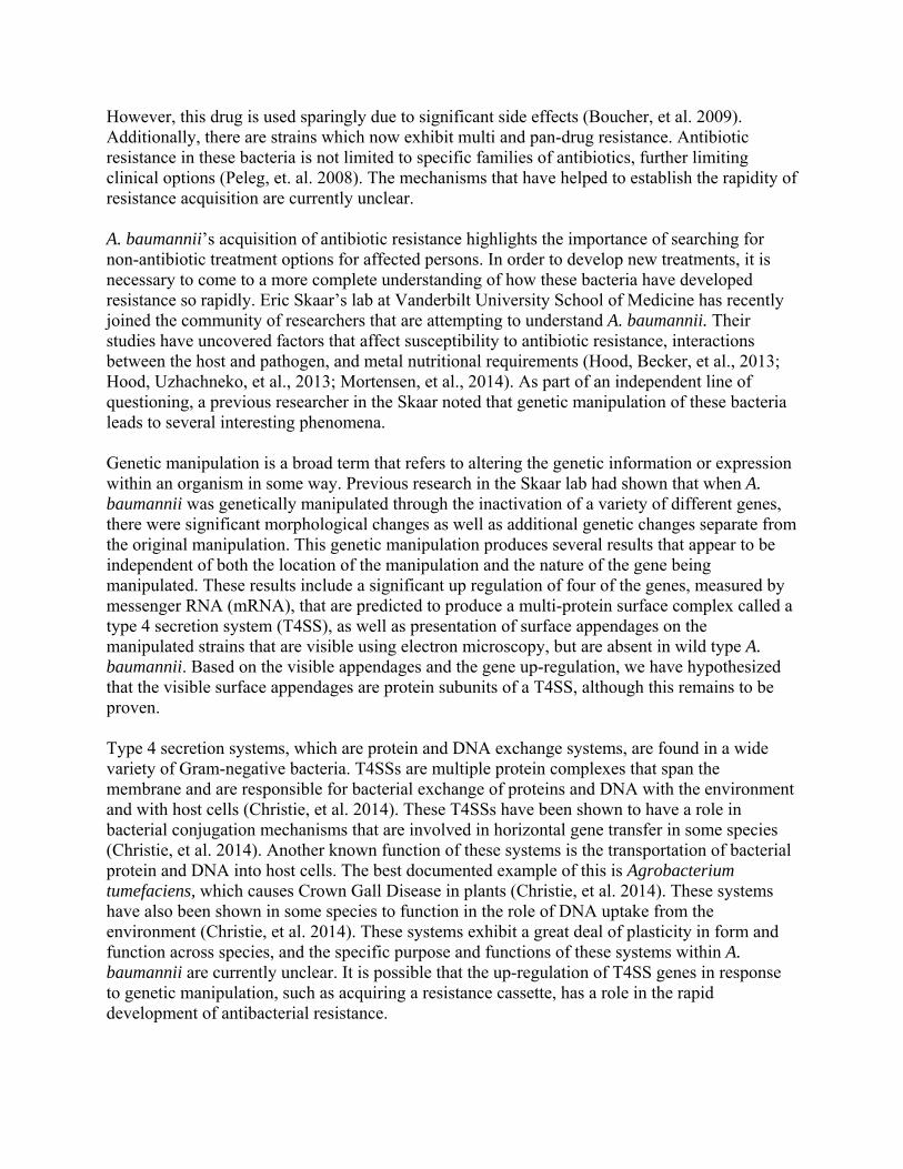
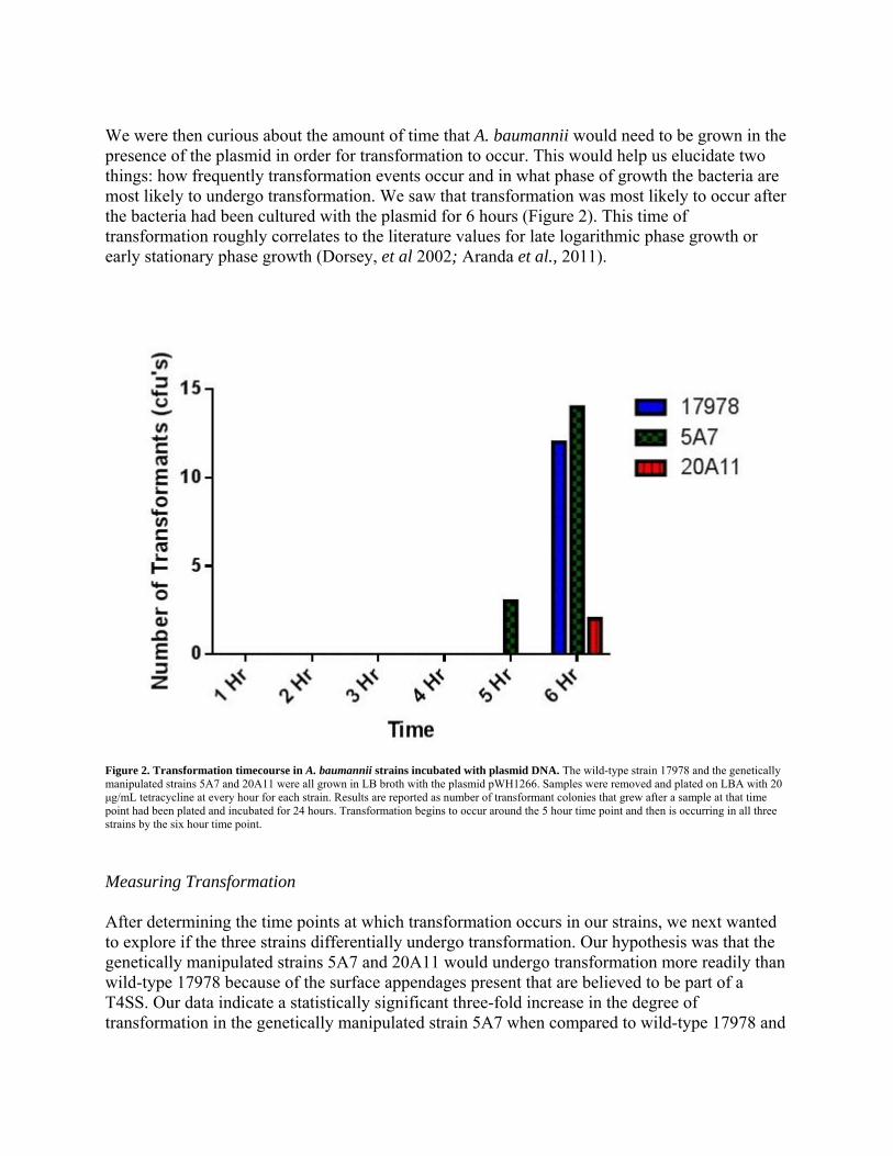
Measuring Transformation After determining the time points at which transformation occurs in our strains, we next wanted to explore if the three strains differentially undergo transformation. Our hypothesis was that the genetically manipulated strains 5A7 and 20A11 would undergo transformation more readily than wild-type 17978 because of the surface appendages present that are believed to be part of a T4SS. Our data indicate a statistically significant three-fold increase in the degree of transformation in the genetically manipulated strain 5A7 when compared to wild-type 17978 and
a non-significant difference between the genetically manipulated strain 20A11 and wild-type 17978 (Figure 3). To be certain that the observed growth on LBA plus 20 μg/mL of tetracycline was due to transformation, we confirmed the presence of the introduced plasmid in the transformant colonies. This was completed by performing a plasmid preparation and gel electrophoresis with UV imaging (Figure 4).
Tra
nsf
orm
ant
colo
nie
s /g
DN
A
1797
85A
7
20A11
0
5
10
15
Figure 3. Difference in occurrence of transformation among the three A. baumannii strains. All three strains were back-diluted from overnight cultures and then grown with the plasmid pWH1266, which contains a tetracycline resistance cassette, for six hours. These samples were then plated on LBA with 20 μg/mL tetracycline. Occurrence of transformation was enumerated by performing a colony count and then confirming for plasmid presence. Results were analyzed using paired t-test, * indicates a p-value of p<0.05 (n=6).
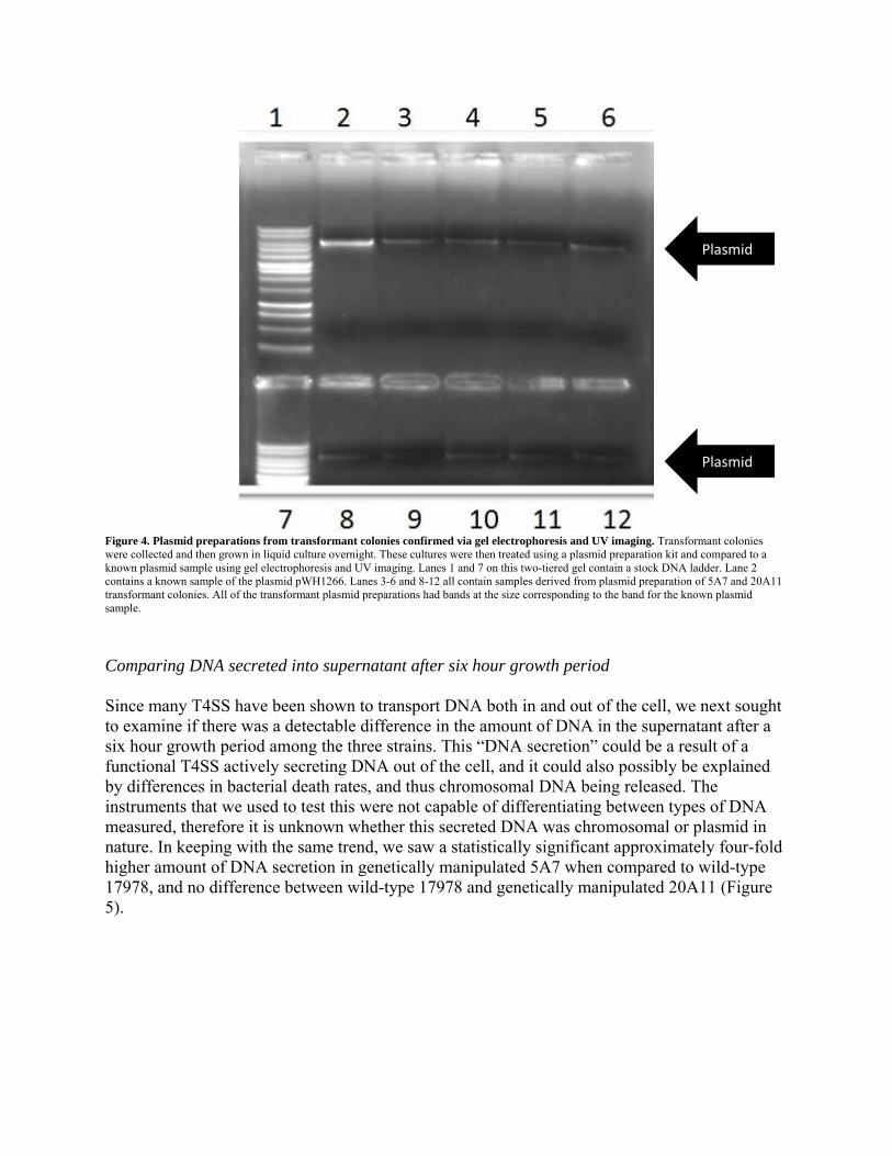
Figure 4. Plasmid preparations from transformant colonies confirmed via gel electrophoresis and UV imaging. Transformant colonies were collected and then grown in liquid culture overnight. These cultures were then treated using a plasmid preparation kit and compared to a known plasmid sample using gel electrophoresis and UV imaging. Lanes 1 and 7 on this two-tiered gel contain a stock DNA ladder. Lane 2 contains a known sample of the plasmid pWH1266. Lanes 3-6 and 8-12 all contain samples derived from plasmid preparation of 5A7 and 20A11 transformant colonies. All of the transformant plasmid preparations had bands at the size corresponding to the band for the known plasmid sample.
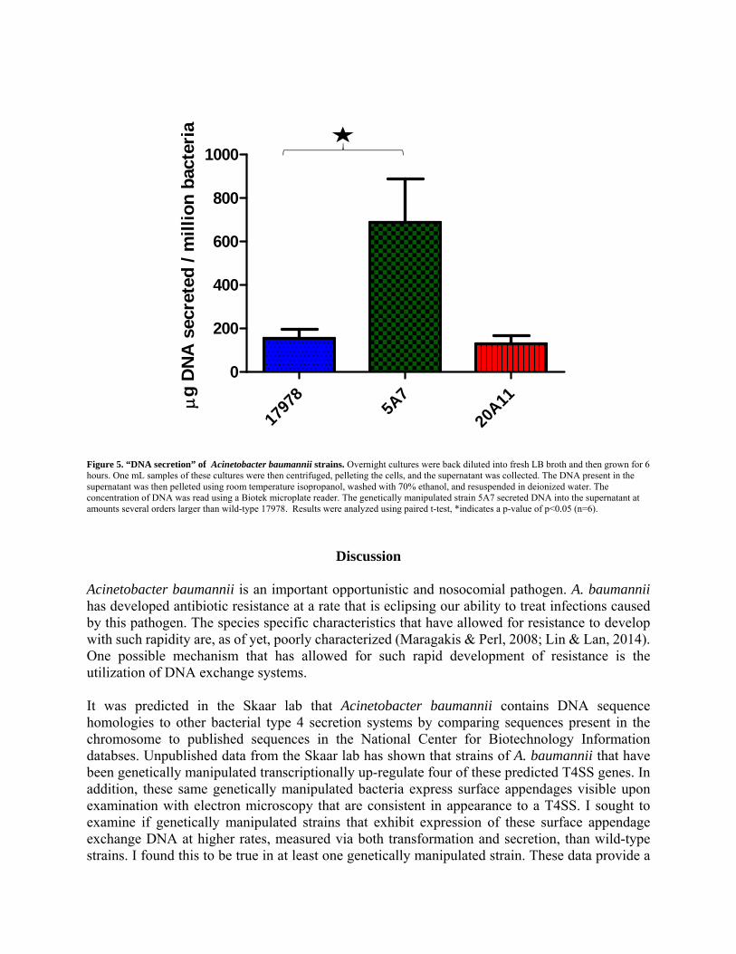
Comparing DNA secreted into supernatant after six hour growth period Since many T4SS have been shown to transport DNA both in and out of the cell, we next sought to examine if there was a detectable difference in the amount of DNA in the supernatant after a six hour growth period among the three strains. This “DNA secretion” could be a result of a functional T4SS actively secreting DNA out of the cell, and it could also possibly be explained by differences in bacterial death rates, and thus chromosomal DNA being released. The instruments that we used to test this were not capable of differentiating between types of DNA measured, therefore it is unknown whether this secreted DNA was chromosomal or plasmid in nature. In keeping with the same trend, we saw a statistically significant approximately four-fold higher amount of DNA secretion in genetically manipulated 5A7 when compared to wild-type 17978, and no difference between wild-type 17978 and genetically manipulated 20A11 (Figure 5).
Plasmid
Plasmid
g
DN
A s
ecre
ted
/ m
illi
on
bac
teri
a
1797
85A
7
20A11
0
200
400
600
800
1000
Figure 5. “DNA secretion” of Acinetobacter baumannii strains. Overnight cultures were back diluted into fresh LB broth and then grown for 6 hours. One mL samples of these cultures were then centrifuged, pelleting the cells, and the supernatant was collected. The DNA present in the supernatant was then pelleted using room temperature isopropanol, washed with 70% ethanol, and resuspended in deionized water. The concentration of DNA was read using a Biotek microplate reader. The genetically manipulated strain 5A7 secreted DNA into the supernatant at amounts several orders larger than wild-type 17978. Results were analyzed using paired t-test, *indicates a p-value of p<0.05 (n=6).
Discussion Acinetobacter baumannii is an important opportunistic and nosocomial pathogen. A. baumannii has developed antibiotic resistance at a rate that is eclipsing our ability to treat infections caused by this pathogen. The species specific characteristics that have allowed for resistance to develop with such rapidity are, as of yet, poorly characterized (Maragakis & Perl, 2008; Lin & Lan, 2014). One possible mechanism that has allowed for such rapid development of resistance is the utilization of DNA exchange systems. It was predicted in the Skaar lab that Acinetobacter baumannii contains DNA sequence homologies to other bacterial type 4 secretion systems by comparing sequences present in the chromosome to published sequences in the National Center for Biotechnology Information databses. Unpublished data from the Skaar lab has shown that strains of A. baumannii that have been genetically manipulated transcriptionally up-regulate four of these predicted T4SS genes. In addition, these same genetically manipulated bacteria express surface appendages visible upon examination with electron microscopy that are consistent in appearance to a T4SS. I sought to examine if genetically manipulated strains that exhibit expression of these surface appendage exchange DNA at higher rates, measured via both transformation and secretion, than wild-type strains. I found this to be true in at least one genetically manipulated strain. These data provide a
possible mechanistic explanation for the population-level acquisition of antibiotic resistance determinants, but this may not hold true in all strains of A. baumannii. By exploring two avenues of DNA exchange with other cells and the environment, namely transformation and secretion, we have sought to uncover the function of these surface appendages in an attempt to understand the complex mechanisms that have evolved in these bacteria that allow them to share DNA. Our data indicate that DNA exchange is occurring at statistically higher rates in at least one of the strains that have been genetically manipulated. Traditional studies of bacterial transformation indicate that the end of exponential and beginning of the stationary growth phase is the canonical time for transformation to occur, which is consistent with our data (Goodgal, 1961). Although it is unclear why these effects did not hold equally true in both genetically manipulated strains, an interesting phenomenon worth further study has been uncovered. It is possible that the genetically manipulated strain 20A11 may be presenting a non-functional T4SS on the cell surface. It is also possible that some sort of protein regulatory pathway differs between the two genetically manipulated strains that allow for 5A7 to exchange DNA in a manner that 20A11 is not able to. These results correlate the presence of the surface appendages and the transcriptional up-regulation of the putative T4SS genes with increased DNA exchange in at least some strains of A. baumannii. Utilization of various secretion systems in Acinetobacter baumannii is only beginning to be understood and appreciated. A team recently demonstrated that Acinetobacter baumannii contains a type VI section system (T6SS) for the purpose of bacterial competition (Carruthers, et al., 2013). Additionally, clinical isolates of carbapenem-resistant Acinetobacter baumannii were found that contained a plasmid encoding a type IV secretion system (Liu, et al. 2014). To the best of our knowledge, a chromosomally-located type IV secretion system has yet to be investigated in A. baumannii. A number of other Gram-negative pathogens utilize a type IV secretion system during infection and colonization of the host, including Helicobacter pylori (Hatakeyama & Higashi, 2005), Legionella pneumophila (Vogel, 1998), and Bordetella pertussis (Burns, 2003). Exploration of these systems in a variety of pathogens has begun and is likely to continue to help create broad-based knowledge about how bacteria cause disease, develop resistance, and the evolutionary course that these species have taken. The next steps in understanding this system are to look at other mechanisms of DNA exchange, ultimately, to establish causation. Bacterial conjugation through the process of cell-to-cell contact is a well-established method of propagating antibiotic resistant determinants and virulence factors in a variety of different organisms (Christie, et al., 2014). An assay that is able to demonstrate and determine differences in conjugation between wild-type and genetically manipulated strains is currently ongoing. Following that, we aim to successfully knock out these putative T4SS genes in all three of our strains and test for loss of the DNA exchange phenotype in those knockout strains. As a negative control, we also intend to knockout a gene that encodes for part of a type IV conjugative pilus, which is an appendage that is responsible for DNA exchange in other Gram-negative bacteria (Carter, et al., 2010). Additionally, it is assumed that knockout strains lacking T4SS genes would cease to express the visible surface appendages, although these appendages could still be present even if the T4SS is nonfunctional because of the multimeric nature of this protein system. This shall be tested by performing further scanning electron microscopy studies.
How Acinetobacter baumannii has developed antibiotic resistance is an inherently interesting question both because of the rapidity of the development of resistance and also the degree that this species has reemerged as an important global pathogen. If these bacteria contain a system that causes them to exchange DNA at higher rates after exposure to genetic manipulation, such as acquisition of determinants that confer antibiotic resistance, then that could help to explain both of the above observations. Identification of such a system would not merely answer these questions but it could ultimately lead to the creation of a therapy that could target these DNA exchange systems specifically. By targeting systems that are only present in resistant strains, antibiotics could become more selective and hopefully apply less of the evolutionary selective pressure that has led to the development of widespread antibiotic resistance in the first place.
Acknowledgments
I would like to thank the Belmont University Honors Program and the Belmont University Department of Biology for supporting me immensely throughout my time as an undergraduate. The opportunities that both of these groups have provided me is unparalleled and extraordinary. I would also like to thank all the members of the Skaar lab for all the assistance and guidance that I have received. I would like to thank all of my fellow students that have provided feedback on this project, especially “Team Thomas” and my roommates. None of my successes would be possible without the support of so many people.
Supporting Grants
This work has been made possible due to the following grant: Department of Veterans Affairs Merit Award INFB-024-13F to E.P.S.
Works Cited
Abraham, E., and Chain, E. (1940). An Enzyme From Bacteria Able To Destroy Penicillin. Nature, 146, 837-837. Acinetobacter baumannii Infections Among Patients at Military Medical Facilities Treating
Injured U.S. Service Members, 2002-2004. MMWR Morb Mortal Wkly Rep. 2004;53(45):1063-6.
Acinetobacter in Healthcare Settings. (2010, November 24). Retrieved January 26, 2015, from http://www.cdc.gov/HAI/organisms/acinetobacter.html American Chemical Society. (1995). Discovery and Development of Penicillin [Booklet]. London, United Kingdom: The Alexander Fleming Laboratory Museum. American Chemical Society. (2005). Selman Waksman and Antibiotics [Booklet]. New Brunswick, United States: Rutgers University. Aminov, R., and Mackie, R. (2007). Evolution And Ecology Of Antibiotic Resistance Genes. FEMS Microbiology Letters, 271, 147-161. Aranda, J., Bardina, C., Beceiro, A., Rumbo, S., Cabral, M., Barbe, J., and Bou, G. (2011).
Acinetobacter baumannii RecA Protein in Repair of DNA Damage, Antimicrobial Resistance, General Stress Response, and Virulence. Journal of Bacteriology, 3740-3747.
Boucher HW, Talbot GH, Bradley JS, Edwards JE, Gilbert D, Rice LB, et al. Bad bugs, no Drugs: no ESKAPE! An Update from the Infectious Diseases Society of America. Clinical Infectious Disease. 2009 48(1):1-12.
Burns, D. (2003). Type IV Transporters of Pathogenic Bacteria. Current Opinion in Microbiology, 6(1), 29-34. Carruthers, M., Nicholson, P., Tracy, E., Munson, R., and Cascales, E. (2013). Acinetobacter
baumannii Utilizes a Type VI Secretion System for Bacterial Competition. PLoS ONE, 8(3), E59388-E59388.
Carter, M., Chen, J., and Lory, S. (2010). The Pseudomonas Aeruginosa Pathogenicity Island PAPI-1 Is Transferred Via A Novel Type IV Pilus. Journal of Bacteriology, 192(13), 3249-3258.
Cassir, N., Rolain, J. and Brouqui, P. (2014). A New Strategy to Fight Antimicrobial Resistance: the Revival of Old Antibiotics. Frontiers in Microbiology, 5, 1-15. Centers for Disease Control and Prevention. (2011). World Health Day: Media Fact Sheet.
Atlanta, Georgia. Accessed at http://www.cdc.gov/media/releases/2011/f0407_antimicrobialresistance.pdf
Chain, E., Florey, H., Gardner, A., Heatley, N., Jennings, M., Orr-Ewing, J., and Sanders, A. (1940). Penicillin As A Chemotherapeutic Agent. The Lancet, 226-228. Crichton, B. (Director). (2013). Pain, Pus, and Poison: Pus. In M. Mosley. London, United Kingdom: BBC. Christie, P., Whitaker, N., and Gonzalez-Rivera, C. (2014). Mechanism and Structure of the Bacterial Type IV Secretion Systems. Biochimica Et Biophysica Acta, 1843, 1578-1591. Clostridium difficile Infection. (2013, March 1). Retrieved February 21, 2015, from http://www.cdc.gov/HAI/organisms/cdiff/Cdiff_infect.html Copper poisoning: MedlinePlus Medical Encyclopedia. (2015, March 2). Retrieved March 6, 2015, from http://www.nlm.nih.gov/medlineplus/ency/article/002496.htm Davenport, D. (2012). The war against bacteria: How Were Sulphonamide Drugs used by Britain During World War II? Medical Humanities, 38, 55-58.
Dorsey, C., Tomaras, A., and Actis, L. (2002). Genetic and Phenotypic Analysis of Acinetobacter baumannii Insertion Derivatives Generated with a Transposome System. Applied and Environmental Microbiology, 6353-6360.
European Antibiotic Awareness Day. (n.d.). Retrieved February 24, 2015, from http://ecdc.europa.eu/en/EAAD/Pages/Home.aspx European Medicines Agency. (2011). Trends in the Sales of Veterinary Antimicrobial Agents in Nine European Countries. London, United Kingdom: European Medicines Agency. Fair, R., and Tor, Y. (2014). Antibiotics and Bacterial Resistance in the 21st Century. Perspectives in Medicinal Chemistry, 6, 25-64. FDA Takes Steps to Protect Public Health. (2013, May 13). Retrieved February 24, 2015, from http://www.fda.gov/NewsEvents/Newsroom/PressAnnouncements/ucm299802.htm Fischbach, M., and Walsh, C. (2009). Antibiotics For Emerging Pathogens. Science, 325(5944), 1089-1093. Fishman, N. (2006). Antimicrobial Stewardship. The American Journal of Medicine, 119(6A), S53-S61. Fleming, A. (1929). On the Antibacterial Action of Cultures of a Penicillium, with Special
Reference to their Use in the Isolation of B. influenzæ. British Journal of Experimental Pathology, 10(3), 226-236.
Garnacho-Montero, J., Ortiz-Leyba, C., Fernández-Hinojosa, E., Aldabó-Pallás, T., Cayuela, A., Marquez-Vácaro, J., ... Jiménez-Jiménez, F. (2005). Acinetobacter baumannii Ventilator-Associated Pneumonia: Epidemiological and Clinical Findings. Intensive Care Medicine, 649-655.
Gaynes, R., and Edwards, J. (2005). Overview Of Nosocomial Infections Caused By Gram‐ Negative Bacilli. Clinical Infectious Diseases, 848-854. Get Smart About Antibiotics Week. (2014, October 21). Retrieved February 24, 2015, from http://www.cdc.gov/getsmart/week/index.html Goodgal, S. (1961). Studies on Transformations of Hemophilus influenzae: I. Competence. The Journal of General Physiology, 44, 1201-1227. Hager, T. (2006). The Demon Under the Microscope: From Battlefield Hospitals to Nazi Labs,
One Doctor's Heroic Search for the World's First Miracle Drug. New York: Harmony Books.
Harris, G. (2012, April 11). U.S. Tightens Rules on Antibiotics Use for Livestock. The New York Times. Hatakeyama, M., and Higashi, H. (2005). Helicobacter pylori CagA: A New Paradigm for Bacterial Carcinogenesis. Cancer Science, 96(12), 835-843. Hood, M., Becker, K., Roux, C., Dunman, P., and Skaar, E. (2013). Genetic Determinants of
Intrinsic Colistin Tolerance in Acinetobacter baumannii. Infection and Immunity, 81(2), 469-469.
Hood, M., Uzhachenko, R., Boyd, K., Skaar, E., and Ivanova, A. (2013). Loss of Mitochondrial Protein Fus1 Augments Host Resistance to Acinetobacter baumannii Infection. Infection and Immunity, 81(12), 4461-4469.
How Vaccines Work. (2013, July 23). Retrieved March 24, 2015, from http://www.vaccines.gov/more_info/work/index.html Humphreys, H. (2014). Self-Disinfecting and Microbiocide-Impregnated Surfaces and Fabrics:
What Potential in Interrupting the Spread of Healthcare-Associated Infection? Clinical Infectious Diseases, 58(6), 848-853.
Hunger, M., Schmucker, R., Kishan, V., and Hillen, W. (1990). Analysis and Nucleotide Sequence of an Origin of DNA Replication in Acinetobacter calcoaceticus and its Use for Escherichia coli Shuttle Plasmids. Gene, 87(1), 45-51.
Iyer, V., Sharma, R., Pathania, R., and Pavani, N. (2012). Small RNAs of Pathogenic Bacteria: Not Small Enough to be Overlooked for Therapeutics. Molecular and Cellular Pharmacology,4(1), 17-30.
Jones, D., Podolsky, S., Greene, S. (2012) The Burden of Disease and the Changing Task of Medicine. New England Journal of Medicine, 366 (25), 2333-2338. Karlsson, E., Kwiatkowski, D., and Sabeti, P. (2014). Natural Selection and Infectious Disease in Human Populations. Nature Reviews Genetics, 15, 379-393. Lane, N. (2003, January 1). New Light on Medicine. Scientific American, 80-87. Lewis, K. (2012). Antibiotics: Recover the Lost Art of Drug Discovery. Nature, 485, 439-440. Lewis, K. (2013). Platforms for Antibiotic Discovery. Nature Reviews Drug Discovery, 12, 371- 387. Lin, M., and Lan, C. (2014). Antimicrobial Resistance in Acinetobacter baumannii: From Bench to Bedside. World Journal of Clinical Cases, 2(12), 787-814. Ling, L., Schneider, T., Peoples, A.J., Spoering, A.L.,Engels, I., Conlon, B.P., Mueller, A.,
Scha¨berle, T.F., Hughes, D.E., Epstein, S., Jones, M., Lazarides, L., Steadman, V.A., Cohen, D.R., Felix, D.R., Fetterman, K.A, Millett, W.P., Nitti, A.G., Zullo, A.M., Chen, C. and Kim Lewis. (2015) A new Antibiotic Kills Pathogens without Detectable Resistance. Nature, 000, 1-5.
Liu, C., Kuo, H., Tang, C., Chang, K., and Liou, M. (2014). Prevalence and Mapping of a Plasmid Encoding a Type IV Secretion System in Acinetobacter baumannii. Genomics, 104(3), 215-223.
Luo, G., Lin, L., Ibrahim, A., Baquir, B., Pantapalangkoor, P., Bonomo, R., ... Filion, L. (2012). Active and Passive Immunization Protects against Lethal, Extreme Drug Resistant-Acinetobacter baumannii Infection. PLoS ONE, 7(1), E29446-E29446.
Maisch, T., Spannberger, F., Regensburger, J., Felgenträger, A., and Bäumler, W. (2014). Fast and Effective: Intense Pulse Light Photodynamic Inactivation of Bacteria. PloS ONE, 9(12), 1013-1021.
Maragakis, L., and Perl, T. (2008). Acinetobacter baumannii: Epidemiology, Antimicrobial Resistance, and Treatment Options. Clinical Infectious Diseases, 46(8), 1254-1263. Mehta, K.C., Dargad, R.R., Borade, D.M, and Swami, O.C. (2014). Burden of Antibiotic
Resistance in Common Infectious Diseases: Role of Antibiotic Combination Therapy. Journal of Clinical and Diagnostic Research, 8,6, 1-8.
Mission Critical: Preventing Antibiotic Resistance. (2014, April 28). Retrieved February 23, 2015, from http://www.cdc.gov/features/antibioticresistance/ Mortensen, B., Rathi, S., Chazin, W., and Skaar, E. (2014). Acinetobacter baumannii Response
to Host-Mediated Zinc Limitation Requires the Transcriptional Regulator Zur. Journal of Bacteriology, 196(14), 2616-2626.
Mouton, J. (2013). Controlling Antimicrobial Resistance: Interfering in the Process of Natural Selection. Antimicrobial Resistance and Infection Control, 2, 32-32. Munoz-Price, L., and Weinstein, R. (2008). Acinetobacter Infection. New England Journal of Medicine, 1271-1281. Murray, C., Hinkle, M., and Yun, H. (2008). History of Infections Associated With Combat- Related Injury. The Journal of Trauma, 64, S221-S231
National Institute of Allergy and Infectious Diseases. Understanding of Microbes in Sickness and in Health (NIH Publication no. 09-4914). Washington, D.C. Online. Ogle, M. (2013, September 3). Riots, Rage and Resistance: A Brief History of How Antibiotics Arrived on the Farm. Scientific American. O'Shea, M. (2012). Acinetobacter in Modern Warfare. International Journal of Antimicrobial Agents, 39, 363-375. Otten, H. (1986). Domagk and the Development of Sulphonamides. Journal of Antimicrobial Chemotherapy, 17, 689-690. Otto, M. (2009). Staphylococcus Epidermidis — The 'Accidental' Pathogen. Nature Reviews Microbiology, 555-567. Peleg, A., Seifert, H., and Paterson, D. (2008). Acinetobacter baumannii: Emergence of a Successful Pathogen. Clinical Microbiology Reviews, 21(3), 538-582. Planta, M. (2007). The Role of Poverty in Antimicrobial Resistance. Journal of American Board of Family Medicine, 20(6), 533-539. Pneumonia-American Lung Association. (n.d.). Retrieved September 19, 2014. Ralston, A. (2008) Simultaneous Gene Transcription and Translation in Bacteria. Nature Education 1(1):4 Schatz, A., Bugle, E., and Waksman, S. (1944). Streptomycin, a Substance Exhibiting Antibiotic
Activity Against Gram-Positive and Gram-Negative Bacteria. Experimental Biology and Medicine, 55(1), 66-69.
Sharma, R., Arya, S., Patil, S., Sharma, A., Jain, P., Navani, N., and Pathania, R. (2014). Identification of Novel Regulatory Small RNAs in Acinetobacter baumannii. PLoS ONE, 9(4), E93833-E93833.
TB. (2014, December 16). Retrieved February 21, 2015, from http://www.cdc.gov/tb/ “The Nobel Prize in Physiology or Medicine 1908". Nobelprize.org. Nobel Media AB 2014. Web. 21 Feb 2015. http://www.nobelprize.org/nobel_prizes/medicine/laureates/1908/ Types of Vaccines. (2013, July 23). Retrieved March 24, 2015, from http://www.vaccines.gov/more_info/types/ Vincent JL, Rello J, Marshall J, Silva E, Anzueto A, Martin CD, et al. International Study of the
Prevalence and Outcomes of infection in intensive care units. Journal of the American Medical Association. 2009, 302(21):2323-9.
Vogel, J. (1998). Conjugative Transfer by the Virulence System of Legionella pneumophila. Science, 279(5352), 873-876. Yarnell, A. (2005, June 20). Salvarsan. Chemical and Engineering News. Yasuyuki, M., Kunihiro, K., Kurissery, S., Kanavillil, N., Sato, Y., and Kikuchi, Y. (2010).
Antibacterial properties of nine pure metals: A laboratory study using Staphylococcus aureus and Escherichia coli. Biofouling, 26(7), 851-858.
YouGov. (2015). Antibiotics [Data file]. Retrieved from http://cdn.yougov.com/cumulus_uploads/document/kc5axd98bx/tabs_OPI_antibiotics_20150112.pdf