parallel imaging ms/ms tof-sims instrument...parallel imaging ms/ms tof-sims instrument gregory l....
TRANSCRIPT
Parallel imaging MS/MS TOF-SIMS instrumentGregory L. Fisher, John S. Hammond, Paul E. Larson, Scott R. Bryan, and Ron M. A. Heeren Citation: Journal of Vacuum Science & Technology B 34, 03H126 (2016); doi: 10.1116/1.4943568 View online: http://dx.doi.org/10.1116/1.4943568 View Table of Contents: http://scitation.aip.org/content/avs/journal/jvstb/34/3?ver=pdfcov Published by the AVS: Science & Technology of Materials, Interfaces, and Processing Articles you may be interested in ToF-SIMS analysis of amyloid beta aggregation on different lipid membranes Biointerphases 11, 02A314 (2016); 10.1116/1.4940706 Latest applications of 3D ToF-SIMS bio-imaging Biointerphases 10, 018902 (2015); 10.1116/1.4907727 Multivariate ToF-SIMS image analysis of polymer microarrays and protein adsorption Biointerphases 10, 019005 (2015); 10.1116/1.4906484 Biomedical studies by TOF-SIMS imaging Biointerphases 10, 018901 (2015); 10.1116/1.4901511 TOF-SIMS quantification of low energy arsenic implants through thin SiO 2 layers AIP Conf. Proc. 550, 692 (2001); 10.1063/1.1354478
Redistribution subject to AVS license or copyright; see http://scitation.aip.org/termsconditions. IP: 207.250.199.132 On: Tue, 22 Mar 2016 15:01:17
Parallel imaging MS/MS TOF-SIMS instrument
Gregory L. Fisher, John S. Hammond, Paul E. Larson, and Scott R. Bryana)
Physical Electronics, Inc., Chanhassen, Minnesota 55317
Ron M. A. HeerenMaastricht University, The Maastricht Multi-Modal Molecular Imaging Institute, Universiteitssingel 50,6211 ER, Maastricht, Netherlands
(Received 15 November 2015; accepted 26 February 2016; published 16 March 2016)
The authors have developed a parallel imaging MS/MS capability for the PHI nanoTOF II
time-of-flight secondary ion mass spectrometry (TOF-SIMS) instrument. The unique design allows
a 1 Da wide precursor mass window to be extracted from a stream of mass separated secondary
ions while all other secondary ions are detected in the normal manner at the standard TOF-SIMS
detector. The selected precursor ions are deflected into an activation cell where they are fragmented
using high energy collision induced dissociation and mass analyzed in a separate linear TOF mass
spectrometer. This TOF-TOF approach allows MS/MS to be accomplished at a high speed
maintaining the primary ion beam repetition rates used in TOF-SIMS. The new MS/MS capability
enables molecular identification to be extended to higher mass ions where the mass accuracy of
TOF-SIMS is not sufficient to unambiguously identify molecular structure. The ability to acquire
TOF-SIMS and MS/MS data simultaneously from the identical analytical volume is a powerful
new method for mass spectrometry imaging. VC 2016 American Vacuum Society.
[http://dx.doi.org/10.1116/1.4943568]
I. INTRODUCTION
The time-of-flight secondary ion mass spectrometry
(TOF-SIMS) has become a well-established technique for
surface analysis due to its unique attributes such as high sen-
sitivity, high mass resolution, submicron spatial resolution,
and ability to provide both elemental and molecular informa-
tion. The widespread adoption of new cluster primary ion
beams over the past decade has significantly improved the
ability to sputter and detect intact molecular ions at higher
mass.1–3 This increase in sensitivity has translated directly into
better spatial resolution molecular imaging as the minimum
pixel size with an acceptable signal intensity has become corre-
spondingly smaller. The use of a bismuth liquid metal ion
source provides cluster primary ion beams (e.g., Bi3þ2) with a
beam size of 70 nm at low mass resolution and 500 nm at high
mass resolution, which are both significantly smaller than the
probe diameter of other imaging mass spectrometry techniques
such as matrix assisted laser desorption/Ionization (Ref. 4) or
nanospray desorption electrospray ionization mass spectrome-
try.5 The developments in cluster primary ion beams have
resulted in TOF-SIMS making increasingly important contribu-
tions to the understanding of complex molecular distributions
in biological tissues and cells and for surface and thin film anal-
ysis of polymers and other engineered organic materials.
The ability to detect higher mass ions using cluster pri-
mary ion beams has made the challenge of peak identifica-
tion more problematic. The mass accuracy in commercial
TOF-SIMS instruments is not sufficient to provide unambig-
uous high mass peak identification. A VAMAS round-robbin
study by Gilmore et al. concluded that using an optimized
procedure for mass calibration, a 10 ppm mass accuracy
could be achieved only up to 140 m/z.6 This makes it
impossible to distinguish CH2 from N or NH2 from O when
trying to identify the chemical structure of higher mass peaks
based on the exact mass measurement.
It has been recognized for many years that MS/MS is
needed in TOF-SIMS in order to be certain of high mass peak
identifications, especially for complex biological samples. This
prompted the development of MS/MS on SIMS instruments
based on a DC primary ion beam and a pulsed TOF mass
spectrometer by research groups led by Winograd7 and
Vickerman.8 The instrument presented here differs from these
previous instruments in two ways: (1) the primary ion beam is
pulsed rather than DC; and (2) the entire TOF-SIMS spectrum
(except the precursor ions) is detected simultaneously with the
MS/MS spectrum. In our implementation, no secondary ions
are discarded while operating in the MS/MS mode of analysis.
We refer to this as “Parallel Imaging MS/MS TOF-SIMS”
because secondary ions are recorded at the standard TOF-
SIMS detector at the same time that MS/MS fragment ions are
recorded at the linear TOF detector. Because there are two
mass spectrometers and two spectra are acquired within a sin-
gle acquisition, we use the following nomenclature for clarity:
MS1 ¼ the TOF analyzer used for TOF� SIMS;
MS2 ¼ the linear TOF analyzer used for MS=MS;
MS1 ¼ the secondary ion mass spectrum;
MS2 ¼ the MS=MS fragment ion spectrum:
The secondary ions detected in MS1 and MS2 are produced
from the same primary ion pulses striking the sample sur-
face. The x-y position of the primary ion beam is recorded
for every count detected in both MS1 and MS2 spectra. In
this way, images produced from MS1 and MS2 are in perfect
registration and are from the identical analytical volume of
the surface.a)Electronic mail: [email protected]
03H126-1 J. Vac. Sci. Technol. B 34(3), May/Jun 2016 2166-2746/2016/34(3)/03H126/4/$30.00 VC 2016 American Vacuum Society 03H126-1
Redistribution subject to AVS license or copyright; see http://scitation.aip.org/termsconditions. IP: 207.250.199.132 On: Tue, 22 Mar 2016 15:01:17
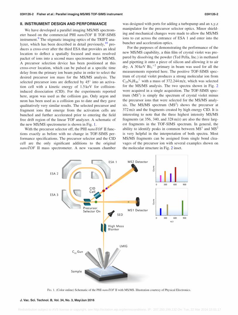
II. INSTRUMENT DESIGN AND PERFORMANCE
We have developed a parallel imaging MS/MS spectrom-
eter based on the commercial PHI nanoTOF II TOF-SIMS
instrument.9 The stigmatic imaging optics of the TRIFT ana-
lyzer, which has been described in detail previously,10 pro-
duces a cross-over after the third ESA that provides an ideal
location to deflect a spatially focused and mass resolved
packet of ions into a second mass spectrometer for MS/MS.
A precursor selection device has been positioned at this
cross-over location, which can be pulsed at a specific time
delay from the primary ion beam pulse in order to select the
desired precursor ion mass for the MS/MS analysis. The
selected precursor ions are deflected by 45� into an activa-
tion cell with a kinetic energy of 1.5 keV for collision-
induced dissociation (CID). For the experiments reported
here, argon was used as the collision gas. Only argon and
neon has been used as a collision gas to date and they gave
qualitatively very similar results. The selected precursor and
fragment ions that emerge from the activation cells are
bunched and further accelerated prior to entering the field
free drift region of the linear TOF analyzer. A schematic of
the new MS/MS spectrometer is shown in Fig. 1.
With the precursor selector off, the PHI nanoTOF II func-
tions exactly as before with no change in TOF-SIMS per-
formance specifications. The precursor selector and the CID
cell are the only significant additions to the original
nanoTOF II mass spectrometer. A new vacuum chamber
was designed with ports for adding a turbopump and an x,y,z
manipulator for the precursor selector optics. Minor shield-
ing and mechanical changes were made to allow the MS/MS
ions to cut across the entrance of ESA 1 and enter into the
buncher and acceleration optics.
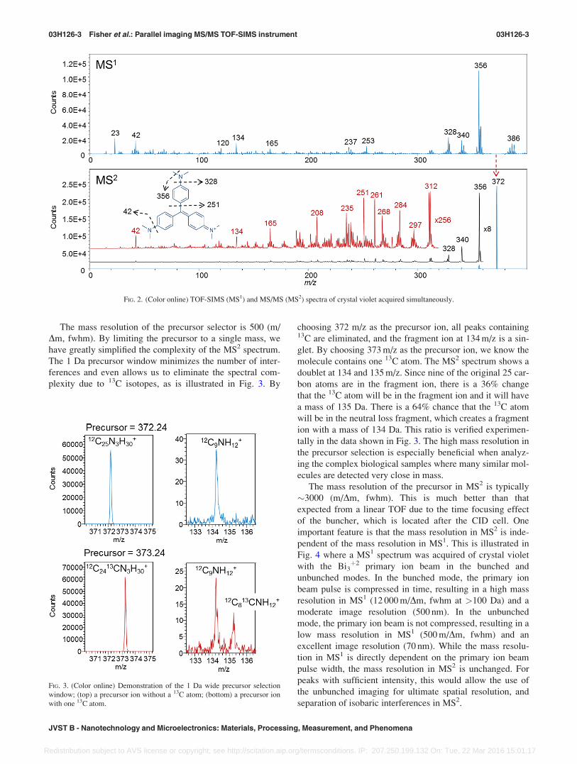
For the purposes of demonstrating the performance of the
new MS/MS capability, a thin film of crystal violet was pre-
pared by dissolving the powder (Ted Pella, Inc.) in methanol
and pipetting it onto a piece of silicon and allowing it to air
dry. A 30 keV Bi3þ2 primary in beam was used for all the
measurements reported here. The positive TOF-SIMS spec-
trum of crystal violet produces a strong molecular ion from
C25N3H30þ with a mass of 372.244 m/z, which was selected
for the MS/MS analysis. The two spectra shown in Fig. 2
were acquired in a single acquisition. The TOF-SIMS spec-
trum (MS1) is simply the spectrum of crystal violet minus
the precursor ions that were selected for the MS/MS analy-
sis. The MS/MS spectrum (MS2) shows the precursor at
372 m/z and the fragments created by high energy CID. It is
interesting to note that the three highest intensity MS/MS
fragments (at 356, 340, and 328 m/z) are also the three larg-
est fragments in the TOF-SIMS spectrum. In general, the
ability to identify peaks in common between MS1 and MS2
is very helpful in the interpretation of both spectra. Most
MS/MS fragments can be assigned from single bond clea-
vages of the precursor ion with several examples shown on
the molecular structure in Fig. 2 inset.
FIG. 1. (Color online) Schematic of the PHI nanoTOF II with MS/MS. Illustration courtesy of Physical Electronics.
03H126-2 Fisher et al.: Parallel imaging MS/MS TOF-SIMS instrument 03H126-2
J. Vac. Sci. Technol. B, Vol. 34, No. 3, May/Jun 2016
Redistribution subject to AVS license or copyright; see http://scitation.aip.org/termsconditions. IP: 207.250.199.132 On: Tue, 22 Mar 2016 15:01:17
The mass resolution of the precursor selector is 500 (m/
Dm, fwhm). By limiting the precursor to a single mass, we
have greatly simplified the complexity of the MS2 spectrum.
The 1 Da precursor window minimizes the number of inter-
ferences and even allows us to eliminate the spectral com-
plexity due to 13C isotopes, as is illustrated in Fig. 3. By
choosing 372 m/z as the precursor ion, all peaks containing13C are eliminated, and the fragment ion at 134 m/z is a sin-
glet. By choosing 373 m/z as the precursor ion, we know the
molecule contains one 13C atom. The MS2 spectrum shows a
doublet at 134 and 135 m/z. Since nine of the original 25 car-
bon atoms are in the fragment ion, there is a 36% change
that the 13C atom will be in the fragment ion and it will have
a mass of 135 Da. There is a 64% chance that the 13C atom
will be in the neutral loss fragment, which creates a fragment
ion with a mass of 134 Da. This ratio is verified experimen-
tally in the data shown in Fig. 3. The high mass resolution in
the precursor selection is especially beneficial when analyz-
ing the complex biological samples where many similar mol-
ecules are detected very close in mass.
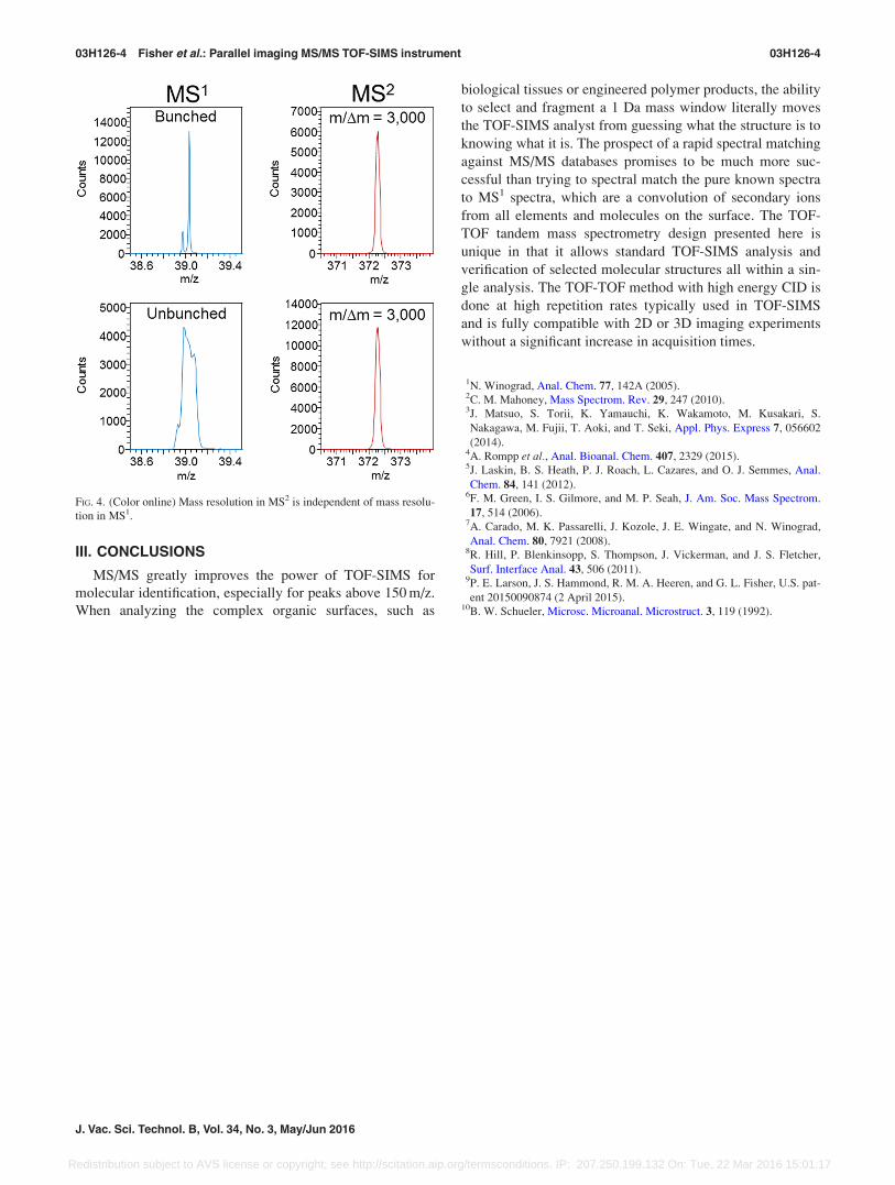
The mass resolution of the precursor in MS2 is typically
�3000 (m/Dm, fwhm). This is much better than that
expected from a linear TOF due to the time focusing effect
of the buncher, which is located after the CID cell. One
important feature is that the mass resolution in MS2 is inde-
pendent of the mass resolution in MS1. This is illustrated in
Fig. 4 where a MS1 spectrum was acquired of crystal violet
with the Bi3þ2 primary ion beam in the bunched and
unbunched modes. In the bunched mode, the primary ion
beam pulse is compressed in time, resulting in a high mass
resolution in MS1 (12 000 m/Dm, fwhm at >100 Da) and a
moderate image resolution (500 nm). In the unbunched
mode, the primary ion beam is not compressed, resulting in a
low mass resolution in MS1 (500 m/Dm, fwhm) and an
excellent image resolution (70 nm). While the mass resolu-
tion in MS1 is directly dependent on the primary ion beam
pulse width, the mass resolution in MS2 is unchanged. For
peaks with sufficient intensity, this would allow the use of
the unbunched imaging for ultimate spatial resolution, and
separation of isobaric interferences in MS2.
FIG. 2. (Color online) TOF-SIMS (MS1) and MS/MS (MS2) spectra of crystal violet acquired simultaneously.
FIG. 3. (Color online) Demonstration of the 1 Da wide precursor selection
window; (top) a precursor ion without a 13C atom; (bottom) a precursor ion
with one 13C atom.
03H126-3 Fisher et al.: Parallel imaging MS/MS TOF-SIMS instrument 03H126-3
JVST B - Nanotechnology and Microelectronics: Materials, Processing, Measurement, and Phenomena
Redistribution subject to AVS license or copyright; see http://scitation.aip.org/termsconditions. IP: 207.250.199.132 On: Tue, 22 Mar 2016 15:01:17
III. CONCLUSIONS
MS/MS greatly improves the power of TOF-SIMS for
molecular identification, especially for peaks above 150 m/z.
When analyzing the complex organic surfaces, such as
biological tissues or engineered polymer products, the ability
to select and fragment a 1 Da mass window literally moves
the TOF-SIMS analyst from guessing what the structure is to
knowing what it is. The prospect of a rapid spectral matching
against MS/MS databases promises to be much more suc-
cessful than trying to spectral match the pure known spectra
to MS1 spectra, which are a convolution of secondary ions
from all elements and molecules on the surface. The TOF-
TOF tandem mass spectrometry design presented here is
unique in that it allows standard TOF-SIMS analysis and
verification of selected molecular structures all within a sin-
gle analysis. The TOF-TOF method with high energy CID is
done at high repetition rates typically used in TOF-SIMS
and is fully compatible with 2D or 3D imaging experiments
without a significant increase in acquisition times.
1N. Winograd, Anal. Chem. 77, 142A (2005).2C. M. Mahoney, Mass Spectrom. Rev. 29, 247 (2010).3J. Matsuo, S. Torii, K. Yamauchi, K. Wakamoto, M. Kusakari, S.
Nakagawa, M. Fujii, T. Aoki, and T. Seki, Appl. Phys. Express 7, 056602
(2014).4A. Rompp et al., Anal. Bioanal. Chem. 407, 2329 (2015).5J. Laskin, B. S. Heath, P. J. Roach, L. Cazares, and O. J. Semmes, Anal.
Chem. 84, 141 (2012).6F. M. Green, I. S. Gilmore, and M. P. Seah, J. Am. Soc. Mass Spectrom.
17, 514 (2006).7A. Carado, M. K. Passarelli, J. Kozole, J. E. Wingate, and N. Winograd,
Anal. Chem. 80, 7921 (2008).8R. Hill, P. Blenkinsopp, S. Thompson, J. Vickerman, and J. S. Fletcher,
Surf. Interface Anal. 43, 506 (2011).9P. E. Larson, J. S. Hammond, R. M. A. Heeren, and G. L. Fisher, U.S. pat-
ent 20150090874 (2 April 2015).10B. W. Schueler, Microsc. Microanal. Microstruct. 3, 119 (1992).
FIG. 4. (Color online) Mass resolution in MS2 is independent of mass resolu-
tion in MS1.
03H126-4 Fisher et al.: Parallel imaging MS/MS TOF-SIMS instrument 03H126-4
J. Vac. Sci. Technol. B, Vol. 34, No. 3, May/Jun 2016
Redistribution subject to AVS license or copyright; see http://scitation.aip.org/termsconditions. IP: 207.250.199.132 On: Tue, 22 Mar 2016 15:01:17