paneth cell defects induce microbiota dysbiosis in mice
TRANSCRIPT

HAL Id: hal-01603303https://hal.archives-ouvertes.fr/hal-01603303
Submitted on 26 May 2020
HAL is a multi-disciplinary open accessarchive for the deposit and dissemination of sci-entific research documents, whether they are pub-lished or not. The documents may come fromteaching and research institutions in France orabroad, or from public or private research centers.
L’archive ouverte pluridisciplinaire HAL, estdestinée au dépôt et à la diffusion de documentsscientifiques de niveau recherche, publiés ou non,émanant des établissements d’enseignement et derecherche français ou étrangers, des laboratoirespublics ou privés.
Paneth cell defects induce microbiota dysbiosis In miceand promote visceral hypersensitivity
Ambre Riba, Maïwenn Olier, Sonia Lacroix-Lamandé, Corinne Lencina,Valérie Alquier-Bacquié, Cherryl Harkat, Marion Gillet, Marine Baron,
Caroline Sommer, Virginie Mallet, et al.
To cite this version:Ambre Riba, Maïwenn Olier, Sonia Lacroix-Lamandé, Corinne Lencina, Valérie Alquier-Bacquié, etal.. Paneth cell defects induce microbiota dysbiosis In mice and promote visceral hypersensitivity. Gas-troenterology, WB Saunders, 2017, 153, pp.1594-1606. �10.1053/j.gastro.2017.08.044�. �hal-01603303�

Ver
sion
pos
tprin
t
Comment citer ce document :Riba, A., Olier, M., Lacroix Lamandé, S., Lencina, C., Bacquié, V., Harkat, C., Gillet, M.,
Baron, M., Sommer, C., Mallet, V., Salvador Cartier, C., Laurent, F., Théodorou, V., Ménard, S.(2017). Paneth cell defects induce microbiota dysbiosis In mice and promote visceral
hypersensitivity. Gastroenterology, 153 (6), 1594-1606. DOI : 10.1053/j.gastro.2017.08.044
Accepted Manuscript
Paneth Cell Defects Induce Microbiota Dysbiosis In Mice And Promote VisceralHypersensitivity
Ambre Riba, Maïwenn Olier, Sonia Lacroix-Lamandé, Corinne Lencina, ValérieBacquié, Cherryl Harkat, Marion Gillet, Marine Baron, Caroline Sommer, VirginieMallet, Christel Salvador-Cartier, Fabrice Laurent, Vassilia Théodorou, SandrineMénard
PII: S0016-5085(17)36077-8DOI: 10.1053/j.gastro.2017.08.044Reference: YGAST 61390
To appear in: GastroenterologyAccepted Date: 24 August 2017
Please cite this article as: Riba A, Olier M, Lacroix-Lamandé S, Lencina C, Bacquié V, Harkat C, GilletM, Baron M, Sommer C, Mallet V, Salvador-Cartier C, Laurent F, Théodorou V, Ménard S, Paneth CellDefects Induce Microbiota Dysbiosis In Mice And Promote Visceral Hypersensitivity, Gastroenterology(2017), doi: 10.1053/j.gastro.2017.08.044.
This is a PDF file of an unedited manuscript that has been accepted for publication. As a service toour customers we are providing this early version of the manuscript. The manuscript will undergocopyediting, typesetting, and review of the resulting proof before it is published in its final form. Pleasenote that during the production process errors may be discovered which could affect the content, and alllegal disclaimers that apply to the journal pertain.

Ver
sion
pos
tprin
t
Comment citer ce document :Riba, A., Olier, M., Lacroix Lamandé, S., Lencina, C., Bacquié, V., Harkat, C., Gillet, M.,
Baron, M., Sommer, C., Mallet, V., Salvador Cartier, C., Laurent, F., Théodorou, V., Ménard, S.(2017). Paneth cell defects induce microbiota dysbiosis In mice and promote visceral
hypersensitivity. Gastroenterology, 153 (6), 1594-1606. DOI : 10.1053/j.gastro.2017.08.044
MANUSCRIP
T
ACCEPTED
ACCEPTED MANUSCRIPT
1
PANETH CELL DEFECTS INDUCE MICROBIOTA DYSBIOSIS IN MICE AND
PROMOTE VISCERAL HYPERSENSITIVITY
Short title: Role of Paneth cells in visceral hypersensitivity
Ambre Riba1, Maïwenn Olier1, Sonia Lacroix-Lamandé2, Corinne Lencina1, Valérie Bacquié1,
Cherryl Harkat1, Marion Gillet1, Marine Baron1, Caroline Sommer1, Virginie Mallet1, Christel
Salvador-Cartier1, Fabrice Laurent2, Vassilia Théodorou1, Sandrine Ménard1.
1 INRA, ToxAlim (Research Centre in Food Toxicology), team Neuro-Gastroenterology and
Nutrition, Université de Toulouse, INRA, ENVT, INP-Purpan, UPS, Toulouse, France
2 Equipe Apicomplexes et Immunité Mucosale (AIM), UMR 1282 INRA/Université-
Infectiologie et Santé Publique (ISP), Centre INRA Val de Loire, Nouzilly, FRANCE
Abbreviations: AMA, antimicrobial activity; AMP, antimicrobial peptides; CD, Crohn’s
Disease; CFU, Colony Forming Unit; EMG, Electromyograph; GVHD, Graft-versus-host
disease; GULDA, GUt Low-Density Array; IBD, Inflammatory Bowel Disease; IBS, Irritable
Bowel Syndrome; MS, Maternal Separation; NEC, Necrotizing enterocolitis; PC, Paneth
cells; PLS-DA, Partial least-squares discriminant analysis; TNFα, Tumor Necrosis Factor α;
VIP, Variable Importance in Projection.
Correspondence: Sandrine Ménard, INRA, ToxAlim (Research Centre in Food Toxicology),
team Neuro-Gastroenterology and Nutrition, Université de Toulouse, INRA, ENVT, INP-

Ver
sion
pos
tprin
t
Comment citer ce document :Riba, A., Olier, M., Lacroix Lamandé, S., Lencina, C., Bacquié, V., Harkat, C., Gillet, M.,
Baron, M., Sommer, C., Mallet, V., Salvador Cartier, C., Laurent, F., Théodorou, V., Ménard, S.(2017). Paneth cell defects induce microbiota dysbiosis In mice and promote visceral
hypersensitivity. Gastroenterology, 153 (6), 1594-1606. DOI : 10.1053/j.gastro.2017.08.044
MANUSCRIP
T
ACCEPTED
ACCEPTED MANUSCRIPT
2
Purpan, UPS, 180 Chemin de Tournefeuille, 31027 Toulouse, France ;
[email protected]; Phone : + 33 5 82 06 64 08 ; Fax: + 33 5 61 28 51 45
Disclosures: The authors disclose no conflicts
Authors contributions: Conceived and designed the experiments: SM AR VT. Performed the
experiments: AR MO SLL VB CH MG MB CS VM CSC SM. Analyzed the data: AR MO
VT SM. SM wrote the manuscript, with AR, VT, MO, SLL and FL providing critical output.
Acknowledgments: The authors gratefully acknowledge Yannick Lippi and Claire Naylies
from Transcriptomic impact of Xenobiotics platform for technical assistance for microbiota
analysis.
Ver
sion
pos
tprin
t
Comment citer ce document :Riba, A., Olier, M., Lacroix Lamandé, S., Lencina, C., Bacquié, V., Harkat, C., Gillet, M.,
Baron, M., Sommer, C., Mallet, V., Salvador Cartier, C., Laurent, F., Théodorou, V., Ménard, S.(2017). Paneth cell defects induce microbiota dysbiosis In mice and promote visceral
hypersensitivity. Gastroenterology, 153 (6), 1594-1606. DOI : 10.1053/j.gastro.2017.08.044
MANUSCRIP
T
ACCEPTED
ACCEPTED MANUSCRIPT
3
ABSTRACT
Background & Aims: Separation of newborn rats from their mothers induces visceral
hypersensitivity and impaired epithelial secretory cell lineages when they are adults. Little is
known about the mechanisms by which maternal separation causes visceral hypersensitivity
or its relationship with defects in epithelial secretory cell lineages.
Methods: We performed studies with C3H/HeN mice separated from their mothers as
newborns and mice genetically engineered (Sox9flox/flox-vil-cre on C57BL/6 background) to
have deficiencies in Paneth cells. Paneth cells deficiency was assessed by lysozyme staining
of ileum tissues and lysozyme activity in fecal samples. When mice were 50 days old, their
abdominal response to colorectal distension was assessed by electromyography. Fecal
samples were collected and microbiota were analyzed using GULDA quantitative PCR.
Results: Mice with maternal separation developed visceral hypersensitivity and defects in
Paneth cells, as reported from rats, compared to mice without maternal separation.
Sox9flox/flox-vil-Cre mice also had increased visceral hypersensitivity compared to control
littermate Sox9flox/flox mice. Fecal samples from mice with maternal separation and from
Sox9flox/flox-vil-cre mice had evidence for intestinal dysbiosis of the microbiota, characterized
by expansion of Escherichia coli. Daily gavage of conventional C3H/HeN adult mice with
109 commensal E. coli induced visceral hypersensitivity. Conversely, daily oral administration
of lysozyme prevented expansion of E. coli during maternal separation and visceral
hypersensitivity.
Conclusions: Mice with defects in Paneth cells (induced by maternal separation or
genetically engineered) have intestinal expansion of E. coli leading to visceral
hypersensitivity. These findings provide evidence that Paneth cell function and intestinal
dysbiosis are involved in visceral sensitivity.
Keywords: Stress, antimicrobial activity, abdominal pain, lysozyme.

Ver
sion
pos
tprin
t
Comment citer ce document :Riba, A., Olier, M., Lacroix Lamandé, S., Lencina, C., Bacquié, V., Harkat, C., Gillet, M.,
Baron, M., Sommer, C., Mallet, V., Salvador Cartier, C., Laurent, F., Théodorou, V., Ménard, S.(2017). Paneth cell defects induce microbiota dysbiosis In mice and promote visceral
hypersensitivity. Gastroenterology, 153 (6), 1594-1606. DOI : 10.1053/j.gastro.2017.08.044
MANUSCRIP
T
ACCEPTED
ACCEPTED MANUSCRIPT
4
INTRODUCTION
The intestine is colonized by trillions of commensal microorganisms that constitute a complex
microbial community 1–3. These microorganisms live in symbiotic and mutualistic relationship
with the host and, as such, the microbiota is essential for mediating physiology, metabolism
and host immune response. Intestinal homeostasis relies on a tightly regulated crosstalk
between commensal bacteria, intestinal epithelial cells and mucosal immune cells. Innate
immunity provides the first line of defense against invading microorganisms and confers
protection by triggering inflammatory and antimicrobial responses.
Paneth cells producing enteric antimicrobial peptides are important players in small intestine
innate immunity. Paneth cells located at the bottom of crypts produce and secrete various
antimicrobial proteins or peptides (AMP) like lysozyme, phospholipase A2 (PLA2), Reg3
lectins and α-defensin named cryptdin in mice (for review 4). Many studies performed in
rodent models highlight Paneth cells contribution in establishment of an appropriate
colonization with commensal microbiota 5 and host protection from enteric pathogens 6,7.
Studies show that both, gut microbiota profile 8 and AMP repertoire 8,9 are dependent on
mouse strain suggesting the role of Paneth cell AMP in shaping intestinal microbiota. Even
though some AMPs are dependent on microbiota colonization, Paneth cells derived AMPs
such as cryptdin 10 lysozyme and PLA2 11,12 are expressed independently of microbiota
colonization.
A defect in Paneth cell numbers and/or antimicrobial activity (functionality) has been
incriminated in susceptibility to enteric infections as well as in multifactorial organic gastro-
intestinal disorders like necrotizing enterocolitis (NEC) 13 and Crohn's diseases (CD) 14,15 both
characterized by intestinal microbiota dysbiosis. One hypothesis is that Paneth cell failure

Ver
sion
pos
tprin
t
Comment citer ce document :Riba, A., Olier, M., Lacroix Lamandé, S., Lencina, C., Bacquié, V., Harkat, C., Gillet, M.,
Baron, M., Sommer, C., Mallet, V., Salvador Cartier, C., Laurent, F., Théodorou, V., Ménard, S.(2017). Paneth cell defects induce microbiota dysbiosis In mice and promote visceral
hypersensitivity. Gastroenterology, 153 (6), 1594-1606. DOI : 10.1053/j.gastro.2017.08.044
MANUSCRIP
T
ACCEPTED
ACCEPTED MANUSCRIPT
5
triggers microbiota dysbiosis in favor of opportunistic bacteria that might trigger intestinal
disorders in predisposed individuals.
Furthermore, it is well accepted that genetic and environmental factors contribute to the
development of organic gastrointestinal disorders like CD 16,17 but also functional ones like
Irritable bowel syndrome (IBS). Despite high discrepancies concerning the severity of
intestinal inflammation between CD and IBS some common pathophysiological features such
as visceral hypersensitivity 18 and microbiota dysbiosis 19 have been reported. Interestingly,
among environmental factors affecting the course of these diseases, early life adverse events
play a crucial role 20,21. Indeed, maternal separation (MS) performed in rat decreases epithelial
secretory cell lineages including Paneth cells 22 and induces visceral hypersensitivity 23.
However, a mechanistic link between Paneth cell defect and visceral hypersensitivity has
never been investigated.
In the present study, by establishing a mouse model of MS and using various mouse models
including Sox9flox/flox-vil-Cre mice genetically engineered to be deficient in Paneth cells, we
sought to assess the direct influence of Paneth cell antimicrobial functions on host fecal
microbiota composition and subsequent consequences on intestinal pathophysiology.
Our results reveal for the first time a key role for Paneth cell-produced lysozyme on visceral
sensitivity regulation through the prevention of E. coli expansion in intestinal microbiota.

Ver
sion
pos
tprin
t
Comment citer ce document :Riba, A., Olier, M., Lacroix Lamandé, S., Lencina, C., Bacquié, V., Harkat, C., Gillet, M.,
Baron, M., Sommer, C., Mallet, V., Salvador Cartier, C., Laurent, F., Théodorou, V., Ménard, S.(2017). Paneth cell defects induce microbiota dysbiosis In mice and promote visceral
hypersensitivity. Gastroenterology, 153 (6), 1594-1606. DOI : 10.1053/j.gastro.2017.08.044
MANUSCRIP
T
ACCEPTED
ACCEPTED MANUSCRIPT
6
MATERIALS AND METHODS
Mouse models
All experimental protocols described in this study were approved by the local Animal Care
Use Committee (Comité d'Ethique de Pharmacologie-Toxicologie de Toulouse - Midi-
Pyrénées, France) registered as N°86 at the Ministry of Research and Higher Education (N°
0029/SMVT), and conducted in accordance with the European directive 2010/63/UE. All
experiments were performed on D50 mice. More details are in Supplementary Materials and
Methods.
Maternal Separation protocol
To minimize cannibalism induced by perinatal stress, we used C3H/HeN mice known to be
excellent breeders. Pups were separated from their dam and the rest of the litter 3 hours per
day. MS was repeated for 10 working days, weekend excluded, between D2 and D15. Control
pups were left with their dam (Figure S1A). More details are in Supplementary Materials and
Methods.
Sox9flox/flox-vil-cre mice
Heterozygote Villin-Cre (vil-Cre) mice (kindly given by S. Robine) in which the Cre
recombinase is expressed specifically in the intestinal epithelium were crossed with
Sox9flox/flox mice (kindly given by T. Pedron), which have both Sox9 alleles flanked by floxP
sequences. This generated Sox9flox/flox-vil-Cre mice, with an intestinal epithelium lacking
Sox9 protein, indicating effective vil-Cre–mediated recombination of the Sox9flox allele.
Sox9flox/flox-vil-Cre mice developed as their control littermates (Sox9flox/flox).

Ver
sion
pos
tprin
t
Comment citer ce document :Riba, A., Olier, M., Lacroix Lamandé, S., Lencina, C., Bacquié, V., Harkat, C., Gillet, M.,
Baron, M., Sommer, C., Mallet, V., Salvador Cartier, C., Laurent, F., Théodorou, V., Ménard, S.(2017). Paneth cell defects induce microbiota dysbiosis In mice and promote visceral
hypersensitivity. Gastroenterology, 153 (6), 1594-1606. DOI : 10.1053/j.gastro.2017.08.044
MANUSCRIP
T
ACCEPTED
ACCEPTED MANUSCRIPT
7
Oral gavage of commensal E. coli streptomycin resistant
Live E. coli were isolated from feces of naïve healthy C3H/HeN mice by culture on
selective ChromID coli plates (Biomérieux, Marcy L’étoile, France). In order to facilitate
monitoring of this commensal E. coli isolate in feces of mice after gavage, a spontaneous
streptomycin resistant mutant of the commensal isolate was generated. More details are in
Supplementary Materials and Methods.
Nulliparous female 35 days old C3H/HeN mice (Janvier, Roubaix, France) were
randomized in two groups: vehicle, which received 100µl of bicarbonate buffer (0.2M pH8.2)
per os per day, and E. coli gavage, which received 109 Colony Forming Unit (CFU) of
streptomycin resistant E. coli in 100 µl of bicarbonate buffer per os per day. Animals were
analyzed after fifteen days of daily gavage (Figure S1B).
Lysozyme treatment
Mice submitted or not to the MS protocol received an oral gavage with lysozyme
(SIGMA, ref L6876) from D35 to D50. Animals were randomized in two groups: vehicle,
which received 200µl of bicarbonate buffer per os per day, and lysozyme gavage, which
received 240 U of active lysozyme in 200 µl of bicarbonate buffer per os per day (Figure
S1C).
Visceral sensitivity
Mice were equipped with 3 NiCr wire electrodes implanted into the abdominal
external oblique muscle at D47 and kept individually after surgery. The electromyographic

Ver
sion
pos
tprin
t
Comment citer ce document :Riba, A., Olier, M., Lacroix Lamandé, S., Lencina, C., Bacquié, V., Harkat, C., Gillet, M.,
Baron, M., Sommer, C., Mallet, V., Salvador Cartier, C., Laurent, F., Théodorou, V., Ménard, S.(2017). Paneth cell defects induce microbiota dysbiosis In mice and promote visceral
hypersensitivity. Gastroenterology, 153 (6), 1594-1606. DOI : 10.1053/j.gastro.2017.08.044
MANUSCRIP
T
ACCEPTED
ACCEPTED MANUSCRIPT
8
(EMG) activity was recorded and analyzed with a Powerlab Chart from AD instrument. EMG
recordings began 3 days after surgery. Mice were placed in polypropylene tunnels. A balloon
consisting of an arterial embolectomy catheter (Fogarty, 4F, Edwards Laboratories, Santa
Ana, CA) was introduced into the rectum at 2.5 cm from the anus and fixed at the base of the
tail. The balloon was progressively inflated during 15 seconds by step of 0.02 ml, from 0.02
to 0.1 ml, with 10 minutes wait between each step. The Fogarty embolectomy catheter balloon
was calibrated using an electronic caliper gauge and the maximal pressure applied
(corresponding to 0.1 ml) was calculated as 63.1±1.7 mmHg meaning that volume progressive
distension corresponds to a range of pressures between 0 and 63.1±1.7 mmHg . Basal EMG
activity was subtracted from the EMG activity registered during the periods of distension. The
use of Forgaty probe and volumes rather than barostat and pressures was selected to obtain
reliable VMR at low volumes.
TNFα and IFNγγγγ measurement in the ileum
TNFα and IFNγ were measured in supernatant on ileal fragments resuspended in RIPA
buffer (0.5% deoxycholate, 0.1% SDS and 1% Igepal in TBS) containing complete anti
protease cocktail (Roche), protein concentrations were measured using BCA uptima kit
(Interchim).
TNFα and IFNγ concentrations were assayed using commercial enzyme linked
immunosorbent assays (ELISA kits; Duoset R&D Systems, Lille, France).
Enteric antimicrobial analysis
Lysozyme expression in Paneth cells

Ver
sion
pos
tprin
t
Comment citer ce document :Riba, A., Olier, M., Lacroix Lamandé, S., Lencina, C., Bacquié, V., Harkat, C., Gillet, M.,
Baron, M., Sommer, C., Mallet, V., Salvador Cartier, C., Laurent, F., Théodorou, V., Ménard, S.(2017). Paneth cell defects induce microbiota dysbiosis In mice and promote visceral
hypersensitivity. Gastroenterology, 153 (6), 1594-1606. DOI : 10.1053/j.gastro.2017.08.044
MANUSCRIP
T
ACCEPTED
ACCEPTED MANUSCRIPT
9
We used 1:100 diluted rabbit anti-mouse lysozyme antibody (Abcam, Paris, France),
0.75µg/ml Alexa fluor 488-conjugated donkey anti-rabbit IgG (Jackson, Sullfolk) and
10µg/ml Alexa fluor 594-conjugated Wheat Germ Agglutinin (WGA) (Life Technology,
Cergy Pontoise, France), mounted in Prolong gold antifade mounting medium with DAPI
(Invitrogen) to examine lysozyme expression in Paneth cells. More details are in
Supplementary Materials and Methods.
Lysozyme activity in fecal content
Activity of lysozyme against the peptidoglycan was determined in feces suspended in
PBS using the EnzChek® Lysozyme Assay Kit (Molecular probes, life technology, St Aubin,
France). More details are in Supplementary Materials and Methods.
Antimicrobial activity of fecal content against commensal E. coli
Antibacterial activity of fecal supernatant resuspended in 20% ethanol was tested
against commensal E. coli isolated from our mice. 105 E. coli CFU grown to mid-logarithmic
phase were coincubated 2h at 37°C in 10mM phosphate buffer containing 1% LB with 2mg of
fecal material (20µl in 100µl final volume, 4% ethanol final so 0.2mg/ml). The resulting
viable bacteria (CFU) were then enumerated by plating serial dilutions on LB agar plate
incubated 24h at 37°C. The growth control was made with 4% ethanol.
Fecal microbiota composition analysis
Total community DNA was extracted from stool samples and adjusted to 1 ng/µl prior
to use as described in 24. Changes in the relative abundance of 21 relevant microbial 16S
rRNA gene targets (supplementary method) were obtained using the GULDA platform
approach 25,26 with minor adaptations 24 described in Supplementary Materials and Methods.

Ver
sion
pos
tprin
t
Comment citer ce document :Riba, A., Olier, M., Lacroix Lamandé, S., Lencina, C., Bacquié, V., Harkat, C., Gillet, M.,
Baron, M., Sommer, C., Mallet, V., Salvador Cartier, C., Laurent, F., Théodorou, V., Ménard, S.(2017). Paneth cell defects induce microbiota dysbiosis In mice and promote visceral
hypersensitivity. Gastroenterology, 153 (6), 1594-1606. DOI : 10.1053/j.gastro.2017.08.044
MANUSCRIP
T
ACCEPTED
ACCEPTED MANUSCRIPT
10
The normalized No-values were reported per g feces, log10-transformed and processed by
MixOmics package (5.2.0 version) with RStudio software (0.99.902 version) to build a partial
least-squares discriminant analysis (PLS-DA) 27. PLS-DA is a multivariate supervised
approach that operates by projecting the samples (X) onto a low-dimensional space of so-
called latent variables that maximizes the separation between different groups of samples
according to their class labels (Y= mice treatments). Missing normalized No-values were
reconstituted using the NIPALS algorithm and leave-one-out cross-validation was used to
select the optimal number of latent variables for PLS-DA models with minimal error rate.
Variable Importance in Projection (VIP, weighted sum of squares of the PLS loadings) scores
were estimated and allowed to classify the microbial amplicon groups according to their
explanatory power of class label 28.
Monitoring of fecal E. coli loads
E. coli was quantified by plating tenfold serial dilution of feces homogenates in PBS
on selective ChromID coli plates (Biomérieux, Marcy L’étoile, France). For experiments of
oral gavage with streptomycin resistant commensal E. coli, stools samples were plated on
selective ChromID coli plates supplemented with 50µg/ml streptomycin. Plates were
incubated at 37°C and CFU were numerated after 24 hours.
Statistical analyses
Statistical analysis was performed using GraphPad Prism version 6.0h (GraphPad
Software, San Diego, California, USA). Results for single comparisons were displayed as box
plots [min to max] and analyzed by performing Mann-Whitney test. Multiple groups were

Ver
sion
pos
tprin
t
Comment citer ce document :Riba, A., Olier, M., Lacroix Lamandé, S., Lencina, C., Bacquié, V., Harkat, C., Gillet, M.,
Baron, M., Sommer, C., Mallet, V., Salvador Cartier, C., Laurent, F., Théodorou, V., Ménard, S.(2017). Paneth cell defects induce microbiota dysbiosis In mice and promote visceral
hypersensitivity. Gastroenterology, 153 (6), 1594-1606. DOI : 10.1053/j.gastro.2017.08.044
MANUSCRIP
T
ACCEPTED
ACCEPTED MANUSCRIPT
11
displayed either as kinetics with SEM or box plots [min to max] and compared per family by
Holm-Sidak’s multiple comparison test or by Newman-Keuls multiple comparison test as
appropriate after a significant two-way ANOVA. Differences were considered significant for
p<0.05.

Ver
sion
pos
tprin
t
Comment citer ce document :Riba, A., Olier, M., Lacroix Lamandé, S., Lencina, C., Bacquié, V., Harkat, C., Gillet, M.,
Baron, M., Sommer, C., Mallet, V., Salvador Cartier, C., Laurent, F., Théodorou, V., Ménard, S.(2017). Paneth cell defects induce microbiota dysbiosis In mice and promote visceral
hypersensitivity. Gastroenterology, 153 (6), 1594-1606. DOI : 10.1053/j.gastro.2017.08.044
MANUSCRIP
T
ACCEPTED
ACCEPTED MANUSCRIPT
12
RESULTS
Maternal Separation (MS) in mice induced visceral hypersensitivity.
As a first step in this study, we established a MS model in C3H/HeN mice.
No weight differences were observed among groups of mice from D2 to D50.
The electromyographic (EMG) response to graded colorectal distension (CRD) was
used as an index of visceral sensitivity. MS led to allodynia at day 50 in response to colorectal
distension with low volume (p=0.028 at 0.02 ml; p=0.007 at 0.04 ml; p=0.003 at 0.06 ml
compared to control; Figure 1A; representative EMG recording is presented in supplementary
Figure 2A). Accordingly, Area Under the Curve (AUC) of EMG in response to increasing
volumes of CRD was higher in MS mice compared to control mice (p=0.048; Figure 1B).
Maternal Separation (MS) reduced lysozyme expression in Paneth cells and fecal
antimicrobial activity.
MS decreased ileal expression of lysozyme by Paneth cells (p=0,0043; Figure 2A and
B) without modification of the number of crypts producing lysozyme (Figure S3). Those
results differ from observations in rats where MS reduced Paneth cell number.
Moreover, fecal anti-peptidoglycan activity of lysozyme was decreased in MS mice
compared to controls (p=0.031 of fecal proteins in control; Figure 2C). MS also decreased
antimicrobial activity of fecal supernatant against commensal E. coli (p=0.022; Figure 2D). In
order to assess whether MS leads to inflammation that might be responsible for visceral
hypersensitivity and/or Paneth cells defect, cytokines production was measured in mice ileum.
Ver
sion
pos
tprin
t
Comment citer ce document :Riba, A., Olier, M., Lacroix Lamandé, S., Lencina, C., Bacquié, V., Harkat, C., Gillet, M.,
Baron, M., Sommer, C., Mallet, V., Salvador Cartier, C., Laurent, F., Théodorou, V., Ménard, S.(2017). Paneth cell defects induce microbiota dysbiosis In mice and promote visceral
hypersensitivity. Gastroenterology, 153 (6), 1594-1606. DOI : 10.1053/j.gastro.2017.08.044
MANUSCRIP
T
ACCEPTED
ACCEPTED MANUSCRIPT
13
The defect of lysozyme expression in ileal Paneth cells was associated with higher
concentrations of TNFα in ileum of MS mice compared to control (p=0.033; Figure 2E) but
not IFNγ (Figure 2F). Of note, myeloid cells as source for the elevated TNFα concentrations
in ileum had been considered but immunofluorescence of lysozyme positive cells did not
provide evidence for an influx of myeloid cells as shown in Figure S4. Furthermore, no
modification of IL17 nor IL22 nor IL10 concentration was observed in response to MS in
ileum (Figure S5).
Loss of Paneth cells in Sox9flox/flox-vil-cre mice was associated with visceral
hypersensitivity.
Sox9flox/flox-vil-cre mice presenting a decrease of the goblet cell lineage and an absence
of Paneth cells 29,30 displayed visceral hypersensitivity at day 50 mimicking the effect
observed in MS mice. Indeed, Sox9flox/flox-vil-cre mice responded with higher EMG activity to
colorectal distension at the volume of 0.04 ml compared to littermate Sox9flox/flox (p=0.0014;
Figure 3A). Representative EMG recording is presented in supplementary Figure 2B. More
generally, AUC of EMG activity in response to increasing volumes of distension was higher
in Sox9flox/flox-vil-cre mice compared to littermate (p=0.021; Figure 3B).
Paneth cell depletion in Sox9flox/flox-vil-cre mice led to a smaller antimicrobial activity of fecal
supernatant against commensal E. coli (p=0.01, Figure 3C). However, no modification of
TNFα concentrations was observed in ileum of Sox9flox/flox-vil-cre compared to littermate
mice (Figure 3D).

Ver
sion
pos
tprin
t
Comment citer ce document :Riba, A., Olier, M., Lacroix Lamandé, S., Lencina, C., Bacquié, V., Harkat, C., Gillet, M.,
Baron, M., Sommer, C., Mallet, V., Salvador Cartier, C., Laurent, F., Théodorou, V., Ménard, S.(2017). Paneth cell defects induce microbiota dysbiosis In mice and promote visceral
hypersensitivity. Gastroenterology, 153 (6), 1594-1606. DOI : 10.1053/j.gastro.2017.08.044
MANUSCRIP
T
ACCEPTED
ACCEPTED MANUSCRIPT
14
Both C3H/HeN submitted to MS and Sox9flox/flox-vil-cre mice induced fecal dysbiosis
characterized by an expansion of E. coli positively correlated with visceral
hypersensitivity.
Based on measurement of the relative abundance of 21 microbial taxa in feces, two
distinct partial least-squares discriminant analysis (PLS-DA) were built to depict fecal
microbial signature associated with MS (Figure 4A) and Paneth cell depletion in Sox9flox/flox-
vil-cre (Figure 4B). Both score plots revealed a separation of microbial mice profile according
to MS and genotype respectively. Associated VIP scores (Figure 4C and D) allowed to rank
key microbial phylotypes based on their importance in discrimination between mouse groups.
Fecal microbial signature associated with MS consisted in an increase of the relative
abundances of Methanobrevibacter smithii (p=0.028), Ruminococcus gnavus (p=0.043),
Eubacterium hallii (p=0.18) and E. coli (p=0.1) associated with a decrease of the Roseburia at
the genus level (p=0.36) (Figure 4C, Figure S6). Increase of the relative abundances of
Bacteroides fragilis (p=0.0014) and E. coli (p= 0.041) (Figure 4D) in Sox9flox/flox-vil-cre mice
was accompanied by a global increase of bacteria mainly belonging to Bacteroidetes phylum
(Bacteroides spp. and Prevotella spp., p=0.05 and p=0.019 respectively) and Enterococcus
spp. and a decrease of relative abundance of Bifidobacterium spp. (Figure 4D, Figure S7).
Among the main contributors to dysbiosis (VIP>1), fecal E. coli expansion represents
a common feature associated with MS and Paneth cell defect in Sox9flox/flox-vil-cre mice.
These qPCR-based analyzes of the relative abundance of E. coli (Figure 5A and 5B) were
confirmed using bacterial culture. Increase of E. coli abundance was indeed observed by
plating fecal pellet on selective agar in feces of mice submitted to MS (Figure 5C, p=0.0002)
and Sox9flox/flox-vil-cre mice (Figure 5D, p=0.042).

Ver
sion
pos
tprin
t
Comment citer ce document :Riba, A., Olier, M., Lacroix Lamandé, S., Lencina, C., Bacquié, V., Harkat, C., Gillet, M.,
Baron, M., Sommer, C., Mallet, V., Salvador Cartier, C., Laurent, F., Théodorou, V., Ménard, S.(2017). Paneth cell defects induce microbiota dysbiosis In mice and promote visceral
hypersensitivity. Gastroenterology, 153 (6), 1594-1606. DOI : 10.1053/j.gastro.2017.08.044
MANUSCRIP
T
ACCEPTED
ACCEPTED MANUSCRIPT
15
E.coli increase in MS and Sox9flox/flox-vil-cre positively correlated with high AUC of
EMG attesting of visceral hypersensitivity (Figure 5E and F).
E. coli gavages induced fecal dysbiosis associated with visceral hypersensitivity without
inflammation
By comparing key microbial features (VIP) previously identified as important in
discrimination of mouse microbiota according to their antimicrobial activity, we noted that
E. coli expansion was observed in both mouse models (MS and Sox9flox/flox-vil-cre mice.) We
wondered if the defect in Paneth cells associated with fecal dysbiosis in favor of E. coli could
be responsible for the observed visceral hypersensitivity. To answer this question, C3H/HeN
mice was submitted to 15 days of oral gavage with an autochthonous commensal E. coli
resistant for streptomycin in order to follow its implantation (Figure S1B). After 15 days of
gavage, streptomycin resistant E. coli represented 90% of fecal E. coli as enumerated on
ChromID agar plates supplemented or not with 50µg/ml streptomycin.
Variance in selected fecal microbial taxa abundances discriminated daily E. coli fed mice
compared with vehicle treated mice (Figure 6A). As expected, E. coli was the most important
contributor responsible for difference observed in microbial profile of mice (Figure 6B).
Increase of relative abundance of fecal E. coli was observed on mice after oral gavage by
plating fecal pellet on ChromID agar (p=0.0007; Figure 6C) and by qPCR (p=0.017; Figure
6D).
The resulting dysbiosis in favor of E. coli induced visceral hypersensitivity in response
to colorectal distension at the volume of 0.04 ml (p=0.017) and 0.06 ml (p=0.0003; Figure
6E). Representative EMG recording is presented in supplementary Figure 2C. More generally,

Ver
sion
pos
tprin
t
Comment citer ce document :Riba, A., Olier, M., Lacroix Lamandé, S., Lencina, C., Bacquié, V., Harkat, C., Gillet, M.,
Baron, M., Sommer, C., Mallet, V., Salvador Cartier, C., Laurent, F., Théodorou, V., Ménard, S.(2017). Paneth cell defects induce microbiota dysbiosis In mice and promote visceral
hypersensitivity. Gastroenterology, 153 (6), 1594-1606. DOI : 10.1053/j.gastro.2017.08.044
MANUSCRIP
T
ACCEPTED
ACCEPTED MANUSCRIPT
16
AUC of EMG activity in response to increasing volumes of distension was higher in mice
submitted to E. coli oral gavage compared to vehicle-treated mice (p=0.002; Figure 6F).
E. coli gavage did not trigger ileal inflammation as TNFα ileal concentrations remained
unchanged after E. coli gavages (Figure 6G) and no modification of IFNγ was observed (data
not shown).
Oral treatment of young MS mice with lysozyme prevented stress-induced E. coli
expansion and associated visceral hypersensitivity in adult.
We wondered if an oral supplementation of MS mice with lysozyme could conversely
prevent E. coli expansion and visceral hypersensitivity. Daily oral gavage of MS mice with
240U of lysozyme from day 35 to day 50 (Figure S1C) was performed. By plating fecal pellet
on selective agar at day 35, no difference in E. coli counting was observed between MS and
control (data not shown). Fifteen days of lysozyme treatment of MS mice prevented fecal
increase of E. coli (p<0.001; Figure 7A).
However, increase of TNFα in ileum of MS mice was not prevented by lysozyme
treatment (p<0.05; Figure 7B).
Lysozyme treatment also prevented visceral hypersensitivity in 50 days old adult MS mice
(p<0.0045 at 0,04 ml; p<0.0001 at 0.06ml; p<0.0001 at 0.08 ml; Figure 7C). Representative
EMG recording is presented in supplementary Figure 2A. Accordingly, AUC of EMG activity
in response to increasing volumes of distension was higher in MS mice compared to MS mice
orally treated with lysozyme (p=0.0006; Figure 7D).

Ver
sion
pos
tprin
t
Comment citer ce document :Riba, A., Olier, M., Lacroix Lamandé, S., Lencina, C., Bacquié, V., Harkat, C., Gillet, M.,
Baron, M., Sommer, C., Mallet, V., Salvador Cartier, C., Laurent, F., Théodorou, V., Ménard, S.(2017). Paneth cell defects induce microbiota dysbiosis In mice and promote visceral
hypersensitivity. Gastroenterology, 153 (6), 1594-1606. DOI : 10.1053/j.gastro.2017.08.044
MANUSCRIP
T
ACCEPTED
ACCEPTED MANUSCRIPT
17
Discussion
Paneth cells producing enteric antimicrobial peptides are important players in small intestine
innate immunity. In mice, Paneth cells appear only 2 weeks after birth 31–34 and they are
sensitive to environmental factors including enteric infections 35,36 and early life stress 22.
Indeed, MS performed in rat decreases epithelial secretory cell lineages number including
Paneth cells 22. Furthermore, neonatal maternal separation (MS) induces visceral
hypersensitivity at adulthood 23. This observation questioned a potential role of Paneth cell
defect triggered by an environmental factor like MS in the induction of visceral
hypersensitivity.
In the mice model of MS established in this study, we show that MS impaired Paneth cell
functions attested by a decrease of lysozyme expression but not the number of Paneth cells as
observed in rats. We also demonstrated that impaired Paneth cell functionality induced by
maternal separation or Paneth cell deficiency in genetic engineered Sox9flox/flox-vil-Cre mice
led to fecal dysbiosis in favor of E. coli and visceral hypersensitivity in response to colorectal
distension.
Regarding these results, we wondered if the observed defect of lysozyme expression in Paneth
cells in MS mice might be responsible for visceral hypersensitivity. To answer this question,
Sox9flox/flox-vil-Cre mice deficient for Paneth cells were used. Visceral hypersensitivity in
response to colorectal distension was observed in Sox9flox/flox-vil-Cre mice compared to
littermate Sox9flox/flox mimicking the phenotype of MS mice. Then, we aimed at deciphering
the mechanisms by which Paneth cell defect can induce visceral hypersensitivity.
The role of Paneth cells derived antimicrobial compounds is not limited to the small intestine
where they are produced and secreted but they also act in lumen of caecum and colon 37–39.

Ver
sion
pos
tprin
t
Comment citer ce document :Riba, A., Olier, M., Lacroix Lamandé, S., Lencina, C., Bacquié, V., Harkat, C., Gillet, M.,
Baron, M., Sommer, C., Mallet, V., Salvador Cartier, C., Laurent, F., Théodorou, V., Ménard, S.(2017). Paneth cell defects induce microbiota dysbiosis In mice and promote visceral
hypersensitivity. Gastroenterology, 153 (6), 1594-1606. DOI : 10.1053/j.gastro.2017.08.044
MANUSCRIP
T
ACCEPTED
ACCEPTED MANUSCRIPT
18
Consequently, we undertook to address the consequences of Paneth cell defect in Sox9flox/flox-
vil-Cre mice and MS mice on fecal microbiota. Interestingly, both mice models MS and
Sox9flox/flox-vil-Cre mice developed a dysbiosis and the common hallmark of this dysbiosis
was a specific increase of E. coli, a representative strain of Enterobacteriaceae family. This
characterization was obtained by performing the previously described GUt Low Density
Array platform 25, and confirmed by bacterial culture. The qPCR GULDA method used only
focused on luminal microbiota. A potential involvement of adherent bacteria in MS-induced
dysbiosis was therefore questioned and in particular the role of Segmented Filamentous
Bacteria (SFB), a representative adherent bacteria known to be a strong inducer of Th17
response in intestinal lamina propria 40,41. MS did not induce any modification of IL17
production in ileum suggesting no major modification of SFB population by MS.
E. coli expansion in both mouse models (MS and Sox9flox/flox-vil-Cre mice) is in agreement
with literature showing in various rodent models that impaired Paneth cell functions
potentiate Enterobacteriaceae growth. Defects in Paneth cell granule composition including
lysozyme observed in Cd1d-/- mice result in E. coli expansion not only in small intestine
content, but also in feces 42. In the mouse graft-versus-host disease (GVHD) model inducing
Paneth cell defect in number and function, a reduced diversity of the fecal microbiota is
observed, as well as an overwhelming expansion of E. coli leading to GVHD mice death due
to E. coli-induced septicemia 43. Finally, dithizone-induced Paneth cell depletion is
detrimental to host defense against E. coli infection in rat neonatal small intestine 44.
Interestingly, previous works showed that MS induces microbiota dysbiosis in favor of
coliforms in rats 45 and monkeys 46. Finally, in 8 week old rats, MS induced microbiota
dysbiosis characterized by reduced diversity and higher representation of Proteobacteria
phylum (including Enterobacteriaceae family) 47. All these data strongly support the role of
Paneth cells in control of E. coli population in small and large intestine.

Ver
sion
pos
tprin
t
Comment citer ce document :Riba, A., Olier, M., Lacroix Lamandé, S., Lencina, C., Bacquié, V., Harkat, C., Gillet, M.,
Baron, M., Sommer, C., Mallet, V., Salvador Cartier, C., Laurent, F., Théodorou, V., Ménard, S.(2017). Paneth cell defects induce microbiota dysbiosis In mice and promote visceral
hypersensitivity. Gastroenterology, 153 (6), 1594-1606. DOI : 10.1053/j.gastro.2017.08.044
MANUSCRIP
T
ACCEPTED
ACCEPTED MANUSCRIPT
19
E. coli expansion in our models of Paneth cell defect (MS and Sox9flox/flox-vil-Cre) was
observed without a broader microbiota overgrowth (Figure S8). This is in agreement with
previous studies showing that Paneth cell defect induced microbial dysbiosis occurs without
microbiota overgrowth 5,48.
Interestingly, in both models, MS and Sox9flox/flox-vil-Cre a strong positive correlation
was observed between E. coli fecal CFU and AUC of EMG attesting of visceral sensitivity.
Next, we aimed to decipher if visceral hypersensitivity triggered by Paneth cell genetic or
functional defect in Sox9flox/flox-vil-Cre and MS mice respectively was a consequence of E.
coli expansion. Fifteen days of oral gavage on conventional mice with autochthonous E. coli
induced fecal E. coli expansion and visceral hypersensitivity, showing that the E. coli
expansion is responsible for the visceral hypersensitivity occurrence. Interestingly, early life
vancomycin treated rats displayed an increase in Proteobacteria associated with visceral
hypersensitivity at adulthood 49. Here, we further dissected the mechanism and demonstrated
for the first time that E. coli, a member of this phylum, is responsible for visceral
hypersensitivity.
The mechanism involved in Paneth cell functional defect observed in MS model is still
unknown. As, TNFα is known to damage Paneth cells 50, one can hypothesizes that MS-
induced ileal inflammation attested by an increase in ileal TNFα concentration may be
responsible for Paneth cell dysfunction. However, the source of the increased TNFα
concentration is still unknown and will warrant further investigations. In this study we have
not observed changes in ileal concentrations of other pro-inflammatory cytokines known to
impair Paneth cells such as IFNγ 36. On the other hand, the ileal increase of TNFα was only
observed in the MS model but neither in Sox9flox/flox-vil-Cre mice nor in mice receiving E. coli
by oral gavage excluding a direct role of TNFα in the visceral hypersensitivity observed in
these different models.

Ver
sion
pos
tprin
t
Comment citer ce document :Riba, A., Olier, M., Lacroix Lamandé, S., Lencina, C., Bacquié, V., Harkat, C., Gillet, M.,
Baron, M., Sommer, C., Mallet, V., Salvador Cartier, C., Laurent, F., Théodorou, V., Ménard, S.(2017). Paneth cell defects induce microbiota dysbiosis In mice and promote visceral
hypersensitivity. Gastroenterology, 153 (6), 1594-1606. DOI : 10.1053/j.gastro.2017.08.044
MANUSCRIP
T
ACCEPTED
ACCEPTED MANUSCRIPT
20
Thereafter, we undertook to clarify the link between the Paneth cell functional defect, namely
the decrease of lysozyme expression in the MS model and the associated E. coli expansion. In
our MS model on mice, Paneth cell function defect was attested by a decrease of lysozyme
expression. However, lysozyme is not the only antimicrobial compound produced by Paneth
cells. Indeed, Paneth cells produce and secrete other antimicrobial proteins or peptides (AMP)
like phospholipase A2 (PLA2), Reg3 lectins and α-defensin (for review 4). Nevertheless,
lysozyme oral treatment could prevent MS-induced E. coli overgrowth and visceral
hypersensitivity. Prevention of E. coli expansion by LZM treatment is either the result of
LZM anti-microbial activity against E. coli or indirectly, a modification of microbiota
composition repressing E. coli expansion. Neither the less LZM treatment results in
preventing E. coli overgrowth and visceral hypersensitivity induced by MS. Interestingly,
lysozyme oral treatment prevented MS-induced E. coli expansion but not TNFα increase in
ileum suggesting that inflammation driven by TNFα release may be responsible for Paneth
cell defect and E. coli expansion and not the opposite i.e. E. coli overwhelming inducing
intestinal inflammation. This claim is supported by studies demonstrating that intestinal
inflammation boosts E. coli growth 51. Furthermore, De Palma et al. demonstrated that MS-
induced changes in host physiology are the cause of intestinal dysbiosis and not the opposite.
Indeed, control mice receiving MS mouse dysbiotic microbiota by oral gavage were not able
to maintain this microbiota and failed to develop anxiety-like behavior observed in MS mice
52.
Interestingly, even though mouse gut microbiota is strain-specific 8, E. coli expansion was
observed in both tested models of Paneth cell deficiency: MS in C3H/HeN mice and genetic
engineered intestinal depletion of Sox9 in C57BL/6 strain. E. coli expansion due to Paneth
cell number and function defects has also been observed independently of mouse genetic
background in a model of GVHD 43. Those observations highlight the major role of Paneth

Ver
sion
pos
tprin
t
Comment citer ce document :Riba, A., Olier, M., Lacroix Lamandé, S., Lencina, C., Bacquié, V., Harkat, C., Gillet, M.,
Baron, M., Sommer, C., Mallet, V., Salvador Cartier, C., Laurent, F., Théodorou, V., Ménard, S.(2017). Paneth cell defects induce microbiota dysbiosis In mice and promote visceral
hypersensitivity. Gastroenterology, 153 (6), 1594-1606. DOI : 10.1053/j.gastro.2017.08.044
MANUSCRIP
T
ACCEPTED
ACCEPTED MANUSCRIPT
21
cells in controlling E. coli intestinal population independently of mouse strain and mouse
antimicrobial repertoire 8,9.
Taken together the results of this study showed that (i) both MS mice and Sox9flox/flox-vil-Cre
mice exhibit fecal E. coli expansion and visceral hypersensitivity, (ii) oral gavage of
conventional mice with E. coli induces fecal E.coli increase and visceral hypersensitivity and
(iii) lysozyme oral treatment of MS mice can prevent E. coli fecal expansion and visceral
hypersensitivity, we propose the following cascade of events: the Paneth cells antimicrobial
defect induces microbiota dysbiosis characterized mainly by E. coli population expansion,
responsible for visceral hypersensitivity in response to colorectal distension.
From a clinical point of view, the findings reported in this study are of particular importance
and highlight the role of Paneth cells in shaping intestinal microbiota and its consequences on
visceral hypersensitivity. For the first time, we establish a cause-effect relationship between
Paneth cell-induced dysbiosis and visceral hypersensitivity in preclinical models. Our results
raise the interesting possibility that Paneth cells may be an important player in the pain
symptomatology of a variety of multifactorial organic (CD) and functional (IBS)
gastrointestinal diseases sharing at least three common features: visceral hypersensitivity,
microbiota dysbiosis and stress as contributor to the pathology. The allodynic response to
colorectal distension observed in MS mice enhances the relevance of our data in IBS
pathophysiology, since IBS patients exhibit lower discomfort and visceral pain thresholds to
colorectal distension compared to healthy controls53–55. Furthermore, it has been described
that intestinal tissues from NEC infants have fewer LZM-producing intestinal cells compared
with healthy infants13 and that NEC incidence is decreased in breast fed infant 56. In a pig
model producing human LZM in milk, human LZM milk can inhibit E. coli growth in small
intestine of suckling piglet 57 highlighting the role of LZM in controlling E. coli expansion.

Ver
sion
pos
tprin
t
Comment citer ce document :Riba, A., Olier, M., Lacroix Lamandé, S., Lencina, C., Bacquié, V., Harkat, C., Gillet, M.,
Baron, M., Sommer, C., Mallet, V., Salvador Cartier, C., Laurent, F., Théodorou, V., Ménard, S.(2017). Paneth cell defects induce microbiota dysbiosis In mice and promote visceral
hypersensitivity. Gastroenterology, 153 (6), 1594-1606. DOI : 10.1053/j.gastro.2017.08.044
MANUSCRIP
T
ACCEPTED
ACCEPTED MANUSCRIPT
22
REFERENCES
1. Bäckhed F, Ley RE, Sonnenburg JL, et al. Host-bacterial mutualism in the human
intestine. Science 2005;307:1915–20. Available at:
http://www.sciencemag.org/cgi/doi/10.1126/science.1104816%5Cnhttp://www.ncbi.nl
m.nih.gov/pubmed/15790844.
2. Gill S, Pop M, DeBoy R, et al. Metagenomic analysis of the human distal gut
microbiome. Science (80- ) 2006;312:1355–1359. Available at:
http://www.sciencemag.org/content/312/5778/1355.short.
3. Turnbaugh PJ, Ley RE, Hamady M, et al. Feature The Human Microbiome Project.
Nature 2007;449:804–810.
4. Bevins CL, Salzman NH. Paneth cells, antimicrobial peptides and maintenance of
intestinal homeostasis. Nat Rev Microbiol 2011;9:356–368. Available at:
http://dx.doi.org/10.1038/nrmicro2546.
5. Salzman NH, Hung K, Haribhai D, et al. Enteric defensins are essential regulators of
intestinal microbial ecology. Nat Immunol 2010;11:76–83. Available at:
http://dx.doi.org/10.1038/ni.1825.
6. Brandl K, Plitas G, Schnabl B, et al. MyD88-mediated signals induce the bactericidal
lectin RegIIIγ and protect mice against intestinal Listeria monocytogenes infection. J
Exp Med 2007;204:1891–1900. Available at:
http://www.pubmedcentral.nih.gov/articlerender.fcgi?artid=2118673&tool=pmcentrez
&rendertype=abstract%5Cnhttp://www.jem.org/lookup/doi/10.1084/jem.20070563.
7. Wilson CL. Regulation of Intestinal -Defensin Activation by the Metalloproteinase
Matrilysin in Innate Host Defense. Science (80- ) 1999;286:113–117. Available at:
http://www.sciencemag.org/cgi/doi/10.1126/science.286.5437.113.

Ver
sion
pos
tprin
t
Comment citer ce document :Riba, A., Olier, M., Lacroix Lamandé, S., Lencina, C., Bacquié, V., Harkat, C., Gillet, M.,
Baron, M., Sommer, C., Mallet, V., Salvador Cartier, C., Laurent, F., Théodorou, V., Ménard, S.(2017). Paneth cell defects induce microbiota dysbiosis In mice and promote visceral
hypersensitivity. Gastroenterology, 153 (6), 1594-1606. DOI : 10.1053/j.gastro.2017.08.044
MANUSCRIP
T
ACCEPTED
ACCEPTED MANUSCRIPT
23
8. Gulati AS, Shanahan MT, Arthur JC, et al. Mouse background strain profoundly
influences paneth cell function and intestinal microbial composition. PLoS One 2012;7.
9. Shanahan MT, Tanabe H, Ouellette AJ. Strain-specific polymorphisms in paneth cell α-
defensins of C57BL/6 mice and evidence of vestigial myeloid α-defensin pseudogenes.
Infect Immun 2011;79:459–573.
10. Pütsep K, Axelsson LG, Boman A, et al. Germ-free and colonized mice generate the
same products from enteric prodefensins. J Biol Chem 2000;275:40478–40482.
11. Hooper L V, Gordon JI. Commensal host-bacterial relationships in the gut. Science
2001;292:1115–1118.
12. O’Neil D a, Porter EM, Elewaut D, et al. Expression and regulation of the human beta-
defensins hBD-1 and hBD-2 in intestinal epithelium. J Immunol 1999;163:6718–6724.
13. Coutinho HB, Mota HC da, Coutinho VB, et al. Absence of lysozyme (muramidase) in
the intestinal Paneth cells of newborn infants with necrotising enterocolitis. J Clin
Pathol 1998;51:512–514.
14. Wehkamp J, Harder J, Weichenthal M, et al. NOD2 (CARD15) mutations in Crohn’s
disease are associated with diminished mucosal alpha-defensin expression. Gut
2004;53:1658–64. Available at:
http://www.ncbi.nlm.nih.gov/pubmed/15479689%5Cnhttp://www.pubmedcentral.nih.g
ov/articlerender.fcgi?artid=PMC1774270.
15. Wehkamp J. Reduced Paneth cell a-defensins in ileal Crohn’s disease. Proc Natl Acad
Sci 2005;102:18129–18134.
16. Halfvarson J. Genetics in twins with Crohn’s disease: Less pronounced than previously
believed? Inflamm Bowel Dis 2011;17:6–12.
17. Schreiber S, Rosenstiel P, Albrecht M, et al. Genetics of Crohn disease, an archetypal
inflammatory barrier disease. Nat Rev Genet 2005;6:376–388.

Ver
sion
pos
tprin
t
Comment citer ce document :Riba, A., Olier, M., Lacroix Lamandé, S., Lencina, C., Bacquié, V., Harkat, C., Gillet, M.,
Baron, M., Sommer, C., Mallet, V., Salvador Cartier, C., Laurent, F., Théodorou, V., Ménard, S.(2017). Paneth cell defects induce microbiota dysbiosis In mice and promote visceral
hypersensitivity. Gastroenterology, 153 (6), 1594-1606. DOI : 10.1053/j.gastro.2017.08.044
MANUSCRIP
T
ACCEPTED
ACCEPTED MANUSCRIPT
24
18. Halpin SJ, Ford AC. Prevalence of symptoms meeting criteria for irritable bowel
syndrome in inflammatory bowel disease: systematic review and meta-analysis. Am J
Gastroenterol 2012;107:1474–82. Available at: http://dx.doi.org/10.1038/ajg.2012.260.
19. Chang C, Lin H. Dysbiosis in gastrointestinal disorders. Best Pract Res Clin
Gastroenterol 2016;30:3–15.
20. Videlock EJ, Adeyemo M, Licudine A, et al. Childhood Trauma Is Associated With
Hypothalamic-Pituitary-Adrenal Axis Responsiveness in Irritable Bowel Syndrome.
Gastroenterology 2009;137:1954–1962.
21. Wegman HL, Stetler C. A meta-analytic review of the effects of childhood abuse on
medical outcomes in adulthood. Psychosom Med 2009;71:805–812.
22. Estienne M, Claustre J, Clain-Gardechaux G, et al. Maternal deprivation alters
epithelial secretory cell lineages in rat duodenum: role of CRF-related peptides. Gut
2010;59:744–751.
23. Barreau F, Ferrier L, Fioramonti J, et al. Neonatal maternal deprivation triggers long
term alterations in colonic epithelial barrier and mucosal immunity in rats. Gut
2004;53:501–6. Available at:
http://www.pubmedcentral.nih.gov/articlerender.fcgi?artid=1774003&tool=pmcentrez
&rendertype=abstract.
24. Yvon S, Olier M, Leveque M, et al. Donkey milk consumption exerts anti-
inflammatory properties by normalizing antimicrobial peptides levels in Paneth’s cells
in a model of ileitis in mice. Eur J Nutr 2016. Available at:
http://link.springer.com/10.1007/s00394-016-1304-z.
25. Bergström A, Licht TR, Wilcks A, et al. Introducing GUt Low-Density Array
(GULDA) - a validated approach for qPCR-based intestinal microbial community
analysis. FEMS Microbiol Lett 2012;337:38–47.
Ver
sion
pos
tprin
t
Comment citer ce document :Riba, A., Olier, M., Lacroix Lamandé, S., Lencina, C., Bacquié, V., Harkat, C., Gillet, M.,
Baron, M., Sommer, C., Mallet, V., Salvador Cartier, C., Laurent, F., Théodorou, V., Ménard, S.(2017). Paneth cell defects induce microbiota dysbiosis In mice and promote visceral
hypersensitivity. Gastroenterology, 153 (6), 1594-1606. DOI : 10.1053/j.gastro.2017.08.044
MANUSCRIP
T
ACCEPTED
ACCEPTED MANUSCRIPT
25
26. Bergström A, Skov TH, Bahl MI, et al. Establishment of intestinal microbiota during
early life: A longitudinal, explorative study of a large cohort of Danish infants. Appl
Environ Microbiol 2014;80:2889–2900.
27. Dejea S, Gonzalez I, With K-ALC. Omics Data Integration Projec. R Packag version
2012;5.0:3.
28. Tenenhaus M. La regression PLS:theorie et pratique. Technip Ed. Paris; 1998.
29. Mori-Akiyama Y, Born M van den, Es JH van, et al. SOX9 Is Required for the
Differentiation of Paneth Cells in the Intestinal Epithelium. Gastroenterology
2007;133:539–546.
30. Bastide P, Darido C, Pannequin J, et al. Sox9 regulates cell proliferation and is required
for Paneth cell differentiation in the intestinal epithelium. J Cell Biol 2007;178:635–
648.
31. Es JH van, Jay P, Gregorieff A, et al. Wnt signalling induces maturation of Paneth cells
in intestinal crypts. Nat Cell Biol 2005;7:381–386.
32. Bry L, Falk P, Huttner K, et al. Paneth cell differentiation in the developing intestine of
normal and transgenic mice. Proc Natl Acad Sci U S A 1994;91:10335–10339.
33. Ménard S, Förster V, Lotz M, et al. Developmental switch of intestinal antimicrobial
peptide expression. J Exp Med 2008;205:183–93. Available at:
http://jem.rupress.org/content/205/1/183.full.
34. Darmoul D, Ouellette AJ. Positional specificity of defensin gene expression reveals
Paneth cell heterogeneity in mouse small intestine. Am J Physiol 1996;271:G68–G74.
35. Rodriguez NRM, Eloi MD, Huynh A, et al. Expansion of paneth cell population in
response to enteric salmonella enterica serovar typhimurium infection. Infect Immun
2012;80:266–275.
36. Raetz M, Hwang S-H, Wilhelm CL, et al. Parasite-induced TH1 cells and intestinal

Ver
sion
pos
tprin
t
Comment citer ce document :Riba, A., Olier, M., Lacroix Lamandé, S., Lencina, C., Bacquié, V., Harkat, C., Gillet, M.,
Baron, M., Sommer, C., Mallet, V., Salvador Cartier, C., Laurent, F., Théodorou, V., Ménard, S.(2017). Paneth cell defects induce microbiota dysbiosis In mice and promote visceral
hypersensitivity. Gastroenterology, 153 (6), 1594-1606. DOI : 10.1053/j.gastro.2017.08.044
MANUSCRIP
T
ACCEPTED
ACCEPTED MANUSCRIPT
26
dysbiosis cooperate in IFN-γ-dependent elimination of Paneth cells. Nat Immunol
2013;14:136–42. Available at:
http://www.ncbi.nlm.nih.gov/pubmed/23263554%5Cnhttp://www.pubmedcentral.nih.g
ov/articlerender.fcgi?artid=PMC3552073.
37. Mastroianni JR, Ouellette AJ. α-Defensins in enteric innate immunity. Functional
paneth cell α-defensins in mouse colonic lumen. J Biol Chem 2009;284:27848–27856.
38. Mastroianni JR, Costales JK, Zaksheske J, et al. Alternative luminal activation
mechanisms for paneth cell alpha-defensins. J Biol Chem 2012;287:11205–11212.
39. Nakamura K, Sakuragi N, Ayabe T. A monoclonal antibody-based sandwich enzyme-
linked immunosorbent assay for detection of secreted alpha-defensin. Anal Biochem
2013;443:124–131.
40. Ivanov II, Atarashi K, Manel N, et al. Induction of Intestinal Th17 Cells by Segmented
Filamentous Bacteria. Cell 2009;139:485–498.
41. Gaboriau-Routhiau V, Rakotobe S, Lecuyer E, et al. The Key Role of Segmented
Filamentous Bacteria in the Coordinated Maturation of Gut Helper T Cell Responses.
Immunity 2009;31:677–689.
42. Nieuwenhuis EES, Matsumoto T, Lindenbergh D, et al. Cd1d-dependent regulation of
bacterial colonization in the intestine of mice. J Clin Invest 2009;119:1241–1250.
43. Eriguchi Y, Takashima S, Oka H, et al. Graft-versus-host disease disrupts intestinal
microbial ecology by inhibiting Paneth cell production of alpha-defensins. Blood
2012;120:223–231.
44. Sherman MP, Bennett SH, Hwang FFY, et al. Paneth cells and antibacterial host
defense in neonatal small intestine. Infect Immun 2005;73:6143–6146.
45. O’Mahony SM, Marchesi JR, Scully P, et al. Early Life Stress Alters Behavior,
Immunity, and Microbiota in Rats: Implications for Irritable Bowel Syndrome and
Ver
sion
pos
tprin
t
Comment citer ce document :Riba, A., Olier, M., Lacroix Lamandé, S., Lencina, C., Bacquié, V., Harkat, C., Gillet, M.,
Baron, M., Sommer, C., Mallet, V., Salvador Cartier, C., Laurent, F., Théodorou, V., Ménard, S.(2017). Paneth cell defects induce microbiota dysbiosis In mice and promote visceral
hypersensitivity. Gastroenterology, 153 (6), 1594-1606. DOI : 10.1053/j.gastro.2017.08.044
MANUSCRIP
T
ACCEPTED
ACCEPTED MANUSCRIPT
27
Psychiatric Illnesses. Biol Psychiatry 2009;65:263–267.
46. Bailey MT, Coe CL. Maternal separation disrupts the integrity of the intestinal
microflora in infant rhesus monkeys. Dev Psychobiol 1999;35:146–155.
47. Zhou X-Y. Visceral hypersensitive rats share common dysbiosis features with irritable
bowel syndrome patients. World J Gastroenterol 2016;22:5211. Available at:
http://www.wjgnet.com/1007-9327/full/v22/i22/5211.htm.
48. Vaishnava S, Behrendt CL, Ismail AS, et al. Paneth cells directly sense gut
commensals and maintain homeostasis at the intestinal host-microbial interface. Proc
Natl Acad Sci 2008;105:20858–20863. Available at:
http://www.pnas.org/cgi/doi/10.1073/pnas.0808723105.
49. O’Mahony SM, Felice VD, Nally K, et al. Disturbance of the gut microbiota in early-
life selectively affects visceral pain in adulthood without impacting cognitive or
anxiety-related behaviors in male rats. Neuroscience 2014;277:885–901.
50. Hauwermeiren F Van, Vandenbroucke RE, Grine L, et al. TNFR1-induced lethal
inflammation is mediated by goblet and Paneth cell dysfunction. Mucosal Immunol
2014:1–13. Available at: http://www.ncbi.nlm.nih.gov/pubmed/25425265.
51. Winter SE, Winter MG, Xavier MN, et al. Host-derived nitrate boosts growth of E. coli
in the inflamed gut. Science 2013;339:708–11. Available at:
http://www.ncbi.nlm.nih.gov/pubmed/23393266.
52. Palma G De, Blennerhassett P, Lu J, et al. Microbiota and host determinants of
behavioural phenotype in maternally separated mice. Nat Commun 2015;6:7735.
Available at: http://www.nature.com/doifinder/10.1038/ncomms8735.
53. Chang L, Mayer E a, Labus JS, et al. Effect of sex on perception of rectosigmoid
stimuli in irritable bowel syndrome. Am J Physiol Regul Integr Comp Physiol
2006;291:R277-84.

Ver
sion
pos
tprin
t
Comment citer ce document :Riba, A., Olier, M., Lacroix Lamandé, S., Lencina, C., Bacquié, V., Harkat, C., Gillet, M.,
Baron, M., Sommer, C., Mallet, V., Salvador Cartier, C., Laurent, F., Théodorou, V., Ménard, S.(2017). Paneth cell defects induce microbiota dysbiosis In mice and promote visceral
hypersensitivity. Gastroenterology, 153 (6), 1594-1606. DOI : 10.1053/j.gastro.2017.08.044
MANUSCRIP
T
ACCEPTED
ACCEPTED MANUSCRIPT
28
54. Bradette M, Delvaux M, Staumont G, et al. Evaluation of colonic sensory thresholds in
IBS patients using a barostat. Definition of optimal conditions and comparison with
healthy subjects. Dig Dis Sci 1994;39:449–457.
55. Ludidi S, Conchillo JM, Keszthelyi D, et al. Rectal hypersensitivity as hallmark for
irritable bowel syndrome: Defining the optimal cutoff. Neurogastroenterol Motil
2012;24.
56. Lucas A, Cole TJ. Breast milk and neonatal necrotising enterocolitis. Lancet
1990;336:1519–1523.
57. Lu D, Li Q, Wu Z, et al. High-level recombinant human lysozyme expressed in milk of
transgenic pigs can inhibit the growth of Escherichia coli in the duodenum and
influence intestinal morphology of sucking pigs. PLoS One 2014;9.
Ver
sion
pos
tprin
t
Comment citer ce document :Riba, A., Olier, M., Lacroix Lamandé, S., Lencina, C., Bacquié, V., Harkat, C., Gillet, M.,
Baron, M., Sommer, C., Mallet, V., Salvador Cartier, C., Laurent, F., Théodorou, V., Ménard, S.(2017). Paneth cell defects induce microbiota dysbiosis In mice and promote visceral
hypersensitivity. Gastroenterology, 153 (6), 1594-1606. DOI : 10.1053/j.gastro.2017.08.044
MANUSCRIP
T
ACCEPTED
ACCEPTED MANUSCRIPT
29
Legends
Figure 1 – Visceral sensitivity on maternally separated (MS) adult mice. (A)
Representative curves of electromyograph (EMG) in response to colorectal distension with
increasing volumes (0.02 to 0.1ml) in MS (grey squares plain line) compared to control (white
circles dashed line) (n = 12 to 21 mice per group). * P<0,05 and ** P<0,01 compared to
control (Two-way ANOVA followed by Holm-Sidak’s post-test). (B) Responses to colorectal
distension are depicted as Area Under the Curve (AUC) (n = 12 to 21 mice per group). *
P<0,05 compared to identical volumes for controls (Mann-Whitney test).
Figure 2 - Antimicrobial response on maternally separated (MS) adult mice. (A)
Immunostaining of ileum paraffin section with anti-lysozyme-(FITC), Wheat germ agglutinin
(WGA)-Texas red and DAPI. (B) Lysozyme fluorescence intensity per crypt cell in MS (grey)
compared to control (white) (n = 5 – 6 mice per group). ** P<0,01 compared to control. (C)
Enzymatic activity of lysozyme against peptidoglycan in feces supernatants (n = 7 to 9 mice
per group). * P<0,05 compared to control. (D) Fecal antimicrobial activity against commensal
E. coli (fecal protein concentration: 20mg/ml) (n = 9 - 10 mice per group). *: P<0.05
compared control. (E) TNFα concentration in the ileum fragment measured by ELISA (n = 15
to 17 mice per group). * P<0.05 compared control. (F) IFNγ concentration in the ileum
fragment measured by ELISA (n = 8 to 9 mice per group). (Mann-Whitney test).
Figure 3 – Visceral sensitivity on adult Sox9flox/flox-vil-cre mice. (A) Representative curves
of EMG in response to colorectal distension with increasing volumes (0.02 to 0.1ml) in
Sox9flox/flox-vil-cre (grey square plain line) compared to littermate (white circle dashed line) (n
= 14 to 15 mice per group). ** P<0,01 compared to identical volumes for controls. (Two-way
ANOVA followed by Holm-Sidak’s post-test) (B) Responses to colorectal distension are

Ver
sion
pos
tprin
t
Comment citer ce document :Riba, A., Olier, M., Lacroix Lamandé, S., Lencina, C., Bacquié, V., Harkat, C., Gillet, M.,
Baron, M., Sommer, C., Mallet, V., Salvador Cartier, C., Laurent, F., Théodorou, V., Ménard, S.(2017). Paneth cell defects induce microbiota dysbiosis In mice and promote visceral
hypersensitivity. Gastroenterology, 153 (6), 1594-1606. DOI : 10.1053/j.gastro.2017.08.044
MANUSCRIP
T
ACCEPTED
ACCEPTED MANUSCRIPT
30
depicted as AUC (n = 14 to 15 mice per group). * P<0,05 compared to littermate (C) Fecal
antimicrobial activity against commensal E. coli (fecal protein concentration: 20 mg/ml) (n =
9 to 10 mice per group) * P<0,05 compared to littermate. (D) TNFα concentration in the
ileum fragment measured by ELISA (n = 14 to 15 mice per group). (Mann-Whitney test).
Figure 4 - Fecal microbiota analysis on maternally separated (MS) and Sox9flox/flox-vil-
cre adult mice. PLS-DA score plot of the relative quantitative abundances (Log10 No) of 21
microbial taxa in feces of MS and control mice (n= 10 mice per group, confidence ellipse
level =0.5) (A) and of Sox9flox/flox-vil-cre mice and littermate (n= 10 mice per group,
confidence ellipse level =0.5) (B). VIP plot representing important features (microbial taxa)
identified by PLS-DA in a descending order of importance (Increase of relative abundances
appeared in red and decrease in green) in MS and control mice (C) and in Sox9flox/flox-vil-cre
mice and littermate (D).
Figure 5 – Fecal E. coli analysis on maternally separated (MS) and Sox9flox/flox-vil-cre
adult mice correlated with AUC of EMG. Relative abundances per g feces of E. coli by
real-time qPCR in MS and control mice (A) and Sox9flox/flox-vil-cre mice and littermate (B)
(n= 9 – 10 mice per group) *: P<0.05 compared to littermate. Counting of E. coli colonies on
ChromID coli selective plates reported per g feces in MS and control mice (n = 8 mice per
group) ***: P<0.001 compared to control (C) and Sox9flox/flox vil-cre mice and littermate (n=
11 to 13 mice per group) *: P<0.05 compared to littermate (D). (Mann-Whitney test).
Correlation between E. coli CFU and AUC of EMG of control and MS mice (Pearson
r=0.6949, P=0.0028) (E) and littermate and Sox9flox/flox-vil-cre (Pearson r=0.5513, P=0.0331)
(F).
Figure 6 - Fecal microbiota, inflammation and visceral sensitivity analysis on adult mice
receiving commensal E. coli by oral gavage. (A) PLS-DA score plot of the relative

Ver
sion
pos
tprin
t
Comment citer ce document :Riba, A., Olier, M., Lacroix Lamandé, S., Lencina, C., Bacquié, V., Harkat, C., Gillet, M.,
Baron, M., Sommer, C., Mallet, V., Salvador Cartier, C., Laurent, F., Théodorou, V., Ménard, S.(2017). Paneth cell defects induce microbiota dysbiosis In mice and promote visceral
hypersensitivity. Gastroenterology, 153 (6), 1594-1606. DOI : 10.1053/j.gastro.2017.08.044
MANUSCRIP
T
ACCEPTED
ACCEPTED MANUSCRIPT
31
quantitative abundances (Log10 No) of 21 microbial taxa in feces of mice receiving
commensal E. coli or vehicle by oral gavage (n= 10 mice per group, confidence ellipse level
=0.5). (B) VIP plot representing important features (microbial taxa) identified by PLS-DA in
a descending order of importance (Increase of relative abundances appeared in red and
decrease in green). (C) Counting of fecal E. coli colonies on ChromID coli selective plates
after 15 days of oral gavage with E. coli. (n = 11 - 12 mice per group) ***: P<0.001 compared
to control. (Mann-Whitney test) (D) Relative abundances per g feces of E. coli by real-time
PCR (n = 10 - 11 mice per group) *: P<0.05 compared to control. (Mann-Whitney test) (E)
Representative curves of EMG in response to colorectal distension with increasing volumes
(0.02 to 0.1ml) in mice receiving E. coli by oral gavage (grey squares plain line) compared to
control/vehicle (white circles dashed line) (n = 8 mice per group). **: P<0,01 and ***:
P<0.001 compared to identical volumes for controls. (Two-way ANOVA followed by Holm-
Sidak’s post-test). (F) Responses to colorectal distension are depicted as Area Under the
Curve (AUC) (n = 8 mice per group). ***: P<0,001 compared to vehicle. (G) TNFα
concentration in the ileum fragment measured by ELISA (n = 10 to 11 mice per group).
(Mann-Whitney test).
Figure 7 - Fecal microbiota, inflammation and visceral sensitivity analysis on MS adult
mice orally treated or not with lysozyme for 15 days. (A) Counting of E. coli colonies on
ChromID selective plates after 15 days of oral lysozyme treatment (D50/G15) (n = 14 to 15
mice per group). *: P<0,05 compared to controls; µµµ: P<0.001 compared to MS+LZM; #:
P<0.05 compared to control+LZM; ##: P<0.01 compared to control+LZM. controls. (Two-
way ANOVA followed by Newman-keuls post-test). (B) TNFα concentration in the ileum
fragment measured by ELISA (n = 14 to 22 mice per group). *P<0.05 compared to control.
(Two-way ANOVA followed by Newman-keuls post-test). (C) Representative curves of
EMG in response to colorectal distension with increasing volumes (0.02 to 0.1ml) in MS

Ver
sion
pos
tprin
t
Comment citer ce document :Riba, A., Olier, M., Lacroix Lamandé, S., Lencina, C., Bacquié, V., Harkat, C., Gillet, M.,
Baron, M., Sommer, C., Mallet, V., Salvador Cartier, C., Laurent, F., Théodorou, V., Ménard, S.(2017). Paneth cell defects induce microbiota dysbiosis In mice and promote visceral
hypersensitivity. Gastroenterology, 153 (6), 1594-1606. DOI : 10.1053/j.gastro.2017.08.044
MANUSCRIP
T
ACCEPTED
ACCEPTED MANUSCRIPT
32
(grey square), MS treated with LZM (pale grey squares), control (white circles) and control
treated with LZM (grey circles) (n = 8-9 mice per group). *: P<0,05 compared to identical
volumes for controls; ** P<0,01 compared to identical volumes for controls; *** P<0,001
compared to identical volumes for controls; µµ: P<0.01 compared to MS+LZM; µµµ:
P<0.001 compared to MS+LZM; ## P<0.01 compared to control+LZM; ### P<0.001
compared to control+LZM. (Two-way ANOVA followed by Holm-Sidak’s post-test) (D)
Responses to colorectal distension are depicted as AUC (n = 8 - 9 mice per group). ***:
P<0,001 compared to control; µµµ: p<0.001 compared to MS+LZM and # p<0.05 compared
to control+LZM. (Two-way ANOVA followed by Holm-Sidak’s post-test).

Ver
sion
pos
tprin
t
Comment citer ce document :Riba, A., Olier, M., Lacroix Lamandé, S., Lencina, C., Bacquié, V., Harkat, C., Gillet, M.,
Baron, M., Sommer, C., Mallet, V., Salvador Cartier, C., Laurent, F., Théodorou, V., Ménard, S.(2017). Paneth cell defects induce microbiota dysbiosis In mice and promote visceral
hypersensitivity. Gastroenterology, 153 (6), 1594-1606. DOI : 10.1053/j.gastro.2017.08.044
MANUSCRIP
T
ACCEPTED
ACCEPTED MANUSCRIPT
1
Supplementary Materials and Methods
Mouse models
In all experiments, mice were kept at a constant temperature (22+/-1°C) and maintained on a 12:12h light/dark cycle (light on at 7h30 am). Food (Harlan, Gannat, France) and water were available ad libitum. After delivery (D1), litters were homogenized to 6±1 pups. Weaning was performed on D21, siblings were sex matched.
Maternal Separation protocol
Nulliparous female C3H/HeN mice (Janvier, Roubaix, France) were mated with male for 4 days. Maternal separation (MS) was performed daily for three consecutive hours (from 9 am to 12 pm). Pups were kept in controlled temperature (27+/-1°C). MS was repeated for 10 working days, weekend excluded, between D2 and D15. Control pups were left with their dam. From D15 to D21, all pups were maintained with their dam.
Oral gavage of commensal E. coli streptomycin resistant
Live E. coli were isolated from feces of naïve healthy C3H/HeN mice by culture on selective ChromID coli plates (Biomérieux, Marcy L’étoile, France). Grown colonies belonged to B1 phylogenetic group. Further characterization of one representative isolate revealed to belong to the O18 serogroup and none of checked virulence genes was harbored (lt, sTa, sTb, stx1, stx2, eae, hly, cnf, Afa, cdt, pks). In order to facilitate monitoring of this commensal E. coli isolate in feces of mice after gavage, a spontaneous streptomycin resistant mutant of the commensal isolate was generated by plating a PBS-washed overnight culture on LB 1% agar plates supplemented with 50µg/ml streptomycin. Growing colonies were then subcultured either on LB 1% agar plate alone, LB 1% agar supplemented with 50µg/ml or 500µg/ml streptomycin in parallel. Colony able to growth on all three conditions was selected and frozen at -80°C until use for gavage.
IL17, IL22 and IL10 measurement in the ileum
IL17, IL22 and IL10 were measured in supernatant of ileal fragments previously treated as follow. Frozen ileal fragments were suspended in RIPA buffer (0.5% deoxycholate, 0.1% SDS and 1% Igepal in TBS) containing complete anti protease cocktail (Roche), protein concentrations were measured using BCA uptima kit (Interchim).
IL17, IL22 and IL10 present in ileal tissues were assayed using commercial enzyme linked immunosorbent assays (ELISA kits; Duoset R&D Systems, Lille, France). Cytokines concentrations were normalized per mg of proteins in the supernatant and results expressed in pg of cytokine per mg of proteins.

Ver
sion
pos
tprin
t
Comment citer ce document :Riba, A., Olier, M., Lacroix Lamandé, S., Lencina, C., Bacquié, V., Harkat, C., Gillet, M.,
Baron, M., Sommer, C., Mallet, V., Salvador Cartier, C., Laurent, F., Théodorou, V., Ménard, S.(2017). Paneth cell defects induce microbiota dysbiosis In mice and promote visceral
hypersensitivity. Gastroenterology, 153 (6), 1594-1606. DOI : 10.1053/j.gastro.2017.08.044
MANUSCRIP
T
ACCEPTED
ACCEPTED MANUSCRIPT
2
Lysozyme expression in Paneth cells
Ileal samples were fixed in 4% formol, dehydrated through graded ethanol and embedded in paraffin. Sections (5µm) were rehydrated and submerged in antigen retrieval solution (citrate buffer, 10mM, pH6, 99°C) for 30 minutes. After incubation in blocking solution (PBS 0,01% Tween 20, 1% bovine serum albumin and 2% donkey normal serum) for 15 min, sections were incubated with rabbit anti-mouse lysozyme antibody (1/100, overnight, +4°C ;Abcam, Paris, France) followed by a Alexa fluor 488-conjugated donkey anti-rabbit IgG (0.75µg/ml, 1H, Room temperature, Jackson, Sullfolk). Antibody used would stained the mouse M and P lysozyme isoforms. Sections were incubated with Alexa fluor 594-conjugated Wheat Germ Agglutinin (WGA, 10µg/ml, 45 min; Invitrogen, Life Technology, Cergy Pontoise, France), mounted in Prolong gold antifade mounting medium with DAPI (Invitrogen) and examined under a Nikon 90i fluorescence microscope. Lysozyme fluorescence intensity in Paneth cells were quantified employing the software Nis-Elements Ar (Nikon, Champigny sur Marne, France) and results were expressed in fluorescence intensity per cell. Analyses were done on five well preserved crypts per animal and on five animals from each group.
Lysozyme activity in fecal content
Fecal proteins were mechanically extracted after rehydration of frozen feces in 500µl of PBS (10mM; pH7.2). After 10 minutes of centrifugation at 1600g, supernatants were sterilized with a 0.22 µm filter and frozen (-80°C). Fecal protein concentration was measured using BCA protein Assay kit, Uptima (Interchim, Montluçon, France). Activity of lysozyme against the peptidoglycan was determined using the EnzChek® Lysozyme Assay Kit (Molecular probes, life technology, St Aubin, France).
Fecal microbiota composition
Changes in the relative abundance of 21 relevant microbial 16S rRNA gene targets (Supplementary table 1) was obtained using the GULDA platform approach (Bergström et al, 2012b, 2012a) with minor adaptations (Yvon et al, 2016). According to this method, the universal bacterial primer set (All phyla) was included as the reference gene (four technical replicas of each amplification). Each 384-well PCR plate (MicroAmp optical reaction plates, Applied Biosystems, Naerum, Denmark) accommodated simultaneous analysis of 8 DNA samples per group. Quantitative real-time PCR was performed in duplicate on an ViiA7 from Applied Biosystems in a total volume of 5 µl containing 2,5 µl 2x Power SYBR Green PCR Master Mix (Applied biosystems), 0,18 µl of each primer (10 µM), 1 µl template DNA, and 1,14 µl nuclease-free water. Liquid handling was performed with a Bravo platform (Agilent Technologies, Santa Clara, USA). Following the previously described thermocycling program, the raw fluorescence data recorded by the ViiA7 RUO Software were exported to the LinRegPCR program to perform baseline correction, calculate the mean PCR efficiency per amplicon group and calculate the initial quantities No (arbitrary fluorescence units) for each amplicon. The relative abundance of the 21 specific amplicon groups were obtained by normalization to the No value obtained for the universal bacterial amplicon group determined

Ver
sion
pos
tprin
t
Comment citer ce document :Riba, A., Olier, M., Lacroix Lamandé, S., Lencina, C., Bacquié, V., Harkat, C., Gillet, M.,
Baron, M., Sommer, C., Mallet, V., Salvador Cartier, C., Laurent, F., Théodorou, V., Ménard, S.(2017). Paneth cell defects induce microbiota dysbiosis In mice and promote visceral
hypersensitivity. Gastroenterology, 153 (6), 1594-1606. DOI : 10.1053/j.gastro.2017.08.044
MANUSCRIP
T
ACCEPTED
ACCEPTED MANUSCRIPT
3
in the same array (No, specific/No, universal). A limit of detection of 10-6 (No, specific/No, universal) was set and samples below this limit were set to 5.10-6.
REFERENCES
Bergström A, Kristensen MB, Bahl MI, Metzdorff SB, Fink LN, Frøkiær H & Licht TR Nature of bacterial colonization influences transcription of mucin genes in mice during the first week of life. BMC Res. Notes (2012a) 5: 402
Bergström A, Licht TR, Wilcks A, Andersen JB, Schmidt LR, Grønlund HA, Vigsnæs LK, Michaelsen KF & Bahl MI Introducing GUt Low-Density Array (GULDA) - a validated approach for qPCR-based intestinal microbial community analysis. FEMS Microbiol. Lett. (2012b) 337: 38–47
Yvon S, Olier M, Leveque M, Jard G, Tormo H, Haimoud-Lekhal DA, Peter M & Eutamène H Donkey milk consumption exerts anti-inflammatory properties by normalizing antimicrobial peptides levels in Paneth’s cells in a model of ileitis in mice. Eur. J. Nutr. (2016) Available at: http://link.springer.com/10.1007/s00394-016-1304-z

Ver
sion
pos
tprin
t
Comment citer ce document :Riba, A., Olier, M., Lacroix Lamandé, S., Lencina, C., Bacquié, V., Harkat, C., Gillet, M.,
Baron, M., Sommer, C., Mallet, V., Salvador Cartier, C., Laurent, F., Théodorou, V., Ménard, S.(2017). Paneth cell defects induce microbiota dysbiosis In mice and promote visceral
hypersensitivity. Gastroenterology, 153 (6), 1594-1606. DOI : 10.1053/j.gastro.2017.08.044
MANUSCRIP
T
ACCEPTED
ACCEPTED MANUSCRIPT
Supplementary Table 1

Ver
sion
pos
tprin
t
Comment citer ce document :Riba, A., Olier, M., Lacroix Lamandé, S., Lencina, C., Bacquié, V., Harkat, C., Gillet, M.,
Baron, M., Sommer, C., Mallet, V., Salvador Cartier, C., Laurent, F., Théodorou, V., Ménard, S.(2017). Paneth cell defects induce microbiota dysbiosis In mice and promote visceral
hypersensitivity. Gastroenterology, 153 (6), 1594-1606. DOI : 10.1053/j.gastro.2017.08.044
MANUSCRIP
T
ACCEPTED
ACCEPTED MANUSCRIPT
D0Gestation (3weeks)
D1 D15 D21
Birth
MaternalSeparationMS(10days)
WeaningMating
D50
Sacrifice
A
D50
Sacrifice
D21
Weaning
D35 Gavageof109resistantstreptomycinE.coliperos/day
B
D0 D1 D15 D21
Birth
MS(10days)
Weaning
D50
Sacrifice
D35GavageofLysozyme240Uperos/day
C
Supplementary Figure 1‐ Experimental protocols. Experimental protocol of Maternal Separation (MS) (A). Experimental protocol of E. coli gavage (B). Experimental protocol of lysozyme gavage (C)
Supplementary Figure 1

Ver
sion
pos
tprin
t
Comment citer ce document :Riba, A., Olier, M., Lacroix Lamandé, S., Lencina, C., Bacquié, V., Harkat, C., Gillet, M.,
Baron, M., Sommer, C., Mallet, V., Salvador Cartier, C., Laurent, F., Théodorou, V., Ménard, S.(2017). Paneth cell defects induce microbiota dysbiosis In mice and promote visceral
hypersensitivity. Gastroenterology, 153 (6), 1594-1606. DOI : 10.1053/j.gastro.2017.08.044
MANUSCRIP
T
ACCEPTED
ACCEPTED MANUSCRIPT
Control
MS
MS + LZM
Vehicle
E. coli fed
Littermate
Sox9flox/flox‐vil‐cre
Supplementary Figure 2‐ Representative, illustrative EMG recording of one mice per group: control, MS mice of MS mice receiving oral gavage of LZM (A) littermate and Sox9flox/flox‐vil‐cre (B) and vehicle or mice receiving E. coli by oral gavage (C).
C
B
A
Supplementary Figure 2
0,02 ml 0,04 ml 0,06 ml 0,08 ml 0,1 ml
0,02 ml 0,04 ml 0,06 ml 0,08 ml 0,1 ml
0,02 ml
0,02 ml
0,02 ml
0,02 ml
0,02 ml
0,04 ml
0,04 ml
0,04 ml
0,04 ml
0,04 ml
0,06 ml
0,06 ml
0,06 ml
0,06 ml
0,06 ml
0,08 ml
0,08 ml
0,08 ml
0,08 ml
0,08 ml
0,1 ml
0,1 ml
0,1 ml
0,1 ml
0,1 ml

Ver
sion
pos
tprin
t
Comment citer ce document :Riba, A., Olier, M., Lacroix Lamandé, S., Lencina, C., Bacquié, V., Harkat, C., Gillet, M.,
Baron, M., Sommer, C., Mallet, V., Salvador Cartier, C., Laurent, F., Théodorou, V., Ménard, S.(2017). Paneth cell defects induce microbiota dysbiosis In mice and promote visceral
hypersensitivity. Gastroenterology, 153 (6), 1594-1606. DOI : 10.1053/j.gastro.2017.08.044
MANUSCRIP
T
ACCEPTED
ACCEPTED MANUSCRIPT
Supplementary Figure 3‐ Number of crypts stained with lysozyme in small intestine on MS mice (n=6 per group)
Supplementary Figure 3

Ver
sion
pos
tprin
t
Comment citer ce document :Riba, A., Olier, M., Lacroix Lamandé, S., Lencina, C., Bacquié, V., Harkat, C., Gillet, M.,
Baron, M., Sommer, C., Mallet, V., Salvador Cartier, C., Laurent, F., Théodorou, V., Ménard, S.(2017). Paneth cell defects induce microbiota dysbiosis In mice and promote visceral
hypersensitivity. Gastroenterology, 153 (6), 1594-1606. DOI : 10.1053/j.gastro.2017.08.044
MANUSCRIP
T
ACCEPTED
ACCEPTED MANUSCRIPT
Supplementary Figure 4
Supplementary Figure 4‐ Immunostaining of ileum paraffin section with anti‐lysozyme‐(FITC) (Green) Wheat germ agglutinin‐(Texas red) (red) and DAPI (blue) (10 fold magnification) in control mice (A) and maternally separated mice (B) (n=5‐6 per group)
A B

Ver
sion
pos
tprin
t
Comment citer ce document :Riba, A., Olier, M., Lacroix Lamandé, S., Lencina, C., Bacquié, V., Harkat, C., Gillet, M.,
Baron, M., Sommer, C., Mallet, V., Salvador Cartier, C., Laurent, F., Théodorou, V., Ménard, S.(2017). Paneth cell defects induce microbiota dysbiosis In mice and promote visceral
hypersensitivity. Gastroenterology, 153 (6), 1594-1606. DOI : 10.1053/j.gastro.2017.08.044
MANUSCRIP
T
ACCEPTED
ACCEPTED MANUSCRIPT
[IL17
] (pg
/mg
of il
eal p
rote
in)
[IL22
] (pg
/mg
of il
eal p
rote
in)
[IL10
] (pg
/mg
of il
eal p
rote
in)
Supplementary Figure 5
Supplementary Figure 5‐ Ileal concentration of IL17, IL22 and IL10 measured by ELISA (n=12‐21 per group)

Ver
sion
pos
tprin
t
Comment citer ce document :Riba, A., Olier, M., Lacroix Lamandé, S., Lencina, C., Bacquié, V., Harkat, C., Gillet, M.,
Baron, M., Sommer, C., Mallet, V., Salvador Cartier, C., Laurent, F., Théodorou, V., Ménard, S.(2017). Paneth cell defects induce microbiota dysbiosis In mice and promote visceral
hypersensitivity. Gastroenterology, 153 (6), 1594-1606. DOI : 10.1053/j.gastro.2017.08.044
MANUSCRIP
T
ACCEPTED
ACCEPTED MANUSCRIPT
Supplementary Figure 6‐ Relative abundances of fecal microbial communities identified as important contributors in addition to these of E. coli to differences between MS mice and control (n=9 to 10 mice per group). *: p<0.05 with control.
Supplementary Figure 6

Ver
sion
pos
tprin
t
Comment citer ce document :Riba, A., Olier, M., Lacroix Lamandé, S., Lencina, C., Bacquié, V., Harkat, C., Gillet, M.,
Baron, M., Sommer, C., Mallet, V., Salvador Cartier, C., Laurent, F., Théodorou, V., Ménard, S.(2017). Paneth cell defects induce microbiota dysbiosis In mice and promote visceral
hypersensitivity. Gastroenterology, 153 (6), 1594-1606. DOI : 10.1053/j.gastro.2017.08.044
MANUSCRIP
T
ACCEPTED
ACCEPTED MANUSCRIPT
Supplementary Figure 7 ‐ Relative abundances of fecal microbial communities identified as important contributors in addition to these of E. coli to differences between Sox9 flox/flox‐vil‐cre mice and littermate (n=9 to 10 mice per group). *: p<0.05 with littermate.
Supplementary Figure 7
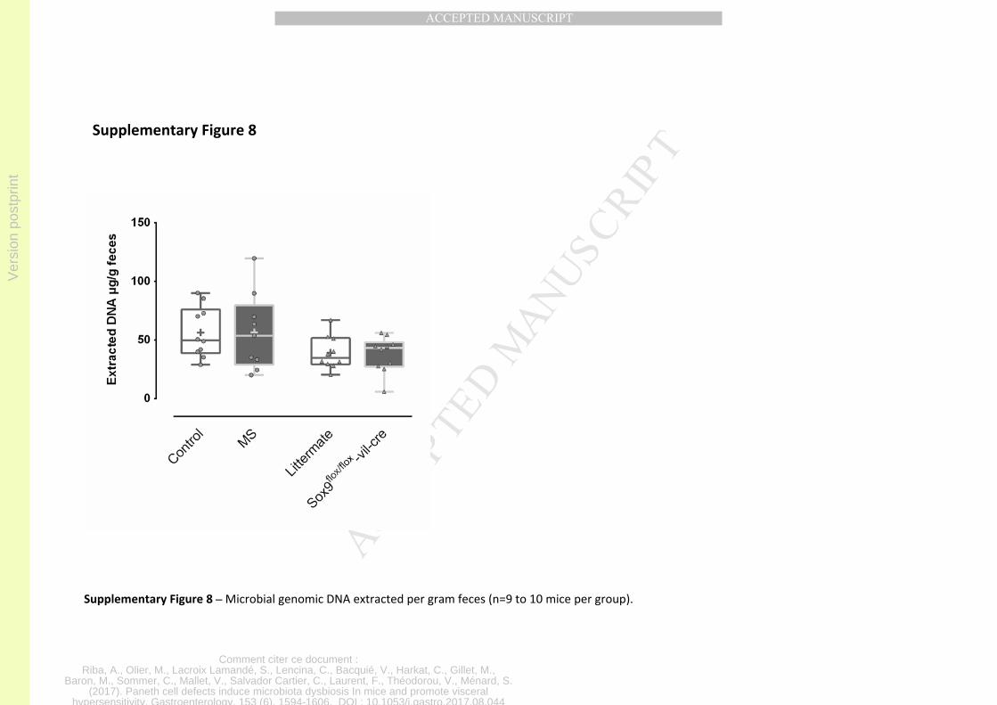
Ver
sion
pos
tprin
t
Comment citer ce document :Riba, A., Olier, M., Lacroix Lamandé, S., Lencina, C., Bacquié, V., Harkat, C., Gillet, M.,
Baron, M., Sommer, C., Mallet, V., Salvador Cartier, C., Laurent, F., Théodorou, V., Ménard, S.(2017). Paneth cell defects induce microbiota dysbiosis In mice and promote visceral
hypersensitivity. Gastroenterology, 153 (6), 1594-1606. DOI : 10.1053/j.gastro.2017.08.044
MANUSCRIP
T
ACCEPTED
ACCEPTED MANUSCRIPT
Supplementary Figure 8
Supplementary Figure 8 – Microbial genomic DNA extracted per gram feces (n=9 to 10 mice per group).