pacemakers and icdscvisymposium.com/sitebuildercontent/sitebuilderfiles/pacemakers... ·...
TRANSCRIPT

Pacemakers and ICDs
John Cogan, MD, FACC, FHRS
Arrhythmia Consultants of South
Florida
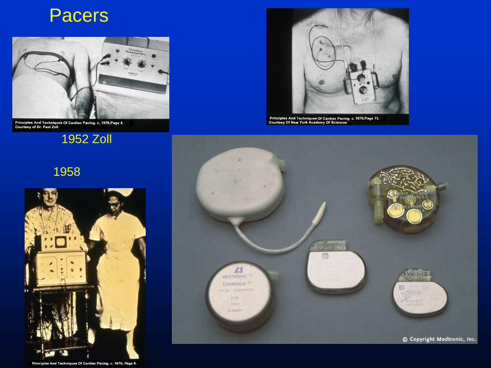
1952 Zoll
1958
Pacers
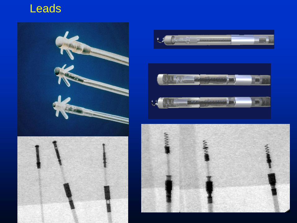
Leads

Unipolar vs. Bipolar
• Unipolar
– Larger pacing spikes on EKG
– Small diameter lead body
– Less rigid lead body
– More susceptible to oversensing
– May produce muscle and nerve stimulation
• Bipolar – Larger diameter lead
body
– Tend to be stiffer
– Less susceptible to oversensing
– Unipolar programmable
– Less likely to produce muscle and nerve stimulation

Basics
• Pulse generator:
– Provides energy, and has an advanced timer
with circuitry and memory chips
• Leads:
– Pace and sense
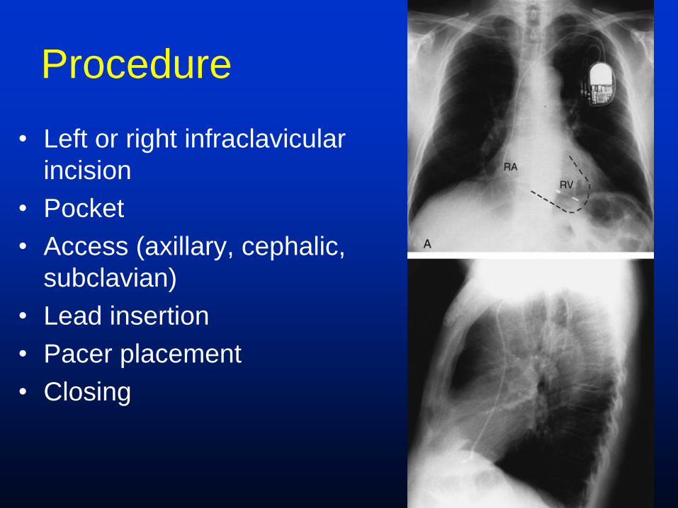
Procedure
• Left or right infraclavicular
incision
• Access (axillary, cephalic,
subclavian)
• Lead insertion
• Pacer placement
• Closing
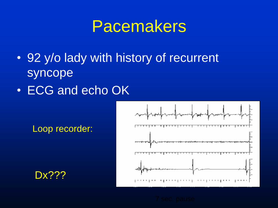
Pacemakers
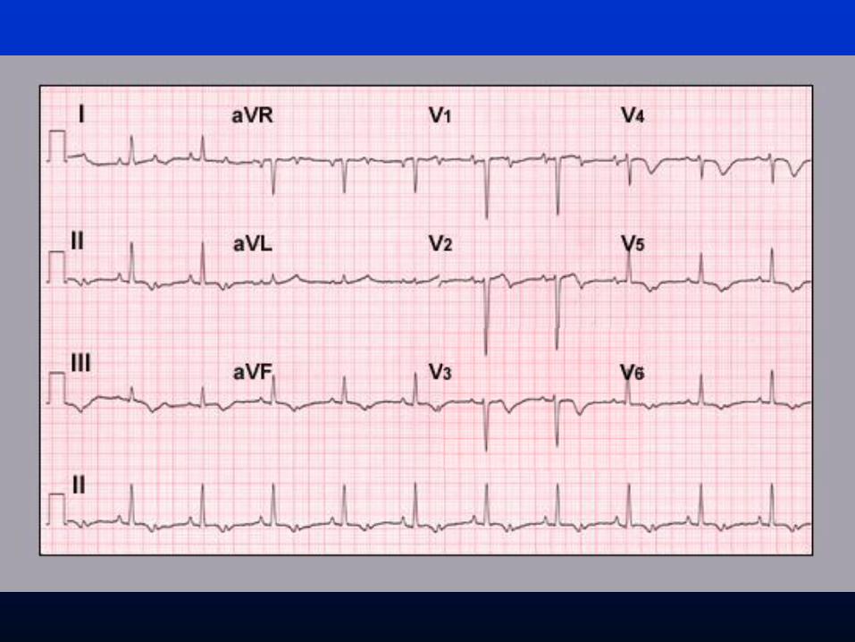
• 92 y/o lady with history of recurrent
syncope
• ECG and echo OK 0.4
mV
0.2
0.0
-0.2
-0.4
0.4
0.2
0.0
-0.2
-0.4
0.4
0.2
0.0
-0.2
-0.4 :45 :44 :43 :42 :41 :40 :39 :38 :37
:37 :36 :35 :34 :33 :32 :31 :30 :29
:29 :28 :27 :26 :25 :24 :23 :22 :21
08:23:2
1
8:23:29
08:23:3
7
Loop recorder:
7 sec. pause
Dx???

Indications
• SSS
• The short story:
– NO SYMPTOMS: NO PACEMAKER
– Symptomatic brady – not iatrogenic
– Chronotropic incompetence
– Syncope with evidence of SSS
– Pauses (3 sec) or very slow HR (<40) while awake and symptoms
Indications
• AV Block
– 3rd degree AVB, or 2nd degree AV block Mobitz II
– Any 2nd degree AV block with symptoms
– Mobitz I, proven to be intra or infra hisian with EPS
– Some neuromuscular diseases (even without symptoms)
– 2nd degree AV Mobitz I, no symptoms No pacer
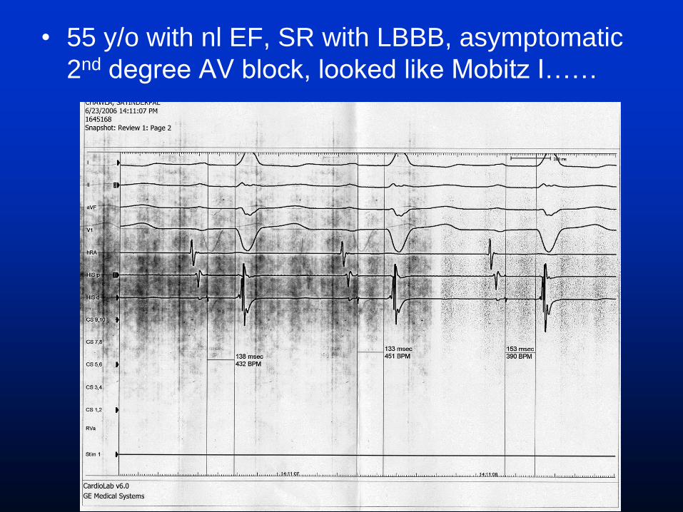
• 55 y/o with nl EF, SR with LBBB, asymptomatic
2nd degree AV block, looked like Mobitz I……

His
Indications
• Syncope with HV >70
• Asymptomatic with HV>100
• Infrahisian block, not physiologic
• Long QT
– Same as others, plus pts with pause
dependant VT
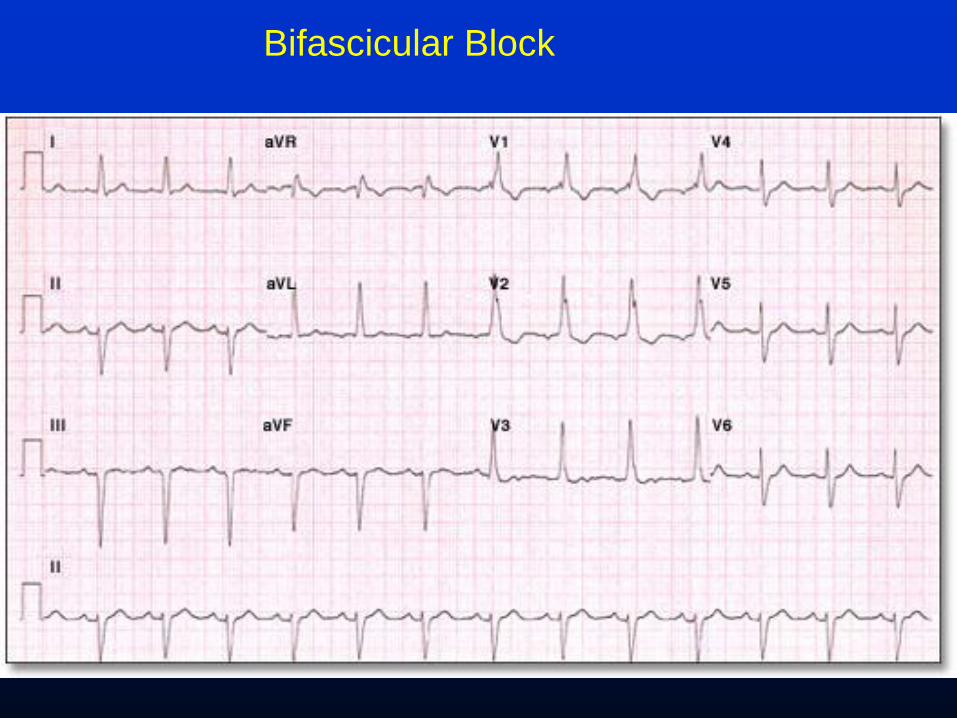
Bifascicular Block
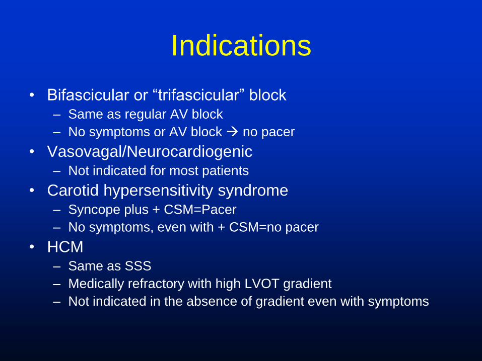
Indications
• Bifascicular or “trifascicular” block – Same as regular AV block
– No symptoms or AV block no pacer
• Vasovagal/Neurocardiogenic – Not indicated for most patients
• Carotid hypersensitivity syndrome – Syncope plus + CSM=Pacer
– No symptoms, even with + CSM=no pacer
• HCM – Same as SSS
– Medically refractory with high LVOT gradient
– Not indicated in the absence of gradient even with symptoms
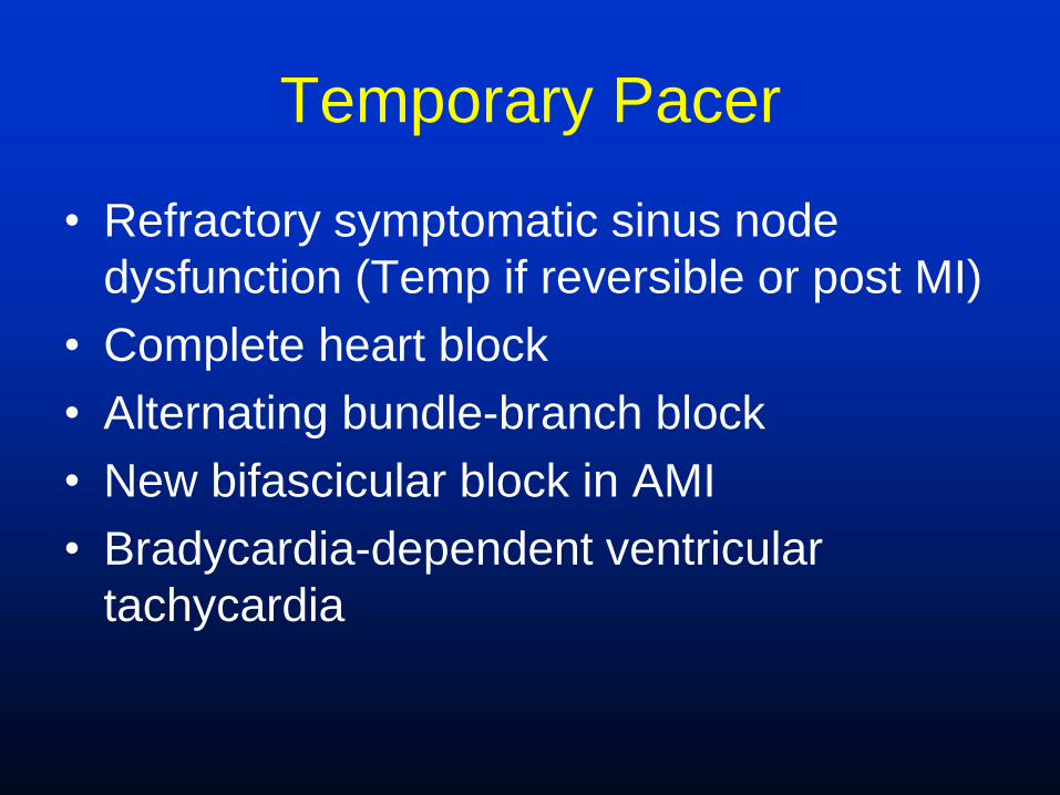
Temporary Pacer
• Refractory symptomatic sinus node
dysfunction (Temp if reversible or post MI)
• Complete heart block
• Alternating bundle-branch block
• New bifascicular block in AMI
• Bradycardia-dependent ventricular
tachycardia
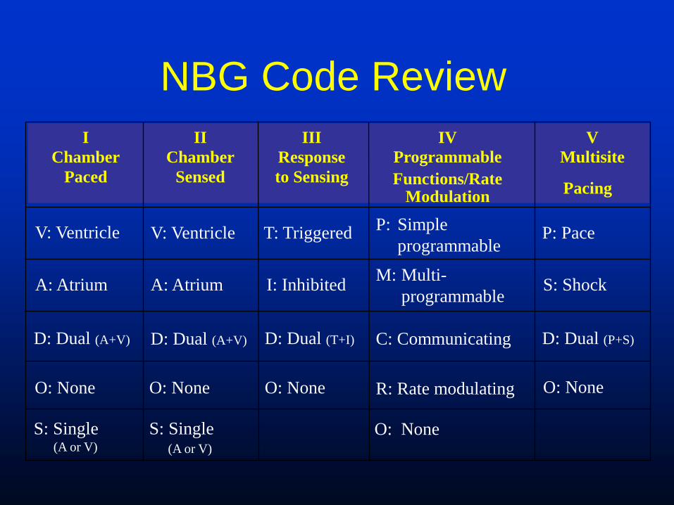
NBG Code Review
I
Chamber
Paced
II
Chamber
Sensed
III
Response
to Sensing
IV
Programmable
Functions/Rate Modulation
V
Multisite
V: Ventricle V: Ventricle T: Triggered P: Simple
programmable P: Pace
A: Atrium A: Atrium I: Inhibited M: Multi-
programmable S: Shock
D: Dual (A+V) D: Dual (A+V) D: Dual (T+I) C: Communicating D: Dual (P+S)
O: None O: None O: None R: Rate modulating O: None
S: Single (A or V)
S: Single
(A or V)
O: None
Pacing
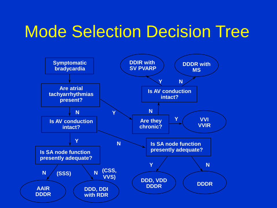
Mode Selection Decision Tree
DDIR with SV PVARP
DDDR with MS
N
VVI VVIR
Are they chronic?
Y
Y N
DDD, VDD DDDR DDDR
Y N
Is AV conduction intact?
Is SA node function presently adequate?
Symptomatic bradycardia
Are atrial tachyarrhythmias
present?
Is SA node function presently adequate?
Is AV conduction intact?
Y
Y N
AAIR DDDR
DDD, DDI with RDR
N N (SSS) (CSS,
VVS)
N
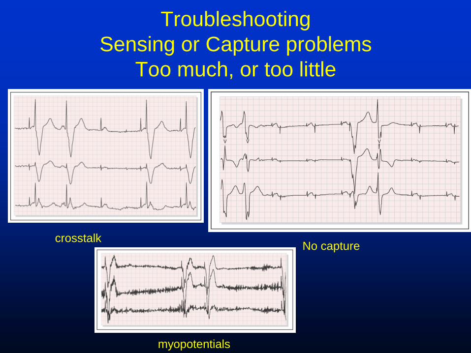
Troubleshooting
Sensing or Capture problems
Too much, or too little
crosstalk No capture
myopotentials

Diagnostics
VT
AFIB
Pacemaker syndrome
• Symptoms:
– syncope or near-syncope, orthostatic dizziness, fatigue, exercise intolerance, weakness, lethargy, chest fullness or pain, cough, uncomfortable pulsations in the neck or abdomen, right upper quadrant pain, and other nonspecific symptoms.
• Cause:
– Loss of AV synchromy, most common in VVI or DDI mode.
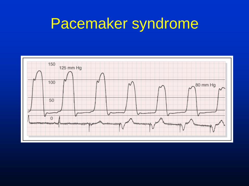
Pacemaker syndrome

ICDs
•1966: Device conception
•1980: First human implant at Johns Hopkins
Hospital. To meet criteria, the patients had to
have survived two episodes of cardiac arrest
not associated with an infarction and VF had to
be documented at least once.
History of ICD Therapy
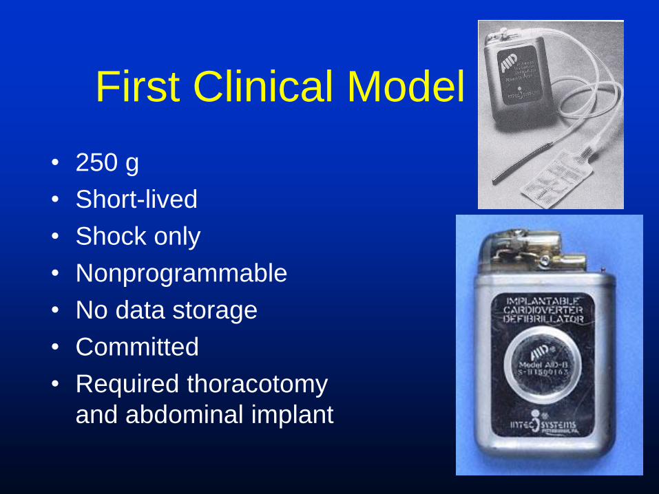
First Clinical Model
• 250 g
• Short-lived
• Shock only
• Nonprogrammable
• No data storage
• Committed
• Required thoracotomy
and abdominal implant
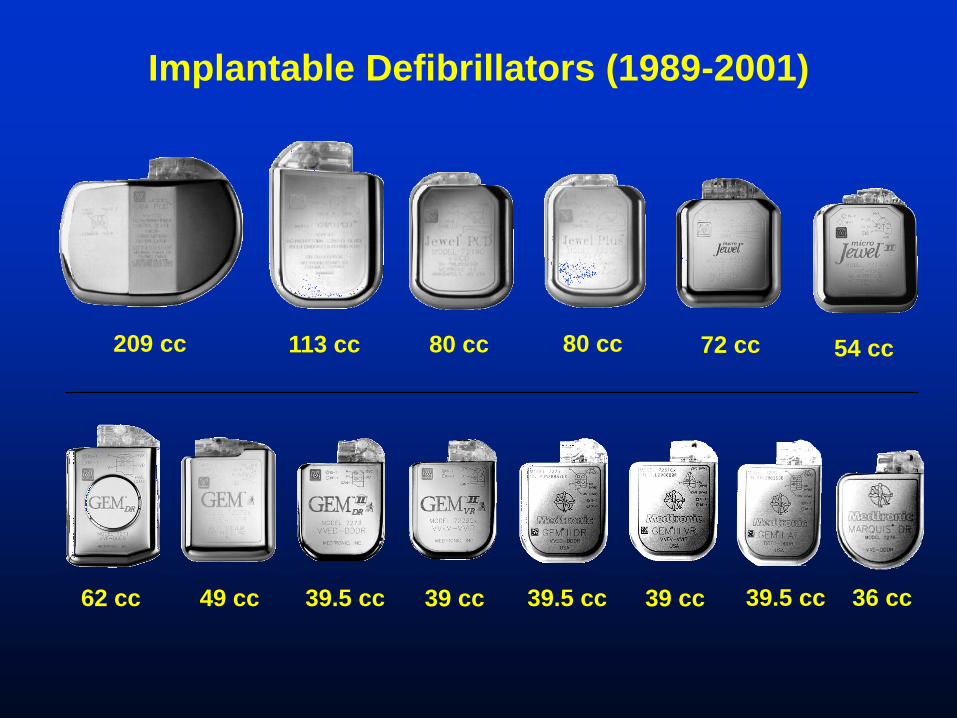
Implantable Defibrillators (1989-2001)
209 cc 113 cc 80 cc 80 cc 72 cc 54 cc
62 cc 49 cc 39.5 cc 39 cc 39.5 cc 39.5 cc 39 cc 36 cc
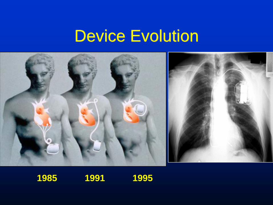
Device Evolution
1985 1991 1995
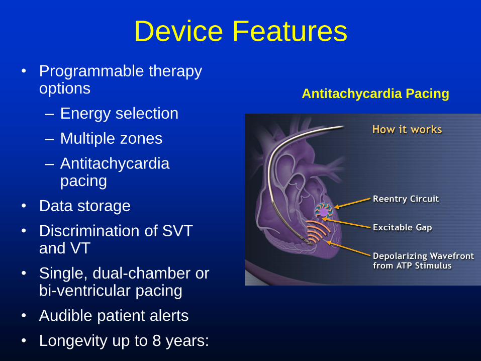
Device Features
• Programmable therapy options
– Energy selection
– Multiple zones
– Antitachycardia pacing
• Data storage
• Discrimination of SVT and VT
• Single, dual-chamber or bi-ventricular pacing
• Audible patient alerts
• Longevity up to 8 years:
Antitachycardia Pacing

Shock Pathway Impedance
Shocking Circuit
Example of Dual Coil: AX > B (Active Can + SVC HV Coil > RV HV Coil)
Vectors

Implantable Cardioverter Defibrillator Trials for Secondary Prevention of SCD
Mortality
STUDY GROUP Control ICDs Rel RR P Value
AVID VF, sustained VT; 24% 15.8% 30% 0.02
EF 40%
ICD vs amiodarone
Mean EF 35%
F/U: 18 mo
CIDS VF, symptomatic VT; 29.6% 25.3% -30% 0.14
EF 35%, CL < 400ms
Mean EF 34%
F/U: 36 mo
CASH Survivors of SCD (VF/VT) 44.4% 36.4% 23% 0.08
propafenone/metoprolol/
amiodarone/ICD
Mean EF 45%, F/U: 57 mo
Meta-analysis 28% 0.006
AVID = Antiarrhythmics vs Implantable Defibrillators. NEJM 1997; 337:1576 (terminated early)
CIDS = Canadian ICD study. Circulation 2000;101:1297
CASH = Cardiac Arrest Study of Hamburg. Circulation 2000;102:748
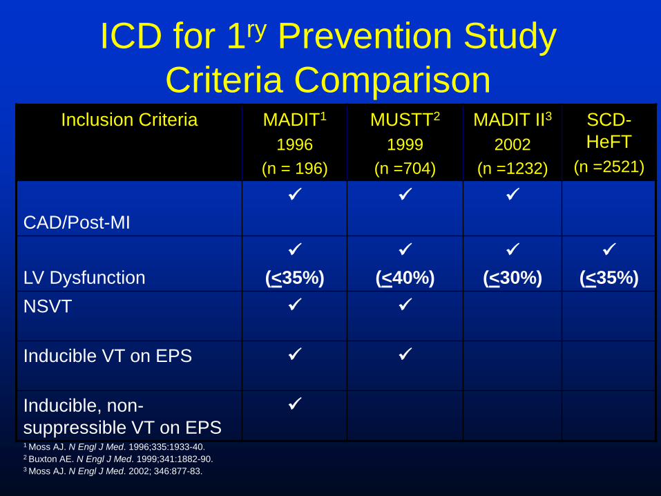
ICD for 1ry Prevention Study
Criteria Comparison Inclusion Criteria MADIT1
1996
(n = 196)
MUSTT2
1999
(n =704)
MADIT II3
2002
(n =1232)
SCD-
HeFT
(n =2521)
CAD/Post-MI
LV Dysfunction
(<35%)
(<40%)
(<30%)
(<35%)
NSVT
Inducible VT on EPS
Inducible, non-
suppressible VT on EPS
1 Moss AJ. N Engl J Med. 1996;335:1933-40. 2 Buxton AE. N Engl J Med. 1999;341:1882-90. 3 Moss AJ. N Engl J Med. 2002; 346:877-83.
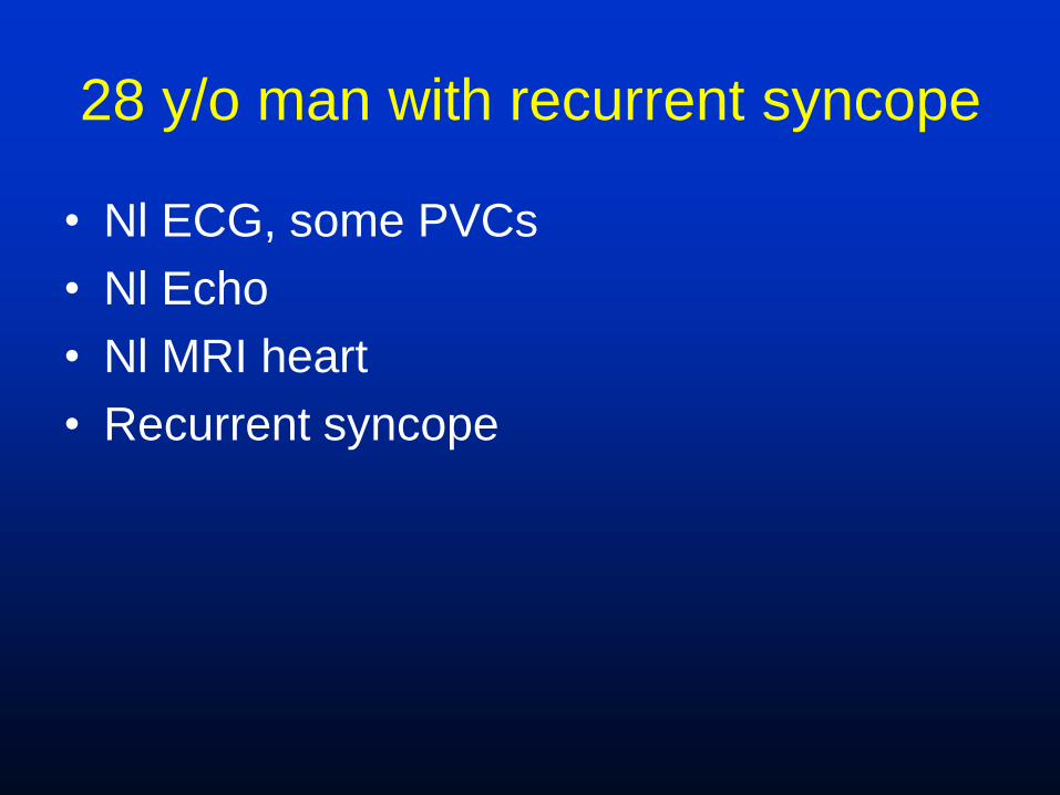
28 y/o man with recurrent syncope
• Nl ECG, some PVCs
• Nl Echo
• Nl MRI heart
• Recurrent syncope
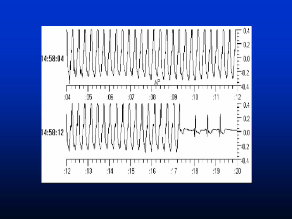
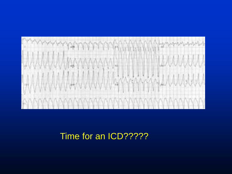
Time for an ICD?????
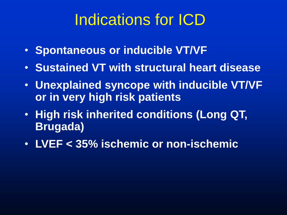
Indications for ICD
• Spontaneous or inducible VT/VF
• Sustained VT with structural heart disease
• Unexplained syncope with inducible VT/VF or in very high risk patients
• High risk inherited conditions (Long QT, Brugada)
• LVEF < 35% ischemic or non-ischemic
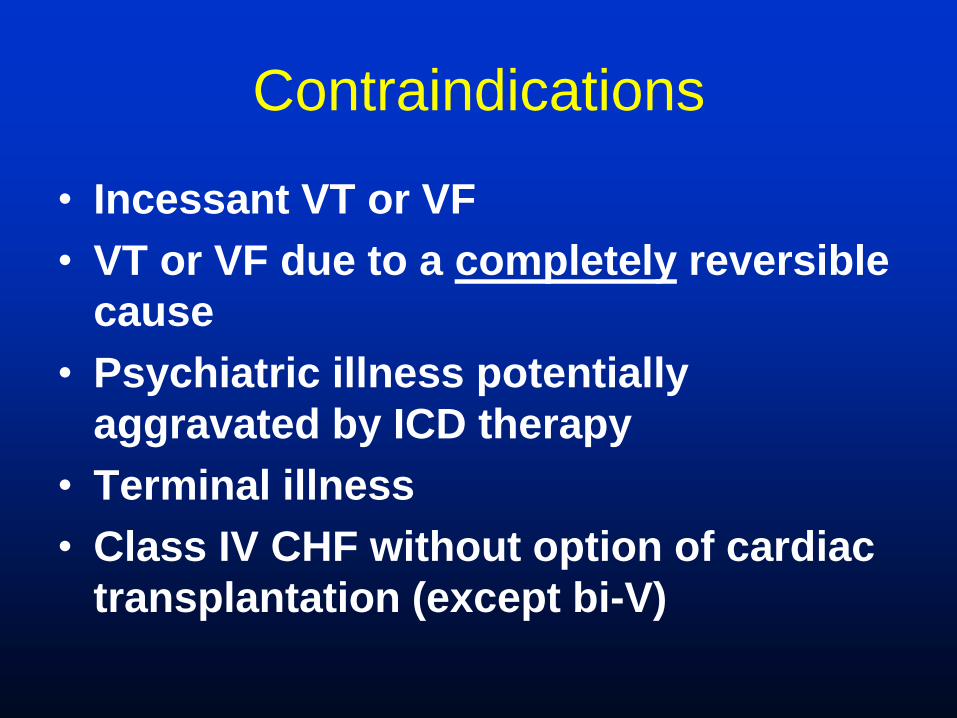
Contraindications
• Incessant VT or VF
• VT or VF due to a completely reversible
cause
• Psychiatric illness potentially
aggravated by ICD therapy
• Terminal illness
• Class IV CHF without option of cardiac
transplantation (except bi-V)
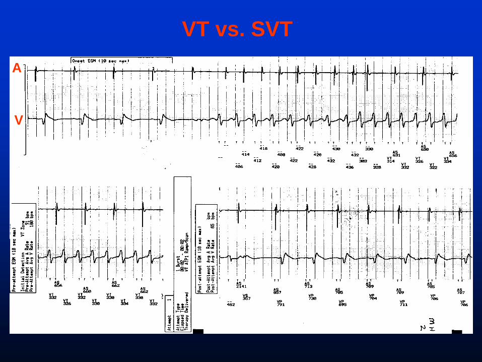
VT vs. SVT
A
V

Sometimes, it’s just bad: Lead fracture,
noise, oversensing (diaphragmatic
myopotentials in this case)
Deep breathing

VT onset ATP onset
Episode duration = 5.3 s
A.
. . . . . . .
VT onset 1st ATP onset
2nd ATP onset
Episode duration = 16.8 s
B.
Accelerated VT 4.8 J shock
Examples of Success (A) and Failure (B)
Wathen M, Sweeney M, DeGroot P. Circulation. 2001; 104: 796-801.
Anti Tachycardia Pacing
Bi-V ICD
Bi-V
• Dyssynchrony is an anatomical-
mechanical event involving:
– Abnormal ventricular activation (EF)
– Decreased ventricular filling
– Abnormal ventricular wall motion
• Up to 50%-70% of patients with HF have
ventricular dyssynchrony

Who Needs CRT?
Classic indication:
• NYHA = III-IV
• QRS duration ≥120 ms
• EF ≤ 35%
New Indication:
• NYHA = I-II
• EF <30%
• LBBB
Bi-V Trials
PATH-CHF
MUSTIC
MIRACLE
Contak CD
Miracle ICD
Companion
Care HF
• These trials show:
– Better exercise tolerance
– Better QOL
– Better NHYA class
– Improved mortality
Pacer/ICD Complications • Acute: Bleeding, pneumothorax, lead
dislodgement, hematoma, lead malposition, perforation, PE, SCV DVT
• Infection or erosion in 1-2% of cases and requires system removal
• Malfunction of device; lead is the weakest link can fracture or fail
• Inappropriate shocks
• Ventricular pacing might be detrimental
– MADIT II demonstrated increased incidence of CHF in defibrillator patients
– DAVID trial demonstrated 3.6% mortality increment with DDD versus backup VVI
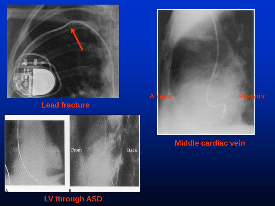
Lead fracture
Middle cardiac vein
LV through ASD
Anterior Posterior
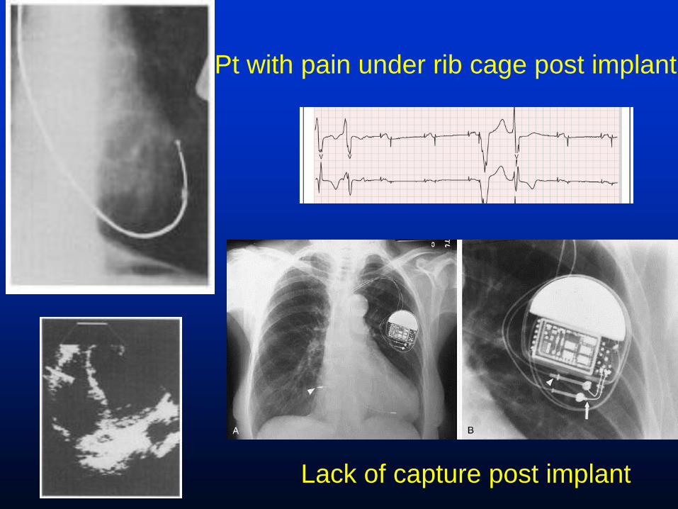
Pt with pain under rib cage post implant
Lack of capture post implant
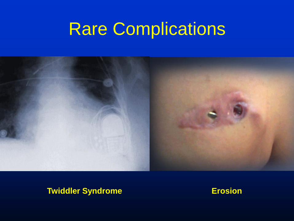
Rare Complications
Twiddler Syndrome Erosion
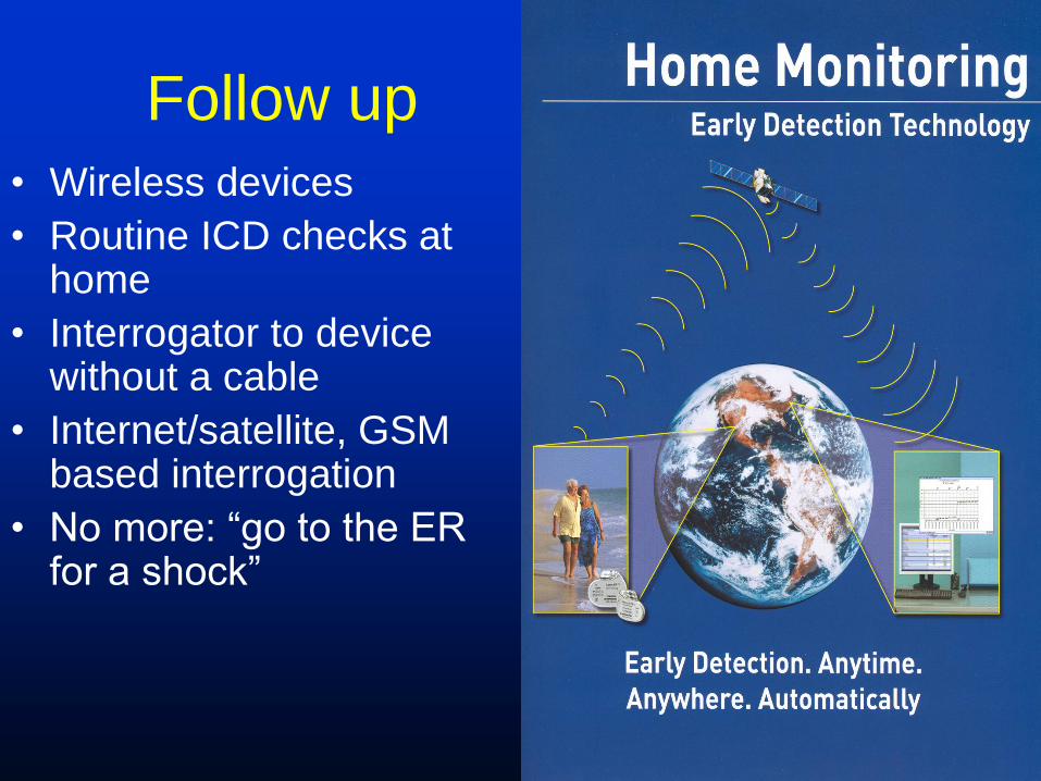
Follow up
• Wireless devices
• Routine ICD checks at home
• Interrogator to device without a cable
• Internet/satellite, GSM based interrogation
• No more: “go to the ER for a shock”
Questions?