overexpression of human apolipoprotein a4 in transgenic ... · overexpression of human...
TRANSCRIPT

Overexpression of human apolipoprotein A4 in transgenic rats and the hyperlipoproteinemia associated with experimental nephrosis
Bryan F. Burkey,'** Dennis France,* Hongxing Wang,? Xiaowen Ma,* Barbara Brand,* Colleen Abuhani,* Margaret R. Diffenderfer,? Julian B. Marsh,? James R. Patemiti, Jr.,* and Edward A. Fisher'+ Department of Metabolic Diseases,* Preclinical Research, Sandoz Research Institute, Sandoz Pharmaceuticals Corporation, East Hanover, NJ 07936, and Biochemistry Department,+ Medical College of Pennsylvania, Philadelphia, PA 19129
Abstract Hyperlipoproteinemia contributes both to kidney disease progression and the development of atherosclerosis. Elevated high density lipoprotein cholesterol and apolipopro- tein A-I (apoA-I) serum levels are independent factors protec- tive against the atherosclerotic process. We examined the effects in a transgenic rat model of human apoA-I expression on the hyperlipoproteinemia and edema after puromycin aminonucleoside-induced nephrosis in three groups of ani- mals: low line (TgR[hAI]low, human plasma apoA-I = 16.0 mg/dl); high line (TgR[hAI]high, 284 mg/dl); and non-trans- genic litter mates (TgR[hAI]non). Nephrosis increased total plasma apoA-I levels 2-fold in TgR[hAI]non rats (75 vs. 162 mg/dl) and 4-fold in the TgR[hAI]low (97 vs. 458 mg/dl) and TgR[hAI]high rats (356 vs. 1,346 mg/dl). In both transgenic lines, this increase was due mainly to elevations of serum human apoA-I. The hepatic steady-state levels of rat apoA-I mRNA increased 5- to ?-fold in all three groups, while human apoA-I mRNA levels increased 21- and 65-fold in the low and high expressing groups, respectively, indicating a different degree of responsiveness of the rat and human genes. While nephrotic TgR[hAI]non and TgR[hAI]low rats showed severe hyperlipoproteinemia and edema, much lower levels of edema and of serum triglycerides, phospholipids, and choles- terol were seen in the TgR[hAI]high group. Urinary excretion of apoA-I, phospholipids, and cholesterol was significantly increased in the TgR[hAI]high group,. indicating this as one possible mechanism for the relatively lower serum levels of these lipids. M We conclude that the human apoA-I gene is responsive to nephrosis and that human apoA-I-transgenic rats with this syndrome provide an animal model for the study of human high density lipoprotein and apoA-I metabo- lism.-Burkey, B. F., D. France, H. Wang, X. Ma, B. Brand, C. Abuhani, M. R. Diffenderfer, J. B. Marsh, J. R. Paterniti, Jr., and E. A. Fisher. Overexpression of human apolipopro- tein A-I in transgenic rats and the hyperlipoproteinemia asso- ciated with experimental nephr0sis.J Lipid Res. 1995. 36: 1463-1473.
Supplementary key words high density lipoprotein - kidney disease puromycin aminonucleoside - hyperlipidemia - proteinuria
Hyperlipoproteinemia is a common feature of the nephrotic syndrome and other glomerular diseases in both humans and experimental animals (1-4). While, in the general population, hyperlipoproteinemia is a strong risk factor for coronary artery disease, in those with renal disease its potential for adverse consequences may be further increased by contributing to the progres- sion of glomerular dysfunction (5 ) .
In previous studies of non-transgenic rats with experi- mental nephrotic syndrome, a hallmark of the hyper- lipoproteinemia has been the overproduction by the liver of apolipoprotein A-I (apoA-I) (4). Thus, in spite of urinary losses of high density lipoprotein (HDL) (6), serum levels of HDL cholesterol typically increase, con- tributing to the hyperlipidemic state. The overproduc- tion of hepatic apoA-I has been shown to result from increased apoA-I synthesis (7), and in more recent stud- ies this was attributed to increased apoA-I gene expres- sion (8-10).
The availability of transgenic rats expressing human apoA-I (h-apoA-I) (1 1) allowed us to determine whether the human gene also responds to the nephrotic state. In addition, by studying lines with different quantitative levels of basal h-apoA-I expression (low and high), we were able to explore relationships among the degree of h-apoA-I production, the serum levels of apoA-I and apoB-containing lipoproteins, and the severity of the nephrotic state.
Abbreviations: TgR, transgenic rat; FPLC, fast protein liquid chromatography; TBS, Tris-buffered saline; PAN, puromycin aminonucleoside; h-apoA-I, human apolipoprotein A-I.
'Reprint requests may be addressed to either author.
Journal of Lipid Research Volume 36, 1995 1463
by guest, on June 19, 2018w
ww
.jlr.orgD
ownloaded from

The results demonstrate that the h-apoA-I gene re- sponded to the nephrotic syndrome in each transgenic group (low and high basal expressors). Furthermore, when nephrosis was induced in the group with high basal expression, compared to non-transgenic controls and low expressors, there was suppression of edema and less hyperlipoproteinemia, associated with increased urinary excretion of apoA-I, cholesterol, and phos- pholipids. Thus, in addition to the value of these two groups of transgenic rats to studies of lipid and apolipo- protein metabolism, their differing responses to neph- rosis could potentially be used to test the effects of plasma lipid and lipoprotein levels on the progression of glomerular dysfunction in experimental renal dis- ease.
MATERIALS AND METHODS
Induction of experimental nephrosis
Transgenic rats were generated by microinjection of a 13 kbp DNA fragment containing the human apoA-I gene plus 10 kbp of 5’ and 1 kbp of 3’ flanking sequence. Two established transgenic rat lines, TgR[OhAI]7 and TgR[OhAI]2, express moderate and high levels of h- apoA-I, respectively. Both lines were originally made using OFA rats (Oncins France Strain A derived from the Sprague-Dawley strain) and subsequently rederived to a Sprague-Dawley background (1 1). TgR[OhAI]7 will be referred to as TgR[hAI]i,,, TgR[OhAI]2 will be re- ferred to as TgR[hAI]hi,h, and non-transgenic litermates will be referred to as TgR[hAIInon. Male rats, 10 per group, age 7- 11 weeks were used. In each group, experi- mental nephrosis was induced in one-half of the rats by bolus intraperitoneal injections of puromycin aminonu- cleoside (PAN) at 65 mg/kg body weight (No P-7103; Sigma Chemical Co., St. Louis, MO) in saline, on 2 consecutive days. All other animals were injected with equivalent volumes of saline. Within 8 days, the re- sponse to PAN treatment was readily apparent as peri- toneal edema, which could be clinically graded (by a blinded observer) over a range of slight to severe. Ani- mals were killed 8 days after the initial injection of PAN or saline. Whole blood for serum isolation and livers were collected at the time of killing. In a separate experiment, urine was collected from four TgR[hAIInon and four TgR[hAI]high rats by placing them in metabolic cages 20 h prior to killing. All animal studies were performed under a protocol approved by the Animal Care Committee of the Medical College of Pennsylvania and the Sandoz Animal Care and Use Committee.
Serum lipid analysis
One part of serum was diluted with four parts of distilled water for the analysis of total serum cholesterol, triglyceride, and phospholipid. In a 96-well microplate (Nunc-Immuno Plate Maxisorb, InterMed) 25 pL of diluted serum was assayed for its lipid content by the addition of 200 pL cholesterol reagent (No. 352-1000, Cholesterol 1000, Sigma), or 200 pL triglyceride reagent (No. 450032; Triglycerides/GB, Boehringer Mannheim Co., Indianapolis, IN), or 200 1L phospholipid reagent (No. 996-54001; Phospholipids B, Wako Pure Chemical Industries, Osaka Japan) as specified by the manufactur- ers. Samples were developed by incubating for 30 min at room temperature and absorbance was determined at 490 nm. Equivalent dilutions of calibrated lipid stand- ards (No. C 0534, Cholesterol Calibrator Set, Sigma) and calibrated controls (No. L 1008, Lipid Control-E and No. L 2008, Lipid Control-N, Sigma) were used as reference standards. Fractionation of serum lipoproteins by Su- perose-6 gel permeation chromatography was per- formed with a robotic FPLC system as previously de- scribed ( 12). Briefly, the column matrix was equilibrated with Tris-buffered saline (TBS, 50 mM Tris, pH 7.4, 0.15 M NaC1) containing 0.01% sodium azide at 0.5 mL per min. Serum, 200 pL, was injected and 40 0.5-mL frac- tions were collected at a flow rate of 0.5 mL/min. The cholesterol profile was determined on an 80-yL fraction with 120 yL of cholesterol reagent (No. 81423, Fast Cholesterol, Sclavo, Wayne, NJ). After incubation at room temperature for 30 min, absorbance was deter- mined at 490 nm.
Urine lipid analysis
Rats were placed in individual metabolic cages and urine was collected overnight. Aliquots were analyzed for cholesterol and phospholipids. Proteins were first precipitated with 10% trichloracetic acid and the pre- cipitates were extracted twice at room temperature with 1 % trichloracetic acid in ethanol. The precipitates were dissolved in 0.1 N NaOH and protein concentrations were measured by the micro-biuret method (13).
The supernatant solution was treated with 3 volumes of ether to precipitate the acid-alcohol-soluble protein. The protein was dissolved in 0.1 N NaOH and its concentration was determined (13). The solvents were removed from the lipid extract, which was then re-dis- solved in chloroform-methanol 2: 1. Water-soluble sub- stances were removed by the method of Folch, Lees, and Sloane Stanley (14). Cholesterol and phospholipid in the chloroform phase of the Folch extract were determined by the methods of Zlatkis, Zak, and Boyle (15) and Sokoloff and Rothblat (16), respectively.
1464 Joumal of Lipid Research Volume 36, 1995
by guest, on June 19, 2018w
ww
.jlr.orgD
ownloaded from

Nondenaturing gradient gel electrophoresis of serum lipoproteins
Total lipoproteins from 200 pL serum, adjusted to d 1.23 g/mL with potassium bromide, were isolated by a 12-h (42,000 rpm) single-spin density ultracentrifuga- tion in a Ti42.2 rotor (Beckman Instruments, Palo Alto, CA). Two parts of lipoprotein fraction (the upper 60 pL sample from the spin) were mixed with one part of Sudan Black B (7 mg/mL in polyethylene glycol) and incubated at room temperature for 2 h. Lipoproteins from 20 pL were loaded onto a nondenaturing polyacry- lamide gradient gel (PAA 2/16; Pharmacia LKB, Uppsala, Sweden) and resolved by electrophoresis at 125 V for 16 h in 89 mM Tris, pH 8.3, 89 mM boric acid, and 2.6 mM EDTA (No. SA 100033, TBE Seprabuff, Integrated Separation Systems, Natick, MA). After lipid- containing bands were identified, apolipoproteins and molecular weight markers were visualized by staining with Coomassie Brilliant Blue (No. B-8647, Brilliant Blue R, Sigma).
Quantitation of serum apoA-I
Levels of human and rat apoA-I were determined by competition ELISA. For the h-apoA-I assay, microplate wells (Nunc-Immuno Plate Maxisorb, Baxter Scientific, Edison, NJ) were coated overnight at 4°C with 0.4 pg per well of purified h-apoA-I (No. A-9284, Sigma), then blocked for 2 h at room temperature in TBS containing 5% bovine serum albumin (BSA). Assay plates were incubated overnight at room temperature with rat se- rum diluted 1:20 in TBS containing 4% Tween-20 (pre- heated to 52"C, 1 h) and goat anti-human apoA-I im- mune serum diluted 1:10,000 in TBS containing 4% Tween-20. Anti-human apoA-I immune serum was gen- erated by immunizing a goat with h-apoA-I isolated from human HDL3 and purified by electroelution of a single band from an SDS polyacrylamide gel (1 7). After wash- ing, bound anti-human apoA-I antibodies were tagged with alkaline phosphataseconjugated rabbit anti-goat IgC (No. A-7650, Sigma) diluted 1:1,000 in TBS contain- ing 2% BSA and detected using 200 pL of a 1 mg/mL phosphatase substrate (No. 1040, Sigma) dissolved in 2% diethanolamine. After incubation at room tempera- ture for 30 min, the absorbance was determined at 405 nm. Standard curves were made by serial dilution of human serum containing known amounts of h-apoA-I. There was no detectable cross-reactivity (by either im- munoblot or ELISA format) of the anti-human apoA-I immune serum for rat apoA-I. The assay was linear over the range of 1.5-24 pg apoA-I per well.
The rat apoA-I ELISA was identical to the above ELISA, except 800 ng of rat apoA-I, purified from rat HDL by preparative SDS PAGE (17), was used to coat each well of the microtiter plate. Diluted rat serum
samples were not preheated, and goat anti-rat apoA-I immune serum was diluted 1:7,500. Anti-rat apoA-I immune serum was generated by immunizing a goat with rat apoA-I isolated from rat HDLs and purified by electroelution of a single band from an SDS polyacry- lamide gel (17). Standard curves were made by dilution of rat serum containing known amounts of rat apoA-I. Minor cross-reactivity of anti-rat apoA-I antiserum for h-apoA-I was removed by affinity chromatography over a human serum affinity column. The assay was linear in the range of 0.9-15 pg of apoA-I per well.
Non-reducing SDS-PAGE analysis of serum and urine proteins
Rat serum or urine (3 pL) was mixed into Tris-SDS sample solubilization buffer (No. SA100051, Tris-SDS SepraSOL, Integrated Separation Systems) to yield a 50 pL final volume. Samples were heat-denatured at 80°C for 15 min, and loaded on an 11% SDS polyacrylamide gel. Proteins, resolved by electrophoresis at 75 V for 6 h, were visualized by staining with Coomassie Brilliant Blue.
Density gradient ultracentrifugation of total serum lipoproteins
Pools of serum (1 mL) from each treatment group were density adjusted to 1.23 g/mL with potassium bromide in 14 x 95 mm polyallomer centrifuge tubes (No. 331374, Beckman). Four potassium bromide solu- tions of descending density from 1.15 to 1.01 g/mL were layered over the serum sample and ultracentri- fuged in a Beckman SW40Ti rotor at 35,000 rpm for 36 h at 4°C. Lipoproteins were collected by pumping a 1.30 g/mL potassium bromide solution at a rate of 0.4 mL/min through a 20-gauge needle inserted through the tube wall at the bottom of the centrifuge tube. Fractions of 0.2 mL were collected for analysis of total cholesterol, h-apoA-I and rat apoA-I.
Isolation and analysis of hepatic RNA
Total RNA was isolated from rat liver by a modified guanidine salt-based procedure previously described (18). Ten-pg aliquots of RNA were mixed in loading buffer (50% formamide, 16% formaldehyde, 50 mM MOPS, 2 mM EDTA, pH 7.0), heated to 56°C for 30 min, rapidly chilled on ice, and electrophoresed at 50 V for 3-5 h on a 1% agarose gel containing 17.75% formalde- hyde, 50 mM MOPS, 2 mM EDTA, pH 7.0. Resolved RNA was transferred to a nylon membrane (Zetaprobe, Bio- Rad, Melville, NY) by passive capillary transfer in 50 mM NaOH and UV crosslinked. Oligonucleotide probes complementary to the rat and human apoA-I mRNAs were labeled by the T4 polynucleotide kinase technique (19) with [p2P]ATP (3000 Ci/mmol, New England Nu-
Burkey et al. Nephrotic hyperlipoproteinemia and apoA-I transgenic expression 1465
by guest, on June 19, 2018w
ww
.jlr.orgD
ownloaded from

clear, Boston, MA). All blots were prehybridized for 4-5 h at 65°C in 5 X SSC, 20 mM sodium phosphate, pH 7.0, 10 x Denhardt’s solution, 7% SDS, with 100 pg/mL denatured salmon sperm DNA. Heat-denatured probes were hybridized to the immobilized RNA at 65°C in hybridization buffer (the same as prehybridization buff- er, but with the addition of 10% dextran sulfate). The blots were washed twice with 0.5 x SSC, 0.1% SDS at room temperature, then once with 0.1 x SSC, 0.1% SDS at 65°C. Blots were then exposed to a phosphor screen overnight and scanned on a Molecular Dynamics Phos- phoimager. The intensity of the hybridization signal was normalized to elongation factor 1 a, as described by Lu and Werner (20), and expressed as relative to this con- trol.
Values are given as the mean k standard error of the mean. Statistical differences between treatment groups were sought by using Student’s 1 test.
RESULTS
Rat and human apoA-I protein and mRNA levels As previously reported (21, 22), total serum apoA-I
increased after the induction of nephrosis (Table 1). However, a more pronounced total apoA-I elevation occurred in TgR[hA111,, and especially in TgR[hAI]high nephrotic rats, where apoA-I replaced albumin as the major serum protein (see Fig. 4A). The increase in total apoA-I in the TgR[hAI]l,, and TgR[hAI]high nephrotic rats was primarily due to increased h-apoA-I levels. Rat
TABLE 1. Rat and human apoA-I serum levels 8 days after PAN (nephrotic) or saline (control) injection
Serum ApoA-I Levels
Group Rat ApoA-I Human ApoA-I Total ApoA-I
TgR[hAIlnon Control Nephrotic N/C
TgR[hAI]b, Control Nephrotic N/C
Control Nephrotic N/C
TgR[hAI]high
75.2 * 10.0 162.4 f 26.6
2.16a
81.2 f 10.4 127.4 f 9.1
1.57
71.8 * 17.7 38.3 * 5.9
0.53=
mg/dL
75.2 f 10.0 162.4 f 26.6
2.1@
97.2 * 8.7 458.2 f 44.0
16.0 f 4.7 330.8 f 51.7
20.68b 4.7Ih
284.2 f 113.4 356.0 * 96.1 1307.5 * 186.0 1345.8 -L 191.5
4.60h 3.78h
Human and rat apoA-I were measured by species-specific competi- tion ELISA. Values are given as mean f SEM, n = 5; total apoA-I is the sum of rat and human values. The change from control in each group is expressed as the ratio of N/C (nephrotic/control).
a P < 0.05; bP < 0.005; cno significant difference, where the two groups are statistically different as determined by Student’s t-test.
apoA-I actually decreased in the nephrotic TgK[ hAI]h,,,, group.
We have shown previously that the genetic construct used to produce TgR[hA111,, and TgR[hA1]1,,,1, rats drove the expression of h-apoA-I in the liver but not in the intestine ( 1 1). Hepatic rat and human apoA-I mRNA levels were measured by Northern blot hybridization in control and nephrotic rats (Fig. 1 and Table 2). Similar to previous reports (8- lo), the induction of experimen- tal nephrosis significantly increased the steady-state lev- els of rat apoA-I mRNA (5- to 7-fold) over the controls. By contrast, h-apoA-I mRNA levels were elevated 2 1-fold in TgR[hAI]i,,, rats and 65-fold in TgR[hA1]1,,,t, rats. These data demonstrate that rat and h-apoA-I mRNA and protein were differentially elevated by the induction of experimental nephrosis.
Effects of human apoA-I on PAN-induced hyperlipoproteinemia
In control TgR[hAI]high rats, expression of h-apoA-I was associated with raised circulating levels of choles- terol (51.2%) and phospholipid (66.5%) compared with TgR[hAIInon rats, while triglycerides were unchanged. The increased serum levels of cholesterol and phos- pholipid in TgR[hAI]high rats reflected expansion of the HDL pool, evident in the FPLC lipoprotein cholesterol profile of Fig. ZA, and were consistent with our previous report (1 1). The minor contribution of h-apoA-I in the control TgR[hA111,, rats was not enough to alter total serum lipid and lipoprotein profiles.
Consistent with the model of experimental nephrosis (4), marked elevations of total cholesterol (+460%), phospholipid (+212%), and triglyceride (+627%) oc- curred in TgR[hAIInon rats after PAN induction. The degree of lipid elevation in nephrotic TgR[hAIInon rats and nephrotic TgR[hA111,, rats was virtually identical (Table 3). By contrast, the induction of experimental nephrosis in TgR[hAI]high animals produced only a mod- est elevation (+52%) of total serum cholesterol and actually decreased phospholipid levels (Table 3). Total serum triglyceride increased 219% (control: 70.1 mg/dL vs. nephrotic: 223.7 mg/dL). However, this elevation was less than one-half of the response of nephrotic TgR[hAIInon or TgR[hA111,, rats, and was not statistically significant ( P = 0.092). Thus, overexpression of h-apoA-I in the TgR[hAI]high rats suppressed the hyperlipoproteinemia associated with experimental nephrosis.
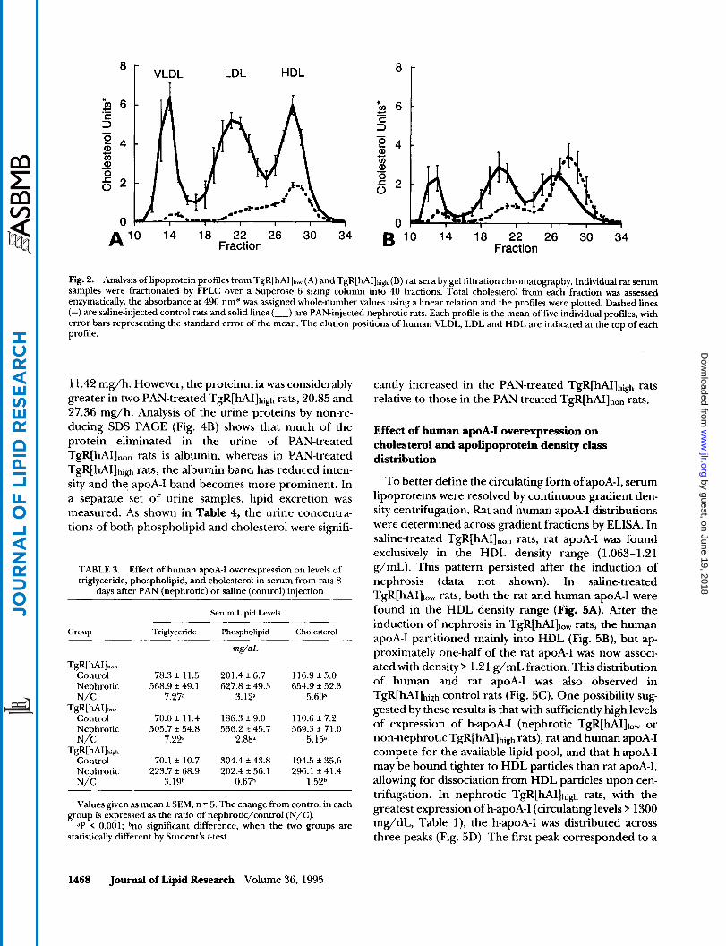
To obtain lipoprotein profiles, serum samples were fractionated by FPLC gel permeation chromatography and lipoprotein peaks were determined by elution vol- ume and cholesterol content. In control TgR[hAI],,, rats, HDL was the predominant lipoprotein species (Fig. 2A). Induction of experimental nephrosis resulted in
1466 Journal of Lipid Research Volume 36, 1995
by guest, on June 19, 2018w
ww
.jlr.orgD
ownloaded from

Rat Apo AI
Human Apo AI
EF1 a
Fig. 1. Northern blot analysis of hepatic rat apoA-I, human apoA-I, and rat EFla. Total RNAwas isolated from livers of TgR[hAI].,,,, TgR[hAI]t,,,, and TgR[hAI]t,,,l, rats killed 8 days after PAN or saline injection. Ten micrograms per lane of total RNA was resolved using a formaldahydedenaturing gel, trans- ferred to nitrocellulose, and hybridized with rat-spe- cific apoA-I, human-specific apoA-I, or EFla radio- labeled probes. Each experimental group, PAN (+) or saline (-) injected rats, is represented by two individuals within each group. - + - + - +
Experimental Treatment
hyperlipoproteinemia evident as increases of all lipopro- tein classes. Lipoprotein profiles of TgR[hA111,, control and nephrotic rats (data not shown) were virtually iden- tical to the corresponding profiles of TgR[hAI]non rats shown in Fig. 2A. Overexpression of h-apoA-I in TgR[hAI]high control rats expanded the HDL choles- terol pool (compare Figs. 2A and 2B). While some increase in non-HDL cholesterol fractions occurred in TgR[hAI]high rats with experimental nephrosis, hyperbe- talipoproteinemia was decreased (compare Figs. 2A and 2B). Surprisingly, in the face of large differences of total serum apoA-I, HDL cholesterol levels in TgR[hAI]high rats were unchanged in controls (142.4 k 33.4 mg/dL)
versus in nephrotics (140.5 f 21.2 mg/dL). However, HDL particles isolated from TgR[hAI]high nephrotic animals tended to elute ahead of control HDL on FPLC, indicating an increased particle size.
The HDL particle size distribution was further exam- ined by non-denaturing gradient gel electrophosis (Fig. 3). In control TgR[hAI]non and TgR[hA111,, rats, a single HDL2 species was present (23). Control TgR[hAI]high rat HDL was polydisperse with distinct HDL1-like, small HDL2, and very small HDLj species. In TgR[hAI]non and TgR[hA111,, rats with experimental nephrosis, two dis- tinct HDL species, comparable to human HDL2 and HDLJ, were observed. Nephrotic TgR[hAI]high rat se- rum contained only large particles that displayed size heterogeneity among individual rats. Smaller HDL par- ticle classes were absent in these animals.
TABLE 2. Levels of hepatic human and rat apoA-I mRNA 8 days after PAN (nephrotic) or saline (control) injection as determined by
Northern blot analysis Effect of overexpression of human apoA-I on albuminemia, edema, and urine composition in PAN-treated rats
Eight days after the initial injection of PAN, rats were killed and serum proteins were resolved on a non-reduc- ing SDS polyacrylamide gel (Fig. 4A). Hypoalbu- minemia was evident in all three PAN-treated groups. Additionally, upon killing, all rats receiving PAN dis- played peritoneal edema while their saline-treated coun- terparts appeared normal. The edema in all TgR[hAI]high nephrotic rats, however, was clinically less severe than that observed in nephrotic TgR[hA111,, and TgR[hAI]non animals. Urine from control (saline- treated) and nephrotic TgR[hAI]non and TgR[hAI]high rats was collected for a 20-h period prior to killing. Urine protein excretion was low from two saline-treated TgR[hAI]non rats (0.54 and 0.55 mg/h) as well as from two saline-treated TgR[hAI]high rats (0.77 and 0.59 mg/h). As expected, significant proteinuria was de- tected in two PAN-treated TgR[hAIInon rats, 5.18 and
Hepatic ApoA-I mRNA
Group Rat ApoA-I Human ApoA-I
TgR[hAI]mn Control 0.41 f 0.04 (3) Nephrotic 2.49 (2.28,2.70) N/C 6.1
TgR[hA1]1ow Control 0.45 f 0.15 (4) 0.15 f 0.06 (4) Nephrotic 3.24 f 0.25 (4) 3.16 f 0.27 (4) N/C 7.2 21.1
Control 0.35 f 0.06 (4) 1.19 f 0.80 (4)
N/C 5.1 64.7
TgR[hAI]high
Nephrotic 1.79 (1.91, 1.67) 77.03 (80.4,73.7)
Rat and human apoA-I mRNAlevels in livers were determined using species-specific oligonucleotide probes and are expressed as phos- phorimager units normalized to constitutively expressed Elongation Factor 1 alpha (EFl a). Each value is the mean f SEM, with the number (n) in parentheses, except where the n was less than 3, when the average of two determinations (values in parentheses) is given. The change from the control in each group is expressed as the ratio of nephrotic/control (N/C).
Burkq et al. Nephrotic hyperlipoproteinemia and apoA-I transgenic expression 1467
by guest, on June 19, 2018w
ww
.jlr.orgD
ownloaded from
VLDL LDL HDL
14 18 22 26 30 34 A l o Fraction
8
5 6 c.’
3 .- tz
14 18 22 26 30 34 Fraction B l o
Fig. 2. Analysis of lipoprotein profiles from TgR[hAIJ,,, (A) and TgR[hAI]hig (B) rat sera by gel filtration chromatography. Individual rat serum samples were fractionated by FPLC over a Superose 6 sizing column into 40 fractions. Total cholesterol from each fraction was assessed enzymatically, the absorbance at 490 nm* was assigned whole-number values using a linear relation and the profiles were plotted. Dashed lines (-) are saline-injected control rats and solid lines (-) are PAN-injected nephrotic rats. Each profile is the mean of five individual profdes, with error bars representing the standard error of the mean. The elution positions of human VLDL, LDL and HDL are indicated at the top of each profile.
11.42 mg/h. However, the proteinuria was considerably greater in two PAN-treated TgR[hAI]high rats, 20.85 and 27.36 mg/h. Analysis of the urine proteins by non-re- ducing SDS PAGE (Fig. 4B) shows that much of the protein eliminated in the urine of PAN-treated TgR[hAIInon rats is albumin, whereas in PAN-treated TgR[hAI]high rats, the albumin band has reduced inten- sity and the apoA-I band becomes more prominent. In a separate set of urine samples, lipid excretion was measured. As shown in Table 4, the urine concentra- tions of both phospholipid and cholesterol were signifi-
TABLE 3. Effect of human apoA-I overexpression on levels of triglyceride, phospholipid, and cholesterol in serum from rats 8
days after PAN (nephrotic) or saline (control) injection
Serum Lipid Levels
Group Triglyceride Phospholipid Cholesterol
mg/dL
TgR[hAI],,, Control 78.3 f 11.5 201.4 f 6.7 116.9 f 5.0 Nephrotic 568.9 f 49.1 627.8 f 49.3 654.9 f 52.3 N/C 7.2F 3.1P 5.6@
TgR[hAI]iOw Control 70.0 f 11.4 186.3 f 9.0 110.6 f 7.2
N/C 7.2P 2.8@ 5.15’ Nephrotic 505.7 f 54.8 536.2 f 45.7 569.3 f 71.0
TgR[hAI]hgh Control 70.1 f 10.7 304.4 f 43.8 194.5 * 35.6 Nephrotic 223.7 f 68.9 202.4 f 56.1 296.1 f 41.4 N/C 3.1gh 0.67h 1.52h
Values given as mean f SEM, n = 5. The change from control in each
dP < 0.001; brio significant difference, when the two groups are group is expressed as the ratio of nephrotic/control (N/C).
statistically different by Student’s t-test.
cantly increased in the PAN-treated TgR[hAI]high rats relative to those in the PAN-treated TgR[hAI]n,n rats.
Effect of human apoA-I overexpression on cholesterol and apolipoprotein density class distribution
To better define the circulating form of apoA-I, serum lipoproteins were resolved by continuous gradient den- sity centrifugation. Rat and human apoA-I distributions were determined across gradient fractions by ELISA. In saline-treated TgR[hAI]non rats, rat apoA-I was found exclusively in the HDL density range (1.063-1.21 g/mL). This pattern persisted after the induction of nephrosis (data not shown). In saline-treated TgR[hAI]lOw rats, both the rat and human apoA-I were found in the HDL density range (Fig. 5A). After the induction of nephrosis in TgR[hA111,, rats, the human apoA-I partitioned mainly into HDL (Fig. 5B), but ap- proximately one-half of the rat apoA-I was now associ- ated with density 1.21 g/mL fraction. This distribution of human and rat apoA-I was also observed in TgR[hAI]high control rats (Fig. 5c). One possibility sug- gested by these results is that with sufficiently high levels of expression of h-apoA-I (nephrotic TgR[hAI]lOw or non-nephrotic TgR[hAI]high rats), rat and human apoA-I compete for the available lipid pool, and that h-apoA-I may be bound tighter to HDL particles than rat apoA-I, allowing for dissociation from HDL particles upon cen- trifugation. In nephrotic TgR[hAI]high rats, with the greatest expression of h-apoA-I (circulating levels 1300 mg/dL, Table l), the h-apoA-I was distributed across three peaks (Fig. 5D). The first peak corresponded to a
1468 Journal of Lipid Research Volume 36, 1995
by guest, on June 19, 2018w
ww
.jlr.orgD
ownloaded from

- - .
rlr
-669 Fig. 3. Nondenaturing gradient gel analysis of tc- tal lipoproteins isolated from TgR[hAI].,,n, TgR[hAll~,,,, and TgR[hAI]hi,+ rat serum by single spin 1.23 g/mL density centrifugation. Lipoprotein fractions, preincubated 2 h with Sudan black 7B to stain neutral lipids, were resolved on PPA 2/16 nondenaturing gradient gels. The prestained lipo- protein profiles were photographed and the gels were then stained and destained in Coomassie Bril- liant Blue to develop the molecular weight stand- ards, shown at the right as M, x IO?. Each experimen- tal treatment group, PAN (+) and saline (-) injected, is represented by three individuals, except the TgR[hAI] Pan-treated group that contains four.
-440
-232
-1 40
+ - - + Experimental Treatment
+ -
buoyant HDL population that co-eluted with a major cholesterol peak, the second one, a denser HDL popu- lation co-eluting with a minor cholesterol peak, and the third, a lipid-free or poor population with density y 1.21 g/mL. Rat apoA-I was now found mainly in the density range > 1.21 g/mL. Thus, the induction of experimental nephrosis in TgR[hAI]hi,h rats resulted in significant proportions of the serum pools of both rat and human apoA-I appearing in the non-lipoprotein density frac- tion.
. - VI
DISCUSSION
Effects of overexpression of human apoA-I on edema in experimental nephrosis
The mechanisms underlying fluid volume expansion in the nephrotic syndrome have been studied exten- sively (24). Hypoalbuminemia decreases plasma oncotic pressure, antagonizing forces in the venular capillary that counteract hydrostatic pressure. Fluid balance is further deranged by changes in capillary permeability
--t̂ - + - + - + - +
* Alb
* AI
- + Experimental Treatment Experimental Treatment A
Fig. 4. Analysis of total serum and urine proteins by non-reducing SDSPAGE. Total proteins from 3 pL of serum (A) or 5 pL of urine (B) per rat were resolved on an 11% non-reducing SDS polyacrylamide gel. The resolved proteins were visualized with Coomassie Brilliant Blue R-250. The locations of albumin and apoA-I bands are indicated to the right. Each experimental group (PAN (+) or saline (-) injected) is represented by three serum samples (panel A) and two urine samples (panel B) from individuals within each group.
Rurkqr et al. Nephrotic hyperlipoproteinemia and apoA-I transgenic expression 1469
by guest, on June 19, 2018w
ww
.jlr.orgD
ownloaded from

TABLE 4. Urine lipid concentrations in nephrotic rats
Urine Lipid Levels
Group Phospholipid Cholesterol
@ml,
TgR[hAI],,,,,, Nephrotic (n = 3) 235.0 + 18.7 162.0 + 14.3
Nephrotic (n = 6) 870.0 + 152.0 348.0 + 43.2
Experimental nephrotic syndrome was induced in TgR[hAI],,,,,, and TgR[hAIlhi,~~ rats and urine samples were collected in metabolic cages. Totdl proteins were precipitated and quantitated. Lipids were ex- tracted by the Folch procedure and cholesterol and phospholipid contents of the extracts were determined. Values are gwen as mean f SEM. The urine protein concentrations averaged 43 k 1.09 and 33 + 2.87 mg/mL in the TgR[hAI],,,,,, and TgR[hAI]t,i,h groups, respectively. P < 0.03 for the group differences in cholesterol and phospholipid.
TgR[hA1]t,,,1,
and an increased natriuretic peptide activity (25). Intra- venous administration of albumin or other plasma ex-
pander macromolecules will decrease edema, at least temporarily (26). Earlier work in humans with nephrotic syndrome has shown that plasma lipid levels will also decline when plasma expanders are given (27).
I n nephrotic animals, overexpression of human apoA- I did not prevent hypoalbuminemia (Fig. 1) or albu- minuria (Fig. 4B). Extreme overproduction of h-apoA-I in the PAN-treated TgR[hAI]higl, animals was associated with decreased edema in the nephrotic state. This can be most simply explained by the support of plasma oncotic pressure by the high serum levels of apoA-I. In other words, in spite of the urinary losses of apoA-I (Fig. 4B and ref. 28), hepatic apoA-I production was so in- creased that the serum level reached 1346 mg/dL. Given the relative molecular weights of albumin (67 kD) and apoA-I (28 kD), this level of apoA-I would exert an oncotic pressure corresponding to 3.2 g/dL of albumin, which is within the normal plasma range. Though this calculation assumes that all apoA-I was in the free or lipid-poor state (d > 1.21) and this was not strictly the
30 30 30
5- U D a 20 20 % E V 2
ki 3
6
D 20 x
2 3
- 0 n n
4- cn 0 - Q) 10 10 5 10 7
v V
0 0 0 A 0 10 20 30 40 50 60 70
Fraction
30 [ 1.063 h $.
1.21 ] 30 c
-1 U \ E 20 v - 2 4- Q) u) % 10
5 0
20 -g e D
- 3 '0,
10 p V
: O
B 0 10 20 30 40 50 60 70 Fraction
30
h -I F z 20 V - 2 - a cn % 10
5 0
120
80 $ D
e 3
40 F h
Y
0 c 0 10 20 30 40 50 60 70 D 0 10 20 30 40 50 60 70 Fraction Fractlon
Fig. 5 . Distribution of apoA-I and cholesterol by density gradient ultracentrifugation. Pooled sera from A) TgR[hA111,,,, saline-injected, B) TgR[hAI],,,, PAN-injected, C) TgR[hAI]h,,t, sdlineinjected, and D) TgR[hAI]h,,l, PAN-injected groups were subjected to density gradient ultracentrifugation. Profiles were fractionated for the analysis of total cholesterol (-), rat apoA-I (A), and human apoA-I ( A ) . The densities of the fractions were determined and reference densities 1.063 g/mL and 1.21 g/mL are shown above each profile.
1470 Journal of Lipid Research Volume 36, 1995
by guest, on June 19, 2018w
ww
.jlr.orgD
ownloaded from

case, nonetheless, a large fraction of apoA-I in nephrotic TgR[hAI]high rats was indeed found in the d 1.21 fraction of serum (Fig. 5D). In contrast, while nephrotic TgR[hAI]l,, rats expressed over 450 mg/dL of apoA-I, almost all of it was associated with HDL particles (Fig. 5B). Thus, because of both the lower concentration of serum apoA-I and its presence predominantly in a form (HDL) that contributes little to oncotic pressure, it is not surprising that in contrast to nephrotic TgR[hAI]high rats, these animals showed the same degree of edema as the non-transgenic nephrotic controls.
Effects of experimental nephrosis on apoA-I gene expression
Although the detailed molecular mechanisms under- lying the hepatic response to the nephrotic syndrome are unknown, there is an increased steady-state level of apoA-I mRNA upon the induction of nephrosis in rats (8-10). As the apoA-I mRNA level is a critical determi- nant of apoA-I production rate (29, 30), an increased level of apoA-I mRNA most likely explains the elevated synthesis and secretion of hepatic apoA-I observed in nephrotic rats (7).
In the present study, non-nephrotic animals in all three lines had essentially identical levels of rat hepatic apoA-I mRNA (Table 2). In agreement with the litera- ture (8-lo), after the induction of nephrosis, these levels rose significantly (5- to 7-fold) in all the groups. There were also effects of nephrosis on the expression of the h-apoA-I mRNA. PAN treatment resulted in elevations of h-apoA-I mRNA of 21-fold in TgR[hAI]l,, and 65-fold in TgR[hAI]high animals. Thus, both transgenic lines showed a striking differential response of rat and human apoA-I mRNA levels to experimental nephrosis, with increased sensitivity of the human mRNA.
Prior studies in the PAN nephrosis model (8), the Heymann nephritis model, and the Nagase analbu- minemic rat (10) indicated that elevation of rat hepatic apoA-I mRNA was associated with an increased tran- scriptional activity of the apoA-I gene. This is most likely also to be the basis for the increase in the h-apoA-I mRNA in the transgenic livers, as preliminary experi- ments (L. Sambucetti and E. A. Fisher, unpublished observations) indicate that the stability of the human apoA-I message was not changed in nephrosis. Why the human gene may be more sensitive than the rat gene to transcriptional activation is currently under study. Hy- potheses include activation of additional transcription units in a highcopy cluster of tandemly repeated human apoA-I genes, increased efficacy of rodent factors to trans-activate the heterologous human &elements, and regulatory information in the 5'-flanking sequence of the human gene not present in the rat gene. Nonethe- less, despite the need for additional studies to elucidate
molecular mechanisms, the present results clearly dem- onstrate for the first time that the human apoA-I gene responds in the experimental nephrotic syndrome.
Effects of apoA-I overexpression on apoA-I and lipoprotein metabolism in nephrotic rats
Rubin et al. (31) have reported that in transgenic mice expressing human apoA-I there is a decrease in the serum level of mouse apoA-I. They hypothesized that HDL containing both species of apoA-I are unstable, and this instability leads to the selective loss of mouse apoA-I. In this study, TgR[hAI]high control rats displayed an inverse relationship between circulating levels of rat and human apoA-I (r* = -0.97, P < 0.005, n = 5). This line of rats normally displays a significant decrease of rat apoA-I levels when h-apoA-I becomes greater than 200 m g / d (unpublished data). TgR[hA111,, rats rendered nephrotic had circulating levels of h-apoA-I in excess of 300 mg/dl, yet rat apoA-I levels did not decrease, and actually increased significantly (Table 1). However, TgR[hAI]high rats rendered nephrotic had massive in- creases in h-apoA-I serum levels and a corresponding decrease (47%) in the level of serum rat apoA-1 (Table 1). In the experimental nephrosis model, higher levels of h-apoA-I (> 1,300 mg/dl) are needed to observe a decrease in circulating levels of rat apoA-I, possibly reflecting a greater stability of HDL containing rat and human apoA-I as compared to HDL containing the mouse and human proteins. Other potential explana- tions include differences in the metabolic parameters that impact the forces responsible for displacement of rat apoA-I from HDL, such as the amount of phos- pholipid associated with apoA-I, the fractional catabolic rate of rat and h-apoA-I, the structure of the nascent apoA-I particle produced by the liver, and alterations in plasma lipase activity associated with nephrosis. Such changes may result in displacement from HDL of rat apoA-I by the large pool of h-apoA-I, with subsequent loss in the urine, or the formation of HDL species, containing rat apoA-I, small enough to be lost in the urine, resulting in the excretion of cholesterol and phospholipids in addition to the apoA-I. Both of these explanations are supported by the gel analysis of urine (Fig. 4), the urine composition analysis (Table 3), and the analyses of serum by non-denaturing gradient gels (Fig. 3) and FPLC (Fig. 5). Associated with the nephrotic state in TgR[hAI]high animals were large urinary losses of apoA-I, cholesterol, and phospholipids and the disap pearance from the serum of a small HDL3-like lipopro- tein species. This disappearance could be explained by increased loss of this small particle into the urine. Fi- nally, the FPLC profile demonstrated that a large frac- tion of the rat apoA-I had a buoyant density > 1.21, not
Burkey et al. Nephrotic hyperlipoproteinemia and apoA-I transgenic expression 147 1
by guest, on June 19, 2018w
ww
.jlr.orgD
ownloaded from

associated with HDL and therefore in a form that might be readily lost in the urine.
Nephrotic TgR[hAI],,, and TgR[hA111,, rats showed the expected increases in all lipoprotein classes, includ- ing triglyceride-rich fractions. However, the results in the TgR[hAI]high rats were dramatically different; al- though overexpression of h-apoA-I in non-nephrotic TgR[hAI]high rats produced an increase in HDL choles- terol and serum phospholipid (compared to non-neph- rotic animals of the other two groups), no further in- crease in HDL cholesterol was observed after PAN treatment (Table 2). In addition, triglyceride and non- HDL cholesterol levels showed only modest increases with nephrosis, and the associated lipoprotein peaks were blunted (Table 3 and Fig. 3).
Reasoning similar to that just discussed above (i.e., increased urinary losses of HDL or its components) may also explain these results. Alternatively, it is also possible that they reflect a smaller than expected increase in the hepatic production of lipoproteins in nephrotic TgR[hAI]high rats. One potential explanation for this is found in the work of Appel et al. (32), who have shown a significant inverse relationship between plasma cho- lesterol and oncotic pressure in patients with nephrotic syndrome. This relationship probably reflects a regula- tory role of oncotic pressure on hepatic lipoprotein
human apoA-I gene can be significantly stimulated. Also, a number of effects of this stimulated expression on the metabolism of both HDL and non-HDL lipopro- teins were observed. Further investigation of the mecha- nisms responsible for the effects of nephrosis in trans- genic rats expressing h-apoA-I are likely to lead to new insights concerning the regulation of rodent and human apoA-I gene expression as well as the formation and catabolism of lipoproteins. In addition, these animals also represent a potentially useful model for the study of progressive renal disease and its amelioration. For example, we have presented preliminary evidence that apoA-I overexpression in rats decreased smooth muscle cell accumulation in the aorta after balloon injury (34) and Rubin et al. (31) have demonstrated that the over- expression of h-apoA-I in transgenic mice suppressed the formation of aortic foam cells in animals fed an atherogenic diet. Given that a major feature of many models of the atherosclerotic lesion (for example, see ref. 35) is proliferation of smooth muscle cells and accumulation of macrophages, similar to the effects in the aorta, perhaps overexpression of apoA-I in the neph- rotic syndrome could also inhibit those changes in glomerular lesions (36) that are hallmarks of the pro- gressive deterioration in renal function. m
. .
Production (lo). As the overproduction Of apoA-l re- duced edema in TgR[UI]high nephrotic rats, it is reason- able to assume that this was due to the support of oncotic pressure in these animals, perhaps reducing the hepatic stimulation Of lipoprotein production by this factor. Another contributing factor is implied by studies by Verkade et al. (33) demonstrating that when newly
The authors wish to thank Dr. David Weinstein for his helpful comments. This work was supported by grants (to JBM and EAF) from the Howard Heinz Endowment and the National Institutes of Health (HL-22633). Manuscript received 14 September 1994 and in revised form 30 March 1995.
synthesized phospholipid becomes limiting, there is de- creased hepatic secretion of apoB-containing trigly- ceride-rich lipoproteins. Thus, if the massive overpro- duction of apoA-I in the nephrotic TgR[hAI]high resulted in a metabolic diversion of phospholipid due to HDL formation, the assembly and secretion of non-HDL lipo- proteins may suffer as a consequence. Finally, the over- utilization of the protein synthetic machinery, such as ribosomes and translocation channels, by the highest levels of apoA-I production may directly interfere with the translation and processing of other proteins, such as apoB. Though in this section we have offered a number of plausible explanations, many of the issues raised by the urine and serum data, particularly in the nephrotic TgR[hAI]high rats, such as the attribution of results to changes in clearance versus production, are currently being investigated.
Concluding remarks
The induction of the nephrotic syndrome in trans- genic rats has demonstrated that the expression of the
REFERENCES
1. Keane, W. F., B. L. Kasiske, and M. P. O’Donnell. 1988. Hyperlipoproteinemia and the progression of renal dis- ease. Am. J. Clin. Nutr. 47: 157-160.
2. Wheeler, D. C., V. Zachariah, andJ. F. Moorhead. 1989. Hyperlipoproteinemia in nephrotic syndrome. Am. J. Nephrol. S(supp1 I): 78-84.
3. Kasiske, B. T., M. P. O’Donnell, P. G. Schmitz, Y . Kim, and W. F. Keane. 1990. Renal injury of diet-induced hypercholesterolemia in rats. Kzdnqr Int. 3% 880-891.
4. Marsh, J. B. 1984. Lipoprotein metabolism in experimen- tal nephr0sis.J. Lipid Res. 25: 1619-1623.
5. Diamond, J. R., and M. J. Karnofsky. 1992. Aputative role of hypercholesterolemia in progressive renal injury. Annu. Rev. Med. 43: 83-92.
6. de Mendoza, S. G., M. L. Kashyap, C. Y . Chen, and R. F. Lutmer. 1976. High density lipoproteinuria in nephrotic syndrome. Metabolism. 25: 1143- 1149.
7. Marsh, J. B., and C. E. Sparks. 1979. Hepatic secretion of lipoproteins in the rat and effect of experimental neph- r0sis.J. Clin. Invest. 6 4 1229-1237.
8. Marshall, J. F., J. J. Apostolopoulos, C. M. Brack, and G. J. Howlett. 1990. Regulation of apolipoprotein gene ex- pression and plasma highdensity lipoprotein composi-
1472 Journal of Lipid Research Volume 36, 1995
by guest, on June 19, 2018w
ww
.jlr.orgD
ownloaded from
tion in experimental nephrosis. Biochim. Biophys. Acta.
9. Tarugi, P., S. Calandra, and L. Chan. 1986. Changes in apolipoprotein A-I mRNA levels in the liver of rats with experimental nephrotic syndrome. Biochim. Biophys. Acta.
10. Sun, X., H. Jones, Jr., J. A. Joles, A. Van Tol, and G. A. Kaysen. 1992. Apolipoprotein gene expression in analbu- minemic rats and in rats with Heymann nephritis. Am. J. Physiol. 262: F755-F76 1.
11. Swanson, M. E., T. E. Hughes, I. St. Denny, D. S. France, J. R. Paterniti, C. Tapparelli, P. Gfeller, and K. Burki. 1992. High level expression of human apolipoprotein A-I in transgenic rats raises total serum high density lipopro- tein cholesterol and lowers rat apolipoprotein A-I. Trans- genic Res. 1: 142-147.
12. France, D. S., R. B. Quimby, J. Babiak, D. C. Lapen, R. J. Paterniti, and D. B. Weinstein. 1990. Clinical chemistry applications of microplate robotic systems in a lipopro- tein laboratory. Lab. Robotics &Automation. 2: 155-173.
13. Itzhaki, R. F., and D. M. Gill. 1964. A micro-biuret method for estimating proteins. Anal. Biochem. 9: 401.
14. Folch, J. M., M. Lees, and G. H. Sloane Stanley. 1957. A simple method for the isolation and purification of total lipids from animal tissues. J. Biol. Chem. 226: 497-509.
15. Zlatkis, A., B. Zak, and J. A. Boyle. 1953. A new method for the direct determination of serum cholesterol. J. Lab. Clin. Med. 41: 486-492.
16. Sokoloff, L., and G. H. Rothblat. 1974. Sterol to phos- pholipid molar ratio of L-cells with qualitative and quan- titative variations of cellular sterols. Proc. SOC. Exp. Biol. Med. 146: 1166-1172.
17. Brewer, H. B., R. Ronan, M. Meng, and C. Bishop. 1986. Isolation and characterization of apolipoprotein A-I, A-11, and A-IV. Methods Enzymol. 128: 223-246.
18. Zolfaghari, R., X. Chen, and E. A. Fisher. 1993. Simple method for extracting RNA from cultured cells and tissue with guanidine salts. Clin. Chem. 39: 1408-141 1.
19. Maniatis, T., E. F. Fritsch, and J. Sambrook. 1982. Molecu- lar Cloning: a Laboratory Manual. Cold Spring Harbor Laboratory, Cold Spring Harbor, NY. 122.
20. Lu, X., and D. Werner. 1989. The complete cDNA se- quence of mouse elongation factor 1 alpha mRNA. Nu- cleic Acids Res. 17: 442.
21. Calandra, S., E. Gherardi, M. Fainaru, A. Guaitani, and I. Bartosek. 1981. Secretion of lipoproteins, apolipoprotein A-I and apolipoprotein E by isolated and perfused liver of rat with experimental nephrotic syndrome. Biochim. Bio- phys. Acta. 665: 331-338.
22. Marsh, J. B., and C. E. Sparks. 1979. Lipoproteins in experimental nephrosis: plasma levels and composition. Metabolism. 28: 455-465.
23. Nichols, A. V., R. M. Krauss, and T. A. Musliner. 1986. Nondenaturing polyacrylamide gradient gel electropho- resis. Methods Enzymol. 128: 4 17-43 1.
1042: 271-279.
868: 51-61.
24. Skorecki, K. L., S. P. Nadler, K. F. Badr, and B. M. Brenner. 1991. Renal and systemic manifestations of glomerular disease. In The Kidney. Vol. 1. 4th ed. B. M. Brenner and F. C. Rector, Jr. editors. W. B. Saun- ders/Philadelphia, Pennsylvania. 891-928.
25. Pericl, N., and G. Remuzzi. 1993. Edema of the nephrotic syndrome: the role of the atrial peptide system. Am. J. Kidney Db 22: 355-366.
26. Allen, J. C., J. H. Baxter, and H. C. Goodman. 1961. Effects of dextran, polyvinylpyrrolidone and gamma globulin on the hyperlipidemia of experimental neph- r0sis.J Clin. Invest. 40: 499-508.
27. Baxter, J. H., H. C. Goodman, and J. C. Allen. 1961. Effects of infusions of serum albumin on serum lipids and lipoproteins in nephrosis. J. Clin. Invest. 40: 490-498.
28. Glass, C. K., R. C. Pittman, G. A. Keller, and D. Steinberg. 1983. Tissue sites of degradation of apolipoprotein A-I in the rat. J. Biol. Chem. 259: 7161-7167.
29. Sorci-Thomas, M., M. M. Prack, N. Dashti, F. Johnson, L. L. Rudel, and D. L. Williamson. 1988. Apolipoprotein (apo) A-I production and mRNA abundance explain plasma apolipoprotein A-I and high density lipoprotein differences between two nonhuman primate species with high and low susceptibilities to diet-induced hyper- cholesterolemia. J. Biol. Chem. 263 5183-5189.
30. Hughes, T. E., B. F. Nottage, M. D. Mone, and J. R. Paterniti, Jr. 1990. Apolipoprotein A-I production is con- trolled at the mRNA level in transgenic mice and three cell lines. Artm’osclerosis. 1 0 774a.
31. Rubin, E. M., R. M. Krauss, E. A. Spangler, J. G. Verstuyft, and S. M. Clift. 1991. Inhibition of early atherogenesis in transgenic mice by human apolipoprotein A-I. Nature (London) 353: 265-267.
32. Appel, G. B., C. B. Blum, S. Chien, C. L. Kunis, and A. S. Appel. 1985. The hyperlipoproteinemia of the nephrotic syndrome: relation to plasma albumim concentration, oncotic pressure, and viscosity. N. Engl. J. Med. 312:
33. Verkade, H. J., D. G. Fast, A. E. Rusinol, D. G. Scraba, and D. E. Vance. 1993. Impaired biosynthesis of phosphatidyl- choline causes a decrease in the number of very low density lipoprotein particles in the Golgi but not in the endoplasmic reticulum of rat liver. J. Biol. Chem. 268
34. Burkey, B. F., N. Vlasic, D. France, T. E. Hughes, M. Drelich, X. Ma, M. B. Stemerman, and J. R. Paterniti. 1992. Elevated apolipoprotein A-I (apolipoprotein A-I) pools in human apolipoprotein A-I transgenic rats de- creases aortic smooth muscle cell proliferation following balloon angioplasty. Circulation. 86: 1-472.
35. Ross, R. 1993. The pathogenesis of atherosclerosis: a perspective for the 1990s. Nature. 362: 801-809.
36. Diamond, J. R., G. Ding, J. Frye, and I-P. Diamond. 1992. Glomerular macrophages and the mesangial proliferative response in the experimental nephrotic syndrome. Am. J. Pathol. 141: 887-894.
1544-1548.
24990-24996.
Burkey et al. Nephrotic hyperlipoproteinemia and apoA-I transgenic expression 1473
by guest, on June 19, 2018w
ww
.jlr.orgD
ownloaded from