overexpression of hgf retards disease … of hgf retards disease progression and prolongs life span...
TRANSCRIPT

Overexpression of HGF Retards Disease Progression and ProlongsLife Span in a Transgenic Mouse Model of ALS
Woong Sun,* Hiroshi Funakoshi,* and Toshikazu Nakamura
Division of Molecular Regenerative Medicine, Course of Advanced Medicine, Osaka University Graduate School ofMedicine, B-7, Osaka 565-0871, Japan
Amyotrophic lateral sclerosis (ALS) is a fatal neurodegenerativedisease characterized by a progressive loss of motoneuronsand degeneration of motor axons. We show that overexpres-sion of hepatocyte growth factor (HGF) in the nervous systemattenuates motoneuron death and axonal degeneration andprolongs the life span of transgenic mice overexpressing mu-tated Cu2�/Zn2� superoxide dismutase 1. HGF prevented in-duction of caspase-1 and inducible nitric oxide synthase (iNOS)in motoneurons and retained the levels of the glial-specific
glutamate transporter (excitatory amino acid transporter 2/glu-tamate transporter 1) in reactive astrocytes. We propose thatHGF may be the first example of an endogenous growth factorthat can alleviate the symptoms of ALS by direct neurotrophicactivities on motoneurons and indirect activities on glial cells,presumably favoring a reduction in glutamatergic neurotoxicity.
Key words: HGF; c-Met; amyotrophic lateral sclerosis;caspase-1; EAAT2; motoneuron
Amyotrophic lateral sclerosis (ALS) is a progressive fatal diseaseinvolving loss of motoneurons and degeneration of motor axons.Approximately 5–10% of patients have familial ALS (FALS),and of those �15–25% carry a mutation(s) in the gene encodingCu 2�/Zn 2� superoxide dismutase (SOD1). Because transgenicmice with mutant SOD1 activity develop deficits found in bothFALS and sporadic ALS, we used transgenic mice overexpressingmutated SOD1G93A (G93A mice) as a model for ALS (Gurney etal., 1994). Although motoneuronal degeneration is thought to bea primary event in disease progression and many treatment ap-proaches have focused on either directly supporting the survivalof motoneurons or preventing their death, astrocytic alterationsare also apparent in ALS. For example, selective loss or reductionin activity of the glial-specific glutamate transporter [excitatoryamino acid transporter 2 (EAAT2)/glutamate transporter 1(GLT-1)] has been found in FALS and sporadic ALS patientsand in the animal model of ALS (Rothstein et al., 1992, 1995;Bruijn et al., 1997); hence glial cells can also be important targetsfor ALS therapy. Indeed, introduction of neither neuron-specificnor astrocyte-specific mutant SOD1 is sufficient to present theALS-like phenotype (Pramatarova et al., 2001); therefore a mul-tipotential molecule capable of restoring the function of astro-cytes and directly rescuing motoneurons would be ideal for thetreatment of patients with ALS.
Hepatocyte growth factor (HGF) was first identified as a potent
mitogen for mature hepatocytes and was molecularly cloned in1989 (Nakamura et al., 1984, 1989). HGF prevents endotoxin-induced lethal hepatic failure in mice with fulminant hepatitis viaits anti-apoptotic activity, and HGF gene therapy is capable ofimproving the survival rate of rats with lethal liver cirrhosis(Kosai et al., 1999; Ueki et al., 1999). In addition to its role as ahepatotrophic factor, extensive expression and functional studies,including knock-out/knock-in mouse strategies, revealed HGF tobe a novel neurotrophic factor (Matsumoto and Nakamura, 1997;Maina and Klein, 1999). This factor exerts neurotrophic activitiesin the hippocampus, cerebral cortex, midbrain dopaminergic,cerebellar granular, sensory, and motoneurons, and sympatheticneuroblasts (Honda et al., 1995; Maina and Klein, 1999). Fur-thermore, HGF is one of the most potent survival-promotingfactors for motoneurons, comparable to glial cell line-derivedneurotrophic factor (GDNF) in vitro (Ebens et al., 1996). Neuro-trophic effects on embryonic spinal motoneurons during develop-ment and on adult motoneurons after axotomy of the hypoglossalnerve have been shown in vivo (Okura et al., 1999; Novak et al.,2000). Therefore, we explored the neuroprotective and molecularevents related to the beneficial effects of HGF as a potent endog-enous factor that may improve ALS. For this purpose, we crossedtransgenic mice overexpressing HGF in a neuron-specific mannerwith transgenic mice overexpressing mutated SOD1G93A (G93Amice). Here we report that introduction of the HGF gene intoneurons of ALS-model mice attenuates motoneuronal degener-ation and increases the life span of these mice. Our evidencesuggests that HGF may act on both astrocytes and motoneurons.
MATERIALS AND METHODSTransgenic mice. Full-length rat HGF cDNA tagged with the KT3epitope was amplified by PCR and inserted into a pUC/SR�-expressionvector. Recombinant HGF-KT-3 showed similar potency to human re-combinant HGF in Madin-Darby canine kidney scattering assays and invitro survival assays of primary hippocampal neuronal cultures (data notshown). HGF-KT-3-poly(A) was excised from the vector and inserteddownstream of the neuron-specific enolase promoter (NSE) promoter inthe pNSE-Ex vector (kindly provided by Dr. P. Doherty, Department ofExperimental Pathology, United Medical and Dental Schools, Guy’s
Received Oct. 16, 2001; revised April 1, 2002; accepted May 2, 2002.This work was supported in part by Research Grants from Center of Excellence
to T.N. and the Ministry of Education, Science, Technology, Sports and Culture ofJapan and the Ministry of Health and Welfare to T.N. and H.F. W.S. was apostdoctoral fellow from the Japan Society for Promotion of Science. We aregrateful to Dr. P. Doherty for the pNSE-Ex vector and to Dr. C. F. Ibanez and M.Ohara for critical comments on this manuscript.
*W.S. and H.F. contributed equally to this work.Correspondence should be addressed to Dr. Toshikazu Nakamura, Division of
Molecular Regenerative Medicine, Course of Advanced Medicine, B-7, OsakaUniversity Graduate School of Medicine, Osaka 565-0871, Japan. E-mail:[email protected].
W. Sun’s present address: Department of Neurobiology and Anatomy, WakeForest University School of Medicine, Winston-Salem, NC 27157.Copyright © 2002 Society for Neuroscience 0270-6474/02/226537-12$15.00/0
The Journal of Neuroscience, August 1, 2002, 22(15):6537–6548

Hospital, London, UK). Transgenic mice were generated and analyzedfor transgene integration, essentially as described, but with some modi-fications (Funakoshi et al., 1998). Briefly, a transgenic cassette wasexcised from the vector by SalI digestion, and DNA was injected intoC57B6 embryos with a genetic background that matched that of G93Atransgenic mice. Progenies of NSE-HGF transgenic mice were crossedwith G93A transgenic mice [B6SJL-TgN(SOD1-G93A)lGur dl], pur-chased from Jackson Laboratory (Gurney et al., 1994), This mouse strainhas fewer copy numbers of SOD-1 G93A and shows delayed onset and alonger life span, thus resembling findings in FALS patients. Each litterwas housed in the same cage until the time of onset of the phenotypes.After the first sign of onset, animals were separated. Food and water wereplaced at the bottom of the cage allowing the animals ad libitum accessto food and water. When an animal could not stand up within 30 sec, thistime was noted as the time of death. Experiments were conducted inaccordance with the guidelines of Osaka University Animal EthicalCommittee.
RNase protection assay. RNase protection assays were performed asdescribed (Funakoshi et al., 1993). A 326 bp fragment encompassing the3� end of HGF, KT-3, and part of poly(A) was inserted into a pGEM-Tvector and used to generate antisense cRNA specific for HGF mRNA inthe presence of �-[ 32P]CTP. Exogenous HGF RNA gives a 326 bpprotected band, whereas endogenous HGF RNA gives a shorter pro-tected band (251 bp), because it lacks the KT-3 and poly(A) sequences.
Astrocyte cell cultures. Primary astrocytes were cultured from cerebralcortex of postnatal day 2 G93A or wild-type littermates, as described(Naveilhan et al., 1996; H. Funakoshi and T. Nakamura, unpublishedobservations). Seven days after plating when cultures became confluent,cells were washed twice with PBS and treated with recombinant humanHGF (Nakamura et al., 1989; Seki et al., 1990; Funakoshi et al., 2001) (30ng/ml) for 3 d. The purity of astrocytes usually exceeded 95% after 7 din culture, as determined by double immunostaining with NSE/glialfibrillary acidic protein (GFAP).
RT-PCR. Quantitative competitive RT-PCR for the ventral horn ofspinal cord was performed, as described (Sun et al., 2000).
Behavioral testing. Tests were administered to 15 animals of eachgenotype from six families. Footprints were collected by letting themouse walk on a straight path after the hindpaw had been dipped inblack ink. Stride was measured within the area showing regular walking.Examiners were unaware of the genotypes or ages of the mice.
Enzyme-linked immunosorbent assay. Levels of HGF in tissues orplasma were measured using an anti-rat HGF polyclonal antibody (To-kushu Meneki, Tokyo, Japan). The rat HGF ELISA system specificallydetects rat and mouse HGF, with a similar affinity.
Immunoblotting analysis. Lumbar spinal cord lysates were prepared inRIPA buffer (150 mM NaCl, 1% NP-40, 0.5% deoxycholate, 0.1% SDS,50 mM Tris, pH 8.0). Equal amounts of proteins of lysates (50 �g) wereresolved by SDS-PAGE, transferred to a polyvinylidene difluoride mem-brane, and immunoblotted. The primary antibodies used were as follows:anti-EAAT2 (Chemicon International, Temecula, CA), anti-GFAP (Sig-ma, St. Louis, MO), anti-human SOD (Sigma), anti-c-Met (Santa CruzBiotechnology, Santa Cruz, CA), anti-inducible nitric oxide synthase(iNOS) (Sigma), anti-Bcl-xL/S (Santa Cruz), and anti-Bcl-2 (Santa Cruz).After incubation of membranes with HRP-coupled secondary antibodies,proteins were visualized by ECL (Amersham), and band intensities weremeasured using a Fluorochem image analyzer (IS-8000).
Histolog ical and immunocytochemical analyses. Motoneuron countswere performed in spinal cords embedded in paraffin and serially sec-tioned (14 �m) from L5 to L4. Numbers of motoneurons (within L4-L5)in the ventral horn were counted from 20 sections in every seventhsection. We counted neurons densely stained with Cresyl violet with aclear nucleolus and in a defined area of the ventral horn. For immuno-histochemistry, antibodies specific for c-Met (polyclonal) (Sun et al.,1999; Funakoshi and Nakamura, 2001), HGF (polyclonal) (TokushuMeneki), human SOD (monoclonal) (Sigma), GFAP (monoclonal) (Sig-ma), �-tubulin (�III) (monoclonal) (Babco, Richmond, CA), andcaspase-1 (polyclonal) (Santa Cruz), were applied to sections for 1–4 hrat room temperature or overnight at 4°C after blocking with 5% goatserum and mouse IgG blocking reagents (M.O.M Kit, Vector Laborato-ries, Burlingame, CA). After washing in PBS, biotin- or fluorescence-labeled secondary antibodies were applied for 15 min. Sections incubatedwith the biotinylated secondary antibody were subsequently washed andincubated with ABC solution (Vector) and developed with DAB.Fluorescence-immunostained sections were observed under the fluores-cent microscope after counterstaining with Hoechst33342. Images were
captured and digitized using a CCD camera (Hamamatsu), and thefluorescence level was measured using Adobe PhotoShop. The specificityof our antibody for c-Met was determined in an absorption test using anexcess amount of immunized peptide [blocking peptide/m-Met (SP260)P,Santa Cruz] for immunostaining and Western blot analysis using anti-c-Met antibody. The specificity of antibody for HGF was determined in anabsorption test using recombinant rat HGF and in immunoprecipitationand subsequent Western blot analysis using anti-HGF antibody as de-scribed previously (Nakamura et al., 2000; Sun et al., 2000; Funakoshiand Nakamura, 2001). The staining specificity of the other antibodies wasalso assessed by (1) preadsorption of the primary antibody with excesspeptide, (2) omission of the primary antibody, or (3) replacement of theprimary antibody with normal rabbit IgG.
The L5 root was dissected and fixed overnight with 4% paraformalde-hyde/0.25% glutaraldehyde. After postfixing with osmium tetraoxide,roots were dehydrated and embedded in Epon812. Embedded roots weresectioned (1 �m) and stained with toluidine blue, and the morphologywas examined under a light microscope.
Statistical analysis. For all statistical analyses we used the Student’s ttest using Statview software (SAS Institute, Cary, NC).
RESULTSExpression and regulation of c-Met/HGF receptor inthe spinal cord of wild-type and mutant (G93A)transgenic miceTo examine the role of HGF in ALS, we first determined whetherthe c-Met/HGF receptor was expressed and regulated in G93Amice. These mice have a low copy number of SOD-1G93A, thusresembling FALS patients. Immunohistochemical analysis re-vealed that, at 2 months of age, c-Met/HGF receptor-like immu-noreactivity (c-Met-IR) was specifically localized in large mo-toneurons of G93A mice, similar to that of wild-type littermates(Fig. 1A). It is noteworthy that at 8 months of age c-Met-IR wasfound in surrounding astrocyte-like small cells, as well as in theremaining motoneurons at a slightly higher level in G93A mice.Double-immunostaining analysis of the spinal cord, using anti-GFAP and anti-c-Met antibodies, revealed that c-Met-IR waslocalized in the remaining large neurons and small GFAP-positive reactive astrocytes (Fig. 1A). These results suggest thatHGF can act on motoneurons during all stages of disease and onreactive astrocytes at the end stage. Quantification of the levels ofexpression of c-met and HGF RNAs in the ventral horn of thespinal cord where motoneurons are localized revealed increasedexpression levels during progression of ALS in G93A mice (Fig.1B). These results suggest the possibility that HGF is involved inretarding disease progression, most likely as an endogenous in-jury response factor.
Generation and characterization of HGF-overexpressing transgenic miceTo explore the neuroprotective effects of HGF on ALS, we firstgenerated transgenic mice overexpressing rat HGF under theregulatory control of the NSE promoter (HGF mice). Of the 12different tissues examined for exogenous HGF RNA expression,RNase protection assay (RPA) showed that only brain and spinalcord were positive (Fig. 2A). Serum levels of HGF in HGF micewere comparable to those of wild-type littermates (Fig. 2B). Thelevels of HGF protein progressively increased in the spinal cordpostnatally only after completion of major differentiation of spi-nal cord neurons (Fig. 2C). We did not find differences in size,weight, gross morphology, or behavior between the nongeno-typed HGF mice and their littermates. In the motor nervoussystem, no developmental changes in the number of motoneuronsand astrocytes or muscle weight were observed (Figs. 2D,3A,B,E) as a result of the introduction of HGF. This demon-strates the successful introduction of HGF into the nervous
6538 J. Neurosci., August 1, 2002, 22(15):6537–6548 Sun et al. • HGF Retards Progression of ALS Mice via Dual Mechanisms

system without developmental modification of the motor nervoussystem.
Neuroprotective effects of HGF in a transgenic murinemodel of ALSTo determine the potential neuroprotective role(s) of HGF inALS, we crossed hemizygous HGF mice with hemizygous G93Amice. This type of mating results in the generation of four groupsof mice: (1) wild type, (2) HGF single transgenic (HGF), (3)G93A single transgenic (G93A), and (4) G93A and HGF doubletransgenic (G93A/HGF).
Single transgenic G93A mice began to lose motoneurons in the
lumbar spinal cord at 6 months of age, and only 40% of motoneu-rons remained at 8 months of age, compared with wild-type orHGF single transgenic littermates (Fig. 3A,B). The remainingmotoneurons in the G93A mice were atrophic. In contrast, doubletransgenic (G93A/HGF) mice retained a significantly larger num-ber of spinal motoneurons with a healthier morphology thanG93A mice at 7 and 8 months of age. Because in previous studiesthe survival-promoting activity of HGF on motoneurons was seenat both the cervical and lumbar levels in rats, but not at cervicallevels in chicken embryos in vitro (Yamamoto et al., 1997; Novaket al., 2000), we also determined whether introduction of HGF
Figure 1. Expression and regulation of HGF and c-Met inG93A transgenic mice and wild-type littermates (A–C) andcharacterization of transgenic mice overexpressing HGF(D–F). A, Two-month-old wild-type or G93A mice showedc-Met-IR in the motoneurons. At 8 months of age (end stageof disease), c-Met-IR was observed in many cells of theventral horn in G93A transgenic mice, whereas age-matchedwild-type mice showed c-Met-IR similar to that seen in2-month-old animals. The bottom panel shows a merged viewof c-Met (red) and GFAP ( green) in the spinal cord of8-month-old mice. In wild-type mice, c-Met-IR was detectedmainly in motoneurons, whereas G93A mice showedc-Met-IR in both remaining motoneurons and reactive as-trocytes (inset shows magnified view). B, Induction of c-metand HGF mRNA wild-type littermates using a quantitativecompetitive RT-PCR (n � 4 in each group). Arrowheadsindicate endogenous c-met or HGF; large dashes indicatecompetitors.
Sun et al. • HGF Retards Progression of ALS Mice via Dual Mechanisms J. Neurosci., August 1, 2002, 22(15):6537–6548 6539

would attenuate motoneuronal death at the cervical level. In8-month-old G93A mice, 55% of cervical motoneurons remained(Fig. 3C). The smaller number of motoneuronal deaths at thecervical level than at the lumbar level is characteristic of thismouse model. Because a significantly larger number of motoneu-rons (87.8 � 2.4%) in double-transgenic mice remained at thecervical level (Fig. 3C), HGF was shown to be effective for bothlumbar and cervical motoneurons.
We next assessed the effects of HGF on axonal degeneration.Degeneration of the ventral root was evident in 8-month-oldG93A mice, whereas in double transgenic littermates degenera-tion of the ventral root was slight and morphology of the dorsalroot was virtually normal (Fig. 3D). Therefore, HGF appears toexert neuroprotective effects against ALS-related neurotoxicitynot only on motoneurons, but also on ventral and dorsal roots.Neuroprotective effects of HGF are also indicated from thedelayed loss of weight of the gastrocnemius muscle (Fig. 3E).
HGF improves motor performance, delays onset ofparalysis, and prolongs life span of a transgenicmurine model of ALSNext, we determined whether introduction of HGF would affectmotor performance, onset of paralysis, and life span of G93Atransgenic mice. Strides of animals were decreased in G93A micefrom 6 months of age and decreased to one-fourth at 8 months ofage, whereas strides were well conserved in G93A/HGF doubletransgenic mice until 8 months of age (Fig. 4A). Onset of paralysiswas observed at the mean age of 243.8 � 4.7 d in the hemizygousG93A mice, whereas in G93A/HGF mice onset was significantly
delayed to 271.9 � 5.6 d (median � 242 � 14 vs 282.5 � 9.5 d; p �0.004) (Fig. 4B). The mean survival for G93A mice was 259.5 �5.0 d, and survival was also extended by 27.3 d to 286.8 � 6.5 d inG93A/HGF mice (median � 259 � 11 d vs 294 � 14.5 d; p �0.003) (Fig. 4C). Levels of HGF protein in spinal cords in all fourgroups of mice during the progression of ALS are shown inFigure 5. The HGF level was approximately twofold higher at 2and 6 months of age in HGF and G93A/HGF mice, demonstrat-ing that even small amounts of HGF improved motor function,delayed onset of paralysis, and prolonged life span by 1 month.
We also examined the effects of HGF in G93A�/� homozygousmice, which show a more severe phenotype of ALS. Onset andmortality of G93A�/� mice was 137.8 � 2.4 d (n � 4) and 147.5 �5.7 d (n � 4), respectively (Table 1). In contrast, onset andmortality of the G93A�/�/HGF�/� mice were 161.5 � 3.4 d (n �4) and 175.3 � 6.5 d (n � 4), respectively (Table 1). Therefore, inG93A�/�/HGF�/� mice, onset and life span were prolonged by23.7 d (17.2%) and 27.8 d (18.8%), respectively.
Taken together, these results show that the introduction of theHGF gene into ALS neurons significantly extended the life spanof and motor functions in a transgenic mouse model of ALS.
HGF does not modify the amount and aggregation ofmutant SOD1 in the spinal cord of a transgenic murinemodel of ALSTo explore possible mechanisms of the HGF neuroprotectiveeffects, we determined whether HGF would modify the initial andlater events of ALS or the rate of disease progression withoutaffecting specific events. Because aggregation of mutant SOD1
Figure 2. A, Tissue distribution of endogenous and introduced (exogenous) HGF in HGF transgenic mice compared with wild-type littermates. RPArevealed that exogenous HGF was introduced exclusively in neural tissues of HGF transgenic mice (arrow), whereas a similar level of endogenous HGFwas expressed in tissues from HGF transgenic and wild-type mice (large dash). B, Plasma HGF levels, analyzed by ELISA, showed no difference betweenwild-type and HGF transgenic mice at 2 months of age (n � 6). C, In whole spinal cords of HGF transgenic mice, analyzed by ELISA, levels of HGFwere increased from postnatal day 14 (P14 ). D, There was no difference in the number of spinal motoneurons and muscle weight between HGF transgenicand wild-type mice. GCM, Gastrocnemius muscle; Br, brain; Lu, lung; Ht, heart; Li, liver; Kd, kidney; Sp, spleen; St, stomach; Sc, spinal cord; Ts, testes;Ms, muscle; In, intestine; Sk, skin; Yt, yeast tRNA.
6540 J. Neurosci., August 1, 2002, 22(15):6537–6548 Sun et al. • HGF Retards Progression of ALS Mice via Dual Mechanisms

had been reported to be the earliest event in this animal model ofALS and SOD1, aggregation was observed in FALS patients(Bruijn et al., 1998). We first examined the aggregation of mutantSOD1 in our transgenic mice. Western blot analysis of spinal cordextracts detected mutant SOD1 in 2-month-old mice with a sim-ilar increase in amounts of mutant SOD1 in both G93A andG93A/HGF mice, with a similar time course (Fig. 4A). Theaggregation of mutant SOD1 in the spinal cord of G93A miceseemed to be preferential in the ventral horn as early as 4 months,and the extent of aggregation was markedly increased at age 8months (Fig. 6B). Similarly, aggregation of mutant SOD1 indouble transgenic mice was evident at age 4 months and wasslightly lower compared with G93A littermates at age 8 months(Fig. 6B). These results suggest that, in our model, HGF does notmodify the origin of neurotoxicity and initial events of disease.
The neuroprotective effect of HGF is independent ofinduction of the Bcl-2 familyBecause HGF induces Bcl-xL, a member of the Bcl-2 family, andblocks massive apoptosis of hepatocytes in the liver in fulminanthepatitis models (Kosai et al., 1999), we determined whether
HGF would exert effects via induction of the Bcl-2 family in G93Atransgenic mice. Western blot analyses revealed that neitherBcl-xL nor Bcl-2 proteins were induced in the spinal cord of8-months-old animals (Fig. 6C).
HGF attenuates induction of caspase-1 in spinal cordsof a transgenic murine model of ALSWe then determined whether HGF modifies the induction ofcaspase-1, to address the question as to which stage of ALS can bemodified by HGF. During the middle stages of ALS, caspase-1 isthought to play an important role in disease progression, because itis induced or activated in motoneurons of transgenic mice overex-pressing mutated SOD1 (Pasinelli et al., 1998; Li et al., 2000), andintroduction of a dominant negative inhibitor for caspase-1 inG93A mice delayed mortality for �3 weeks (Friedlander et al.,1997; Li et al., 2000). We did double-immunohistochemical analy-sis using antibodies against caspase-1 (red) and tubulin �III ( green;marker of mature neurons) (Fig. 7A). Caspase-1-IR was underthe detection limit in both wild-type and HGF littermates at alltime points examined (data not shown). In 6-month-old G93Amice, introduced caspase-1-IR colocalized in large tubulin III-
Figure 3. Protective effects of HGF against mutant SOD1 neurotoxicity. A, Cresyl violet-stained paraffin sections of the ventral horn of the lumbar spinalcord at the end stage of disease showed a markedly reduced number of motoneurons in G93A mice (G93A) with an atrophic morphology, whereas doubletransgenic mice (G93A/HGF ) showed a much higher number of motoneurons with a healthy morphology. Wild-type and single transgenic HGF mice(HGF ) showed a similar number of healthy motoneurons. B, C, Quantitative graph showing an increase in the number of motoneurons in doubletransgenic mice at the lumbar level, compared with G93A mice (n � 6 in each group) and at the cervical level at 8 months of age (n � 6 in each group).E, Wild type; F, HGF single transgenic; �, G93A; Œ G93A/HGF mice. D, Cross section (1 �m) of ventral (V, top panel ) and dorsal (D, bottom panel )roots at 8 months of age (n � 3 in each group). The morphology of the ventral roots showed severe degeneration of large axons in G93A mice, comparedwith findings in wild-type or HGF mice. In double transgenic littermates, a greater number of large axons remained intact. The morphology of the dorsalroots showed moderate degeneration in G93A mice, whereas in double transgenic littermates the dorsal root was intact. Root morphologies wereexamined in at least three independent animals per group. E, Wet weight of the gastrocnemius muscle. Loss of muscle weight was slowed in G93A/HGFmice during the progression of ALS.
Sun et al. • HGF Retards Progression of ALS Mice via Dual Mechanisms J. Neurosci., August 1, 2002, 22(15):6537–6548 6541

immunostained cells, thus indicating the induction of caspase-1 inmotoneurons. The levels of caspase-1-IR in 8-month-old G93Amice were decreased, and only a faint caspase-1-IR was detected.In contrast to the G93A mice, 6-month-old double transgenicmice showed much lower levels of caspase-1-IR in large tubulinIII-IR cells (Fig. 7A). Differences of the caspase-1-IR in mo-toneurons between G93A mice and G93A/HGF mice were �2.5-fold (Fig. 7B). At 7–8 months of age, double transgenic mice
showed faint caspase-1-IR (data not shown). HGF apparentlyreduced the levels of caspase-1 induction in motoneurons at themiddle stage of ALS. To clarify whether HGF could modify thelevels of the active form of caspase-1, Western blot analysis ofcaspase-1 was performed using an antibody that recognizes bothpro-caspase-1 and the active fragment of caspase-1 (p20). Thisanalysis revealed that production of the activated form ofcaspase-1 was induced in G93A mice, but its production wassuppressed in G93A/HGF mice compared with that in G93Amice, suggesting that HGF supports survival of motoneuronspartly because of the prevention of active caspase-1 induction,predominantly seen in motoneurons of G93A mice (Fig. 7C).
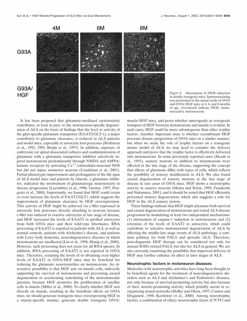
Attenuation of the induction in levels of iNOS in atransgenic murine model of ALSOxidative stress and induction of iNOS are thought to play animportant role in the progression of ALS. Therefore, we ad-dressed whether HGF would affect the levels of iNOS induced inmotoneurons caused by the upregulation of nitric oxide and freeradicals in ALS model mice. Immunohistochemical analysis ofiNOS revealed that, consistent with previous reports, iNOS wasinduced in the motoneurons of G93A mice at 6 months of age,whereas this induction was attenuated in G93A/HGF mice, indi-
Table 1. Comparison of onset of paralysis, length, and mortality amongG93A�/�, G93A�/�/HGF�/�, G93A�/�, and G93A�/�/HGF�/� mice
G93A(�/�) G93A(�/�)/HGF(�/�) p value
Onset 243.8 � 4.7 271.9 � 5.6 0.004*Length 15.7 � 1.6 14.8 � 1.6 0.686Mortality 259.5 � 5.0 286.8 � 6.5 0.003*
G93A(�/�) G93A(�/�)/HGF(�/�) p value
Onset 137.8 � 2.4 161.5 � 3.4 0.007*Length 12.0 � 2.0 13.8 � 3.4 0.457Mortality 147.5 � 5.7 175.3 � 6.5 0.007*
*Statistical significance was evaluated by t test.Top panel, G93A �/� mice; bottom panel, G93A �/� mice. Length is expressed asthe duration (days) between onset and mortality.
Figure 4. Improved motor performance, onset of paralysis, and mortalityin G93A/HGF mice. A, Comparison of motor performance in wild-type,HGF, G93A, and G93A/HGF mice determined by stride; using a footprint test revealed that the stride was markedly decreased in G93A/HGFmice, whereas the stride was retained in G93A/HGF mice. E, Wild type(n � 14); F, HGF (n � 15); Œ, G93A (n � 16); and �, G93A/HGF mice(n � 16). B, Onset of paralysis, scored as the first observation of paralysisof bilateral limbs, was delayed in G93A/HGF mice compared with G93Amice. �, G93A (n � 15); Œ, G93A/HGF mice (n � 16). C, Probability ofsurvival showed an extended life span in G93A/HGF mice compared withG93A mice.
Figure 5. HGF protein levels in the spinal cords of wild-type, HGF,G93A, and G93A/HGF mice. Protein levels of HGF were determined byELISA.
6542 J. Neurosci., August 1, 2002, 22(15):6537–6548 Sun et al. • HGF Retards Progression of ALS Mice via Dual Mechanisms

cating that HGF could also modify the levels of iNOS in mo-toneurons in addition to those of caspase-1 (Fig. 8).
Reduction of gliosis and attenuation of the reductionin levels of EAAT2/GLT1 in a transgenic murine modelof ALSBecause we found c-Met-IR in reactive astrocytes in G93A miceat the end stage of disease (Fig. 1A), we next focused on effects ofHGF on astrocytes. In wild-type or HGF single transgenic mice,astrocytes were localized mainly in the white matter or cellssurrounding the central canal and marginally in the ventral horn(Fig. 9A). Reactive astrocytes progressively proliferated in theventral horn when motoneuronal death was not yet evident in6-month-old G93A mice (Fig. 9A). In contrast, double transgeniclittermates showed strikingly smaller numbers of reactive astro-cytes in the ventral horn. Quantification of the relative intensityof GFAP-IR showed an �40 and 60% decrease in the ventralhorn, compared with findings in G93A mice at 6 and 8 months ofage, respectively (Fig. 9B).
Because EAAT2 levels are reduced at the end stage of ALS,which is thought to be one of the critical events to increaseglutamatergic neurotoxicity on motoneurons, we next determinedwhether EAAT2 levels were modified in G93A transgenic mice.At 8 months of age, EAAT2 levels in G93A mice were reduced to40% compared with wild-type or HGF single transgenic litter-mates (Fig. 9C,D), which is consistent with reported data. Incontrast, double transgenic mice showed even higher levels ofEAAT2 (140%) in the lumbar spinal cord compared with wild-type or HGF single transgenic littermates. To assess whethereffects of HGF on EAAT2 levels are caused by a direct effect ofHGF on astrocytes, we determined whether HGF treatmentwould modify levels of EAAT2 in primary cultured astrocytes.After 7 d, only marginal levels of EAAT2 were detected inastrocytes from wild-type or G93A transgenic mice. In contrast,higher levels of EAAT2 were detected in astrocytes treated withHGF, suggesting that reduction in EAAT2 levels is caused by adirect activity of HGF on astrocytes through astrocytic foot
processes attached to motoneurons (Fig. 9E,F). Taken together,these results indicate that single transgenic G93A mice overpro-duce nonfunctional astrocytes in terms of glutamate clearance,whereas double transgenic mice maintain more functionalastrocytes.
DISCUSSIONWe investigated whether gene transfer of HGF, a pleiotrophiccytokine with a highly potent neurotrophic activity for motoneu-rons, specifically to neurons of ALS model mice may play a rolein disease retardation. Local and sustained delivery of the HGFgene into ALS neurons was achieved by generating transgenicmice overexpressing HGF in a neuron-specific manner (HGFmice) and by crossing HGF mice with transgenic mice expressinglow copy number of mutant SOD1G93A (G93A mice), a mousemodel of ALS. The HGF gene was introduced postnatally inneurons of HGF mice, and no differences could be seen inparameters of the motor nervous system examined, thus indicat-ing comparable development of transgenic and control mice. Inthis system, exogenous HGF protein can be introduced intoneurons at postnatal developmental stages of G93A mice. Only atlater stages when reactive astrocytes proliferate and extend theirfoot processes to motoneurons can those astrocytes expressingc-Met at late stage of disease receive exogenous HGF proteinproduced in motoneurons.
Using this system, we provide the first evidence that HGFattenuates degeneration of both motoneuronal death and axonaldegeneration, resulting in improved motor performance, delayeddisease progression, and extension of the life span of transgenicmice overexpressing mutant SOD1G93A, via functions not only onmotoneurons but also on astrocytes presumably through the footprocesses facing motoneurons.
Bifunctional role for HGF in delaying ALS progressionAlthough the mechanisms of motoneuron-specific disease pro-gression are not fully understood, regulation or accumulation ofa series of molecules, such as SOD1, caspase-1, neurofilament,
Figure 6. Modification of human SOD1, caspase-1, Bcl-xL, and Bcl-2 in the lumbar spinal cord during disease progression. A, Immunoblotting of humanSOD1 in the lumbar spinal cord at 2, 6, and 8 months of age. The total amount of mutant SOD1 protein was not modified by overexpression of HGFat any time point examined. W, Wild type; H, HGF; G, G93A; G/H, G93A/HGF mice. B, Immunostaining of human SOD1 in the lumbar spinal cordat 4 months of age showed aggregation of SOD1 similar to that seen in G93A transgenic and G93A/HGF double transgenic mice (n � 3 in each group).SOD1 aggregation was accumulated more densely in G93A or G93A/HGF mice at 8 months of age. A representative picture is shown of six sectionsat the lumbar level of each animal, and three mice per group were examined. C, Immunoblotting of Bcl-xL and Bcl-2 in the lumbar spinal cord at 8 monthsof age. No induction of Bcl-2 and Bcl-xL was evident in the spinal cord of G93A/HGF mice. W, Wild type; H, HGF; G93A/HGF, double transgenic mice;spl, spleen as a positive control.
Sun et al. • HGF Retards Progression of ALS Mice via Dual Mechanisms J. Neurosci., August 1, 2002, 22(15):6537–6548 6543

and members of the Bcl-2 family, are thought to be involved inmotoneuronal degeneration (Pasinelli et al., 1998; Strong, 1999;Vukosavic et al., 1999; Al-Chalabi and Leigh, 2000). One of thepossible events affected by HGF is the regulation of the expres-sion levels of members of the bcl-2 family, because we reportedthat HGF could induce Bcl-xL and attenuates massive cell deathof hepatocytes of the fulminant hepatitis model in mice. Further-more, it has been noted that overexpression of the anti-apoptoticoncoprotein bcl-2 in a neuron-specific manner prolongs the lifespan of ALS mice (Kostic et al., 1997), suggesting that HGF mayprevent motoneuronal cell death via induction of the Bcl-2 familyof proteins. Because we found the absence of induction of Bcl-2and Bcl-xL at the end stage of ALS in G93A/HGF doubletransgenic mice, distinct mechanisms may play a role in the effectsof HGF overexpression and Bcl-2 in ALS mice. We also showedin this study that aggregation of mutant SOD1, which occurred at
a relatively early stage of the disease, was not modified. On theother hand, we found that a middle stage event (i.e., induction ofcaspase-1 seen predominantly in motoneurons) was diminished inG93A/HGF double transgenic mice. Therefore it seems likelythat attenuation of caspase-1 induction in motoneurons may beresponsible for HGF activity in preventing motoneuronal deathat the middle stage of disease. This idea is supported by theevidence that intraventricular application of a dominant negativemutant of caspase-1 or a broad caspase inhibitor “zVAD-fmk”also attenuates motoneuronal death and prolongs the life span ofALS mice through a direct action on motoneurons (Friedlanderet al., 1997; Li et al., 2000). In addition, attenuation of theinduction of iNOS in motoneurons was evident by expression ofHGF. Such direct neurotrophic activities of HGF on motoneu-rons might be of great importance in the attenuation of theprogression of ALS.
Figure 7. Attenuation of caspase-1 induction in double transgenic mice. A, Immunostaining of tubulin III ( green) and caspase-1 (red) in the lumbarspinal cord at 6 months of age (n � 3 in each group). Caspase-1 was specifically induced in large tubulin III-positive neurons of G93A transgenic mice,whereas double transgenic mice showed much lower levels of caspase-1. Inset is magnified view. B, Comparison of the fluorescence intensity of caspase-1in motoneurons. Fluorescence intensity of caspase-1-IR in each motoneuron was determined by image analysis using Photoshop after collecting theconfocal image; average � SE of 40 motoneurons was expressed. C, Western blot analysis of caspase-1 in the spinal cords of wild-type, HGF, G93A, andG93A/HGF mice.
6544 J. Neurosci., August 1, 2002, 22(15):6537–6548 Sun et al. • HGF Retards Progression of ALS Mice via Dual Mechanisms
It has been proposed that glutamate-mediated excitotoxicitycontributes, at least in part, to the motoneuron-specific degener-ation of ALS on the basis of findings that the level or activity ofthe glial-specific glutamate transporter (EAAT2/GLT-1), a majorcontributor to glutamate clearance, is reduced in ALS patientsand model mice, especially at astrocytic foot processes (Rothsteinet al., 1992, 1995; Bruijn et al., 1997). In addition, exposure ofembryonic rat spinal dissociated cultures and coadministration ofglutamate with a glutamate transporter inhibitor selectively in-jured motoneurons predominantly through NMDA and AMPA/kainate receptors by activating Ca2�/calmodulin-neuronal NOSbut did not injure nonmotor neurons (Urushitani et al., 2001).Partial phenotypic improvement and prolongation of the life spanof ALS model mice and patients by riluzole, a glutamate inhibi-tor, indicated the involvement of glutamatergic neurotoxicity indisease progression (Lacomblez et al., 1996; Gurney, 1997; Pon-gratz et al., 2000). Importantly, we found that HGF could retainor even increase the levels of EAAT2/GLT1, which suggests theimprovement of glutamate clearance by HGF overexpression.This activity of HGF might be achieved via c-Met expressed inastrocytic foot processes directly attaching to neurons becausec-Met was induced in reactive astrocytes at late stage of disease,and HGF increased the levels of EAAT2 in purified astrocytesfrom both G93A mice and their wild-type littermates. RNAprocessing of EAAT2 is reported in patients with ALS, as well asnormal controls, patients with Alzheimer’s disease, and patientswith Lewy body dementia, neurodegenerative diseases in whichmotoneurons are unaffected (Lin et al., 1998; Honig et al., 2000).However, such processing does not occur for all RNA species. Inaddition, RNA processing of EAAT2 is not reported in G93Amice. Therefore, retaining the levels of or obtaining even higherlevels of EAAT2 in G93A/HGF mice may be beneficial forreducing the glutamate neurotoxicities on motoneurons. An al-ternative possibility is that HGF acts on muscle cells, indirectlysupporting the survival of motoneurons and preventing axonaldegeneration or accelerating remodeling of the neuromuscularjunction, because HGF promotes the proliferation of satellitecells in muscle (Miller et al., 2000). To clarify whether HGF actsdirectly on muscle, resulting in the beneficial effects on G93Amice, we should generate transgenic mice overexpressing HGF ina muscle-specific manner, generate double transgenic G93A/
muscle-HGF mice, and prove whether anterograde or retrogradetransport of HGF between motoneurons and muscle is evident. Insuch cases, HGF could be more advantageous than other trophicfactors. Another important issue is whether recombinant HGFprevents disease progression of G93A mice in a similar manner,but when we study the role of trophic factors on a transgenicmouse model of ALS, we may need to consider the deliveryapproach and prove that the trophic factor is effectively deliveredinto motoneurons. In some previously reported cases (Heads etal., 1991), sensory neurons in addition to motoneurons wereaffected at the late stage of the disease, supporting the conceptthat effects of glutamate differ with types of cells, which reflectsthe possibility of sensory modification in ALS. We also foundaxonal degeneration of sensory neurons at the late stage ofdisease in rare cases of G93A mice. HGF shows a neurotrophicactivity in sensory neurons (Maina and Klein, 1999; Funakoshiand Nakamura, 2001), and it should be noted that HGF efficientlyprevented sensory degeneration, which also suggests a role forHGF in the ALS sensory system.
These findings indicate that HGF might attenuate both survivaland axonal degeneration of motoneurons and retard ALS diseaseprogression by modulating at least two independent mechanisms:(1) attenuation of caspase-1 induction in motoneurons and (2)retention of the levels of EAAT2 in astrocytes, which couldcontribute to selective motoneuronal degeneration of ALS byaffecting the middle-late stage events of ALS pathology, a com-mon pathway for both FALS and sporadic ALS. Therefore,post-diagnostic HGF therapy can be considered not only formutant SOD1-related FALS, but also for ALS in general. We arealso currently examining the possibility that improved delivery ofHGF may further enhance its effect at later stages of ALS.
Neurotrophic factors in motoneuron diseasesMolecules with neurotrophic activities have long been thought tobe beneficial agents for the treatment of neurodegenerative dis-orders such as ALS and Alzheimer’s and Parkinson’s diseases,not only because of survival-promoting activity but also becauseof their neurite-promoting activity, which possibly assists in re-organizing neural networks (Hoffer and Olson, 1997; Connor andDragunow, 1998; Kordower et al., 2000). Among neurotrophicfactors, a combination of ciliary neurotrophic factor (CNTF) and
Figure 8. Attenuation of iNOS inductionin double transgenic mice. Immunostainingwas performed in the spinal cords of G93Aand G93A/HGF mice at 4, 6, and 8 monthsof age. Arrowheads indicate iNOS- immu-noreactive motoneurons.
Sun et al. • HGF Retards Progression of ALS Mice via Dual Mechanisms J. Neurosci., August 1, 2002, 22(15):6537–6548 6545

brain-derived neurotrophic factor or CNTF and neurotrophin-3are therapeutics for motoneuron diseases, as determined inmodel mice such as pmn or wobbler mice, respectively (Mitsumotoet al., 1994; Haase et al., 1997). In these models, combinations ofneurotrophic factors slowed both motoneuronal death and axonaldegeneration (Mitsumoto et al., 1994; Haase et al., 1997). GDNFhas been shown to promote survival of motoneurons of G93Amice in vitro (Derby et al., 2000). In contrast to CNTF, GDNFsignificantly reduced the loss of facial motoneurons by 50%, anumber similar to what was observed when CNTF was adminis-tered to the pmn mice. Counts of myelinated axons revealed thatGDNF-secreting cells had no effect on axonal degeneration anddid not increase the lifespan of pmn/pmn mice (Sagot et al.,1996). Despite its potential role in rescuing motoneuron cell
bodies, it was suggested that the ability of GDNF to preventnerve degeneration might be restricted to cotreatment withother factors that act on nerve processes (Sagot et al., 1996).Use of other neurotrophic factors, such as neurotrophin-4, hasalso been considered for ALS treatment because it promotessurvival, neurite extension, and sprouting of motoneurons inan activity-dependent manner (Funakoshi et al., 1995). Al-though the potential benefits of using these neurotrophic fac-tors in these ALS models are evident, it is also apparent thata combination of these neurotrophic factors is necessary. Themechanism(s) by which neurotrophic factors contribute to thesurvival and axonal regeneration of motoneurons is not fullyunderstood, yet it is possible that these neurotrophic factorsmay function directly via their cognate receptors expressed on
Figure 9. Attenuation of astrocytosis and downregulation of EAAT2 in double transgenic mice. A, Immunohistochemical analysis of GFAP in theventral horn of the lumbar spinal cord at 8 months of age revealed that, in contrast to the marked proliferation of astrocytes in G93A mice, doubletransgenic mice showed fewer astrocytes in the ventral horn, whereas wild-type and HGF transgenic mice showed only a small number of astrocytes. B,Quantitative values of GFAP immunoreactivities in different mice. Relative intensity of GFAP-IR in wild type (E), HGF single transgenic (F), G93A(�), and double transgenic (Œ) (n � 3 in each group). C, Immunoblotting of EAAT2, GFAP, and c-Met in the spinal cord at 2, 6, and 8 months of ageshows induction of GFAP from 6 months of age in both G93A and G93A/HGF mice. EAAT2 was downregulated in G93A mice and c-Met wasupregulated in G93A and G93A/HGF mice specifically at 8 months of age. The total level of EAAT2 was maintained in G93A/HGF mice at 8 monthsof age. D, Relative levels of EAAT2 at 8 months of age (n � 4 in each group). E, Immunoblotting of EAAT2 and c-Met in primary astrocytes. HGFtreatment resulted in upregulation of EAAT2 in primary astrocytes from both wild-type and G93A mice. F, Quantitative results are shown (n � 3 ineach group).
6546 J. Neurosci., August 1, 2002, 22(15):6537–6548 Sun et al. • HGF Retards Progression of ALS Mice via Dual Mechanisms

motoneurons, because their receptors are present in motoneu-ron and direct neurotrophic activity on motoneurons has beenshown in vitro. Evidence of beneficial actions of these factorson glial cells is not evident.
In summary, because HGF acts on both survival and axonalregeneration of motoneurons and astrocytes, we propose thatHGF is the first example of an endogenous cytokine that plays adual role in ALS. This could take place through direct neurotro-phic activities on motoneurons and indirect modification of glu-tamate neurotoxicity in motoneurons by maintaining appropriatelevels of glutamate transporters in astrocytes. These functions ofHGF raise the possibility for a therapeutic use of this factor inALS and related disorders.
REFERENCESAl-Chalabi A, Leigh PN (2000) Recent advances in amyotrophic lateral
sclerosis. Curr Opin Neurol 13:397–405.Bruijn LI, Becher MW, Lee MK, Anderson KL, Jenkins NA, Copeland
NG, Sisodia SS, Rothstein JD, Borchelt DR, Price DL, Cleveland DW(1997) ALS-linked SOD1 mutant G85R mediates damage to astro-cytes and promotes rapidly progressive disease with SOD1-containinginclusions. Neuron 18:327–338.
Bruijn LI, Houseweart MK, Kato S, Anderson KL, Anderson SD, OhamaE, Reaume AG, Scott RW, Cleveland DW (1998) Aggregation andmotor neuron toxicity of an ALS-linked SOD1 mutant independentfrom wild-type SOD1. Science 281:1851–1854.
Connor B, Dragunow M (1998) The role of neuronal growth factors inneurodegenerative disorders of the human brain. Brain Res Brain ResRev 27:1–39.
Derby ML, Giuliano R, Figlewicz DA, Bohn MC (2000) GDNF is tro-phic for mouse motoneurons that express a mutant superoxide dis-mutase (SOD-1) gene. Amyotroph Lateral Scler Other Motor NeuronDisord 1:113–122.
Ebens A, Brose K, Leonardo ED, Hanson Jr MG, Bladt F, Birchmeier C,Barres BA, Tessier-Lavigne M (1996) Hepatocyte growth factor/scat-ter factor is an axonal chemoattractant and a neurotrophic factor forspinal motor neurons. Neuron 17:1157–1172.
Friedlander RM, Brown RH, Gagliardini V, Wang J, Yuan J (1997)Inhibition of ICE slows ALS in mice. Nature 388:31.
Funakoshi H, Nakamura T (2001) Identification of HGF-like protein asa novel neurotrophic factor for avian dorsal root ganglion sensoryneurons. Biochem Biophys Res Commun 283:606–612.
Funakoshi H, Frisen J, Barbany G, Timmusk T, Zachrisson O, VergeVM, Persson H (1993) Differential expression of mRNAs for neuro-trophins and their receptors after axotomy of the sciatic nerve. J CellBiol 123:455–465.
Funakoshi H, Belluardo N, Arenas E, Yamamoto Y, Casabona A, Pers-son H, Ibanez CF (1995) Muscle-derived neurotrophin-4 as anactivity-dependent trophic signal for adult motor neurons. Science268:1495–1499.
Funakoshi H, Risling M, Carlstedt T, Lendahl U, Timmusk T, Metsis M,Yamamoto Y, Ibanez CF (1998) Targeted expression of a multifunc-tional chimeric neurotrophin in the lesioned sciatic nerve acceleratesregeneration of sensory and motor axons. Proc Natl Acad Sci USA95:5269–5274.
Gurney ME (1997) The use of transgenic mouse models of amyotrophiclateral sclerosis in preclinical drug studies. J Neurol Sci 152:S67–73.
Gurney ME, Pu H, Chiu AY, Dal Canto MC, Polchow CY, AlexanderDD, Caliendo J, Hentati A, Kwon YW, Deng HX (1994) Motorneuron degeneration in mice that express a human Cu, Zn superoxidedismutase mutation. Science 264:1772–1775.
Haase G, Kennel P, Pettmann B, Vigne E, Akli S, Revah F, SchmalbruchH, Kahn A (1997) Gene therapy of murine motor neuron diseaseusing adenoviral vectors for neurotrophic factors. Nat Med 3:429–436.
Heads T, Pollock M, Robertson A, Sutherland WH, Allpress S (1991)Sensory nerve pathology in amyotrophic lateral sclerosis. Acta Neuro-pathol (Berl) 82:316–320.
Hoffer B, Olson L (1997) Treatment strategies for neurodegenerativediseases based on trophic factors and cell transplantation techniques.J Neural Transm [Suppl] 49:1–10.
Honda S, Kagoshima M, Wanaka A, Tohyama M, Matsumoto K, Naka-mura T (1995) Localization and functional coupling of HGF andc-Met/HGF receptor in rat brain: implication as neurotrophic factor.Brain Res Mol Brain Res 32:197–210.
Honig LS, Chambliss DD, Bigio EH, Carroll SL, Elliott JL (2000)Glutamate transporter EAAT2 splice variants occur not only in ALS,but also in AD and controls. Neurology 55:1082–1088.
Kordower JH, Emborg ME, Bloch J, Ma SY, Chu Y, Leventhal L,McBride J, Chen EY, Palfi S, Roitberg BZ, Brown WD, Holden JE,Pyzalski R, Taylor MD, Carvey P, Ling Z, Trono D, Hantraye P,Deglon N, Aebischer P (2000) Neurodegeneration prevented by len-tiviral vector delivery of GDNF in primate models of Parkinson’sdisease. Science 290:767–773.
Kosai K, Matsumoto K, Funakoshi H, Nakamura T (1999) Hepatocytegrowth factor prevents endotoxin-induced lethal hepatic failure inmice. Hepatology 30:151–159.
Kostic V, Jackson-Lewis V, de Bilbao F, Dubois-Dauphin M, Przedbor-ski S (1997) Bcl-2: prolonging life in a transgenic mouse model offamilial amyotrophic lateral sclerosis. Science 277:559–562.
Lacomblez L, Bensimon G, Leigh PN, Guillet P, Meininger V (1996)Dose-ranging study of riluzole in amyotrophic lateral sclerosis.Amyotrophic lateral sclerosis/riluzole study group II. Lancet347:1425–1431.
Li M, Ona VO, Guegan C, Chen M, Jackson-Lewis V, Andrews LJ,Olszewski AJ, Stieg PE, Lee JP, Przedborski S, Friedlander RM(2000) Functional role of caspase-1 and caspase-3 in an ALS transgenicmouse model. Science 288:335–339.
Lin CL, Bristol LA, Jin L, Dykes-Hoberg M, Crawford T, Clawson L,Rothstein JD (1998) Aberrant RNA processing in a neurodegenera-tive disease: the cause for absent EAAT2, a glutamate transporter, inamyotrophic lateral sclerosis. Neuron 20:589–602.
Maina F, Klein R (1999) Hepatocyte growth factor, a versatile signal fordeveloping neurons. Nat Neurosci 2:213–217.
Matsumoto K, Nakamura T (1997) HGF: its organotrophic role andtherapeutic potential. Ciba Found Symp 212:198–211.
Miller KJ, Thaloor D, Matteson S, Pavlath GK (2000) Hepatocytegrowth factor affects satellite cell activation and differentiationin regenerating skeletal muscle. Am J Physiol Cell Physiol 278:C174 –181.
Mitsumoto H, Ikeda K, Klinkosz B, Cedarbaum JM, Wong V, LindsayRM (1994) Arrest of motor neuron disease in wobbler mice cotreatedwith CNTF and BDNF. Science 265:1107–1110.
Nakamura T, Nawa K, Ichihara A (1984) Partial purification and char-acterization of hepatocyte growth factor from serum of hepatecto-mized rats. Biochem Biophys Res Commun 122:1450–1459.
Nakamura T, Nishizawa T, Hagiya M, Seki T, Shimonishi M, Sugimura A,Tashiro K, Shimizu S (1989) Molecular cloning and expression ofhuman hepatocyte growth factor. Nature 342:440–443.
Nakamura T, Mizuno S. Matsumoto K, Sawa Y, Matsuda H, NakamuraT (2000) Myocardial protection from ischemia/reperfusion injury byendogenous and exogenous HGF. J Clin Invest 106:1511–1519.
Naveilhan P, Neveu I, Baudet C, Funakoshi H, Wion D, Brachet P, MetsisM (1996) 1,25-Dihydroxyvitamin D3 regulates the expression of thelow-affinity neurotrophin receptor. Brain Res Mol Brain Res41:259–268.
Novak KD, Prevette D, Wang S, Gould TW, Oppenheim RW (2000)Hepatocyte growth factor/scatter factor is a neurotrophic survival fac-tor for lumbar but not for other somatic motoneurons in the chickembryo. J Neurosci 20:326–337.
Okura Y, Arimoto H, Tanuma N, Matsumoto K, Nakamura T, Ya-mashima T, Miyazawa T, Matsumoto Y (1999) Analysis of neurotro-phic effects of hepatocyte growth factor in the adult hypoglossal nerveaxotomy model. Eur J Neurosci 11:4139–4144.
Pasinelli P, Borchelt DR, Houseweart MK, Cleveland DW, Brown Jr RH(1998) Caspase-1 is activated in neural cells and tissue with amyotro-phic lateral sclerosis-associated mutations in copper-zinc superoxidedismutase. Proc Natl Acad Sci USA 95:15763–15768.
Pongratz D, Neundorfer B, Fischer W (2000) German open label trial ofriluzole 50 mg b.i.d. in treatment of amyotrophic lateral sclerosis(ALS). J Neurol Sci 180:82–85.
Pramatarova A, Laganiere J, Roussel J, Brisebois K, Rouleau GA,Andreassen OA, Jenkins BG, Dedeoglu A, Ferrante KL, BogdanovMB, Kaddurah-Daouk R, Beal MF (2001) Neuron-specific expressionof mutant superoxide dismutase 1 in transgenic mice does not lead tomotor impairment. J Neurosci 21:3369–3374.
Rothstein JD, Martin LJ, Kuncl RW (1992) Decreased glutamate trans-port by the brain and spinal cord in amyotrophic lateral sclerosis.N Engl J Med 326:1464–1468.
Rothstein JD, Van Kammen M, Levey AI, Martin LJ, Kuncl RW (1995)Selective loss of glial glutamate transporter GLT-1 in amyotrophiclateral sclerosis. Ann Neurol 38:73–84.
Sagot Y, Tan SA, Hammang JP, Aebischer P, Kato AC (1996) GDNFslows loss of motoneurons but not axonal degeneration or prematuredeath of pmn/pmn mice. J Neurosci 16:2335–2341.
Seki T, Ihara I, Sugimura A, Shimonishi M, Nishizawa T, Asami O,Hagiya M, Nakamura T, Shimizu S (1990) Isolation and expression ofcDNA for different forms of hepatocyte growth factor from humanleukocyte. Biochem Biophys Res Commun 172:321–327.
Sun et al. • HGF Retards Progression of ALS Mice via Dual Mechanisms J. Neurosci., August 1, 2002, 22(15):6537–6548 6547

Strong MJ (1999) Neurofilament metabolism in sporadic amyotrophiclateral sclerosis. J Neurol Sci 169:170–177.
Sun W, Funakoshi H, Nakamura T (1999) Differential expression ofhepatocyte growth factor and its receptor, c-Met, in the rat retinaduring development. Brain Res 851:46–53.
Sun W, Funakoshi H, Matsumoto K, Nakamura T (2000) A sensitivequantification method for evaluating the level of hepatocyte growthfactor and c-met/HGF receptor mRNAs in the nervous system usingcompetitive RT-PCR. Brain Res Brain Res Protoc 5:190–197.
Ueki T, Kaneda Y, Tsutsui H, Nakanishi K, Sawa Y, Morishita R,Matsumoto K, Nakamura T, Takahashi H, Okamoto E, Fujimoto J(1999) Hepatocyte growth factor gene therapy of liver cirrhosis in rats.Nat Med 5:226–230.
Urushitani M, Nakamizo T, Inoue R, Sawada H, Kihara T, Honda K,Akaike A, Shimohama S (2001) N-methyl-D-aspartate receptor-mediated mitochondrial Ca(2�) overload in acute excitotoxic motorneuron death: a mechanism distinct from chronic neurotoxicity afterCa(2�) influx. J Neurosci Res 63:377–387.
Vukosavic S, Dubois-Dauphin M, Romero N, Przedborski S (1999) Baxand Bcl-2 interaction in a transgenic mouse model of familial amyotro-phic lateral sclerosis. J Neurochem 73:2460–2468.
Yamamoto Y, Livet J, Pollock RA, Garces A, Arce V, deLapeyriere O,Henderson CE (1997) Hepatocyte growth factor (HGF/SF) is amuscle-derived survival factor for a subpopulation of embryonic mo-toneurons. Development 124:2903–2913.
6548 J. Neurosci., August 1, 2002, 22(15):6537–6548 Sun et al. • HGF Retards Progression of ALS Mice via Dual Mechanisms