ovarian carcinoma by dr najeeb ur rehman
TRANSCRIPT

Najeeb ur rahman08-239Batch-k
Final Year MBBS

Cancer that arises in the epithelium, the tissue that lines the skin and internal organs of the body, and has a malignant potential aswell.

In general ovarian malignancy accounts for 25% of gynaecological cancers.
Among ovarian tumors only 10% are malignant. Malignant tumors are classified into;◦ Epithelial tumors….that accounts for 90% of malignant
ovarian tumors.◦ Non epithelial tumors…that accounts for 10% of malignant
ovarian tumors.
Epithelial malignant tumors◦ Serous carcinoma◦ Mucinous carcinoma◦ Endometroid carcinoma◦ Clear cell carcinoma◦ Undifferentiated carcinoma◦ Unclassified tumors aswell
Not clerly known, however there are some factors which are affiliated with Ca ovary.◦ Incessant ovulation◦ Age◦ Geographic destribution◦ Family history◦ Infective agents◦ Infertility treatment◦ Pelvic irradiation◦ Other factors such as asbestos, tale etc.

Direct spread.◦ Neighbouring organs like fallopian tube, uterus, bladder
and pelvic peritonium. Lymphatic spread.◦ Via lymphatic channel to the diaphragm, omentum,
peritonial surfaces of small and large bowel , liver and perital peritonium throughout the abdominal cavity.◦ Also involves para-aortic and pelvic lymph nodes, and
may metastize to groin and cervical lymph nodes.

Blood metastasis.◦ to the liver, lungs, bones and brain but occurs late in the
course of the disease.
Limited by two factors;◦ Ovary is not an accessable organ.◦ A premalignant stage of ovarian cance has not yet been
recognized.
Screening includes, bimanual pelvic examination, ultrasonography and tumour markers.

Late presentation Pain Abdominal swelling Abnormal uterine bleeding Weight loss Pressure effets
GPE Pale and cachexic Neck and groin examined for enlarged lymph
nodes and breast for primary or secondary tumours
Legs should be examined for DVT Abdominal examinationAbdomen may look distended and may have
prominent veinsOn palpation malignant ovarian tumour appear
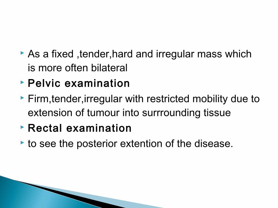
As a fixed ,tender,hard and irregular mass which is more often bilateral
Pelvic examination Firm,tender,irregular with restricted mobility due to
extension of tumour into surrrounding tissue Rectal examination to see the posterior extention of the disease.

Ultrasound. Radiological Investigations.◦ Chest x-ray◦ Barium studies to assess bowel involvement.
Imaiging techniques.◦ CT scan◦ MRI◦ Lymphangio-graphy

Cytology.◦ Fine needle aspiration done in clinically suspicious lymph
nodes in the groin and neck. Tumor markers.◦ CA-125 (<35 IU/mL)
Haematological investigations.◦ Full blood count, urea , creatinine, electrolytes and liver
function tests.

The Federation Internationale de Gynecologie et d'Obstetrique (FIGO) and the American Joint Committee on Cancer (AJCC) have designated staging.

l imited to the ovaries. ◦ Stage IA: tumour l imited to 1 ovary, the
capsule is intact, no tumour on ovarian surface and no malignant cells in ascites or peritoneal washings. ◦ Stage IB: tumour l imited to both ovaries,
capsules intact, no tumour on ovarian surface and no malignant cells in ascites or peritoneal washings. ◦ Stage IC: tumour is l imited to 1 or both
ovaries with any of the following: capsule ruptured, tumour on ovarian surface, malignant cells in ascites or peritoneal washings.

tumors involving 1 or both ovaries with pelvic extension and/or implants. ◦ Stage IIA: extension and/or implants on the
uterus and/or fallopian tubes. No malignant cells in ascites or peritoneal washings. ◦ Stage IIB: extension to and/or implants on
other pelvic t issues. No malignant cells in ascites or peritoneal washings. ◦ Stage IIC: Pelvic extension and/or implants
(stage IIA or stage IIB) with malignant cells in ascites or peritoneal washings.
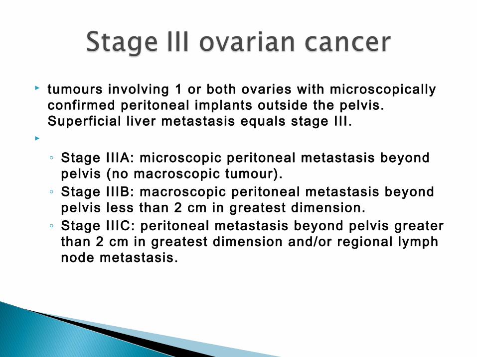
tumours involving 1 or both ovaries with microscopically confirmed peritoneal implants outside the pelvis. Superficial l iver metastasis equals stage III.
◦ Stage IIIA: microscopic peritoneal metastasis beyond
pelvis (no macroscopic tumour). ◦ Stage IIIB: macroscopic peritoneal metastasis beyond
pelvis less than 2 cm in greatest dimension. ◦ Stage IIIC: peritoneal metastasis beyond pelvis greater
than 2 cm in greatest dimension and/or regional lymph node metastasis.
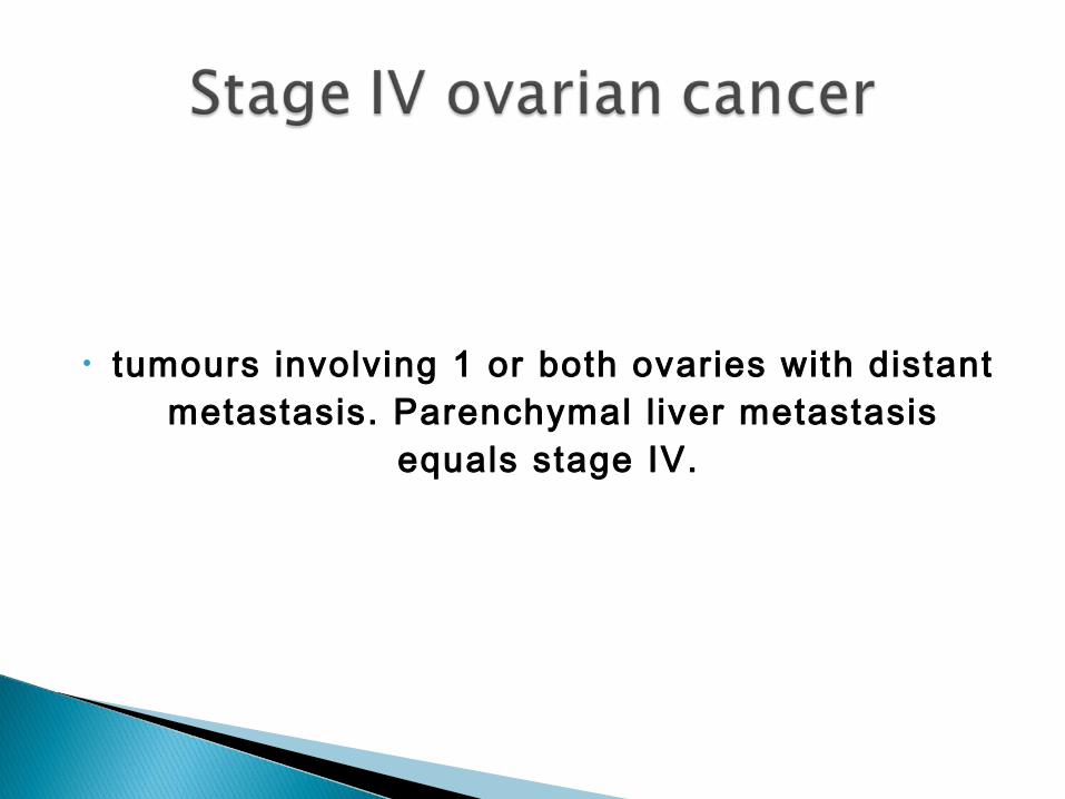
• tumours involving 1 or both ovaries with distant metastasis. Parenchymal l iver metastasis
equals stage IV.

Dysgerminoma Less than 1% of ovarian malignancies Counterpart of testicular seminoma Usually young patients (81% under age 30) 5% associated with gonadal dysgenesis/Swyer syndrome androgen insensitivity or pseudohermaphroditism; rarely associated with
hypercalcemia Metastasize to opposite ovary, retroperitoneal nodes, peritoneal cavity Rarely transforms to yolk sac tumor
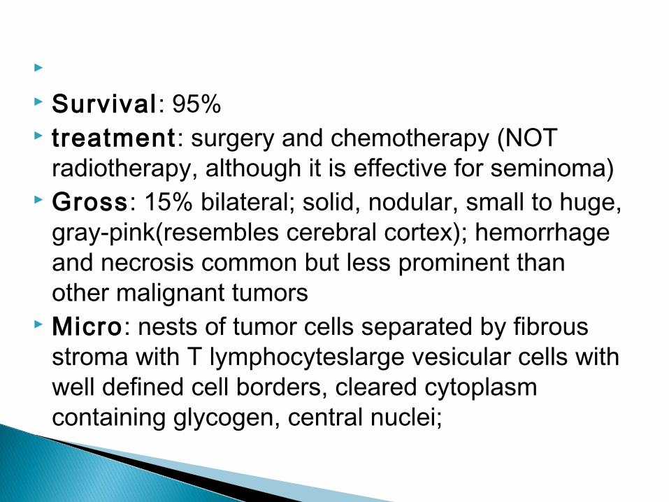
Survival: 95% treatment: surgery and chemotherapy (NOT
radiotherapy, although it is effective for seminoma) Gross: 15% bilateral; solid, nodular, small to huge,
gray-pink(resembles cerebral cortex); hemorrhage and necrosis common but less prominent than other malignant tumors
Micro: nests of tumor cells separated by fibrous stroma with T lymphocyteslarge vesicular cells with well defined cell borders, cleared cytoplasm containing glycogen, central nuclei;
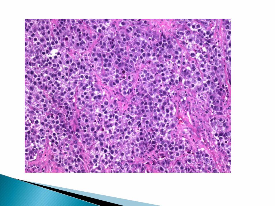
Also called endodermal sinus tumor May be derived from embryonal carcinoma presentation Usually children or young adults (median age 19 years) with abdominal pain and rapidly growing mass, increasing alpha fetoprotein (AFP) and alpha-1-antitrypsin serum levels; negative hCG Gross: mean 15 cm, smooth and glistening external surface, cystic cut surface
with hemorrhage and necrosis; often has benign teratoma component; rarly is found in pelvis unattached to ovary


Malignant tumor, whose tissue resembles embryonal or fetal tissue Usually prepubertal or young women (mean 18 years) Most recurrences within 2 years; presence of yolk sac component is best
predictor of recurrence in pediatric tumors Treatment: surgery, multiagent chemotherapy; better prognosis if only mature
teratoma found after chemotherapy, although abnormal karyotype is maintained in mature teratoma
Gross: bulky, solid or cystic with necrosis, hemorrhage Micro: usually neurogenic elementsmesodermal elements common; some
tumors derived primarily of esophageal, liver and intestina


Also called Sertoli-stromal cell tumor; formerly called androblastoma, arrhenoblastoma
Usually young women (mean age 25 years, 75%<age 30)
Rare, < 0.1% of ovarian neoplasms 80 % produce Androgens hence causing
masculinization

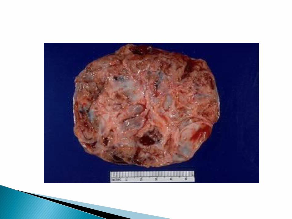
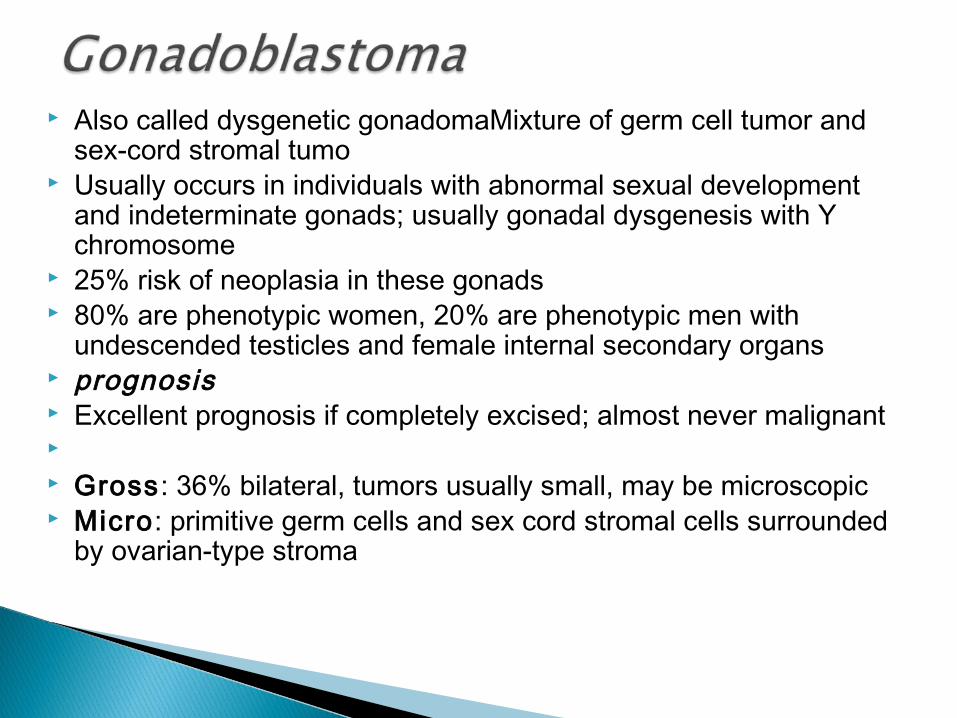
Also called dysgenetic gonadomaMixture of germ cell tumor and sex-cord stromal tumo
Usually occurs in individuals with abnormal sexual development and indeterminate gonads; usually gonadal dysgenesis with Y chromosome
25% risk of neoplasia in these gonads 80% are phenotypic women, 20% are phenotypic men with
undescended testicles and female internal secondary organs prognosis Excellent prognosis if completely excised; almost never malignant Gross: 36% bilateral, tumors usually small, may be microscopic Micro: primitive germ cells and sex cord stromal cells surrounded
by ovarian-type stroma
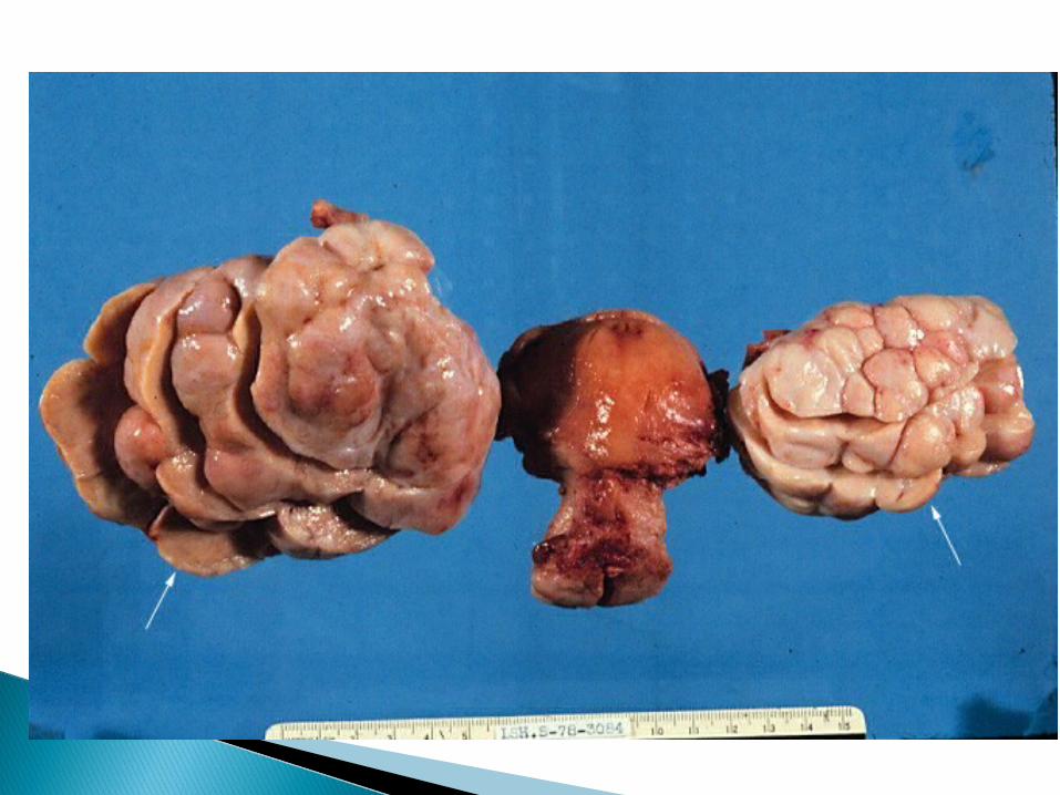
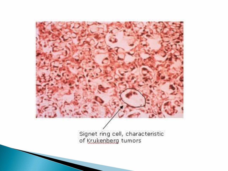
A Krukenberg tumor refers to a malignancy in the ovary that
metastasized from a primary site, classically the gastrointestinal tract, although it can arise in other tissues such as the breast.
[1] Gastric adenocarcinoma, especially at the pylorus, is the most common source.
[2] Krukenberg tumors are often (over 80%)[2] found in both ovaries, consistent with its metastatic nature
Symptoms Krukenberg tumors often come to the attention when they cause abdominal or pelvic pain, bloating, ascites, or pain during sexual intercourse.
Krukenberg tumors can occasionally provoke a reaction of the ovarian stroma which leads to hormone production, that results in vaginal bleeding, a change in menstrual habits, or hirsutismor occasionally vir i l izat ion as a main symptom. Diagnosis Al l these symptoms are non-specif ic and can also arise with a range of problems other than cancer, and a diagnosis can only be made following confirmatory investigations such as computed tomography (CT) scans, laparotomy and/or a biopsy of the ovary.

The treatment of ovarian cancers based on the stage of the disease which is a reflection of the extent or spread of the cancer to other parts of the body.
It also depends on histologic cell type, and the patient's age and overall condit ion.
There are basically three forms of treatment of ovarian cancer:◦ surgery ◦ Chemotherapy ◦ radiation treatment ,
Standard treatment is surgery (staging and optimal debulking) followed by adjuvant chemotherapy in most cases. Even if optimal surgery is not possible, removing as much tumor as possible will provide significant palliation of symptoms.
Borderline lesions may be treated with conservative surgery

Advanced epithelial ovarian cancer is very sensitive to chemotherapy with responses in the range of 70-80% to first-line chemotherapy. The majority, however, relapse and ultimately die of chemotherapy-resistant disease. Second-line chemotherapy to date is disappointing in all forms of epithelial ovarian cancer with virtually no chance of successful second-line treatment following failure of initial regime.


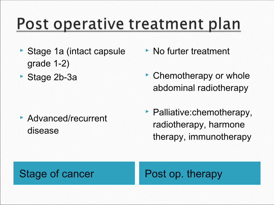
Stage of cancer Post op. therapy
Stage 1a (intact capsule grade 1-2)
Stage 2b-3a
Advanced/recurrent disease
No furter treatment
Chemotherapy or whole abdominal radiotherapy
Palliative:chemotherapy, radiotherapy, harmone therapy, immunotherapy
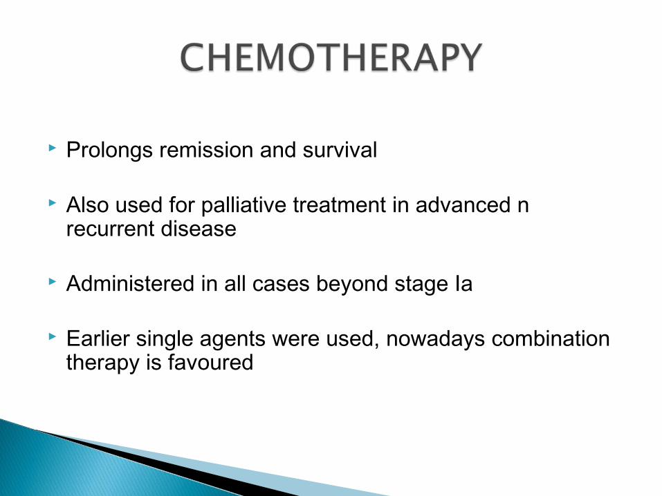
Prolongs remission and survival
Also used for palliative treatment in advanced n recurrent disease
Administered in all cases beyond stage Ia
Earlier single agents were used, nowadays combination therapy is favoured

The drugs used are; Alkylating agents.◦ Cyclophosphamide, chlorambucil
Anti-metabolites.◦ 5-florouracil, methotrexate
Platinum compounds.◦ Cisplatin, carboplatin
Toxoid compounds◦ Paclitaxil (taxol)
Anti tumour antibiotics.

Combination therapy is most beneficial.
Drugs are given at 3 weeks intervals
Intraperitoneal chemotherapy is also done but is very effective.
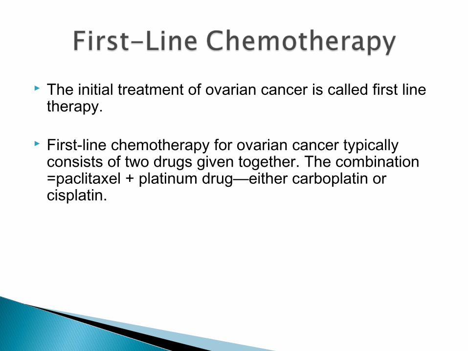
The initial treatment of ovarian cancer is called first line therapy.
First-line chemotherapy for ovarian cancer typically consists of two drugs given together. The combination =paclitaxel + platinum drug—either carboplatin or cisplatin.

Now, has a very small role since platinum based protocols and paclitaxel have improved the median survival.
-However it can be used as a palliative treatment for metastatic bone or brain lesions or of localized recurrence to alleviate the pain.

CA-125 Second look surgery in the form of laprotomy or
laproscopy.
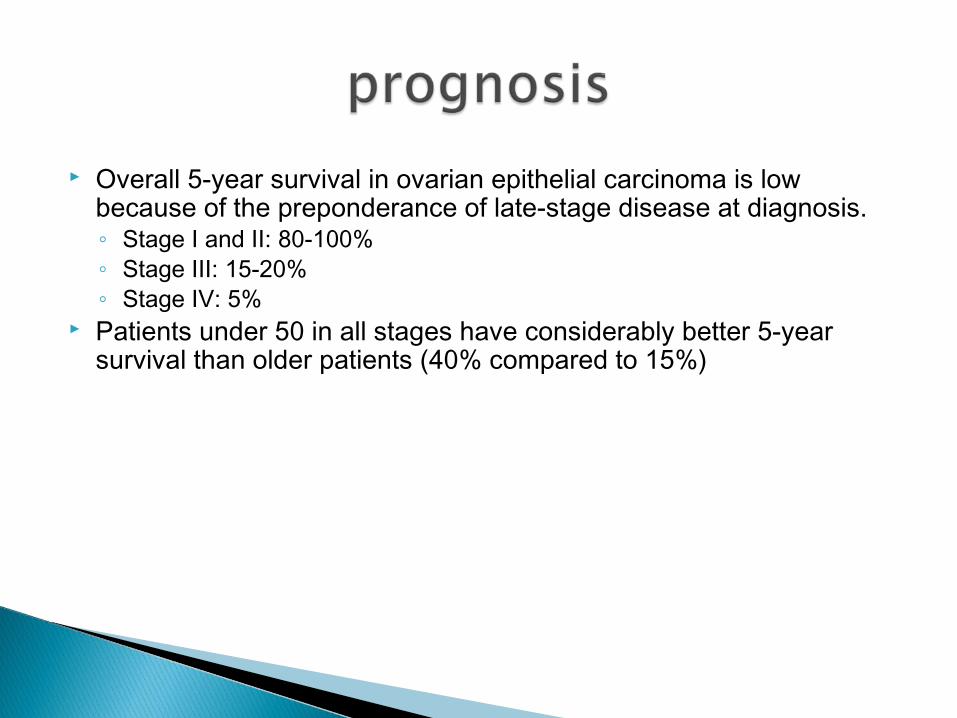
Overall 5-year survival in ovarian epithelial carcinoma is low because of the preponderance of late-stage disease at diagnosis. ◦ Stage I and II: 80-100% ◦ Stage III: 15-20% ◦ Stage IV: 5%
Patients under 50 in all stages have considerably better 5-year survival than older patients (40% compared to 15%)

Diet: a high-fat diet may play a role in the aetiology of ovarian cancer.
Oral contraceptives appear to reduce the risk of ovarian cancer for up to 10 years following cessation of use. This protective effect appears to apply to patients with BRCA mutations as well.
Patients who have used fertility drugs should be counselled as to their possible increase in risk of ovarian cancer.