other-worlds: encounters with the visual perception of ... · lungfishes through biological science...
TRANSCRIPT
By Audrey Maria Appudurai, BSc (Hons)
Supervised by Dr. Ionat Zurr
Professor Shaun P. Collin Associate Professor Nathan S. Hart
This thesis is presented for the degree of Doctor of Philosophy of the University of Western Australia
School of Anatomy, Physiology and Human Biology School of Animal Biology
2015
Other-Worlds: Encounters with the visual perception of lungfishes
through science and art


Abstract
iii
ABSTRACT
Lungfishes are large, freshwater, lobe-finned fishes that have the ability to breathe both
dissolved and atmospheric oxygen through gills and “primitive” lungs, respectively. Six
species of lungfishes remain worldwide; four in Africa (Protopterus spp.), one in South
America (Lepidosiren paradoxa) and one in Australia (Neoceratous forsteri).
Lungfishes are the closest living descendants of the ancestors of all terrestrial
vertebrates, and are vital to the study of the evolution of vision in tetrapods because of
their association with animals that made the remarkable transition from water onto land.
This unique interdisciplinary study explores the visual perception and experience of
lungfishes through biological science and visual art, while also tracing the history of
human cultural interactions with this significant animal. Scientific data generated
through anatomical, electrophysiological and behavioural experimentation contributes
to the emerging field of lungfish visual ecology, and artistic research provides a more
sensorial speculation of the lungfish visual experience in order to reflect on how we
(humans) produce knowledge of non-humans and the limitations of these methods.
To begin, a cultural history of non-human animal visual perception is investigated to
understand and acknowledge the limitations of technologies and our anthropocentric
perception of reality. Human narratives from the nineteenth century to present day are
traced to reveal the similarities and differences in previous attempts to uncover non-
human animal perceptual capabilities, with a particular focus on Jakob von Uexküll and
his concept of the Umwelt. A collaborative art project titled ‘Inperception’ (2012) that
endeavoured to interpret the Umwelten of Australian lungfishes through contemporary
scientific knowledge and art is discussed as a case study.
Abstract
iv
Diverse cultural narratives are discussed, revealing a long history of the lungfish as an
“in-between” and “monstrous” animal. This thesis shows that human understandings of
lungfishes are influenced by a myriad of factors that include, but are not limited to, the
evaluation of scientific data. Personal and cultural biases effect how we make sense of
non-human “Others”. These inescapable biases, formed by ingrained beliefs,
assumptions and ideologies, force a particular understanding between what we “see”
and what we “know” about lungfishes. The crucial importance of recognising these
biases and anthropocentric limitations in any attempt to understand the lungfish Umwelt
and the historical and cultural contexts of scientific exploration more broadly is
revealed.
Having established the limitations and complexities involved in “knowing” the
perception of a non-human “Other”, the biological “machinery” of the lungfish visual
perception is described by two scientific studies. The first presents the results of
anatomical investigations of the retinal photoreceptor cells of two previously
unexamined species, P. dolloi and L. paradoxa, in order to determine their capacity for
colour vision. These findings were compared with previous studies of N. fosteri in order
to present a more comprehensive understanding of the visual ecology of the three
lungfish genera. Previous anatomical studies of N. forsteri have shown that lungfishes
possess the retinal machinery to process colour. This study revealed the presence and
ultrastructure of one rod and two cone photoreceptor types in juvenile P. dolloi and
characterised one rod and one cone photoreceptor type in L. paradoxa. The
topographical distribution of the red cone in a juvenile P. dolloi is also described. The
presence of large oil droplets (or intracellular inclusions that act as filters) in all three
species suggests that the lungfish visual system is adapted for high sensitivity. The
absence of double cones in all three species however, strongly suggests that double
cones may have developed after the evolutionary separation of tetrapods from P. dolloi.

Abstract
v
In addition, the characterisation of the retinal machinery of L. paradoxa by this study
implies that, although both N. fosteri and P. dolloi possess the machinery to process
colour, L. paradoxa does not have the potential for colour vision.
The second scientific study investigated the visual perception of a unique group of
juvenile N. forsteri whose eyes were not externally visible. New findings are presented
that begin to uncover how a unique group of fish experience the world without the
ability to form images, resulting from behavioural, circadian, electrophysiological and
anatomical studies that compared these abnormal N. forsteri with normal Australian
lungfishes. The abnormal fishes all possessed subcutaneous eyes capable of photo-
transduction, although possibly not spatial vision. Clear corneas were absent in the eyes
of all these abnormal N. forsteri, although they all possessed the retina and
photoreceptor complement typical of their species. Variation in the functional integrity
of the eye between individuals was evident however, as behavioural phototaxis was not
demonstrated by all specimens.
Finally, a number of artworks and exhibitions generated through the scientific and
artistic research that extended and complicated issues surrounding the lungfish Umwelt
are discussed. Presented chronologically, these creative outcomes reveal my shifting
understandings of the limitations of technology, objectivity and human perception and
cognition in attempting to interpret the Umwelt of such a different animal. The first set
of artworks, ‘Third Eye’ and ‘In Focus’, produced in 2013 present the “machinery” of
the lungfish eye generated through biological dissection, macro-photography and light
microscopy. These black and white, abstracted micrographs of retinae and
photoreceptors are aestheticised scientific representations produced in my attempt to
discover the “truth” of the lungfish eye. A series of sculptural works produced in 2014
and collectively titled ‘The Forms’ explores role of aesthetics and manipulation in the

Abstract
vi
production and presentation of scientific images through transmission electron
microscopy (TEM), camera-less photography (photograms) and material manipulation.
My frustrated attempts to negotiate the gaps between technology, measurement,
representation and experience are manifest in these objects. Most recently, the 2015
multi-media exhibition, ‘Cernentia: exploring the visual perception of lungfishes
without eyes’ provided visitors with a multi-sensorial insight into the investigation of
the abnormal N. forsteri visual system and a speculative experience of the perceptual
world of these unique animals. Collectively, these artworks trace my own journey with
lungfishes and offer new encounters with the lungfish Umwelt outside of the
mechanistic realm of biological science. Importantly, they reveal the anthropocentric
lens through which an Umwelt of a non-human animal “Other” is explored.
This unique interdisciplinary thesis unravels the variety and complexities of the visual
perception of an important group of non-human animals – lungfishes. The
methodologies of three disparate disciplines – biological science, history and visual art
– are integrated in an attempt to understand the complexity of how non-human animals
perceive their environment, whilst recognising the limitations of each discipline. In a
society which is characterised by increasing specialisation, there is a need for scholars
who can communicate and speculate on the “Other” animal from a rigorous and highly
informed scientific and cultural/artistic position. The unique interdisciplinary and inter-
sensory approach offered by this thesis is therefore, vital when human relations with our
ecology are in crisis and in need of re-negotiation.

Acknowledgements
vii
ACKNOWLEDGEMENTS
This thesis is dedicated to my wonderful parents, Agnes and Richard Appudurai.
Without your sacrifice, unconditional patience, dedication, love and support, I could not
have become the woman I am today. You never stopped believing in me. To my
husband, I have no doubt of the part you have played in all my achievements thus far.
Christo Bester, amidst the rollercoaster of adolescence, adulthood and tertiary education
you have been my cheerleader, my rock, and my love. We have so many more
adventures ahead of us. I have so much love for my family-in-law, Jennifer, Douglas,
Nik, Ash and Scott Bester. I doubt you realise how much you mean to me. I gained
another family while doing this PhD, and how lucky am I that it was this one.
I want to sincerely thank my supervisors, Ionat Zurr, Shaun Collin, and Nathan Hart for
giving me the opportunity to undertake such an interesting, unique and challenging
project. I have learned so much, and without your support and encouragement, my PhD
journey would not have been as rich and fulfilling. Ionat, you have taught me so much
and I am so proud to have been your student. You are a tremendous mentor, a dear
friend, and I owe much of my successes to your careful tutelage.
I am grateful to The University of Western Australia, the Graduate Women of WA, the
Neuroecology Group and SymbioticA for supporting my candidature through the
Australian Postgraduate Award (APA), the UWA Safety-Net Top-Up Scholarship, the
UWA Completion Scholarship, and the Elsie and Mary Stevens Bursary and financially
aiding my experiments, artworks and exhibitions.
I am indebted to Guido Westoff, and Glenn Northcutt for their expertise and kind
donation of lungfish specimens, and Peter Kind, David Roberts and Michael Hutchison
Acknowledgements
viii
for their contribution in sourcing and transporting the infamous blind Australian
lungfishes from Queensland. Without your help, this study would not have been
possible.
I wish to sincerely thank all the scientists involved for their emotional and academic
support throughout this time. Carl Schmidt, you taught me all I know about looking
after lungfishes. I had so much fun building experimental equipment together at BSAU;
I could not have done this without you. Michael Archer, histology extraordinaire, your
patience and dad jokes never failed to amaze me. Thank you for teaching me everything
I know about histology and electron microscopy preparation. To Jan Hemmi, you were
not my supervisor, yet you made such a vital contribution to my project. Thank you for
sharing your knowledge and expertise with me; you took me under your wing when you
did not have to and I am forever grateful. Thank you Caroline Kerr, for your support
and help with grants, ethics and everything in between; you are invaluable to all of us.
Special thanks to you, Lucille, and Victoria for helping me with the lungfishes when I
really needed it. Thank you to the rest of the science crew, J.P., Eduardo, Fanny, Nic,
Tony, Carlos, Laura, Marcin, Rachael, Mikaela, Peter, Jamie, Maria, Kalina, Wissam,
Kristyn, and Paula for sharing the laughs and the tears.
Thank you to Oron Catts, Chris Cobilis and all the SymbioticA friends, postgraduates
and residents, past and present, for your enthusiasm, encouragement, critique and
support. Special thanks to Tarsh Bates for “making me write good” and bringing me out
of the tough times, Devon Ward for his mad art and life skills, Shannon Williamson, my
twitcher-in-crime, for her sage advice and encouragement, and Emily Parsons-Lord, my
photography Mama and endless source of laughter. You are some of my dearest friends
and I am so happy to have you all in my life.

Acknowledgements
ix
Last but not least, I am so thankful to have such amazing friends and family. Special
thanks to my chummies Lauren Hollier, Gemma Bothe, Alex Poulsen, and David
Hansen. You four are my oldest and closest friends; I could not have survived without
your continuous support with those long wine-filled dinners, painfully hilarious Skype
dates, beer and chocolate afternoons by the river, and the short but treasured holidays,
abroad and at home.
Statement Of Contribution
x
STATEMENT OF CANDIDATE CONTRIBUTION
In accordance with the University of Western Australia’s regulations regarding Research Higher Degrees, this thesis is presented as a series of chapters that may be prepared for publication.
This thesis is an account of research undertaken by the candidate during the period of 10th February 2011 to 31st of August 2015 while enrolled as a full-time postgraduate student in The School of Anatomy, Physiology and Human Biology, and The School of Animal Biology at the University of Western Australia.
Lepidosiren paradoxa and Protopterus dolloi specimens were donated by Glenn Northcutt and Guido Westoff, respectively. Except for the occasional assistance from the candidate’s supervisors and other academics of the lab (Shaun P. Collin, Nathan S. Hart, Jan Hemmi, Michael Archer, and Ionat Zurr) in the experimental set-up, training for experimental procedures and analyses; all experiments and data analyses were conducted by the candidate.
The custom software used to analyse electrophysiological data was created by Nathan S. Hart. The custom software used to analyse behavioural and circadian data was created by the candidate and Jan Hemmi.
A critical review of each chapter upon completion was provided by the candidate’s supervisors.
The artwork ‘Inperception’ (2012) was a collaborative project between the candidate and two other artists (Shannon Williamson and Devon Ward), and is credited accordingly. The construction of the headset for the artwork ‘Bloop’ (2015) was in collaboration with Devon Ward and the candidate, and is credited accordingly.
The content of this thesis is the candidate’s own composition, and all relevant sources are acknowledged. This thesis has not been submitted for any other degree in this or another institution.
_______________________ __ Audrey Maria Appudurai Shaun P. Collin Ionat Zurr Candidate Co-ordinating supervisor Co-ordinating supervisor 14th August 2015 18th January 2016 14th August 2015

Table Of Contents
xi
TABLE OF CONTENTS
Abstract ........................................................................................................................... iii Acknowledgements ........................................................................................................ vii Statement Of Contribution............................................................................................. x Table Of Contents .......................................................................................................... xi List Of Figures .............................................................................................................. xiv List Of Tables .............................................................................................................. xvii List Of Abbreviations ................................................................................................ xviii I GENERAL INTRODUCTION ................................................................................... 1 i.1 Non-human animal perception ..................................................................................... 3 i.2 Lungfishes and their visual system ............................................................................. 7
i.2.1 Morphology ................................................................................................... 8 i.2.2 Distribution and habitat ............................................................................... 11 i.2.3 Diet .............................................................................................................. 12 i.2.4 Aestivation .................................................................................................. 14 i.2.5 The visual system of lungfishes .................................................................. 16
i.2.5a The lungfish retina ........................................................................ 18 i.2.5b Spectral characteristics of N. forsteri retinal photoreceptors ........ 20
i.3 Creating an interdisciplinary platform ....................................................................... 24 i.4 References .................................................................................................................. 28
CHAPTER 1 – The visual Umwelten of lungfishes through art and science ........... 32 1.1 Introduction ............................................................................................................... 33 1.2 Visual perception studies: a historical overview ....................................................... 34
1.2.1 Jakob von Uexküll and the Umwelt ........................................................... 34 1.2.2 Visual perceptions and behaviour: Lubbock, von Frisch, and Morrison-
Scott .................................................................................................................... 36 1.3 Visual perception: contemporary .............................................................................. 38
1.3.1 Anatomical, physiological, and behavioural methodologies ..................... 38 1.4 Case study: the lungfish Umwelt ............................................................................... 40 1.5 Inperception: artistic inquiry into the Australian lungfish Umwelt ........................... 43 1.6 Conclusion ................................................................................................................ 55 1.7 References ................................................................................................................. 55 CHAPTER 2 – Lungfishes from a historical perspective: how humans see the “Other” .......................................................................................................................... 58 2.1 Introduction ............................................................................................................... 59 2.2 Lungfishes in indigenous communities: the Dala, the Mmamba, and the Amazonian killer lungfish .................................................................................................................. 61 2.3 The great lungfish controversy: how the discovery of a fish shook the West .......... 70 2.4 The secret of immortality: contemporary cultural influence of N. forsteri ............... 79
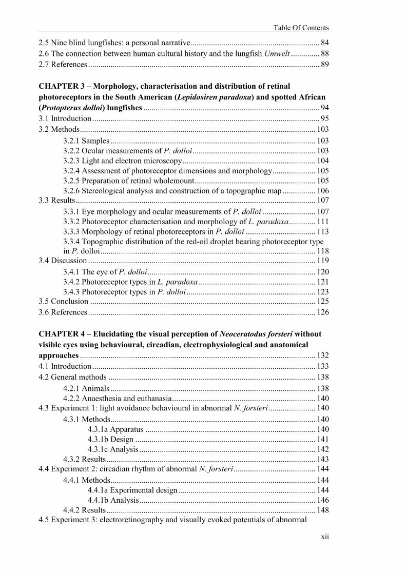
Table Of Contents
xii
2.5 Nine blind lungfishes: a personal narrative ............................................................... 84 2.6 The connection between human cultural history and the lungfish Umwelt .............. 88 2.7 References ................................................................................................................. 89 CHAPTER 3 – Morphology, characterisation and distribution of retinal photoreceptors in the South American (Lepidosiren paradoxa) and spotted African (Protopterus dolloi) lungfishes ...................................................................................... 94 3.1 Introduction ............................................................................................................... 95 3.2 Methods ................................................................................................................... 103
3.2.1 Samples .................................................................................................... 103 3.2.2 Ocular measurements of P. dolloi ............................................................ 103 3.2.3 Light and electron microscopy ................................................................. 104 3.2.4 Assessment of photoreceptor dimensions and morphology ..................... 105 3.2.5 Preparation of retinal wholemount ........................................................... 105 3.2.6 Stereological analysis and construction of a topographic map ................ 106
3.3 Results ..................................................................................................................... 107 3.3.1 Eye morphology and ocular measurements of P. dolloi .......................... 107 3.3.2 Photoreceptor characterisation and morphology of L. paradoxa ............. 111 3.3.3 Morphology of retinal photoreceptors in P. dolloi .................................. 113 3.3.4 Topographic distribution of the red-oil droplet bearing photoreceptor type
in P. dolloi ......................................................................................................... 118 3.4 Discussion ............................................................................................................... 119
3.4.1 The eye of P. dolloi .................................................................................. 120 3.4.2 Photoreceptor types in L. paradoxa ......................................................... 121 3.4.3 Photoreceptor types in P. dolloi ............................................................... 123
3.5 Conclusion .............................................................................................................. 125 3.6 References ............................................................................................................... 126 CHAPTER 4 – Elucidating the visual perception of Neoceratodus forsteri without visible eyes using behavioural, circadian, electrophysiological and anatomical approaches ................................................................................................................... 132 4.1 Introduction ............................................................................................................. 133 4.2 General methods ..................................................................................................... 138
4.2.1 Animals .................................................................................................... 138 4.2.2 Anaesthesia and euthanasia ...................................................................... 140
4.3 Experiment 1: light avoidance behavioural in abnormal N. forsteri ....................... 140 4.3.1 Methods .................................................................................................... 140
4.3.1a Apparatus ................................................................................... 140 4.3.1b Design ........................................................................................ 141 4.3.1c Analysis ...................................................................................... 142
4.3.2 Results ...................................................................................................... 143 4.4 Experiment 2: circadian rhythm of abnormal N. forsteri ........................................ 144
4.4.1 Methods .................................................................................................... 144 4.4.1a Experimental design ................................................................... 144 4.4.1b Analysis ...................................................................................... 146
4.4.2 Results ...................................................................................................... 148 4.5 Experiment 3: electroretinography and visually evoked potentials of abnormal

Table Of Contents
xiii
N. forsteri ...................................................................................................................... 150 4.5.1 Methods .................................................................................................... 150
4.5.1a Apparatus.................................................................................... 150 4.5.1b Analysis ...................................................................................... 153
4.5.2 Results ...................................................................................................... 153 4.5.2a Electroretinograms of normal and abnormal N. forsteri ............ 153 4.5.2b Visually evoked potentials of normal and abnormal N. forsteri 158
4.6 Experiment 4: investigation of the presence or absence of image-forming eyes in abnormal N. forsteri ...................................................................................................... 162
4.6.1 Methods .................................................................................................... 162 4.6.1a Sample preparation ..................................................................... 162 4.6.1b General morphology, ocular dimensions and light microscopy. 163 4.6.1c Assessment of photoreceptor dimensions and morphology ....... 164
4.6.2 Results ...................................................................................................... 164 4.6.2a General morphology and ocular measurements ......................... 164 4.6.2b Photoreceptor dimensions and morphology ............................... 172
4.7 General discussion .................................................................................................. 174 4.7.1 The abnormal N. forsteri eye ................................................................... 175 4.7.2 Visual perception of abnormal N. forsteri................................................ 177 4.7.3 Possible causes for the abnormal morphological deformities in N. forsteri .......................................................................................................... 181
4.8 Conclusion .............................................................................................................. 183 4.9 References ............................................................................................................... 184 CHAPTER 5 – Entering the soap-bubble: integrating science and art to describe encounters with the lungfish Umwelt ......................................................................... 190 5.1 Introduction ............................................................................................................. 191 5.2 ‘Third Eye’ and ‘In Focus’: encounters through dissections and light microscopy .................................................................................................................... 193 5.3 ‘The Forms’: encounters through electron microscopy, sculpture and performance................................................................................................................... 202 5.4 ‘Cernentia’: multi-sensorial encounters with lungfishes lacking visible eyes........ 215 5.5 Conclusion .............................................................................................................. 235 5.6 References ............................................................................................................... 235 II GENERAL DISCUSSION ..................................................................................... 240 ii.1 Introduction ............................................................................................................. 241 ii.2 The visual perception of Protopterus dolloi, Lepidosiren paradoxa and abnormal Neoceratodus forsteri .................................................................................................... 241 ii.3 Encounters with the lungfish Umwelt ..................................................................... 244 ii.4 Conclusion and further directions ........................................................................... 247 ii.5 References ............................................................................................................... 254 III APPENDIX ............................................................................................................ 256

List Of Figures
xiv
LIST OF FIGURES
GENERAL INTRODUCTION i.1 Living specimens of Neoceratodus forsteri ................................................................. 9 i.2 Immature South American lungfish (Lepidosiren paradoxa) .................................... 10 i.3 Adult South American lungfish (Lepidosiren paradoxa) .......................................... 10 i.4 Schematic representation of a fish (teleosts) eye and the direction of incoming light ................................................................................................................................. 17 i.5 A light micrograph of an Australian lungfish (Neoceratodus forsteri) retina ........... 20 i.6 The object colour solid volumes for juvenile and adult Neoceratodus forsteri, with and without spectral filtering affects of yellow ellipsoidal pigmentation and red oil droplets in medium- (MWS) and short-wavelength sensitive (SWS) cones .................. 22 CHAPTER 1 1.1 ‘Inperception’............................................................................................................ 46 1.2 The ‘Inperception’ mural hanging near the lungfishes’ housing tanks during the behavioural experiment ................................................................................................... 46 1.3 Devon Ward’s contribution to the ‘Inperception’ project ........................................ 48 1.4 Retinal wholemount of morphological characteristics in the Australian lungfish photoreceptors ................................................................................................................. 48 1.5 Shannon Williamson’s contribution to the ‘Inperception’ project ........................... 50 1.6 Appudurai first response to the ‘Inperception’ project ............................................. 52 1.7 Audrey Appudurai’s second response to the ‘Inperception’ project ........................ 53 CHAPTER 2 2.1 Adult male L. paradoxa ............................................................................................ 71 2.2 L. annectens specimen examined by Owen .............................................................. 72 2.3 Australian lungfish lying on a cane fishing basket ................................................... 76 2.4 Two fishermen with their Australian lungfish catch, Coomera River, Queensland . 76 2.5 Granddad arrives at the Shedd Aquarium ................................................................. 80 2.6 Granddad in his aquarium after arriving at Shedd .................................................... 80 2.7 Granddad’s 80th birthday cake, Shedd Aquarium ..................................................... 82 2.8 Granddad at his 80th anniversary celebration, Shedd Aquarium ............................... 82 2.9 Mundubbera water tower, Queensland ..................................................................... 84 2.10 A juvenile Australian lungfish, Neoceratodus forsteri ........................................... 85 2.11 An abnormal juvenile Neoceratodus forsteri .......................................................... 86 2.12 Confirming the visual deformity in abnormal Neoceratodus forsteri: the eye is clearly absent................................................................................................................... 87
List Of Figures
xv
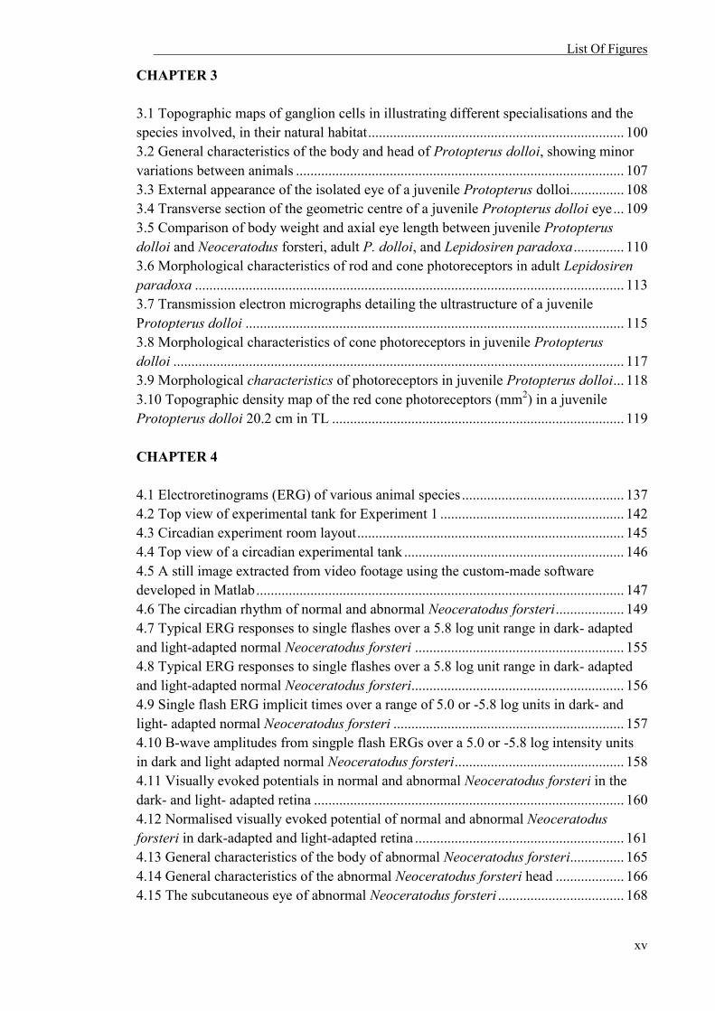
CHAPTER 3 3.1 Topographic maps of ganglion cells in illustrating different specialisations and the species involved, in their natural habitat ....................................................................... 100 3.2 General characteristics of the body and head of Protopterus dolloi, showing minor variations between animals ........................................................................................... 107 3.3 External appearance of the isolated eye of a juvenile Protopterus dolloi............... 108 3.4 Transverse section of the geometric centre of a juvenile Protopterus dolloi eye ... 109 3.5 Comparison of body weight and axial eye length between juvenile Protopterus dolloi and Neoceratodus forsteri, adult P. dolloi, and Lepidosiren paradoxa .............. 110 3.6 Morphological characteristics of rod and cone photoreceptors in adult Lepidosiren paradoxa ....................................................................................................................... 113 3.7 Transmission electron micrographs detailing the ultrastructure of a juvenile Protopterus dolloi ......................................................................................................... 115 3.8 Morphological characteristics of cone photoreceptors in juvenile Protopterus dolloi ............................................................................................................................. 117 3.9 Morphological characteristics of photoreceptors in juvenile Protopterus dolloi ... 118 3.10 Topographic density map of the red cone photoreceptors (mm2) in a juvenile Protopterus dolloi 20.2 cm in TL ................................................................................. 119 CHAPTER 4 4.1 Electroretinograms (ERG) of various animal species ............................................. 137 4.2 Top view of experimental tank for Experiment 1 ................................................... 142 4.3 Circadian experiment room layout .......................................................................... 145 4.4 Top view of a circadian experimental tank ............................................................. 146 4.5 A still image extracted from video footage using the custom-made software developed in Matlab ...................................................................................................... 147 4.6 The circadian rhythm of normal and abnormal Neoceratodus forsteri ................... 149 4.7 Typical ERG responses to single flashes over a 5.8 log unit range in dark- adapted and light-adapted normal Neoceratodus forsteri .......................................................... 155 4.8 Typical ERG responses to single flashes over a 5.8 log unit range in dark- adapted and light-adapted normal Neoceratodus forsteri........................................................... 156 4.9 Single flash ERG implicit times over a range of 5.0 or -5.8 log units in dark- and light- adapted normal Neoceratodus forsteri ................................................................ 157 4.10 B-wave amplitudes from singple flash ERGs over a 5.0 or -5.8 log intensity units in dark and light adapted normal Neoceratodus forsteri ............................................... 158 4.11 Visually evoked potentials in normal and abnormal Neoceratodus forsteri in the dark- and light- adapted retina ...................................................................................... 160 4.12 Normalised visually evoked potential of normal and abnormal Neoceratodus forsteri in dark-adapted and light-adapted retina .......................................................... 161 4.13 General characteristics of the body of abnormal Neoceratodus forsteri............... 165 4.14 General characteristics of the abnormal Neoceratodus forsteri head ................... 166 4.15 The subcutaneous eye of abnormal Neoceratodus forsteri ................................... 168
List Of Figures
xvi
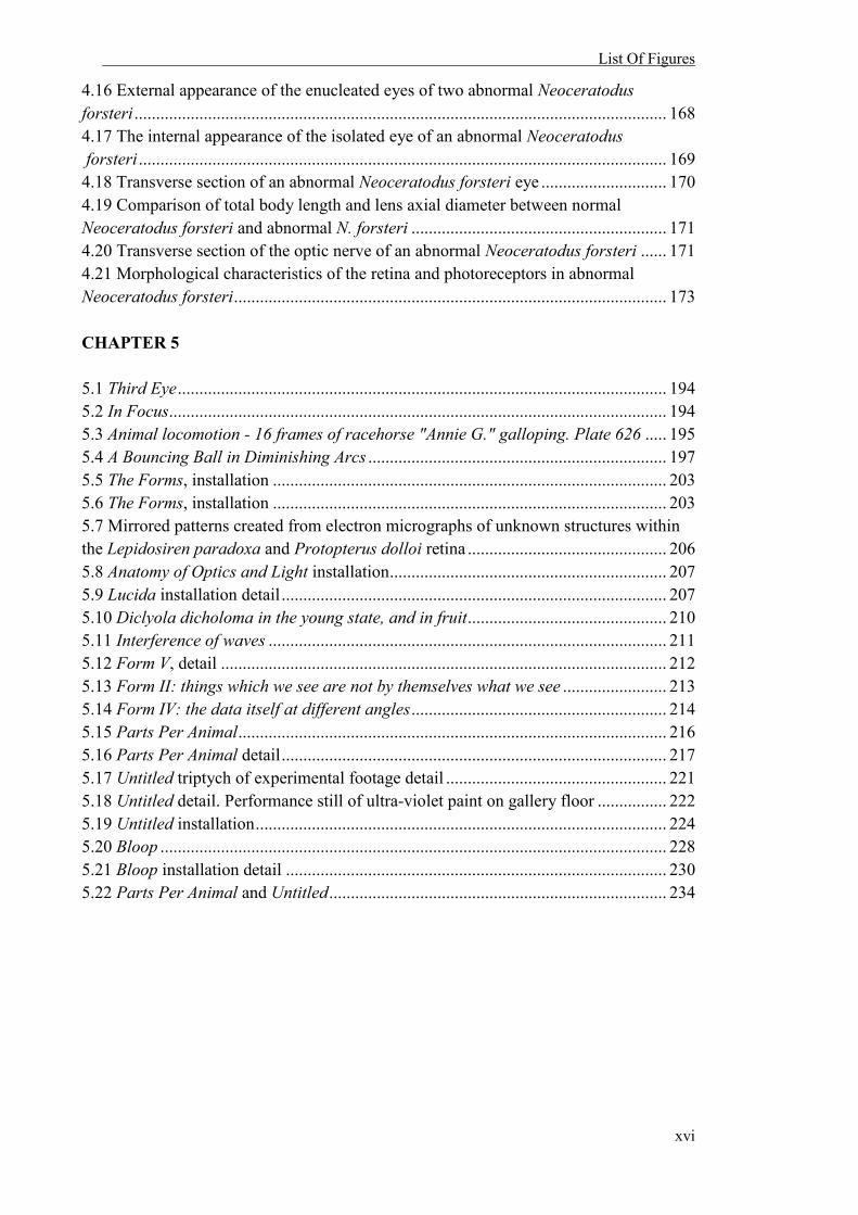
4.16 External appearance of the enucleated eyes of two abnormal Neoceratodus forsteri ........................................................................................................................... 168 4.17 The internal appearance of the isolated eye of an abnormal Neoceratodus forsteri .......................................................................................................................... 169 4.18 Transverse section of an abnormal Neoceratodus forsteri eye ............................. 170 4.19 Comparison of total body length and lens axial diameter between normal Neoceratodus forsteri and abnormal N. forsteri ........................................................... 171 4.20 Transverse section of the optic nerve of an abnormal Neoceratodus forsteri ...... 171 4.21 Morphological characteristics of the retina and photoreceptors in abnormal Neoceratodus forsteri .................................................................................................... 173 CHAPTER 5 5.1 Third Eye ................................................................................................................. 194 5.2 In Focus................................................................................................................... 194 5.3 Animal locomotion - 16 frames of racehorse "Annie G." galloping. Plate 626 ..... 195 5.4 A Bouncing Ball in Diminishing Arcs ..................................................................... 197 5.5 The Forms, installation ........................................................................................... 203 5.6 The Forms, installation ........................................................................................... 203 5.7 Mirrored patterns created from electron micrographs of unknown structures within the Lepidosiren paradoxa and Protopterus dolloi retina .............................................. 206 5.8 Anatomy of Optics and Light installation ................................................................ 207 5.9 Lucida installation detail ......................................................................................... 207 5.10 Diclyola dicholoma in the young state, and in fruit .............................................. 210 5.11 Interference of waves ............................................................................................ 211 5.12 Form V, detail ....................................................................................................... 212 5.13 Form II: things which we see are not by themselves what we see ........................ 213 5.14 Form IV: the data itself at different angles ........................................................... 214 5.15 Parts Per Animal ................................................................................................... 216 5.16 Parts Per Animal detail ......................................................................................... 217 5.17 Untitled triptych of experimental footage detail ................................................... 221 5.18 Untitled detail. Performance still of ultra-violet paint on gallery floor ................ 222 5.19 Untitled installation ............................................................................................... 224 5.20 Bloop ..................................................................................................................... 228 5.21 Bloop installation detail ........................................................................................ 230 5.22 Parts Per Animal and Untitled .............................................................................. 234
List Of Tables
xvii
LIST OF TABLES
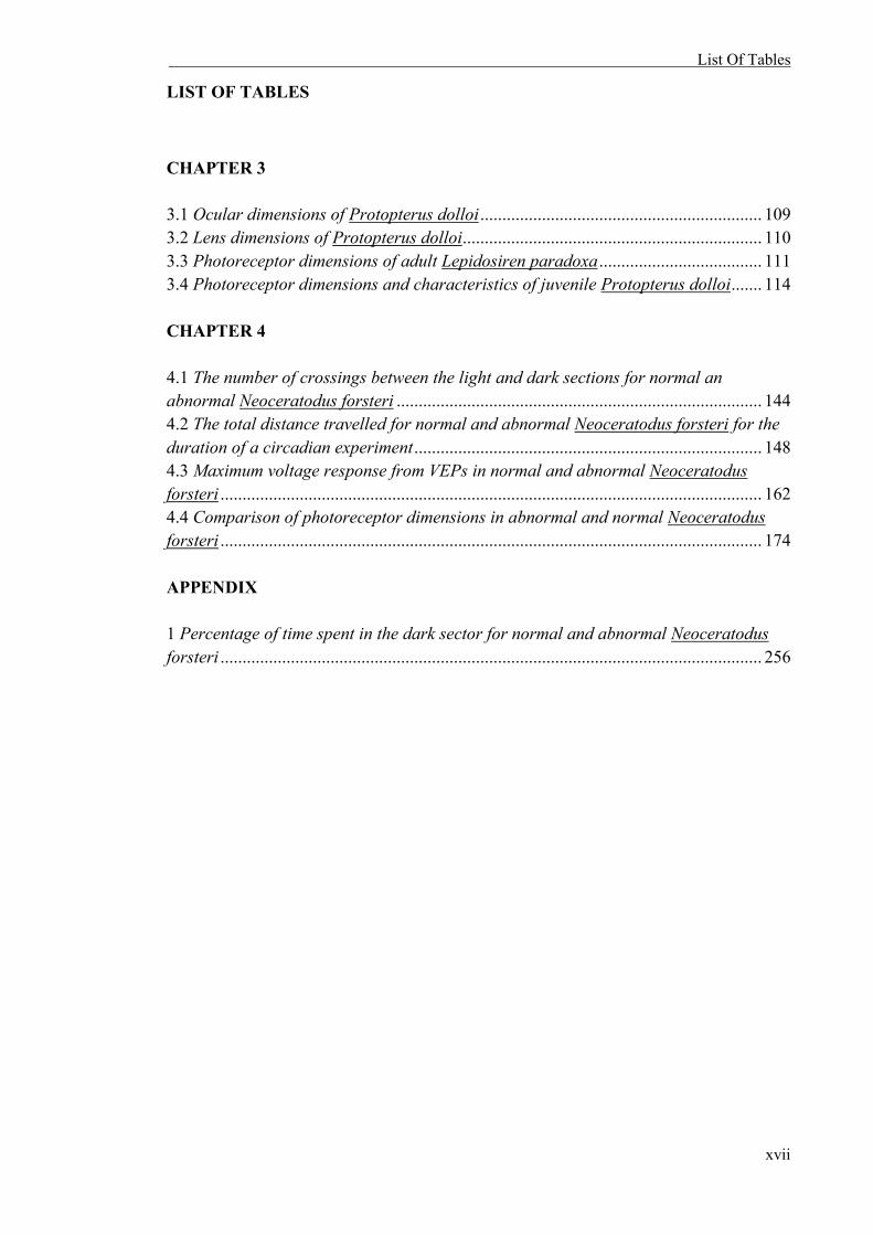
CHAPTER 3 3.1 Ocular dimensions of Protopterus dolloi ................................................................ 109 3.2 Lens dimensions of Protopterus dolloi .................................................................... 110 3.3 Photoreceptor dimensions of adult Lepidosiren paradoxa ..................................... 111 3.4 Photoreceptor dimensions and characteristics of juvenile Protopterus dolloi ....... 114 CHAPTER 4 4.1 The number of crossings between the light and dark sections for normal an abnormal Neoceratodus forsteri ................................................................................... 144 4.2 The total distance travelled for normal and abnormal Neoceratodus forsteri for the duration of a circadian experiment ............................................................................... 148 4.3 Maximum voltage response from VEPs in normal and abnormal Neoceratodus forsteri ........................................................................................................................... 162 4.4 Comparison of photoreceptor dimensions in abnormal and normal Neoceratodus forsteri ........................................................................................................................... 174 APPENDIX 1 Percentage of time spent in the dark sector for normal and abnormal Neoceratodus forsteri ........................................................................................................................... 256
List Of Abbreviations
xviii
LIST OF ABBREVIATIONS
µm Micrometres µV Microvolts λmax Maximum absorbance a Aperture a Axial length of the lens AC Alternating current Ag/Ag-Cl silver/silver chloride c/cor Cornea cc Clear cone cFFF critical flicker-fusion frequency CO2 Carbon dioxide DAFF Department of Agriculture, Fisheries and Forestry E Eyeless Neoceratodus forsteri EM Electron microscopy ERG Electroretinography EPBC Environment Protection and Biodiversity Conservation Act 1999 fr Focal ratio g Grams gfp Green fluorescent protein H2O Water Hz Hertz ir Iris INL Inner nuclear layer ipl Inner plexiform layer is/IS Inner segment of photoreceptor cell JITL Jindalee International Pty Ltd kV Kilowatts LED Light-emitting diode LM Light microscopy ln Lens LWS Long-wavelength sensitive M Molarity m Mitochondria ms Milliseconds MS222 Tricaine methane sulfonate MSP Microspectrophotometry MWS Medium-wavelength sensitive N Normal Neoceratodus forsteri n Sample size N/A Not available NA Numerical aperture

List Of Abbreviations
xix
NaHCO3 Sodium bicarbonate ND neutral-density filter nm Nanometres O2 Oxygen od/OD Oil droplet on Optic nerve ONL Outer nuclear layer OP Oscillatory potentials opl Outer plexiform layer os/OS Outer segment of photoreceptor cell p Paraboloid Pax6 Paired box protein Pax-6 PC Personal computer PFA 4% paraformaldehyde in 0.1 M phosphate buffer PND Distance between the centre of the lens and the back of the retina at 90° from the posterior nodal distance PR Photoreceptors Qld Queensland, Australia r Radius of curvature rc Red oil droplet containing cone rd Rod r/ret Retina rgc/RGC Retinal ganglion cells rpe/RPE Retinal pigment epithelium scl Sclera SEM Standard error of the mean SD Standard deviation SWS Short-wavelength sensitive TEM Transmission electron microscopy TL Total length UV Ultra-violet UVS Ultra-violet sensitive VEPs Visually evoked potentials yc Yellow-pigment containing cone


General Introduction
1
I ::::::
GENERAL INTRODUCTION
::::::
General Introduction
2
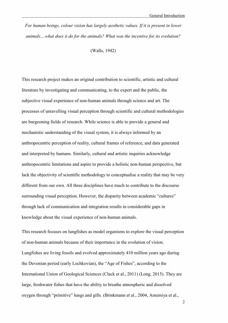
For human beings, colour vision has largely aesthetic values. If it is present in lower
animals....what does it do for the animals? What was the incentive for its evolution?
(Walls, 1942)
This research project makes an original contribution to scientific, artistic and cultural
literature by investigating and communicating, to the expert and the public, the
subjective visual experience of non-human animals through science and art. The
processes of unravelling visual perception through scientific and cultural methodologies
are burgeoning fields of research. While science is able to provide a general and
mechanistic understanding of the visual system, it is always informed by an
anthropocentric perception of reality, cultural frames of reference, and data generated
and interpreted by humans. Similarly, cultural and artistic inquiries acknowledge
anthropocentric limitations and aspire to provide a holistic non-human perspective, but
lack the objectivity of scientific methodology to conceptualise a reality that may be very
different from our own. All three disciplines have much to contribute to the discourse
surrounding visual perception. However, the disparity between academic “cultures”
through lack of communication and integration results in considerable gaps in
knowledge about the visual experience of non-human animals.
This research focuses on lungfishes as model organisms to explore the visual perception
of non-human animals because of their importance in the evolution of vision.
Lungfishes are living fossils and evolved approximately 410 million years ago during
the Devonian period (early Lochkovian), the “Age of Fishes”, according to the
International Union of Geological Sciences (Clack et al., 2011) (Long, 2015). They are
large, freshwater fishes that have the ability to breathe atmospheric and dissolved
oxygen through “primitive” lungs and gills. (Brinkmann et al., 2004, Amemiya et al.,

General Introduction
3
2013). The lungfishes’ evolutionary position makes them vital to the study of the
evolution of vision in land vertebrates (tetrapods) as they are the closest surviving
descendents of those animals that made the transition from water onto land.
This thesis investigates how the sciences and the arts (often opposing disciplines)
explore the visual perception of lungfishes by simultaneously undertaking scientific and
artistic experiments to uncover, represent and interpret the visual experience of these
animals. In addition, it discusses the general anthropocentric and discipline-specific
limitations inhibiting deeper understanding of the visual experience of non-human
animals. This discussion of limitations results in opportunities when these different
academic “cultures” are forced or encouraged to communicate. Therefore, a more
informed view of the lungfish’s visual experience is sought by uncovering limitations
and opportunities provided by previous methods.
i.1 NON-HUMAN ANIMAL PERCEPTION
We polish the animal mirror to look at ourselves
(Haraway, 1991)
Scholars from the sciences and the arts1 have attempted to critically uncover the visual
perception of non-human animals. Scientific scholars focus on quantitative
measurements of perceptual capabilities in other organisms, and rely on direct logical
extrapolations from experimental data to address non-human visual perception. Early
1 I refer to both humanities and visual art disciplines here

General Introduction
4
scientific discoveries that tested the presence and type of colour vision of non-human
animals were achieved by direct observation. John Lubbock and Karl von Frisch were
pioneers in this research, and used behavioural criteria to determine whether non-human
animals were able to differentiate colours (Lubbock, 1889, Von Frisch, 1914). The
“grey card experiment” created by Frisch, established that honeybees (Apis mellifera)
associate colour with food, and can distinguish blue and yellow from thirty shades
(intensities) of grey (Von Frisch, 1914). Experiments following the same design have
subsequently confirmed the ability to discriminate colour in a variety of animals,
including butterflies (belonging to the families Nymphalidae, Pieridae, Satyridae and
Papilionidae), hermit crabs (Eupagurus anachoretus), shrimp (Crangon vulgaris)
domestic chickens (Gallus gallus) (Kelber et al., 2003), and the giant shovelnose ray
(Glaucostegus typus) (Van-Eyk et al., 2011).
Techniques such as microscopy, microspectrophotometry (MSP) and electrophysiology
have also been useful in understanding the mechanistic basis of vision and identifying
differences in visual performance of many species. These techniques enable the
identification and characterisation of different kinds of retinal cells and other retinal
specialisations, and can define the range of wavelengths to which an animal is optimally
sensitive, and whether that animal has the potential for colour vision. The potential for
colour vision and the description of visual specialisations have been reported in many
organisms including the crayfish (Orconectes virilise) (Wald, 1967), the southern
hemisphere lamprey (Geotria australis) (Collin et al., 2003, Collin et al., 2004), the
turtle (Pseudemys scripta elegans) (Kolb and Jones, 1987), the human (Homo sapiens)
(Bowmaker and Dartnall, 1980), the tawny owl (Strix aluco) (Bowmaker and Martin,
1978) and the duck (Anas platyrhynchos) (Jane and Bowmaker, 1988).
General Introduction
5
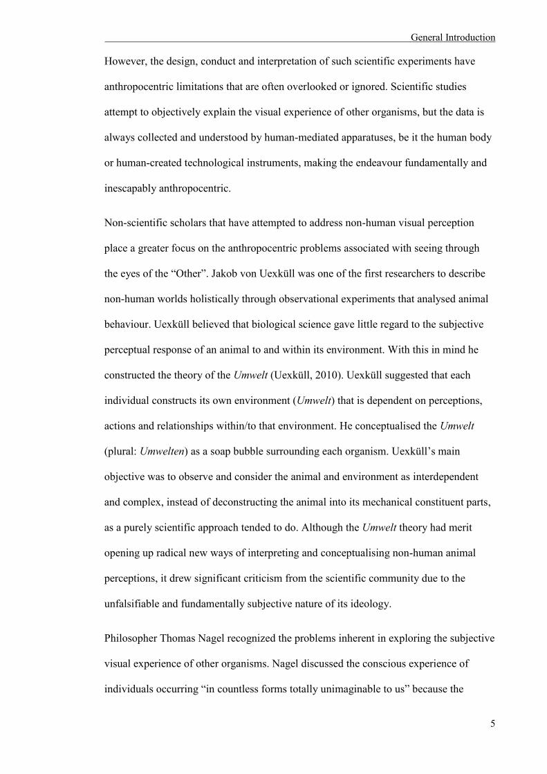
However, the design, conduct and interpretation of such scientific experiments have
anthropocentric limitations that are often overlooked or ignored. Scientific studies
attempt to objectively explain the visual experience of other organisms, but the data is
always collected and understood by human-mediated apparatuses, be it the human body
or human-created technological instruments, making the endeavour fundamentally and
inescapably anthropocentric.
Non-scientific scholars that have attempted to address non-human visual perception
place a greater focus on the anthropocentric problems associated with seeing through
the eyes of the “Other”. Jakob von Uexküll was one of the first researchers to describe
non-human worlds holistically through observational experiments that analysed animal
behaviour. Uexküll believed that biological science gave little regard to the subjective
perceptual response of an animal to and within its environment. With this in mind he
constructed the theory of the Umwelt (Uexküll, 2010). Uexküll suggested that each
individual constructs its own environment (Umwelt) that is dependent on perceptions,
actions and relationships within/to that environment. He conceptualised the Umwelt
(plural: Umwelten) as a soap bubble surrounding each organism. Uexküll’s main
objective was to observe and consider the animal and environment as interdependent
and complex, instead of deconstructing the animal into its mechanical constituent parts,
as a purely scientific approach tended to do. Although the Umwelt theory had merit
opening up radical new ways of interpreting and conceptualising non-human animal
perceptions, it drew significant criticism from the scientific community due to the
unfalsifiable and fundamentally subjective nature of its ideology.
Philosopher Thomas Nagel recognized the problems inherent in exploring the subjective
visual experience of other organisms. Nagel discussed the conscious experience of
individuals occurring “in countless forms totally unimaginable to us” because the

General Introduction
6
experience is exclusive to the organism, and one must “be that organism” in order to
understand it (Nagel, 1974). Although he acknowledged the human mind as the limiting
factor preventing full comprehension of another perceptual world, Nagel did not dismiss
the futile task of attempting to bridge the subjective character of experience. Instead, he
suggested removing human imagination from the interpretation, perhaps through
objective experimentation, as a way to reveal parts of “the subjective character of
experiences in a form comprehensible to beings incapable of having those experiences”
(Nagel, 1974). However, Nagel did not engage with science or suggest and/or articulate
how such experiments could take place.
Non-scientific research into the visual perception of animals rarely engages with science
or the mechanisms and concepts surrounding what are known about visual systems,
while scientific research rarely acknowledges the anthropocentricity and limitations of
the field. This research project acknowledges the importance of the scientific
investigation of non-human animal visual experiences, but also recognises the
anthropocentric limitations of the scientific method highlighted by non-scientific
scholars, and attempts to combine them for a deeper understanding of the lungfish
visual experience. Chapter 1 explores this through a discussion of several historical
scientific and non-scientific attempts to uncover animal perception. With a particular
focus on the similarities and differences of the two fields, the collaborative art project
‘Inperception’ (2012) endeavoured to interpret the Umwelten of Australian lungfishes
through contemporary scientific knowledge and art. In order to uncover the mechanisms
of lungfish perception, chapter 3 and chapter 4 utilizes contemporary scientific
approaches (anatomical dissection, transmission electron microscopy,
electrophysiology, behavioural and experiments on circadian rhythms) to describe
aspects of the visual system of the spotted African (Protopterus dolloi) and South

General Introduction
7
American (Lepidosiren paradoxa) lungfishes, and compares the visual perception of
normal and abnormal Australian lungfish (Neoceratodus forsteri).
i.2 LUNGFISHES AND THEIR VISUAL SYSTEM
If it hadn't been for the obstinacy of our great-uncle N'ba N'ga, we would have long
since lost all contact with the aquatic world. Yes, we had a great-uncle who was a fish,
on my paternal grandmother's side, to be precise, of the Coelacanthus family of the
Devonian period...
at any season of the year all we had to do was push ourselves over the softer layers of
vegetation until we could feel ourselves sinking into the dampness, and there below, a
few palms' lengths from the edge, we could see the column of little bubbles he sent up,
breathing heavily the way old folk do, or the little cloud of mud scraped up by his sharp
snout, always rummaging around, more out of habit than out of the need to hunt for
anything.
We lay on a sloping bank, all three of us: my great-uncle was nearest the water, but the
two of us were half in and half out, too, so anyone seeing us from the distance, all close
together, wouldn't have known who was terrestrial and who was aquatic.
(Calvino, 1968)
Lungfishes are ideal to explore the visual perception of non-human animals through the
sciences and arts because of their unique evolutionary position as “in-between”
creatures. These animals diverged from the main vertebrate stock around 410 MYA and
along with the coelacanth (Latimeria chalumnae), encompass the surviving lobe-finned

General Introduction
8
fishes (Sarcopterygii) (Hart et al., 2008, Clack et al., 2011). They have the ability to
breathe dissolved and atmospheric oxygen through gills and “primitive” lungs,
respectively, existing between water and land. Linked to humans by being the closest
living relatives to the tetrapods (Brinkmann et al., 2004, Amemiya et al., 2013), they are
vital to the study of the evolution of vision in terrestrial animals.
Six extant species of lungfishes remain worldwide. Four of these are located in Africa,
one in South America and one in Australia. The South American species (L. paradoxa)
and all African species (Protopterus spp.) belong to the family Lepidosirenidae (Walls,
1942). N. forsteri is the only surviving species belonging to the family Ceratodontidae,
and is considered to be the most basal of all lungfishes with a single lung used only
during periods of increased activity or in areas of poor water quality (Kind, 2011).
i.2.1 Morphology
N. forsteri was originally described by Krefft (1870), who thought this large fish to be
an amphibian “allied to the genus Lepidosiren” because of its comparative morphology
and dentition. Morphologically, adult Australian lungfishes are characterised by having
a wide, flat head, a thick heavy body, large scales that decrease in size towards the tail,
a diphycercal tail, and paddle shaped fins (Fig. i.1) (Kemp, 1986, Kind, 2011).

General Introduction
9
Fig. i.1. Living specimens of Neoceratodus forsteri. (A) An adult N. forsteri about to rise to the surface. Note the paddle shaped pelvic fins and diphycercal tail. Scale bar = 1 cm. (B) Two living juveniles of N. forsteri, illustrating the body proportions and differences in morphology when compared to the adult (A), relatively smaller pelvic fins and rounder heads. The dorsal fin also reaches further forward. Scale bar = 1 cm. Reproduced with kind permission from Wiley-Liss Inc. (Kemp, 1986).
L. paradoxa is morphologically similar to Protopterus spp. (Mlewa et al., 2011).
However, its body length is longer and slimmer, with smaller scales, and reduced
pectoral and pelvic fins for sensory functions when compared to N. forsteri (Berra,
2001, Fonesca de Almeida-Val et al., 2011). Juvenile L. paradoxa are predominantly
black and spotted yellow which fades to a brown/gray colour as they mature (Figs. i.2
and i.3).

General Introduction
10
Fig i.2. Immature South American lungfish (Lepidosiren paradoxa). Photo credit: SE Temple, SP Collin, NS Hart and RG Northcutt (2010).
Fig. i.3. Adult South American lungfish (Lepidosiren paradoxa). Photo credit: SE Temple, SP Collin, NS Hart and RG Northcutt (2010).
General Introduction
11
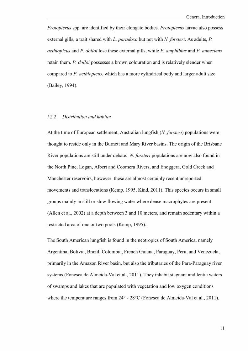
Protopterus spp. are identified by their elongate bodies. Protopterus larvae also possess
external gills, a trait shared with L. paradoxa but not with N. forsteri. As adults, P.
aethiopicus and P. dolloi lose these external gills, while P. amphibius and P. annectens
retain them. P. dolloi possesses a brown colouration and is relatively slender when
compared to P. aethiopicus, which has a more cylindrical body and larger adult size
(Bailey, 1994).
i.2.2 Distribution and habitat
At the time of European settlement, Australian lungfish (N. forsteri) populations were
thought to reside only in the Burnett and Mary River basins. The origin of the Brisbane
River populations are still under debate. N. forsteri populations are now also found in
the North Pine, Logan, Albert and Coomera Rivers, and Enoggera, Gold Creek and
Manchester reservoirs, however these are almost certainly recent unreported
movements and translocations (Kemp, 1995, Kind, 2011). This species occurs in small
groups mainly in still or slow flowing water where dense macrophytes are present
(Allen et al., 2002) at a depth between 3 and 10 meters, and remain sedentary within a
restricted area of one or two pools (Kemp, 1995).
The South American lungfish is found in the neotropics of South America, namely
Argentina, Bolivia, Brazil, Colombia, French Guiana, Paraguay, Peru, and Venezuela,
primarily in the Amazon River basin, but also the tributaries of the Para-Paraguay river
systems (Fonesca de Almeida-Val et al., 2011). They inhabit stagnant and lentic waters
of swamps and lakes that are populated with vegetation and low oxygen conditions
where the temperature ranges from 24° - 28°C (Fonesca de Almeida-Val et al., 2011).

General Introduction
12
All Protopterus species inhabit both lentic (lakes, or standing water) and lotic (river,
stream, or running water) environments that extend throughout most of the continental
African landmass, including all of the Rift Valley lakes, with the exception of Lake
Malawi, the major river systems of tropical Africa and a number of localities therein
(Greenwood, 1987). P. annectens and P. dolloi are predominantly West African, while
P. aethiopicus and P. amphibius are considered East African taxa (Mlewa et al., 2011).
However, P. aethiopicus also occurs in the Zaire basin and P. annectens can extend east
into the middle and lower Zambezi systems as well as the Limpopo system. Although
African lungfishes are known to reside in open waters of lakes and river systems, they
are generally associated with the shallow areas and swamps surrounding the margins of
these water bodies including marginal grass-, water lily, and papyrus swamps.
i.2.3 Diet
All lungfishes are opportunistic omnivores. In their natural habitat, these fish forage
among macrophytes and ingest large quantities of substrate particles and various plants,
which pass through the digestive tract relatively intact (Günther, 1871). They have also
been reported to feed on terrestrial plant seed pods and molluscs, crustaceans, worms,
tadpoles and small fishes (Kemp, 1986, Kemp, 1995, Kind, 2011). The adult South
American lungfish diet consists of small bony fishes, snails, clams, shrimp and some
algae and terrestrial flora (Berra, 2001, Fonesca de Almeida-Val et al., 2011). African
lungfishes are considered to be the most carnivorous of all the Dipnoan fishes
(Greenwood, 1987). They consume a variety of prey including molluscs, crustaceans,
small fishes, worms and aquatic insects as well as plant material found in their natural
habitat (Curry-Lindahl, 1956).

General Introduction
13
The diet of Australian lungfishes changes over time. Juvenile N. forsteri tend to be sit-
and-wait carnivorous predators, whereas adults are opportunistic omnivores (Kind,
2011). Evidence of this change in diet within N. forsteri is evident in dentition;
juveniles possess sharp grasping teeth that change to broad crushing plates in adults
(Kemp, 1977).
i.2.4 Aestivation
Five of the six lungfish species undergo aestivation, with N. forsteri being the only
exception. Aestivation can be described as a “light” state of dormancy, characterised by
the construction of a cocoon, withdrawal into a state of torpor, and characteristic cardio-
respiratory and metabolic changes (Fishman et al., 1987). The process enables
lungfishes to avoid damage from high temperatures and desiccation during the dry
season without leaving the swamps for permanent water, like other fishes (Greenwood,
1986).
L. paradoxa and P. dolloi employ different methods of aestivation. The South American
species creates a burrow 0.5 metres into the muddy clay substrate of the swamp by
forcing its way vertically or obliquely using undulating body movements (Carter and
Beadle, 1930) and leaving several holes to facilitate air-breathing (Fonesca de Almeida-
Val et al., 2011). There the fish lies coiled in an oval expansion of the burrow and
lowers all metabolic function. The burrow’s substrate is predominantly clay which is
characteristically impervious to water. This provides the fish with an ideal burrow that
is partially filled with water to prevent it from drying out (Carter and Beadle, 1930).
Although L. paradoxa does not form a mucous seal over its water-filled burrow, the
burrow is sufficient to sustain the fish throughout the dry season (Fonesca de Almeida-
Val et al., 2011).

General Introduction
14
Aestivation patterns in Protopterus vary between species (Greenwood, 1987),
P. annectens being the most dependant on aestivation (Johnels and Svensson, 1954,
Greenwood, 1987, Mlewa et al., 2011). With the arrival of the dry season and the
resulting decrease in water level, P. annectens (40 mm and over in length) individuals
excavate a vertical burrow into the still submerged mud of the swamp bed by body
movements and biting into the soil(Johnels and Svensson, 1954). Once the burrow is
between 30 – 250 mm in depth (depending on the fish’s length), the fish turns 180° on
its long axis, creating a bulb-shaped chamber approximately twice the width of its body
in diameter, and rests with its snout pointing upwards (Greenwood, 1987). Once this
chamber is formed, the fish makes its mouth into a tube in order to breathe until the
water table falls below the snout level of the fish, whereby it will cease respiration and
remain coiled in the chamber. As the ambient water decreases, the fish secretes mucus
that, as the mud surrounding the chamber dries, hardens to produce a waterproof cocoon
that completely engulfs the lungfish apart from the respiratory opening in the snout
(Mlewa et al., 2011). P. annectens remains in this state until the water level rises again
and the cocoon is submerged by the coming wet season. An individual can aestivate for
periods up to 7 or 8 months. However, some captive and laboratory reared lungfishes
have been reported to have aestivated for 7 years (Smith, 1931, Greenwood, 1987,
Mlewa et al., 2011). There is little evidence that on P. aethiopicus aestivates in the wild,
although their burrows have been found in various places including north of Lake
Kyoga, Uganda, and closely resemble those described for L. paradoxa (Wasawo, 1959).
P. dolloi has developed a different approach to surviving the dry season, burrowing a
vertical shaft into the raft-like substrate of its swamp habitat. This shaft is partially filled
with water that is rich in organic matter, with a high CO2 and low O2 content. P. dolloi
inhabits this burrow until the next wet season (Greenwood, 1987). Along with the
cessation of respiration during aestivation, physiological processes characteristic of

General Introduction
15
aestivation in Protopterus include reduced metabolic rate, renal shutdown and
mechanisms that prevent the harmful effects of fat and protein metabolism while fasting
without access to water (Fishman et al., 1987, Mlewa et al., 2011). Aestivation allows
the lungfish to permanently occupy seasonal habitats and decreases the mortality rates
and resource limitations of migrating, non-resident species.
i.2.5 The visual system of lungfishes
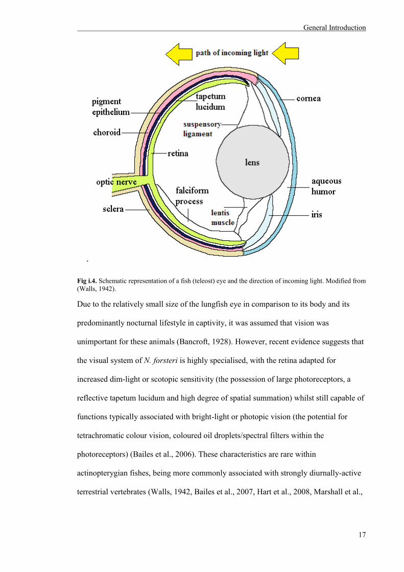
The aquatic vertebrate eye (Fig. i.4) is adapted to detect light and form images in a
range of light environments. Light first passes through the transparent cornea, differing
from terrestrial vertebrates by imparting only limited refractive power to the eye due to
the similarities of the refractive indices of the cornea and the surrounding water. A
much larger and more highly refractile crystalline lens in aquatic vertebrates
compensates for this lack of refractive power (Charman, 1991, Collin and Collin, 2001).
After entering the eye through the cornea, light passes through the lens to and strikes the
photoreceptor cells in the retina located at the back of the eyecup. Photoreceptor cells
transform light energy into a biochemical signal through a process called
phototransduction, and these visual signals are transmitted through the interneurons of
the retina (bipolar, horizontal and amacrine cells) to reach the ganglion cells. The axons
of ganglion cells are then responsible for conveying this information to the visual
centres of the brain via the optic nerve.
The rods and cone photoreceptors are not only morphologically distinct, but have
different physiological properties in order to operate effectively under different levels of
illumination. Rods are used for vision in low light conditions, as they are highly
sensitive and saturate in bright light. Cones are used for vision in bright light because
they saturate only under very intense illumination. If two or more types of

General Introduction
16
photoreceptor (usually cones) that are maximally sensitive to different parts of the
visible spectrum are present in the retina, their outputs can be compared in order to
perceive colour because a single photoreceptor type cannot distinguish between changes
in intensity and changes in wavelength2. The relative stimulation of photoreceptors with
differing spectral sensitivities must be compared by higher order neurons (such as the
retinal ganglion cells or output cells that send messages to the brain) in order to
discriminate objects on the basis of spectral reflectance (‘colour’) rather than just
intensity.
Humans have three different types of cone photoreceptors and one type of rod
photoreceptor. These cones have peak sensitivities (λmax values) at 562 nm (long-
wavelength sensitive [LWS], or red), 533 nm (medium-wavelength sensitive [MWS], or
green), and 420 nm (short-wavelength sensitive [SWS], or blue), while the single rod
photoreceptor type is MWS with a λmax of 497 nm (Bowmaker and Dartnall, 1980).
Opponent comparisons of the relative stimulation of the different types of cone
photoreceptors enable us to perceive colour.
2 This is known as the theory of univariance. See RUSHTON, W. 1972. Review lecture. Pigments and signals in colour vision. The Journal of Physiology, 220, 1-31..
General Introduction
17
Fig i.4. Schematic representation of a fish (teleost) eye and the direction of incoming light. Modified from (Walls, 1942).
Due to the relatively small size of the lungfish eye in comparison to its body and its
predominantly nocturnal lifestyle in captivity, it was assumed that vision was
unimportant for these animals (Bancroft, 1928). However, recent evidence suggests that
the visual system of N. forsteri is highly specialised, with the retina adapted for
increased dim-light or scotopic sensitivity (the possession of large photoreceptors, a
reflective tapetum lucidum and high degree of spatial summation) whilst still capable of
functions typically associated with bright-light or photopic vision (the potential for
tetrachromatic colour vision, coloured oil droplets/spectral filters within the
photoreceptors) (Bailes et al., 2006). These characteristics are rare within
actinopterygian fishes, being more commonly associated with strongly diurnally-active
terrestrial vertebrates (Walls, 1942, Bailes et al., 2007, Hart et al., 2008, Marshall et al.,

General Introduction
18
2011). The sophistication of its ocular system suggests that the visual sense of N.
forsteri is crucial for many aspects of its behavioural ecology (Bailes et al., 2007).
Considerable gaps in our knowledge about the visual system in lungfishes are evident,
and although there has been considerable interest in N. forsteri over the last ten years,
the South American and African species have been somewhat neglected. Therefore, this
project investigated and compared the visual systems of these lungfish species with that
of N. forsteri to determine if the complex visual system of N. forsteri is universal among
this group of ancient organisms.
i.2.5a The lungfish retina
The retinae of lungfishes have been described as “simple”, although containing very
large cells (Locket, 1970). The retinae of South American and African lungfishes
previously characterised by light microscopy revealed a well pigmented epithelial layer,
with rod and cone photoreceptors possessing oil droplets. The “monstrous” rods and
cones are difficult to distinguish by size, as their morphology is similar (Kerr, 1902,
Walls, 1942, Pfeiffer, 1968, Ali and Anctil, 1973). All lungfish retinae contain both rods
and cones. Protopterus spp. appear to have single and double cones (Pfeiffer, 1968),
unlike L. paradoxa and N. forsteri, which show no evidence of double cones (Ali and
Anctil, 1973, Bailes et al., 2006). In the case of N. forsteri, the rod (53%) and cone
(47%) proportions are similar, suggesting the animal balances both scotoptic and
photopic3 visual requirements (Bailes et al., 2006). Conversely, the number of rods is
inferior to cones in the case of L. paradoxa (Ali and Anctil, 1973). N. forsteri
photoreceptors are composed of a single rod type and three (adults) or four (juvenile)
morphologically and spectrally distinct cone types (Fig. i.5). The retina of this species
3 Scotopic is vision under low light conditions, and photopic is vision under bright light conditions.
General Introduction
19
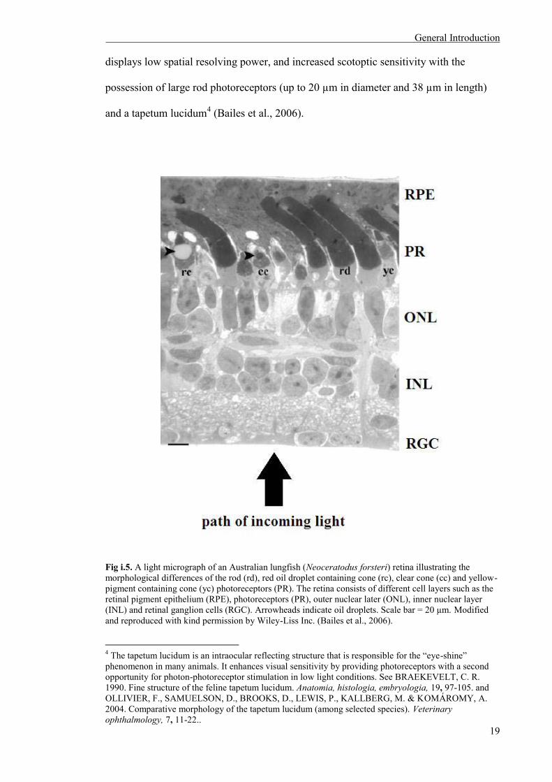
displays low spatial resolving power, and increased scotoptic sensitivity with the
possession of large rod photoreceptors (up to 20 µm in diameter and 38 µm in length)
and a tapetum lucidum4 (Bailes et al., 2006).
Fig i.5. A light micrograph of an Australian lungfish (Neoceratodus forsteri) retina illustrating the morphological differences of the rod (rd), red oil droplet containing cone (rc), clear cone (cc) and yellow-pigment containing cone (yc) photoreceptors (PR). The retina consists of different cell layers such as the retinal pigment epithelium (RPE), photoreceptors (PR), outer nuclear later (ONL), inner nuclear layer (INL) and retinal ganglion cells (RGC). Arrowheads indicate oil droplets. Scale bar = 20 µm. Modified and reproduced with kind permission by Wiley-Liss Inc. (Bailes et al., 2006).
4 The tapetum lucidum is an intraocular reflecting structure that is responsible for the “eye-shine” phenomenon in many animals. It enhances visual sensitivity by providing photoreceptors with a second opportunity for photon-photoreceptor stimulation in low light conditions. See BRAEKEVELT, C. R. 1990. Fine structure of the feline tapetum lucidum. Anatomia, histologia, embryologia, 19, 97-105. and OLLIVIER, F., SAMUELSON, D., BROOKS, D., LEWIS, P., KALLBERG, M. & KOMÁROMY, A. 2004. Comparative morphology of the tapetum lucidum (among selected species). Veterinary ophthalmology, 7, 11-22..

General Introduction
20
i.2.5b Spectral characteristic of N.forsteri retinal photoreceptors
Five visual pigment opsin genes, orthologous to the five visual opsin groups in other
jawed vertebrates have been characterised in N. forsteri. Along with a single type of rod
photoreceptor with a wavelength of maximum absorbance (λmax) of 540nm, the juvenile
N. forsteri visual system contains four individual cone photoreceptor types with visual
pigments with λmax value at 366 nm (ultra-violet sensitive [UVS]), 479 nm (SWS),
558 nm (MWS) and 623 nm (LWS) (Hart et al., 2008). All five photoreceptor types are
observed only in juveniles. The adult fish loses the UVS photoreceptor, perhaps as a
result of increased filtering of short wavelengths by the ocular media (cornea) in adults
that prevents UV light from entering the eye (Hart et al., 2008). This suggests that
juvenile N. forsteri have the potential for tetrachromatic vision. However, the influence
of all characterised photoreceptor types on lungfish behaviour remains to be
investigated.
In addition to the five distinct photoreceptor types, N. forsteri also possess red oil
droplets in the ellipsoid of their LWS cones, one or multiple colourless oil droplets in
the inner segments of their UVS and SWS cones, and yellow ellipsoidal pigmentation in
their MWS cones. The yellow pigmentation and red oil droplets shift peak sensitivities
of the photoreceptor types to 584 nm and 656 nm, respectively (Hart et al., 2008).
As depicted in Fig. i.6, spectral filters such as intracellular oil droplets enhance colour
discrimination (Vorobyev, 2003) but also decrease in the absolute quantum catch of the
visual pigment, reducing overall light sensitivity (Hart et al., 2008). Although their
behavioural function is unknown, the presence of spectral filters within lungfish
photoreceptors suggests that these animals may lead a more diurnal lifestyle than

General Introduction
21
previously thought. In addition, N. forsteri also go through ontogenetic changes in their
diet, from sit-and-wait carnivores in their juvenile years to opportunistic omnivores as
adults (Kemp, 1977). These changes in lifestyle may be involved in the ontogenetic
shifts observed in the lungfish retinal photoreceptors.
Fig. i.6. The object colour solid volumes for juvenile (A, B) and adult (C, D) Neoceratodus forsteri, with (A, C) and without (B, D) the spectral filtering affects of yellow ellipsoidal pigmentation and red oil droplets in medium- (MWS) and short-wavelength sensitive (SWS) cones. The tetrachromatic colour space of juvenile lungfish (A, B) is shown by two projections of the spaces identified by the quantum catches of the ultraviolet-wavelength sensitive (UVS), SWS and MWS cones and also the SWS, MWS and long-wavelengthL sensitive (LWS) cones. Note that there is an increase in the volume of object-colour solids in both juveniles (B) and adults (D) with spectral filters relative to those projections without spectral filters (A and C, respectively). Reproduced with kind permission from Shaun P. Collin. (Hart et al., 2008).

General Introduction
22
The visual machinery of lungfishes, predominantly at the ocular and retinal levels, is
investigated through scientific experimentation in chapter 3 and chapter 4 of this thesis.
Chapter 3 reveals aspects of the morphology, density and topographic distribution of
retinal photoreceptors in L. paradoxa and P. dolloi, and chapter 4 investigates and
compares the visual perception of abnormal N. forsteri lacking external eyes with
normal N. forsteri possessing external eyes. These two chapters reiterate that science is
an important tool and methodology to make sense of lungfishes’ visual perception.
However, as Bruno Latour says, “scientific activity is just one social arena in which
knowledge is constructed” (Latour and Woolgar, 1986) and Donna Haraway reminds us
that, “biology is a discourse, not the living world itself” (Haraway, 1992). Therefore,
chapter 2 of this thesis delves into historical and contemporary stories about lungfishes
to show that many factors, aside from the evaluation of scientific data, influence our
understanding of lungfishes. Just as scientific research is one “social arena” in which
knowledge about lungfish visual perception can be attained, cultural ideologies and
assumptions also influence how humans comprehend the visual experience of
lungfishes. Chapter 5 brings scientific and artistic investigation together by
contextualising a number of artworks that create new encounters and relationships with
non-human animals through the exploration of perception. These artworks poetically
and sensorially extended the complicated understandings and limitations surrounding
the lungfish Umwelt to provide speculation that lies outside the mechanistic realm of
scientific research.

General Introduction
23
i.3 CREATING AN INTERDISCIPLINARY PLATFORM
“To deny the reality or logical significance of what we can never describe or
understand is the crudest form of cognitive dissonance”
(Nagel, 1974)
The sciences and the arts have both made important contributions to how we understand
the visual experience of non-human animals. However, the two fields rarely understand
or communicate with one another on equal terms. The differing research methodologies
adopted by each field create discipline-specific boundaries that, while enabling depth of
disciplinary investigation, may restrict understanding, especially when the subject is
non-human animal perception; a problematic topic because the reality of the beholder is
subjective. Chemist C.P. Snow (1959) was one of the first to describe science and the
arts (“literary intellectuals”) as “two cultures”. He wrote:
constantly I felt I was moving among two groups – comparable in intelligence,
identical in race, not grossly different in social origin, earning about the same incomes,
who had almost ceased to communicate at all, who in intellectual, moral and
psychological climate had so little in common that...one might have crossed the ocean.
(Snow, 1998)
According to Snow, non-scientists described scientists as “shallowly optimistic, unaware
of man’s condition” and lacked “imaginative understanding”. To scientists, practitioners
of the arts were “lacking in foresight”, “anti-intellectual, (and) anxious to restrict both
art and thought to the existential moment” (Snow, 1998). Snow described the sciences
and arts cultures as “ignorant specialists” that, through their beliefs and oftentimes

General Introduction
24
misinterpretations of the opposing culture, became self-impoverished as a result (Snow,
1998). Snow’s main concern was the communication gap formed by the disparity
between the two cultures to the detriment of society and to further knowledge: “Closing
the gap between our cultures is a necessity in the most abstract intellectual sense, as well
as in the most practical” because they are “of great importance for our intellectual and
moral health” (Snow, 1998). Snow recognized a need to address the methodological
limitations of each culture so he proposed the emergence of a “third culture” to close
this unbridgeable gap. In Snow’s “third culture”, the “literary intellectuals” would reach
out to scientists and “some of the difficulties of communication will at last be softened:
for such a culture has, just to do its job, to be on speaking terms with the scientific one”
(Snow, 1998).
Other, more contemporary theorists also recognized a need to address the limitations
within the scientific and non-scientific fields. Literary agent and author John Brockman
used Snow’s “third culture” to describe a space where members of the “science culture”,
such as Richard Dawkins, Brian Goodwin, Marvin Minsky, Roger Penrose and Stephen
Jay Gould, could communicate directly with the general public through books and the
media (Brockman, 1991). In Brockman’s “third culture”, Snow’s “literary intellectuals”
are replaced rather than cooperated with. He writes, “third-culture thinkers tend to avoid
the middleman and endeavour to express their deepest thoughts in a manner accessible
to the intelligent reading public” and are “taking the place of the traditional intellectual
in rendering visible the deeper meanings of our lives, redefining who are and what we
are” (Brockman, 1996).
Theorists and philosophers Donna Haraway, Giles Deleuze and Félix Guattari approach
the limitations of the sciences and the arts by addressing the gap between humans and
non-human animals in order to better understand the world. For them (and others in the
General Introduction
25
budding field of critical animal studies), attempting to “perceive” like the “Other” (non-
human animal) involves tearing down pre-conceived notions about what it is to be
human. They believe that there is a growing need for humans to reconnect with the
environment and become “more worldly” (Haraway, 2008). Deleuze and Guattari
described this process as “becoming animal”; the human being’s opportunity to think
themselves “other-than-in-identity” and provide “the way out, the line of escape” (cited
in Baker, 2000). Haraway described this discourse as a way to understand that the
human species is part of – and interdependent on – the environment as all animals are,
and refute the anthropocentric assumption that we have dominion over it (Haraway,
1991, Haraway, 2008).
The importance of this process of “becoming animal” lies in the opportunity to think
differently about humans and animals. It is to leave the purely human and directly
engage with the “Other” (be it animal or its environment, for the purpose of this thesis)
to gain new understanding (Deleuze and Guattari, 1987, Baker, 2000). Deleuze and
Guttari write,
the relationships between animals are object not only for science but also of dreams,
symbolism, art and poetry, practice and practical use...[and] are bound up with the
relations between man and animal, man and woman, man and child, man and the
elements, man and the physical and microphysical universe (Deleuze and Guattari,
1987)
This engagement can be done in many ways that should not be exclusive to any single
discipline, and when investigating subjective experience, the objectivity of science is “a
direction in which understanding can travel” (Nagel, 1974). Snow also spoke of this
potential, stating that, “the clashing point of two subjects, two disciplines, two cultures –
of two galaxies, so far as it does – ought to produce creative chances” (Snow, 1998).
General Introduction
26
The underlying philosophy that grounds Haraway, Deleuze and Guattari is
posthumanism. Posthumanism began as cynicism at how “aimless and arbitrary” human
intellect appears in the natural world. This is more evident as we become more aware
that the humanist perspective that privileges humanity above all other forms of existence
will not prevent our extinction (Heidegger, 1993, Wolfe, 2010). This causes us to realise
that there is an urgent need for a new kind of thought that is less anthropocentric, and
positions humans as part of, connected to, and interdependent on the environment. If
humanism is the belief that humans are the pinnacle of existence, then posthumanism is
one that rethinks the position of humanity, and recontextualises it in terms of other
species (Wolfe, 2010). Posthumanist scholar Cary Wolfe (2010) observed that
posthumanism is “acknowledging that we are fundamentally prosthetic creatures that
have co-evolved with various forms of technicity and materiality, forms that are
radically ‘non-human’ and yet have nevertheless made the human what it is.” The
posthuman aspires to understand the world from multiple, heterogeneous perspectives
and manifest him/herself in consideration with the different identities.
This thesis creates an interdisciplinary platform that is different from the “third culture”
Snow predicted and Brockman has created. Here, rather than Snow’s “literary
intellectuals” repairing communication, I connect science with art, and the humanities.
Like members of Brockman’s “third culture”, a part of this research does involve
scientific experimentation and directly communicating it to a general audience, but it
does not do this by casting the arts aside. Instead, the arts are an integral part of the
methodology. Deleuze and Guttari’s “becoming animal” is purely philosophical, and
although they suggest a way to leave the human by inventing an “animal personality”,
they do not engage with scientific concepts that might be useful to accomplish it.
Haraway, who has a PhD in primatology, engages with both the sciences and the arts to
further human knowledge about our connection to non-human animals. Unlike Haraway

General Introduction
27
however, this research also involved art-making as a tool to access the visual experience
of lungfishes, and embody the process of communication beyond the verbal and
cognitive to other senses (sight, sound touch etc.).
Instead of creating a new definition for a “third culture” or “space” that connects science and the arts, and humans and non-human animals, this research project is posthumanist in its attempt to understand the visual perception of lungfishes from multiple heterogeneous perspectives (scientific, philosophical/cultural, and art-making). The success of this posthumanist endeavour is considered in the general discussion chapter which integrates all the thesis chapters and considers the strengths and weaknesses of combining the “two cultures” in this way. In addition, each chapter conforms to its discipline-specific language and style conventions to respect and maintain discipline conventions (e.g. chapter 3 and chapter 4 follow scientific convention with methodology, results and discussion, while chapters 1, 2 and 5 are purposefully narrative driven).
General Introduction
28
i.4 REFERENCES
ALI, M. A. & ANCTIL, M. 1973. Retina of the South American lungfish, Lepidosiren paradoxa Fitzinger. Canadian Journal of Zoology, 51, 969-972.
ALLEN, G. R., MIDGLEY, S. H. & ALLEN, M. 2002. Field guide to the freshwater fishes of Australia,
Western Australian Museum Perth. AMEMIYA, C. T., ALFÖLDI, J., LEE, A. P., FAN, S., PHILIPPE, H., MACCALLUM, I., BRAASCH,
I., MANOUSAKI, T., SCHNEIDER, I. & ROHNER, N. 2013. The African coelacanth genome provides insights into tetrapod evolution. Nature, 496, 311-316.
BAILES, H. J., DAVIES, W. L., TREZISE, A. E. O. & COLLIN, S. P. 2007. Visual pigments in a living
fossil, the Australian lungfish Neoceratodus forsteri. BMC Evolutionary Biology, 7, 200. BAILES, H. J., ROBINSON, S., TREZISE, A. E. O. & COLLIN, S. P. 2006. Morphology,
characterization, and distribution of retinal photoreceptors in the Australian lungfish Neoceratodus forsteri (Krefft, 1870). The Journal of Comparative Neurology, 494, 381-397.
BAILEY, R. 1994. Guide to the fishes of the River Nile in the Republic of the Sudan. Journal of Natural
History, 28, 937-970. BAKER, S. 2000. The Postmodern Animal, London, Reaktion Book Ltd. BANCROFT, T. On the life history of Ceratodus. Proceedings of the Linnean Society of New South
Wales, 53, 315-317. BERRA, T. M. 2001. Freshwater fish distribution, Academic Pr. BOWMAKER, J. & DARTNALL, H. 1980. Visual pigments of rods and cones in a human retina. The
Journal of Physiology, 298, 501-511. BOWMAKER, J. & MARTIN, G. 1978. Visual pigments and colour vision in a nocturnal bird, Strix
aluco (tawny owl). Vision Research, 18, 1125-1130. BRAEKEVELT, C. R. 1990. Fine structure of the feline tapetum lucidum. Anatomia, histologia,
embryologia, 19, 97-105. BRINKMANN, H., VENKATESH, B., BRENNER, S. & MEYER, A. 2004. Nuclear protein-coding
genes support lungfish and not the coelacanth as the closest living relatives of land vertebrates. Proceedings of the National Academy of Sciences of the United States of America, 101, 4900.
BROCKMAN, J. 1991. The Third Culture. Available from: https://edge.org/conversation/the-third-culture
[Accessed 18 May 2015]. BROCKMAN, J. 1996. Introduction to The Third Culture: Beyond the Scientific Revolution. Available
from: https://edge.org/conversation/the-emerging [Accessed 29 May 2015]. CALVINO, I. 1968. Cosmicomics, trans. William Weaver (Abacus: London, 1982). CARTER, G. & BEADLE, L. 1930. Notes on the habits and development of Lepidosiren paradoxa.
Journal of the Linnean Society of London, Zoology, 37, 197-203. CHARMAN, W. 1991. The vertebrate dioptric apparatus. In: JR, C.-D. & GREGORY, R. (eds.) Vision
and Visual Dysfunction. London: Macmillan Press. CLACK, J. A., SHARP, E. L. & LONG, J. A. 2011. The Fossil Record of Lungfishes. In: JORGENSEN,
J. M. & JOSS, J. (eds.) The Biology Of Lungfishes. Enfield: Science Publishers. COLLIN, S. P. & COLLIN, H. 2001. The fish cornea: adaptations for different aquatic environments.
Sensory Biology of Jawed Fishes. New Insights, 57-96.

General Introduction
29
COLLIN, S. P., HART, N. S., SHAND, J. & POTTER, I. C. 2003. Morphology and spectral absorption
characteristics of retinal photoreceptors in the southern hemisphere lamprey (Geotria australis). Visual Neuroscience, 20, 119-130.
COLLIN, S. P., HART, N. S., WALLACE, K. M., SHAND, J. & POTTER, I. C. 2004. Vision in the
southern hemisphere lamprey Mordacia mordax: Spatial distribution, spectral absorption characteristics, and optical sensitivity of a single class of retinal photoreceptor. Visual Neuroscience, 21, 765-773.
CURRY-LINDAHL, K. 1956. On the ecology, feeding behaviour and territoriality of the African
lungfish, Protopterus aethiopicus Heckel. Arkiv for Zoologi, 9, 479-497. DELEUZE, G. & GUATTARI, F. 1987. Becoming-Intense, Becoming-Animal, Becoming-Imperceptible.
A Thousand Plateaus : capitalism and schizophernia. Minneapolis: University of Minnesota Press.
FISHMAN, A., PACK, A., DELANEY, R. & GALANTE, R. 1987. Estivation in Protopterus. In:
BEMIS, W. E., BURGGREN, W. W. & KEMP, N. E. (eds.) The Biology and Evolution of Lungfishes. New York: Alan R Liss Inc.
FONESCA DE ALMEIDA-VAL, V., NOZAWA, S., LOPES, N., ARIDE, P., MESQUITA-SAAD, L.,
MAZARE PAULA DA SILVA, M., HONDA, R., FERREIRA-NOZAWA, M. & VAL, A. 2011. Biology of the South American lungfish, Lepidosiren paradoxa. In: JORGENSEN, J. & JOSS, J. (eds.) The Biology of Lungfishes. Enfield: Science Publishers.
GREENWOOD, P. 1987. The natural history of African lungfishes. In: BEMIS, W. E., BURGGREN, W.
W. & KEMP, N. E. (eds.) The Biology and Evolution of Lungfishes. New York: Alan R. Liss Inc.
GREENWOOD, P. H. 1986. The natural history of African lungfishes. Journal of Morphology, 190, 163-
179. GÜNTHER, A. 1871. Description of Ceratodus, a genus of ganoid fishes, recently discovered in rivers of
Queensland, Australia. Philosophical Transactions of the Royal Society of London, 161, 511-571.
HARAWAY, D. J. 1991. Simians, Cyborgs, and Women : The Reinvention of Nature, New York,
Routledge. HARAWAY, D. J. 1992. The Promise of Monsters: A regenerative politics for inappropriate/d Others. In:
GROSSBERG, L., NELSON, C. & TREICHLER, P. A. (eds.) Cultural Studies. New York: Routledge.
HARAWAY, D. J. 2008. When species meet, University Of Minnesota Press. HART, N. S., BAILES, H. J., VOROBYEV, M., MARSHALL, N. J. & COLLIN, S. P. 2008. Visual
ecology of the Australian lungfish (Neoceratodus forsteri). BMC ecology, 8, 21. HEIDEGGER, M. 1993. Letter on Humanism. In: KRELL, D. (ed.) Basic Writings from Being and Time
(1927) to The Task of Thinking (1964). New York: HarperCollins Publishers. JANE, S. & BOWMAKER, J. 1988. Tetrachromatic colour vision in the duck (Anas platyrhynchos L.):
microspectrophotometry of visual pigments and oil droplets. Journal of Comparative Physiology A, 162, 225-235.
JOHNELS, A. & SVENSSON, G. 1954. On the biology of Protopterus annectens (Owen). Arkiv för
Zoologi, 7, 131-164. KELBER, A., VOROBYEV, M. & OSORIO, D. 2003. Animal colour vision—behavioural tests and
physiological concepts. Biological Reviews, 78, 81-118.
General Introduction
30
KEMP, A. 1986. The biology of the Australian lungfish, Neoceratodus forsteri (Krefft 1870). Journal of Morphology, 190, 181-198.
KEMP, A. 1995. Threatened fishes of the world: Neoceratodus forsteri (Kreft, 1870)
(Neoceratodontidae). Environmental Biology of Fishes, 43, 310-310. KERR, J. 1902. The development of Lepidosiren paradoxa. III. Development of the skin and its
derivatives. Quarterly Journal of Microscopial Sciences, 46, 417 - 459. KIND, P. 2011. The natural history of the Australian lungfish Neoceratodus forsteri (Krefft, 1870). In:
JORGENSEN, J. & JOSS, J. (eds.) The Biology of Lungfishes. Enfield: Science Publishers. KOLB, H. & JONES, J. 1987. The distinction by light and electron microscopy of two types of cone
containing colorless oil droplets in the retina of the turtle. Vision Research, 27, 1445-1458. LATOUR, B. & WOOLGAR, S. 1986. Laboratory Life: The Construction of Scientific Facts., Princeton,
Princeton University Press. LOCKET, N. 1970. Mitosis in mature lungfish retina. Zoological Journal of the Linnean Society, 49, 1-4. LONG, J.A. 2nd November 2015, Type to APPUDURAI, A. M. LUBBOCK, S. J. 1889. On the Senses, Instincts, and Intelligence of Animals: with Special Reference to
Insects, K. Paul, Trench & co. MARSHALL, J., COLLIN, S. P., HART, N. & BAILES, H. 2011. Vision in lungfish. In: JORGENSEN,
J. & JOSS, J. (eds.) The Biology of Lungfishes. New Hampshire: Science Publishers. MLEWA, C., GREEN, J. & DUNBRACK, R. 2011. The general natural history of African lungfishes. In:
JORGENSEN, J. & JOSS, J. (eds.) The Biology of Lungfishes. Enfield: Science Publishers. NAGEL, T. 1974. What Is It Like To Be A Bat? The Philosophical Review, 83, 435-450. O'CONNOR, D. 1897. Report on preservation of Ceratodus. Proceedings of the Royal Society of
Queensland, 12, 102 - 102. OLLIVIER, F., SAMUELSON, D., BROOKS, D., LEWIS, P., KALLBERG, M. & KOMÁROMY, A.
2004. Comparative morphology of the tapetum lucidum (among selected species). Veterinary ophthalmology, 7, 11-22.
PFEIFFER, W. 1968. Retina und Retinomotorik der Dipnoi und Brachiopterygii. Cell and Tissue
Research, 89, 62-72. RUSHTON, W. 1972. Review lecture. Pigments and signals in colour vision. The Journal of Physiology,
220, 1-31. SMITH, H. W. 1931. Observations on the African lungfish, Protopterus aethiopicus, and on evolution
from water to land environments. Ecology, 12, 164-181. SNOW, C. P. 1998. The Two Cultures, New York, Cambridge University Press. UEXKÜLL, J. V. 2010. A Foray Into The Worlds Of Animals And Humans With A Theory Of Meaning,
Minneapolis, University of Minnesota Press. VAN-EYK, S. M., SIEBECK, U. E., CHAMP, C. M., MARSHALL, J. & HART, N. S. 2011.
Behavioural evidence for colour vision in an elasmobranch. The Journal of Experimental Biology, 214, 4186-4192.
VON FRISCH, K. 1914. Der farbensinn und formensinn der biene. Zoologische Jahrbücher. Abieilung
für Allgemeine Zoologie und Physiologie., 35, 1-188.

General Introduction
31
VOROBYEV, M. 2003. Coloured oil droplets enhance colour discrimination. Proceedings of the Royal Society of London Series B-Biological Sciences, 270, 1255-1261.
WALD, G. 1967. Visual pigments of crayfish. Nature, 215, 1131-1133. WALLS, G. L. 1942. The Vertebrate Eye and its Adaptive Radiation, Bloomfield Hills, The Cranbook
Press. WASAWO, D. 1959. A dry season burrow of Protopterus aethiopicus Heckel. Revue de Zoologie et de
Botanique Africaines, 60, 65-71. WOLFE, C. 2010. What is Posthumanism?, Minneapolis, University of Minnesota Press.

Visual Umwelten Of Lungfishes
32
CHAPTER 1
::::::
THE VISUAL UMWELTEN
OF LUNGFISHES
THROUGH ART AND
SCIENCE
::::::

Visual Umwelten Of Lungfishes
33
1.1 INTRODUCTION
Aristotle famously inferred that the whole is greater than the sum of its parts5. He may
have been contextualising this in terms of metaphysics, but the same applies to
scientific research and the study of non-human animal behaviour. The process of
unravelling the visual perceptual experience of non-human animals using anatomical,
physiological and behavioural approaches is an established yet burgeoning field of
scientific research. Scientists have relied on mostly anthropocentric, technologically
unaided observation and logical extrapolations when investigating the visual reality of
the non-human animal. However, technological advances over the last 100 years have
allowed scientists to further uncover the visual reality of non-human animals.
Techniques such as electron microscopy and electrophysiology unravel the anatomical
and physiological mechanisms of vision, and behavioural experiments enable us to
observe how non-human animals react to visual stimuli within their environment.
Although it is possible to obtain a mechanistic understanding of non-human perception
by the exploration of visually guided behaviours, and neuronal circuitry within the eye
and brain, there are still large gaps in our understanding of how to conceptualise a
reality that is fundamentally different from our own, if only because our understanding
of non-human perception is constructed and understood by our embedded human
perception and its inherent biases and limitations.
This chapter explores some of the scientific and non-scientific history of the visual
perception of non-human animals from the nineteenth century to present day by
drawing on some of the similarities and differences researchers shared in their attempts
to uncover non-human animal perceptual capabilities. By recognizing the limitations of
technology and our anthropocentric perception of reality, this chapter will attempt to
5 See Aristotle, Metaphysics Book VIII 1045a1-2ff
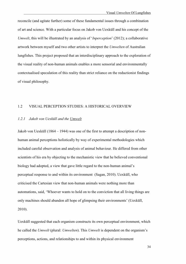
Visual Umwelten Of Lungfishes
34
reconcile (and agitate further) some of these fundamental issues through a combination
of art and science. With a particular focus on Jakob von Uexküll and his concept of the
Umwelt, this will be illustrated by an analysis of ‘Inperception’ (2012); a collaborative
artwork between myself and two other artists to interpret the Umwelten of Australian
lungfishes. This project proposed that an interdisciplinary approach to the exploration of
the visual reality of non-human animals enables a more sensorial and environmentally
contextualised speculation of this reality than strict reliance on the reductionist findings
of visual philosophy.
1.2 VISUAL PERCEPTION STUDIES: A HISTORICAL OVERVIEW
1.2.1 Jakob von Uexküll and the Umwelt
Jakob von Uexküll (1864 – 1944) was one of the first to attempt a description of non-
human animal perceptions holistically by way of experimental methodologies which
included careful observation and analysis of animal behaviour. He differed from other
scientists of his era by objecting to the mechanistic view that he believed conventional
biology had adopted, a view that gave little regard to the non-human animal’s
perceptual response to and within its environment (Sagan, 2010). Uexküll, who
criticised the Cartesian view that non-human animals were nothing more than
automations, said, ‘Whoever wants to hold on to the conviction that all living things are
only machines should abandon all hope of glimpsing their environments’ (Uexküll,
2010).
Uexküll suggested that each organism constructs its own perceptual environment, which
he called the Umwelt (plural: Umwelten). This Umwelt is dependent on the organism’s
perceptions, actions, and relationships to and within its physical environment

Visual Umwelten Of Lungfishes
35
(Buchanan, 2008). Umwelten are depicted by Uexküll as a series of ‘soap bubbles’
encircling each organism, where context and reality are determined by the individual’s
behaviour and perception, and to which no other organism has full ‘access’ or complete
comprehension because it is completely subjective (Uexküll, 2010). Uexküll firmly
believed that all animals were capable of identifying meaning from cues that originated
from the environment beyond an instinctual or innate level. As Buchanan (2008)
described, ‘Instead of interpreting organisms based on natural selection...Uexküll sought
to understand them with respect to the designs that they represented in relation to
meaningful signs’. This has always been considered a radical approach in the attempt to
understand and relate to non-human perception.
Uexküll’s theory of the Umwelt as an organism-environment unit opened up radical new
ways of interpreting and conceptualising non-human animal perception. However, its
conception drew a large amount of criticism from the scientific community, primarily
because his theories were seen as unfalsifiable and too subjective to be useful in
science. Despite this justified criticism, Uexküll was one of the first researchers to
recognise the importance of studying non-human animal perception (including visual
perception) and its relationship to the animal’s environment, and developed experiments
to test non-human animal cognition and capabilities. Uexküll’s contribution to the
understanding of beings as active agents, although lacking scientific integrity due to its
subjective nature, is still a useful conceptual tool in the approach and interpretation of
visual perception in non-human animals.
Visual Umwelten Of Lungfishes
36
1.2.2 Visual perceptions and behaviour: Lubbock, von Frisch, and Morrison-Scott
Early scientific experiments that tested the colour vision capabilities of non-human
animals were conducted by observation of the animals’ behaviour. John Lubbock and
Karl von Frisch were pioneers in developing behavioural criteria to establish whether
non-human animals possessed the ability to differentiate colours (for full review on
behavioural tests on animal colour vision, see Kelber et al., 2003). Lubbock observed
that various species of Daphnia (planktonic crustaceans) prefer yellow to white or
unfiltered light (Lubbock, 1889). He also claimed that honeybees (Apis mellifera)
associate colour with food (Lubbock, 1889), and this was later confirmed by Frisch.
Frisch established that honeybees discriminate blue and yellow from 30 different shades
of grey (Von Frisch, 1914). This experiment, coined the ‘grey card experiment’, is still
used today to determine colour discrimination in non-human animals. An animal is first
taught to associate a reward with a colour. This colour is then placed within a sample of
many shades of grey. At least one of these grey shades has a similar brightness to the
test colour, which renders brightness as an unreliable cue. In order to gain the food
reward, the animal must identify the colour by hue alone. In an experiment of similar
design, Morrison-Scott’s 1954 study on the satin bowerbird (Ptilonorhynchus
violaceus), observed colour preferences of male birds by the choice of coloured fabrics
with which they preferred to adorn their bowers (Morrison Scott, 1937). Whether the
bowerbird or honeybee colour preference is a product of instinct and learned behaviour,
or determined by the relationship between animal and its Umwelt is still unknown.
Behavioural experiments following the same design principle of von Frisch’s grey card
experiments have confirmed the ability to discriminate colour in a number of species
including various species of butterflies (belonging to the families Nymphalidae,
Pieridae, Satyridae and Papilionidae), hermit crabs (Eupagurus anachoretus), shrimp

Visual Umwelten Of Lungfishes
37
(Crangon vulgaris) domestic chickens (Gallus gallus) (Kelber et al., 2003), and the
giant shovelnose ray (Glaucostegus typus) (Van-Eyk et al., 2011).
Lubbock, Frisch, and Morrison-Scott’s findings were solely based on human
observation, and as Uexküll argued, did not take into account the full subjectivity of the
non-human animal and its environment because both were continuously being defined
through human perception and its inescapable bias. This anthropocentrism is based on
an assumed correlation between stimulus and behaviour, and between the meanings of
expressed behaviour. This meaning is usually based on human experience, which has
led to assumptions about animal behaviour that may not be consistent with what the
animal is actually or fully experiencing. For instance, the human expression of a smile
to be one of pleasure, happiness and amusement can be a gross misinterpretation in
other animals such as canids and primates. In wolves, the horizontal retraction of lips
across the teeth can be a submissive gesture to dominant members of the group, and the
vertical lip retraction given by dominant animals is a sign of aggression (Fox, 1970).
Similarly, bared teeth displays of chimpanzees illustrate submission and/or fear in
response to aggression or uncertainty regarding the intent of conspecifics (Waller and
Dunbar, 2005).
Are we able to ascertain and explain the experience of a non-human animal, and its
visual capabilities, if we cannot see or experience them? New scientific technologies
may be able to overcome some of the limitations of behavioural studies and describe
non-human animal visual perceptions more accurately by pointing to the anatomical and
physiological attributes that enable an organism to perceive.

Visual Umwelten Of Lungfishes
38
1.3 VISUAL PERCEPTION: CONTEMPORARY
In addition to behavioural studies, techniques such as electron microscopy,
microspectrophotometry, and electrophysiology have been used since the middle of the
20th century to study the vision of human and non-human animals. These techniques
have proven to be particularly useful in understanding the mechanistic basis of vision
and interspecific differences in visual ability/performance. Although other techniques,
such as molecular genetics, are increasingly being used to explore the sensory worlds of
other non-human animals, this review will focus on these three methods in particular.
See the general introduction for a review of the vertebrate eye and its components.
1.3.1 Anatomical, physiological, and behavioural methodologies
Microscopy enables the identification and characterisation of the different kinds of
retinal photoreceptor cells based on morphological characteristics such as size, shape
and ultrastructure. Light microscopy is typically used to characterise cells to a
resolution of microns (10-6 m), while electron microscopy enables cells to be examined
at an even higher resolution of nanometers (10-9 m) (Williams and Carter, 1996).
Transmission electron microscopy (TEM), which passes an electron beam through the
retinal tissue, allows for ultrastructural differentiation in cell morphology, i.e. the size of
mitochondria (organelles within cells that are primarily responsible in generating
cellular energy) within the outer segment, and the number and type of connections
(synapses) between photoreceptors and bipolar cells.
Physiological techniques such as microspectrophotometry (MSP) and
electroretinography (ERG) are able to characterise photoreceptor cells by function. MSP
allows the precise measurement of spectral absorption characteristics in the visual

Visual Umwelten Of Lungfishes
39
pigments and intracellular spectral filters found in individual photoreceptors (Liebman
and Entine, 1964, Liebman, 1972, Brown, 1968). This information not only defines the
range of wavelengths to which an animal is sensitive but also reveals whether multiple
spectral types of photoreceptor are present in the retina, and thus, the potential for
colour vision.
ERG is used to quantify the response of an animal’s eyes to visual stimuli (Brown,
1968). Not limited to ophthalmologists who routinely utilize this technique on humans
to diagnose various retinal diseases, ERG can be applied to other animals by measuring
the massed electrical response of the eye to light stimuli in order to determine its
absolute retinal sensitivity, spectral sensitivity and temporal responses properties
(Blough et al., 1972, Goldsmith, 1986). ERG can also determine how sensitive the eye
is to temporal changes in illumination; potentially revealing how fast an animal can
react to predators or prey under different conditions of illumination (dark or light)
(Cavonius, 1964). This is often determined by identifying the critical flicker-fusion
frequency (cFFF); the frequency at which a train of light flashes can no longer be seen
as individual flashes, but appears as a continuous light stimulus (Hecht et al., 1933,
Jenssen and Swenson, 1974). The photopic cFFF of the human eye in bright light is 50
to 70 Hz (Bowles and Kraft, 2012). In contrast, some birds have a much higher photopic
cFFF, which enables them to resolve flicker greater than 100 Hz (Nuboer et al., 1992).
This difference may be because birds often move quickly when flying and need superior
motion vision relative to humans. Superior motion vision is achieved by improving the
temporal response properties (reflected by a high cFFF) of the retina, otherwise moving
objects would appear to ‘blur’ as they move across the visual field. At low light
intensities, cFFF is reduced, and so is the ability to see motion. This is why cricket and
baseball matches, for example, are abandoned in poor light because the cFFF of the

Visual Umwelten Of Lungfishes
40
scoptopic rod system is much lower than the photopic cone system (Bowles and Kraft,
2012).
1.4 CASE STUDY: THE LUNGFISH UMWELT
As previously discussed, lungfishes are large, freshwater dipnoan6 fishes that have the
ability to breathe dissolved and atmospheric air through gills and “primitive” lungs.
Both internal and external nostrils are found in lungfishes; an uncommon feature of
fishes, which usually possess two pairs of external nostrils. Lungfishes are classified as
Sarcopterygii, along with coelacanths and tetrapods (Clack et al., 2011), and through a
whole genome analysis of the African coelacanth, it was recently confirmed that they
are the closest living ancestors of all terrestrial vertebrates (Amemiya et al., 2013). By
investigating the only living source to the origin of vision on land, many lines can be
drawn to connect animal Umwelten, may they be human or not.
Dipnoan fish are important in the debate surrounding the evolution of vision in
terrestrial vertebrates because their visual adaptations and evolutionary history places
them in the divide between life in the water and on land. In general, the visual
machinery of lungfishes resembles that of terrestrial vertebrates, which suggests that
several of the ocular characteristics these animals possess may have evolved in shallow
water before the transition onto land (Collin, 2010). One adaptation is the corneal
surface microprojections which are important for vision on land and are present in both
terrestrial vertebrates and in the Australian lungfish (Neoceratodus forsteri). These
appear as a thick carpet of microvilli extending over the dermal cornea, which stabilise
6 ‘Dipnoan’ originates from the Greek dipnoos, meaning ‘double-breathing’.
Visual Umwelten Of Lungfishes
41
the corneal tear film in terrestrial vertebrates, necessary for clear aerial vision (Collin
and Collin, 2001).
Six species of lungfishes are extant worldwide; four in Africa (Protopterus spp.), one in
South America (Lepidosiren paradoxa) and one in Australia (N. forsteri). Previous
anatomical studies have shown that lungfishes possess a number of different types of
large photoreceptors in their eyes, demonstrating that they have the retinal machinery to
process colour (Walls, 1942, Pfeiffer, 1968, Ali and Anctil, 1973). Although the full
complement of photoreceptors has yet to be established in the South American and
African lungfishes, the retina of the Australian lungfish – which is considered to be the
most primitive of the extant lungfishes – has been studied in detail (Marshall, 1986,
Tokita et al., 2005, Bailes et al., 2006a, Bailes et al., 2006b, Bailes et al., 2007b, Bailes
et al., 2007a).
As previously discussed, there are up to five spectrally distinct types of retinal
photoreceptors present in the Australian lungfish: one type of rod (λmax 540nm) and four
types of cones (UVS λmax 366nm, SWS λmax 479nm, MWS λmax 558nm and LWS λmax
623nm). UVS cones are only found in the retinae of juvenile N. forsteri, suggesting that
sensitivity to ultraviolet light is lost during maturation (Bailes et al., 2006a, Bailes et al.,
2007a, Hart et al., 2008). The possession of four types of cones by the juvenile
lungfishes implies that they have the potential for tetrachromatic vision; unusual for an
animal that has previously been considered predominantly nocturnal or crepuscular7
(Dean, 1906, Grigg, 1965, Kemp, 1986).
L. paradoxa and Protopterus spp. possess coloured and colourless oil droplets in their
cone and rod photoreceptors, respectively (Kerr, 1902) (Walls, 1942, Pfeiffer, 1968, Ali
and Anctil, 1973), whereas N. forsteri only possess colourless oil droplets within some
7 Active predominantly during twilight (dawn and dusk).
Visual Umwelten Of Lungfishes
42
types of cone photoreceptors and coloured oil droplets and/or coloured pigments in
other cone types (Bailes et al., 2006a). The presence of coloured oil droplets (and the
potential for tetrachromatic colour vision) in the lungfish retina is surprising because
these characteristics are typically found only in strongly diurnal animals such as birds
and reptiles. N. forsteri is thought to have a predominantly nocturnal lifestyle, often
living in turbid, and therefore relatively dim, underwater light environments. Colourless
oil droplets are found in marsupial mammals which are generally nocturnal, crepuscular
or cathemeral8 and are thought to improve light gathering ability and thus, overall
sensitivity. Coloured oil droplets act as miniature spectral filters within each
photoreceptor and, by narrowing the spectral sensitivity function of the photoreceptors,
are thought to improve colour discrimination by reducing overlap with adjacent spectral
classes of cone (Vorobyev et al., 1998). This potentially enables the animal to perceive
a far greater number of distinct colour “hues” in the visual spectrum than they would
without the coloured oil droplets (Govardovskii, 1983, Hart et al., 2006).
The lungfish eye is something of a conundrum. The large size of the photoreceptors
suggests that lungfish eyes are adapted for maximising the amount of light detected by
the retina. N. forsteri also possesses a tapetum lucidum, a reflective layer behind the
retina that enables light to pass through the photoreceptors a second time, thereby
increasing photon capture (i.e. sensitivity). This structure is typically seen in
crepuscular, nocturnal species or animals that live in dim habitats and is consistent with
the lungfish environment. However, the complex visual apparatus of the lungfish eye, in
particular the presence of coloured oil droplets and the impressive complement of cone
photoreceptors for colour vision, suggests that colour vision is more important to
8Active sporadically at random intervals during the day or night dependant on food availability, predation pressures and other environmental cues such as temperature.
Visual Umwelten Of Lungfishes
43
lungfishes than previously thought,9 or indicated by their relatively low light
environment.
1.5 INPERCEPTION: ARTISTIC INQUIRY INTO THE AUSTRALIAN
LUNGFISH UMWELT
The difficulty for humans to perceive the visual Umwelten of aquatic animals is inherent
in the subjectivity of visual perception. Coupled with the need to perceive an
environment through water instead of air, with visual machinery that is optimised for a
particular habitat, the quest of seeing through the eyes of a lungfish becomes even more
problematic. Philosopher Thomas Nagel articulated that the “conscious experience” of a
non-human animal (in his example, the bat) exists “only if there is something it is like to
be that organism – something it is like for that organism,” but due to the “subjective
character of experience” the “something it is like” can never be completely revealed
(Nagel, 1974). He recognized that the restriction resides in our own minds, which is
inadequate to understand another perceptual world because of its subjectivity. However,
he called for attempts to bridge this subjective character of experience, claiming that “to
deny the reality or logical significance of what we can never describe or understand is
the crudest form of cognitive dissonance” (Nagel, 1974). Therefore, there is always
merit in the gesture of trying to pierce Umwelten despite the apparent impossibility of
doing so.
Nagel speculated that the only way to move forward is to take imagination out of
interpretation and form a more objective approach that will not give the entire picture,
9Early lungfish studies suggested that these fish were almost blind, and possessed degenerate eyes DEAN, B. Notes on the living specimens of the Australian lungfish, Ceratodus forsteri, in the Zoological Society's collection. Proceedings of the Zoological Society of London, 1906. Wiley Online Library, 168-178..

Visual Umwelten Of Lungfishes
44
but may provide a description of ‘the subjective character of experiences in a form
comprehensible to beings incapable of having those experiences’ (Nagel, 1974) .
‘Inperception’ (2012), a collaborative artistic interpretation and translation of scientific
data onto a large mixed media mural, extended Nagel’s argument to aquatic organisms,
and considers the additional complication of vision in water rather than air.
Previously, contemporary artists Olly and Suzi explored the interaction of the human
and aquatic non-human Umwelten in the installation, ‘Shark Bite’ (1997). Along with
photographer Greg Williams, the duo tracked and painted Great White sharks off the
coast of South Africa. Once the human artists had painted the animal’s portrait, the
animal was encouraged to provide its “voice”, or point of view, by interacting
physically with its portrait. One shark responded dramatically by biting its portrait; a
performative signature. For Olly and Suzi, this signature was a powerful message that
documented “the passing of animals and habitats that are here now but may not be for
much longer” (Olly and Suzi, 2002). This can also illustrate the dissonance between
crossing the Umwelten of humans and aquatic animals, illustrated by the antagonistic
reaction/interaction between human outreach and non-human response. Although
‘Shark Bite’ (1997) documented a human/aquatic non-human interaction, it did not
bridge the gap between Umwelten, demonstrating the futility of the pursuit in a
spectacular gesture.
In an attempt to understand the perceptual Umwelten of Australian lungfishes, three
artists engaged in the ‘Inperception’ project (2012), a collaborative interpretation and
translation of scientific data onto a large mixed media mural. Unlike ‘Shark Bite’
(1997), ‘Inperception’ (2012) endeavoured to tease out the nuances of the futility of
crossing Umwelten by focusing on human attempts to understand the Umwelten of
lungfishes, banal aquatic organisms. Audrey Appudurai, and two other contemporary

Visual Umwelten Of Lungfishes
45
artists, Shannon Williamson, and Devon Ward, responded to scientific data generated as
part of the broader research into the visual perception of the Australian lungfish and
Uexküll’s theory of Umwelten as “soap bubbles”. The artists were provided with
scientific research papers which included data in the form of words, anatomical
photographs, diagrams and figures about N. forsteri10. Williamson is a painter who
usually works with watercolour, charcoal and paper, Ward is a graphic designer and
photographer, and Appudurai is an artist who primarily uses pencils, pens, and paper.
Appudurai provided the participants with a large canvas, acrylic, watercolour paints,
charcoal, oil pastels and graphite as materials to use. The artists then translated the
scientific data onto a large canvas that was suspended around the water pools housing
Australia lungfishes (Fig. 1.1 and 1.2). These lungfishes were being used for
behavioural and physiological experiments. Each artist worked separately on sections of
the canvas. Williamson also completed a written reflection on her attempt to interpret
the Umwelt of the non-human other.
This artistic inquiry humbly aspired towards an informed subjectivity. It was an attempt
to create a shared experience which allowed for a scientifically informed glimpse into a
non-human vision while also acknowledging the limitations of human perceptions and
knowledge systems. ‘Inperception’ (2012) aimed to bridge the disparate disciplines of
biological science and visual art, analysing data generated through anatomical and
behavioural studies to provide alternate interpretations of the visual perception of
lungfishes.
10 For resources given to artists see Bailes et al. (2006) and Hart et al. (2008).

Visual Umwelten Of Lungfishes
46
Fig. 1.1 ‘Inperception’ (2012) (Audrey Appudurai et al., 2013).
Fig. 1.2. The ‘Inperception’ (2012) mural hanging near the lungfishes’ housing tanks during the behavioural experiment (Audrey Appudurai et al., 2013).
Through informed subjectivity, ‘Inperception’ (2012) was interested in the following
research question: What might be produced when three humans are provoked to
Visual Umwelten Of Lungfishes
47
consider how an Australian lungfish might see its world? It was an exploration of the
overlap between various human Umwelten and the visual Umwelten of Australian
lungfishes. Ultimately, the artistic responses showed the difficulty in navigating and
piercing individual human Umwelten, which made the pursuit of doing the same to an
aquatic animal even more problematic. Although there are numerous reflections in the
mural, we will be focusing on three different pieces from each contributor in this
analysis.
Devon Ward’s contribution (on the far left of the mural) is shown in Fig. 1.3. He used
predominantly green, blue and yellow circular dots of varying size, with some red
punctuating the piece. Small, densely placed circles to the left of the canvas became
bigger and more separated as the eye moves to the right (Fig. 3). The abstract dots are
evocative of the visual ‘machinery’ of the lungfish’s eye, similar to the retinal
wholemount photograph (Fig. 1.4) presented to each artist during the project brief.
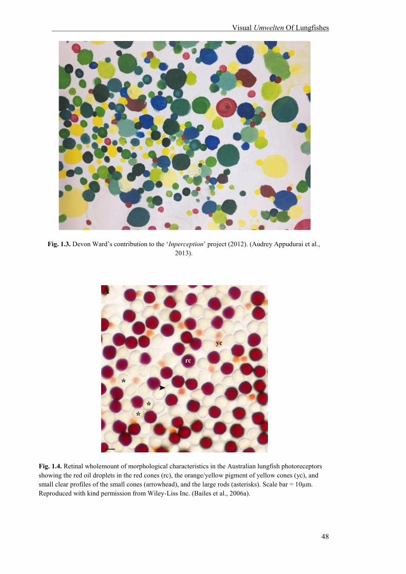
Visual Umwelten Of Lungfishes
48
Fig. 1.3. Devon Ward’s contribution to the ‘Inperception’ project (2012). (Audrey Appudurai et al., 2013).
Fig. 1.4. Retinal wholemount of morphological characteristics in the Australian lungfish photoreceptors showing the red oil droplets in the red cones (rc), the orange/yellow pigment of yellow cones (yc), and small clear profiles of the small cones (arrowhead), and the large rods (asterisks). Scale bar = 10µm. Reproduced with kind permission from Wiley-Liss Inc. (Bailes et al., 2006a).
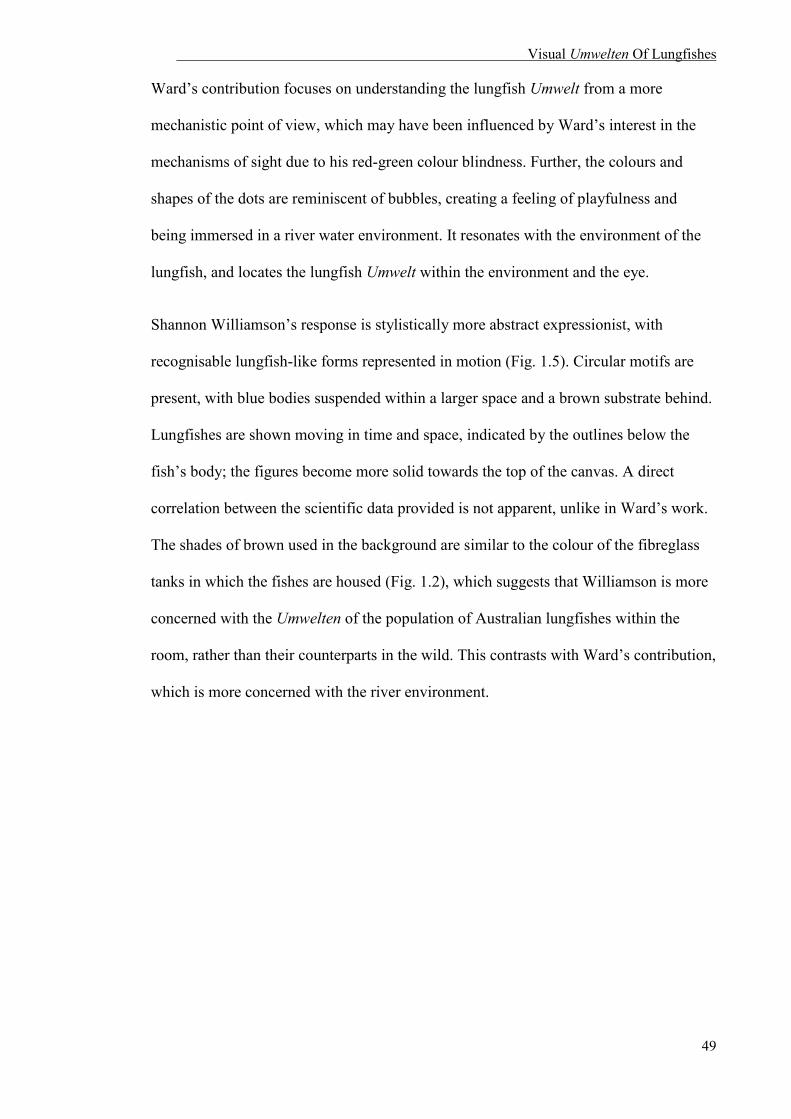
Visual Umwelten Of Lungfishes
49
Ward’s contribution focuses on understanding the lungfish Umwelt from a more
mechanistic point of view, which may have been influenced by Ward’s interest in the
mechanisms of sight due to his red-green colour blindness. Further, the colours and
shapes of the dots are reminiscent of bubbles, creating a feeling of playfulness and
being immersed in a river water environment. It resonates with the environment of the
lungfish, and locates the lungfish Umwelt within the environment and the eye.
Shannon Williamson’s response is stylistically more abstract expressionist, with
recognisable lungfish-like forms represented in motion (Fig. 1.5). Circular motifs are
present, with blue bodies suspended within a larger space and a brown substrate behind.
Lungfishes are shown moving in time and space, indicated by the outlines below the
fish’s body; the figures become more solid towards the top of the canvas. A direct
correlation between the scientific data provided is not apparent, unlike in Ward’s work.
The shades of brown used in the background are similar to the colour of the fibreglass
tanks in which the fishes are housed (Fig. 1.2), which suggests that Williamson is more
concerned with the Umwelten of the population of Australian lungfishes within the
room, rather than their counterparts in the wild. This contrasts with Ward’s contribution,
which is more concerned with the river environment.

Visual Umwelten Of Lungfishes
50
Fig. 1.5. Shannon Williamson’s contribution to the ‘Inperception’ project (2012). (Audrey Appudurai et al., 2013).
The presence of more than one body in Williamson’s contribution suggests that the
perception of an individual fish is affected by the relationships with other fishes; in fact,
recognition of conspecifics and social interaction is an important aspect of the lungfish
Umwelt. Lungfishes live in small groups in the wild and as social animals, recognition
of other individuals of their group and species would be important. Like Ward,
Williamson gives the impression of playfulness as well as serenity and calmness,
indicated by the use of muted colours, tones and space. The space between individuals
and the white circles are suggestive of air and breathing. There is also a sense of
movement in the work, depicted by the transition between transparent and solid forms
and the streaks down the right of the canvas. An element of observation is also apparent
that is resonant with scientific experiments – we are outsiders observing the lungfishes
in their community. Ultimately, Williamson’s contribution suggests the importance of
relationships between individuals, their housing and their environment.

Visual Umwelten Of Lungfishes
51
Finally, I provided two separate responses, before and after the other artists. My initial
response (Fig. 1.6) depicts a single figure of a large, salmon pink lungfish. The green
and yellow figures (Fig. 1.7) were a response to the other artworks. My contribution is
focussed on the individualism of each fish. This differs from Williamson’s response,
where each body is relatively uniform in colour with no distinguishing features and can
either be interpreted as a different individuals or the same individual at different time
points. My focus on individuals was influenced by being the primary caretaker of the
fishes as each fish had a unique set of behaviours (“personality”) and identifying
features only apparent through time spent caring for them.
The pink lungfish was not a response to the same scientific data given to others, but to
prior research that suggests the visual importance of a pink underbelly in mate selection.
In this case, the pink colour is inverted, where the entire fish is presenting itself. This
suggests that the whole body is sensual in those moments, and that perception is not
restricted to certain parts. Although some aspects of the green and blue lungfishes were
responses to artistic data, the colours are suggestive of a river environment rather than
the brown fibreglass tanks of the laboratory. This response is also sensual; the bodies
are touching, individuals are distinctive but together: I found lungfishes I cared for to be
playful and romantic. Like the other artist’s response, there is a feeling of air and breath;
here illustrated by their heads facing upwards towards the surface and their supply of air
and/or food. My interpretation of the lungfish Umwelt was not directly about sight, but a
perception of spatial orientation and bodies, influenced by my knowledge as the primary
caregiver of these fishes. Or perhaps she was driven by a desire for them to perceive us.
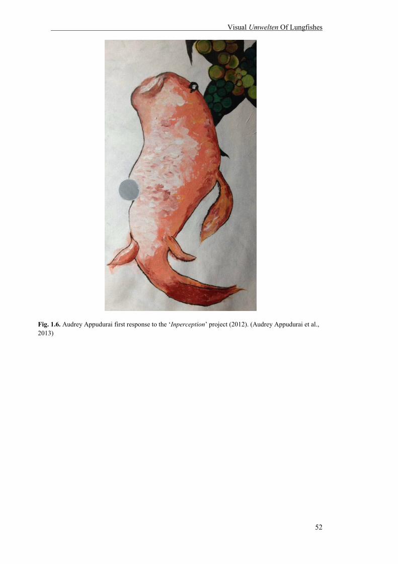
Visual Umwelten Of Lungfishes
52
Fig. 1.6. Audrey Appudurai first response to the ‘Inperception’ project (2012). (Audrey Appudurai et al., 2013)

Visual Umwelten Of Lungfishes
53
Fig. 1.7. Audrey Appudurai’s second response to the ‘Inperception’ project (2012). (Audrey Appudurai et al., 2013).
All artists were present at the same time and gave their initial response concurrently,
each choosing a space on the canvas, which did not initially overlap. However, as the
sessions progressed, Williamson’s contribution diffused into Ward’s work and my
contribution diffused into Williamson’s. Ward’s spots became further apart, giving way
to Williamson’s work to the right of the mural. Recurring motifs permeate the mural in
the forms of colours, circles and feelings of playfulness. As Williamson (2013) wrote,
“being a collaborative project it was impossible to ponder and express our own versions
of lungfish perception without taking into account the modes of visualisation that had
been adopted by our peers.” The resultant “seeping” of one artist’s interpretation into
another created a dialogue between the humans, and between human and aquatic non-
humans to discover the informed subjective nature of perception that transcends
individuals, species, and their shared environments.

Visual Umwelten Of Lungfishes
54
Despite this, the mural lacks aesthetic cohesion. Each response is blurred at the edges,
but clearly separate, illustrating isolation in Umwelten between individuals. Ward’s
contribution is disconnected from Williamson’s by broad streaks of bright yellow.
Although my fishes play at the edges of Williamson’s pool, they are cut off by brown
streaks. Williamson (2013) expanded on this dissonance, saying, “There were multiple
perspectives that could be taken. With an undefined audience, the adaption of any
specific mode was challenging”. The chaos revealed the dynamic responses to the
Australian lungfish Umwelt from individuals presented with the same scientific data,
and the alternative agents that may be involved in an aquatic animal’s perceptual
experience. Additionally, it illustrated the frustration associated with trying to do so,
even with informed subjectivity.
‘Inperception’ was a project that attempted to discover new information about the
Australian lungfish’s visual experience by revisiting Uexküll’s concept of the “soap
bubble”, and utilizing scientific data “as a means for generating questions and
considerations of a world beyond an exclusively human concept” (Williamson, pers.
comm. 2013). The project showed that the Umwelten of lungfishes goes beyond the
machinery of the eye. A dialogue exists between fish, between the fish and their human
observers, and ultimately between the fish and their environment.

Visual Umwelten Of Lungfishes
55
1.6 CONCLUSION
Although the Umwelten of lungfishes may never be completely unravelled to our
satisfaction, attempts to demystify it is important and insightful research. Scientific data
contributes to the growing knowledge of lungfish visual ecology, while artistic
interpretations speculate on and communicate missing pieces of the puzzle. Both
attempts reflect our own limitations in perceiving the “Other”. By combining these two
approaches, the scientific and artistic, the lungfish Umwelt is revealed from different
perspectives and new ways of exploration are recognised. Such interdisciplinary
research is important to understanding of the fundamental similarities and differences in
vision between human and non-human aquatic animals in order to connect us with the
world and understand our place within it. It also helps us to appreciate the limitations
involved in considerations of what lies outside our visual experience and, in doing so,
increases awareness of organisms and environments under threat due to human impact.
1.7 REFERENCES
ALI, M. A. & ANCTIL, M. 1973. Retina of the South American lungfish, Lepidosiren paradoxa Fitzinger. Canadian Journal of Zoology, 51, 969-972.
AMEMIYA, C. T., ALFÖLDI, J., LEE, A. P., FAN, S., PHILIPPE, H., MACCALLUM, I., BRAASCH,
I., MANOUSAKI, T., SCHNEIDER, I. & ROHNER, N. 2013. The African coelacanth genome provides insights into tetrapod evolution. Nature, 496, 311-316.
APPUDURAI, A.M., WILLIAMSON, S., WARD, D. 2013. Inperception. Mixed media on canvas. 100 x
270cm. BAILES, H. J., DAVIES, W. L., TREZISE, A. E. O. & COLLIN, S. P. 2007a. Visual pigments in a living
fossil, the Australian lungfish Neoceratodus forsteri. BMC Evolutionary Biology, 7, 200. BAILES, H. J., ROBINSON, S., TREZISE, A. E. O. & COLLIN, S. P. 2006a. Morphology,
characterization, and distribution of retinal photoreceptors in the Australian lungfish Neoceratodus forsteri (Krefft, 1870). The Journal of Comparative Neurology, 494, 381-397.
BAILES, H. J., TREZISE, A. E. O. & COLLIN, S. P. 2006b. The number, morphology, and distribution
of retinal ganglion cells and optic axons in the Australian lungfish Neoceratodus forsteri (Krefft 1870). Visual Neuroscience, 23, 257-273.

Visual Umwelten Of Lungfishes
56
BAILES, H. J., TREZISE, A. E. O. & COLLIN, S. P. 2007b. The optics of the growing lungfish eye: lens shape, focal ratio and pupillary movements in Neoceratodus forsteri. Visual Neuroscience, 24, 377-387.
BLOUGH, P. M., RIGGS, L. A. & SCHAFER, K. L. 1972. Photopic spectral sensitivity determined
electroretinographically for the pigeon eye. Vision Research, 12, 477-485. BOWLES, K. E. & KRAFT, T. W. 2012. ERG Critical Flicker Frequency Assessment in Humans. In:
LAVAIL, M. M., ASH, J. D., ANDERSON, R. E., HOLLYFIELD, J. G. & GRIMM, C. (eds.) Retinal Degenerative Diseases. Springer US.
BROWN, K. T. 1968. The electroretinogram: its components and their origins. Vision Research, 8, 633-
IN6. BUCHANAN, B. 2008. Onto-Ethologies: The Animal Environments of Uexkill, Heidegger, Merleau-
Ponty, and Deleuze, Albany, State Univerity of New York Press. CAVONIUS, C. R. 1964. Color sensitive responses in the human flicker—ERG. Documenta
Ophthalmologica, 18, 101-113. CLACK, J. A., SHARP, E. L. & LONG, J. A. 2011. The Fossil Record of Lungfishes. In: JORGENSEN,
J. M. & JOSS, J. (eds.) The Biology Of Lungfishes. Enfield: Science Publishers. COLLIN, S. P. 2010. Evolution and Ecology of Retinal Photoreception in Early Vertebrates. Brain
Behavior and Evolution, 75, 174-185. COLLIN, S. P. & COLLIN, H. 2001. The fish cornea: adaptations for different aquatic environments.
Sensory Biology of Jawed Fishes. New Insights, 57-96. DEAN, B. Notes on the living specimens of the Australian lungfish, Ceratodus forsteri, in the Zoological
Society's collection. Proceedings of the Zoological Society of London, 1906. Wiley Online Library, 168-178.
FOX, M. 1970. A comparative study of the development of facial expressions in canids; wolf, coyote and
foxes. Behaviour, 49-73. GOLDSMITH, T. H. 1986. Interpreting trans-retinal recordings of spectral sensitivity. Journal of
Comparative Physiology A, 159, 481-487. GOVARDOVSKII, V. 1983. On the role of oil drops in colour vision. Vision Research, 23, 1739-1740. GRIGG, G. C. 1965. Studies on the Queensland lungfish, Neoceratodus forsteri (Krefft). 3. Aerial
respiration in relation to habits. Australian Journal of Zoology, 13, 413-422. HART, N. S., BAILES, H. J., VOROBYEV, M., MARSHALL, N. J. & COLLIN, S. P. 2008. Visual
ecology of the Australian lungfish (Neoceratodus forsteri). BMC ecology, 8, 21. HART, N. S., LISNEY, T. J. & COLLIN, S. P. 2006. Cone photoreceptor oil droplet pigmentation is
affected by ambient light intensity. Journal of Experimental Biology, 209, 4776. HECHT, S., SHLAER, S. & VERRIJP, C. D. 1933. Intermittent stimulation by light II. The measurement
of critical fusion frequency for the human eye. The Journal of General Physiology, 17, 237-249. JENSSEN, T. A. & SWENSON, B. 1974. An ecological correlate of critical flicker-fusion frequencies for
some Anolis lizards. Vision Research, 14, 965-970. KELBER, A., VOROBYEV, M. & OSORIO, D. 2003. Animal colour vision—behavioural tests and
physiological concepts. Biological Reviews, 78, 81-118. KEMP, A. 1986. The biology of the Australian lungfish, Neoceratodus forsteri (Krefft 1870). Journal of
Morphology, 190, 181-198.
Visual Umwelten Of Lungfishes
57
KERR, J. 1902. The development of Lepidosiren paradoxa. III. Development of the skin and its derivatives. Quarterly Journal of Microscopial Sciences, 46, 417 - 459.
LIEBMAN, P. 1972. Microspectrophotometry of photoreceptors. Photochemistry of vision. Springer. LIEBMAN, P. & ENTINE, G. 1964. Sensitive low-light-level microspectrophotometer: detection of
photosensitive pigments of retinal cones. JOSA, 54, 1451-1458. LUBBOCK, S. J. 1889. On the Senses, Instincts, and Intelligence of Animals: with Special Reference to
Insects, K. Paul, Trench & co. MARSHALL, C. R. 1986. A list of fossil and extant dipnoans. Journal of Morphology, 190, 15-23. MORRISON SCOTT, T. C. S. 1937. Experiments on colour vision in the satin bower bird (Ptilono
rhynchus violaceus), with other observations. Proceedings of the Zoological Society of London, 107, 41-49.
NAGEL, T. 1974. What Is It Like To Be A Bat? The Philosophical Review, 83, 435-450. NUBOER, J., COEMANS, M. & VOS, J. 1992. Artificial lighting in poultry houses: do hens perceive the
modulation of fluorescent lamps as flicker? British Poultry Science, 33, 123-133. OLLY & SUZI. 2002. Untamed [Online]. Available: http://www.ollysuzi.com/projects/untamed.php
[Accessed 06/02/2012. OLLY & SUZI. 1997. Shark Bite. C-type photograph mounted on alluminium with perspex. 48"x32". PFEIFFER, W. 1968. Retina und Retinomotorik der Dipnoi und Brachiopterygii. Cell and Tissue
Research, 89, 62-72. SAGAN, D. 2010. Introduction: Umwelt after Uexküll. A Foray Into The Worlds Of Animals And
Humans. London: University of Minnesota Press. TOKITA, M., OKAMOTO, T. & HIKIDA, T. 2005. Evolutionary history of African lungfish: a
hypothesis from molecular phylogeny. Molecular Phylogenetics and Evolution, 35, 281. UEXKÜLL, J. V. 2010. A Foray Into The Worlds Of Animals And Humans With A Theory Of Meaning,
Minneapolis, University of Minnesota Press. VAN-EYK, S. M., SIEBECK, U. E., CHAMP, C. M., MARSHALL, J. & HART, N. S. 2011.
Behavioural evidence for colour vision in an elasmobranch. The Journal of Experimental Biology, 214, 4186-4192.
VON FRISCH, K. 1914. Der farbensinn und formensinn der biene. Zoologische Jahrbücher. Abieilung
für Allgemeine Zoologie und Physiologie., 35, 1-188. VOROBYEV, M., OSORIO, D., BENNETT, A., MARSHALL, N. & CUTHILL, I. 1998.
Tetrachromacy, oil droplets and bird plumage colours. Journal of Comparative Physiology A, 183, 621-633.
WALLER, B. M. & DUNBAR, R. I. 2005. Differential behavioural effects of silent bared teeth display
and relaxed open mouth display in chimpanzees (Pan troglodytes). Ethology, 111, 129-142. WALLS, G. L. 1942. The Vertebrate Eye and its Adaptive Radiation, Bloomfield Hills, The Cranbook
Press. WILLIAMS, D. B. & CARTER, C. B. 1996. The Transmission Electron Microscope, New York,
Springer. WILLIAMSON, S. Personal interview, 24 March 2013.
Lungfishes From A Historical Perspective
58
CHAPTER 2
::::::
LUNGFISHES FROM A
HISTORICAL
PERSPECTIVE: HOW
HUMANS SEE THE
“OTHER”
::::::

Lungfishes From A Historical Perspective
59
2.1 INTRODUCTION
Three hundred and ninety million years ago during the Devonian period, the Earth was
devoid of four legged creatures. The land was colonised with primitive avascular plants
and arthropods, and the Devonian seas were dominated by marine invertebrates and
armoured fish. Within the freshwater basins of Gondwanaland lungfishes evolved,
organisms that represent the transition between life in the water and atmospheric oxygen
breathing life on land. These humble animals have contributed to shaping cultural
groups across the world and throughout history. Human narratives about lungfishes
reveal multiple aspects and concerns with this intriguing animal and act as mirrors
which present biases (both personal and social) in our attempt to understand the
“Other”.
For the Indigenous peoples of Africa and Australia, this creature has provided spiritual
and physical sustenance, was a mythical harbourer of spirits that walked on land when
children slept, and became a powerful clan totem that commanded respect. Its discovery
by Western civilization in the 19th century created pandemonium for the taxonomic
organisation and classification of animals, and added fuel to the controversy that
surrounded Charles Darwin’s epochal release of ‘On the Origin of Species’. In the 21st
century, these fishes are celebrated as national treasures. Sculptures are raised in their
honour: they are seen as hallmarks of evolutionary success, and beacons to the
conservation effort in sub-tropical Australia. In the United States of America, half a
world away from where they are found endemically, one Australian lungfish is famous
for being the oldest living animal in captivity. It is recognised as an ambassador to its
species, solidifying international relations between countries. The fame has also
bizarrely resulted in the belief that this long-lived individual may hold the secret to
everlasting life. In Perth, Western Australia, within a laboratory in the most isolated
capital city in the world, research conducted on the visual system of the Australian

Lungfishes From A Historical Perspective
60
species, has attempted to uncover the visual reality of these animals. The narrative of
this research project includes an ironic, unexpected turn of events that illustrates the
intimate connection between ingrained assumptions and understandings about
Australian lungfishes.
This chapter delves into historical and contemporary stories concerning lungfishes.
Indigenous stories are used to reveal alternative frames of knowledge, diverse attempts
to understand and make meanings out of this curious animal which lives in water but
possesses lungs. Stories from 19th century Europe illustrate how lungfishes, as
“transitional animals”, contributed to the shift from a predominantly religious to a
secular based taxonomy and world view. These stories confirm that science is an
important tool and methodology to make sense of the “Other” animal but is always a
reflection of the society within which it operates. Contemporary stories reveal other
obsessions of Western societies as they reflect, through the lungfishes, national pride
and human obsession with celebrity and desire for long life. My own personal narrative
about a small group of Australian lungfishes reveals how ingrained assumptions result
in looking but not seeing. In general, the chapter explores the rich human cultural
archives of lungfish stories which reflect the human desire to make sense out of a
transitional animal that, in turn, can help reveal anthropocentric limitations in the quest
for the non-human Umwelt. In other words, these human narratives all show how
personal and social biases influence how we choose to understand lungfishes and by
inference, other non-human beings.
Perceptions of organisms, including humans, are constructed both through biological
apparatuses as well as subjective experience and interpretation. Therefore, each
organism constructs/forms their own subjective reality. As Thomas Nagel stipulated in
‘What Is It Like To Be A Bat?’ (Nagel, 1974) and Jakob von Uexküll suggested with his
theory of the Umwelt (Uexküll, 2010), that when it comes to unravelling visual
Lungfishes From A Historical Perspective
61
experience, especially of non-human animals, it is usually expected that scientific
methodology holds the key because objectivity is a fundamental aspect of its approach
(Kitcher, 1993). However, the construction and outcomes of scientific research are
always framed by humans within a certain perspective that forces a particular
understanding of the world. Bruno Latour reminds us, “scientific activity is just one
social arena in which knowledge is constructed” (Latour and Woolgar, 1986), and as
Donna Haraway said, “biology is a discourse, not the living world itself” (Haraway,
1992). Therefore, science can provide an important aspect of the puzzle, but not all the
pieces to see into the Umwelten of lungfishes. Other discourses based on human stories
also offer new perspectives of the lungfish Umwelt and provide additional (or
alternative) pieces of the puzzle together.
2.2 LUNGFISHES IN INDIGENOUS COMMUNITIES: THE DALA, THE
MMAMBA, AND THE AMAZONIAN KILLER LUNGFISH
Before lungfishes captured international attention after their modern Western11
discovery, their presence and unique position was well known to Indigenous
communities. To the Gubbi Gubbi people12 of Queensland, Australia, the Australian
lungfish, or Dala in the Gubbi Gubbi tongue, are still sacred animals. The Dreamtime
story of Dala, woven by Dr. Eve Fesl, a Gubbi Gubbi elder tells a story of a fish
destined to be the ancestor to all vertebrates:
Many years ago, thousands of years ago, there were no animals on the Earth, and the
Earth Mother and our ancestral spirits looked down and decided that they wanted to
have animals on the Earth, which would be their children. So, they designated that
11 By Western, I refer mainly to the Judeo-Christian version of perceiving/explaining life. 12 Sometimes referred to as ‘the Dala’ people by other Aboriginal groups See: FESL, E. M. D. n.d. Extinction is Forever! A bid to save "Dala", the Queensland lungfish, an endangered species in the Mary River across whcih a dam is proposed to be built. [Online]. Brisbane. Available: http://www.savethemaryriver.com/_mgxroot/page_10786.html [Accessed 20 May 2014 2014].

Lungfishes From A Historical Perspective
62
Dala would, first of all, get a lung, then it could get vertebrae and be able to walk, and
that would be the forefather and foremother, really, of all the animals in the world. The
breeding place would be near the edges of the river, so that the animals could come out
and so we have Dala, the lungfish which only has one lung. It’s the only place in the
world you could find it, and this place where we stand is one of the most viable
breeding places (Franklin, 2007).
Although Fesl was not aware of the scientific importance of the Australian lungfish
when growing up, she understood that the Gubbi Gubbi people were their sacred
custodians. She had been told about the cultural importance of Dala from a very young
age, remembering a family meeting by the Mary River that involved lungfishes
“swimming around...in the water below” and highlighted the value of such an encounter
because it was “our sacred fish”(Franklin, 2007). Dala is even now a totem to the Gubbi
Gubbi people. Fesl elaborates and says, “We were taught not to hurt it, to protect its
habitat, where it feeds and swims” because Dala “are ancestor spirits” (Queensland,
2009).
This intimate tie that Fesl, and the Gubbi Gubbi people have to the lungfish is also
linked to their relationship to the Mary River. Both Fesl’s mother and uncle were born
on the banks of the Mary River in traditional style, where, “when giving birth, the
mother has to have her sisters by her side to assist her” (Franklin, 2007). In another
anecdote Fesl speaks of her grandmother, and the stories told about Dala:
My grandmother wove shelter covers from bladey grass and used paperbark as a soft
bed to lie on. At night the family would see the reflection of the stars in the water, and
hear the ripple of the water and the occasional explosion of air as Dala, our sacred fish,
chose to come to the surface to expel the breath from his lung. It was at these times,
the children would be told the story of Dala. As they lay under the stars and beside the
rainforest, with its night bird calls, the children listened to the story of the beginning of
our culture (JerryinBrisbane, 2008).

Lungfishes From A Historical Perspective
63
The cultural importance of Dala, created by the close cultural relationships to the river
landscape encompassing the Mary River, family, and community, influenced Fesl’s
understanding of the Australian lungfish. To Australian Aboriginal groups, oral
narratives and the meanings they convey are “owned by the individual storytellers in
accordance with their position within their social and spiritual universe” (Klapproth,
2004) and are “items of exchange in a cultural arena” (Muecke, 1983). In Fesl’s case,
these narratives are intertwined with anthropomorphic attitudes towards Dala. Fesl often
mentions that “Dala will come up beside the canoe and you can stroke Dala. That’s what
a friendly creature it is” (Queensland, 2009) and when greeting them in her native
tongue, does not forget to let them know that she is “part of the family” (Franklin,
2007). This does not necessarily suggest that Fesl is making Dala more human-like, but
rather that non-human animals like the lungfish are important members in the human
world. Gubbi Gubbi stories present an alternative position for the human living within
the world. The human animal is integrated as part of the natural world and therefore
must employ responsibility and respect to other animals.
The liminal nature of Dala, as a fish with a lung,13 is not a “problem” to cultural beliefs.
Rather, this view of the world embraces the concept of fluidity in animals’ forms and
shapes. Dala was chosen as a totem and sacred animal because of its unusual nature.
Fesl says, “Our people were very good at anatomy and physiology, and those things.
They had to be to be able to survive, and so it’s been perceived a long time ago that this
was something special” (Franklin, 2007). The Gubbi Gubbi people were aware of Dala’s
ability to breathe atmospheric oxygen air long before the Western scientific community
and unlike this community did not seek to disprove or condemn it (as will be illustrated
in the stories from 19th century England, in a later part of this chapter).
13 The Australian lungfish, N. forsteri, is the only species that possesses one lung, all other lungfish speces possess a pair of lungs.
Lungfishes From A Historical Perspective
64
The responsibility Fesl felt towards Dala played a major role in the cancellation of the
Traveston Crossing Dam project in 2009. It is important to note that although this
project did increase awareness of the Australian lungfish, there is debate whether the
main motivations was to help the species directly, or rather use it to polarise opinions
and/or to aid development lobbies and conservation groups in realising their own
agendas (personal communication, Kind, 2015). The dam, proposed in 2006 after the
worst drought Queensland had experienced in a century, was to be constructed
approximately 16 kilometres south of Gympie (Meteorology, 2006, Franklin, 2007). The
dam would have affected the surrounding built up areas north of Brisbane, and flooded
the major breeding grounds of the Australian lungfish in the Mary River. This angered
the local and international community. The importance of the Australian lungfish was
articulated to the government through rigorous protests, petitions and media reports. At
one such protest, then Greens leader, Senator Bob Brown said, “I’ve told the minister
there is no way he should allow the major breeding ground for the Australian lungfish or
the Mary River turtle, or the Mary River cod, to be obliterated” at a protest of “hundreds
of farmers and local residents” (McGrath, 2008).
Fesl was a guest speaker at many of these rallies and at one, expressed how the
government was attempting to buy support from the Indigenous communities to take
away this sacred land. In the rebuttal to the proposal, on behalf of the Gubbi Gubbi
people she said, “We would have nothing to do with it. We’re not going to sign away
our history, our culture, the Mary Valley. No way.” (Crow_Boy, 2007). At the Climate
Torch Relay 2008, Fesl told the Dreamtime story of Dala and finished with, “flowing
through time, our duty is to care for Mummabulla (Mary River) and to care for Dala, and
it is your duty too. She is the symbol of our past, our present, and our future”
(JerryinBrisbane, 2008) to the cheers of supporters. Fesl campaigned through media
reports and proposals to the Premier of Queensland about Dala, emphasising the

Lungfishes From A Historical Perspective
65
negative effects of Traveston Crossing on the ‘migration over long distances they need
during spawning’ and asking, “Do people in Queensland, Australia and around the globe
care enough to protect Dala, the Queensland lungfish and its habitat?” (Fesl, n.d.).
An Australian Broadcasting Corporation radio show titled ‘A Whisper From The Past’
was dedicated to the conservation effort (Franklin, 2007). Professor Jean Joss was also
involved in the campaign against the dam, and stated that “the lungfish has been
described as being the most important vertebrate because it sits at the transition between
water and land” (Franklin, 2007) and “the Mary River dam will destroy its spawning and
nursery habitat and threaten it with extinction; and this will be a scientific scandal that
the scientific community will neither forgive nor forget, and which will permanently
stain Australia's conservation record in the eyes of the world” (Joss, 2007a). In a petition
by Joss, over 4,000 signatories were accumulated in its first week (Joss, 2007b) and the
international scientific community was alerted through editorials published in Nature
(Pearson, 2006, 2006). Finally in 2009, due to this growing pressure from popular media
and members of the community, the project was refused approval by the federal
Environment Minister, Peter Garrett, on the grounds of “Listed Threatened species and
communities” under the Environment Protection Biodiversity Conservation Act 1999
(Garrett, 2009).
Similarly, Protopterus aethiopicus, one of the African lungfish species, is of special
value to one of the biggest clans of Buganda, the largest sub-national kingdom within
Uganda. More than fifty clans call Buganda home, and each clan can trace its lineage
back to the historical waves of immigrations into the kingdom. The first of these
brought the Nansangwa, or indigenous people, into Buganda. The Nansangwa are
comprised of the six oldest clans in Buganda. One of these is Mmamba Gabunga, which
translates to the native name for P. aethiopicus, also known as the marbled lungfish.
Like N. forsteri to the Gubbi Gubbi, P. aethiopicus is a totem to the Mmamba people, a

Lungfishes From A Historical Perspective
66
spiritual emblem to remind them of their ancestry and mythic past. In the Mmamba
origin story, the clan’s founder Mabiru, had close ties with the rivers, and gained the
king, Kabaka14 Kintu’s favour by offering his talents as an expert navigator and canoe
maker, which resulted in Kabaka Kintu appointing many of Mabiru’s clansmen as
chiefs in his kingdom. The Mmamba remain one of the largest and most influential clan
of Buganda, but do not eat their namesake due to its symbolism of their clan (2014a).
Like the story of Dala, the Mmamba understand of the human as an integral part of the
living environment.
While both employ some taxonomical systems in regard to the world around them, the
Indigenous populations of Australia and Africa allow for the fluidity of nature in the
living world; liminality and difference are accepted (and sometimes celebrated). The
Western view appears to be more rigid in terms of order within a fixed hierarchy.
Therefore, it is not surprising that Western discovery of the lungfishes during
colonization resulted in many mythologies featuring the lungfish as “monsters” due to
their novel and bizaare nature. After the 1837 discovery of the South American lungfish
by Johann Natterer, reports flooded in about a mythical beast, the minhocao15 of Brazil,
and its possible ties to the lungfish. The tales of minhocôes stem from Goyaz, a state in
central Brazil, where in the deep lakes of Padre Aranda and Feia, minhocôes torment the
locals by dragging horses, mules and cattle into the water. Saint Hilaire, a scientific
journalist visiting the area in the mid 19th century, investigated these reports by
residents near the lake and described the creature thus:
The word minhocao, is an augmentative of minhoca, which, in Portuguese, signifies
earth-worm; and, indeed, they state that the monster in question absolutely resembles
these worms, with this difference, that it has a visible mouth; they also add, that it is
14 ‘King’ in the Mmamba language 15 Singular of minhocôes

Lungfishes From A Historical Perspective
67
black, short, and of enormous size; that it does not rise to the surface of the water, but
that it causes animals to disappear by seizing them by the belly (Saint Hilaire, 1847).
Further investigation led Saint Hilare to the Governor of Meiapoint, M. Joaquim Alvez
de Olveira. De Olveira had heard of these monsters, and advised Saint Hilaire that
fishermen reported the minhocao “was a true fish, provided with fins”. Saint Hilaire’s
interest was further piqued, and turned his research to organisms that had recently been
discovered and identified by Western science. This search led him to Professor Gervais,
who directed Hilare to Richard Owen and Mikkel Bischoff’s papers on the newly
discovered African species Lepidosiren annectens (Owen, 1840, Edwards, 1841) and
Natterer and Fitzinger’s recent discovery of the South American L.paradoxa (Natterer,
1837, Bischoff, 1840). To Saint Hilaire, both these animals fit the description given by
the fishermen, and L. paradoxa shared the habitat of the minhocao. He quoted Bischoff
in his paper of the dentition of L. annectens and wrote, “These characters agree
extremely well with those which we must of necessity admit in the minhocao, since it
seizes very powerfully upon large animals, and drags them away to devour them”.
Finally, he continued with great confidence that “it is, therefore, probably that the
minhocao is an enormous species of Lepidosiren”. (Saint Hilaire, 1847). Saint Hilare’s
story illustrates how the “different” can become monstrous and result in exaggerated
tales; in this case, created by Western cultural biases surrounding the exotic and the
unknown of newly colonised land.
More recently in the mid 20th century, the buru, an ancient animal of the Ziro valley of
Arunachal Pradesh, India was thought to be a lungfish. At the end of World War II,
explorer Charles Stonor and anthropologist J. P. Mills travelled to the valley and heard
the legend of the buru, a now extinct group of creatures that were a blight to the Apatani
people of the Ziro valley. According to Apatani elders, the valley’s marshes were
populated by these creatures and when their people settled in the valley, livestock and
Lungfishes From A Historical Perspective
68
the residents were attacked by these large, reptilian-like water dwelling monsters. The
Apatani people were so afraid of the buru that all marshes were drained of water, which
eventually caused the extinction of burus; who either perished without water or
burrowed deep into the underground springs of the valley. In 1948, Stonor accompanied
news correspondent Ralph Izzard to the neighbouring Rilo valley, where locals insisted
that burus still existed. After an extensive search, Izzard and Stonor failed to find
evidence that burus were still at large.
Many cryptozoologists have tried to identify this mythical beast. The general contenders
are reptiles, such as modern day dinosaurs, crocodiles or water-dwelling monitor
lizards. However, cryptozoologist Karl Shuker believes that the buru was a now extinct
species of lungfish; an Asian counterpart to the species inhabiting Africa, Australia and
the South America. The lungfishes’ aquatic lifestyle, behaviour, morphology and
association with burrowing into the earth for long periods of time have convinced
Shuker that the buru may have been a more primitive lungfish. More interestingly, a
personal experience with an African species of lungfish may have cemented his beliefs:
One of the most popular exhibits of the ichthyological practicals during my days as a
zoology student at university was a living specimen of an African lungfish
Protopterus, which was sometimes placed on display in order that we could observe its
behaviour. As it happened, for much of the time there was actually very little that we
could observe, because it would spend most of the practical resting motionless at the
bottom of its tank. Every so often, however, and usually when everyone’s attention
was diverted elsewhere, it would solemnly perform its pièce de resistance. All at once,
without any prior warning, it would raise the front part of its large body upwards, until
its head just touched the surface of the water. Sometimes it would then simply nudge
the tip of its snout above the water, but if we were lucky (by now, everyone would
have rushed up to its tank to watch its celebrated performance) it would actually raise
its entire head, after which it would remain in this position for several minutes,
ventilating (Shuker, 2012).

Lungfishes From A Historical Perspective
69
This experience, articulated in detail, left a lasting impression on Shuker. The locals of
the Himalayan valley have recalled that the buru would occasionally raise its head out
of the water to make a bellowing noise. Lungfishes, when respiring through their lungs
do the same, and Shuker comments that “this scenario [of the buru’s bellowing] is one
that has strong lungfish associations for me” even though others argue against the buru
being a lungfish. Shuker’s personal and cultural background, that included a personal
encounter with the captive animal, left a lasting impression that may have influenced
why he chose to believe the lungfish to be the ancestor to a mythical beast. Although
separated by time and context, this story is not unlike that of Saint Hilare and the
minhocao. In both narratives, the “monstrous” nature of the buru and minohocao led
Saint Hilare and Shuker, respectively, to associate them with the lungfish, a transitional
animal – a fish that can breathe air – with no prior record of attacking locals and
dragging animals into the water. The lungfish was suspect due to its inability to fit
within a clear hierarchical taxonomy.
These stories also represent the clash between the Indigenous worldview and the Western colonialist one – the loved and respected animal became a dangerous beast: the human within and equal to the rest of the living world assumed dominion and control, and anything that did not fit within the order were deemed to be monstrous.

Lungfishes From A Historical Perspective
70
2.3 THE GREAT LUNGFISH CONTROVERSY: HOW THE DISCOVERY OF A
FISH SHOOK THE WEST
In 1817, the young zoologist Johann Natterer was chosen to be part of one of the biggest
scientific expeditions to leave Austrian soil (Barreto and Machado, 2001). In order to
increase Austria’s influence over South America, Prince Klemens Wenzel von
Metternich, an enthusiastic student of natural history and one of the most influential
statesmen of Europe, arranged the marriage between the Austrian Archduchess Maria
Leopoldina and Don Pedro, who would eventually become the first Emperor of Brazil
(Riedl-Dorn and Natterer, 1999). Fourteen scientists and artists were selected to
accompany the Emperor’s daughter to Rio de Janeiro and explore and document as
much of the continent as they could. The expedition lasted four years, but due to
political unrest in Brazil, the Austrian government was forced to officially call off the
venture in 1821 (Taylor, 2013). Natterer however, chose to stay. During the next
eighteen years, Natterer amassed a collection of over twelve thousand specimens from
South America’s natural landscape (2013). One of these was an animal he found so
peculiar that he brought it to Leopold Fitzinger, curator of reptiles at the Imperial
Museum of Vienna, for a closer inspection in 1837. Natterer had found two specimens,
one in a swamp on the left bank of the Amazon and another in a pond in the river
Madeira (Natterer, 1837, Gunther, 1871). The animals were long, slender, and had
mottled brown patterns upon their backs. The head and bodies were decidedly eel-like.
Their fins were extremely delicate for their size and descended from the creatures’
bodies like vestigial appendages when they relaxed, motionless, on the river bed. By all
accounts this animal was a fish at first glance. However, something caused Natterer to
question this classification, so he handed over identification to Fitzinger. When
Fitzinger examined the creatures he discovered, as Natterer had in the wilderness of the
Amazon, they possessed what was undeniably a pair of lungs. In Fitzinger’s day,

Lungfishes From A Historical Perspective
71
amphibians fell under the umbrella of “reptile”, but even classified amphibians such as
particular salamander species (or “sirens”) that teetered upon the line of fish and reptile
with their feather-like gills, had limbs with digits that resembled the fingers and toes of
terrestrial reptiles. This creature had no such appendages. So bewildered was he by this
unique animal, Fitzinger christened it Lepidosiren paradoxa: “lepido” for the scales,
“siren” for the eel-like salamanders it resembled, “paradoxa” for his confusion, and
firmly placed the animal within the classification of reptile (Fig. 2.2) (Bischoff, 1840,
Marshall, 1986, Zimmer, 1998).
Fig.2.1. Adult male L. paradoxa. (Jordan, 1907).
Although Charles Darwin’s “On the Origin of Species” had not yet been published,
evolution as an explanation to new findings in natural history was already being
discussed in the scientific community. Their personal stances upon the matter are
unknown, but Fitzinger’s confusion and uneasiness about L. paradoxa is reflected in the
naming of the South American lungfish as a “paradox”. It is important to note that even
in his recognition of an animal that did not fit into pre-existing taxonomy, he still did
not venture to change the boundaries and acknowledge this animal as being transitional.
Fitzinger and Natterer were prisoners of 19th century Western scientific assumptions and
the ideology of fixed species created by God’s will or plan. Nevertheless, the South
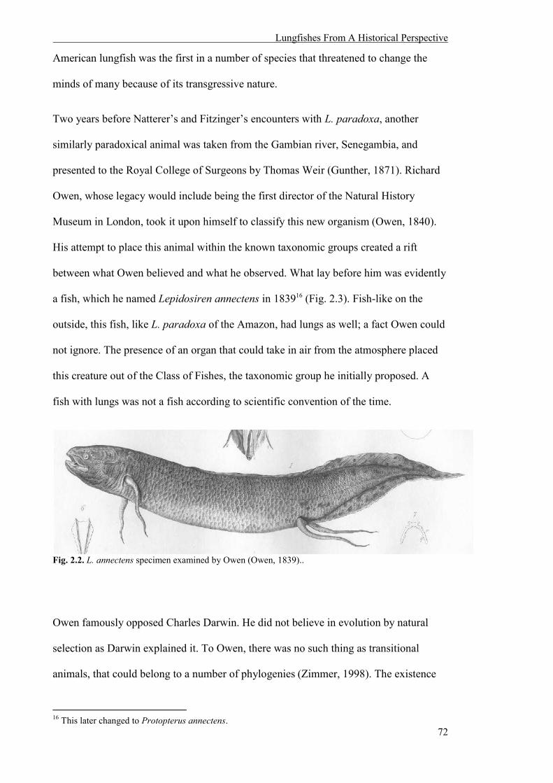
Lungfishes From A Historical Perspective
72
American lungfish was the first in a number of species that threatened to change the
minds of many because of its transgressive nature.
Two years before Natterer’s and Fitzinger’s encounters with L. paradoxa, another
similarly paradoxical animal was taken from the Gambian river, Senegambia, and
presented to the Royal College of Surgeons by Thomas Weir (Gunther, 1871). Richard
Owen, whose legacy would include being the first director of the Natural History
Museum in London, took it upon himself to classify this new organism (Owen, 1840).
His attempt to place this animal within the known taxonomic groups created a rift
between what Owen believed and what he observed. What lay before him was evidently
a fish, which he named Lepidosiren annectens in 183916 (Fig. 2.3). Fish-like on the
outside, this fish, like L. paradoxa of the Amazon, had lungs as well; a fact Owen could
not ignore. The presence of an organ that could take in air from the atmosphere placed
this creature out of the Class of Fishes, the taxonomic group he initially proposed. A
fish with lungs was not a fish according to scientific convention of the time.
Fig. 2.2. L. annectens specimen examined by Owen (Owen, 1839)..
Owen famously opposed Charles Darwin. He did not believe in evolution by natural
selection as Darwin explained it. To Owen, there was no such thing as transitional
animals, that could belong to a number of phylogenies (Zimmer, 1998). The existence
16 This later changed to Protopterus annectens.

Lungfishes From A Historical Perspective
73
of such animals would further prove Darwin’s theory of gradual change through time;
that groups were not fixed and evolution occurred through natural selection. Owen was
determined that this creature was not one of these animals. In other words, because of
the lungfish, Owen (consciously or not) disregarded true scientific reporting in favour of
his religious and cultural beliefs. As a result, Owen observations and notes about the
lungfish’s nose were congruent with these beliefs. He wished to prove that the lungfish
was not using its lung to breathe, as only then could this animal truly be a fish. “The
nostrils...” he wrote, “...appear as two small perforations leading to blind sacs.” This
was his proof: the nostrils did not lead to the lungs, rendering the lungs useless. He
hypothesised that these organs were “swim- or air-bladder(s)”; hence the lungfish was a
fish after all. His final statement after describing the various systems of animal in
excruciating detail was as follows:
In the organ of smell we have, at least, a character which is absolute in reference to the
distinction of Fishes from Reptiles. In every Fish it is a shut sac communicating only
with the external surface; in every Reptile it is a canal with both an external and an
internal opening.
According to this test, the Lepidosiren is a Fish: by its nose it is known not to be a
Reptile: in other words, it may be said that the Lepidosiren is proved to be a Fish, not
by its gills, not by its air-bladders, not by its spiral intestine, not by its unossified
skeleton, not by its generative apparatus, nor its extremities, nor its skin, nor its eyes,
not its ears, but simply by its nose (Owen, 1840).
However, Owen’s conclusions were questioned in 1840 when M. Bishoff examined
members of the same species and concluded that the blind sacs spoken of by Owen did
in fact connect to the mouth, and thus this animal was a reptile (Bischoff, 1840,
Edwards, 1841). Twenty years later, the Irish anatomist Robert M’Donnel examined
another specimen of L. annectens and also found that the passageway from the
lungfish’s nose did not lead to the blind sacs as observed by Owen (Zimmer, 1998). He
Lungfishes From A Historical Perspective
74
concluded that this group of fishes were indeed the transitional creatures Owen fought
to deny. He wrote, “I know of no animal more calculated leading to the adoption of the
theory of Darwin than the Lepidosiren.” (Zimmer, 1998).
These narratives surrounding the Western discovery of the transitional lungfishes
illustrate how religious ideologies fed into the classification and naming conventions of
19th century science. The Judo-Christian teleological influences stemmed from Carl
Linnaeus (1707-1778) and his widely accepted system of plant and animal classification
by binomial nomenclature introduced in the 18th century. Linnaeus was a devout and
orthodox Christian who believed early in his career as a botanist, physician and
zoologist, that as Man, he had been ordered by God to study nature and its laws
(Ereshefsky, 2001, Gardiner, 2001). He then created what we know as the Linnaean
system of ordered taxa that includes genus and species. Linnaeus believed that every
species was decided by God’s original creation, writing “every genus is natural, created
as such at the beginning – hence not to be rashly spilt up or stuck together by whim or
according to anyone’s theory” (Translation by Ramsbottom, 1938). He also mentioned
Noah’s ark as a reason for such wide diversity in the natural world and asserted that “it
is not credible that the Creator should fill the world with animals only to destroy them
all shortly afterwards through the deluge – with the exception of a single pair of each
species preserved in the ark” (Translation by Ramsbottom, 1938). Although later in his
career, Linnaeus adopted the theory of transformism17 and hybridization in speciation,
he still did not alter his belief that the natural world consisted of systematically ordered
works of Creation.18 Linnaeus’s fixism19 was widely adopted by the scientific
community despite his creationist views because, for the first time, clear and consistent
17 For more information on the concepts of transformism, see Corsi, Pietro. 2005. ‘Before Darwin: Transformist concepts in European natural history.’ Journal of the History of Biology, 38, 67-83. 18 See Gardiner, B. G. 2001. ‘Linneaus's species concept and his views on evolution.’ The Linnean, 17, 24-36. 19 Fixism is a non-religious theory that all species alive today are identical to those in the past, and these organisms emerged already adapted to the environment without undergoing changes (Ereshefsky, 2001).

Lungfishes From A Historical Perspective
75
rules for classification were possible. For Fitzinger, these biases are evident in his
classification of L. paradoxa as an anomaly that could still firmly fit within a pre-
existing taxonomic Class, and more explicitly in Owen’s incorrect identification of L.
annectens.20
On the other side of the world, Gerard Krefft, director of the Australian Museum, was
the first scientist to see and name the Australian counterpart of lungfishes. The
Queensland lungfish was offered to Krefft by William Forster, a squatter from
Queensland who had acquired a position at the Australian Museum as a cook. Forster
had been trying to alert Krefft to a new species of fish unknown to science for a number
of years (Olsen, 2010, Semon, 1899). Known as baramoonda or baramoondi by the
local indigenous populace (Fig. 2.4) (Sclater, 1870), the white colonisers of Queensland
called it the Burnett, or Dawson salmon (Fig. 2.5) (for where it is found), but Krefft did
not seem interested at first. Finally, Krefft decided it was time to get some proof. Krefft
related the situation to a colleague in 1880:
During ten years, I dined with Mr Wm. Forster, the late Agent General for New
South Wales...once a week or so, and every time William Forster tantalized me with
his stories of ‘fresh water salmon’. Well, when a thing of this kind is repeated too
often, a man requires proof. Then the Honorable gentleman sent me a message about
11 years that THE fish had come to hand. I attended, and a messenger brought a box
which I opened and found two fishes in it. Said Mr Forster, ‘Well Krefft, what are
these fish?’ Said I, ‘I cannot tell till you allow me to examine them.’ Said Forster:
‘Do so, you are welcome to them. I present them to you, if you will name them after
me.’ I replied: ‘I will’, took my knife out, exposed the teeth and told Forster: ‘never
saw anything to equal this in my life.’ ‘Are they new?’ said Forster. ‘No,’ I said,
‘They are as old as the mountains of Australia, and if you will let me alone we will
make a fortune with these fishes.’...’Well take them away, do what you like with
them, but make the discovery known in tomorrow’s Herald.’ (Monteith, 2001)
20 It is interesting to note that lungfishes still remain in Owen’s Class of choice, the Fishes.

Lungfishes From A Historical Perspective
76
True to his word, Krefft christened the fish Ceratodus forsteri21 in 1870 after Forster
(Krefft, 1870, Marshall, 1986, Olsen, 2010).
Fig. 2.3. Australian lungfish lying on a cane fishing basket (Anon. 1895).
Fig. 2.4. Two fishermen with their Australian lungfish catch, Coomera River, Queensland. (Anon, 1917).
21 The name eventually changed to Neoceratodus forsteri.
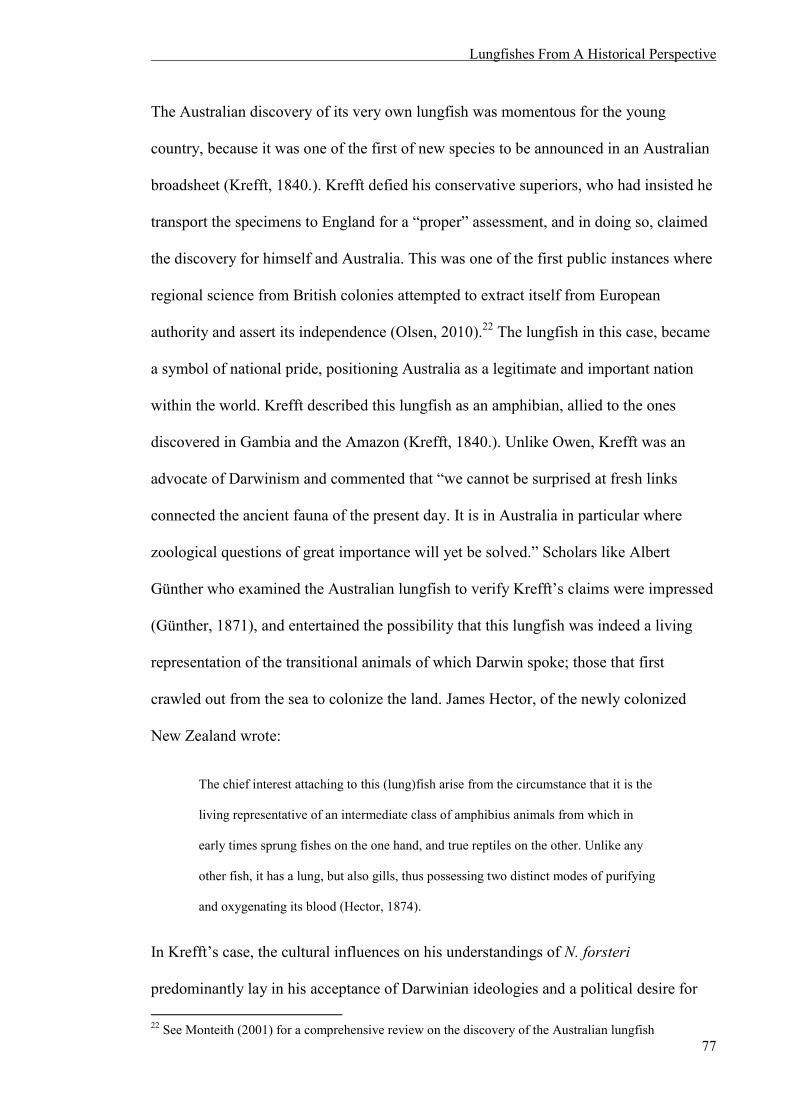
Lungfishes From A Historical Perspective
77
The Australian discovery of its very own lungfish was momentous for the young
country, because it was one of the first of new species to be announced in an Australian
broadsheet (Krefft, 1840.). Krefft defied his conservative superiors, who had insisted he
transport the specimens to England for a “proper” assessment, and in doing so, claimed
the discovery for himself and Australia. This was one of the first public instances where
regional science from British colonies attempted to extract itself from European
authority and assert its independence (Olsen, 2010).22 The lungfish in this case, became
a symbol of national pride, positioning Australia as a legitimate and important nation
within the world. Krefft described this lungfish as an amphibian, allied to the ones
discovered in Gambia and the Amazon (Krefft, 1840.). Unlike Owen, Krefft was an
advocate of Darwinism and commented that “we cannot be surprised at fresh links
connected the ancient fauna of the present day. It is in Australia in particular where
zoological questions of great importance will yet be solved.” Scholars like Albert
Günther who examined the Australian lungfish to verify Krefft’s claims were impressed
(Günther, 1871), and entertained the possibility that this lungfish was indeed a living
representation of the transitional animals of which Darwin spoke; those that first
crawled out from the sea to colonize the land. James Hector, of the newly colonized
New Zealand wrote:
The chief interest attaching to this (lung)fish arise from the circumstance that it is the
living representative of an intermediate class of amphibius animals from which in
early times sprung fishes on the one hand, and true reptiles on the other. Unlike any
other fish, it has a lung, but also gills, thus possessing two distinct modes of purifying
and oxygenating its blood (Hector, 1874).
In Krefft’s case, the cultural influences on his understandings of N. forsteri
predominantly lay in his acceptance of Darwinian ideologies and a political desire for 22 See Monteith (2001) for a comprehensive review on the discovery of the Australian lungfish

Lungfishes From A Historical Perspective
78
Australia to be legitimised and independent from governing English authorities in
matters of animal biology.
The origin of terrestrial vertebrates was a major and important focus in the history of
scientific ideas. The discovery of lungfishes prompted excitement and heated debate on
the existence of evolution by natural selection. Academics like William Cladwell
flocked to Australia to study transitional animals like the lungfish and platypus
(Cladwell, 1884). Another researcher, Richard Semon, spent two years in Australia
attempting to collect and study the Australian lungfish’s eggs (Semon, 1901). Carl
Gegenbaur, who theorised that evidence of evolution could be found in the anatomy of
organisms, constructed his model using the Australian lungfish as a template, a
primitive creature with gills, a lung, and leaf-shaped fins that may have become fingers
(Gegenbaur, 1878). Papers announcing this new species requested any new specimens
to be sent to London and beseeched those in the lungfish’s native land to resist
temptation to hunt and eat this valuable species. P. C. Sclater placed such a note in
Nature, writing:
In conclusion, I may express a hope that this short notice may have the effect of
calling the attention of some of the colonists of Queensland to the wonderful nature of
this relic of the Denovian epoch that is now swimming about beneath their noses, and
that they will cease, for the present at least, to kill it and eat it as ‘salmon’. Any
specimens that may ‘rise to their fly’ should be carefully kept out of the way of the
cook, preserved in alcohol and transmitted to the British Museum or some other
scientific institution. When the existence of Ceratodus forsteri becomes more widely
known, there will be no lack of applicants for examples of it (Sclater, 1870).
This time, the new Australian immigrants “colonised” the unique animals of the
continent to establish their hold on the land. Despite the lungfishes’ “discovery”, long
before, by the Indigenous population, it had to be ‘rediscovered’ to the rest of the

Lungfishes From A Historical Perspective
79
Western world by Western colonists. While the Indigenous people respected the lungfish
within its environment, the colonialists captured and shipped it to be displayed in other
countries (dead or alive); removing it from its environment and transforming it into a
“commodity” of the human.
2.4 THE SECRET OF IMMORTALITY: CONTEMPORARY CULTURAL
INFLUENCES OF N.FORSTERI
The Chicago World’s Fair of 1933 was named “A Century of Progress International
Exposition” to commemorate the city’s centennial. The fair’s motto “Science Finds,
Industry Applies, Man Conforms” celebrated technological innovation, but one
particular exhibit had little to do with technology. Two Australian lungfishes, a male
and female, had arrived from Sydney to the recently opened Shedd aquarium to be
witnessed by Chicagoans for the first time. Walter Chute, the aquarium’s director,
wanted rare, precious fish to attract visitors to the fair; a testament to the human
fascination with the exotic. This act illustrated that despite all technological innovation
and control, there were still things beyond our understanding, such as a fish with lungs.
When Chute discovered the steamship collecting exotic fish from Hawaii was also
headed to Australia, he promptly asked permission from Sydney’s Taronga Zoo for one
of the continent’s own “mud-fish” to become a permanent resident of Shedd. Not long
after, Shedd’s railroad car, The Nautilus, collected thirty containers of exotic fishes
from Los Angeles to Chicago (2014b), including the fish Chute had requested, one of
which was christened Granddad (Fig. 2.6). Although the age of this lungfish is
unknown, as he came to Shedd as an adult (Fig. 2.7), the 80th anniversary of his
captivity was celebrated on September 17th 2013, making him the oldest living animal
in any aquarium in history since the death of George, the Galapagos tortoise (Hassler,
2013).
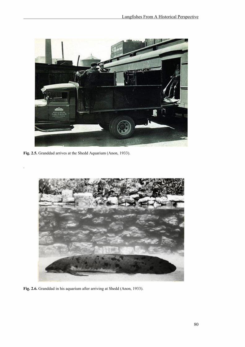
Lungfishes From A Historical Perspective
80
Fig. 2.5. Granddad arrives at the Shedd Aquarium (Anon, 1933).
.
Fig. 2.6. Granddad in his aquarium after arriving at Shedd (Anon, 1933).
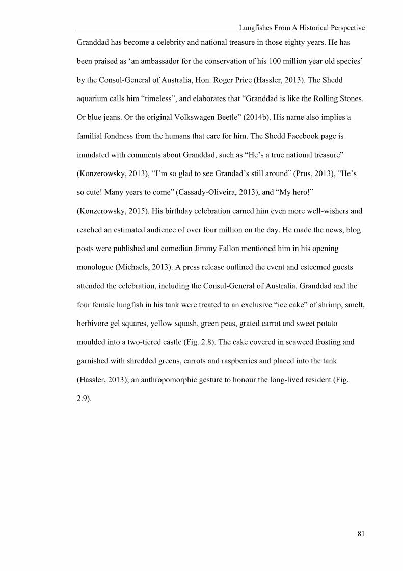
Lungfishes From A Historical Perspective
81
Granddad has become a celebrity and national treasure in those eighty years. He has
been praised as ‘an ambassador for the conservation of his 100 million year old species’
by the Consul-General of Australia, Hon. Roger Price (Hassler, 2013). The Shedd
aquarium calls him “timeless”, and elaborates that “Granddad is like the Rolling Stones.
Or blue jeans. Or the original Volkswagen Beetle” (2014b). His name also implies a
familial fondness from the humans that care for him. The Shedd Facebook page is
inundated with comments about Granddad, such as “He’s a true national treasure”
(Konzerowsky, 2013), “I’m so glad to see Grandad’s still around” (Prus, 2013), “He’s
so cute! Many years to come” (Cassady-Oliveira, 2013), and “My hero!”
(Konzerowsky, 2015). His birthday celebration earned him even more well-wishers and
reached an estimated audience of over four million on the day. He made the news, blog
posts were published and comedian Jimmy Fallon mentioned him in his opening
monologue (Michaels, 2013). A press release outlined the event and esteemed guests
attended the celebration, including the Consul-General of Australia. Granddad and the
four female lungfish in his tank were treated to an exclusive “ice cake” of shrimp, smelt,
herbivore gel squares, yellow squash, green peas, grated carrot and sweet potato
moulded into a two-tiered castle (Fig. 2.8). The cake covered in seaweed frosting and
garnished with shredded greens, carrots and raspberries and placed into the tank
(Hassler, 2013); an anthropomorphic gesture to honour the long-lived resident (Fig.
2.9).

Lungfishes From A Historical Perspective
82
Fig. 2.7. Granddad’s 80th birthday cake, Shedd Aquarium (Brenna Hernandez, 2013).
Fig. 2.8. Granddad at his 80th anniversary celebration, Shedd Aquarium (Brenna Hernandez, 2013).
Bonnie McGrath illustrated the bond felt between human and lungfish when she wrote
of Granddad in an article released about the lungfish’s anniversary:
When I met Granddad in 1978, he was just about to celebrate his 45th anniversary as
a South Loop resident. And I thought he was old then! He’s been living in the South
Loop a lot longer than I have. I’ve only been here 19 years. But we both came to live

Lungfishes From A Historical Perspective
83
here during the month of September, although I arrived 61 years later (McGrath,
2013).
Michelle Sattler of Shedd said, “A lot of people have a great relationship with
[Granddad], and a lot invested in him. So, we hope to see him for years to come here at
the aquarium,” and Ken Ramirez, Shedd’s executive vice president of animal care added
“Granddad makes people happy. Everybody knows him” (Coffey, 2013).
After his milestone as the oldest fish in captivity, Granddad enthusiasts are pondering if
he holds the secret to immortality. Robert H. Jordan reported, “Scientists are hoping that
by studying [Granddad] and other lungfish, they may unlock the secrets to longevity,
which can then be applied to us” (Jordon, 2013). Such stories are reveal the nature of
human desires in contemporary Western society, illustrating our obsession with
celebrity: we care more for one “famous” lungfish, than the environment that sustains
the species. In addition, due to this celebrity, we create anthropocentric items of ritual
celebration that have no meaning for the fish, like a birthday cake and Facebook
comments, rather than choosing to release it back into the wild. They highlight our
“need” for the “exotic” and the “Other” in order to feel “normal”, as well as our
obsession with longevity and immortality. We see other animals and the living world as
technology and tools that can be utilised for human desire.
Back on Australian soil, the Australian lungfish is a mascot of Queensland. Its influence
and signature can be seen in a number of art forms scattered all over the state. In
Bundaberg, “The Ceratodus”, a large sculpture of the lungfish made entirely of recycled
material by John Olsen, is a landmark of the city’s riverside parklands. Electricity poles
in the town of Gayndah (the town where the original specimens Krefft used to name the
Australian lungfish came from) have lungfishes painted on them, and in Mundubbera, a
mural covers the entirety of a water tower (Fig. 2.10). Painted in 1997, this mural

Lungfishes From A Historical Perspective
84
depicts the meeting of the Auburn, Boyne and Burnett rivers, and large lungfishes are
seen swimming amongst other iconic flora and fauna of the area.
Fig. 2.9. Mundubbera water tower, Queensland (Mattinbgn, year unknown).
The signature of the lungfish is apparent all over the world; from contemporary
sculptures and art that celebrate the lungfish and its legacy, to Granddad, whose
presence has been in the hearts and minds of Chicago for nearly a hundred years. These
narratives have shaped the way contemporary Western culture interprets and
understands itself through the lungfish.
2.5 NINE BLIND LUNGFISHES: A PERSONAL NARRATIVE
The last four years of my life have concentrated on the visual perception of lungfishes.
Although my research touches on several species, my encounters with lungfishes in the
flesh have been with the Australian species, N. forsteri (Fig. 2.11). Part of my research
into their visual perception involved testing the colour vision capabilities of N. forsteri
(including the ultra-violet in juveniles) to confirm if they utilize their potential for
colour vision. As discussed in the following chapters, my research shows that their eyes
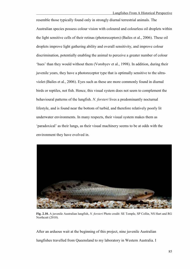
Lungfishes From A Historical Perspective
85
resemble those typically found only in strongly diurnal terrestrial animals. The
Australian species possess colour vision with coloured and colourless oil droplets within
the light sensitive cells of their retinas (photoreceptors) (Bailes et al., 2006). These oil
droplets improve light gathering ability and overall sensitivity, and improve colour
discrimination, potentially enabling the animal to perceive a greater number of colour
‘hues’ than they would without them (Vorobyev et al., 1998). In addition, during their
juvenile years, they have a photoreceptor type that is optimally sensitive to the ultra-
violet (Bailes et al., 2006). Eyes such as these are more commonly found in diurnal
birds or reptiles, not fish. Hence, this visual system does not seem to complement the
behavioural patterns of the lungfish. N. forsteri lives a predominantly nocturnal
lifestyle, and is found near the bottom of turbid, and therefore relatively poorly lit
underwater environments. In many respects, their visual system makes them as
‘paradoxical’ as their lungs, as their visual machinery seems to be at odds with the
environment they have evolved in.
Fig. 2.10. A juvenile Australian lungfish, N. forsteri Photo credit: SE Temple, SP Collin, NS Hart and RG Northcutt (2010).
After an arduous wait at the beginning of this project, nine juvenile Australian
lungfishes travelled from Queensland to my laboratory in Western Australia. I

Lungfishes From A Historical Perspective
86
personally checked on their welfare every day, feeding them and making sure the water
quality in their tanks was optimal. I noticed subtle differences between the personalities
of each fish. It is difficult to remain indifferent when caring for an organism, human or
otherwise, and I was no exception. I anthropomorphised my charges, grew fond of
them, and looked forward to observing their responses to different coloured lights. After
four months, a photographer visited our research group to document the various animals
we were studying. I took advantage of this rare opportunity to photograph my own fish,
preparing a freshly clean glass tank in a room bathed in light. I picked one fish and
carefully brought it down to model for the camera. As the photographer was setting up
the equipment, I noticed something peculiar. Amidst the bright lights and through a
freshly cleaned glass, the head of this particular individual looked strange: the fish
appeared to have no eyes. I was numb with shock. The shape and colour of the
lungfish’s head were normal, but in the area its eyes were not where they should have
been (Fig. 2.12). There was no scarring, no indication that eyes had been removed or
deformed in some way; they seemed simply to have not developed at all (Fig. 2.13).
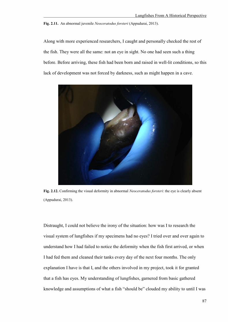
Lungfishes From A Historical Perspective
87
Fig. 2.11. An abnormal juvenile Neoceratodus forsteri (Appudurai, 2013).
Along with more experienced researchers, I caught and personally checked the rest of
the fish. They were all the same: not an eye in sight. No one had seen such a thing
before. Before arriving, these fish had been born and raised in well-lit conditions, so this
lack of development was not forced by darkness, such as might happen in a cave.
Fig. 2.12. Confirming the visual deformity in abnormal Neoceratodus forsteri: the eye is clearly absent
(Appudurai, 2013).
Distraught, I could not believe the irony of the situation: how was I to research the
visual system of lungfishes if my specimens had no eyes? I tried over and over again to
understand how I had failed to notice the deformity when the fish first arrived, or when
I had fed them and cleaned their tanks every day of the next four months. The only
explanation I have is that I, and the others involved in my project, took it for granted
that a fish has eyes. My understanding of lungfishes, garnered from basic gathered
knowledge and assumptions of what a fish “should be” clouded my ability to until I was

Lungfishes From A Historical Perspective
88
faced with an undeniable “truth”. Like Owen, my expectation of a certain “truth”
inhibited an understanding possible only when these expectations were cast aside.
2.6 THE CONNECTION BETWEEN HUMAN CULTURAL HISTORY AND
THE LUNGFISH UMWELT
In this chapter, I have outlined different stories about lungfishes and the humans who
invented and told these stories. To the Indigenous peoples of parts of Australia and
Africa, the liminal nature of lungfishes have made them celebrated creatures of the
living world, and illustrated an understanding of the embeddedness of the human within
the natural environment. In the mythical stories of the minhocao and the buru spread by
colonialists of the New World however, the differences of lungfishes as transitional
animals made them monstrous. For Richard Owen in Victorian England, the fiercely
embedded ideologies of a Linnaean creationist taxonomy combined with Owen’s own
personal biases resulted in an incorrect description of L. annectens, a transitional
creature that did not fit the established natural order of 19th century European science. In
newly colonised Australia, the “rediscovered” lungfish was a commodity that shored up
the position of “Australian” science. The narratives about Granddad illustrate a
contemporary self-absorption and obsession with celebrity and immortality that
positions lungfishes as commodities and tools for human advancement. Finally, my own
story reveals how reality is obfuscated by preconceived assumptions of what a lungfish
“should” be.
These human narratives show that many factors, aside from the evaluation of scientific
data, influence our understanding of lungfishes. These inescapable biases are formed by
ingrained beliefs, assumptions and ideologies which often subconsciously, force a
particular understanding of what lungfishes are and/or should be. John Berger discusses

Lungfishes From A Historical Perspective
89
this intimate connection between what we “see” and what we “know”, saying, “The way
we see things is affected by what we know or what we believe...we never look at just
one thing; we are always looking at the relation between things and ourselves” (Berger,
2003). Such human cultural narratives of lungfishes show that these biases are prevalent
across cultures and throughout history, and thereby influence how humans choose to
understand liminal creatures like lungfishes because, “[narrative] accounts are not
simply representations of the world; they are part of the world they describe”
(Hammersley and Atkinson, 2007). In addition, these narratives also illustrate that non-
human animals are capable of eliciting an unsettling response in humans, especially
when they threaten the security of fundamental religious ideologies and definitions of
life. Just as scientific research is one “social arena” in which knowledge about how a
lungfish may visually perceive the world may be examined, cultural ideologies and
assumptions influence how humans comprehend the visual experience of lungfishes. It
is important therefore, to recognize these multiple discourses to obtain a complex
understanding of lungfish Umwelten.
2.7 REFERENCES
2006. Save the lungfish. Nature, 442, 224-224. 2013. History of the fish collection; Johann Natterer (1787-1843) [Online]. Vienna: Naturhistorisches
museum wien. Available: http://www.nhm-wien.ac.at/en/research/_zoology_vertebrates/fish_collection_/history [Accessed 16th Jan 2014 2014].
2014a. Buganda clans and their totems - Emmamba Gabunga [Online]. Buganda Kingdom. Available:
http://www.buganda.or.ug/index.php/buganda-clans-and-their-totems-v15-523/emmamba-gabunga [Accessed 11.09.2014 2014].
2014b. Granddad: the aquarium's oldest fish [Online]. Chicago: Shedd Aquairum. Available:
http://www.sheddaquarium.org/granddad.html [Accessed 16 Jan 2014]. BAILES, H. J., ROBINSON, S., TREZISE, A. E. O. & COLLIN, S. P. 2006. Morphology,
characterization, and distribution of retinal photoreceptors in the Australian lungfish Neoceratodus forsteri (Krefft, 1870). The Journal of Comparative Neurology, 494, 381-397.

Lungfishes From A Historical Perspective
90
BARRETO, C. & MACHADO, J. S. 2001. Exploring the Amazon, explaining the unknown: Views from the past. In: MCEWAN, C., BARRETO, C. & NEVES, E. (eds.) Unknown Amazon, nature and Culture in Ancient Brazil. London: British Museum Press.
BERGER, J. 2003. Ways of seeing. The Feminism and Visual Culture Reader, 37-39. BISCHOFF, T. L. W. 1840. Lepidosiren paradoxa: Anatomisch untersucht und beschrieben, Leipzig, L.
Voss. CASSADY-OLIVEIRA, E. 2013. Social networking group [Online]. (Facebook). 19 September.
Available: https://www.facebook.com/sheddaquarium/photos/a.10151668601582843.1073741860.48206282842/10151668601762843/?type=3&theater [Accessed 14 April 2015].
CLADWELL, W. 1884. On the development of the Monotremes and Ceratodus. Proceedings of the Royal
Society of New South Wales, 18, 117-122. COFFEY, L. T. 2013. Let him eat cake! 'Granddad' the fish celebrates 80 years at aquarium. today.com
[Online]. Available from: http://www.today.com/pets/let-him-eat-cake-granddad-fish-celebrates-80-years-aquarium-4B11187459 [Accessed 26 June 2015].
CROW_BOY. 2007. The many faces of Eve. Sunshine Coast Daily, December 3, 2007. EDWARDS, M. M. 1841. LIV.—On the natural affinities of the Lepidosiren; and on the differing
opinions of Mr. Owen and M. Bischoff with regard to them. Annals and Magazine of Natural History, including Zoology, Botany, and Geology: SERIES 1, 6, 466-468.
ERESHEFSKY, M. 2001. The Poverty of the Linnaean Hierarchy, Cambridge, Cambridge University
Press. FESL, E. M. D. n.d. Extinction is Forever! A bid to save "Dala", the Queensland lungfish, an endangered
species in the Mary River across whcih a dam is proposed to be built. [Online]. Brisbane. Available: http://www.savethemaryriver.com/_mgxroot/page_10786.html [Accessed 20 May 2014 2014].
A Whisper From The Past, 2007. Radio. Directed by FRANKLIN, N. Australia: ABC National Radio. GARDINER, B. G. 2001. Linneaus's' species concept and his views on evolution. The Linnean, 17, 24 -
36. GARRETT, P. 2009. Refusal of Approval: Traveston Crossing Dam, Mary River, Queensland, (EPBC
2006/3150). In: DEPARTMENT OF THE ENVIRONMENT, W., HERITAGE AND THE ARTS (ed.). Canberra.
GEGENBAUR, C. 1878. Elements of comparative anatomy, Macmillan and Co. GÜNTHER, A. 1871. The new Ganoid fish (Ceratodus) recently discovered in Queensland. Nature, 4,
428-429. GÜNTHER, A. 1871. Description of Ceratodus, a genus of ganoid fishes, recently discovered in rivers of
Queensland, Australia. Philosophical Transactions of the Royal Society of London, 161, 511-571.
HAMMERSLEY, M. & ATKINSON, P. 2007. Ethnography: Principles in practice, Routledge. HARAWAY, D. J. 1992. The Promise of Monsters: A regenerative politics for inappropriate/d Others. In:
GROSSBERG, L., NELSON, C. & TREICHLER, P. A. (eds.) Cultural Studies. New York: Routledge.
HASSLER, M. 2013. Shedd celebrates anniversary of oldest fish in any aquarium. HECTOR, J. 1874. Notice of Ceratodus forsteri, Krefft or Barramunda of the Queensland Rivers.
Transactions and Proceedings of the Royal Zoological Society of New Zealand, 7, 490-491.

Lungfishes From A Historical Perspective
91
Save Mary - Eve Fesl, 2008. Directed by JERRYINBRISBANE. Australia: YouTube. JORDON, R. H. 2013. Shedd honors 80 years with lungfish "Granddad". WGNtv, 17 September. JOSS, J. 2007a. Standing Committee on Rural and Regional Affairs and Transport Inquiry: Additional
Water Supplies for South East Queensland – Traveston Crossing Dam. In: DEPARTMENT OF SENATE, P. H. (ed.). Canberra: Senate Printing Unit.
JOSS, J. 2007b. A Very Special Fish 2013 Australian Lungfish under Threat [Online]. Brisbane:
thepetitionsite. Available: http://www.thepetitionsite.com/1/a-very-special-fish-australian-lungfish-under-threat/ [Accessed November 12 2014].
KITCHER, P. 1993. Legend's Legacy. Advancement of Science: Science without Legend, Objectivity
without Illusions. Oxford: Oxford University Press. KLAPPROTH, D. M. 2004. Narrative as social practice: Anglo-Western and Australian Aboriginal oral
traditions, Walter de Gruyter. KONZEROWSKY, M. 2013. Social networking group [Online]. (Facebook). 8 November. Available:
https://www.facebook.com/sheddaquarium/photos/a.10151774840257843.1073741872.48206282842/10151774840272843/?type=3&theater [Accessed 14 April 2014].
KONZEROWSKY, M. 2015. Social networking group [Online]. (Facebook). 15 June. Available:
https://www.facebook.com/sheddaquarium/photos/a.55982732842.69877.48206282842/10152988362467843/?type=3&theater [Accessed 26 June 2015].
KREFFT, G. 1870. Description of a gigantic amphibian allied to Genus Lepidosiren, from the Wide Bay
District, Queensland. Proceedings of the Scientific Meetngs of the Zoological Society of London, 16, 221-224.
KREFFT, J. L. G. 1840. Letter to the Editor. Sydney Morning Herald, 18 Jan 1870. LATOUR, B. & WOOLGAR, S. 1986. Laboratory Life: The Construction of Scientific Facts., Princeton,
Princeton University Press. MARSHALL, C. R. 1986. A list of fossil and extant dipnoans. Journal of Morphology, 190, 15-23. MCGRATH, B. 2013. Australian lungfish "Granddad" celebrates his 80th anniversary at the Shedd
(many more to come) [Online]. Chicago: chicagonow.com. [Accessed 16 Jan 2014]. MCGRATH, N. 2008. Bob Brown leads Traveston Dam protest. The Sydney Morning Herald, September
6, 2008. METEOROLOGY, B. O. 2006. Drought intensifies over eastern and southern Australia as spring rains
fail. Queensland: BOM. MICHAELS, L. 2013. Late Night with Jimmy Fallon. (television broadcast) 21 September 2013. New
York, Broadway Videos and Universal Television. MONTEITH, G.B. 2001. Gayndah, lungfish and beetles - a fishy tale. The Queensland Naturalist, 39, 5-
32. MUECKE, S. 1983. Ideology reiterated - the uses of Aboriginal oral narrative. Southern Review Adelaide,
16, 86-101. NABEGEYO, B. (Aborigine, lived 1949-2009). Untitled (Lung fish). Natural earth pigments on
eucalyptus bark. 78.5x29cm. NAGEL, T. 1974. What Is It Like To Be A Bat? The Philosophical Review, 83, 435-450. NATTERER, J. 1837. Lepidosiren paradoxa, eine neue Gattung aus der Familie der fischähnlichen
Reptilien. Ann Wien Mus, II, 165-170.
Lungfishes From A Historical Perspective
92
OLSEN, P. 2010. Burnett Salmon (Australian lungfish). Upside Down World: Early European
Impressions Of Austraila's Curious Animals. Canberra National Library of Australia. OWEN, S. R. 1840. XX. Description of the Lepidosiren annectens. Transactions of the Linnean Society
of London, 18, 327-361. PEARSON, H. 2006. Dam project threatens living fossil. Nature, 442, 232-233. PRUS, D. M. 2013. Social networking group [Online]. (Facebook). 19 September. Available:
https://www.facebook.com/media/set/?set=a.10151668601582843.1073741860.48206282842&type=3 [Accessed 14 April 2014].
The Importance of Dala: The Queensland Lungfish, 2009. Online. Directed by QUEENSLAND, S. L. O.
Australia: YouTube. RAMSBOTTOM, J. 1938. Linnaeus and the species concept. Proceedings of the Linnean Society of
London, 150, 192-220. RIEDL-DORN, C. & NATTERER, J. 1999. Johann Natterer und die Österreichische Brasilien
expedition, Editora Index, Vienna. SAINT HILAIRE, M. A. 1847. On the Minhocao of Goyanese, an enormous species of Lepidosiren. The
Edinburgh New Philosophical Journal, 42, 278-281. SCLATER, P. 1870. The new Australian mud-fish. Nature, 2, 106-107. SEMON, R. 1899. The Auburn Camp. In the Australian Bush and on the Coast of the Coral Sea: Being
the experiences and observations of a naturalist in Australia, New Guinea and the Moluccas. London: MacMillan and Co. Limited.
SEMON, R. 1901. Normentafel zur Entwicklungsgeschichte des Ceratodus forsteri, Germany, Jena, G.
Fischer. SHUKER, K. 2012. Riddle of the buru, and the lungfish link. Available from:
http://karlshuker.blogspot.com.au/2012/08/riddle-of-buru-and-lungfish-link.html [Accessed 20 May 2014].
TAYLOR, M. 2013. Johann Natterer (1787-1843). Available from:
http://exhibitions.blogs.lib.lsu.edu/?page_id=1822 [Accessed 23 June 2013]. UEXKÜLL, J. V. 2010. A Foray Into The Worlds Of Animals And Humans With A Theory Of Meaning,
Minneapolis, University of Minnesota Press. VOROBYEV, M., OSORIO, D., BENNETT, A., MARSHALL, N. & CUTHILL, I. 1998.
Tetrachromacy, oil droplets and bird plumage colours. Journal of Comparative Physiology A, 183, 621-633.
ZIMMER, C. 1998. At The Water's Edge: Macroevolution and the Transformation of Life, New York,
The Free Press.


Retinal Photoreceptors in the South American and Spotted African Lungfishes
94
CHAPTER 3
:::::: MORPHOLOGY,
CHARACTERISATION AND
DISTRIBUTION OF RETINAL
PHOTORECEPTORS IN THE
SOUTH AMERICAN
(LEPIDOSIREN PARADOXA) AND
SPOTTED AFRICAN
(PROTOPTERUS DOLLOI)
LUNGFISHES
::::::

Retinal Photoreceptors in the South American and Spotted African Lungfishes
95
3.1 INTRODUCTION
In this chapter, the use of anatomical techniques to investigate visual photoreception in
the South American (Lepidosiren paradoxa) and spotted African (Protopterus dolloi)
lungfishes is described. New findings concerning aspects of the morphology, density
and topographic distribution of their retinal photoreceptors are presented that are
essential in unravelling the visual mechanisms which influence how these lungfishes
experience the world visually.
Of 70 lungfish species described through fossilised remains (Clack et al., 2011), six
species remain in existence. The spotted African lungfish (P. dolloi), the marbled
lungfish (P. aethiopicus), the West African lungfish (P. annectens) and the gilled
lungfish (P. amphibius) are members of the family Protopteridae and are all found in
Africa. The South American lungfish (L. paradoxa) within the family Lepidosirenidae
inhabits the Amazon River and Parana-Paraguay River basins, and the Australian
lungfish (Neoceratodus forsteri) resides in the rivers of Queensland, Australia.
The South American lungfish resides in the neotropics of South America and has the
most extensive distribution of all lungfish species (Fonesca de Almeida-Val et al.,
2011). Its range extends through Argentina, Bolivia, Colombia, Brazil, Paraguay,
Venezuela and French Guiana, and although it is found in the Parana-Paraguay River
system, it is preferentially located within the Amazon River basin (Fonesca de Almeida-
Val et al., 2011). All African lungfishes are endemic to the river systems of a large part
of the African continental landmass, and P. dolloi primarily inhabits the Congo River
basin.
The species’ used as model organisms in this research, the spotted African (P. dolloi)
and South American (L. paradoxa) lungfishes have eel-like elongate bodies,
filamentous pelvic and pectoral fins and a continuous diphycercal tail formed by the
Retinal Photoreceptors in the South American and Spotted African Lungfishes
96
fusion of the dorsal, caudal and anal fins. All lungfishes possess paired pelvic and
pectoral fins, although the paired fins of L. paradoxa and P. dolloi are thinner compared
to other species. These paired fins have a minimal contribution to propulsion, but aid in
stabilisation of the body, orientation and prey capture through tactile and chemosensory
input (Johnels and Svensson, 1954, Curry-Lindahl, 1956, Thomson, 1969, Greenwood,
1986, Fonesca de Almeida-Val et al., 2011). It is not known at what depth in the
shallow swamps of the Congo River the spotted African lungfish spends its time
(Greenwood, 1986), but the South American lungfish tends to spend most of its time
near the surface of the water (Fonesca de Almeida-Val et al., 2011). Both species swim
using anguilliform23 locomotion and may utilize their paired fins to navigate when
moving over the bottom of the riverbed.
Both species are omnivorous carnivores, and are considered to be opportunistic feeders
that consume a wide, generalist diet. For Protopterus, molluscs are the primary food
source, but both the African and South American species also ingest small fish,
crustaceans, aquatic insects, worms and vegetation (algae, weeds and terrestrial plant
stems) (Greenwood, 1987, Fonesca de Almeida-Val et al., 2011, Mlewa et al., 2011).
All lungfish species, except for the gilled lungfish (P. amphibius)24 are considered to be
large. Greenwood (1986) reported P. dolloi to reach a length of up to 1.3 metres, and L.
paradoxa to average 1.25 metres in total length (Arratia, 2003). In addition, there are no
known natural predators of lungfishes that have been quantified outside of anecdotal
accounts which may be a contributing factor, along with their diet, to their large adult
body size.
23 A type of fish locomotion using muscle contractions in the body to generate undulating body movements. 24 Greenwood (1986) reported the largest known adult P. amphibius to be 44.3 centimetres.
Retinal Photoreceptors in the South American and Spotted African Lungfishes
97
The eyes of lungfishes, although relatively small compared to their body size, are well-
developed and provide a clear image of the world. The optical image gives rise to a
neural image at the level of the retina and more specifically, the photoreceptors.
Characterisation of the complement of photoreceptor types based on size, shape, and
spectral sensitivity and their topographic distribution across the retina are important
criteria and powerful predictors of retinal, and therefore visual function in animals.
Previous studies of N. forsteri show that at least some species of lungfishes possess a
complex colour vision system that strongly resembles that of diurnal vertebrates such as
birds and reptiles, which suggests that several of the ocular characteristics these animals
possess may have evolved in shallow water before the transition of tetrapods onto land
(Collin, 2010). One such characteristic is the presence of corneal surface
microprojections that are important for clear vision in air, and which are present in both
terrestrial vertebrates and in N. forsteri. As discussed previously, in N. forsteri, these
form a thick carpet of microvilli extending over the dermal cornea, which stabilise the
corneal tear film. As discussed in the General Introduction, some species of lungfishes
may have evolved this characteristic in order to move onto land to aestivate and may
require clear aerial vision (Collin and Collin, 2001).
Previous anatomical studies have shown that the eyes of lungfishes possess a number of
different types of large photoreceptors, demonstrating that they have the retinal
machinery to process colour (Walls, 1942, Pfeiffer, 1968, Ali and Anctil, 1973). The
retina of N. forsteri – which is considered to be the most primitive of the extant
lungfishes – is the only species that has been studied in detail (Marshall, 1986, Tokita et
al., 2005, Bailes et al., 2006). Superficial examination of the other lungfish families has
suggested that these species may have more complex ocular characteristics than
N. forsteri. By describing the ocular mechanics of representatives of the other lungfish
Retinal Photoreceptors in the South American and Spotted African Lungfishes
98
families, this study therefore fills a neglected, yet crucial gap in knowledge of the
evolution of terrestrial vision.
As previously discussed up to five spectrally distinct types of large retinal
photoreceptors have been found in N. forsteri: one type of rod (λmax 540nm) and four
types of cones (UVS λmax 366nm, SWS λmax 479nm, MWS λmax 558nm and LWS λmax
623nm). UVS cones are only found in the retinas of juvenile N. forsteri, suggesting that
sensitivity to ultraviolet light is lost during maturation (Bailes et al., 2006, Hart et al.,
2008). The possession of four types of cones in juvenile lungfishes implies that they
have the potential for tetrachromatic vision, which is perhaps unusual for an animal that
has previously been considered predominantly nocturnal or crepuscular25 (Dean, 1906,
Dean, 1912, Grigg, 1965, Kemp, 1986). To date, the retinae of the South American and
spotted African lungfishes have only been superficially characterised by using light
microscopy and both species are thought to possess one type of rod and at least two
types of cone, suggesting that these species may also have the capacity for colour
vision. P. dolloi appears to possess both single and double cones, while L. paradoxa and
N. forsteri possess only single cones (Ali and Anctil, 1973, Bailes et al., 2006).
P. dolloi and L. paradoxa possess oil droplets in both their cone and rod photoreceptors
(Kerr, 1902, Walls, 1942, Pfeiffer, 1968, Ali and Anctil, 1973), whereas N. forsteri only
possesses colourless oil droplets in their SWS cones, coloured yellow pigment in their
MWS cones and red oil droplets in their LWS cones (Bailes et al., 2006). Colourless oil
droplets are found in marsupial mammals which are generally nocturnal, crepuscular or
cathemeral26 and are thought to improve light gathering ability and thus, overall
sensitivity. Coloured oil droplets act as miniature spectral filters within each
photoreceptor and, by narrowing the spectral sensitivity function of the photoreceptors,
25 Active predominantly during twilight (dawn and dusk). 26Active sporadically at random intervals during the day or night dependant on food availability, predation pressures and other environmental cues such as temperature.
Retinal Photoreceptors in the South American and Spotted African Lungfishes
99
are thought to improve colour discrimination by reducing overlap with adjacent spectral
classes of cone (Hart et al., 2008, Vorobyev et al., 1998). This potentially enables the
animal to perceive a far greater number of distinct colour ‘hues’ in the visual spectrum
than would be possible without the coloured oil droplets (Govardovskii, 1983, Hart et
al., 2008). The presence of coloured oil droplets in the lungfish retina is surprising
because these characteristics are found only in strongly diurnal animals such as birds
and reptiles, and the spotted African and South American lungfishes occupy swamps
and lakes that consist of lentic (stagnant) water bodies associated with poor visibility,
weedy vegetation, low oxygen content and seasonal drying (Greenwood, 1986, Mlewa
et al., 2011).
In most animals with image-forming eyes, the densities of photoreceptor cells are not
uniform across the retina (Collin, 1999). The topographic distribution of photoreceptors
is instead species-specific, and usually reflects key features of their visual behaviour in
respect to their physical environment, as well as determining the visual acuity or
“spatial resolving power” of their eyes. Analysis of the topographic distribution of
photoreceptors in the retina is a powerful way to gain insights as to the importance each
species places on observing objects in specific regions of their visual field.
The area centralis and the horizontal streak are the two main topographical
specialisations in the retina. The area centralis is a concentric increase in retinal cell
density in a specific retinal location that allows for increased spatial sampling in the
corresponding region of visual space (Fig. 3.1A). An area centralis is more commonly
found in species that live in enclosed environments such as coral reefs and river systems
(Fig. 1B). A horizontal streak is characterised by a band of increased cell density across
the central meridian of the retina (Fig. 3.1C). This retinal arrangement facilitates
increased spatial sampling of a large part of the visual image in panoramic
environments such as open ocean or land, without the need for the extensive eye

Retinal Photoreceptors in the South American and Spotted African Lungfishes
100
movements that would be required to scan the same area of visual space with an area
centralis (Fig. 1D).
Fig. 3.1. Topographic maps of ganglion cells in illustrating different specialisations and the species involved, in their natural habitat. (A) The area centralis in the coral cod, Cephalopholis miniata (<10.0 to 47.0 x 10³ ganglion cells per mm2. Adapted from (Collin and Pettigrew, 1988b). (B) The coral cod, C. miniata in its natural habitat in amongst the marine reefs of the Indo-West and Central Pacific. Image taken by Erik Schlögl (Schlögl, 2002). (C) The horizontal streak in a small-spotted catshark, Scyliorhinus canicula (<0.5 to 2.4 x 10³ ganglion cells per mm2). Adapted from (Bozzano and Collin, 2000).(D) The small-spotted catshark, S. canicula in its natural habitat in the open marine environment of the Northest Atlantic ocean. Image taken by Uwe Waller (Waller, 1998).
The areas that exhibit an increase in photoreceptor density in the N. forsteri retina vary
as the fish matures (Bailes et al., 2006). The highest density of rods in the juvenile and
sub-adult N. forsteri lie in the temporal retina, implying an increase in retinal sensitivity
in the frontal visual field. As the fish matures, this becomes two areas of increased rod
density in the more central retina in adults, forming a weak horizontal band across the
retinal meridian. In general, the increased density of cone photoreceptors is
predominantly in the dorso-temporal retina and the ventral-nasal retina in all growth
stages of N. forsteri, indicating a downwardly directed visual axis (Bailes et al., 2006).
However, the topographic specializations in L. paradoxa or Protopterus species are not
yet known.

Retinal Photoreceptors in the South American and Spotted African Lungfishes
101
Both Protopterus spp. and L. paradoxa undergo aestivation during the dry season in
their relevant habitats. Aestivation can be described as a ‘light’ state of dormancy,
characterised by inactivity and a lowered metabolic rate that can be quickly reversed if
the right conditions are met. Five out of the six species of lungfishes undergo
aestivation, with the Australian species N. forsteri, being the only exception. This
process enables these ancient fishes to avoid damage from high temperatures and
desiccation during the dry season without leaving the swamps for permanent water, like
some teleost fishes (Greenwood, 1986). In their respective burrows, both species of
lungfishes remain until the onset of the wet season, which may be for up to eight
months. It is not known if vision plays a significant role in their lifestyle during this
aestivating phase, since normal metabolic function and activity is not triggered until the
wet season begins.
Consequently, the visual system in lungfishes is something of a conundrum. As for
N. forsteri, it appears the habitat and lifestyle of P. dolloi and L. paradoxa are also not
heavily reliant on their visual system. Little is known about the visual behaviour of any
lungfish species, and it has been stated a number of times that vision does not contribute
heavily to their lifestyle, at least in prey capture and navigation (Carter and Beadle,
1930, Johnels and Svensson, 1954, Curry-Lindahl, 1956, Pfeiffer, 1968, Greenwood,
1986, Owen, 1840). The photic habitat of the swamps and rivers of the Amazon and
Congo is limited, allowing only poor visibility, with a substantial amount of sediment
and mineral deposits contributing to turbid, and therefore relatively dim, underwater
light environments. Combined with their aestivating behaviour, where they remain
dormant for most of the year, it would appear that vision does not play an essential role
in survival. However, the complex visual apparatus of the lungfish eye, including a well
developed lens and active accommodatory apparatus, the presence of coloured oil
droplets and the presence of four spectral types of cone photoreceptor in N. forsteri

Retinal Photoreceptors in the South American and Spotted African Lungfishes
102
(Bailes et al., 2006, Bailes et al., 2007), suggests that colour vision is more important to
lungfishes than previously thought27.
This chapter endeavours to fill crucial knowledge gaps regarding the visual capabilities
of P. dolloi and L. paradoxa through the characterisation of specific photoreceptor types
based on their size, ultrastructure and topographic distribution. There have only been
two published papers specifically focused on the retina of P. dolloi (Pfeiffer, 1968) and
L. paradoxa (Ali and Anctil, 1973) using retinal tissue prepared in paraffin wax for light
microscopy. This will be the first report on the retinal photoreceptors of P. dolloi and
L. paradoxa examining resin embedded retinal tissue which significantly increases
resolution, enabling the ultrastructure of the photoreceptors to be described at the level
of the transmission electron microscope. The study reveals that based on morphology;
there are at least three different photoreceptor types in juvenile P. dolloi, and one rod
and one cone photoreceptor type in L. paradoxa. The topographical distribution of a
cone photoreceptor type containing a red oil droplet is also presented for juvenile P.
dolloi. This suggests that juvenile P. dolloi have the potential for dichromatic colour
vision, and L. paradoxa may be the only lungfish species without the potential for
colour vision. These findings provide novel data that contributes to our understanding of
the visual experience of P. dolloi and L. paradoxa, and provide insight to the role vision
plays in their behavioural ecology.
27Early lungfish studies suggested that these fish were almost blind, and possessed degenerate eyes (Dean 170).

Retinal Photoreceptors in the South American and Spotted African Lungfishes
103
3.2 METHODS
3.2.1 Samples
All lungfish eye samples were donated by international collaborators who had obtained
the animals through the international pet trade. Eyes from three juvenile P. dolloi (total
length, TL 20.6 cm, 20.2 cm and 24. cm; eye codes Pd5L, Pd3R and Pd4R) were
obtained from Dr. Guido Westhoff from the University Of Bonn, Germany. Two eyes
from an adult L. paradoxa (TL 96 cm) were obtained from Prof. Glenn Northcutt from
the University of California, San Diego (UCSD), USA. Following enucleation, the lens
of L. paradoxa was removed. However due to the small size of the P. dolloi eyes, the
lens was not removed and the eyes were immersion fixed in toto. All samples were
preserved in Karnovsky’s fixative (2% paraformaldehyde, 2.5% glutaraldehyde in 0.1
M sodium cacodylate buffer, pH 7.4) prior to being transported to UWA.
3.2.2 Ocular measurements of P. dolloi
Total body length and the axial length of the eye were measured prior to fixation for all
P. dolloi samples. The axial length of a vertebrate eye has a linear allometric
relationship to body weight on a log-log plot (Howland et al., 2004). The relationship
between axial length of P. dolloi eyes and weight were compared to the regression slope
of N. forsteri, P. dolloi, and L. paradoxa by Bailes et al. (2007). The lens dimensions of
Pd3R and Pd4R were measured after fixation in Karnovsky’s. The ocular and lens
dimensions of Pd5L were measured following fixation with Karnovsky’s and osmium
tetroxide, dehydration and embedding in araldite. Dimensions were measured on a
digital photograph of a section representing the geometric centre (widest diameter) of
the eye. Radii of curvature (r) were calculated using the formula:
Retinal Photoreceptors in the South American and Spotted African Lungfishes
104
where r is the radius of curvature, y is half of the axial diameter and s is the sagittal
diameter of the curved surface when assumed to be spherical (Bailes et al., 2007). From
these measurements, the focal ratio (fr) was calculated using this formula:
where PND is the distance between the centre of the lens and the back of the retina at
90° from the plane of the cornea (posterior nodal distance), and a is the axial length of
the lens (Bailes et al., 2007).
3.2.3 Light and electron microscopy
All eye samples were processed for both light and electron microscopy to examine the
micro- and ultra-structural morphology of the photoreceptors within the retina of
P. dolloi and L. paradoxa. Due to the fragility of the photoreceptors, the retina was not
removed from the eye cup, but significant amount of connective tissue was removed
prior to embedding in Procure-Araldite (ProSciTech). However, under some
circumstances, the retina of L. paradoxa still peeled away from the underlying pigment
epithelium, subsequently damaging the photoreceptors. The eyecup was then postfixed
for one hour with 1% osmium tetroxide in 0.15 M phosphate buffer, dehydrated through
an alcohol and propylene oxide series and infiltrated with Procure-Araldite
(ProSciTech). For light microscopy, semi-thin (1 µm) sections were cut with a glass
knife using a LKB Bromma Ultratome NOVA. Semi-thin sections were then
deplastinated with a wash in a solution of sodium ethoxide, 70% alcohol and double
distilled H2O and stained with 4% Toluidine blue. Light micrographs were taken using

Retinal Photoreceptors in the South American and Spotted African Lungfishes
105
an Olympus camera (Model DP70) mounted on an Olympus compound light
microscope (Model BX50F4). For transmission electron microscopy, ultrathin sections
(110 nm) were cut using a diamond knife and mounted on a 200 mesh copper grid.
Examination of ultrathin sections was done using a JEOL 2100 transmission electron
microscope operating at 120 kV and photographed using an 11 megapixel Gatan Orius
digital camera.
3.2.4 Assessment of photoreceptor dimensions and morphology
All measurements were conducted from digital images of light and electron
micrographs using ImageJ 1.46r (National Institute of Health, USA). To allow for
comparison between photoreceptor cells, the outer segment diameter was measured at
the base, and the ellipsoidal diameter was measured at the widest point. Shrinkage of L.
paradoxa eye tissue could not be determined due to unrecorded pre-fixation
dimensions. Due to measurements of the P. dolloi eye tissue pre-fixation, shrinkage
post-fixation was calculated to be 6.3% using the methodology outlined by Bailes et al.
(2006). All measurements are quoted as mean ± standard deviation (SD) followed by
sample size (n).
3.2.5 Preparation of retinal wholemount
In order to analyse the topographic distribution of the large red oil droplet containing
cone photoreceptor type , one retina of a juvenile P. dolloi (24.3 cm in TL) was
dissected and fixed in 4% paraformaldehyde in 0.1 M phosphate buffer (PFA, pH 7.4).
A retinal wholemount was prepared according to standard protocols (Stone, 1981,
Coimbra et al., 2006). An incision was made in the pupil of the eye and the cornea
Retinal Photoreceptors in the South American and Spotted African Lungfishes
106
removed. A dorsal orientation slit was created by cutting towards the back of the eye
cup far enough that an incision was made in the retina. The retina was carefully
removed from the eye cup using No. 3 watchmaker’s forceps and a fine natural-hair
paint brush, and additional radial cuts were made in order to flatten the retina onto a
subbed glass slide. The flattened retina was then rinsed in phosphate buffer and
mounted onto a glass slide with the photoreceptor layer facing upwards in 100%
glycerol. The cover slip was sealed with nail polish to prevent the retina from drying
out. As the photoreceptor layer was facing upwards, the cones were visible in axial view
and could be distinguished from rods (based on their size and tapered outer segment) by
changing the fine focus on the microscope at a magnification of x1000 using a 1.40
numerical aperture (NA) oil immersion objective.
3.2.6 Stereological analysis and construction of a topographic map
The topographic distribution of the oil droplet containing photoreceptor in P. dolloi was
assessed using the modified optical fractionator technique detailed in Coimbra et al.
(2009). Using StereoInvestigator software (Microbrightfield Inc., USA) on a Windows
7 PC connected to a compound microscope (Olympus BX50) equipped with a
motorised stage (MAC200, Ludle Electronic Products, USA) and digital video camera
(MicroFIRE, OPTRONICS), the outline of the retinal wholemount was digitised using a
x4 objective (NA 0.13). Using an x100 oil immersion objective (1.40 NA), all red cone
photoreceptors in the retinal wholemount were counted.
The retinal outline and cell count data were exported as an Extensible Markup
Language (.xml) format file and analysed using the open source statistical program R
v2.15.1 (R Foundation for Statistical Computing 2012) modified with additional

Retinal Photoreceptors in the South American and Spotted African Lungfishes
107
packages and a custom script according to Garza-Gisholt et al. (2014) to construct the
retinal topographical map.
3.3 RESULTS
3.3.1 Eye morphology and ocular measurements of P. dolloi
Each juvenile P. dolloi used in this study was normal and healthy with minor variations
in body and head shape (Fig. 3.2). Eyes of juvenile P. dolloi are dorsally positioned,
contain a dermal and scleral cornea (Fig. 3.2B, D), and are antero-posterially flattened
(Fig. 3.3A). The dermal and scleral cornea appears to be non-pigmented, and the
conjunctiva and iris are heavily pigmented with melanin granules. The sclera is also
pigmented with clusters of melanin granules that form freckles around the periphery of
the eye, and contains a dorsal indentation towards the iris. The iris also possesses
silver/gold-reflecting material that is partly obscured by the pigmented sclera (Fig.
3.3B).
Fig. 3.2. General characteristics of the body and head of Protopterus dolloi, showing minor variations between animals. Images by Guido Westoff. (A) Pd3 body post-euthanasia. Scale bar = 1 cm. (B) Head of Pd3 post-euthanasia with dorsally positioned eyes. Scale bar = 1 cm. (C) Pd4 body post-euthanasia. Scale bar = 1 cm. (D) Head of Pd4 post-euthanasia with dorsally positioned eyes. Scale bar = 1 cm.

Retinal Photoreceptors in the South American and Spotted African Lungfishes
108
Fig. 3.3. External appearance of the isolated eye of a juvenile Protopterus dolloi. (A) The eye is antero-posterially flattened . The optic nerve (on) is visible, as is the cornea (c) where the pupil is situated. Scale bar = 1 mm (B) Front view of a juvenile P. dolloi eye. The cornea is over the iris (ir) and pupil, where the lens sits underneath. The iris and sclera (scl) is heavily pigmented, with bigger spots of pigment on the sclera, and a silver/gold-reflecting material on the iris, and a dorsal indentation (arrowhead) of the sclera towards the iris is visible. Image by Guido Westoff. Scale bar = 500 µm.
The ocular lens of P. dolloi is relatively large and is positioned more dorsally within the
eye (Fig. 3.4), which is similar to the juvenile N. forsteri. The radii of curvature and
ocular dimensions were measured in fixed tissue prepared for light and electron
microscopy (Fig. 3.4), and therefore will have suffered from some degree of shrinkage
(Table 3.1.) The lenses of juvenile P. dolloi were measured following Karnovsky’s
fixation with an average shrinkage of 6.3%. The lenses are spherical with a ratio of
0.991-0.994 (Table 3.2).
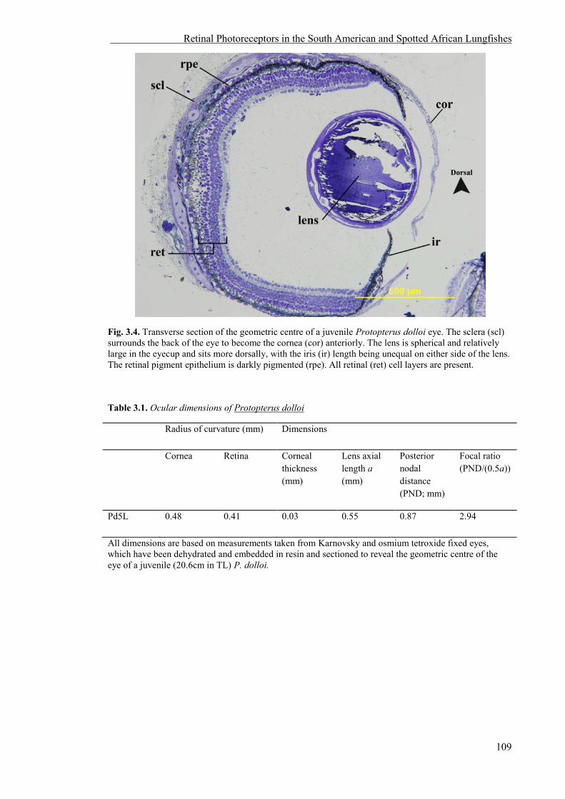
Retinal Photoreceptors in the South American and Spotted African Lungfishes
109
Fig. 3.4. Transverse section of the geometric centre of a juvenile Protopterus dolloi eye. The sclera (scl) surrounds the back of the eye to become the cornea (cor) anteriorly. The lens is spherical and relatively large in the eyecup and sits more dorsally, with the iris (ir) length being unequal on either side of the lens. The retinal pigment epithelium is darkly pigmented (rpe). All retinal (ret) cell layers are present.
Table 3.1. Ocular dimensions of Protopterus dolloi
Radius of curvature (mm) Dimensions
Cornea Retina Corneal thickness (mm)
Lens axial length a (mm)
Posterior nodal distance (PND; mm)
Focal ratio (PND/(0.5a))
Pd5L 0.48 0.41 0.03 0.55 0.87 2.94
All dimensions are based on measurements taken from Karnovsky and osmium tetroxide fixed eyes, which have been dehydrated and embedded in resin and sectioned to reveal the geometric centre of the eye of a juvenile (20.6cm in TL) P. dolloi.
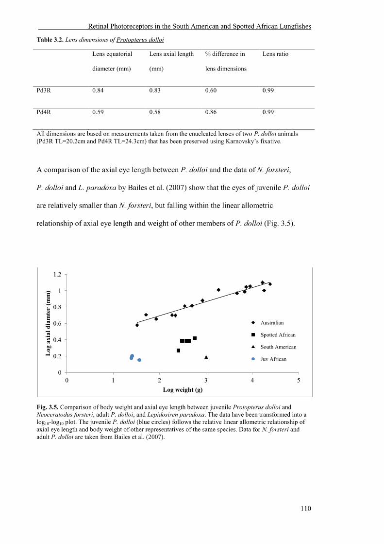
Retinal Photoreceptors in the South American and Spotted African Lungfishes
110
Table 3.2. Lens dimensions of Protopterus dolloi
Lens equatorial
diameter (mm)
Lens axial length
(mm)
% difference in
lens dimensions
Lens ratio
Pd3R 0.84 0.83 0.60 0.99
Pd4R 0.59 0.58 0.86 0.99
All dimensions are based on measurements taken from the enucleated lenses of two P. dolloi animals (Pd3R TL=20.2cm and Pd4R TL=24.3cm) that has been preserved using Karnovsky’s fixative.
A comparison of the axial eye length between P. dolloi and the data of N. forsteri,
P. dolloi and L. paradoxa by Bailes et al. (2007) show that the eyes of juvenile P. dolloi
are relatively smaller than N. forsteri, but falling within the linear allometric
relationship of axial eye length and weight of other members of P. dolloi (Fig. 3.5).
Fig. 3.5. Comparison of body weight and axial eye length between juvenile Protopterus dolloi and Neoceratodus forsteri, adult P. dolloi, and Lepidosiren paradoxa. The data have been transformed into a log10-log10 plot. The juvenile P. dolloi (blue circles) follows the relative linear allometric relationship of axial eye length and body weight of other representatives of the same species. Data for N. forsteri and adult P. dolloi are taken from Bailes et al. (2007).
0
0.2
0.4
0.6
0.8
1
1.2
0 1 2 3 4 5
Log
axi
al d
iam
ter
(mm
)
Log weight (g)
Australian
Spotted African
South American
Juv African

Retinal Photoreceptors in the South American and Spotted African Lungfishes
111
3.3.2 Photoreceptor characterisation and morphology of L. paradoxa
The photoreceptors of adult L. paradoxa are large and tightly packed. Based on
morphological characteristics at both the light and electron microscope levels and
dimensions of the photoreceptors, there is one rod and at least one cone photoreceptor
type in adult L. paradoxa (Fig. 3.6A). Both rods and cones possess oil droplets. No
double cones were observed. Oil droplets visible in the electron micrographs of
photoreceptors appear to contain electron-dense material (Fig. 3.6B). All light
micrograph images do not contain inclusions within the oil droplets (Fig. 3.6A). We
believe that the electron dense inclusions in the electron micrographs are artefacts, as
the tissue that was observed at the ultrastructral level was not stained and did not
undergo a washing stage, as it did in the preparation of the sections for light
microscopy. All photoreceptor dimensions were measured in the light-adapted retina
and are presented in Table 3.3.
Table 3.3. Photoreceptor dimensions of adult Lepidosiren paradoxa1
Basal diameter of outer segment (OS) (µm)
Length of OS (µm)
Diameter of ellipsoid (µm)
Length of ellipsoid (µm)
Oil droplet (OD) (Present/Absent)
Size of OD (µm2)
Rod 10.1 ± 0.8 (14)
8.0 ± 1.4 (13)
18.0 ± 1.8 (13)
27.7 ± 4.1 (10)
Present 268.7 ± 51.0 (12)
Cone 7.5 ± 0.7 (16)
13.6 ± 2.8 (15)
10.5 ± 1.8 (10)
22.7 ± 2.4 (6)
Present 82.8 ± 22.0 (7)
1All values are the mean ± SD with number of cells sampled in parentheses. All measurements are from one individual 96cm in TL.
The rod photoreceptor is large, and possesses a tapered outer segment that is usually
shorter in length than the outer segment of the cones (8.0 ± 1.4 µm [n = 13], with a
basal diameter of 10.1 ± 0.8 µm, n = 14) (Fig. 3.6A). The ellipsoid contains a larger oil

Retinal Photoreceptors in the South American and Spotted African Lungfishes
112
droplet than the cones (268.7 ± 51.0 µm2 [n = 12] in rods, compared to 82. ± 22.0 µm2
[n = 7] in cones), with the majority of mitochondria and the paraboloid located closer to
the nucleus (Fig. 3.6A). It is more difficult to differentiate rods and cones in
L. paradoxa in comparison to N. forsteri and P. dolloi. However, rods can be identified
by incisures within a more cylindrical outer segment.
Like all other species of lungfishes described, the cones are large (13.6 ± 2.8 µm outer
segment length [n = 15], and 10.5 ± 1.5 µm ellipsoid diameter [n = 10] in a 96 cm
individual) and contain one oil droplet (82.8 ± 21.9 µm2 [n = 7]) within the ellipsoid.
There are no additional oil droplets embedded within the mitochondria in the ellipsoid
region in the cones (Fig. 3.6B). The mitochondria are distributed more centrally within
the cone ellipsoid, close to the oil droplet (Fig. 3.6B) with a paraboloid that consists of
an aggregation of granules (Fig. 3.6B).
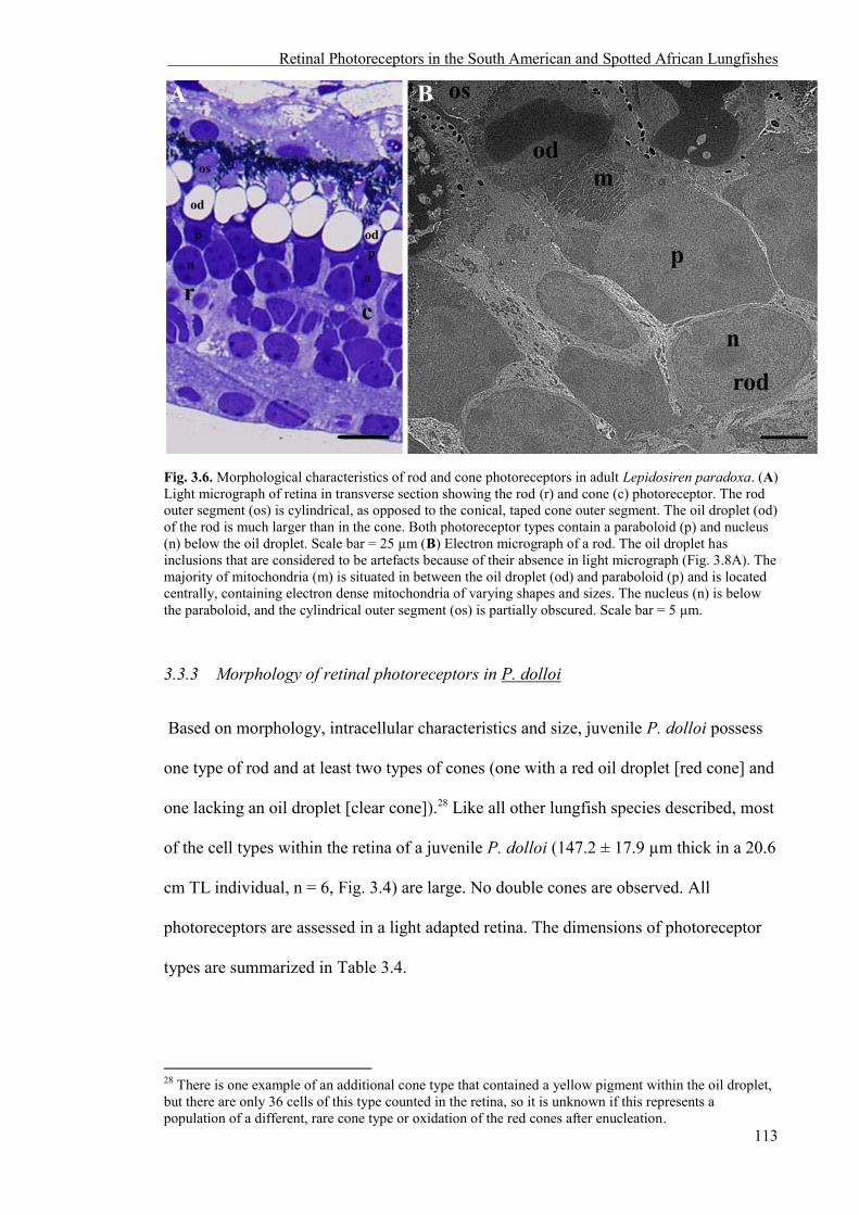
Retinal Photoreceptors in the South American and Spotted African Lungfishes
113
Fig. 3.6. Morphological characteristics of rod and cone photoreceptors in adult Lepidosiren paradoxa. (A) Light micrograph of retina in transverse section showing the rod (r) and cone (c) photoreceptor. The rod outer segment (os) is cylindrical, as opposed to the conical, taped cone outer segment. The oil droplet (od) of the rod is much larger than in the cone. Both photoreceptor types contain a paraboloid (p) and nucleus (n) below the oil droplet. Scale bar = 25 µm (B) Electron micrograph of a rod. The oil droplet has inclusions that are considered to be artefacts because of their absence in light micrograph (Fig. 3.8A). The majority of mitochondria (m) is situated in between the oil droplet (od) and paraboloid (p) and is located centrally, containing electron dense mitochondria of varying shapes and sizes. The nucleus (n) is below the paraboloid, and the cylindrical outer segment (os) is partially obscured. Scale bar = 5 µm.
3.3.3 Morphology of retinal photoreceptors in P. dolloi
Based on morphology, intracellular characteristics and size, juvenile P. dolloi possess
one type of rod and at least two types of cones (one with a red oil droplet [red cone] and
one lacking an oil droplet [clear cone]).28 Like all other lungfish species described, most
of the cell types within the retina of a juvenile P. dolloi (147.2 ± 17.9 µm thick in a 20.6
cm TL individual, n = 6, Fig. 3.4) are large. No double cones are observed. All
photoreceptors are assessed in a light adapted retina. The dimensions of photoreceptor
types are summarized in Table 3.4.
28 There is one example of an additional cone type that contained a yellow pigment within the oil droplet, but there are only 36 cells of this type counted in the retina, so it is unknown if this represents a population of a different, rare cone type or oxidation of the red cones after enucleation.

Retinal Photoreceptors in the South American and Spotted African Lungfishes
114
Table 3.4. Photoreceptor dimensions and characteristics of juvenile Protopterus dolloi1
Basal diameter of outer segment (OS) (µm)
Length of OS (µm)
Diameter of ellipsoid (µm)
Length of ellipsoid (µm)
Oil droplet (OD) (Present/Absent)
Size of OD (µm2)
Rod 9.2 ± 1.0 (32)
16.0 ± 2.1 (32)
11.7 ± 1.3 (34)
22.2 ± 2.7 (33)
Present 118.4 ± 23.6* (33)
Red cone 4.7 ± 0.8 (15)
6.4 ± 0.7 (14)
7.4 ± 1.8 (14)
12.7 ± 2.0 (12)
Present 39.0 ± 10.3 (13)
Clear cone 3.8 ± 1.0 (13)
5.6 ± 1.4 (13)
9.1 ± 1.0 (15)
12.3 ± 0.7 (14)
Absent N/A
1All values are the mean ± SD with number of cells sampled in parentheses. All measurements are from one individual 20.6cm in TL. *measurements were taken of largest oil droplet in the rod.
The rods are considered large (16.0 ± 2.1 µm outer segment length [n = 32], and 11.7 ±
1.3 µm ellipsoid diameter [n = 34], in a 20.6 cm TL individual) and contain one large
oil droplet situated within the sclerad section of the ellipsoid of the inner segment
amongst the mitochondria (118.4 ± 23.6 µm2 oil droplet area, n = 33), and unlike
L. paradoxa, a number of smaller oil droplets (between 0-5, n = 12) closer to the
nucleus (Fig. 3.7A). The outer segment is cylindrical in shape, and consists of scalloped
discs surrounded by a plasma membrane (Fig. 3.7B). The presence of rods is confirmed
by the presence of incisures in the outer segment discs (Fig. 3.7C). There are also
unknown inclusions within the rod outer segment (Fig. 3.7A). The ellipsoid of the rod is
similar to that of L. paradoxa and consists of electron-dense, tightly packed
mitochondria of various shapes and sizes that become smaller towards the nucleus
(outer nuclear layer), and lack discernible internal structure apart from a few remaining
cristae. A large paraboloid containing an aggregation of granules lies between the
mitochondria and nucleus of the rod, but unlike L. paradoxa, there is a space (that may
be an artefact) between the mitochondria and nucleus (Fig. 3.7A).
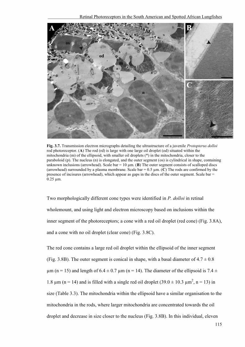
Retinal Photoreceptors in the South American and Spotted African Lungfishes
115
Fig. 3.7. Transmission electron micrographs detailing the ultrastructure of a juvenile Protopterus dolloi rod photoreceptor. (A) The rod (rd) is large with one large oil droplet (od) situated within the mitochondria (m) of the ellipsoid, with smaller oil droplets (*) in the mitochondria, closer to the paraboloid (p). The nucleus (n) is elongated, and the outer segment (os) is cylindrical in shape, containing unknown inclusions (arrowhead). Scale bar = 10 µm. (B) The outer segment consists of scalloped discs (arrowhead) surrounded by a plasma membrane. Scale bar = 0.5 µm. (C) The rods are confirmed by the presence of incisures (arrowhead), which appear as gaps in the discs of the outer segment. Scale bar = 0.25 µm.
Two morphologically different cone types were identified in P. dolloi in retinal
wholemount, and using light and electron microscopy based on inclusions within the
inner segment of the photoreceptors; a cone with a red oil droplet (red cone) (Fig. 3.8A),
and a cone with no oil droplet (clear cone) (Fig. 3.8C).
The red cone contains a large red oil droplet within the ellipsoid of the inner segment
(Fig. 3.8B). The outer segment is conical in shape, with a basal diameter of 4.7 ± 0.8
µm (n = 15) and length of 6.4 ± 0.7 µm (n = 14). The diameter of the ellipsoid is 7.4 ±
1.8 µm (n = 14) and is filled with a single red oil droplet (39.0 ± 10.3 µm2, n = 13) in
size (Table 3.3). The mitochondria within the ellipsoid have a similar organisation to the
mitochondria in the rods, where larger mitochondria are concentrated towards the oil
droplet and decrease in size closer to the nucleus (Fig. 3.8B). In this individual, eleven

Retinal Photoreceptors in the South American and Spotted African Lungfishes
116
smaller cones with a small red oil droplet were counted along the nasal edge of the
retina. These may be red cones that were damaged by tissue fixation and/or processing
or a developmental stage of the red cone receptor type as they resemble the large red-oil
droplet bearing cones in all other characteristics.
The cone without an oil droplet has a tapered, conical outer segment with a basal
diameter of 3.8 ± 1.0 µm (n = 13) and a length of 5.6 ± 1.4 µm (n = 13) in an individual
of 20.6 cm in TL. The ellipsoid is 9.1 ± 1.0 µm (n = 15) in diameter and is filled with
mitochondria of varying sizes, the largest of which are positioned closest to the outer
segment (Fig. 3.8C and 3.9A). Like the rods, the paraboloid of the clear cone consists of
granules that sit between the mitochondria and nucleus (Fig. 3.9B).

Retinal Photoreceptors in the South American and Spotted African Lungfishes
117
Fig. 3.8. Morphological characteristics of cone photoreceptors in juvenile Protopterus dolloi. (A) Retinal wholemount of an unstained P. dolloi retina showing the red cones (arrowhead) at the level of the ellipsoid. Scale bar = 20 µm. (B) Electron micrograph showing the ultrastructure of a red cone (rc). The outer segment (os) is tapered with an oil droplet (od) located in the ellipsoid region above the majority of mitochondria (m), with the nucleus (n) more vitread. Scale bar = 5 µm (C) Electron micrograph of the outer segment (os) and mitochondria (m) containing the ellipsoid of the clear cone. Scale bar = 2 µm.
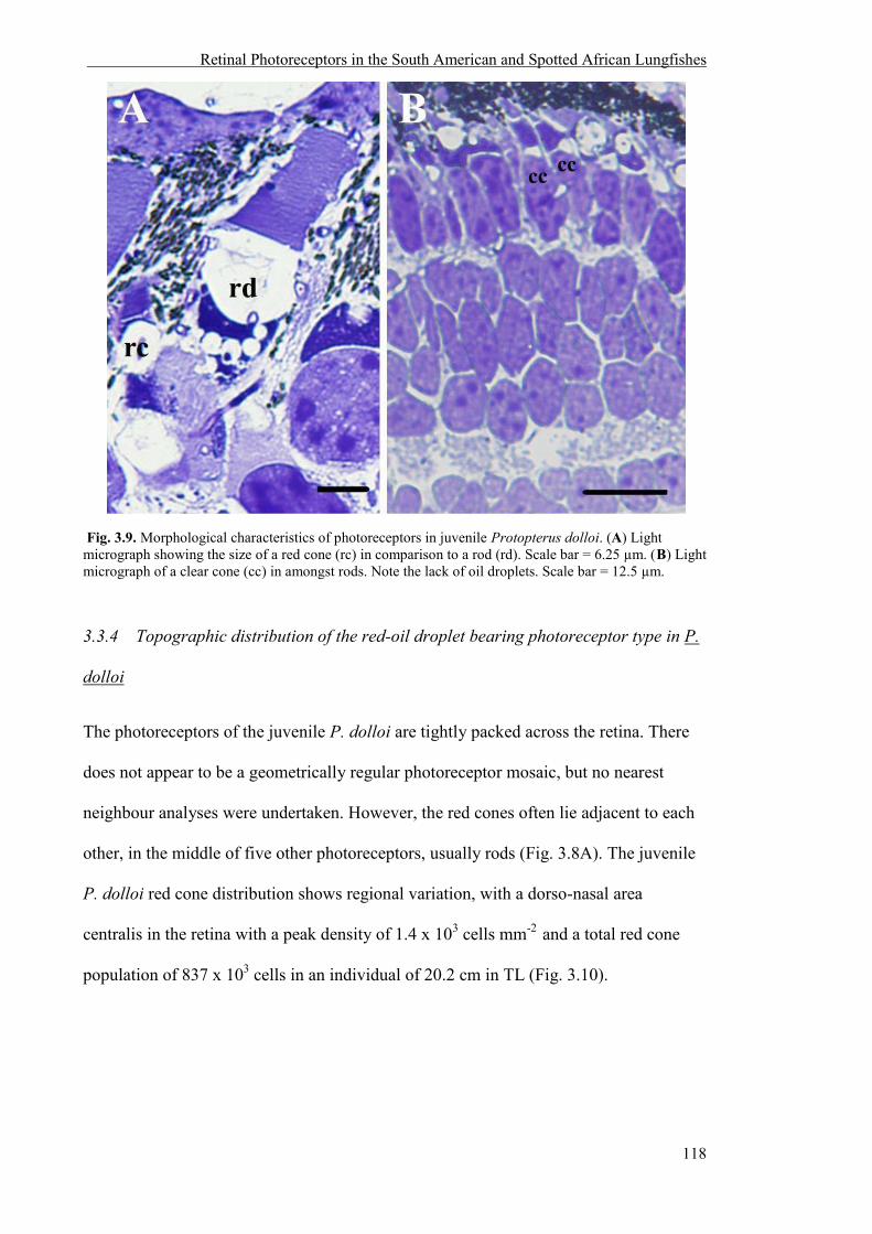
Retinal Photoreceptors in the South American and Spotted African Lungfishes
118
Fig. 3.9. Morphological characteristics of photoreceptors in juvenile Protopterus dolloi. (A) Light micrograph showing the size of a red cone (rc) in comparison to a rod (rd). Scale bar = 6.25 µm. (B) Light micrograph of a clear cone (cc) in amongst rods. Note the lack of oil droplets. Scale bar = 12.5 µm.
3.3.4 Topographic distribution of the red-oil droplet bearing photoreceptor type in P.
dolloi
The photoreceptors of the juvenile P. dolloi are tightly packed across the retina. There
does not appear to be a geometrically regular photoreceptor mosaic, but no nearest
neighbour analyses were undertaken. However, the red cones often lie adjacent to each
other, in the middle of five other photoreceptors, usually rods (Fig. 3.8A). The juvenile
P. dolloi red cone distribution shows regional variation, with a dorso-nasal area
centralis in the retina with a peak density of 1.4 x 10³ cells mm-2 and a total red cone
population of 837 x 103 cells in an individual of 20.2 cm in TL (Fig. 3.10).

Retinal Photoreceptors in the South American and Spotted African Lungfishes
119
Fig. 3.10. Topographic density map of the red cone photoreceptors (mm2) in a juvenile Protopterus dolloi 20.2 cm in TL. Note that the greatest density of red cones is located centrally in the dorso-nasal region of the retina.
3.4 DISCUSSION
This chapter reveals the presence and ultrastructure of one rod and two cone
photoreceptor types in juvenile P. dolloi, the topographical distribution of the red cone
in a juvenile P. dolloi, and the characterisation of one rod and one cone type in adult
L. paradoxa. Like the photoreceptors of N. forsteri, the photoreceptors of P. dolloi and
L. paradoxa are large in absolute terms and their large inner segment cross sectional
area and outer segment length will aid in maximising photon capture in dim light. This
has been confirmed behaviourally in the cichlid (Haplochromis sauvagei) where an
increase in photic sensitivity is associated with increased photoreceptor size (Van der
Meer, 1994).
3.4.1 The eye of P. dolloi
The overall shape of the eyes in a juvenile P. dolloi is spherical, which is similar to the
eyes of juvenile N. forsteri. However, the eye of N. forsteri is spherical in juveniles and

Retinal Photoreceptors in the South American and Spotted African Lungfishes
120
becomes more ellipsoidal in adults (Bailes et al., 2007). This ontogenetic change is also
seen in some amphibian species that undergo metamorphosis such as in the toad (Bufo
americanus) and newt (Notophthalmus viridescens) (Sivak et al., 1985, Sivak, 1988,
Mathis et al., 1988). The optics of the growing P. dolloi eye have not been described, so
it is unknown if the same ontogenetic changes are observed in this species, although this
may be predicted given the plasticity that occurs in N. forsteri, the most basal species of
lungfish, and in amphibians.
The spherical lens of juvenile P. dolloi is typical of those seen in most teleosts because
it allows for greater refraction of light in the absence of any refractive power in the
cornea (Walls, 1942, Sivak, 1990, Sivak et al., 1999). In aquatic animals, the lens is
usually the only effective refractive element of the eye because the similarity of the
refractive index between the cornea and the surrounding water negates any focusing
ability. Most terrestrial species possess aspherical lenses because of the greater
difference in refractive index between the air and the cornea to form a sharp image
(Walls, 1942, Sivak, 1990, Charman, 1991, Land, 1991, Collin and Collin, 2001).
However, the lens of N. forsteri is spherical in juveniles and becomes more elliptical in
shape as it grows, despite spending its entire life in water (Bailes et al., 2007). The same
ontogenetic change may occur in P. dolloi, and because aspherical lenses are also seen
in other aquatic animals such as lampreys (Collin and Fritzsch, 1993, Collin et al.,
1999), sharks (Gilbert, 1963) and some species of teleosts (Pettigrew and Collin, 1995,
Douglas et al., 2002), it appears that lens shape may depend on visual needs on a
species specific basis.
Retinal Photoreceptors in the South American and Spotted African Lungfishes
121
3.4.2 Photoreceptor types in L. paradoxa
This study showed L. paradoxa possesses one rod and one cone photoreceptor type.
This is in accordance with the description by Ali and Anctil (1973) using light
microscopy and Zeiss et al (2011) using immunohistochemistry that identified the
presence of LWS cones but no SWS cones. However, no immunostaining for MWS
cones was undertaken (Zeiss et al., 2011). During aestivation, L. paradoxa remains
dormant and does not feed during the dry, winter months that can potentially last for
eight months a year. This is unlike the lifestyle of P. dolloi, which make frequent
journeys to the top of their burrows during aestivation to breathe, and N. forsteri that do
not aestivate at all (see the General Introduction for a review). This differing lifestyle
may have resulted in less dependency on colour vision, as L. paradoxa is frequently
described as the least fish-like, with the least developed retina (Ali and Anctil, 1973),
but precisely what the role of colour vision is in all lungfish species is still a matter of
speculation.
The morphology of the oil droplet bearing rod and cone photoreceptors in L. paradoxa
closely resembles those of P. dolloi, and follows what was described in an
ultrastructural study by Ali and Anctil (1973). However, Zeiss et al. (2011) described
the rods of L. paradoxa as containing an ellipsoid without an oil droplet. This finding is
in contrast with Ali and Anctil’s (1973) report on rods despite specimens being
processed in the same way. In processing the L. paradoxa retina for this study, unknown
inclusions within the oil droplets of rods and cones appeared in the transmission
electron micrographs that disappeared when sections were deplastinated and stained for
light microscopy. This was the same method used to process P. dolloi specimens, but
without the same artefact being produced. This may be an artefact due to the same
phenomenon occurring in both rod and cone photoreceptors, or may indicate a
difference in the composition of the ellipsoid and/or the oil droplet in the photoreceptors

Retinal Photoreceptors in the South American and Spotted African Lungfishes
122
in comparison to P. dolloi. In order to determine if the inclusions within the inner
segment of L. paradoxa photoreceptors are oil droplets, the retina can be stained with
Oil Red O to show the presence of neutral fats (triglycerides), as was carried out with
N. forsteri (Bailes et al., 2006), but due to the similarity of the photoreceptors in
L. paradoxa to those in P. dolloi and P. aethiopicus, and the presence of the inclusions
in both rods and cones, it is presumed that this is an artefact.
Of all lungfishes, N. forsteri is the only known species that does not possess oil droplets
in their rod photoreceptors. Many species of vertebrates contain colourless oil droplets,
such as the turtle Pseudymys scripta (Kolb and Jones, 1987), the green frog Rana
clamitans (Hailman, 1976), and the strawberry poison frog Dendrobates pumilio
(Siddiqi et al., 2004). It is speculated that retinal oil droplets were originally coloured,
but where species have subsequently become nocturnal these pigments were lost (Hart
et al., 2006). This is presumably because the benefits of spectral tuning conferred by
coloured oil droplets are outweighed by the reduction in absolute sensitivity that would
make vision in dim light more difficult. Chickens (Gallus gallus domesticus) reared in
dim light develop less dense pigmentation in their coloured oil droplets compared to
those reared in bright light, presumably to maintain absolute sensitivity at the expense
of spectral tuning (Hart et al., 2006), so it may be possible that adopting nocturnality
provided the selective pressure to lose coloured pigments within oil droplets. These
clear/colourless oil droplets can either contain an ultraviolet-absorbing pigment that will
absorb scattered short wavelengths and sharpen contrast detection as it does in
Pseudymys and Rana, or can be truly colourless without any wavelength absorbing
characteristics as in Dendrobates. All oil droplets can potentially act as micro-lenses to
increase photon capture (Sivak et al., 1999); although more research is needed to
determine what role they play in P. dolloi and L. paradoxa.
Retinal Photoreceptors in the South American and Spotted African Lungfishes
123
3.4.3 Photoreceptor types in P. dolloi
The photoreceptors of the juvenile P. dolloi contain at least one type of rod and two
types of cones (red oil droplet containing cones and clear, no oil droplet containing
cones) that suggest it has the potential for dichromatic colour vision. The photoreceptor
arrangement did not show regular mosaics, like N. forsteri and L. paradoxa (Ali and
Anctil, 1973, Bailes et al., 2006). The description of oil droplet containing rods, and
cones with and without oil droplets is in accordance to Pfeiffer (1968). However, our
investigation did not reveal double cones. Pfeiffer (1968) described the retina of
P. dolloi in paraffin wax processed tissue, and reported single cones with oil droplets,
and double cones with only one member containing an oil droplet. Our investigation of
P. dolloi photoreceptors did not show a clear cone next to a cone of a different type that
was not another clear cone, rod, or red cone. In fact, there were many occasions where
two clear cones were situated next to each other. Ali and Anctil’s (1973) description of
L. paradoxa photoreceptors from tissue processed in paraffin wax did note the presence
of pairs of visual cells that may have been unequal double cones. However, they
concluded that these instances were random associations of a single cone and rod (Ali
and Anctil, 1973). The superior processing of our tissue in resin for greater resolution
may account for artefacts that resulted in Pfeiffer’s (1968) report of double cones, but
without a reference image of P. dolloi double cones, this is difficult to confirm. Double
cones are present in many teleost fishes (Stell and Hárosi, 1976, Collin, 1997, Collin
and Shand, 2003, Pignatelli et al., 2010), and most terrestrial vertebrates such as birds
(Hart, 2001), amphibians (Mariani, 1986) and diurnal reptiles (Detwiler and Laurens,
1920, Sillman et al., 1997), and some marsupials and monotremes (Young and
Pettigrew, 1991, Ahnelt and Kolb, 2000, Ebrey and Koutalos, 2001). The function of
double cones is still under consideration, but there is evidence that they aid in the
detection of motion and discrimination in fine spatial detail rather than chromatic visual
Retinal Photoreceptors in the South American and Spotted African Lungfishes
124
tasks in birds (for review see Hart and Hunt, 2007) but are involved in colour
discrimination in reef teleosts such as Rhinecanthus aculeatus (Pignatelli et al., 2010).
In dipnoans, N. forsteri and L. paradoxa do not possess double cones, while double
cones are unconfirmed in Protopterus annectens and Protopterus amphibius. The only
species with described double cones is Protopterus aethiopicus. P. aethiopicus and
P. annectens are the “youngest” members of the extant lungfishes in the
Lepidosirenidae family, and are more closely related to each other than L. paradoxa,
P. dolloi or P. amphibius. The confirmed lack of double cones in N. forsteri,
L. paradoxa, and now P. dolloi, suggests that they may have evolved after the
evolutionary separation from P. dolloi. However, until the photoreceptors in
P. annectens and P. amphibius have been described in greater detail, the reason is
uncertain because visual specialisations or degeneracy in species is greatly dependent
on environmental pressures.
The red cones of P. dolloi are also present in N. forsteri. Coloured oil droplets tend to
act as long-pass cut-off filters, selectively transmitting longer wavelengths of light and
blocking shorter wavelengths. The spectral location of the cut-off varies, but in general
their effect is to narrow the spectral sensitivity function of the cones while
simultaneously increasing colour discrimination (by increasing contrast between
adjacent spectral photoreceptor types) (Vorobyev, 2003). Without decreasing the total
amount of ambient light entering the eye through a coloured lens, as in humans,
coloured oil droplets allow an animal to utilize almost all the light available for vision
without sacrificing clarity (Bowmaker, 1980). In N. forsteri, the red cone oil droplet
absorbs all wavelengths below about 560 nm and, along with the yellow pigmented
cone type, improves the animals’ ability to discriminate colours by a factor of
approximately 1.3 which may aid their discrimination of foliage, prey items,
conspecifics and potential predators (Hart et al., 2008). However, this assumes that we
Retinal Photoreceptors in the South American and Spotted African Lungfishes
125
understand the visual tasks that have driven the evolution of the eye in N. forsteri. Until
the spectral absorption characteristics of the visual pigments and oil droplets of P. dolloi
are measured, it is impossible to assess how much the inclusion of red oil droplets in
one cone type improves colour discrimination. Nevertheless, the presence of red cones
in P. dolloi suggests that greater discrimination of objects at long wavelengths is an
adaptation required for animals living in freshwater rivers and swampland where
substantial sediment and mineral deposits reflect light in the red part of the visible
spectrum (Hart et al., 2008).
The position of the area centralis of the red cones in the retina of P. dolloi suggests the
sampling of light with greater resolving power in the dorso-nasal region of the retina.
This equates to a visual field dorsal and lateral to the lungfish, and may be useful in
predator avoidance. In the African lungfish P. aethiopicus (Curry-Lindahl, 1956,
Greenwood, 1987), an acute zone directed forward and into eccentric visual space may
aid navigation between tight spaces in the dense vegetation of the swamp (Collin and
Pettigrew, 1988a). This is different to the topographic distribution of the cones in
N. forsteri that indicates a downwardly directed visual axis in juveniles (Bailes et al.,
2006). Further study is required to investigate if this increased resolving power in the
dorso-nasal region of the visual field is also reflected in other photoreceptor types and
within the ganglion cell population thereby providing a better indication of how retinal
structures reflects their visual ecology.
3.5 CONCLUSION
This study has revealed that the retina of a juvenile P. dolloi contains one rod and two
cone photoreceptor types, with increased retinal sensitivity in the dorso-nasal region of
the retina based on the topographical distribution of the red cones. It has also
Retinal Photoreceptors in the South American and Spotted African Lungfishes
126
characterised of one rod and one cone type in adult L. paradoxa. The large size of the
photoreceptors and the presence of oil droplets in P. dolloi and L. paradoxa closely
resembles the situation in N. forsteri, despite the lack of aestivation in the Australian
species, and suggests a visual system that is adapted for high sensitivity, while the
presence of one cone photoreceptor type infers that L. paradoxa is the only lungfish
species described without the potential for colour vision. However, further study is
needed to establish the importance of colour discrimination, and the selective pressures
involved in the specializations of the retina of P. dolloi and L. paradoxa in order to
establish the role vision plays in these species’ behavioural ecology.
3.6 REFERENCES
AHNELT, P. K. & KOLB, H. 2000. The mammalian photoreceptor mosaic-adaptive design. Progress in Retinal and Eye Research, 19, 711-777.
ALI, M. A. & ANCTIL, M. 1973. Retina of the South American lungfish, Lepidosiren paradoxa
Fitzinger. Canadian Journal of Zoology, 51, 969-972. AMEMIYA, C. T., ALFÖLDI, J., LEE, A. P., FAN, S., PHILIPPE, H., MACCALLUM, I., BRAASCH,
I., MANOUSAKI, T., SCHNEIDER, I. & ROHNER, N. 2013. The African coelacanth genome provides insights into tetrapod evolution. Nature, 496, 311-316.
ARRATIA, G. 2003. Lepidosirenidae (Aestivating lungfishes). In: REIS, R. E., KULLANDER, S. O. &
FERRARIS JR., C. J. (eds.) Checklist of the Freshwater Fishes of South and Central America. Porto Alegre: EDIPUCRS, Brasil.
BAILES, H. J., ROBINSON, S., TREZISE, A. E. O. & COLLIN, S. P. 2006. Morphology,
characterization, and distribution of retinal photoreceptors in the Australian lungfish Neoceratodus forsteri (Krefft, 1870). The Journal of Comparative Neurology, 494, 381-397.
BAILES, H. J., TREZISE, A. E. O. & COLLIN, S. P. 2007. The optics of the growing lungfish eye: lens
shape, focal ratio and pupillary movements in Neoceratodus forsteri. Visual Neuroscience, 24, 377-387.
BOWMAKER, J. 1980. Colour vision in birds and the role of oil droplets. Trends in Neurosciences, 3,
196-199. BOZZANO, A. & COLLIN, S. P. 2000. Retinal ganglion cell topography in elasmobranchs. Brain,
behavior and evolution, 55, 191-208. CARTER, G. & BEADLE, L. 1930. Notes on the habits and development of Lepidosiren paradoxa.
Journal of the Linnean Society of London, Zoology, 37, 197-203.

Retinal Photoreceptors in the South American and Spotted African Lungfishes
127
CHARMAN, W. N. 1991. Optics of the human eye. In: CHARMAN, W. N. (ed.) Vision and Visual Dysfunction: Visual Optics and Instrumentation. London: The Macmillan Press Ltd.
CLACK, J. A., SHARP, E. L. & LONG, J. A. 2011. The Fossil Record of Lungfishes. In: JORGENSEN,
J. M. & JOSS, J. (eds.) The Biology Of Lungfishes. Enfield: Science Publishers. COIMBRA, J. P., MARCELIANO, M., ANDRADE-DA-COSTA, B. & YAMADA, E. S. 2006. The
retina of tyrant flycatchers: topographic organization of neuronal density and size in the ganglion cell layer of the great kiskadee Pitangus sulphuratus and the rusty margined flycatcher Myiozetetes cayanensis (Aves: Tyrannidae). Brain, Behavior and Evolution, 68, 15-25.
COLLIN, S. P. 1999. Behavioural ecology and retinal cell topography. Adaptive Mechanisms in the
Ecology of Vision. Springer. COLLIN, S. P. 1997. Specialisations of the teleost visual system: adaptive diversity from shallow-water
to deep-sea. Acta Physiologica Scandinavica Supplementum, 638, 5. COLLIN, S. P. 2010. Evolution and Ecology of Retinal Photoreception in Early Vertebrates. Brain
Behavior and Evolution, 75, 174-185. COLLIN, S. P. & COLLIN, H. 2001. The fish cornea: adaptations for different aquatic environments.
Sensory Biology of Jawed Fishes. New Insights, 57-96. COLLIN, S. P. & FRITZSCH, B. 1993. Observations on the shape of the lens in the eye of the silver
lamprey, Ichthyomyzon unicuspis. Canadian Journal of Zoology, 71, 34-41. COLLIN, S. P. & PETTIGREW, J. 1988a. Retinal topography in reef teleosts. Brain, Behavior and
Evolution, 31, 269-282. COLLIN, S. P. & PETTIGREW, J. 1988b. Retinal topography in reef teleosts. Brain, Behavior and
Evolution, 31, 283-295. COLLIN, S. P., POTTER, I. C. & BRAEKEVELT, C. R. 1999. The ocular morphology of the southern
hemisphere lamprey Geotria australis Gray, with special reference to optical specialisations and the characterisation and phylogeny of photoreceptor types. Brain, Behavior and Evolution, 54, 96-118.
COLLIN, S. P. & SHAND, J. 2003. Retinal sampling and the visual field in fishes. Sensory Processing in
Aquatic Environments. Springer. CURRY-LINDAHL, K. 1956. On the ecology, feeding behaviour and territoriality of the African
lungfish, Protopterus aethiopicus Heckel. Arkiv for Zoologi, 9, 479-497. DEAN, B. Notes on the living specimens of the Australian lungfish, Ceratodus forsteri, in the Zoological
Society's collection. Proceedings of the Zoological Society of London, 1906. Wiley Online Library, 168-178.
DEAN, B. 1912. Additional notes on the living specimens of the Australian lungfish (Ceratodus forsteri)
in the Zoological Society's collection. Proceedings of the Zoological Society of London, 1912, 607 - 612.
DETWILER, S. R. & LAURENS, H. 1920. Studies on the retina. The structure of the retina of
Phrynosoma cornutum. Journal of Comparative Neurology, 32, 347-356. DOUGLAS, R. H., COLLIN, S. P. & CORRIGAN, J. 2002. The eyes of suckermouth armoured catfish
(Loricariidae, subfamily Hypostomus): pupil response, lenticular longitudinal spherical aberration and retinal topography. Journal of Experimental Biology, 205, 3425-3433.
EBREY, T. & KOUTALOS, Y. 2001. Vertebrate photoreceptors. Progress in Retinal and Eye Research,
20, 49-94. FONESCA DE ALMEIDA-VAL, V., NOZAWA, S., LOPES, N., ARIDE, P., MESQUITA-SAAD, L.,
MAZARE PAULA DA SILVA, M., HONDA, R., FERREIRA-NOZAWA, M. & VAL, A. 2011.

Retinal Photoreceptors in the South American and Spotted African Lungfishes
128
Biology of the South American lungfish, Lepidosiren paradoxa. In: JORGENSEN, J. & JOSS, J. (eds.) The Biology of Lungfishes. Enfield: Science Publishers.
GILBERT, P. W. 1963. The visual apparatus of sharks. In: GILBERT, P. W. (ed.) Sharks and Survival. GOVARDOVSKII, V. 1983. On the role of oil drops in colour vision. Vision Research, 23, 1739-1740. GREENWOOD, P. 1987. The natural history of African lungfishes. In: BEMIS, W. E., BURGGREN, W.
W. & KEMP, N. E. (eds.) The Biology and Evolution of Lungfishes. New York: Alan R. Liss Inc.
GREENWOOD, P. H. 1986. The natural history of African lungfishes. Journal of Morphology, 190, 163-
179. GRIGG, G. C. 1965. Studies on the Queensland lungfish, Neoceratodus forsteri (Krefft). 3. Aerial
respiration in relation to habits. Australian Journal of Zoology, 13, 413-422. HAILMAN, J. P. 1976. Oildroplets in the eyes of adult anuran amphibians: a comparative survey.
Journal of Morphology, 148, 453-468. HART, N. S. 2001. The visual ecology of avian photoreceptors. Progress in Retinal and Eye Research,
20, 675-703. HART, N. S., BAILES, H. J., VOROBYEV, M., MARSHALL, N. J. & COLLIN, S. P. 2008. Visual
ecology of the Australian lungfish (Neoceratodus forsteri). BMC ecology, 8, 21. HART, N. S. & HUNT, D. M. 2007. Avian visual pigments: Characteristics, spectral tuning, and
evolution. American Naturalist, 169, S7-S26. HART, N. S., LISNEY, T. J. & COLLIN, S. P. 2006. Cone photoreceptor oil droplet pigmentation is
affected by ambient light intensity. Journal of Experimental Biology, 209, 4776. HOWLAND, H. C., MEROLA, S. & BASARAB, J. R. 2004. The allometry and scaling of the size of
vertebrate eyes. Vision Research, 44, 2043-2065. JOHNELS, A. & SVENSSON, G. 1954. On the biology of Protopterus annectens (Owen). Arkiv för
Zoologi, 7, 131-164. KEMP, A. 1986. The biology of the Australian lungfish, Neoceratodus forsteri (Krefft 1870). Journal of
Morphology, 190, 181-198. KERR, J. 1902. The development of Lepidosiren paradoxa. III. Development of the skin and its
derivatives. Quarterly Journal of Microscopial Sciences, 46, 417 - 459. KOLB, H. & JONES, J. 1987. The distinction by light and electron microscopy of two types of cone
containing colorless oil droplets in the retina of the turtle. Vision Research, 27, 1445-1458. LAND, M. F. 1991. Optics of the eyes of the animal kingdom. Evolution of the eye and the visual system.
Vision and visual dysfunction. JR Cronly-Dillon, 118-135. MARIANI, A. 1986. Photoreceptors of the larval tiger salamander retina. Proceedings of the Royal
Society of London. Series B, Biological Sciences, 483-492. MARSHALL, C. R. 1986. A list of fossil and extant dipnoans. Journal of Morphology, 190, 15-23. MATHIS, U., SCHAEFFEL, F. & HOWLAND, H. C. 1988. Visual optics in toads (Bufo americanus).
Journal of Comparative Physiology A, 163, 201-213. MLEWA, C., GREEN, J. & DUNBRACK, R. 2011. The general natural history of African lungfishes. In:
JORGENSEN, J. & JOSS, J. (eds.) The Biology of Lungfishes. Enfield: Science Publishers. OWEN, S. R. 1840. XX. Description of the Lepidosiren annectens. Transactions of the Linnean Society
of London, 18, 327-361.

Retinal Photoreceptors in the South American and Spotted African Lungfishes
129
PETTIGREW, J. & COLLIN, S. 1995. Terrestrial optics in an aquatic eye: The sandlance, Limnichthytes
fasciatus (Creediidae, Teleostei). Journal of Comparative Physiology A, 177, 397-408. PFEIFFER, W. 1968. Retina und Retinomotorik der Dipnoi und Brachiopterygii. Cell and Tissue
Research, 89, 62-72. PIGNATELLI, V., CHAMP, C., MARSHALL, J. & VOROBYEV, M. 2010. Double cones are used for
colour discrimination in the reef fish, Rhinecanthus aculeatus. Biology letters, rsbl20091010. SCHLÖGL, E. 2002. Head of a Coral Cod at 'Lighthouse' [Online]. Sydney: Australian Museum.
Available: http://australianmuseum.net.au/image/Head-of-a-Coral-Cod-at-Lighthouse/ [Accessed 19th December 2014].
SIDDIQI, A., CRONIN, T. W., LOEW, E. R., VOROBYEV, M. & SUMMERS, K. 2004. Interspecific
and intraspecific views of color signals in the strawberry poison frog Dendrobates pumilio. Journal of Experimental Biology, 207, 2471-2485.
SILLMAN, A., GOVARDOVSKII, V., RÖHLICH, P., SOUTHARD, J. & LOEW, E. 1997. The
photoreceptors and visual pigments of the garter snake (Thamnophis sirtalis): a microspectrophotometric, scanning electron microscopic and immunocytochemical study. Journal of Comparative Physiology A, 181, 89-101.
SIVAK, J., ANDISON, M. & PARDUE, M. 1999. Vertebrate optical structure. Adaptive Mechanisms in
the Ecology of Vision. Springer. SIVAK, J., LEVY, B., WEBER, A. & GLOVER, R. 1985. Environmental influence on shape of the
crystalline lens: the amphibian example. Experimental Biology, 44, 29-40. SIVAK, J. G. 1988. Optics of amphibious eyes in vertebrates. Sensory Biology of Aquatic Animals.
Springer. SIVAK, J. G. 1990. Optical variability of the fish lens. The Visual System of Fish. Springer. STELL, W. K. & HÁROSI, F. I. 1976. Cone structure and visual pigment content in the retina of the
goldfish. Vision Research, 16, 647-IN4. STONE, J. 1981. The whole mount handbook: a guide to the preparation and analysis of retinal whole
mounts., Sydney, Maitland Publications. THOMSON, K. S. 1969. The biology of the lobe-finned fishes. Biological Reviews, 44, 91-154. TOKITA, M., OKAMOTO, T. & HIKIDA, T. 2005. Evolutionary history of African lungfish: a
hypothesis from molecular phylogeny. Molecular Phylogenetics and Evolution, 35, 281. VAN DER MEER, H. 1994. Ontogenetic change of visual thresholds in the cichlid fish Haplochromis
sauvagei. Brain, Behavior and Evolution, 44, 40-49. VOROBYEV, M. 2003. Coloured oil droplets enhance colour discrimination. Proceedings of the Royal
Society of London Series B-Biological Sciences, 270, 1255-1261. VOROBYEV, M., OSORIO, D., BENNETT, A., MARSHALL, N. & CUTHILL, I. 1998.
Tetrachromacy, oil droplets and bird plumage colours. Journal of Comparative Physiology A, 183, 621-633.
WALLER, U. 1998. Scyliorhinus canicula [Online]. Fishbase. Available:
http://www.fishbase.org/summary/845 [Accessed 19th December 2014]. WALLS, G. L. 1942. The Vertebrate Eye and its Adaptive Radiation, Bloomfield Hills, The Cranbook
Press. YOUNG, H. M. & PETTIGREW, J. D. 1991. Cone photoreceptors lacking oil droplets in the retina of the
echinda, Tachyglossus aculeatus (Monotremata). Visual Neuroscience, 6, 409-420.

Retinal Photoreceptors in the South American and Spotted African Lungfishes
130
ZEISS, C. J., SCHWAB, I. R., MURPHY, C. J. & DUBIELZIG, R. W. 2011. Comparative retinal
morphology of the platypus. Journal of Morphology, 272, 949-957.


Elucidating The Visual Perception Of Neoceratodus forsteri Without Visible Eyes
132
CHAPTER 4
::::::
ELUCIDATING THE VISUAL
PERCEPTION OF
NEOCERATODUS FORSTERI
WITHOUT VISIBLE EYES
USING BEHAVIOURAL,
CIRCADIAN,
ELECTROPHYSIOLOGICAL
AND ANATOMICAL
APPROACHES
::::::

Elucidating The Visual Perception Of Neoceratodus forsteri Without Visible Eyes
133
4.1 INTRODUCTION
This chapter investigates the visual perception of a unique group of juvenile Australian
lungfishes (Neoceratodus forsteri) that do not possess externally visible eyes. Using
behavioural, circadian, electrophysiological and anatomical approaches comparing
abnormal N. forsteri (no visible eyes) and normal N. forsteri (visible eyes), new
findings are presented that begin to uncover how a unique group of fishes experience
the world without the ability to form images.
The external appearance of the head region typically containing the image-forming eyes
of the abnormal N. forsteri was the same in all individuals; there was no scarring, no
indication of damage or deformity except that the eyes appeared not have developed.
Abnormal visual systems can arise from fishes being reared in complete darkness
(Pankhurst, 1992), however, to our knowledge the abnormal lungfishes were developed
in a normal light-dark cycle. Rather than discarding this anomalous resource, it was
considered a unique opportunity to explore the visual Umwelt of a curious group of
lungfishes without external eyes. In order to provide adequate comparison to normal
lungfishes, three normal juvenile N. forsteri were acquired and four experiments were
designed to compare the visual capabilities of the two groups. This chapter is organised
into four separate experiments that investigated the visual Umwelten of abnormal
lungfishes in three complementary ways.
1. The first experiment investigated if, despite the absence of external eyes, the
abnormal N. forsteri were able to behaviourally discriminate between areas of relative
light and relative dark.
The behavioural patterns of abnormal and normal N. forsteri were compared to
determine whether the abnormal N. forsteri moved randomly between areas of light and
dark, and whether these movements differ from the movements of the normal

Elucidating The Visual Perception Of Neoceratodus forsteri Without Visible Eyes
134
N. forsteri. N. forsteri are typically nocturnal and seek shelter where available. In their
natural habitat of the Mary, Burnett and Brisbane River systems of south-east
Queensland, Australia (Kemp, 1986, Kind, 2002, Kind, 2011), N. forsteri typically
shelter by day and exhibit a crepuscular and nocturnal lifestyle, where the majority of
foraging occurs during the night (Dean, 1906, Grigg, 1965b). Observations that wild
N. forsteri spawn at night (Grigg, 1965a) and anecdotal evidence from local fishermen
show that they are most often heard gulping atmospheric air at night, suggesting
nocturnal activity (Illidge, 1894, Spencer, 1892, Grigg, 1965b). When resting,
N. forsteri typically prefer woodpiles, macrophyte beds and are often found amongst
overhanging vegetation in shallow pools (Brooks and Kind, 2002). The observation that
tagged individuals in Mary River prefer a complex shallow-water habitat with surface
shading and avoid open, deep water regions (Kind, 2002), suggests that lungfishes will
actively seek shelter and spend more time in the dark section of the tank in this
experiment. It is possible that the abnormal fishes do not possess light sensitive tissue
capable of perceiving differences in ambient light over short periods of time. Therefore,
it is hypothesised that the abnormal lungfishes will not display the same behaviour as
normal N. forsteri and will be unable to discriminate between areas of relative light and
relative dark.
2. The second experiment was designed to observe and compare the circadian
rhythms of normal and abnormal N. forsteri measured over a 72 hour period.
The physiological network that contributes to the creation of the circadian rhythm is
controlled by a tightly interconnected system of regulatory molecules, cells, circuits and
tissues at several layers of organisation (Kronfeld-Schor et al., 2013). Among the neural
and endocrine centres that autoregulate the entrainment of the circadian clock in fishes
is the pineal gland and the retina (Falcón et al., 2010). Factors such as thermoregulation,

Elucidating The Visual Perception Of Neoceratodus forsteri Without Visible Eyes
135
food availability, and predation can affect the entrainment of circadian rhythms, but
light remains the main synchronizer of the circadian clock (Falcón et al., 2007, Ekström
and Meissl, 1997). The pineal gland possesses melanopsin in its photoreceptors, a non-
image forming visual pigment that detects changes in illumination and responds by
releasing melatonin (Falcón et al., 1992, Cahill and Besharse, 1995, Davies et al.,
2012a, Davies et al., 2012b). Pineal melatonin provides a hormonal signal that affects
the entire organism to regulate circadian patterns and plays a central role in the
entrainment of fishes to changing light environments (Benyassi et al., 2000), while
retinal melatonin is used mostly for local paracrine functions, and may attenuate pineal
melatonin production by enabling the animal of light adapt in advance to actual changes
in illumination (Zhdanova and Reebs, 2005, Li and Dowling, 2000).
The pineal gland develops early in fish embryos. Zebrafishes develop the pineal gland
19 hours post-fertilisation, and, at this stage, the pineal is already responsive to light and
secretes melatonin (Kazimi and Cahill, 1999, Danilova et al., 2004). If the animals are
exposed to a light-dark cycle during this stage of development, the rate of melatonin
secretion follows a distinct circadian pattern that fluctuates over a diel cycle (Danilova
et al., 2004). However, zebrafish embryos kept in constant darkness produce melatonin
at a low level throughout life, and the circadian rhythm never develops (Kazimi and
Cahill, 1999). Since the eyes and the pineal organ are both light sensitive organs and are
involved in the regulation of circadian rhythms, this experiment aimed to establish
whether the pineal is also atypical in abnormal N. forsteri. It is hypothesized that the
circadian rhythm of abnormal N. forsteri will be more arrhythmic than normal
N. forsteri.
3. The third experiment investigated the electrical response of the eye in normal
N. forsteri and the region of the head that would typically contain the eye and brain in

Elucidating The Visual Perception Of Neoceratodus forsteri Without Visible Eyes
136
abnormal N. forsteri to light stimuli using electroretinography (ERG) and visually
evoked potentials (VEPs).
Electrophysiological experiments were conducted to determine whether the eye (normal
condition), and the head region where the eye should be (abnormal condition), show a
functional light response. ERG measures the voltage produced across the eye in
response to light stimulation. ERG waveforms vary in amplitude and pattern between
vertebrate species but consists of basic features common to all species that have been
studied (Fig. 4.1). The ERG response to a flash of light can be separated into several
distinct phases; the a-wave, which reflects the response generated by the outer segment
of the photoreceptors, and the b-wave, which reflects the photocurrent created by the
horizontal, bipolar and Müller cells (Brown, 1968, Asi and Perlman, 1992, Tian and
Slaughter, 1995, Pinto et al., 2007). Oscillatory potentials can also be observed before
the peak of the b-wave (Fig. 4.1D). ERG oscillatory potentials are generated by local
networks of bipolar, amacrine and ganglion cells in the inner retina and are considered
to be sensitive to disturbances in retinal circulation (Speros and Price, 1981, Asi and
Perlman, 1992).
ERGs could not be recorded from any of the abnormal lungfishes. Therefore, it was
decided to examine VEPs from the normal and abnormal lungfishes as well. VEPs are
electrical potentials recorded from an electrode placed subcutaneously on the skin
overlying the optic tecta of the N. forsteri brain, and are used to measure the functional
integrity of the retina and visual pathway to the brain (Creel et al., 1974, Creel, 2012).
Due to the inability of recording ERGs from abnormal lungfishes, it is hypothesised that
it will not be possible to record VEPs in the abnormal N. forsteri as well.
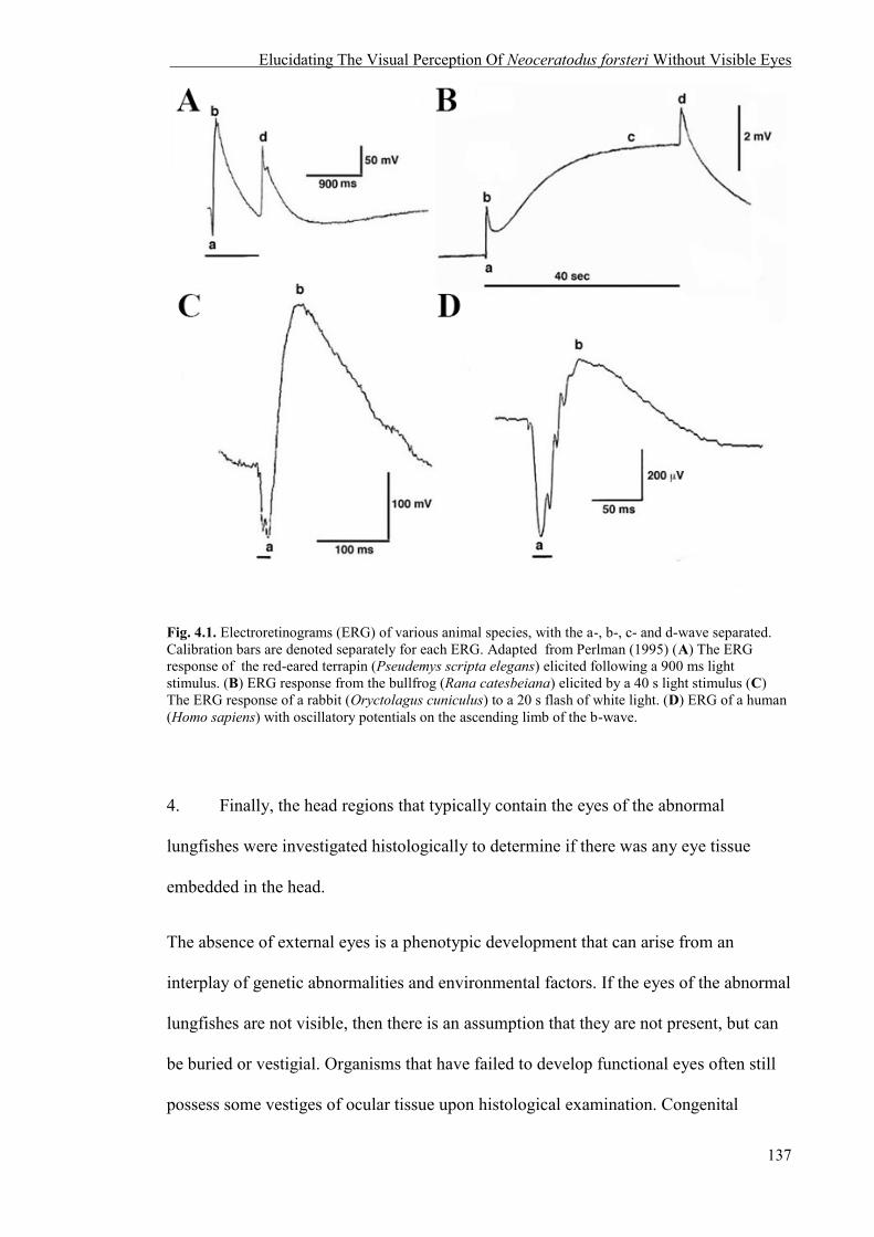
Elucidating The Visual Perception Of Neoceratodus forsteri Without Visible Eyes
137
Fig. 4.1. Electroretinograms (ERG) of various animal species, with the a-, b-, c- and d-wave separated. Calibration bars are denoted separately for each ERG. Adapted from Perlman (1995) (A) The ERG response of the red-eared terrapin (Pseudemys scripta elegans) elicited following a 900 ms light stimulus. (B) ERG response from the bullfrog (Rana catesbeiana) elicited by a 40 s light stimulus (C) The ERG response of a rabbit (Oryctolagus cuniculus) to a 20 s flash of white light. (D) ERG of a human (Homo sapiens) with oscillatory potentials on the ascending limb of the b-wave.
4. Finally, the head regions that typically contain the eyes of the abnormal
lungfishes were investigated histologically to determine if there was any eye tissue
embedded in the head.
The absence of external eyes is a phenotypic development that can arise from an
interplay of genetic abnormalities and environmental factors. If the eyes of the abnormal
lungfishes are not visible, then there is an assumption that they are not present, but can
be buried or vestigial. Organisms that have failed to develop functional eyes often still
possess some vestiges of ocular tissue upon histological examination. Congenital

Elucidating The Visual Perception Of Neoceratodus forsteri Without Visible Eyes
138
defects manifested as primary anophthalmia (the absence of one or both eyes) in viable
births still have remnants of ocular structures, such as an abnormally-sized lens, an
underdeveloped optic tract, optic nerve and optic chiasma in cattle (Leipold and Huston,
1968), and nodules containing condensed scleral tissue and choroidal pigment particles
in humans (Recordon and Griffiths, 1938, Verma and FitzPatrick, 2007).
The vestigial eyes of organisms adapted to subterranean habitats still contain remnants
of ocular tissue. The eyes of the blind cave fish, Astyanax mexicanus, initially develop
in the embryo, but subsequently degenerate in the larval stage and become buried inside
the orbit, covered by epidermis and connective tissue (Cahn, 1958, Strickler et al., 2007,
Wilkens, 2007). Similarly, the troglodytic moles Notorychtes (marsupial mole),
Chrysochloris (golden mole), Eremitalpa (Chrysochloridae; Insectivora) and
Heterocephalus glaber (naked-mole rat) all contain cells representing the lens (Sweet,
1906, Vaughn, 1978, Nikitina et al., 2004). Therefore, histological examination of the
head region that would typically contain the eyes in abnormal N. forsteri is necessary to
determine if there is vestigial ocular tissue present beneath the epidermis that would
mediate some level of light detection.
Since eye tissue has previously been found in clinically described “eyeless” individuals
(anophthalmia) and blind organisms, it is hypothesised that abnormal N. forsteri will
possess ocular tissue beneath the skin.
4.2 GENERAL METHODS
4.2.1 Animals
The two groups of juvenile N. forsteri, abnormal and normal were sourced from
different suppliers from Queensland (Qld), Australia. Three abnormal juvenile

Elucidating The Visual Perception Of Neoceratodus forsteri Without Visible Eyes
139
N. forsteri (25 – 29.5 cm in total length [TL]; fish codes E1, E2 and E3) were chosen
randomly from a cohort of nine fishes kindly donated by Dr. Michael Hutchison of the
Queensland Department of Agriculture, Fisheries and Forestry (DAFF) (Bribie Island)
and Dr. David Roberts of The University of Queensland/Seqwater, Qld. These fishes
were bred in captivity at Professor Jean Joss’ facility at Macquarie University, NSW,
before being moved to DAFF and subsequently transported to UWA under a General
Fisheries Permit authorised by the Qld DAFF (Permit number: 151483).
Three normal juvenile N. forsteri (26.4 – 29.4 cm in TL; fish codes N1, N2 and N3)
were supplied by Jindalee International Pty Ltd (JITL). These fishes were bred in
captivity through the Wildlife Trade Operation (GN22) and Aquaculture Program
(2011/108755) under the provisions of the Environment Protection and Biodiversity
Conservation Act 1999 (EPBC Act) approved to JITL by the Department of
Sustainability, Environment, Water, Population and Communities, Australia. All fishes
could be identified individually by variations in tail length and shape, and individual
markings on the head and/or body.
All fishes were housed indoors in grey fibreglass tanks: abnormal fishes in 190 x 190
cm tanks and normal fishes in a 70 x 160 cm tank. All fishes were maintained in tanks
containing recirculating freshwater to a depth of 40 cm at a constant temperature of
22°C. The fishes were fed every second day on a diet of native fish pellets (52% protein
3 mm sinking pellets, Fishwerx Hobbyist Supplies). Each tank was illuminated using
overhead fluorescent strip lighting on a 12h:12h light:dark cycle. Two experimental
tanks (70 cm x 160 cm x 38 cm) containing one fish each was used for all behavioural
experiments.

Elucidating The Visual Perception Of Neoceratodus forsteri Without Visible Eyes
140
4.2.2 Anaesthesia and Euthanasia
Anaesthesia for Experiment 3 and euthanasia protocols for Experiment 4 were approved
by the UWA Animal Ethics Committee (RA/3/100/1220) and follow a standard protocol
developed by Fritsche et al. (1993). For Experiment 3, lungfishes were initially
anaesthetised in dechlorinated tank water containing 100 mg/L tricaine methane
sulfonate (MS222) buffered with 100 mg/L sodium bicarbonate (NaHCO3) and
subsequently maintained by directing a continuous stream of oxygenated tank water
containing 75 mg/L MS222 buffered with 75 mg/L NaHCO3. Anaesthesia was reversed
by immersion in, and active ventilation with, freshly oxygenated tank water without
MS222. For Experiment 4, abnormal lungfishes were euthanased using an overdose of
MS222 (500 mg/L buffered with 500 mg/L NaHCO3).
4.3 EXPERIMENT 1: LIGHT AVOIDANCE BEHAVIOUR IN ABNORMAL N.
FORSTERI
4.3.1 Methods
4.3.1a Apparatus
The experiment was run simultaneously in two experimental tanks. Each tank housed
one fish, either normal or abnormal. Prior to the experiment, the tank was bisected by a
light barrier in order to create a bright and a dark tank half (Fig. 4.2). The barrier
(5 x 40 cm) was submerged in the water low enough to prevent light from entering the
dark sector but high enough to allow the fish to pass freely between the two halves (the
fish had approximately 20 cm of space to pass through into the other sections). A
removable sheet of black plastic was placed over the top of the dark section of the tank.
The presence and movement patterns of each individual were recorded in the bright
Elucidating The Visual Perception Of Neoceratodus forsteri Without Visible Eyes
141
section of the tank with a Sony Handycam HDR-CX550 that was suspended on a tripod
above the light sector (Fig. 4.2). Before each trial began, the pump in each tank was
turned off and removed to prevent surface reflections from interfering with the clarity of
the video images. The pumps were replaced when the trial was completed (6 hours).
4.3.1b Design
The activity patterns of three normal and three abnormal lungfishes were recorded four
times each for a period of six hours over the course of four days – one period per day.
Each six hour period took place between 0600 – 1200 hrs or 1200 – 1800 hrs (randomly
allocated with 0.5 probability, with the restriction that each fish was observed twice
during each time period). The four trials were conducted over eight days, with a rest and
feeding day in between each experimental day. For each fish, the side of the tank shaded
for the first six hour period (left or right) was chosen randomly (with 0.5 probability
with the same restrictions as above), and swapped to the opposite side of the tank for the
second period of six hours, and so on, to control for possible preferences within the
tank. The fish (abnormal or normal) that inhabited the tank for each trial was randomly
allocated (0.5 probability with above restrictions) and alternated to the other fish type
for the subsequent trials.

Elucidating The Visual Perception Of Neoceratodus forsteri Without Visible Eyes
142
Fig. 4.2. Top view of experimental tank for Experiment 1. The dark section was made dark by the addition of removable black plastic. The light and dark sections were separated by a barrier that allowed the fish to swim freely between sections but inhibited light from entering the dark section. A video camera (vc) recorded the presence of a fish in the light section.
4.3.1c Analysis
Video footage was transferred to a Windows 7 PC and converted to .avi using open-
source software ffmpeg 2.5.3 and down-sampled to eight frames/s to reduce the
processing time. The resulting movies were opened in Matlab 2012a (Mathworks Inc.),
where custom-version software (Jan Hemmi, The University of Western Australia,
modified from Hemmi, 2005 and How et al., 2008) was used to identify the presence of
the fish in each section (either in light or dark) and the duration of time spent within
each section. The presence of the fish in the dark section was inferred from its absence
in the light section. Each video was manually checked to ensure data integrity. Due to
the non-normal distribution of the underlying data, the statistical analysis was based on
randomised permutations (Good, 2005). This non-parametric approach was used to
avoid assumptions about normality for the underlying distribution of the amount of time
each fish spent in a particular section per visit. The statistical analysis was performed in
Elucidating The Visual Perception Of Neoceratodus forsteri Without Visible Eyes
143
Matlab 2012a (Mathworks Inc.) In short, the percentage of relative time spent in light or
dark for each individual was calculated. Each visit, as a block of time in either the light
or dark section, was permutated 10 000 times for each individual lungfish. That means
each visit was randomly assigned to either the light or the dark section, but the overall
number of visits to each section was held constant. After each permutation, a new
percentage score was calculated. The score of the statistic measurement of each
individual’s observed data was then compared to all the scores from the permuted data
sets for the same individual. Statistical significance was determined at p = < 0.05.
4.3.2 Results
Normal fishes spent more than 90% of their time in the dark sector of the experimental
tank during each six hour trial period (93.2 ± 4.72 %) despite the experiment taking
place during the day. However, the abnormal fish spent less time in the dark and
showed more variability in time spent in the dark sector at each trial period (E1 = 60.77
± 22.03 % [n = 4]; E2 = 57.85 ± 9.51 % [n = 4]; E3 = 77.83 ± 15.81 % [n = 4]) (Raw
data is presented in Appendix Table 1).
Randomisation tests show that all normal N. forsteri are significantly non-random (p ≤
0.001) in their choice between sections. None of the 10 000 randomised permutations
produced a bias towards the dark as strongly as observed for all normal fishes.
Abnormal fishes E1 and E2 are not statistically different from random (p = 0.956,
0.278, respectively), but E3 is statistically non-random (p ≤ 0.001), like the normal
lungfishes. The numbers of crossings between sections for each fish is presented in
Table 4.1, and shows that for the abnormal fish, the failure to achieve significance is not
based on lack of choice behaviour. These results indicate that despite E3’s absence of
external eyes, it is still able to distinguish between sections of light and dark non-

Elucidating The Visual Perception Of Neoceratodus forsteri Without Visible Eyes
144
randomly, and all abnormal lungfishes may not possess the same level of visual
disability.
Table 4.1. The number of crossings between the light and dark sections for normal (N) and abnormal (E) Neoceratodus forsteri
Fish # of crossings N1 58 N2 69 N3 128 E1 77 E2 215 E3 156
4.4 EXPERIMENT 2: CIRCADIAN RHYTHM OF ABNORMAL N. FORSTERI
4.4.1 Methods
4.4.1a Experimental design
The circadian rhythm experiment was conducted in total darkness for each fish over 72
hours. The two experimental tanks were confined to a light proof room with a double
containment entry/exit to prevent light from entering the room when accessed (Fig. 4.3).
The filter and pump remained on for the duration of the experiment. A semi-permeable
barrier was placed near the filter and pump (reducing the size of the tank by one quarter)
to prevent the fish from accessing the shelter of the filter/pump. A plastic tube was
placed vertically into the tank underneath the pump to prevent rippling water at the
surface, but still allowing fresh water to diffuse through the tank. This reduced
interference of surface waves with the video analysis. As in Experiment 1, the fish were
recorded using a Sony Handycam HDR-CX550 suspended on a tripod over the tank. To
increase contrast between the fish and the bottom of the tank, the gravel was pushed to
the inaccessible filter/pump side of the tank, and one 48 LED infrared illuminator was

Elucidating The Visual Perception Of Neoceratodus forsteri Without Visible Eyes
145
used in conjunction with the video camera’s ‘low lux’ setting (Fig. 4.4). Infrared light
was used to provide illumination for the camera as N. forsteri are unable to detect light
at that wavelength (Hart et al., 2008). As in Experiment 1, the pairs of fishes used per
72 hour periods were randomised (with 0.5 probability). For example, if Tank 1
contained a normal fish for the first 72 hours, the subsequent experiment contained an
abnormal fish. Each fish was moved to an experimental tank 48 hours prior to the
experiment to acclimatize, and was fed the day prior to commencement of the
experiment and on the morning following the experiment to limit feeding as a variable
affecting activity.
Fig. 4.3. Circadian experiment room layout. The two tanks used in the experiment were placed in a light-proof room. A containment room with a light-proof entry and exit points was constructed to inhibit light entering the experimental room.

Elucidating The Visual Perception Of Neoceratodus forsteri Without Visible Eyes
146
Fig. 4.4. Top view of a circadian experimental tank. Lungfish activity was recorded on a video camera (vc) that was suspended over the experimental arena. Additional light was provided by a 48 LED infrared illuminator (ir) placed near the video camera. The animal was prevented from resting near the filter by a semi-permeable barrier and water ripples were reduced by a plastic tube (t) inserted underneath the pump (p). Video contrast was increased by moving the gravel away from the recording arena.
4.4.1b Analysis
The video footage was transferred to the computer and converted to .avi using open-
source software ffmpeg 2.5.3 and down-sampled to 8 frames/s to reduce the amount of
data to process. The resulting videos were analysed in Matlab 2012a (Mathworks Inc.)
with custom-version software (Jan Hemmi, University of Western Australia, modified
from Hemmi, 2005 and How et al., 2008). The software automatically tracked the fish’s
position, which was subsequently used to calculate activity in the tank. The fish was
distinguished from the background by calculating the difference between the current
frame and an average background image. A background image devoid of the fish’s
presence was obtained by averaging 200 randomly selected images across the entire
experiment. In cases where the infrared lighting changed slightly during an experiment,
a local background image was calculated from 100 frames averaged over a set amount
of frames when lighting remained constant. The fish’s centre of gravity was calculated

Elucidating The Visual Perception Of Neoceratodus forsteri Without Visible Eyes
147
for each frame and used to determine activity. The difference in the centre of gravity
between the current frame and background was filtered and smoothed and the
movement of the fish was determined by taking the difference between the current
centre of gravity and the previous estimate of difference in movement (Fig. 4.5). The
threshold for activity was movement exceeding 3.9 cm/sec. A rest period was defined as
any movement below 1 cm/sec for > 5 mins. All videos were manually checked for
outliers, and to make sure that periods of high activity and little or no activity were
accurate. A custom-made script for Matlab (Mathworks Inc.) was used to visualise the
data.
Fig. 4.5. A still image extracted from video footage using the custom-made software (Jan Hemmi, University of Western Australia) developed in Matlab (Mathworks Inc.). The animal’s outline was visualised by calculating the difference between the current frame and the background image.

Elucidating The Visual Perception Of Neoceratodus forsteri Without Visible Eyes
148
4.4.2 Results
The circadian rhythm of abnormal and normal N. forsteri was observed to investigate if
the pineal gland was still functional in the abnormal group of fishes without visible
image-forming eyes. The normal lungfishes did not exhibit a classical circadian rhythm,
but exhibited two large periods of rest between approximately 24 and 35 hours, and
between 41 and 63 hours, with reduced activity (Fig. 4.6A – C). The abnormal lungfish
E2 also displayed two large periods of rest with reduced activity between 22 and 29
hours, and between 47 and 67 hours (Fig. 4.6E). E3’s larger periods of rest occurred
between 32 and 37 hours, and between 52 and 65 hours (Fig. 4.6F). E1 shows the most
uncharacteristic activity pattern by being active for a continuous period between
approximately 0 and 54 hours, resting only between 54 and 59 hours (Fig. 4.6D).
General observations of all lungfishes showed that even during periods of activity, these
animals liked to take ‘naps’ that generally remained under one hour, and during the
large periods of rest, the lungfishes showed small bursts of activity that lasted a few
minutes at a time (Fig 4.6). However, there is a difference in the distance each group of
lungfishes travelled during the circadian experiment, where the abnormal lungfishes
travelled at least 2.5 times more than the normal N. forsteri (unpaired t(4) = 3.71, p ≤
0.05) (Table 4.2).
Table 4.2. The total distance travelled for normal (N) and abnormal (E) Neoceratodus forsteri for the duration of a circadian experiment.
Fish Distance travelled (km) Fish Distance travelled (km) N1 0.50 E1 4.50 N2 1.70 E2 2.50 N3 0.81 E3 4.60
Mean 1.00 Mean 3.87 Std. Dev 0.62 Std. Dev 1.18

Elucidating The Visual Perception Of Neoceratodus forsteri Without Visible Eyes
149
Fig. 4.6. The circadian rhythm of normal (N1 – N3; A – C, respectively) and abnormal (E1 – E3; D – F, respectively) Neoceratodus forsteri. Activity was measured as the distance travelled between video frames at a speed of 3.9 cm/s over a period of 72 hours. Black arrows indicate periods of rest (prolonged activity less than 1 cm/sec) in each individual.
N1 N2 N3
E1 E2 E3

Elucidating The Visual Perception Of Neoceratodus forsteri Without Visible Eyes
150
4.5 EXPERIMENT 3: ELECTORETINOGRAPHY AND VISUALLY EVOKED
POTENTIALS OF ABNORMAL N. FORSTERI
4.5.1 Methods
4.5.1a Apparatus
ERGs and VEPs were measured in anaesthetised N1 and N2, and all abnormal
lungfishes (see general methods of the present chapter for protocol). Both normal and
abnormal lungfishes were placed in a container of tank water and secured to an inclined
ramp to raise the head of the animal out of the water while keeping the rest of the body
submerged. Oxygenated tank water containing the maintenance dose of anaesthesia (75
mg/L MS222 buffered with 75 mg/L NaHCO3) was pumped from a reservoir to the
container through a small delivery tube inserted into the mouth of the animal to deliver
a continuous flow of water over the gills. Wet paper towels were placed on the animal’s
body to prevent dehydration of any exposed areas of the head and trunk. The exposed
head was also dampened periodically using a pipette filled with water.
The electrophysiology apparatus consisted of a 175 W xenon arc lamp (Spectral
Products) light source producing spectrally broad band ‘white light’ illumination that
was transmitted to a mechanical shutter using a quartz fibre-optic and collimating lens.
Located immediately after the shutter was a computer controlled neutral density (ND)
filter wheel for attenuating the intensity of light (ND 0 – 5.829 in ND 0.2 steps). The
filtered light was presented to the eye using another quartz fibre-optic cable with a
collimating lens (Ocean Optics 74-UV). The collimating lens delivered the light
stimulus to the eye at a distance of 8 mm from the surface of the cornea. For the single-
flash ERG recordings in the normal lungfishes, an active platinum wire electrode was
placed on the surface of the left cornea towards the centre of the eye, ensuring it did not
29 No neutral density filters (0 ND), 1.01 x 10¹⁶ photons cm2.sec1.
Elucidating The Visual Perception Of Neoceratodus forsteri Without Visible Eyes
151
block the beam emanating from the fibre-optic light source. A silver/silver chloride
(Ag/Ag-Cl) wire reference electrode was inserted subcutaneously on the top of the head
5 mm posterior to the midline of the eyes. Ag/Ag-Cl wire active electrodes were used
instead of platinum electrodes in abnormal lungfishes due to the absence of external
eyes. In this case, the recording electrode was placed subcutaneously under the skin
where the left eye should have been.
For visually evoked potentials (VEPs), the active electrode was placed subcutaneously
on the top of the head 20 mm from the midline of the eyes, over the optic tecta.
Differential AC voltages between the active and reference electrodes were amplified
1000 x using a WPI DAM-50 amplifier (World Precision Instruments), and hardware
high-pass and low-pass filtered at 1 Hz and 1 kHz for all normal lungfishes. Differential
AC voltages for abnormal lungfishes were amplified 10000 times with hardware high-
pass and low-pass filters at 0.1 Hz and 1 kHz, respectively. The amplified voltage was
digitised with a National Instruments USB 6353 multifunction data acquisition device,
and stored on a laptop computer running Windows and using custom built software
(Nathan Hart, University of Western Australia). Amplified signals were viewed in real
time using a Tectronix digital oscilloscope.
The animal and recording amplifier were placed within a Faraday cage to reduce pick-
up noise from mains electricity and other sources. The front of the cage was covered
with a black cloth during the experiment to exclude dim ambient room light (e.g. the
computer screen). Once the animal was placed in the container after dark adaptation,
dim red head torches were used to make adjustments to the apparatus or to monitor the
animal. Lungfishes were allowed to dark adapt for 30 minutes and light adapt for 15
minutes prior to ERG and VEP recordings.

Elucidating The Visual Perception Of Neoceratodus forsteri Without Visible Eyes
152
Single flash (50 ms duration) ERGs were recorded at 8 different light intensities over a
5.8 log unit range. The time interval between successive stimulus presentation was
increased as the stimuli increased in brightness (e.g. 5 – 60 s intervals at ND 5.8 – ND
0, respectively) to give the retina adequate time to return to a dark-adapted state. The
number of replicates of each stimulus was also reduced with increasing intensity (e.g.
10 – 3 reps at ND 5.8 – ND 0, respectively) due to the improvement in signal-to-noise
ratio of the responses. Light-adapted VEPs were conducted on all fishes that underwent
ERG recordings, and dark-adapted VEPs were recorded from N1 and all abnormal
lungfishes. Single flash VEPs were only recorded at the highest light intensity (no ND
filters, 1.01 x 10¹⁶ photons cm2.sec1) to maximize the potential response from the
abnormal lungfishes. ERG and VEP recordings were conducted on the left eye of all
fishes. To confirm that the VEPs were from the optic tecta and not the auditory
structures of the brain or from the lateral line that detects sound or vibration from the
mechanical shutter, single flash VEPs were recorded with the light source blocked, but
the shutter opened and closed as normal. To confirm that VEPs from the abnormal
fishes were generated in response to direct stimulation of the head region where the eye
should be, rather than stimulation of other light-sensitive structures by scattered or off
axis light, single flash VEPs were recorded from various parts of the fish, such as the
flank of the animal. The VEPs were confirmed to be caused by stimulation to the retina
rather than the pineal organ by assessing the time-to-peak potential. The pineal organ
photoreceptors are specialised for the detection of slow, gradual changes in the ambient
light environment and cannot discriminate between rapidly changing light (Meissl and
Yanez, 1994). The rainbow trout (Salmo gairdneri) (Meissl and Ekström, 1988), swamp
turtle (Pseudemys scripta elegans) (Baylor and Hodgkin, 1974), and toad (Bufo
marinus) (Cervetto et al., 1977) pineal photoreceptor response is 5 – 6 times longer than
the retinal rod photoreceptor response (Ekstrzm and Meissl, 1997). A comparison
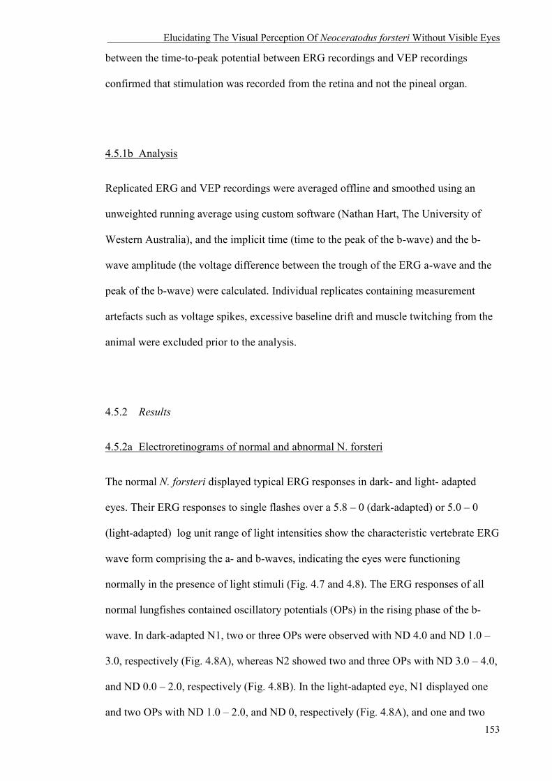
Elucidating The Visual Perception Of Neoceratodus forsteri Without Visible Eyes
153
between the time-to-peak potential between ERG recordings and VEP recordings
confirmed that stimulation was recorded from the retina and not the pineal organ.
4.5.1b Analysis
Replicated ERG and VEP recordings were averaged offline and smoothed using an
unweighted running average using custom software (Nathan Hart, The University of
Western Australia), and the implicit time (time to the peak of the b-wave) and the b-
wave amplitude (the voltage difference between the trough of the ERG a-wave and the
peak of the b-wave) were calculated. Individual replicates containing measurement
artefacts such as voltage spikes, excessive baseline drift and muscle twitching from the
animal were excluded prior to the analysis.
4.5.2 Results
4.5.2a Electroretinograms of normal and abnormal N. forsteri
The normal N. forsteri displayed typical ERG responses in dark- and light- adapted
eyes. Their ERG responses to single flashes over a 5.8 – 0 (dark-adapted) or 5.0 – 0
(light-adapted) log unit range of light intensities show the characteristic vertebrate ERG
wave form comprising the a- and b-waves, indicating the eyes were functioning
normally in the presence of light stimuli (Fig. 4.7 and 4.8). The ERG responses of all
normal lungfishes contained oscillatory potentials (OPs) in the rising phase of the b-
wave. In dark-adapted N1, two or three OPs were observed with ND 4.0 and ND 1.0 –
3.0, respectively (Fig. 4.8A), whereas N2 showed two and three OPs with ND 3.0 – 4.0,
and ND 0.0 – 2.0, respectively (Fig. 4.8B). In the light-adapted eye, N1 displayed one
and two OPs with ND 1.0 – 2.0, and ND 0, respectively (Fig. 4.8A), and one and two

Elucidating The Visual Perception Of Neoceratodus forsteri Without Visible Eyes
154
OPs were observed with ND 0.0 – 2.0, and ND 1.0, respectively in N2 (Fig. 4.8B). N1
and N2 displayed similar implicit times at each light intensity (Fig. 4.9). The b-wave
amplitudes of normal N. forsteri were clinically normal (Perlman, 1995) the voltage
measured in the dark- and light- adapted eye meet at 0.1 stimulus intensity (Fig. 4.10).
No ERG responses were recorded from any abnormal lungfishes.
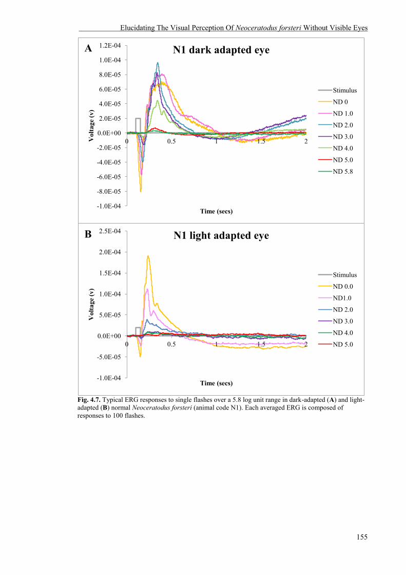
Elucidating The Visual Perception Of Neoceratodus forsteri Without Visible Eyes
155
Fig. 4.7. Typical ERG responses to single flashes over a 5.8 log unit range in dark-adapted (A) and light-adapted (B) normal Neoceratodus forsteri (animal code N1). Each averaged ERG is composed of responses to 100 flashes.
-1.0E-04
-8.0E-05
-6.0E-05
-4.0E-05
-2.0E-05
0.0E+00
2.0E-05
4.0E-05
6.0E-05
8.0E-05
1.0E-04
1.2E-04
0 0.5 1 1.5 2 Vol
tage
(v)
Time (secs)
N1 dark adapted eye
Stimulus
ND 0
ND 1.0
ND 2.0
ND 3.0
ND 4.0
ND 5.0
ND 5.8
-1.0E-04
-5.0E-05
0.0E+00
5.0E-05
1.0E-04
1.5E-04
2.0E-04
2.5E-04
0 0.5 1 1.5 2
Vol
tage
(v)
Time (secs)
N1 light adapted eye
Stimulus
ND 0.0
ND1.0
ND 2.0
ND 3.0
ND 4.0
ND 5.0
A
B

Elucidating The Visual Perception Of Neoceratodus forsteri Without Visible Eyes
156
Fig. 4.8. Typical ERG responses to single flashes over a 5.8 log unit range in dark-adapted (A) and light-adapted (B) normal Neoceratodus forsteri (animal code: N2). Each averaged ERG is composed of responses to 100 flashes.
-1.5E-04
-1.0E-04
-5.0E-05
0.0E+00
5.0E-05
1.0E-04
0 0.5 1 1.5 2
Vol
tage
(v)
Time (s)
N2 dark adpated eye
Stimulus
ND 0.0
ND 1.0
ND 2.0
ND 3.0
ND 4.0
ND 5.0
ND 5.8
-1.5E-04
-1.0E-04
-5.0E-05
0.0E+00
5.0E-05
1.0E-04
1.5E-04
0 0.5 1 1.5 2
Vol
tage
(v)
Time (s)
N2 light adpated eye
Stimulus
ND 0.0
ND 1.0
ND 2.0
ND 3.0
ND 4.0
ND 5.0
B
A

Elucidating The Visual Perception Of Neoceratodus forsteri Without Visible Eyes
157
Fig. 4.9. Single flash ERG implicit times over a range of 5.0 or -5.8 log units in dark- and light- adapted normal Neoceratodus forsteri; N1 (A) and N2 (B).
0
50
100
150
200
250
300
350
400
450
1E-06 1E-05 0.0001 0.001 0.01 0.1 1
Tim
e to
pea
k (m
s)
Stimulus intensity (log10 units)
N1 implicit time
Dark Adapted
Light Adapted
0
50
100
150
200
250
300
350
400
450
1 0.1 0.01 0.001 0.0001 1E-05 1.58E-06
Tim
e to
pea
k (m
s)
Stimulus intensity (log10 units)
N2 implicit time
Dark Adapted
Light Adapted
B
A
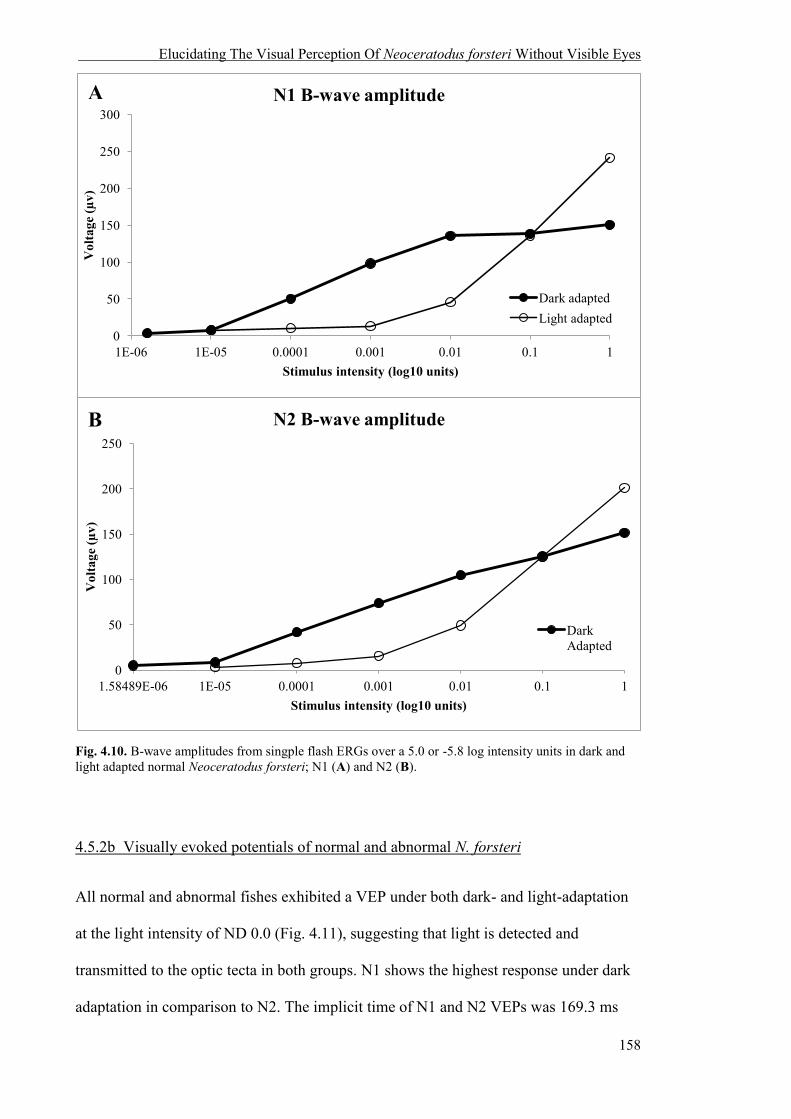
Elucidating The Visual Perception Of Neoceratodus forsteri Without Visible Eyes
158
Fig. 4.10. B-wave amplitudes from singple flash ERGs over a 5.0 or -5.8 log intensity units in dark and light adapted normal Neoceratodus forsteri; N1 (A) and N2 (B).
4.5.2b Visually evoked potentials of normal and abnormal N. forsteri
All normal and abnormal fishes exhibited a VEP under both dark- and light-adaptation
at the light intensity of ND 0.0 (Fig. 4.11), suggesting that light is detected and
transmitted to the optic tecta in both groups. N1 shows the highest response under dark
adaptation in comparison to N2. The implicit time of N1 and N2 VEPs was 169.3 ms
0
50
100
150
200
250
300
1E-06 1E-05 0.0001 0.001 0.01 0.1 1
Vol
tage
(µv)
Stimulus intensity (log10 units)
N1 B-wave amplitude
Dark adapted Light adapted
0
50
100
150
200
250
1 0.1 0.01 0.001 0.0001 1E-05 1.58489E-06
Vol
tage
(µv)
Stimulus intensity (log10 units)
N2 B-wave amplitude
Dark Adapted
B
A
Elucidating The Visual Perception Of Neoceratodus forsteri Without Visible Eyes
159
and 193.5 ms, respectively, and the implicit time of N1 and N2 ERGs of the same light
intensity was 235.7 ms and 251.1 ms, respectively. All abnormal lungfishes displayed a
markedly lower VEP voltage compared to the normal lungfishes (Table 4.3), potentially
due to the limited amount of light penetrating the skin where the eyes should be.
Interestingly, E2, a fish that demonstrated statistically random movement between
sectors of relative light and relative dark exhibited the highest VEP response of all
abnormal lungfishes (Fig. 4.11). Normalisation of the VEPs for each lungfish showed
that, despite the differences in amplitude, they show a similar response duration (Fig.
4.12).

Elucidating The Visual Perception Of Neoceratodus forsteri Without Visible Eyes
160
Fig. 4.11. Visually evoked potentials (VEPs) in normal and abnormal Neoceratodus forsteri in the dark- (A) and light- (B) adapted retina with ND 0.0. Each averaged evoked potential is composed of responses to 100 flashes.
-1.0E-06
-5.0E-07
0.0E+00
5.0E-07
1.0E-06
1.5E-06
2.0E-06
2.5E-06
3.0E-06
3.5E-06
0 0.2 0.4 0.6 0.8 1 1.2 1.4 1.6 1.8 2
Vol
tage
(V)
Time (secs)
Dark adapted VEPs
Stimulus N1 E1 E2 E3
-2.5E-06
-1.5E-06
-5.0E-07
5.0E-07
1.5E-06
2.5E-06
3.5E-06
4.5E-06
5.5E-06
0 0.2 0.4 0.6 0.8 1 1.2 1.4 1.6 1.8 2
Vol
tage
(V)
Time (secs)
Light adapted VEPs Stimulus N1 N2 E1 E2 E3
B
A

Elucidating The Visual Perception Of Neoceratodus forsteri Without Visible Eyes
161
Fig. 4.12. Normalised visually evoked potentials (VEPs) of normal (N) and abnormal (E) Neoceratodus forsteri in dark-adapted (A) and light-adapted (B) retina with ND 0.0 illustrating the VEP waveform. Each averaged evoked potential is composed of responses to 100 flashes.
-1.5
-1
-0.5
0
0.5
1
1.5
0 0.1 0.2 0.3 0.4 0.5 0.6 0.7 0.8 0.9 1
Vol
tage
(uv)
Time (secs)
Normalised VEPs dark adapted
N1
E1
E2
E3
-1
-0.5
0
0.5
1
0 0.1 0.2 0.3 0.4 0.5 0.6 0.7 0.8 0.9 1
Vol
tage
(uv)
Time (secs)
Normalised VEPs light adapted
N1
N2
E1
E2
E3
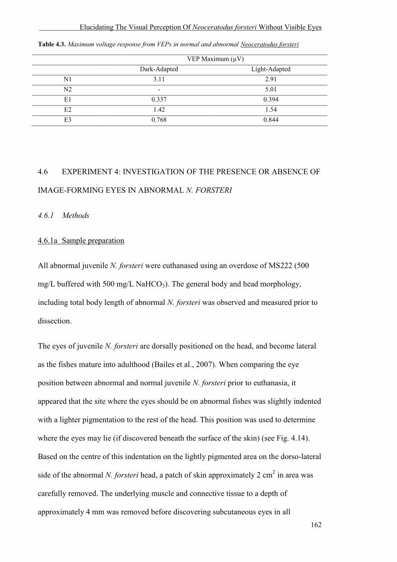
Elucidating The Visual Perception Of Neoceratodus forsteri Without Visible Eyes
162
Table 4.3. Maximum voltage response from VEPs in normal and abnormal Neoceratodus forsteri
VEP Maximum (µV) Dark-Adapted Light-Adapted
N1 3.11 2.91 N2 - 5.01 E1 0.337 0.394 E2 1.42 1.54 E3 0.768 0.844
4.6 EXPERIMENT 4: INVESTIGATION OF THE PRESENCE OR ABSENCE OF
IMAGE-FORMING EYES IN ABNORMAL N. FORSTERI
4.6.1 Methods
4.6.1a Sample preparation
All abnormal juvenile N. forsteri were euthanased using an overdose of MS222 (500
mg/L buffered with 500 mg/L NaHCO3). The general body and head morphology,
including total body length of abnormal N. forsteri was observed and measured prior to
dissection.
The eyes of juvenile N. forsteri are dorsally positioned on the head, and become lateral
as the fishes mature into adulthood (Bailes et al., 2007). When comparing the eye
position between abnormal and normal juvenile N. forsteri prior to euthanasia, it
appeared that the site where the eyes should be on abnormal fishes was slightly indented
with a lighter pigmentation to the rest of the head. This position was used to determine
where the eyes may lie (if discovered beneath the surface of the skin) (see Fig. 4.14).
Based on the centre of this indentation on the lightly pigmented area on the dorso-lateral
side of the abnormal N. forsteri head, a patch of skin approximately 2 cm2 in area was
carefully removed. The underlying muscle and connective tissue to a depth of
approximately 4 mm was removed before discovering subcutaneous eyes in all

Elucidating The Visual Perception Of Neoceratodus forsteri Without Visible Eyes
163
abnormal lungfishes. The enucleated eyes of abnormal and normal N. forsteri were
preserved in Karnovsky’s fixative (2% paraformaldehyde, 2.5% glutaraldehyde in 0.1M
sodium cacodylate buffer, pH 7.4). The lens of E1 and E2 was removed prior to
fixation. The eyes of E3 were fixed with the lens still in situ. The normal N. forsteri
eyes were used as a comparison for the gross morphology of the eye between abnormal
and normal fishes.
4.6.1b General morphology, ocular dimensions and light microscopy
Following the discovery and localisation of the eyes of all abnormal N. forsteri, left
eyes were enucleated and processed for light microscopy to examine and measure the
gross morphology of ocular tissue and photoreceptor cells. The eyes were postfixed for
one hour with 1% osmium tetroxide in 0.15M phosphate buffer, dehydrated through an
alcohol and propylene oxide series and infiltrated with Procure-Araldite (ProSciTech).
Semi-thin (1 µm) sections were cut using a diamond knife on a LKB Bomma Ultratome
NOVA and stained with 4 % Toluidine blue. Ocular tissue sections were examined on
an Olympus compound light microscopy (Model BX50F4) fitted with an Olympus
camera (Model DP70).
The lens of N. forsteri is known to increase with body length in a linear relationship
(Bailes et al., 2007). In order to determine if the lens in the eyes of abnormal N. forsteri
also followed the same relationship as the eyes in normal N. forsteri, the lens axial
length and body length of E3 was compared to the regression slope of N. forsteri,
previously published by Bailes et al. (2007).

Elucidating The Visual Perception Of Neoceratodus forsteri Without Visible Eyes
164
4.6.1c Assessment of photoreceptor dimensions and morphology
All photoreceptor measurements were performed from digital images of light
micrographs using ImageJ 1.46r (National Institute of Health, USA). To allow for
comparison between photoreceptor cells, the inner segment (IS) diameter was measured
at the widest point, and the outer segment (OS) diameter was measured at the base. The
photoreceptor dimensions of abnormal lungfishes were also compared to normal
N. forsteri using an unpaired t-test with data from Bailes et al. (2006) of an individual
31.5 cm in total length. The measurements of abnormal and normal lungfishes are from
different data sets and are statistically not directly comparable. However, large
differences between groups can still be taken as an indication of additional deformities
in the abnormal N. forsteri eye. All measurements are quoted as mean ± standard
deviation (SD) followed by sample size (n).
4.6.2 Results
4.6.2a General morphology and ocular measurements
All abnormal lungfishes are dark brown/green in colour and share a similar head and
body shape. The tails of E1 and E3 are pointed (Fig. 4.13A, C), typical of normal
N. forsteri, but the dorsal and ventral regions of the caudal peduncle of E2 are fused,
resulting in a blunt and squared tail (Fig. 4.13B). All abnormal lungfishes possessed an
excess amount of skin around the mouth and gills. The skin where the eyes should have
been located generally contained a small indention and a lighter pigmentation (Fig.
4.14).
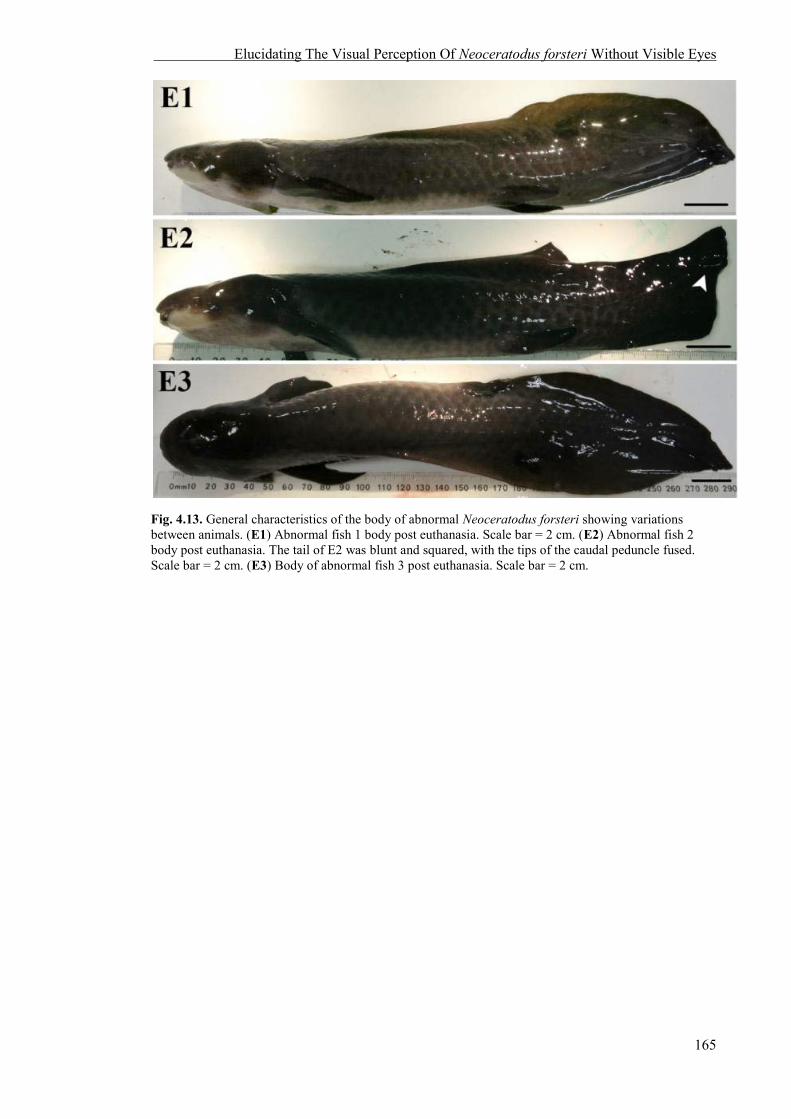
Elucidating The Visual Perception Of Neoceratodus forsteri Without Visible Eyes
165
Fig. 4.13. General characteristics of the body of abnormal Neoceratodus forsteri showing variations between animals. (E1) Abnormal fish 1 body post euthanasia. Scale bar = 2 cm. (E2) Abnormal fish 2 body post euthanasia. The tail of E2 was blunt and squared, with the tips of the caudal peduncle fused. Scale bar = 2 cm. (E3) Body of abnormal fish 3 post euthanasia. Scale bar = 2 cm.
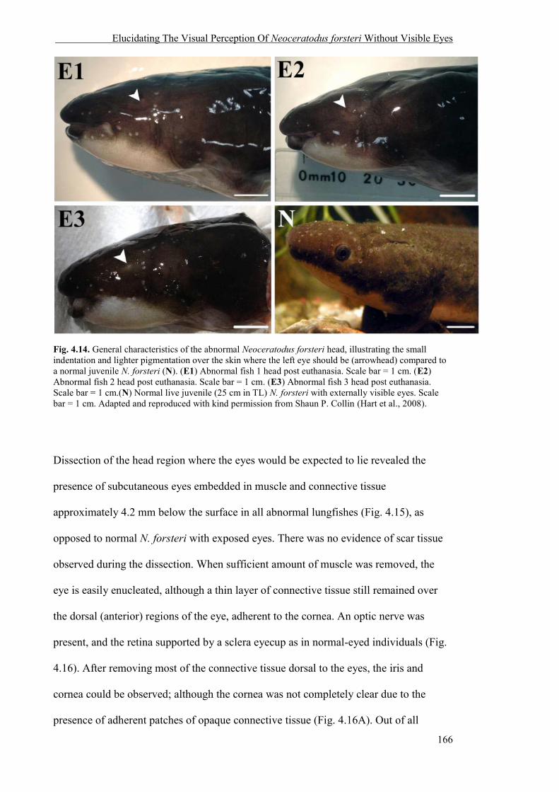
Elucidating The Visual Perception Of Neoceratodus forsteri Without Visible Eyes
166
Fig. 4.14. General characteristics of the abnormal Neoceratodus forsteri head, illustrating the small indentation and lighter pigmentation over the skin where the left eye should be (arrowhead) compared to a normal juvenile N. forsteri (N). (E1) Abnormal fish 1 head post euthanasia. Scale bar = 1 cm. (E2) Abnormal fish 2 head post euthanasia. Scale bar = 1 cm. (E3) Abnormal fish 3 head post euthanasia. Scale bar = 1 cm.(N) Normal live juvenile (25 cm in TL) N. forsteri with externally visible eyes. Scale bar = 1 cm. Adapted and reproduced with kind permission from Shaun P. Collin (Hart et al., 2008).
Dissection of the head region where the eyes would be expected to lie revealed the
presence of subcutaneous eyes embedded in muscle and connective tissue
approximately 4.2 mm below the surface in all abnormal lungfishes (Fig. 4.15), as
opposed to normal N. forsteri with exposed eyes. There was no evidence of scar tissue
observed during the dissection. When sufficient amount of muscle was removed, the
eye is easily enucleated, although a thin layer of connective tissue still remained over
the dorsal (anterior) regions of the eye, adherent to the cornea. An optic nerve was
present, and the retina supported by a sclera eyecup as in normal-eyed individuals (Fig.
4.16). After removing most of the connective tissue dorsal to the eyes, the iris and
cornea could be observed; although the cornea was not completely clear due to the
presence of adherent patches of opaque connective tissue (Fig. 4.16A). Out of all

Elucidating The Visual Perception Of Neoceratodus forsteri Without Visible Eyes
167
enucleated eyes, E3 appeared the most ‘normal’, with a more easily identifiable iris and
pupillary aperture, and the least amount of connective tissue adherent to the cornea (Fig.
4.16B). The enucleated lens of E1 was rostrocaudally flattened with an axial diameter of
1.42 mm and an equatorial lens diameter of 2.25 mm (Fig. 4.17A). Upon examination of
the fundus of the abnormal lungfish eye, the retina was present with axons joining the
optic nerve, which exited the eyecup at the back of the eye (Fig. 4.17B). The ocular lens
of E2 and E3 was spherical. Following examination of the ocular tissue cut in transverse
section, the retinal cell layers appeared normal and intact but with the thickness of the
scleral tissue found to vary around the eye: scleral posterior thickness of 214.72 ± 27.47
µm (n = 40) compared to scleral anterior thickness (“cornea”) of 145.11 ± 14.26 µm (n
= 40) (Fig. 4.18). A comparison of lens axial diameter and total body size between E3
and data of normal N. forsteri (Bailes et al., 2007) showed that the E3 lens, although
slightly smaller than the closest normal N. forsteri of similar size, did follow the relative
linear allometric relationship of lens axial diameter and total body size (Fig. 4.19). The
optic nerve of abnormal N. forsteri was also embedded in connective tissue as was the
case for normal individuals. The diameter of the abnormal N. forsteri optic nerve was
363.97 µm at the widest point (in an individual 25 cm in TL) compared to 543.63 µm in
a subadult normal N. forsteri (in an individual 39.5 cm in TL) (Bailes et al., 2006b). The
fascicles surrounding the axons in the optic nerve of the abnormal eyes were present,
although they were not as clearly defined as in the optic nerve in the normal N. forsteri.
No myelinated axons were observed. Although axons and the nuclei of glial cells were
observed, the optic nerve was not organised and contained many spaces situated
between the axons and astroglial cells (Fig. 4.20).

Elucidating The Visual Perception Of Neoceratodus forsteri Without Visible Eyes
168
Fig. 4.15. The subcutaneous eye of abnormal Neoceratodus forsteri, E3. The eye was embedded within muscle approximately 4.2 mm below the skin and surrounded by connective tissue. The entire eye is covered in scleral tissue, and darkly pigmented with no clear cornea present in this individual. Scale bar = 1 cm.
Fig. 4.16. External appearance of the enucleated eyes of two abnormal Neoceratodus forsteri. The diameter of each eye was approximately 5 mm. (A) The eye of E2 was embedded in more connective tissue than E3. Scleral tissue (scl) surrounded the entire eye in the absence of a clear cornea. An aperture (a) was present and appeared dark, indicating the presence of a pigmented retinal pigment epithelium underlying the retina. Scale bar = 2.5 mm. (B) The eye of E3 appeared to be the most representative of a normal juvenile N. forsteri. The cornea is relatively clear, revealing a clearly defined iris (ir) and the pupillary aperture (a). Scale bar = 2.5 mm.

Elucidating The Visual Perception Of Neoceratodus forsteri Without Visible Eyes
169
Fig. 4.17. The internal appearance of the isolated eye of an abnormal Neoceratodus forsteri. (A) All abnormal lungfish eyes possessed a lens, but the lens (ln) of E1 was rostrocaudally flattened. Scale bar = 2.5 mm (B) The retina (r) was present with axons (arrowhead) converging at the optic nerve head (on) at the back of the eye. Scale bar = 2.5 mm.
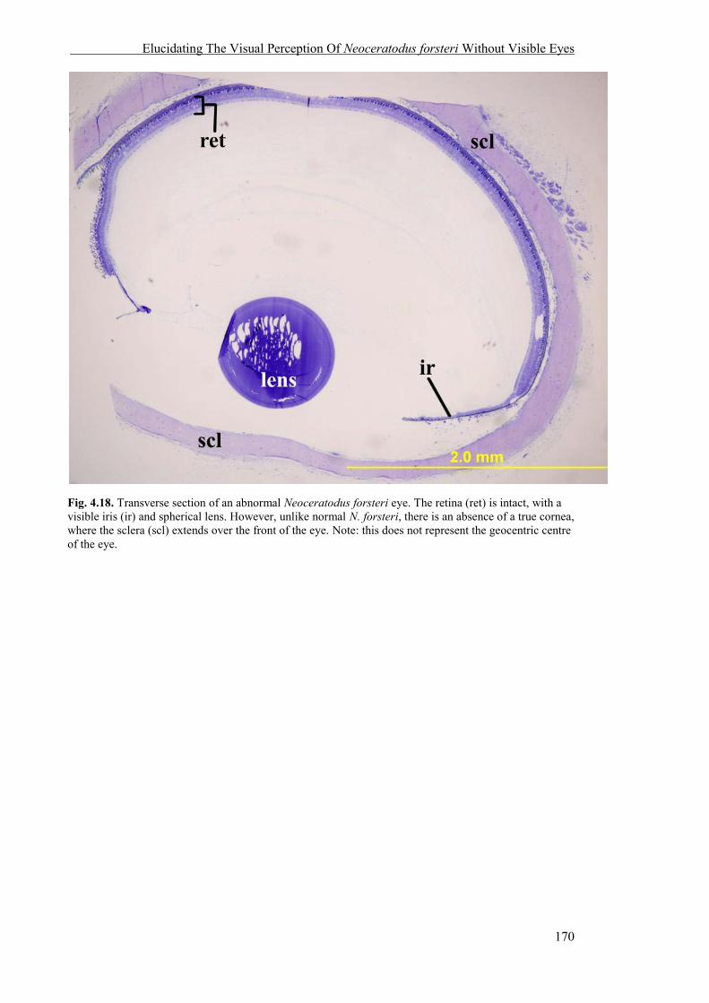
Elucidating The Visual Perception Of Neoceratodus forsteri Without Visible Eyes
170
Fig. 4.18. Transverse section of an abnormal Neoceratodus forsteri eye. The retina (ret) is intact, with a visible iris (ir) and spherical lens. However, unlike normal N. forsteri, there is an absence of a true cornea, where the sclera (scl) extends over the front of the eye. Note: this does not represent the geocentric centre of the eye.
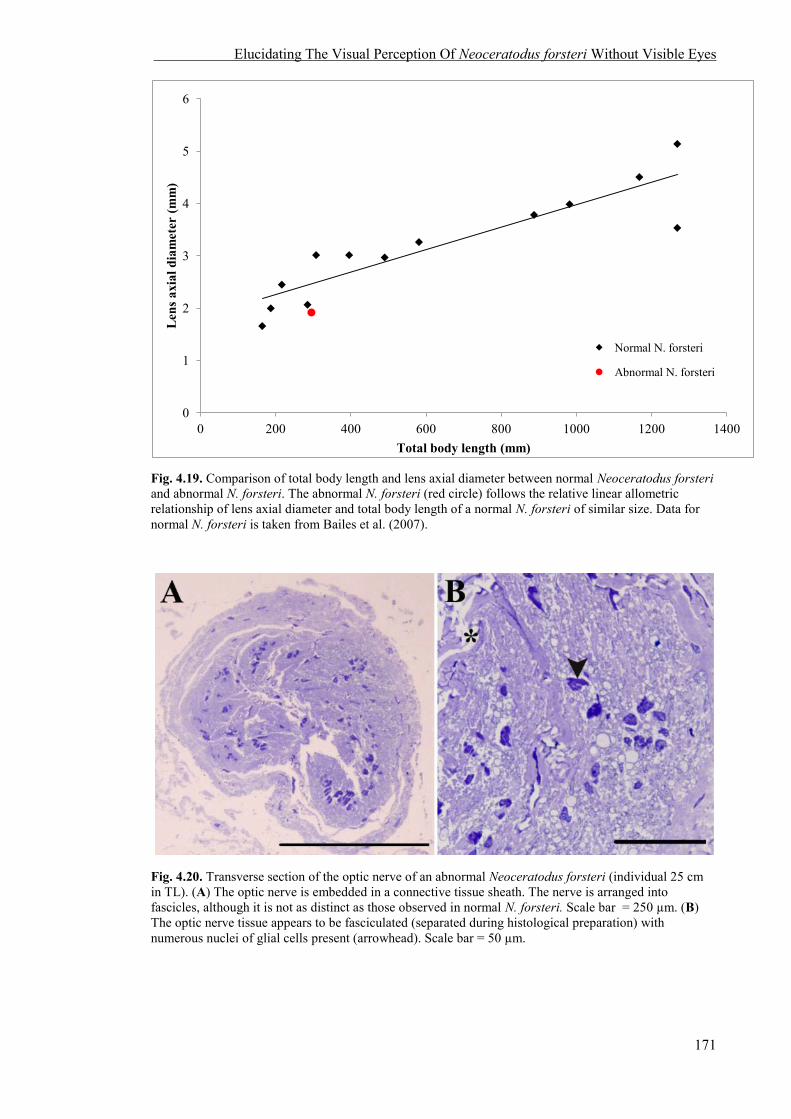
Elucidating The Visual Perception Of Neoceratodus forsteri Without Visible Eyes
171
Fig. 4.19. Comparison of total body length and lens axial diameter between normal Neoceratodus forsteri and abnormal N. forsteri. The abnormal N. forsteri (red circle) follows the relative linear allometric relationship of lens axial diameter and total body length of a normal N. forsteri of similar size. Data for normal N. forsteri is taken from Bailes et al. (2007).
Fig. 4.20. Transverse section of the optic nerve of an abnormal Neoceratodus forsteri (individual 25 cm in TL). (A) The optic nerve is embedded in a connective tissue sheath. The nerve is arranged into fascicles, although it is not as distinct as those observed in normal N. forsteri. Scale bar = 250 µm. (B) The optic nerve tissue appears to be fasciculated (separated during histological preparation) with numerous nuclei of glial cells present (arrowhead). Scale bar = 50 µm.
0
1
2
3
4
5
6
0 200 400 600 800 1000 1200 1400
Len
s axi
al d
iam
eter
(mm
)
Total body length (mm)
Normal N. forsteri
Abnormal N. forsteri

Elucidating The Visual Perception Of Neoceratodus forsteri Without Visible Eyes
172
4.6.2b Photoreceptor dimensions and morphology
The eyes of all abnormal N. forsteri possess the requisite number of retinal cell layers
(Fig. 4.21A) and the complement of photoreceptor types typical of the species; one rod,
and three morphological types of cones (red, yellow and colourless, based on the
appearance of the intracellular inclusions within the ellipsoid) with no differences in
general morphology (Fig. 4.21B, 4.21C) (Bailes et al., 2006a). A comparison of
dimensions between the outer segment (OS) length and inner segment (IS) diameter of
photoreceptors of abnormal (two individuals TL = 25 cm, 29.5 cm) and normal
N. forsteri (TL = 31.5 cm) showed that the IS diameter in all photoreceptor types was
not statistically different. However, the OS length of rods was significantly smaller
(31.73 ± 2.39 µm in abnormal, 34.64 ± 2.45 µm in normal, n = 10), the red cone OS
length, significantly larger (7.67 ± 0.90 µm in abnormal, 6.46 ± 1.31 µm in normal, n =
10) and the clear cone OS length, significantly larger (7.45 ± 0.84 µm in abnormal, 5.78
± 1.54 µm, n = 10) in abnormal compared to normal lungfishes (p < 0.05). However,
these differences may be a reflection of comparing two different datasets generated by
different protocols. The measurements and t-test statistics are summarized in Table 4.4.

Elucidating The Visual Perception Of Neoceratodus forsteri Without Visible Eyes
173
Fig. 4.21. Morphological characteristics of the retina and photoreceptors in abnormal Neoceratodus forsteri. (A) Light micrograph of the retina of the abnormal N. forsteri showing the normal retina with all retinal cell layers. Moving from the top to the bottom of the micrograph lie the photoreceptors, outer nuclear layer (onl), outer plexiform layer (opl), inner nuclear layer (inl), inner plexiform layer (ipl) and retinal ganglion cell layer (rgc). Scale bar = 25 µm. (B) Light micrograph showing the rods (rd) with cylindrical outer segments (os), a yellow cone (yc) with the yellow pigment situated above the paraboloid (p) (based on previous nomenclature published by Bailes et al. 2006), and a red oil droplet (od) containing cone (rc) surrounded by mitochondria (m) in the paraboloid. (C) Light micrograph depicting the clear cones (cc) with multiple oil droplets (arrowheads).

Elucidating The Visual Perception Of Neoceratodus forsteri Without Visible Eyes
174
Table 4.4. Comparison of photoreceptor dimensions in abnormal and normal Neoceratodus forsteri1
Abnormal N. forsteri
Normal N. forsteri
t-stat p-value
OS length
(µm)
Rod 31.73 ± 2.39 34.64 ± 2.45 2.69 0.02* Red cone 7.67 ± 0.90 6.46 ± 1.31 2.41 0.03*
Yellow cone 8.02 ± 1.04 8.03 ± 2.43 0.01 0.99 Clear cone 7.45 ± 0.84 5.78 ± 1.54 3.02 0.01*
IS diameter
(µm)
Rod 10.79 ± 1.79 7.96 ± 1.43 1.24 0.23 Red cone 9.68 ± 1.2 6.16 ± 1.28 1.81 0.09
Yellow cone 6.12 ± 0.65 10.744 ±1.42 1.08 0.94 Clear cone 7.16 ± 0.92 10.12 ± 1.13 1.49 0.15
1All values are the mean ± SD. The number of individual cells measured for all fishes (n) = 10. Measurements from abnormal lungfishes were taken from light micrographs in individuals 25 and 29.5 cm in TL. Data for normal lungfishes was taken from Bailes et al. (2006) in an individual 31.5 cm in TL from electron micrographs. All measurements are raw data that have not been corrected for shrinkage. The two groups of lungfishes were compared using an unpaired t-test with df = 18 (* for p < 0.05).
4.7 GENERAL DISCUSSION
This chapter revealed the presence of subcutaneous eyes in a group of abnormal
N. forsteri, supporting the hypothesis that vestigial ocular tissue is present beneath the
epidermis. Contrary to the initial hypothesis, the recording of VEPs from abnormal
lungfishes was successful, and showed that all eyes possessed a retina that was sensitive
to light despite being embedded underneath muscle and connective tissue. However,
these tests of functional visual integrity did not translate into a consistent behavioural
phototaxis in all abnormal lungfishes, which partially supports the hypothesis that
abnormal lungfishes will not display the same behaviour as normal lungfishes in
response to differences in ambient light. Lastly, the hypothesis that the circadian rhythm
of abnormal lungfishes would not be the same as normal N. forsteri was partially
supported by the large differences in distance travelled by abnormal lungfishes and E1
remaining consistently active for 54 hours.

Elucidating The Visual Perception Of Neoceratodus forsteri Without Visible Eyes
175
4.7.1 The abnormal N. forsteri eye
The enucleated eyes of abnormal N. forsteri contained most of the structures typical of a
vertebrate eye (Walls, 1942) and what has been described for the species (Bailes et al.,
2006a, Bailes et al., 2006b, Bailes et al., 2007). However, the most striking difference in
the eyes of abnormal N. forsteri is the absence of a cornea and the sclera extending over
the anterior pole of the eye that not always provide a clear window for light to enter the
pupil. The vertebrate cornea typically consists of a transparent multi-layered epithelium
over a basement membrane, a thick stroma, Desçemet’s membrane and an endothelium
(Hay, 1980), with some variations in some of these structures that may include
additional layers between species (reviewed in Collin and Collin, 2001). None of the
differentiated layers typical of a cornea were observed in the abnormal N. forsteri eye.
Instead, networks of opaque, dense connective tissue consisting of collagenous and
elastic fibres of the sclera (Rada et al., 2006) extended over the anterior chamber of the
eye in in abnormal N. forsteri. Additionally, the absence of a limbus (also known as
scleral roll or scleral spur) that marks the edge of the sclera and the beginning of the
cornea (Walls, 1942) further suggests the lack of corneal tissue development. The
cornea is one of the last structures to develop in the eye, so development may have been
arrested at the point when the lens vesicle meets the surface ectoderm (Chow and Lang,
2001).
The lens is present in all abnormal N. forsteri and follows the linear allometric
relationship of lens axial diameter and total body length (Bailes et al., 2007), but the
aspherical lens of E1 suggests the lens development in at least one abnormal N. forsteri
was also disrupted at this point in eye morphogenesis, and that there is variation in the
eye phenotype between abnormal individuals.
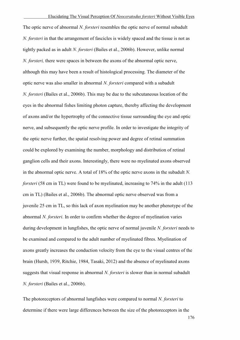
Elucidating The Visual Perception Of Neoceratodus forsteri Without Visible Eyes
176
The optic nerve of abnormal N. forsteri resembles the optic nerve of normal subadult
N. forsteri in that the arrangement of fascicles is widely spaced and the tissue is not as
tightly packed as in adult N. forsteri (Bailes et al., 2006b). However, unlike normal
N. forsteri, there were spaces in between the axons of the abnormal optic nerve,
although this may have been a result of histological processing. The diameter of the
optic nerve was also smaller in abnormal N. forsteri compared with a subadult
N. forsteri (Bailes et al., 2006b). This may be due to the subcutaneous location of the
eyes in the abnormal fishes limiting photon capture, thereby affecting the development
of axons and/or the hypertrophy of the connective tissue surrounding the eye and optic
nerve, and subsequently the optic nerve profile. In order to investigate the integrity of
the optic nerve further, the spatial resolving power and degree of retinal summation
could be explored by examining the number, morphology and distribution of retinal
ganglion cells and their axons. Interestingly, there were no myelinated axons observed
in the abnormal optic nerve. A total of 18% of the optic nerve axons in the subadult N.
forsteri (58 cm in TL) were found to be myelinated, increasing to 74% in the adult (113
cm in TL) (Bailes et al., 2006b). The abnormal optic nerve observed was from a
juvenile 25 cm in TL, so this lack of axon myelination may be another phenotype of the
abnormal N. forsteri. In order to confirm whether the degree of myelination varies
during development in lungfishes, the optic nerve of normal juvenile N. forsteri needs to
be examined and compared to the adult number of myelinated fibres. Myelination of
axons greatly increases the conduction velocity from the eye to the visual centres of the
brain (Hursh, 1939, Ritchie, 1984, Tasaki, 2012) and the absence of myelinated axons
suggests that visual response in abnormal N. forsteri is slower than in normal subadult
N. forsteri (Bailes et al., 2006b).
The photoreceptors of abnormal lungfishes were compared to normal N. forsteri to
determine if there were large differences between the size of the photoreceptors in the

Elucidating The Visual Perception Of Neoceratodus forsteri Without Visible Eyes
177
two groups. In general, the retina of abnormal N. forsteri contained all cell layers and
photoreceptor types typical of the species (Bailes et al., 2006a). Although there were
some significant differences in the outer segment length of the rod, red and clear
photoreceptor types, it is important to note that the comparisons made between normal
and abnormal lungfishes were from different datasets generated from slightly different
protocols, and the differences, although significant, are small. The rod OS length in
Protopterus dolloi (16.0 ± 0.37 SEM µm, n = 32) and Lepidosiren paradoxa (8.0 ± 0.39
SEM µm, n = 13) (Chapter 3) are notably smaller than the rod OS length of normal
N. forsteri (34.64 ± 0.77 SEM µm), and clear differences like this were not found
between normal and abnormal N. forsteri in this study.
4.7.2 Visual perception of abnormal N. forsteri
Despite the subcutaneous location of the eye, the absence of a cornea in some
individuals, and other differences in the size of some of the retinal photoreceptor types
and differences in the optic nerve structure, all abnormal lungfishes exhibited a VEP.
Although the amplitude of the VEPs varied between individuals, this finding
demonstrates that the retina of abnormal N. forsteri is still photoreceptive, that the optic
nerve is relaying this information to the brain, and (presumably) the optic tectum is
receiving this input. The differences between the amplitude measured in normal and
abnormal lungfish VEPs are probably affected by the amount of light stimuli reaching
the retina as the subcutaneous eyes of abnormal lungfishes were buried deep beneath the
skin. Despite the fact that VEPs were measured from all three abnormal lungfishes, only
E3 demonstrated statistically non-random behaviour in the two regions of the tank (light
and dark). This suggests that either the sclera over the anterior of the eye allowed
enough ambient light to pass through and elicit a behavioural response in E3, the light
Elucidating The Visual Perception Of Neoceratodus forsteri Without Visible Eyes
178
intensity in Experiment 1 was not bright enough to elicit a behavioural response in E1
and E2, or the VEPs generated by E1 and E2 were not from the optic tecta. VEPs are
generally produced by the optic tecta, but in human studies, VEPs have been reported to
also be generated by sub-cortical or extrageniculate sources in functionally blind
individuals rather than the visual cortex (Kupersmith and Nelson, 1986, Mackie and
McCulloch, 1995, Clarke et al., 1997). More investigation into the brain of abnormal
lungfishes may provide greater information on how this organ may be affected.
Sensory plasticity has been described in a number of species. Adverse environmental
conditions in early development that results in a deficit in one sense can result in
adaptations that heighten the capacity in another to enable organisms to respond
effectively to environmental change (see the “compensatory plasticity hypothesis” by
Rauschecker and Kniepert, 1994). Guppies (Poecilia reticulata) have been shown to
exhibit sensory plasticity in vision and chemoreception in response to differing light
intensities. Guppies reared in low light environments had a significantly stronger
response to chemical food cues and a weaker response to visual cues in isolation,
compared to those reared in higher light conditions (Chapman et al., 2010). Terrestrial
snakes (Notechis scutatus) reared in an aquatic environment have shown improved
locomotor performance in water compared to snakes reared on land (Aubret et al., 2007,
Aubret and Shine, 2008). These examples suggest that sensory plasticity causes a
stronger response to chemical food cues in guppies, and an increase in locomotor
response in terrestrial snakes reared in aquatic envionrments.
Other senses reported to be important to lungfishes are the lateral line (Lippolis et al.,
2009) and electroreception (Watt et al., 1999), which provide enhanced sensory capacity
at low light conditions. A 72 hour observation of lungfishes in total darkness showed
that the distance abnormal N. forsteri travelled when active was more than 2.5 times

Elucidating The Visual Perception Of Neoceratodus forsteri Without Visible Eyes
179
that of the normal N. forsteri. The increase in distance travelled in abnormal fishes and,
in the case of E1 and E2, the inability to discriminate between light and dark sections of
the holding tank, may be a form of behavioural plasticity that, in the absence of external
eyes, has contributed to a greater dependence on the lateral line and electroreception
systems.
The lateral line system is a mechanoreceptive sense that consists of a series of hair cell
bundles (neuromasts) that occur either on the surface of the skin (superficial) or within
canals lying beneath the skin, where water enters the canals via small pores over the
head and trunk. This system mediates the ability to detect minute hydrodynamic
disturbances and acts as a specially-adapted mechanoreceptive system for aquatic life
(Bleckmann, 1986, Montgomery et al., 1997). The lateral line can aid in obstacle, wave
and chemical detection (Dijkgraaf, 1963, Hashimoto et al., 1970, Müller and Schwartz,
1982, Kawamura and Yamashita, 1983) and in schooling fish, helps maintain the
swimming speed and direction of travel (Partridge and Pitcher, 1980). Studies on the
blind cave-fish Astyanax fasciatus (Bleckmann, 1986) and the nocturnal torrent fish
Cheimarrichthys fosteri (Montgomery and Milton, 1993) demonstrate that this can
provide tactile reference between the substrate and oncoming water current. The lateral
line of N. forsteri is used in predator detection and avoidance (Lippolis et al., 2009) so,
in the absence of vision, this system may also be utilized in general to compensate in
navigation.
Organisms with electroreception possess ampullary receptors, modulated by movement,
that enable the detection and measurement of weak bioelectric fields that surround prey
animals (Kalmijn, 1971, Bullock et al., 1993, Manger and Pettigrew, 1995). All
lungfishes possess passive electroreception and, during foraging, juvenile N. forsteri
utilize this sense to prey on animals buried in the substrate with greater accuracy after
Elucidating The Visual Perception Of Neoceratodus forsteri Without Visible Eyes
180
assessing the general whereabouts of the organism using olfaction (Watt et al., 1999).
The importance of vision in the lifestyle of N. forsteri is unknown, but the reduced
distance travelled by normal N. forsteri compared with abnormal N. forsteri, suggests
that other senses (i.e. lateral line and electroreception) affect how far the fishes travel.
However, E3, the only abnormal fish that could behaviourally discriminate between
light and dark, exhibited the greatest distance travelled over a 72 hour period,
suggesting there may be other reasons associated with this increased activity, such as
the absence of spatial vision. Early exposure to low light levels during development has
been shown to result in a severe impairment of spatial tasks that include locating food
(Axelrod, 1959). Further investigation and comparison into the regions of the brain
responsible for these other senses may provide additional information on how the
abnormal lungfishes are compensating for the lack of image forming vision or the
absence of light intensity information.
Some of the behavioural differences, such as the continuous activity of abnormal
N. forsteri E1 for 54 hours in complete darkness, and the statistically random
movement of E1 and E2 between relative light and dark sections of an aquarium, may
also be due to potential differences in the pineal organ. The general morphology of the
pineal gland in lungfishes has been described as an intermediate between teleost fishes
and amphibians (Holmgren, 1959), and the developmental changes over the first 12
months in N. forsteri resembles that of pre-metamorphic amphibians (Joss et al., 1997a).
Unlike teleosts, lungfishes do not possess a parapineal organ (Holmgren, 1959), and
unlike amphibians, the proportion of photoreceptors decreases and the proportion of
photoreceptors lacking outer segments increases in a developing N. forsteri pineal organ
(Joss et al., 1997a). Some of the functions common to both the amphibian and teleost
pineal organ are setting the circadian locomotor rhythms, entrainment to environmental

Elucidating The Visual Perception Of Neoceratodus forsteri Without Visible Eyes
181
light cycles and free-running rhythms (Adler, 1976, Ekstrzm and Meissl, 1997).
Removal of the pineal organ terminated the circadian free-running locomotor activity
rhythm and caused arrhythmicity in stinging catfish (Heteropneustes fossilis) (Garg and
Sundararaj, 1986). The abnormal N. forsteri E1 was continuously active for 54 hours,
and this arrhythmicity may have been due to an abnormal pineal organ. The teleost
pineal organ can also affect swimming activity and phototactic behaviour. White
suckers (Catostomus commersoni) with shielded pineal regions spent significantly more
time in the illuminated portion of the aquarium (in a two choice light:dark based
system) when kept at a constant temperature (Kavaliers, 1982). The behavioural
differences observed between normal N. forsteri and abnormal N. forsteri (E1 and E2)
in the light and dark portions of the aquarium may also be due to differences in the
pineal organ. The pineal organ of dipnoan fishes has not been extensively researched,
and it is unknown if the pineal photoreceptors of lungfishes function in the same way as
in teleosts and amphibians. However, the close histological and developmental
resemblance of the pineal organ in lungfishes, teleosts and amphibians suggests that a
comparative study between the pineal organs of normal and abnormal N. forsteri may
provide insights into some of the differences seen in the behavioural and circadian
experiments, such as the 54 hours of continuous activity of E1 in complete darkness and
the statistically random behaviour in the two regions of the aquarium (light and dark) in
E1 and E2.
4.7.3 Possible causes for the abnormal morphological deformities in N. forsteri
A clear cause for the morphological deformities in the abnormal lungfishes cannot be
determined from the experiments conducted in this project. However, some speculation
regarding their ocular abnormality can be explored.
Elucidating The Visual Perception Of Neoceratodus forsteri Without Visible Eyes
182
The integrated roles of the environment and/or genetics can cause various diseases and
deformities. In fishes, the environment can adversely influence the development of the
visual system through the absence of light affecting the development of both the image
forming eyes and the pineal organ. The pineal organ of teleosts and amphibians is
responsible for body lightening in the larval stages when raised in complete darkness;
presumably due to melatonin contracting the melanophores in the skin (Bogenschütz,
1965) (Bagnara, 1965) (Joss et al., 1997b). All abnormal fishes possessed a darkly
pigmented body and two out of the three abnormal N. forsteri exhibited a circadian
response similar to normal N. forsteri. This suggests that these fishes perceived a
normal light/dark diel cycle when developing because of the early development of the
pineal gland and its dependence on a consistent light/dark cycle to properly regulate
melatonin secretion, the circadian rhythm and prevent blanching of the skin (Joss et al.,
1997b, Kazimi and Cahill, 1999, Danilova et al., 2004). However, morphological
deformities in fishes can also occur in contaminated water (reviewed in Pereira et al.,
2013) and as a result of poor nutrition (Monaghan, 2008). Morphological deformities
have been observed in other fishes as a result of water quality and temperature, such as
the tilapia Oreochromis spp. (Sun et al., 2009), the japanese eel Anguilla japonica and a
number of species in Skellefteå and Bothnian Bay, Sweden (Bengtsson et al., 1985).
Zebra fish larvae exposed to organic mercury had the greatest concentration of this
heavy metal in the eye (Korbas et al., 2008, Korbas et al., 2010, Korbas et al., 2011,
Korbas et al., 2013) and the accumulation may cause visual impairment, as it does in
humans and other mammals (Warfvinge and Bruun, 1996, Erie et al., 2005). The water
conditions in the development of the abnormal lungfish examined in this study are
unknown and until they are confirmed, cannot be disregarded as a possible cause of
their morphological deformities.

Elucidating The Visual Perception Of Neoceratodus forsteri Without Visible Eyes
183
All the abnormal N. forsteri are from the same brood. The diversity of the genetic pool
of captive lungfishes at Macquarie University is unknown, so the visual (and other
potential deformities) may be due to congenital defects caused by inbreeding. Pax6 is a
one of the best studied transcriptional regulatory genes essential for the normal
development of the brain and eye. It is essential for normal eye development in
Drosophila (Halder et al., 1995) and over- and under-expression of the gene through
mutations can result in severe visual abnormalities (Schedl et al., 1996). Heterozygous
mutations of Pax6 in mice result in the reduction of eye size and iris hypoplasia, and
homozygous mutations result in the lack of eye cavities, nasal cavities, and brain
abnormalities that result in death shortly after birth (Hogan et al., 1988, Ton et al., 1991,
Schedl et al., 1996). Heterozygote mutations in humans result in aniridia, an umbrella
term that describes visual defects of varying degrees such as iris hypoplasia, optic nerve
hypoplasia corneal opacification and cataracts (Glaser et al., 1994). Therefore,
molecular analyses of Pax6 may uncover the genetic basis of the congenital defects
observed in abnormal N. forsteri.
4.8 CONCLUSION
This study compared aspects of the visual Umwelten between groups of normal N.
forsteri and abnormal N. forsteri with ectopic eyes. Through behavioural,
electrophysiological and histological experimentation, it has revealed the presence of
subcutaneous eyes in all abnormal lungfishes that are capable of photo-transduction, but
perhaps not spatial vision. The abnormal lungfish eye lacks a cornea, possesses an
atypical optic nerve, but has a retina and a photoreceptor complement typical of N.
forsteri. In addition, the measured functional integrity of the eye does not translate into

Elucidating The Visual Perception Of Neoceratodus forsteri Without Visible Eyes
184
behavioural phototaxis in all abnormal lungfishes, suggesting there is variation between
individuals. However, further study is needed to uncover other aspects of the abnormal
lungfish visual Umwelt, and establish the genetic and/or environmental basis underlying
their morphological deformities.
4.9 REFERENCES
ADLER, K. 1976. Extraocular photoreception in amphibians. Photochemistry and Photobiology, 23, 275-298.
ASI, H. & PERLMAN, I. 1992. Relationships between the electroretinogram a-wave, b-wave and oscillatory potentials and their application to clinical diagnosis. Documenta Ophthalmologica, 79, 125-139.
AUBRET, F., BONNET, X. & SHINE, R. 2007. The role of adaptive plasticity in a major evolutionary
transition: early aquatic experience affects locomotor performance of terrestrial snakes. Functional Ecology, 21, 1154-1161.
AUBRET, F. & SHINE, R. 2008. Early experience influences both habitat choice and locomotor
performance in tiger snakes. The American Naturalist, 171, 524-531. AXELROD, S. 1959. Effects of early blindness. New York: American Foundation for the Blind. BAGNARA, J. T. 1965. Pineal regulation of body blanching in amphibian larvae. Progress in Brain
Research, 10, 489-506. BAILES, H. J., ROBINSON, S., TREZISE, A. E. O. & COLLIN, S. P. 2006a. Morphology,
characterization, and distribution of retinal photoreceptors in the Australian lungfish Neoceratodus forsteri (Krefft, 1870). The Journal of Comparative Neurology, 494, 381-397.
BAILES, H. J., TREZISE, A. E. O. & COLLIN, S. P. 2006b. The number, morphology, and distribution
of retinal ganglion cells and optic axons in the Australian lungfish Neoceratodus forsteri (Krefft 1870). Visual Neuroscience, 23, 257-273.
BAILES, H. J., TREZISE, A. E. O. & COLLIN, S. P. 2007. The optics of the growing lungfish eye: lens
shape, focal ratio and pupillary movements in Neoceratodus forsteri. Visual Neuroscience, 24, 377-387.
BAYLOR, D. & HODGKIN, A. 1974. Changes in time scale and sensitivity in turtle photoreceptors. The
Journal of Physiology, 242, 729-758. BENGTSSON, B.-E., BENGTSSON, Å. & HIMBERG, M. 1985. Fish deformities and pollution in some
Swedish waters. Ambio, 32-35. BENYASSI, A., SCHWARTZ, C., COON, S. L., KLEIN, D. C. & FALCÓN, J. 2000. Melatonin
synthesis: Arylalkylamine N-acetyltransferases in trout retina and pineal organ are different. Neuroreport, 11, 255-258.
BLECKMANN, H. 1986. Role of the lateral line in fish behaviour. The Behaviour of Teleost Fishes.
Springer.

Elucidating The Visual Perception Of Neoceratodus forsteri Without Visible Eyes
185
BOGENSCHÜTZ, H. 1965. Untersuchungen über den lichtbedingten Farbwechsel der Kaulquappen. Zeitschrift für vergleichende Physiologie, 50, 598-614.
BROOKS, S. G. A. K., P.K. 2002. Ecology and demography of the Queensland lungfish (Neoceratodus
forsteri) in the Burnett Riverm Queensland - with reference to the impacts of Walla Weir and future water infrastructure development. In: 2002, F. R. M. (ed.). Department of Primary Industries Report QO02004. Prepared for the Department of Natural Resources and Mines.
BROWN, K. T. 1968. The electroretinogram: its components and their origins. Vision Research, 8, 633-
IN6. BULLOCK, T. H., BODZNICK, D. A. & NORTHCUTT, R. G. 1993. The phylogenetic distribution of
electroreception: evidence for convergent evolution of a primitive vertebrate sense modality. How do Brains Work? : Springer.
CAHILL, G. M. & BESHARSE, J. C. 1995. Circadian rhythmicity in vertebrate retinas: regulation by a
photoreceptor oscillator. Progress in Retinal and Eye Research, 14, 267-291. CAHN, P. H. 1958. Comparative optic development in Astyanax mexicanus and in two of its blind cave
derivatives. Bulletin of the American Museum of Natural History, 115. CERVETTO, L., PASINO, E. & TORRE, V. 1977. Electrical responses of rods in the retina of Bufo
marinus. The Journal of Physiology, 267, 17-51. CHAPMAN, B. B., MORRELL, L. J., TOSH, C. R. & KRAUSE, J. 2010. Behavioural consequences of
sensory plasticity in guppies. Proceedings of the Royal Society B: Biological Sciences, rspb20092055.
CHOW, R. L. & LANG, R. A. 2001. Early eye development in vertebrates. Annual Review of Cell and
Developmental Biology, 17, 255-296. CLARKE, M., MITCHELL, K. & GIBSON, M. 1997. The prognostic value of flash visual evoked
potentials in the assessment of non-ocular visual impairment in infancy. Eye, 11, 398-402. COLLIN, S. P. & COLLIN, H. 2001. The fish cornea: adaptations for different aquatic environments.
Sensory Biology of Jawed Fishes. New Insights, 57-96. CREEL, D., DUSTMAN, R. E. & BECK, E. C. 1974. Intensity of flash illumination and the visually
evoked potential of rats, guinea pigs and cats. Vision Research, 14, 725-729. CREEL, D. J. 2012. Visually evoked potentials. Webvision: The Organization of the Retina and Visual
System. Salt Lake City: University of Utah Health Sciences Center, 201-229. DANILOVA, N., KRUPNIK, V. E., SUGDEN, D. & ZHDANOVA, I. V. 2004. Melatonin stimulates cell
proliferation in zebrafish embryo and accelerates its development. The FASEB Journal, 18, 751-753.
DAVIES, W. I., TAY, B.-H., ZHENG, L., DANKS, J. A., BRENNER, S., FOSTER, R. G., COLLIN, S.
P., HANKINS, M. W., VENKATESH, B. & HUNT, D. M. 2012a. Evolution and functional characterisation of melanopsins in a deep-sea chimaera (elephant shark, Callorhinchus milii). PLoS One, 7, e51276.
DAVIES, W. L., FOSTER, R. G. & HANKINS, M. W. 2012b. Focus on molecules: Melanopsin.
Experimental Eye Research, 97, 161-162. DEAN, B. Notes on the living specimens of the Australian lungfish, Ceratodus forsteri, in the Zoological
Society's collection. Proceedings of the Zoological Society of London, 1906. Wiley Online Library, 168-178.
DIJKGRAAF, S. 1963. The functioning and significance of the lateral-line organs. Biological Reviews,
38, 51-105.

Elucidating The Visual Perception Of Neoceratodus forsteri Without Visible Eyes
186
EKSTRÖM, P. & MEISSL, H. 1997. The pineal organ of teleost fishes. Reviews in Fish Biology and Fisheries, 7, 199-284.
EKSTRZM, P. & MEISSL, H. 1997. The pineal organ of teleost fishes. Reviews in Fish Biology and
Fisheries, 7, 199-284. ERIE, J. C., BUTZ, J. A., GOOD, J. A., ERIE, E. A., BURRITT, M. F. & CAMERON, J. D. 2005.
Heavy metal concentrations in human eyes. American Journal of Ophthalmology, 139, 888-893. FALCÓN, J., BESSEAU, L., SAUZET, S. & BOEUF, G. 2007. Melatonin effects on the hypothalamo–
pituitary axis in fish. Trends in Endocrinology & Metabolism, 18, 81-88. FALCÓN, J., MIGAUD, H., MUNOZ-CUETO, J. A. & CARRILLO, M. 2010. Current knowledge on the
melatonin system in teleost fish. General and Comparative Endocrinology, 165, 469-482. FALCÓN, J., THIBAULT, C., BEGAY, V., ZACHMANN, A. & COLLIN, J.-P. 1992. Regulation of the
rhythmic melatonin secretion by fish pineal photoreceptor cells. Rhythms in Fishes. Springer. GARG, S. K. & SUNDARARAJ, B. I. 1986. Role of pineal in the regulation of some aspects of circadian
rhythmicity in the catfish, Heteropneustes fossils (Bloch). Chronobiologia. GLASER, T., JEPEAL, L., EDWARDS, J. G., YOUNG, S. R., FAVOR, J. & MAAS, R. L. 1994. PAX6
gene dosage effect in a family with congenital cataracts, aniridia, anophthalmia and central nervous system defects. Nature Genetics, 7, 463-471.
GOOD, P. I. 2005. Permutation, Parametric and Boostrap Tests of Hypotheses, New York, Springer. GRIGG, G. 1965a. Spawning behaviour in the Queensland lungfish, Neoceratodus forsteri. Australian
Natural History, 15, 75. GRIGG, G. C. 1965b. Studies on the Queensland lungfish, Neoceratodus forsteri (Krefft). 3. Aerial
respiration in relation to habits. Australian Journal of Zoology, 13, 413-422. HALDER, G., CALLAERTS, P. & GEHRING, W. J. 1995. Induction of ectopic eyes by targeted
expression of the eyeless gene in Drosophila. Science, 267, 1788-1792. HART, N. S., BAILES, H. J., VOROBYEV, M., MARSHALL, N. J. & COLLIN, S. P. 2008. Visual
ecology of the Australian lungfish (Neoceratodus forsteri). BMC ecology, 8, 21. HASHIMOTO, T., KATSUKI, Y. & YANAGISAWA, K. 1970. Efferent system of lateral-line organ of
fish. Comparative Biochemistry and Physiology, 33, 405-421. HAY, E. D. 1980. Development of the vertebrate cornea, Academic Press. HEMMI, J. M. 2005. Predator avoidance in fiddler crabs: 1. Escape decisions in relation to the risk of
predation. Animal Behaviour, 69, 603-614. HOGAN, B. L., HIRST, E. M., HORSBURGH, G. & HETHERINGTON, C. M. 1988. Small eye (Sey): a
mouse model for the genetic analysis of craniofacial abnormalities. Development, 103, 115-119. HOLMGREN, U. 1959. On the pineal area and adjacent structures of the brain of the dipnoan fish,
Protopterus annectens (Owen). Brev Mus Comp Zool, 108, 108-7. HOW, M. J., HEMMI, J. M., ZEIL, J. & PETERS, R. 2008. Claw waving display changes with receiver
distance in fiddler crabs, Uca perplexa. Animal Behaviour, 75, 1015-1022. HURSH, J. 1939. Conduction velocity and diameter of nerve fibers. American Journal of Physiology. ILLIDGE, T. 1894. On Ceratodus forsteri. Proceedings for the Royal Society of Queensland, 10, 40-44. JOSS, J. M., RAJASEKAR, P. S., RAJ-PRASAD, R. A. & RUITENBERG, K. 1997a. Developmental
endocrinology of the dipnoan, Neoceratodus forsteri. American Zoologist, 37, 461-469.
Elucidating The Visual Perception Of Neoceratodus forsteri Without Visible Eyes
187
JOSS, J. M. P., RAJASEKAR, P. S., RAJ-PRASAD, R. A. & RUITENBERG, K. 1997b. Developmental
endocrinology of the Dipnoan, Neoceratodus forsteri. American Zoologist, 37, 461. KALMIJN, A. J. 1971. The electric sense of sharks and rays. Journal of Experimental Biology, 55, 371-
383. KAVALIERS, M. 1982. Effects of pineal shielding on the thermoregulatory behavior of the white sucker
Catostomus commersoni. Physiological zoology, 155-161. KAWAMURA, T. & YAMASHITA, S. 1983. Chemical sensitivity of lateral line organs in the goby,
Gobius giurinus. Comparative Biochemistry and Physiology Part A: Physiology, 74, 253-257. KAZIMI, N. & CAHILL, G. M. 1999. Development of a circadian melatonin rhythm in embryonic
zebrafish. Developmental Brain Research, 117, 47-52. KEMP, A. 1986. The biology of the Australian lungfish, Neoceratodus forsteri (Krefft 1870). Journal of
Morphology, 190, 181-198. KIND, P. 2002. Movement patterns and habitat use in the Queensland lungfish Neoceratodus forsteri
(Krefft 1870). PhD, University of Queensland. KIND, P. 2011. The natural history of the Australian lungfish Neoceratodus forsteri (Krefft, 1870). In:
JORGENSEN, J. & JOSS, J. (eds.) The Biology of Lungfishes. Enfield: Science Publishers. KORBAS, M., BLECHINGER, S. R., KRONE, P. H., PICKERING, I. J. & GEORGE, G. N. 2008.
Localizing organomercury uptake and accumulation in zebrafish larvae at the tissue and cellular level. Proceedings of the National Academy of Sciences, 105, 12108-12112.
KORBAS, M., KRONE, P. H., PICKERING, I. J. & GEORGE, G. N. 2010. Dynamic accumulation and
redistribution of methylmercury in the lens of developing zebrafish embryos and larvae. JBIC Journal of Biological Inorganic Chemistry, 15, 1137-1145.
KORBAS, M., LAI, B., VOGT, S., GLEBER, S.-C., KARUNAKARAN, C., PICKERING, I. J.,
KRONE, P. H. & GEORGE, G. N. 2013. Methylmercury targets photoreceptor outer segments. ACS chemical biology, 8, 2256-2263.
KORBAS, M., MACDONALD, T. C., PICKERING, I. J., GEORGE, G. N. & KRONE, P. H. 2011.
Chemical form matters: differential accumulation of mercury following inorganic and organic mercury exposures in zebrafish larvae. ACS chemical biology, 7, 411-420.
KRONFELD-SCHOR, N., BLOCH, G. & SCHWARTZ, W. J. 2013. Animal clocks: when science meets
nature. Proceedings of the Royal Society B: Biological Sciences, 280, 20131354. KUPERSMITH, M. J. & NELSON, J. I. 1986. Preserved visual evoked potential in infancy cortical
blindness: Relationship to blindsight. Neuro-ophthalmology, 6, 85-94. LEIPOLD, H. W. & HUSTON, K. 1968. Congenital syndrome of anophthalmia-microphthalmia with
associated defects in cattle. Pathologia Veterinaria Online, 5, 407-418. LI, L. & DOWLING, J. E. 2000. Effects of dopamine depletion on visual sensitivity of zebrafish. The
Journal of Neuroscience, 20, 1893-1903. LIPPOLIS, G., JOSS, J. M. P. & ROGERS, L. J. 2009. Australian Lungfish (Neoceratodus forsteri) : A
Missing Link in the Evolution of Complementary Side Biases for Predator Avoidance and Prey Capture. Brain Behavior and Evolution, 73, 295-303.
MACKIE, R. & MCCULLOCH, D. 1995. Assessment of visual acuity in multiply handicapped children.
The British Journal of Ophthalmology, 79, 290.
Elucidating The Visual Perception Of Neoceratodus forsteri Without Visible Eyes
188
MANGER, P. R. & PETTIGREW, J. D. 1995. Electroreception and the feeding behaviour of platypus (Ornithorhynchus anatinus: Monotremata: Mammalia). Philosophical Transactions of the Royal Society B: Biological Sciences, 347, 359-381.
MEISSL, H. & EKSTRÖM, P. 1988. Photoreceptor responses to light in the isolated pineal organ of the
trout, Salmo gairdneri. Neuroscience, 25, 1071-1076. MEISSL, H. & YANEZ, J. 1994. Pineal photosensitivity. A comparison with retinal photoreception. Acta
Neurobiologiae Experimentalis, 54, 19-19. MONAGHAN, P. 2008. Early growth conditions, phenotypic development and environmental change.
Philosophical Transactions of the Royal Society B: Biological Sciences, 363, 1635-1645. MONTGOMERY, J. & MILTON, R. 1993. Use of the lateral line for feeding in the torrentfish
(Cheimarrichthys fosteri). New Zealand journal of zoology, 20, 121-125. MONTGOMERY, J. C., BAKER, C. F. & CARTON, A. G. 1997. The lateral line can mediate rheotaxis
in fish. Nature, 389, 960-963. MÜLLER, U. & SCHWARTZ, E. 1982. Influence of single neuromasts on prey localizing behavior of
the surface feeding fish, Aplocheilus lineatus. Journal of Comparative Physiology A: Neuroethology, Sensory, Neural, and Behavioral Physiology, 149, 399-408.
NIKITINA, N. V., MAUGHAN‐BROWN, B., O'RIAIN, M. J. & KIDSON, S. H. 2004. Postnatal
development of the eye in the naked mole rat (Heterocephalus glaber). The Anatomical Record Part A: Discoveries in Molecular, Cellular, and Evolutionary Biology, 277, 317-337.
PANKHURST, N. 1992. Ocular morphology of the sweep Scorpis lineolatus and the spotty Notolabrus
celidotus (Pisces: Teleostei) grown in low intensity light. Brain, Behavior and Evolution, 39, 116-123.
PARTRIDGE, B. L. & PITCHER, T. J. 1980. The sensory basis of fish schools: relative roles of lateral
line and vision. Journal of Comparative Physiology, 135, 315-325. PEREIRA, P., RAIMUNDO, J., CANÁRIO, J., ALMEIDA, A. & PACHECO, M. 2013. Looking at the
aquatic contamination through fish eyes–A faithful picture based on metals burden. Marine Pollution Bulletin, 77, 375-379.
PERLMAN, I. 1995. The electroretinogram: ERG. Webvision: The Organization of the Retina and Visual
System. PINTO, L. H., INVERGO, B., SHIMOMURA, K., TAKAHASHI, J. S. & TROY, J. B. 2007.
Interpretation of the mouse electroretinogram. Documenta Ophthalmologica, 115, 127-136. RADA, J. A. S., SHELTON, S. & NORTON, T. T. 2006. The sclera and myopia. Experimental Eye
Research, 82, 185-200. RAUSCHECKER, J. P. & KNIEPERT, U. 1994. Auditory localization behaviour in visually deprived
cats. European Journal of Neuroscience, 6, 149-160. RECORDON, E. & GRIFFITHS, G. M. 1938. A case of primary bilateral anophthalmia (clinical and
histological report). The British Journal of Ophthalmology, 22, 353. RITCHIE, J. 1984. Physiological basis of conduction in myelinated nerve fibers. Myelin. Springer. SCHEDL, A., ROSS, A., LEE, M., ENGELKAMP, D., RASHBASS, P., VAN HEYNINGEN, V. &
HASTIE, N. D. 1996. Influence of PAX6 gene dosage on development: overexpression causes severe eye abnormalities. Cell, 86, 71-82.
SPENCER, W. B. 1892. Notes on the habits of Ceratodus forsteri. Proceedings of the Royal Society of
Victoria, 2, 81-84.

Elucidating The Visual Perception Of Neoceratodus forsteri Without Visible Eyes
189
SPEROS, P. & PRICE, J. 1981. Oscillatory potentials. History, techniques and potential use in the evaluation of disturbances of retinal circulation. Survey of Ophthalmology, 25, 237-252.
STRICKLER, A. G., YAMAMOTO, Y. & JEFFERY, W. R. 2007. The lens controls cell survival in the
retina: evidence from the blind cavefish Astyanax. Developmental biology, 311, 512-523. SUN, P. L., HAWKINS, W. E., OVERSTREET, R. M. & BROWN-PETERSON, N. J. 2009.
Morphological deformities as biomarkers in fish from contaminated rivers in Taiwan. International Journal of Environmental Research and Public Health, 6, 2307-2331.
SWEET, G. 1906. Contributions to our knowledge of the anatomy of Notoryctes typhlops, Stirling. 3, the
eye. Quarterly Journal of Microscopical Science, 547-571. TASAKI, I. 2012. Physiology and Electrochemistry of Nerve Fibers, Elsevier. TIAN, N. & SLAUGHTER, M. M. 1995. Correlation of dynamic responses in the ON bipolar neuron and
theb-wave of the electroretinogram. Vision Research, 35, 1359-1364. TON, C. C., SAUNDERS, G. F. & HASTIE, N. D. 1991. Mouse small eye results from mutations in a
paired-like homeobox-containing gene. Nature, 354, 19. VAUGHN, T. 1978. Mammology, Fort Worth, Saunders College Publishing. VERMA, A. S. & FITZPATRICK, D. R. 2007. Anophthalmia and microphthalmia. Orphanet Journal of
Rare Diseases, 2, 47. WALLS, G. L. 1942. The Vertebrate Eye and its Adaptive Radiation, Bloomfield Hills, The Cranbook
Press. WARFVINGE, K. & BRUUN, A. 1996. Mercury accumulation in the squirrel monkey eye after mercury
vapour exposure. Toxicology, 107, 189-200. WATT, M., EVANS, C. S. & JOSS, J. M. 1999. Use of electroreception during foraging by the
Australian lungfish. Animal Behaviour, 58, 1039-1045. WILKENS, H. 2007. Regressive evolution: ontogeny and genetics of cavefish eye rudimentation.
Biological Journal of the Linnean Society, 92, 287-296. ZHDANOVA, I. V. & REEBS, S. G. 2005. Circadian rhythms in fish. Fish Physiology, 24, 197-238.
Entering The Soap-Bubble
190
CHAPTER 5
::::::
ENTERING THE
SOAP-BUBBLE:
INTEGRATING SCIENCE
AND ART TO DESCRIBE
ENCOUNTERS WITH THE
LUNGFISH UMWELT
::::::

Entering The Soap-Bubble
191
5.1 INTRODUCTION
The visual perception of any organism is multifaceted, and can be explored and
understood in a variety of ways. In scientific research, perception is investigated
through a reductionist approach where the organism or species in question is “broken”
into smaller parts and defined within a strict set of rules that govern scientific
methodology. These rules/methodologies create a framework to obtain a general and
mechanistic understanding of the visual perception of organisms that is detectable by
means beyond the human, such as human-designed technologies, as discussed in
previous chapters. However, understanding the visual experience of lungfishes is
problematic because our interpretation of the knowledge obtained through scientific
experimentation is always informed by our own visual and cognitive apparatus, and is
thereby fundamentally anthropocentric. Nagel acknowledged the failings of human
cognition in this respect, saying, “Yet if I try to imagine (what it is like to be a bat), I am
restricted to the resources of my own mind, and those resources are inadequate to the
task” (Nagel, 1974). Uexküll also touched on this futility, describing the soap-bubbles
of the Umwelt as inaccessible worlds where “certain convictions are able to bar the
entrance to those worlds so securely that not even one ray of all the splendour that
spreads over them can penetrate it” (Uexküll, 2010).
No complete depiction of the visual perception of lungfishes is possible due to the
embedded subjectivity of the organism and the individual, but science and art enable a
multilingual philosophy that uses the strengths and weaknesses of either discipline in
mutual respect, rather than relying solely on one while discounting the other (Anderson,
1995, Mullen, 1998). Scientific data contributes to the growing knowledge of lungfish
visual ecology, and artistic research contributes and connects the knowledge gained that

Entering The Soap-Bubble
192
uses “objectivity as a direction in which understanding can travel” (Nagel, 1974). This
interdisciplinary platform may demystify lungfish perception.
This chapter contextualises several artworks and exhibitions generated through
scientific and artistic research that attempt to extend and complicate issues and
understandings surrounding the lungfish Umwelt. The artwork ‘Inperception’, examined
in chapter 1, focused on human attempts to represent the lungfish Umwelt by inviting
three artists to respond to scientific data on the visual perception of the Australian
lungfish (Neoceratodus forsteri) onto a large mural suspended around the lungfish
pools. In retrospect, the project was anthropocentric because it was explicitly framed by
human interpretations of scientific data generated by humans and their technologies, and
translated through human drawings.
The artworks produced since this early attempt evolved to question the multiple
anthropocentric obstructions that inhibit full immersion into the lungfish Umwelt, and
reflect on the experience of working between science and art. These works are discussed
in chronological order to illustrate this progression. The first set of works, ‘Third Eye’
(2013) and ‘In Focus’ (2013), explored the “machinery” of the lungfish eye through
biological dissection, macro-photography and histology when viewed using light
microscopy. Sculptural works collectively titled ‘The Forms’ (2014), featured in a
group exhibition (2014), used the previous photography works as a departure point to
explore the aesthetics of, and human engagement in, the production and presentation of
scientific images. ‘The Forms’ also chronicles my personal experience of investigating
the lungfishes’ Umwelten as labour fraught with uncertainty; I was both literally and
figuratively, “in the dark”.
The final exhibition ‘Cernentia: exploring the visual perception of lungfishes without
eyes’ (2015) extended beyond photography as visual “evidence” to question the use of
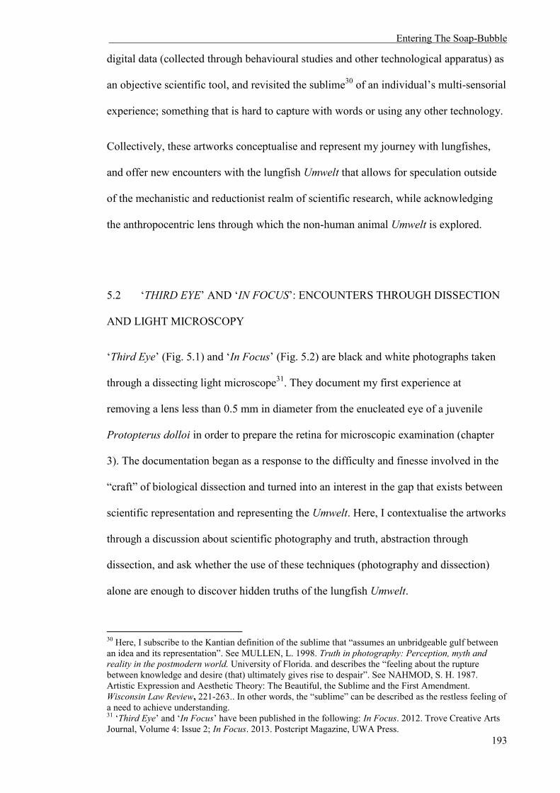
Entering The Soap-Bubble
193
digital data (collected through behavioural studies and other technological apparatus) as
an objective scientific tool, and revisited the sublime30 of an individual’s multi-sensorial
experience; something that is hard to capture with words or using any other technology.
Collectively, these artworks conceptualise and represent my journey with lungfishes,
and offer new encounters with the lungfish Umwelt that allows for speculation outside
of the mechanistic and reductionist realm of scientific research, while acknowledging
the anthropocentric lens through which the non-human animal Umwelt is explored.
5.2 ‘THIRD EYE’ AND ‘IN FOCUS’: ENCOUNTERS THROUGH DISSECTION
AND LIGHT MICROSCOPY
‘Third Eye’ (Fig. 5.1) and ‘In Focus’ (Fig. 5.2) are black and white photographs taken
through a dissecting light microscope31. They document my first experience at
removing a lens less than 0.5 mm in diameter from the enucleated eye of a juvenile
Protopterus dolloi in order to prepare the retina for microscopic examination (chapter
3). The documentation began as a response to the difficulty and finesse involved in the
“craft” of biological dissection and turned into an interest in the gap that exists between
scientific representation and representing the Umwelt. Here, I contextualise the artworks
through a discussion about scientific photography and truth, abstraction through
dissection, and ask whether the use of these techniques (photography and dissection)
alone are enough to discover hidden truths of the lungfish Umwelt.
30 Here, I subscribe to the Kantian definition of the sublime that “assumes an unbridgeable gulf between an idea and its representation”. See MULLEN, L. 1998. Truth in photography: Perception, myth and reality in the postmodern world. University of Florida. and describes the “feeling about the rupture between knowledge and desire (that) ultimately gives rise to despair”. See NAHMOD, S. H. 1987. Artistic Expression and Aesthetic Theory: The Beautiful, the Sublime and the First Amendment. Wisconsin Law Review, 221-263.. In other words, the “sublime” can be described as the restless feeling of a need to achieve understanding. 31 ‘Third Eye’ and ‘In Focus’ have been published in the following: In Focus. 2012. Trove Creative Arts Journal, Volume 4: Issue 2; In Focus. 2013. Postcript Magazine, UWA Press.

Entering The Soap-Bubble
194
Fig. 5.1 Third Eye. (Audrey Appudurai, 2013).
Fig. 5.2. In Focus. (Audrey Appudurai, 2013).
Scientific imaging is profoundly connected to the history of photography. Not long
after its invention, photography was ordained as an objective witness of phenomena
(Marien, 2011). When the technological advances of cameras moved beyond the stiff
poses required for long-exposure cameras, the resulting immediate, realistic images
shifted the way humans were able to perceive and document the world. The camera
could deliver objective renderings of reality; better than human eyes and free from
emotions and the senses. In relation to its use in science, the camera was capable of
capturing a moment in time that our eyes could not see. For example, Muybridge’s
‘Animal Locomotion’ (1830 – 1904) (Fig. 5.3) exposed, for the first time, that all the
hooves of a galloping horse leave the ground in a sequence of motion. Photographs
became credible witnesses of reality (Mullen, 1998), as Ralph Waldo Emerson claimed,
“Photography is distinguished by its immediacy, its authenticity, and the remarkable
fact that its eye sees more than the human eye. The camera shows everything.”
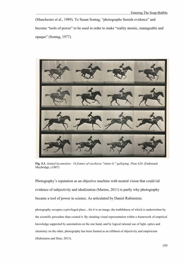
Entering The Soap-Bubble
195
(Manchester et al., 1989). To Susan Sontag, “photographs furnish evidence” and
become “tools of power” to be used in order to make “reality atomic, manageable and
opaque” (Sontag, 1977).
Fig. 5.3. Animal locomotion - 16 frames of racehorse "Annie G." galloping. Plate 626. (Eadweard Muybridge, c1887)
Photography’s reputation as an objective machine with neutral vision that could rid
evidence of subjectivity and idealization (Marien, 2011) is partly why photography
became a tool of power in science. As articulated by Daniel Rubinstein:
photography occupies a privileged place....for it is an image, the truthfulness of which is underwritten by
the scientific procedure than created it. By situating visual representation within a framework of empirical
knowledge supported by automatism on the one hand, and by logical rational use of light, optics and
chemistry on the other, photography has been framed as an offshoot of objectivity and empiricism
(Rubinstein and Sluis, 2013).

Entering The Soap-Bubble
196
Unresolved images of biological samples in early photography were seen as more
truthful than the highly detailed drawings of the same specimens. When Alfred Donné
(1845) published microscopic-daguerreotypes of human cells, he declared them
independent from interpretation because of the “rigorous fidelity of photomicrography
to the original subject”: “the plates show exactly the microscope appearance and the
truth of the details” (Jardim and Peres, 2014). When photographer Berenice Abbott
ventured into the realm of scientific photography, she made mathematics and theoretical
physics visible to the eye through dramatic, minimalist black and white photography
(Fig. 5.4). Abbott believed that photography was a scientific endeavour, and this
methodological, unsentimental approach is evident in her practice; whether it is portraits
of art-world figures in Paris, documentations of the New York cityscape, or illustrating
the decrease of energy in a moving object.

Entering The Soap-Bubble
197
Fig. 5.4. A Bouncing Ball in Diminishing Arcs (Berenice Abbott, 1958)
The reliance on photography as objective truth is evident in my own work as a biologist.
Photography is the primary tool in communicating evidence and experimental findings.
For example, I have used light and electron micrographs to describe the structure of the
retinal photoreceptor types of Protopterus dolloi and Lepidosiren paradoxa (chapter 3),
and videography and histology to observe and describe the behaviour and visual
apparatus of abnormal Neoceratodus forsteri (chapter 4). In ‘Third Eye’ and ‘In Focus’
the photographs capture details of P. dolloi that our eyes cannot resolve by themselves,
such as the pattern of “freckles” on the periphery of the eye and the fibres forming the
crystalline lens, thus creating an avenue to view the tiny eyes of a juvenile lungfish that
is only attainable through the mediation of dissection, microscopy and photography.

Entering The Soap-Bubble
198
However, it is well established that photography is a construction made by humans and
their technological apparatuses. No photograph is without agency, and photography
manipulates reality to reveal truths that are apparent to the critical eye (Mullen, 1998).
Mullen (1998) says, “a photograph, although professing to depict truth, actually
involves manipulation of both object and message. To say that photographers depict the
truth is not correct; what they actually depict are slices of life selected and framed by
the photographer”.
The manipulation of scientific imaging predominantly lies in the framing of the
photograph to focus on a specific subject matter, and an alteration of the images to
enhance the quality, for examples see the micrographs presented in chapters 3 and 4.
However, this close association of photography and truth, and science and truth can lead
to constructed images that have no basis in reality, but are taken as such. For example,
when Eduardo Kac’s ‘GFP Bunny’ (2000) was revealed by the artist in popular media,
the world was confronted with the dystopian and utopian potentials of transgenics.
Kac’s artwork was a gfp rabbit he called Alba; a laboratory rabbit with green florescent
protein (gfp) inserted into its DNA. Kac produced an image of a vividly glowing rabbit
under ultraviolet light. The rabbit was never seen in the flesh, and the only “evidence”
of its existence was through carefully selected images released by the artist. Transgenic
gfp mice used in scientific research do not glow in the way Alba did in the photographs
(Philipkoski, 2002); only living cells express gfp, which does not include the keratin
within fur (Chalfie et al., 1994, Prasher, 1995). Kac’s ‘GFP Bunny’ photograph had
been constructed to enhance the fluorescent properties of gfp, and in doing so, created
an exaggerated spectacle that exploited the idealization of truth presented ‘scientifically’
(Philipkoski, 2002). Similarly, the disseminated image of Laura Cinti’s ‘Cactus Project’
(2001) persuaded people to believe that she had created a cactus capable of growing
human hair due to the clever construction and framing of the object to create a

Entering The Soap-Bubble
199
convincing narrative. These examples (and there are many more) illustrate the
importance of acknowledging the aesthetic choices used in ‘Third Eye’ and ‘In Focus’
in order to discuss the role of photography in exploring the Umwelten of lungfishes.
‘Third Eye’ and ‘In Focus’ both illustrate the disembodied eye and lens of P. dolloi
floating in aqueous space. ‘Third Eye’ (Fig. 5.1) documents the eye before the lens was
removed, and is presented as a triptych with changing lighting, while ‘In Focus’ (Fig.
5.2) is a diptych of the P. dolloi lens in changing light. Both works are an experiment
with scale, and aside from documentation of a scientific methodology (and “craft”),
attempt to bridge the gap between human and non-human animal perceptions by
manipulating scale, presenting a magnified “universal” vertebrate eye and its properties
that are only visible through the mediation of technology.
Ocularcentrism is a term that recognises a perceptual and epistemological bias of vision
over other human senses. Plato and Aristotle argued that sight was directly associated
with reason and the traces of this are still present in Western culture; to ‘see’ is to
‘understand’, to agree is to ‘see eye to eye’ (Jay, 1993). In the context of my research,
these works are naturally ocularcentric because of the focus on visual perception, and
specifically, a comparative investigation of the ocular morphology of different species.
Although perception is never uni-sensory and involves many sensorial apparatuses, my
research parameters at the time did not extend, especially for L. paradoxa and P. dolloi,
to the other senses and how they contribute to their Umwelten. This eventually changed
when I was faced with exploring the visual perception of N. forsteri without visible
eyes, but ‘Third Eye’ and ‘In Focus’ signified the period when I was focused
(obsessed?) with the eyes and the visual perception of lungfishes.

Entering The Soap-Bubble
200
As a documentation of the dissection process used in scientific research, ‘Third Eye’
and ‘In Focus’ are visual apparatuses that are abstracted from context. The floating eyes
are large and all-consuming; they comprise almost the entire photograph. There is an
element of surveillance: as I observe the eye, it observes me. Despite being preserved,
they are “real”, and a trace of the animal that once used them to see. Without context we
do not know what kind of eye this is (if an eye at all); it appears to be universal. In ‘In
Focus’, the abstraction of the eye from the body creates images with open meanings that
go beyond the ocular focus. Here, the lens appears to take the form of a celestial body in
space. The bubble next to the lens alludes to the aquatic nature of this particular
“space”, and the changing light between the images pertains to the changing of time,
making it appear as though the bubble is orbiting the lens, like a planet around a star.
The concept of the Umwelt does not lend itself well to the carefully (human)
constructed, two-dimensional nature of photography. The Umwelt of any organism is
deeply entwined with that organism’s relationship to and within its environment, and
scientifically themed photographic images illustrate the limitations of photography to
represent the Umwelt. Lennart Nilsson’s ‘Drama of Life Before Birth’ (1965) photo
essay and National Geographic’s ‘Extraordinary Animals in the Womb’ (2012) depicts
unborn human and non-human foetuses, respectively, within their Umwelt of the uterus.
The media and public read these images as “real”, but each photograph was carefully
constructed either by modelling with ultrasounds or with dead foetuses, and many
details were manipulated to remove the foetus from its placental Umwelt. Blood is
either absent, or coloured a pleasant red, unlike the dark red of blood within the body. In
all the images, the body of the mother is absent, and especially in Nilsson’s work, so is
the uterine wall. The animals are floating free, either in what appears to be celestial
space or nestled comfortably within the placenta. In all these constructed images, the

Entering The Soap-Bubble
201
foetus is abstracted from its environment, and the Umwelt is superfluous to the “truth”
of the object.
Similarly, ‘Third Eye’ and ‘In Focus’ do not exhibit the all encompassing nature of the
lungfish Umwelt. Firstly, the spatial aspect of the eyes and lens is lost because the image
is two-dimensional. The photographs are in black and white, and although this is
traditionally associated with documentary photography because it “avoids distraction”
from the subject matter, it does obscure the pre-photographic “reality” of the subject.
Most importantly, these photographs are a reductionist fragmentation of the organism
and its Umwelt; the animal and its human experimenter are missing, and the visual
organ and its structures are disembodied and floating in space. The photographs are a
result of a scientific dissection process that takes the eye out of context, and abstracts
body parts into singular forms open to be analysed in many ways.
Sontag describes photographic images as “clouds of fantasy and pellets of information”,
and elaborates that, ‘the ultimate wisdom of the photographic image is to say: ‘There is
the surface. Now think – or rather feel, intuit – what is beyond it, and what the reality
must be like if it looks this way’” (Sontag, 1977). The human acts of concealment and
revealing in photography is precisely why, when used alone, it is inadequate to describe
the Umwelten of lungfishes because the anthropocentric fantasy of what we believe
permeates through the images. With this in mind, ‘Third Eye’ and ‘In Focus’ were an
exercise in experimentation with photography to explore the Umwelt. When used alone,
they are limited in their ability to describe and convey the Umwelten of lungfishes.
However, they were an entry into the gap between scientific representation and
representations of the lungfish Umwelt.

Entering The Soap-Bubble
202
5.3 ‘THE FORMS’: ENCOUNTERS THROUGH ELECTRON MICROSCOPY,
SCULPTURE AND PERFORMANCE
‘The Forms’ (Fig. 5.5 and Fig. 5.6) are a series of sculptures that poetically explore light
and electron-mediated optical media in relation to the micro-/nano-scopic study of the
South American (Lepidosiren paradoxa) and Spotted Africa (Protopterus dolloi)
lungfishes32. ‘Third Eye’ and ‘In Focus’ were used as departure points that explored
human engagement and presence in the production and presentation of scientific
imaging. The works were created through a combination of transmission electron
microscopy (TEM), camera-less photography (photograms) on silk, and physical
manipulation of the image to create three-dimensional sculptures that consciously
exposed the human presence in all forms of scientific exploration into the lungfish
Umwelt. They were also experimentations with scientific and artistic craftsmanship and
materialities.
On a more personal level, the process of creating the sculptures developed into a
performance that reflected my experience working with L. paradoxa, P. dolloi, and
abnormal N. forsteri. ‘The Forms’ is contextualised by drawing on the struggle of
working between science and art, the challenge of using electron microscopy to
compare the visual morphology of L. paradoxa and P. dolloi (chapter 3), and the
discovery of Australian lungfish (Neoceratodus forsteri) without visible eyes (chapter
4). ‘The Forms’ was an attempt to extend the representation of the lungfish Umwelt
beyond the mechanism of scientific imaging by acknowledging anthropocentric
limitations and using performance to embody the lungfish and my lived experiences.
32‘The Forms’ have been exhibited in the following group exhibitions: Material/Immaterial. 2014. ARTLAAB, UWA, Australia; An Introduction to Neolife. 2014. The University Club, UWA, Australia.
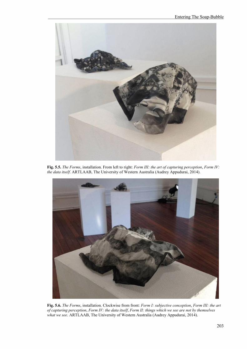
Entering The Soap-Bubble
203
Fig. 5.5. The Forms, installation. From left to right: Form III: the art of capturing perception, Form IV: the data itself. ARTLAAB, The University of Western Australia (Audrey Appudurai, 2014).
Fig. 5.6. The Forms, installation. Clockwise from front: Form I: subjective conception, Form III: the art of capturing perception, Form IV: the data itself, Form II: things which we see are not by themselves what we see. ARTLAAB, The University of Western Australia (Audrey Appudurai, 2014).

Entering The Soap-Bubble
204
The micrographs were collected while acquiring data to characterize and describe the
retinal photoreceptors of L. paradoxa and P. dolloi (presented in chapter 3). TEM is
frequently used in vision studies to comparatively investigate the ultra-structure of
retinal cells in many organisms because the combination of high resolution and high
magnification enables the description and differentiation of biological cells in the
greatest detail. EM is one of the few photographic techniques that are conducted in the
absence of light, within a high vacuum environment (Williams and Carter, 1996).
The electron micrographs captured for ‘The Forms’ are considered a form of digital
imaging. Although the construction of an electron image is not digital because the
image is formed by electrons interacting directly with the specimen, the only way to see
and subsequently analyse the data is through digital capture. Digital photography differs
from traditional analogue photography in the sense that the images are algorithmic, and
composed of fixed information in the form of discrete pixels with precise gradients of
spatial and tonal resolution (Rubinstein and Sluis, 2013, Mullen, 1998). Digital imaging
is often considered “less authentic” compared to traditional photography because of the
ease and readily accessible ways to manipulate the images, but it is argued to have
solved the ethical qualms surrounding photography because it explicitly acknowledges
the manipulative nature inherent in all forms of the practice, forcing an understanding
that “truth” is rarely represented in a still image. Fred Ritchin, in the context of truth
integrity in photography and journalism says, “Saying, ‘the camera never lies’ is as
foolish as asserting that the computer always does. Just because words can be fictional
does not require the outlawing of news articles; similarly with photographs” (Ritchin,
1999). Batchen expands by saying, “Digitization abandons even the rhetoric of truth
that has been an important part of photography’s cultural success...newspapers have of
course always manipulated their images in one way or another. The much-heralded
advent of digital imaging simply means having to admit it” (quoted in Mullen, 1998).

Entering The Soap-Bubble
205
However, another argument may push in favour of digital objectivity – digital data is
represented as the ultimate form of abstraction (digital code) because it avoids the
“biases” of looking at a photographic image – the data gathered is presumably objective
beyond the human eye. However, the data is still a construction made by technology
created by humans for humans.
The digital images in ‘The Forms’ were manipulated by mirroring the micrographs to
create interesting patterns from some of the unknown sub-cellular structures in the
retina of L. paradoxa and P. dolloi (Fig. 5.7). The pattern making reveals a desire to
understand and extrapolate ideas from forms that would otherwise be meaningless.

Entering The Soap-Bubble
206
Fig. 5.7. Mirrored patterns created from electron micrographs of unknown structures within the Lepidosiren paradoxa and Protopterus dolloi retina. Clockwise from top left: Form I: subjective conception, Form II: things which we see are not by themselves what we see, Form III: the art of capturing perception, Form IV: the data itself,. (Audrey Appudurai, 2014).
Nina Sellars explores how perceptions of images and phenomenological experiences
with light and anatomical representations are articulated and engaged with outside of a
scientific context in ‘Anatomy of Optics and Light’ (2009) (Fig. 5.8) and ‘Lumen’
(2011) ‘Lucida’ (2012) (Fig. 5.9) (Sellars, 2012) .

Entering The Soap-Bubble
207
Fig. 5.8. Anatomy of Optics and Light installation (Nina Sellars, 2009)
Fig. 5.9. Lucida installation detail (Nina Sellars, 2012)

Entering The Soap-Bubble
208
Both of Sellars’ works present representations of the anatomical body in a
diagrammatical or an abstract language, either through the creation of drawn images
depicting a schematic of the sacral plexus or “plausible, fictional anatomies that
reference cytology” (Sellars, 2012). For Sellars, the structures presented and the
technical processes involved in contemporary biological imaging is secondary to the
phenomenological engagement and discourses surrounding the anatomical body in
culture and science (Sellars, 2010). Similarly, the act of creating patterns with electron
micrographs for ‘The Forms’ to abstract anatomical information was a way to focus on
how we engage with the scientific representation (analogue or digital) rather than a need
to understand or describe the structures presented. As ‘Anatomy of Optics and Light’
simplified the anatomical sacral plexus as a schematic network, ‘Lumen’ and ‘Lucida’
went a step further and removed scientific context entirely. Sellars explains:
‘Lumen’ is an appropriated phenomenological experience, which has been removed from a scientific
context and stripped of its original protagonists,
My working methodology has led me to focus not on things as objects but on our engagement with
things, to a point where the original things are no longer needed. References to people, cadavers,
anatomical images, medical equipment, all start to disappear from my work. Instead, what becomes more
important is the attempt to elicit and unfold that initial, first-hand phenomenological experience so that it
exists and resonates as if it were the original one (Sellars, 2012)
However, unlike ‘Lumen’ and ‘Lucida’, ‘The Forms’ is not a purely phenomenological
experience because the anatomical information is still present; and although the images
are mirrored, all the information necessary to identify and describe the ultrastructure of
the L. paradoxa and P. dolloi retinal photoreceptor cells is contained in the forms.
An interest in the differences and similarities between digital and analogue scientific
imaging, and how they can influence our engagement with scientific representation
motivated me to combine both forms of photography and explicitly add physical
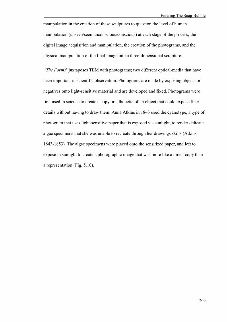
Entering The Soap-Bubble
209
manipulation in the creation of these sculptures to question the level of human
manipulation (unseen/seen unconscious/conscious) at each stage of the process; the
digital image acquisition and manipulation, the creation of the photograms, and the
physical manipulation of the final image into a three-dimensional sculpture.
‘The Forms’ juxtaposes TEM with photograms; two different optical-media that have
been important in scientific observation. Photograms are made by exposing objects or
negatives onto light-sensitive material and are developed and fixed. Photograms were
first used in science to create a copy or silhouette of an object that could expose finer
details without having to draw them. Anna Atkins in 1843 used the cyanotype, a type of
photogram that uses light-sensitive paper that is exposed via sunlight, to render delicate
algae specimens that she was unable to recreate through her drawings skills (Atkins,
1843-1853). The algae specimens were placed onto the sensitized paper, and left to
expose in sunlight to create a photographic image that was more like a direct copy than
a representation (Fig. 5.10).

Entering The Soap-Bubble
210
Fig. 5.10. Diclyola dicholoma in the young state, and in fruit. (Anna Atkins 1843 – 1853).
Photographer Berenice Abbott used photograms to illustrate physical phenomena, such
as the interference pattern of energy as it pulses through water. Here, the interference
pattern of waves could only be visible by a step-wise exposure of projected shadows
formed by oscillating waves in water (Fig. 5.11).

Entering The Soap-Bubble
211
Fig. 5.11. Interference of waves (Berenice Abbott, 1958 – 1961).
Photograms are interesting because the images are more than a representation; they
possess the essence of the object (algae specimen) or phenomena (interference pattern)
being photographed. In ‘The Forms’, the essence was not of the retinal cells themselves,
but my (the human) presence in the process of creating the photographic image. The
material needed to be prepared with silver gelatine in a darkroom, and any
imperfections, which cannot be corrected, are visible in the resulting images; the human
could not be erased (Fig. 5.12).

Entering The Soap-Bubble
212
Fig. 5.12. Form V, detail. ARTLAAB, The University of Western Australia (Audrey Appudurai, 2014).
The white silk was used as the support to expose the negative patterns and create the
sculptures (Fig. 5.13). The production of silk is analogous to the transformation of
biological tissue for TEM (see methods section in chapter 3). Silk is produced by
unwinding the delicate filaments of a silkworm’s cocoon. The reeled filaments are then
put through a number of washing, drying and treatment stages before the thread is
woven into material. Similarly, the retina needs to be fixed, dehydrated, infiltrated with
resin and cut into ultrathin (110 nm) sections before it can be observed using a
transmission electron microscope. These processes change the origins of these materials
as parts of organisms: they became materials of craft and data acquisition.

Entering The Soap-Bubble
213
Fig. 5.13. Form II: things which we see are not by themselves what we see. ARTLAAB, The University of Western Australia (Audrey Appudurai, 2014).
The physical manipulation of the photograms into sculptures gave the images physical
and tactile dimensions, allowing them to “escape” the photographic document. The
construction of three-dimensional sculptures from images brought back dimensionality
and tangibility that was lost in the two-dimensional photographs of ‘Third Eye’ and ‘In
Focus’. The physical manipulation of the silk illustrates the human agency in the
process and presentation of scientific imaging. In addition, sculptures allow for the
micrographs to be observed from different angles, changing the information that is
perceived (Fig. 5.14).

Entering The Soap-Bubble
214
Fig. 5.14. Form IV: the data itself at different angles. (Audrey Appudurai, 2014).
The process of creating the photograms and sculptures of ‘The Forms’ developed into a
personal performance that reflected my attempt to experience how the abnormal
N. forsteri lacking external eyes (chapter 4) sense their environment. The performance
was conducted entirely in the confines of a darkroom, and represented a hybridisation of
the metaphorical and literal darkness of uncovering the Umwelten of lungfishes. I drew
particularly on the struggle of working in between science and art, and the difficulty in
creating a discourse between the two cultures in order to describe the visual world of
lungfishes. Although the works directly related to the histological examination of
L. paradoxa and P. dolloi retinal photoreceptors, I had recently acquired the abnormal
N. forsteri, and wished to conceptualise my journey into the lungfish Umwelt through an
attempt to embody the lungfishes without visible eyes. I lost my sight in that room, and
had to rely primarily on touch and memory to navigate and complete the process of
creating the sculptures.
‘The Forms’ are a poetic exploration into human engagement with the lungfish
Umwelt. By explicitly acknowledging the anthropocentric limitations within scientific
imaging, and combining it with a performance that changed my perception (dark room)
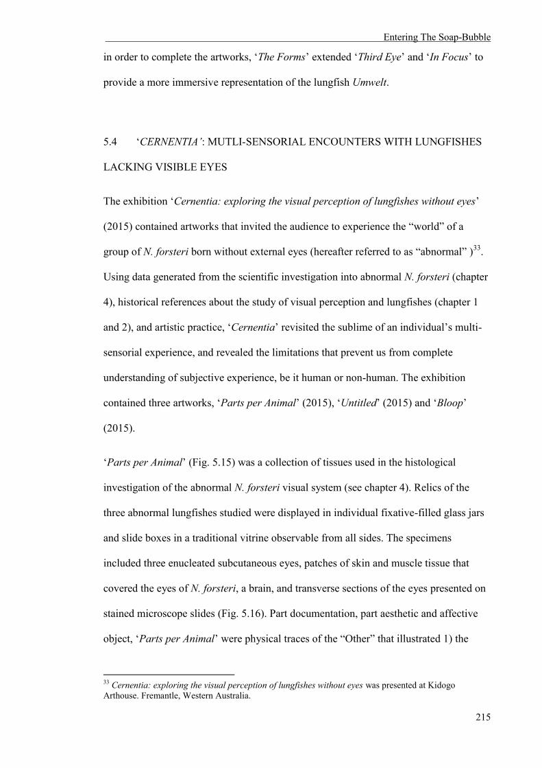
Entering The Soap-Bubble
215
in order to complete the artworks, ‘The Forms’ extended ‘Third Eye’ and ‘In Focus’ to
provide a more immersive representation of the lungfish Umwelt.
5.4 ‘CERNENTIA’: MUTLI-SENSORIAL ENCOUNTERS WITH LUNGFISHES
LACKING VISIBLE EYES
The exhibition ‘Cernentia: exploring the visual perception of lungfishes without eyes’
(2015) contained artworks that invited the audience to experience the “world” of a
group of N. forsteri born without external eyes (hereafter referred to as “abnormal” )33.
Using data generated from the scientific investigation into abnormal N. forsteri (chapter
4), historical references about the study of visual perception and lungfishes (chapter 1
and 2), and artistic practice, ‘Cernentia’ revisited the sublime of an individual’s multi-
sensorial experience, and revealed the limitations that prevent us from complete
understanding of subjective experience, be it human or non-human. The exhibition
contained three artworks, ‘Parts per Animal’ (2015), ‘Untitled’ (2015) and ‘Bloop’
(2015).
‘Parts per Animal’ (Fig. 5.15) was a collection of tissues used in the histological
investigation of the abnormal N. forsteri visual system (see chapter 4). Relics of the
three abnormal lungfishes studied were displayed in individual fixative-filled glass jars
and slide boxes in a traditional vitrine observable from all sides. The specimens
included three enucleated subcutaneous eyes, patches of skin and muscle tissue that
covered the eyes of N. forsteri, a brain, and transverse sections of the eyes presented on
stained microscope slides (Fig. 5.16). Part documentation, part aesthetic and affective
object, ‘Parts per Animal’ were physical traces of the “Other” that illustrated 1) the
33 Cernentia: exploring the visual perception of lungfishes without eyes was presented at Kidogo Arthouse. Fremantle, Western Australia.
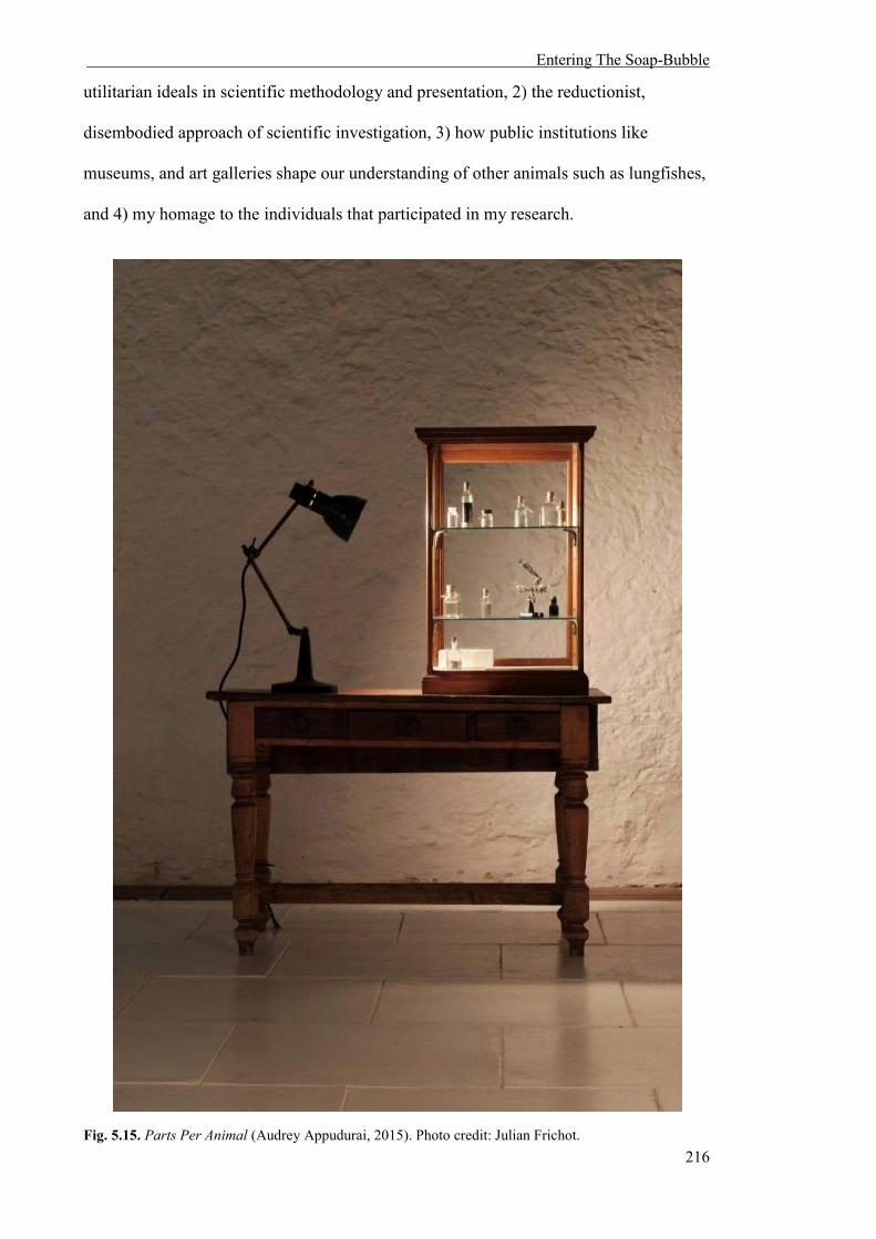
Entering The Soap-Bubble
216
utilitarian ideals in scientific methodology and presentation, 2) the reductionist,
disembodied approach of scientific investigation, 3) how public institutions like
museums, and art galleries shape our understanding of other animals such as lungfishes,
and 4) my homage to the individuals that participated in my research.
Fig. 5.15. Parts Per Animal (Audrey Appudurai, 2015). Photo credit: Julian Frichot.
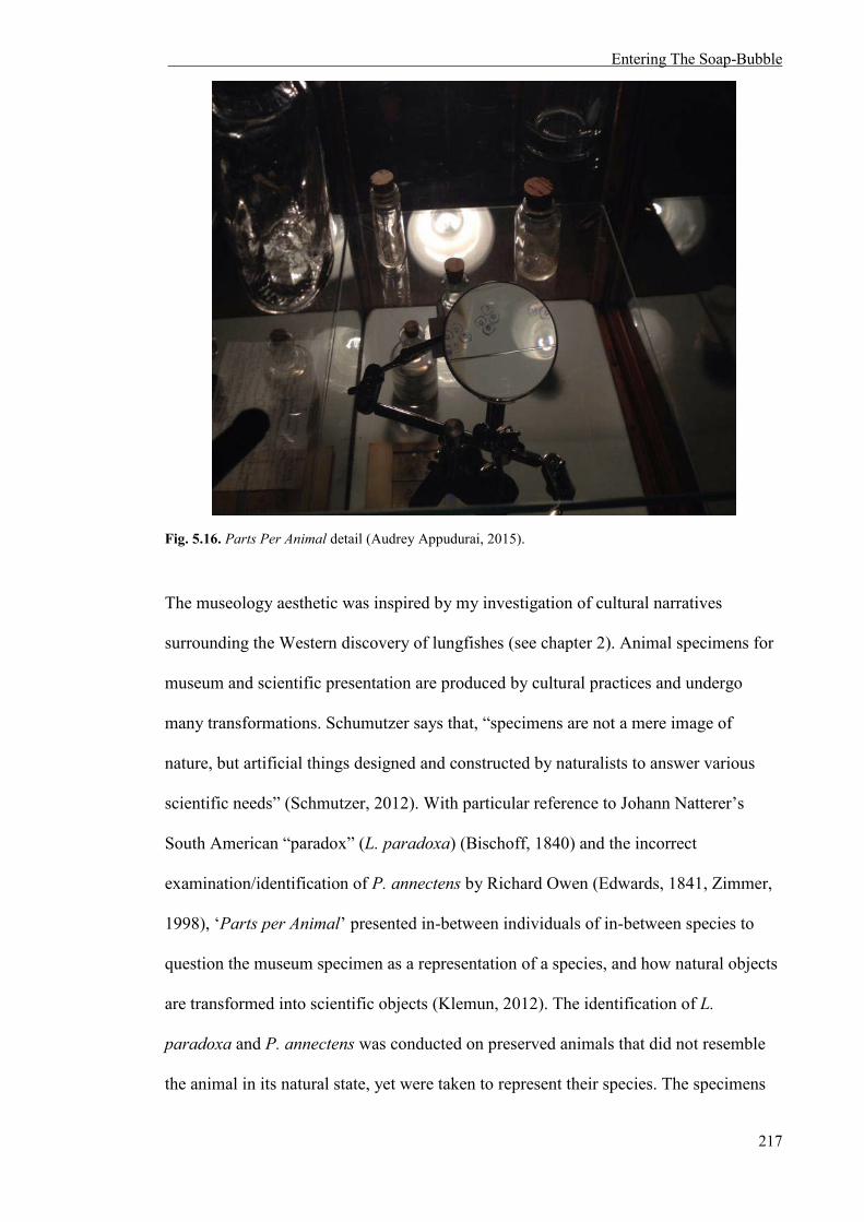
Entering The Soap-Bubble
217
Fig. 5.16. Parts Per Animal detail (Audrey Appudurai, 2015).
The museology aesthetic was inspired by my investigation of cultural narratives
surrounding the Western discovery of lungfishes (see chapter 2). Animal specimens for
museum and scientific presentation are produced by cultural practices and undergo
many transformations. Schumutzer says that, “specimens are not a mere image of
nature, but artificial things designed and constructed by naturalists to answer various
scientific needs” (Schmutzer, 2012). With particular reference to Johann Natterer’s
South American “paradox” (L. paradoxa) (Bischoff, 1840) and the incorrect
examination/identification of P. annectens by Richard Owen (Edwards, 1841, Zimmer,
1998), ‘Parts per Animal’ presented in-between individuals of in-between species to
question the museum specimen as a representation of a species, and how natural objects
are transformed into scientific objects (Klemun, 2012). The identification of L.
paradoxa and P. annectens was conducted on preserved animals that did not resemble
the animal in its natural state, yet were taken to represent their species. The specimens

Entering The Soap-Bubble
218
used to identify L. paradoxa did not possess any internal organs, having been removed
by Natterer, which contributed to Fitzinger’s frustration at classifying the animal as a
fish or a reptile (Fitzinger, 1837, Schmutzer, 2012). The specimens Owen used to
classify P. annectens were desiccated, which may have contributed to his incorrect
documentation of the nostrils not being connected to the lungs (Bischoff, 1840, Owen,
1840, Edwards, 1841). Although methods of preservation and documentation have
improved considerably since the 19th century, it is still important to recognize that
specimens used for histological examinations are far removed from the original
organism and its environment. ‘Parts per Animal’ explicitly acknowledges the
processes involved in order to make sense of the ‘Other’ and understand the perception
of lungfishes.
Other artists that incorporate scientific presentations of non-human animals in their art
practice include Brandon Ballengée, Mark Dion and Janet Laurence. As a biologist and
artist, Ballengée’s main interest is the impact of human-caused phenomena, such as
pollution and climate change, on the natural world and its inhabitants (Ballengée,
2015a). His work ‘Collapse’ (2012) responded to the 2010 BP Deepwater Horizon oil
disaster that subjected the Gulf of Mexico’s food web to the largest accidental marine
oil spill of the petroleum industry’s history (Roberston and Krauss, 2010). A seven
tiered pyramid consisted of 26,162 preserved specimens representing 370 species of
aquatic organisms in gallon jars dominated the room. Some of the jars were empty,
representing species in decline, forgotten or already extinct and other containers
presented deformed organisms affected by pollution (Cotter, 2012). According to
Ballengée, the scientific presentations of organisms were used as “a sculptural
expression of the complexity of the issue, and also the complexity of the food chain
itself” (Beitiks, 2012). In ‘Collapse’, the dead organisms, preserved whole and
suspended in clear buffer or fixative were a sterile reminder of the slow destruction of

Entering The Soap-Bubble
219
our environment (Ballengée, 2015b). ‘Parts per Animal’ does not touch on ecological
crises directly, but can be seen as a “sculptural expression” of ways of understanding
other organisms, in particular a group of captive N. forsteri whose deformities may have
been caused by humans.
Mark Dion applies archaeological and scientific methods of collecting, documenting,
ordering and presenting objects to explore cultural representations of nature (Dion,
2015). His arrangements of natural and man-made objects displayed in curiosity
cabinets (see ‘Marine Invertebrates’ [2013]) explore the distinctions between scientific
methods and subjective, irrational influences. The carefully designed sets containing
artefacts involved in knowledge acquisition and dissemination of the natural world
(such as ‘Department of Marine animal Identification of the City of New York
[Chinatown Division]’ [1992] and ‘Tate Thames Dig’ [1999]) reveals how public
institutions shape our understandings, and questions the power structures behind the
Linnaean taxonomic system (see Chapter 2) (Fiske and Bottinelli, 2002). Dion believes
“nature is one of the most sophisticated arenas for the production of ideology” (Stonard,
2000) and through art, he questions the authoritative role of the scientific voice that
underlies environmental politics and public policy in contemporary society (Dion). Like
Dion, ‘Parts per Animal’ examines the role that animal specimens, as cultural artefacts,
play in the construction of knowledge, thereby shaping our understanding of lungfish
perception and the natural world.
Janet Laurence’s ‘Fugitive’ (2013) was an installation that includes life/non-life
human/non-human objects, such as taxidermy and preserved non-human animals, to
create “a place of healing, hope, breeding and care for our native animals” (Gibson and
Laurence, 2015). The immobile specimens are surrounded by photographs and the
sounds of their natural habitat (Umwelt), which give voice to non-human organisms

Entering The Soap-Bubble
220
under threat. Laurence’s work is grounded in posthumanism through the act of care. She
believes the current “ecological crisis demands us to shift our focus from the human-
centred perspective to a broader multi-species, environmental approach” and
“participates in discourses of agency and realities outside a singular human experience
by considering the point of view of animals, even in death” by archiving nature
artistically through the museology aesthetic (Gibson and Laurence, 2015). Similarly,
‘Cernentia’ and this thesis consider the point of view of lungfishes through their
subjective experience. Care is certainly part of the embodied experience, as seen in ‘The
Forms’ and ‘Untitled’ (discussed below), but ‘Parts per Animal’ does not do this alone.
The sounds and the sights of the lungfishes’ environment, their Umwelt, were
encompassed within the other works in the gallery, ‘Untitled’ and ‘Bloop’. It is
important to note that ‘Cernentia’ did not contain any elements of the N. forsteri native
habitat. I did not want to give the illusion that these fishes were ever from the wild; the
rivers of Queensland were never their habitat. Nevertheless, I wanted to stress that their
Umwelt, the aquariums at the University of Western Australia, is still an important
feature in understanding their perception.
‘Parts per Animal’ cannot fully describe or embody the Umwelt of the abnormal N.
forsteri born without external eyes. It was more a trace and sculptural expression of how
humans attempt to understand the subjective experience of non-human animals. The
Umwelt of the lungfishes was lost when the animal was broken apart and its visual
system examined in isolation. Nonetheless, it is still important to openly acknowledge
that this is one “aspect of knowing” the abnormal N. forsteri Umwelt, complete with its
research opportunities and limitations.
‘Untitled’ was an artwork that evolved from the investigation of the circadian rhythm of
the N. forsteri born without visible eyes’ over a period of 72 hours in complete darkness

Entering The Soap-Bubble
221
(see experiment 3, chapter 4) and my own experience observing their behaviour in real
time. It was presented in two parts: 1) upon a wall as a triptych of video footage of the
three abnormal N. forsteri and, 2) throughout the gallery as marks made in UV paint
upon the floor that invites the audience to follow in my tracks (Fig. 5.17 and 5.18,
respectively).
Fig. 5.17. Untitled triptych of experimental footage detail. (Audrey Appudurai. 2015). Photo credit: Julian Frichot.

Entering The Soap-Bubble
222
Fig. 5.18. Untitled detail. Performance still of ultra-violet paint on gallery floor. (Audrey Appudurai, 2015). Photo credit: Julian Frichot.
The video footage is a bird’s eye surveillance of the fishes in complete darkness over 72
hours to measure their activity patterns. The movies were recorded in low lux and are
silent, showing the slow, undulating movements of each fish as it circulated within its
minimal, experimental environment. The surveillance-style videos of ‘Untitled’ taken as

Entering The Soap-Bubble
223
part of scientific behavioural studies (chapter 4) illustrate the relationship and power
dynamic between experimenter and subject prevalent in scientific investigations of the
“Other”. On a more personal note, as I watched these fishes through a computer
interface, I became intrigued by the way they moved around the perimeter of their tanks,
sometimes without stopping. The fishes’ endless “going around in circles” prompted a
metaphorical interpretation of the inability to pierce the Umwelt; any effort to perceive
subjective experience was met by anthropocentric limitations (chapter 2). Therefore, the
futility of “watching” from a distance as a scientific experimenter led to a second
attempt of understanding through performance: the tracks of UV paint.
Prior to the opening of the exhibition, I traced the fishes’ movements during the
circadian experiment (experiment 2, chapter 4). On hands and knees and in complete
darkness, I felt my way around the floor of the gallery space and marked the abnormal
lungfishes’ direction and positions of rest with fingers dipped in UV paint. During the
exhibition, two black-light LED torches were placed near the video footage, and
audience members were invited to follow my, and the lungfishes’ tracks around the
gallery (Fig. 5.19).

Entering The Soap-Bubble
224
Fig. 5.19. Untitled installation. (Audrey Appudurai. 2015) Photo credit: Julian Frichot.
Without the black-lights to aid them, my tracings and the abnormal N. forsteri tracks
were not perceivable to the human eye. Unlike humans however, juvenile N. forsteri
have the potential to perceive ultra-violet light (Hart et al., 2008). In reference to some
Entering The Soap-Bubble
225
of the differences between the human and lungfish visual system, I chose an
indiscernible medium to create my markings; one only visible to the human visitors
with technological intervention (black-light torches). I created marks around the gallery
space with my fingers, one of the most sensitive parts of the human body based on the
divisions of the primary somatosensory cortex (Marieb and Hoehn, 2007). The results
of chapter 4 suggested that, in the absence of external eyes, the abnormal N. forsteri
may use their lateral line to compensate for navigation (chapter 4). I focused on my/our
tactile senses to navigate around the gallery space to allude to this, and because it is
comparable to the lateral line of fishes (Dijkgraaf, 1963, Bleckmann 1986, Montgomery
et al., 1997). Inviting the audience to follow the trace and to enact this performance
extended the isolated performance of ‘The Forms’, allowing others to take part. This
pilgrimage, following me following the lungfishes, formed a dialogue between the
audience, experimenter/artist, and subject/non-human animal that created a temporary
subjective experience exclusive to each individual taking part, and illustrated 1) the
physical limitations of our senses (our inability to perceive ultra-violet light), and 2)
how technology (black-light LED torches) offers us a fleeting, inadequate glimpse into
the Umwelten of non-human animals.
Richard Long and Shannon Williamson also utilize mapping and traces of themselves in
their art practice. Long is a British land artist who redefined traditional approaches to
sculpture. One of his most famous works, ‘A Line Made by Walking’ (1967) produced
alterations in a meadow by a repetitive journey back and forth across grass. Long’s
walking artworks redefined the line between an action through performance, and
sculpture (Burgon, 2012). Long’s mud paintings are often site-specific, impermanent
examples of his attempt to bring his experiences back into public institutions like
museums and galleries (O'Hagan, 2009). Most of Long’s works that include mud as his
primary medium (such as ‘Waterfall Line’ [2000]) involve direct contact with the

Entering The Soap-Bubble
226
artwork by using his fingers and hands as tools to create marks upon the walls, floor, or
paper. Long believes that using mud and his hands add another form of physicality to
his work, as “the wateriness of the mud, and water is the main subject and content of
these works, they show its nature” (Brettell and Friis-Hansen, 1996). Long’s
interventions in the landscape, either within the field or in a gallery space are “transient
and humble” (Burgon, 2012), and in most cases, reveal the impermanency of nature and
our environment (O'Hagan, 2009).
The performative aspect of ‘Untitled’ is a site-specific, impermanent work that attempts
to embody the subjective experience of non-human animals. However, ‘Untitled’ is not
land art, but rather refers to the aquatic environment of abnormal N. forsteri. Richard
Long stressed that “the meaning of his work lay in the visibility of his actions rather
than the representation of a particular landscape” (Costa) and “represents a trace of a
corporeal presence and bodily action” (Burgon, 2012). Like, Long’s mud drawings, I
am not attempting to recreate or represent the lungfishes’ aquaria within the gallery
space, but to render visible their embodiment through marks that audience members can
track. It was important for me to create an intimate dialogue with the abnormal N.
forsteri through performance and touch, and make that experience temporarily
accessible to others. Tactility, in some form, is a sense shared by all organisms and does
not require verbal language or the possession of eyes.
Shannon Williamson utilizes mapping and physical traces to navigate personal
narratives (Williamson and George, 2013). Like Richard Long’s mud drawings, and
‘Untitled’, Williamson’s 2013 work ‘No More On Broken Bones’ and ‘Flight Path’
were site-specific impermanent drawings/sculptures on the floor and walls of a gallery
space. These works responded to Williamson’s personal experience with displacement
and sudden loss after the 2010 New Zealand earthquakes, with a particular focus on the

Entering The Soap-Bubble
227
anatomy and migration patterns of sea birds that frequent the southern island of New
Zealand and Australia (Mullen, 2013).
In the 2014 series titled ‘Night Shift’, Williamson revisited mapping to trace the
intimate and personal accounts of bodily experience as a shift worker in a long-term
relationship (Williamson, 2014). ‘Cartography of one sided sleep (work ii, series i)’
overlaid Williamson’s estranged circadian rhythm after working a number of night
shifts as a sleep technician (Williamson, 2014). ‘Untitled’ continues with the theme of
circadian rhythm mapping, except the “estranged circadian rhythm” is that of non-
human animals lacking external eyes. Like Williamson, I navigated personal
geographical and psychological terrain (Mullen, 2013) with the performative aspect of
‘Untitled’, but in a way that juxtaposed the two-dimensional, objective view of
experimentation seen in the video footage. A map activated through audience
participation attempted to relieve some of the frustration caused by passively watching
these animals within an experimental context.
The final artwork in ‘Cernentia’, ‘Bloop’ (in collaboration with Devon Ward), was an
immersive sound and light installation contained within a headset; a symbolic gesture
that attempts to embody the Umwelt (see chapter 1) of abnormal N. forsteri during
experimentation (see chapter 4) (Fig. 5.20).

Entering The Soap-Bubble
228
Fig. 5.20. Bloop (Audrey Appudurai in collaboration with Devon Ward, 2015) Photo credit: Julian Frichot.
The visual/sonic piece was derived from the electrophysiological investigation of
normal and abnormal N. forsteri outlined in experiment 3, chapter 4. In that experiment,
the functional integrity of the retina and visual pathway to the brain was measured
through the recording of visually evoked potentials (VEPs) in abnormal N. forsteri
(Creel et al., 1974, Creel, 2012). The subjective experience of an individual during
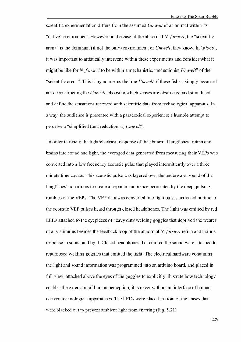
Entering The Soap-Bubble
229
scientific experimentation differs from the assumed Umwelt of an animal within its
“native” environment. However, in the case of the abnormal N. forsteri, the “scientific
arena” is the dominant (if not the only) environment, or Umwelt, they know. In ‘Bloop’,
it was important to artistically intervene within these experiments and consider what it
might be like for N. forsteri to be within a mechanistic, “reductionist Umwelt” of the
“scientific arena”. This is by no means the true Umwelt of these fishes, simply because I
am deconstructing the Umwelt, choosing which senses are obstructed and stimulated,
and define the sensations received with scientific data from technological apparatus. In
a way, the audience is presented with a paradoxical experience; a humble attempt to
perceive a “simplified (and reductionist) Umwelt”.
In order to render the light/electrical response of the abnormal lungfishes’ retina and
brains into sound and light, the averaged data generated from measuring their VEPs was
converted into a low frequency acoustic pulse that played intermittently over a three
minute time course. This acoustic pulse was layered over the underwater sound of the
lungfishes’ aquariums to create a hypnotic ambience permeated by the deep, pulsing
rumbles of the VEPs. The VEP data was converted into light pulses activated in time to
the acoustic VEP pulses heard through closed headphones. The light was emitted by red
LEDs attached to the eyepieces of heavy duty welding goggles that deprived the wearer
of any stimulus besides the feedback loop of the abnormal N. forsteri retina and brain’s
response in sound and light. Closed headphones that emitted the sound were attached to
repurposed welding goggles that emitted the light. The electrical hardware containing
the light and sound information was programmed into an arduino board, and placed in
full view, attached above the eyes of the goggles to explicitly illustrate how technology
enables the extension of human perception; it is never without an interface of human-
derived technological apparatuses. The LEDs were placed in front of the lenses that
were blacked out to prevent ambient light from entering (Fig. 5.21).

Entering The Soap-Bubble
230
Fig. 5.21. Bloop installation detail. (Audrey Appudurai in collaboration with Devon Ward, 2015). Photo credit: Julian Frichot.
‘Bloop’ was inspired by Uexküll’s own experiments to describe the Umwelten “soap
bubble” of other non-human animals. Uexküll used experiments to illustrate how a
limpet may navigate “home”, how time might pass for a fast moving fish versus a snail,
and how the environment looks through the eyes of a housefly or mollusc by modifying

Entering The Soap-Bubble
231
a camera or speculating through different drawings and watercolour (Uexküll, 2010).
‘Bloop’ endeavours to create an Umwelt that is not defined by verbal, written, or drawn
description, which are mainly associated with human expression, but through immersive
experience. This experience is characterised (and obscured) by the individual
participant’s sensory systems (their own Umwelt), and addressed the
objective/subjective dilemma of visual perception by incorporating human speculation
with scientific data of the abnormal N. forsteri visual and neurological machinery.
The artist Vaughn Bell and designer Kenichi Okada present artistic examples of
individual augmented Umwelten, and created new experiences that changed
relationships between humans and their environment. Bell’s ‘Personal Home
Biosphere’ (2004) and ‘Personal Portable’ (2008) are biospheres (closed ecological
systems) that individuals place their heads into to experience with eyes, ears, and nose a
miniature natural world amid an urban environment. Other than “playing with the idea
of what we really yearn for when we’re in an urban location”, Bell says there is satire in
her works because “you could wear (the biospheres) over your head and it has this green
horizon that was this layer of mosses right in front of your nose. You wouldn’t have to
smell the exhaust fumes from the cars and everything would be muffled, so it’s a really
personal piece of nature, but also absurd” (Mitchell, 2008). Bell’s biospheres are about
creating meaningful relationships between humans and plants that are also deeply
personal because of the isolated, individual experience within each miniature ecosystem
(Bell). In other words, the new Umwelt within Bell’s biospheres creates a new
experience that can change your relationship with the “Other”. Like Bell, I am
interested in creating new, personal relationships and experiences with ‘Bloop’ by
altering the Umwelten of the audience. However, I was not interested in the literal
borders created with Bell’s biospheres that cut the individual off from the surrounding
environment. ‘Bloop’ encountered lungfish perception by playing with the subtleties of

Entering The Soap-Bubble
232
sensation and the Umwelt; the person was still present within their environment, but
some of the senses were selectively obstructed or stimulated.
Okada is a designer who created ‘Animal Superpowers’ (2007), a series of children’s
toys that transformed an individual’s “self” to reflect the Umwelten of other non-human
animals (Okada). ‘Ant’, a helmet containing a screen that projects footage of a video
microscope attached to the wearer’s hands, mimicked the way ants utilize feelers in
order to navigate and sense their microscopic Umwelten. ‘Giraffe’ enabled the wearer to
feel and sound taller than they are by using a periscope, mirrors and a voice changer to
alter their perception. ‘Animal Superpowers’ modified what Uexküll would describe as
the “effect space”; the knowledge of where our limbs, body and self are at all times
(Uexküll, 2010). By changing vision using mirrors and altering our sense of scale, ‘Ant’
and ‘Giraffe’ created a disorientating experience that changed the wearer’s relationship
with the environment. ‘Bloop’ also modified how the wearer received sensory
information to create new experiences of non-human animals through a technological
interface. However, unlike ‘Animal Superpowers’, the experience is not constructed as
child’s play or a naïve experience, but rather individual-to-individual, or person-to-
person, human-and-lungfish.
The artworks of ‘Cernentia’ represented my journey and the challenges faced when
investigating a group of abnormal N. forsteri without external eyes. Parts Per Animal’,
‘Untitled’, and ‘Bloop’ revisited the sublime of subjective sensory perception and
created new encounters with the Umwelten of other animals through scientific and
speculative investigation (Fig. 5.22). These works explicitly revealed the
anthropocentric limitations that prevents us from complete comprehension of subjective
experience, either through an examination of how knowledge is collected and
understood about lungfishes (‘Parts Per Animal’), embodying the lungfish experience

Entering The Soap-Bubble
233
through sensory deprivation and mapping (‘Untitled’), and creating an immersive
abnormal N. forsteri experience mediated directly through technological apparatuses
(‘Bloop’). I was not trying to create the N. forsteri Umwelt in the literal sense; my works
were more poetic and humble gestures that explored alternate experiences.

Entering The Soap-Bubble
234
Fig. 5.21. Parts Per Animal and Untitled (Audrey Appudurai, 2015). Photo credit: Julian Frichot.

Entering The Soap-Bubble
235
5.5 CONCLUSION
This chapter contextualised artworks that poetically and sensorially extended and
complicated understandings and limitations surrounding the Umwelten of lungfishes.
Generated through scientific and artistic research, the works revealed that understanding
the subjective experience of other animals is multifaceted, and can be explored and
interpreted in a number of ways. ‘Third Eye’, ‘In Focus’, ‘The Forms’, and the artworks
from ‘Cernentia: exploring the visual perception of lungfishes without eyes’ all
reflected on the often challenging experience of working between science and art, and
the different obstructions that inhibit full immersion into the Umwelten of non-human
animals through photography, scientific documentation, sculpture and embodied
performance.
Understanding the Umwelten of lungfishes is futile because of our (human) visual and
cognitive apparatus and individual embedded subjectivity. However, art allows for
speculation outside the mechanistic realm of scientific research, thus creating a
discourse that generates new encounters and relationships between human and non-
human “Others”.
5.6 REFERENCES
Material Immaterial, 2014. held at Artlaab, University of Western Australia: Perth. ABBOTT, B. 1958-1961. A Bouncing Ball in Diminishing Arcs. Silver gelatin print. 54.3x47.5cm.
Collection of MIT Museum, Massachusetts. ABBOTT, B. 1958-1961. Interference of Waves. Gelatin silver print. 27x31.9cm. Museum of Modern Art
(MoMA). New York. ANDERSON, W. T. 1995. The truth about the truth: De-confusing and re-constructing the postmodern
world, Putnam New York. APPUDURAI, A. M. 2013. Third Eye. Print photograph. Perth. APPUDURAI A. M. 2013. In Focus. Print photograph. Kidogo Art Institute. Fremantle.

Entering The Soap-Bubble
236
APPUDURAI A. M. 2014. The Forms. Silver gelatin photogram on silk. Kidogo Art Institute. Fremantle. APPUDURAI A. M. 2015. Parts per Animal. Preserved specimens in glass jars, phosphate buffer saline,
antique glass and wood cabinet. Kidogo Art Institute. Fremantle. APPUDURAI A. M. 2015. Untitled. Performance installation, ultra-violet (UV) paint, UV toches and
video projection, colour, sound. 72 hour looped projections. Kidogo Art Institute. Fremantle. APPUDURAI A. M. 2015. Bloop. In collaboration with Devon Ward. Headset, arduino, LEDs, sound
generated from visually evoked potentials. 3 min loops. Perth. ATKINS, A. 1843-1853. Photographs of British Algae: Cyanotype Impressions, Halstead Place,
Sevenoaks, privately published. BALLENGÉE, B. 2015a. Biography [Online]. Available: http://brandonballengee.com/bio/ [Accessed
16th June 2015]. BALLENGÉE, B. 2015b. Collapse [Online]. Available: http://brandonballengee.com/collapse/ [Accessed
16th June 2015]. BEITIKS, M. M. 2012. Brandon Ballengée's 'Collapse' at Ronald Feldman Gallery. Available:
http://www.culturebot.org/2012/06/13654/brandon-ballengees-collapse-at-ronald-feldman-gallery/ [Accessed 16 June 2015].
BELL, V. n.d. Portable Personal Biosphere [Online]. Available: http://www.vaughnbell.net/biosphere/
[Accessed 25th June 2015]. BISCHOFF, T. L. W. 1840. Lepidosiren paradoxa: Anatomisch untersucht und beschrieben, Leipzig, L.
Voss. BLECKMANN, H. 1986. Role of the lateral line in fish behaviour. The Behaviour of Teleost Fishes.
Springer. BRETTELL, R. R. & FRIIS-HANSEN, D. 1996. Richard Long :Circles Cycles Mud Stones, exhibition
catalogue. Contemporary Arts Museum. Houston. BURGON, R. 2012. Richard Long. A Line Made by Walking (1967) summary [Online]. London: Tate.
Available: http://www.tate.org.uk/art/artworks/long-a-line-made-by-walking-ar00142/text-summary [Accessed 22 June 2015].
CHALFIE, M., TU, Y., EUSKIRCHEN, G., WARD, W. W. & PRASHER, D. C. 1994. Green fluorescent
protein as a marker for gene expression. Science, 263, 802-805. COSTA, V. Richard Long born 1945. Artist biography [Online]. London: Tate Britain. Available:
http://www.tate.org.uk/art/artists/richard-long-1525 [Accessed 22 June 2015]. COTTER, H. 2012. Brandon Ballengée: ‘Collapse: The Cry of Silent Forms’. The New York Times, 14th
June. CREEL, D., DUSTMAN, R. E. & BECK, E. C. 1974. Intensity of flash illumination and the visually
evoked potential of rats, guinea pigs and cats. Vision Research, 14, 725-729. CREEL, D. J. 2012. Visually evoked potentials. Webvision: The Organization of the Retina and Visual
System. Salt Lake City: University of Utah Health Sciences Center, 201-229. DIJKGRAAF, S. 1963. The functioning and significance of the lateral-line organs. Biological Reviews,
38, 51-105. DION, M. Mark Dion biography [Online]. New York: Tanya Bonakdar Gallery. Available:
http://www.tanyabonakdargallery.com/artists/mark-dion/series#series-sculpture-and-installation/series [Accessed 16th June 2015].

Entering The Soap-Bubble
237
DION, M. Mark Dion biography. 2015 New York. Columbia University. EDWARDS, M. M. 1841. LIV.—On the natural affinities of the Lepidosiren; and on the differing
opinions of Mr. Owen and M. Bischoff with regard to them. Annals and Magazine of Natural History, including Zoology, Botany, and Geology: SERIES 1, 6, 466-468.
FISKE, T. & BOTTINELLI, G. 2002. Mark Dion, Tate Thames Dig (1999) summary. Available:
http://www.tate.org.uk/art/artworks/dion-tate-thames-dig-t07669/text-summary [Accessed 16 June 2015].
FITZINGER, L. 1837. Vorläufiger Bericht über eine höchst interessante Entdeckung Dr. Natterer in
Brasil. Isis [Oken], 30, 379-380. GIBSON, P. & LAURENCE, J. 2015. Janet Laurence: Aesthetics of Care. Antennae: The Journal of
Nature in Visual Culture, 1, 39-52. HART, N. S., BAILES, H. J., VOROBYEV, M., MARSHALL, N. J. & COLLIN, S. P. 2008. Visual
ecology of the Australian lungfish (Neoceratodus forsteri). BMC ecology, 8, 21. JARDIM, M. E. & PERES, M. 2014. Photographing microscopic preparations in the nineteenth century:
Techniques and instrumentation. In: GRANATO, M. & LOURENÇO, M. C. (eds.) XXXI Symposium of the Scientific Instruments Commission. Rio de Janeiro, Brazil: Museu de Astronomia e Ciências Afins (Mast).
JAY, M. 1993. Downcast eyes: The denigration of vision in twentieth-century French thought, Univ of
California Press. KLEMUN, M. 2012. Intoduction: 'Moved' natural objects - 'Spaces in between'. HOST, 5, 9-16. MANCHESTER, W., LACOUTURE, J., RITCHIN, F. & PHOTOS, M. 1989. In Our Time: the world as
seen by Magnum photographers, American Federation of Arts. MARIEB, E. N. & HOEHN, K. 2007. Human anatomy & physiology, Pearson Education. MARIEN, M. W. 2011. 100 Ideas That Changed Photography, London, Laurence King Publishing Ltd. MITCHELL, J. E. 2008. A piece of land to call your own. North Adams Transcript 30th May. MONTGOMERY, J. C., BAKER, C. F. & CARTON, A. G. 1997. The lateral line can mediate rheotaxis
in fish. Nature, 389, 960-963. MULLEN, K. 2013. Shannon Williamson and Teelah George: Thematic Mapping, exhibition catalogue.
Moana project space. Perth. MULLEN, L. 1998. Truth in photography: Perception, myth and reality in the postmodern world.
University of Florida. MUYBRIDGE, E. c1887. Animal locomotion - 16 frames of racehorse "Annie G." galloping. Plate 626.
Photomechanical print: collotype. Library of Congress Prints and Photographs Division, Washington D.C.
NAGEL, T. 1974. What Is It Like To Be A Bat? The Philosophical Review, 83, 435-450. NAHMOD, S. H. 1987. Artistic Expression and Aesthetic Theory: The Beautiful, the Sublime and the
First Amendment. Wisconsin Law Review, 221-263. O'HAGAN, S. 2009. One step beyond. The Observer [Online]. Available:
http://www.theguardian.com/artanddesign/2009/may/10/art-richard-long [Accessed 10 May]. OKADA, K. Animal Superpowers (2007) [Online]. Available:
http://kenichiokada.com/projects/2008/animalsuperpowers.html [Accessed 25th June 2015].
Entering The Soap-Bubble
238
OWEN, S. R. 1840. XX. Description of the Lepidosiren annectens. Transactions of the Linnean Society
of London, 18, 327-361. PHILIPKOSKI, K. 2002. RIP: Alba, the Glowing Bunny. Wired Magazine. http://archive.wired.com:
Condé Nast. PRASHER, D. C. 1995. Using GFP to see the light. Trends in Genetics, 11, 320-323. RITCHIN, F. 1999. In Our Own Image: The coming revolution in photography, New York, Aperture. ROBERSTON, C. & KRAUSS, C. 2010. Gulf spill is the largest of its kind, scientists say. The New York
Times, 2nd August. RUBINSTEIN, D. & SLUIS, K. 2013. ?The digital image in photographic culture: algorithmic
photography and the crisis of representation. In: LISTER, M. (ed.) The Photographic Image in Digital Culture. 2nd ed. Oxon: Routledge.
SCHMUTZER, K. 2012. Metamorphosis between field and museum: collections in the making. HOST, 5,
68-83. SELLARS, N. 2009. Anatomy of Optics and Light. Installation detail, cervical plexus diagram; inverse camera obscura image; two inverse camera obscuras. Shifted Gallery. Melbourne. SELLARS, N. 2010. Anatomy of Optics and Light. Culture Machine, 11. SELLARS, N. 2012. The Optics Of Anatomy And Light: A studio-based investigation of the construction
of anatomical images. PhD, Monash University. SELLARS, N. 2012. Lucida. Installation detail. Dimensions: height 130cm x variable width. Dimensions
of glass: 9cm x 9cm. Fehily Contemporary. Melbourne. SONTAG, S. 1977. On photography, Macmillan. STONARD, J. P. 2000. Mark Dion artist biography. Available: http://www.tate.org.uk/art/artists/mark-
dion-2789 [Accessed 16th June 2015]. UEXKÜLL, J. V. 2010. A Foray Into The Worlds Of Animals And Humans With A Theory Of Meaning,
Minneapolis, University of Minnesota Press. WILLIAMS, D. B. & CARTER, C. B. 1996. The Transmission Electron Microscope, New York,
Springer. WILLIAMSON, S. 2014. Night Shift [Online]. Perth. Available: http://swilliamsonstudio.net/night-shift
[Accessed 22 June 2015]. WILLIAMSON, S. & GEORGE, T. 2013. Thematic Mapping artist talk. Moana, ARI. Perth, Western
Australia. ZIMMER, C. 1998. At The Water's Edge: Macroevolution and the Transformation of Life, New York,
The Free Press.


General Discussion
240
II ::::::
GENERAL DISCUSSION ::::::

General Discussion
241
ii.1 INTRODUCTION
The main aim of this thesis was to provide an interdisciplinary platform to better
understand the visual perception of dipnoan fishes through science and art. This study
was divided into two objectives that utilized cross-disciplinary approaches to 1)
investigate scientifically the visual machinery of three different species of lungfish and,
2) provide artistically a larger context, including philosophical and cultural background
to provide a sensorial speculation of lungfish perception that also highlights exploration
and limitations in the human attempt to unravel the lungfish visual experience.
ii.2 THE VISUAL PERCEPTION OF PROTOPTERUS DOLLOI, LEPIDOSIREN
PARADOXA AND ABNORMAL NEOCERATODUS FORSTERI
The first objective of this thesis was to investigate visual photoreception, predominantly
at the ocular and retinal levels through anatomical, electrophysiological and behavioural
methodologies (chapter 1) of representative species from the three dipnoan families;
Protopterus dolloi, Lepidosiren paradoxa (chapter 3), and normal (with external eyes)
and abnormal (lacking external eyes) Neoceratodus forsteri (chapter 4). Findings from
these studies confirmed that the overall eye shape and the photoreceptors of all three
species are adapted for high sensitivity, and aid in maximising photon capture in dim
light environments (chapters 1 and 3). Due to the range of environments and lifestyles
of all lungfish species, findings suggest that “variety” is an important descriptor of the
lungfish visual system. In addition, it confirms that the Order Ceratodontiformes, which
contains N. forsteri, is the only dipnoan Order lacking oil droplet-bearing rod
photoreceptors, and this adaptation may have evolved after the separation of the Order
Lepidosireniformes, of which L. paradoxa and Protopterus spp. are a part (chapter 3).

General Discussion
242
In chapter 3, it was revealed that adult L. paradoxa possess one oil droplet-containing
rod and one oil droplet-containing cone photoreceptor type, and may be the only
lungfish species without the potential for colour vision. The visual system of animals
deeply reflects each species’ ecology and lifestyle, and L. paradoxa has been described
as the least fish-like, with the least differentiated retina out of all the lungfish species
(Ali and Anctil, 1973). Although the reason for this lack of colour vision is still
unknown, it may be due to its lifestyle as this species is the only dipnoan to remain
dormant within its aestivation cocoon for up to eight months a year (Carter and Beadle,
1930, Fonesca de Almeida-Val et al., 2011).
The juvenile P. dolloi eye contains at least one type of rod and two types of cones (a red
oil droplet-containing cone and a clear, no oil droplet-containing cone), suggesting that
it has the potential for dichromatic colour vision. The presence of red oil droplet-
containing cones suggests that P. dolloi is able to discriminate objects at long
wavelengths because of its freshwater, swampland habitat that often contains sediment
and mineral deposits that reflect light in the red part of the spectrum (Hart et al., 2008).
Coloured oil droplets allow an organism to tune the spectral sensitivity of its retinal
photoreceptors without sacrificing clarity (Bowmaker, 1980, Vorobyev, 2003), and may
improve P.dolloi’s ability to discriminate foliage, prey items, conspecifics and predators
(Hart et al., 2008). Based on the topographic distribution of the red cones, juvenile P.
dolloi have increased visual acuity in the dorso-nasal region of the retina that receives
information from the ventro-lateral region of its visual field, which may be useful in
predator avoidance. Contrary to previous studies, double cones were not observed in the
juvenile P. dolloi eye (Pfeiffer, 1968). N. forsteri and L. paradoxa also do not possess
double cones (Ali and Anctil, 1973, Bailes et al., 2006a); P. aethiopicus is the only
species confirmed to have this specialised photoreceptor types (Walls, 1942). P.
annectens and P. aethiopicus are the most recently evolved lungfishes, and are more

General Discussion
243
closely related to each other compared to the other members of the family
Lepidosirenidae (L. paradoxa, P. dolloi, and P. amphibious). The confirmed lack of
double cones in L. paradoxa, N. forsteri and now P. dolloi suggests that this
specialisation may have evolved after the separation of L .paradoxa and N. forsteri from
P. dolloi.
The study focussing on the abnormal N. forsteri (chapter 4) confirmed histologically the
presence of subcutaneous eyes embedded beneath muscle and connective tissue that
contained most of the structures typical of eyes of normal N. forsteri. The successful
recordings of VEPs in all abnormal N. forsteri demonstrates that the retina is still
photoreceptive, and that the optic tectum received this input despite the eyes being
buried under skin and the absence of a clear cornea. However, only individual E3
demonstrated statistically non-random, behavioural responses to light and dark,
suggesting that the sclera over the anterior of the eye allowed enough ambient light for
phototaxis.
Some of the behavioural differences observed in abnormal and normal N. forsteri may
be due to sensory plasticity and potential differences in the sensitivity of the pineal
organ. A 72 hour observation of N. forsteri in complete darkness revealed that the
abnormal lungfishes travelled more than 2.5 times further than normal lungfishes. This
increase in distance travelled may be a form of sensory plasticity (Rauschecker and
Kniepert, 1994) that, in the absence of external eyes, resulted in a greater dependence
on other senses such as electroreception (Watt et al., 1999) and the lateral line (Lippolis
et al., 2009), where movement increases the levels of afferent input, the localisation of
food and enhanced navigational abilities. Although the importance of vision in the
lifestyle of lungfishes is unknown, the increase in the distance travelled in abnormal N.
forsteri compared to normal N. forsteri suggests that these senses play a role in
navigation, prey capture and predator avoidance.

General Discussion
244
ii.3 ENCOUNTERS WITH THE LUNGFISH UMWELT
The second objective of this thesis focused on the wider contextual underpinnings of the
human exploration into non-human visual experience. This objective drew primarily
from the conceptual ideas about subjective experience of Thomas Nagel and Jakob von
Uexküll, and combined these with scientific research into the visual perception of
lungfishes.
Investigation into historical (scientific and non-scientific) attempts at uncovering non-
human animal perceptual experiences, and human narratives about lungfishes revealed
that any attempt to conceptualise a reality that is different from our own is affected by
the limitations of technology, our anthropocentric perception of reality, and cultural
frames of reference (chapters 1 and 2). Through the collaborative art project
‘Imperception’ (2012) that endeavoured to interpret the N. forsteri Umwelt through
contemporary scientific knowledge, it was revealed that, despite aspiring towards
informed subjectivity, the work was built upon anthropocentric foundations because it
was 1) informed by scientific data generated by humans and their technologies, 2)
framed by human interpretations of scientific data and, 3) translated through the human
expression of drawings (chapter 1). In addition, human narratives about lungfishes
across cultures and throughout history showed that, despite our attempts at objectivity
through the evaluation of scientific data, inescapable cultural factors influence how we
understand lungfishes, and highlight the idea that scientific research can be seen as one
‘social arena’ in which the visual experience of lungfishes can be examined (chapter 2)
(Latour and Woolgar, 1986, Haraway, 1992, Cole, 1998). Any attempt at demystifying
the lungfish Umwelt through scientific and artistic research, while recognizing
anthropocentric limitations, is important and insightful because it establishes a more
informed discourse that connects us with the world and enables us to better understand
our place within it.

General Discussion
245
The multifaceted nature of the lungfish visual experience outlined in chapters 1 - 4 led
to the creation of artworks that attempted to extend, “embody” and complicate issues
and knowledge surrounding the lungfish Umwelt, and reflect on the experience of
working between science and art (chapter 5).
‘Third Eye’ (2013) and ‘In Focus’ (2013) explored the scientific methodologies of
chapter 3, and the conversation that exists between scientific representation and
representing the Umwelt through biological dissection, macro-photography and
histology viewed using light microscopy. Despite the close relationship between science
and photography, photography (a human- made, and interpreted technology) still may
be seen as anthropocentric, and the abstraction of the lungfish through dissection
showed that, when used alone, photography is a very limited tool with regard to the
ability to convey the Umwelten of lungfishes.
In light of ‘Third Eye’ and ‘In Focus’, ‘The Forms’ was a more poetic exploration into
the human engagement with the lungfish Umwelt through light- and electron-
microscopy (chapter 3), sculpture and performance. ‘The Forms’ was an attempt to
extend the representation of the lungfish Umwelt beyond the mechanisms of scientific
imaging and photography by acknowledging anthropocentric limitations (chapters 1 and
2) and using performance to embody the lungfish, particularly the abnormal N. forsteri
of chapter 4. ‘The Forms’ extended ‘Third Eye’ and ‘In Focus’ to provide a more
immersive representation of the lungfish Umwelt by explicitly acknowledging the
anthropocentric limitations within scientific imaging, and combining it with a
performance that changed my environment in order to complete the artworks.
The final exhibition ‘Cernentia: exploring the visual perception of lungfishes without
eyes’ (2015) contained artworks generated from the scientific investigation of abnormal
N. forsteri lacking external eyes (chapter 4), and historical references about visual

General Discussion
246
perception and lungfishes (chapters 1 and 2). The three artworks, ‘Parts per animal’
(2015), ‘Untitled’ (2015) and ‘Bloop’ (2015) invited the audience to experience the
sublime of an individual’s multi-sensorial experience and, at the same time, reveal the
limitations that prevent us from complete embodiment.
‘Parts Per Animal’ was a collection of tissues used in the histological investigation of
the abnormal N. forsteri visual system (see experiment 4, chapter 4) presented in a
wood and glass vitrine. The museology/scientific aesthetic was inspired by my research
into cultural narratives about the Western discovery of lungfishes (see chapter 2), and
also acted as a homage to the individuals that participated in this study. ‘Untitled’ was a
triptych of surveillance footage observing the three abnormal N. forsteri over 72 hours
in complete darkness (see experiment 2, chapter 4), and included an invisible trace of
their movements in the gallery space. The video footage illustrated the power dynamic
between experimenter and subject and the apparent “transparency” of the minimal
Umwelt of experimental conditions. However, like ‘The Forms’ there was a
performance element in order to embody the lungfishes and their Umwelt. I traced and
marked the mapped movements of the fishes in the tanks within the gallery space using
invisible UV paint in darkness, and during the exhibition, the audience were invited to
follow the fishes, and my footsteps using a black light torch. ‘Bloop’ provided an
immersive experience of the abnormal N. forsteri undergoing electrophysiology
examination (see experiment 3, chapter 4) through sound- and light- mediated
technology over the eyes and ears. Sounds of the lungfishes’ aquaria were punctuated
with the deep rumblings of their brain’s response to light stimuli with red LEDs
flickering in time to their visually evoked potentials. ‘Bloop’ was an attempt to provide
a multi-sensory experience of the abnormal N. forsteri perception that incorporated data
generated by the animal and its environment. However, it simultaneously questions the
use of digital data as the new objective scientific tool because it explicitly shows the

General Discussion
247
technological interface needed to achieve it, illustrating the ‘sublime’ of non-human
subjective experience.
ii.4 CONCLUSIONS AND FURTHER DIRECTIONS
Ultimately, this thesis focused on the human exploration of non-human animal visual
experiences through scientific methodologies (chapters 3 and 4), history (chapters 1 and
2), and the creation of artworks (chapters 1 and 5), while highlighting the
anthropocentric limitations that prevent complete embodiment (chapter 2).
From a scientific perspective, these results have contributed to our understanding of the
visual perception of lungfishes by confirming that lungfishes possess a visual system
adapted for high sensitivity (Bailes et al., 2006a, Bailes et al., 2006b, Ali and Anctil,
1973, Walls, 1942), and reveals that the ectopic eyes of abnormal N. forsteri still
provide some level of light detection, but do not have the ability to form images.
However, a lot remains to be investigated about the visual perception of this
evolutionarily important group of fishes. The most important is to establish the role
vision plays in these species’ behavioural ecology. The relatively small eyes of
lungfishes are well-developed, and strongly resemble those of diurnal vertebrates such
as birds and reptiles (Bailes et al., 2006a). Yet, little is known about the visual
behaviour of any lungfish species besides their proclivity for nocturnal activity.
Curiously, it has been stated a number of times that vision does not contribute greatly to
their lifestyle, at least in prey capture and navigation (Carter and Beadle, 1930, Johnels
and Svensson, 1954, Curry-Lindahl, 1956, Pfeiffer, 1968, Greenwood, 1986).
L. paradoxa may be the only lungfish species described without the potential for colour
vision. This supports the descriptions of Ali and Anctil (1973) and Zeiss et al (2011).

General Discussion
248
However, immunohistochemical examination of the retina of L. paradoxa only targeted
LWS and SWS cones, and not MWS cones (Zeiss et al., 2011). The intracellular
inclusions within rod photoreceptors of L. paradoxa are still unconfirmed. Our
observation of oil droplets within the rods in this species follows the description by Ali
and Anctil (1973), although Zeiss et al. (2011) described the inner segment of
L. paradoxa rods containing an ellipsoid lacking an oil droplet. In order to determine if
the inclusion within the L. paradoxa rod photoreceptors are oil droplets, the retina could
be stained with Oil Red O to show the presence of triglycerides, as was conducted in
N. forsteri (Bailes et al., 2006a).
Characterisation of the complement of photoreceptor types of the remaining African
species P. annectens, P. aethiopicus and P. amphibius in greater detail will help
establish the selective pressures underlying the specialisation of the retina of N. forsteri,
L. paradoxa and P. dolloi, such as the lack of double cones, and if other species, such as
L. paradoxa, do not have the potential for colour vision because of their long periods of
aestivation. The topographic distribution of the red cone photoreceptors in P. dolloi
suggests that there is an area of increased retinal sensitivity in the dorso-lateral visual
field of this species, which may be useful in predator avoidance. This is different to the
downwardly-directed visual axis of all the cone photoreceptor types in N. forsteri
(Bailes et al., 2006a). Further study to investigate the areas of increased resolving power
of other photoreceptor types and the ganglion cell population in P. dolloi will provide a
better indication of how retinal structure and cell arrangement reflects this species’
visual ecology.
The most important questions still remaining about the abnormal N. forsteri lacking
external eyes is to investigate how the brain and other senses are affected, and establish
the genetic and/or environmental factors underlying their morphological deformities.
Interestingly, there were no myelinated axons observed in the optic nerve of the
General Discussion
249
abnormal N. forsteri (25 cm in TL), suggesting that the conduction velocity of retinal
response from the eye to the visual centres of the brain is slower than in normal
N. forsteri (Hursh, 1939, Tasaki, 2012). Eighteen percent of optic nerve axons in the
subadult N. forsteri (58 cm in TL) are myelinated, increasing to 74% in the adult (113
cm in TL) (Bailes et al., 2006b). The lack of myelination in the optic nerve axons in the
25 cm long individual examined in this species may be another phenotype of the
abnormal fish, but in order to confirm whether the degree of myelination varies during
development in lungfishes, the optic nerve of normal juvenile N. forsteri needs to be
examined and compared to the number of myelinated fibres in adults.
A comparative study of the brains of normal and abnormal N. forsteri may explain some
of the differences observed in the behavioural and circadian experiments, and provide
insights into how the brain may be affected in cases of anophthalmia and/or
environmental induction of vestigial eyes in lungfishes. For example, the pineal organ
of teleosts can affect swimming activity and phototactic behaviour. White suckers
(Catostomus commersoni) with shielded pineal regions spend more time in the
illuminated portions of an aquarium (in a two choice light:dark based system)
(Kavaliers, 1982) and pinealectomy in stinging catfishes (Heteropneustes fossilis)
results in circadian arrhythmicity (Garg and Sundararaj, 1986). Moreover, the proposed
sensory plasticity in abnormal N. forsteri may be further explored by comparing the
optic tecta and areas of the brain that receive electroreceptive and lateral line
information in normal N. forsteri.
Finally, all abnormal lungfishes originated from the same brood and their deformity
may be due to congenital defects caused by inbreeding. Molecular investigation into
important transcriptional regulatory genes involved in normal eye development, such as
Pax6, may uncover the genetic basis of the congenital abnormalities observed in the
abnormal N. fosteri (Glaser et al., 1994, Halder et al., 1995, Schedl et al., 1996).

General Discussion
250
These studies attempted to combine scientific and artistic methodologies to create a
robust platform to interrogate ideas exploring the subjective visual experience of
lungfishes. Following upon C. P. Snow and John Brockman’s views about a “third
culture”, where disparate cultures could meet, Snow’s third culture was a place where
non-scientists (“literary intellectuals”) would engage and close the communication gap
with scientists to further the progress of knowledge and to benefit society (Snow, 1998).
Brockman’s third culture replaces literary intellectuals entirely with scientists that
render “visible the deeper meanings of our lives, redefining who and what we are”
(Brockman, 1991) by directly communicating to the general public through books and
media without the intervening assistance of practitioners of the arts (Brockman and
Gratzer, 1995). In a way, this thesis is the point where the scientific and non-scientific
cultures meet. However, it is not the third culture Snow predicted, nor is it the one
Brockman uses to describe the scientific outreach to the public forum. Here, an
interdisciplinary platform is created where science and the arts meet on equal grounds to
investigate the multifaceted nature of lungfish visual perception. This is not always a
seamless integration, and there are many points along this journey when the different
disciplines challenged each other’s understandings, language, and methodologies. As a
scientist, I am subscribing to Brockman’s view of innovation in communicating with the
general public, but not in a way that displaces the non-scientific intellectuals. As an
artist, science is an important and fascinating tool/methodology to gain knowledge and
to reflect on vital questions surrounding subjective experience and the ability to bring it
outside of specialised research communities, but it is still a place for speculation on how
our own (human) perception has a direct effect on how we understand the natural world.
Investigation into the subjective nature of visual experience through the sciences and the
arts is a vast area of research, and it is important to assess if this new interdisciplinary
venture was successful. It provided a deeper understanding of, and created new

General Discussion
251
encounters with, the visual perception of lungfishes that lies outside of disciplinary
boundaries, and created new narratives in the quest for informed knowledge about non-
human animal visual perception. Scientific data progresses the knowledge of lungfish
visual ecology and artistic research contributes and connects the knowledge gained,
using “objectivity as a direction in which understanding can travel” (Nagel, 1974).
From a posthuman perspective that rethinks the stature placed on humanity, the
connections made between humans and lungfishes through evolutionary relationships,
embodiment through art practice, and human cultural narratives, reinforced ideas that
we are part of the environment and interdependent on it (Wolfe, 2010). This study
achieved posthumanism because it also aspires to understand the world from multiple,
heterogenous perspectives and manifests itself in consideration with the different
identities (Wolfe, 2010). In doing so, it allows us (humans) to understand more about
the organisms we share the world with, and our place within it. The creation and
exhibiting of artworks enabled this research to extend outside the academic community,
and encourage more open ended and speculative understanding of cultural ideas about
subjective experience to a wider and diverse audience. Despite lungfishes being
humans’ closest evolutionary link to aquatic animals, there are still large gaps in our
knowledge about lungfishes and their visual ecology. Pragmatically, artworks about the
visual perception of lungfishes create curiosity about non-human animals. Additionally,
the artworks address and provoke aesthetic, cultural and philosophical questions
concerned with new knowledge about the visual ecology of lungfishes, as well as
shifting understandings of how humans traverse the terrain of visual perception,
especially in regards to lungfishes.
However, this interdisciplinary investigation about the subjective visual experience is
not without its challenges. At a fundamental level, aspects of Snow’s “two cultures” are
still apparent in academia today. Scientists and “literary intellectuals” (including artists)

General Discussion
252
still have difficulty in communicating because of their highly specialist language, and
have vastly different, often opposing approaches when addressing common questions,
such as visual perception (Snow, 1998). There was considerable difficulty in creating a
platform to present ideas to the scientific and artistic community in a robust, relevant
and accessible way. This was especially evident in the challenge of contextualising
artworks and exhibitions to the scientific community, and describing scientific concepts
to non-scientists. In fact, as a reader of this thesis, you are currently engaged in this
experimental pursuit. This challenge in working between two cultures is more clearly
seen in the style and language choices of chapters 1 – 5. The changes in writing style
between chapters showed, more explicitly, how the visual perception was examined
through different “lenses”. However, some chapters were also purposefully written to
follow discipline conventions to respect and maintain discipline autonomy. For
example, all scientific chapters followed the “scientific style” of communicating that
include utilitarian structure and objective language (chapters 3 and 4), and chapter 5 was
purposefully non-didactic to conform to the style of the arts. Chapter 1 and perhaps
chapter 4 attempted to bring aspects of both disciplines together. There is limited
language that is accessible to both artists and scientists on equal grounds, and it is often
a requirement to be educated in both cultures in order to traverse this terrain. Members
of Brockman’s third culture have succeeded somewhat in creating a bridge between
science and popular culture in the form of written and oral works (Brockman, 1991,
Brockman and Gratzer, 1995), but there is still much to be learned and improved upon
to combine multiple disciplines in order to further human knowledge. Nevertheless, this
interdisciplinary endeavour created a platform that encouraged members of one culture
to understand disciplines of another culture. The scientists and artists that have either
contributed to or collaborated with aspects of this project are now more comfortable to
discuss art and science, respectively. These researchers, including myself, have grown

General Discussion
253
to accept the limitations of their specialty discipline. Preconceived notions of what “art”
and “science” are, some of which are unchanged since Snow’s discussion in “The Two
Cultures”, has shifted for academics and artists, resulting in the beginning of what could
be bridging the gap.
Utilizing an interdisciplinary, posthumanist approach to subjective topics like the visual
perception of non-humans is still useful despite its challenges. This study explored the
visual experience of some aquatic, non-human animals. A similar interdisciplinary
framework can also be applied for the pursuit of understanding visual perception of
other non-humans. This would be particularly useful in species with vastly different
visual systems to humans. For example, some bioluminescent deep-sea fishes have
retinae composed of only rod photoreceptors that are sensitive to dim light and viewing
bioluminescence (Locket, 1977, Marshall, 1979). On the other hand, reptiles such as the
chameleon (Chamaeleo) (Bowmaker et al., 2005) and gecko (Gonatodes albogularis)
(Ellingson et al., 1995) that have evolved in high light environments have retinae
composed of only cone photoreceptors. A similar research framework can also be
applied to the Umwelt of animals that do not utilize vision, such as strictly subterranean
mammals like troglodytic moles such as Notorychtes (the marsupial mole), and
Chrysochloris (the golden mole), and Heterocephalus glaber (naked-mole rat) (Sweet,
1906, Vaughn, 1978, Nikitina et al., 2004) and the blind cave fish Astyanax mexicanus
(Cahn, 1958). In addition to scientific experiments that uncover the Umwelten of these
animals, there is much room to explore other understandings of these animals through
art-making, cultural studies and philosophy to highlight human limitations and extend
perception across species.

General Discussion
254
ii.5 REFERENCES
ALI, M. A. & ANCTIL, M. 1973. Retina of the South American lungfish, Lepidosiren paradoxa Fitzinger. Canadian Journal of Zoology, 51, 969-972.
BAILES, H. J., ROBINSON, S., TREZISE, A. E. O. & COLLIN, S. P. 2006a. Morphology,
characterization, and distribution of retinal photoreceptors in the Australian lungfish Neoceratodus forsteri (Krefft, 1870). The Journal of Comparative Neurology, 494, 381-397.
BAILES, H. J., TREZISE, A. E. O. & COLLIN, S. P. 2006b. The number, morphology, and distribution
of retinal ganglion cells and optic axons in the Australian lungfish Neoceratodus forsteri (Krefft 1870). Visual Neuroscience, 23, 257-273.
BOWMAKER, J. 1980. Colour vision in birds and the role of oil droplets. Trends in Neurosciences, 3,
196-199. BOWMAKER, J. K., LOEW, E. R. & OTT, M. 2005. The cone photoreceptors and visual pigments of
chameleons. Journal of Comparative Physiology A, 191, 925-932. BROCKMAN, J. 1991. The Third Culture. Available from: https://edge.org/conversation/the-third-culture
[Accessed 18 May 2015]. BROCKMAN, J. & GRATZER, W. 1995. The third culture: Beyond the scientific revolution. Nature,
375, 743-743. CAHN, P. H. 1958. Comparative optic development in Astyanax mexicanus and in two of its blind cave
derivatives. Bulletin of the American Museum of Natural History, 115. CARTER, G. & BEADLE, L. 1930. Notes on the habits and development of Lepidosiren paradoxa.
Journal of the Linnean Society of London, Zoology, 37, 197-203. COLE, K. 1998. The Universe and the Teacup: The Mathematics of Truth and Beauty, Houghton Mifflin
Harcourt. CURRY-LINDAHL, K. 1956. On the ecology, feeding behaviour and territoriality of the African
lungfish, Protopterus aethiopicus Heckel. Arkiv for Zoologi, 9, 479-497. ELLINGSON, J., FLEISHMAN, L. & LOEW, E. 1995. Visual pigments and spectral sensitivity of the
diurnal gecko Gonatodes albogularis. Journal of Comparative Physiology A, 177, 559-567. FONESCA DE ALMEIDA-VAL, V., NOZAWA, S., LOPES, N., ARIDE, P., MESQUITA-SAAD, L.,
MAZARE PAULA DA SILVA, M., HONDA, R., FERREIRA-NOZAWA, M. & VAL, A. 2011. Biology of the South American lungfish, Lepidosiren paradoxa. In: JORGENSEN, J. & JOSS, J. (eds.) The Biology of Lungfishes. Enfield: Science Publishers.
GARG, S. K. & SUNDARARAJ, B. I. 1986. Role of pineal in the regulation of some aspects of circadian
rhythmicity in the catfish, Heteropneustes fossils (Bloch). Chronobiologia. GLASER, T., JEPEAL, L., EDWARDS, J. G., YOUNG, S. R., FAVOR, J. & MAAS, R. L. 1994. PAX6
gene dosage effect in a family with congenital cataracts, aniridia, anophthalmia and central nervous system defects. Nature Genetics, 7, 463-471.
GREENWOOD, P. H. 1986. The natural history of African lungfishes. Journal of Morphology, 190, 163-
179. HALDER, G., CALLAERTS, P. & GEHRING, W. J. 1995. Induction of ectopic eyes by targeted
expression of the eyeless gene in Drosophila. Science, 267, 1788-1792. HARAWAY, D. J. 1992. The Promise of Monsters: A regenerative politics for inappropriate/d Others. In:
GROSSBERG, L., NELSON, C. & TREICHLER, P. A. (eds.) Cultural Studies. New York: Routledge.
General Discussion
255
HART, N. S., BAILES, H. J., VOROBYEV, M., MARSHALL, N. J. & COLLIN, S. P. 2008. Visual
ecology of the Australian lungfish (Neoceratodus forsteri). BMC ecology, 8, 21. HURSH, J. 1939. Conduction velocity and diameter of nerve fibers. American Journal of Physiology. JOHNELS, A. & SVENSSON, G. 1954. On the biology of Protopterus annectens (Owen). Arkiv för
Zoologi, 7, 131-164. KAVALIERS, M. 1982. Effects of pineal shielding on the thermoregulatory behavior of the white sucker
Catostomus commersoni. Physiological zoology, 155-161. LATOUR, B. & WOOLGAR, S. 1986. Laboratory Life: The Construction of Scientific Facts., Princeton,
Princeton University Press. LIPPOLIS, G., JOSS, J. M. P. & ROGERS, L. J. 2009. Australian Lungfish (Neoceratodus forsteri) : A
Missing Link in the Evolution of Complementary Side Biases for Predator Avoidance and Prey Capture. Brain Behavior and Evolution, 73, 295-303.
LOCKET, N. 1977. Adaptations to the deep-sea environment. The visual system in vertebrates. Springer. MARSHALL, N. 1979. Development in deep-sea biology. Blandford, London. NAGEL, T. 1974. What Is It Like To Be A Bat? The Philosophical Review, 83, 435-450. NIKITINA, N. V., MAUGHAN‐BROWN, B., O'RIAIN, M. J. & KIDSON, S. H. 2004. Postnatal
development of the eye in the naked mole rat (Heterocephalus glaber). The Anatomical Record Part A: Discoveries in Molecular, Cellular, and Evolutionary Biology, 277, 317-337.
PFEIFFER, W. 1968. Retina und Retinomotorik der Dipnoi und Brachiopterygii. Cell and Tissue
Research, 89, 62-72. RAUSCHECKER, J. P. & KNIEPERT, U. 1994. Auditory localization behaviour in visually deprived
cats. European Journal of Neuroscience, 6, 149-160. SCHEDL, A., ROSS, A., LEE, M., ENGELKAMP, D., RASHBASS, P., VAN HEYNINGEN, V. &
HASTIE, N. D. 1996. Influence of PAX6 gene dosage on development: overexpression causes severe eye abnormalities. Cell, 86, 71-82.
SNOW, C. P. 1998. The Two Cultures, New York, Cambridge University Press. SWEET, G. 1906. Contributions to our knowledge of the anatomy of Notoryctes typhlops, Stirling. 3, the
eye. Quarterly Journal of Microscopical Science, 547-571. TASAKI, I. 2012. Physiology and Electrochemistry of Nerve Fibers, Elsevier. VAUGHN, T. 1978. Mammology, Fort Worth, Saunders College Publishing. VOROBYEV, M. 2003. Coloured oil droplets enhance colour discrimination. Proceedings of the Royal
Society of London Series B-Biological Sciences, 270, 1255-1261. WALLS, G. L. 1942. The Vertebrate Eye and its Adaptive Radiation, Bloomfield Hills, The Cranbook
Press. WATT, M., EVANS, C. S. & JOSS, J. M. 1999. Use of electroreception during foraging by the
Australian lungfish. Animal Behaviour, 58, 1039-1045. WOLFE, C. 2010. What is Posthumanism?, Minneapolis, University of Minnesota Press. ZEISS, C. J., SCHWAB, I. R., MURPHY, C. J. & DUBIELZIG, R. W. 2011. Comparative retinal
morphology of the platypus. Journal of Morphology, 272, 949-957.
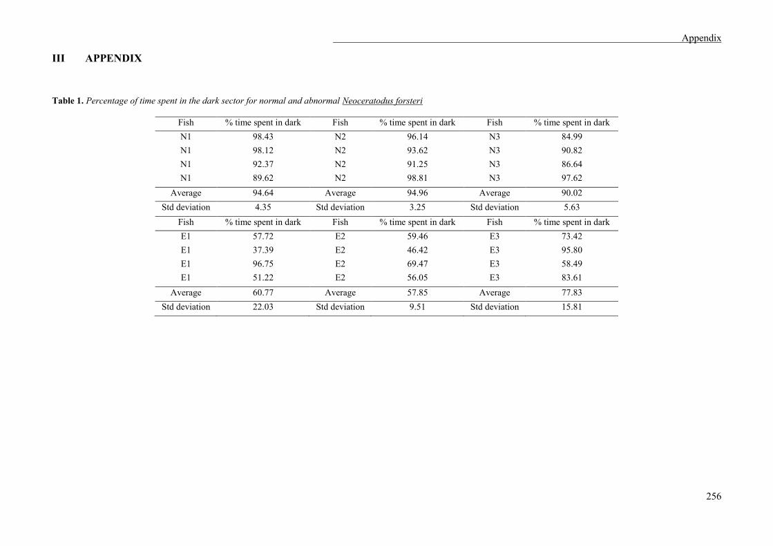
Appendix
256
III APPENDIX
Table 1. Percentage of time spent in the dark sector for normal and abnormal Neoceratodus forsteri
Fish % time spent in dark Fish % time spent in dark Fish % time spent in dark N1 98.43 N2 96.14 N3 84.99 N1 98.12 N2 93.62 N3 90.82 N1 92.37 N2 91.25 N3 86.64 N1 89.62 N2 98.81 N3 97.62
Average 94.64 Average 94.96 Average 90.02 Std deviation 4.35 Std deviation 3.25 Std deviation 5.63
Fish % time spent in dark Fish % time spent in dark Fish % time spent in dark E1 57.72 E2 59.46 E3 73.42 E1 37.39 E2 46.42 E3 95.80 E1 96.75 E2 69.47 E3 58.49 E1 51.22 E2 56.05 E3 83.61
Average 60.77 Average 57.85 Average 77.83 Std deviation 22.03 Std deviation 9.51 Std deviation 15.81