osce exam simulation with the ideal answer read the question and make your own answer and then...
TRANSCRIPT

OSCE EXAM SIMULATION WITH THE IDEAL ANSWER
READ THE QUESTION AND MAKE YOUR OWN ANSWER AND THEN
COMPARE IT WITH THE ATTACHED IDEAL ANSWER.
Dr Saleh W Alharby [email protected] http://faculty.ksu.edu.sa/DrSalehAlharby
Dr Saleh W Alharby [email protected] http://faculty.ksu.edu.sa/DrSalehAlharby
Prepared by
Dr Saleh WaslAllah Alharby , Dept of Orthopedics,
King Saud University.
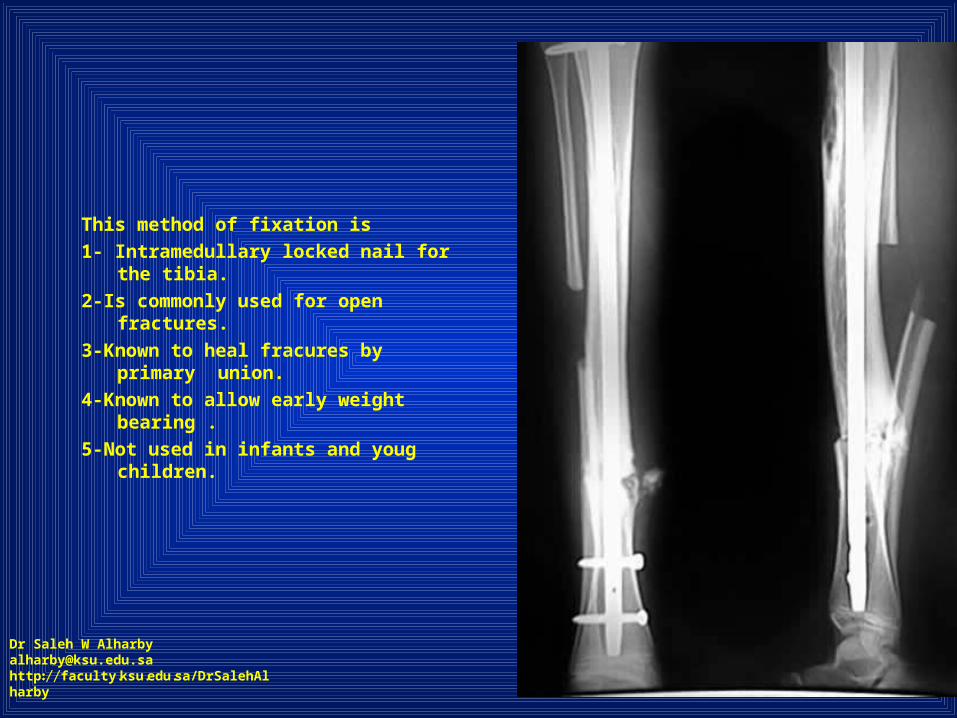
This method of fixation is
1- Intramedullary locked nail for the tibia.
2-Is commonly used for open fractures.
3-Known to heal fracures by primary union.
4-Known to allow early weight bearing .
5-Not used in infants and youg children.
Dr Saleh W Alharby [email protected] http://faculty.ksu.edu.sa/DrSalehAlharby
Answer
This method of fixation is
1- Intramedullary locked nail for the tibia.
4-Known to allow early weight bearing .
5-Not used in infants and young children.
Dr Saleh W Alharby [email protected] http://faculty.ksu.edu.sa/DrSalehAlharby

This injury is
1- Caused by axial torsional forces on the leg.
2- Best treated by cast immobilization.
3-Known to have delayed union.
4-Has higher incidence to be open fracture.
5-Spiral type tibial fracture.
Dr Saleh W Alharby [email protected] http://faculty.ksu.edu.sa/DrSalehAlharby

Answer
This injury is
1- Caused by axial torsional forces on the leg.
3-Known to have delayed union.
4-Has higher incidence to be open fracture.
5-Spiral type tibial fracture.
Dr Saleh W Alharby [email protected] http://faculty.ksu.edu.sa/DrSalehAlharby

This young male has
1-Left leg shortening.
2-Varus leg deformity.
3-Dysuse atrophy of the left lower limb.
4-Congenital pathology.
5-Limping and inability to put weight on it.
Dr Saleh W Alharby [email protected] http://faculty.ksu.edu.sa/DrSalehAlharby

This young male has
1-Left leg shortening.
2-Varus leg deformity.
3-Dysuse atrophy of the left lower limb.
4-Congenital pathology.
5-Limping and inability to put weight on it.
Dr Saleh W Alharby [email protected] http://faculty.ksu.edu.sa/DrSalehAlharby

This X-ray shows
1-Mid shaft recent femoral fracture.
2-Evedence of nonunion.
3-Faint callus formation at mid shaft.
4-Osteopenia at the distal femur.
5-Valgus deformity at proximal femur.
Dr Saleh W Alharby [email protected] http://faculty.ksu.edu.sa/DrSalehAlharby

Answer
This X-ray shows
2-Evedence of nonunion.
3-Faint callus formation at mid shaft.
4-Osteopenia at the distal femur.
Dr Saleh W Alharby [email protected] http://faculty.ksu.edu.sa/DrSalehAlharby

X-ray of the pelvis shows
1-Mild pelvic tilt.
2-Loss of contour of left femoral head.
3-Wide and short left femoral neck.
4-Dysuse atrophy of left femoral shaft.
5-Picture of avascular necrosis of left femoral head.
Dr Saleh W Alharby [email protected] http://faculty.ksu.edu.sa/DrSalehAlharby

Answer
X-ray of the pelvis shows
1-Mild pelvic tilt.
2-Loss of contour of left femoral head.
3-Wide and short femoral neck.
4-Dysuse atrophy of left femoral shaft.
5-Picture of avascular necrosis of femoral head.
Dr Saleh W Alharby [email protected] http://faculty.ksu.edu.sa/DrSalehAlharby

This child abnormality is
1-Short left leg.
2-Long right thigh.
3-Long left thigh.
4-Shortening of the whole left lower limb.
5-lengthening of right lower limb.
Dr Saleh W Alharby [email protected] http://faculty.ksu.edu.sa/DrSalehAlharby

Answer
This child abnormality is
1-Short left leg.
4-Shortening of the whole left lower limb.
Dr Saleh W Alharby [email protected] http://faculty.ksu.edu.sa/DrSalehAlharby

8 yrs old girl injured her left knee at 4 yrs of age. Her X ray shows
1-Varus left knee.
2-Adduction left hip.
3-Shortening left femur.
4-Growth plate abnormality left femur.
5-Growth plate injury left femur in the past.
Dr Saleh W Alharby [email protected] http://faculty.ksu.edu.sa/DrSalehAlharby
Answer
8 yrs old girl injured her left knee at 4 yrs of age. Her X ray shows
2-Adduction left hip.
3-Shortening left femur.
4-Growth plate abnormality left femur.5-Growth plate injury left femur in the past.
Dr Saleh W Alharby [email protected] http://faculty.ksu.edu.sa/DrSalehAlharby