original article pyrogallol induces apoptosis in human ... · protein synthesis, and undergo...
TRANSCRIPT

Folia Biologica (Praha) 64, 23-30 (2018)
Original Article
Pyrogallol Induces Apoptosis in Human Platelets(apoptosis / γ-glutamyl transpeptidase / glutathione / mitochondrial membrane potential / platelets / thrombocytopenia / pyrogallol / polyphenol)
G. BRUGES1, W. VENTURINI1, G. CRESPO1, M. LÓPEZ ZAMBRANO1,2
1Laboratorio de Hemostasia y Genética Vascular. Centro de Biofísica y Bioquímica. Instituto Venezolano de Investigaciones Científicas, Caracas, Venezuela2Institute of Biochemistry, Medical School, Justus Liebig University, Giessen, Germany
Abstract. Pyrogallol is a polyphenol that generates the superoxide anion. In this study, we investigated the influence of pyrogallol on human platelets. Our data showed that exposure of platelets to pyrogallol induced numerous manifestations of apoptosis includ-ing depolarization of mitochondrial inner membrane and release of cytochrome c from the mitochondria. Pyrogallol also induced downstream extra-mitochon-drial apoptotic responses, including activation of caspase-3 and phosphatidylserine exposure on the outer leaflet of the plasma membrane. Addition of glutathione significantly rescued cells from pyro-gallol-induced apoptosis, as evidenced by a decrease of all markers of apoptosis. Thus, pyrogallol appears to produce depletion of intracellular glutathione con-tent in platelets, the main non-protein antioxidant in the cells. Furthermore, inhibition of γ-glutamyl trans-peptidase, an enzyme that plays the main role in the cellular supply of glutathione, reverted the glutathione (GSH) protection over platelet apoptosis. Our results indicate that pyrogallol induces apoptosis by sup-pressing the natural anti-oxidation in human plate-lets.
Received June 10, 2016. Accepted April 5, 2018
Corresponding author: Mercedes López Zambrano, Instituto Vene-zolano de Investigaciones Científicas. Centro de Biofísica y Bio-química, Lab. de Hemostasia y Genética Vascular. Carretera Pan-americana Km 11. Caracas 1020A, Venezuela. Phone: (+58) 212-504-1903; Fax: (+58) 212-504-1093; e-mail: [email protected]
Abbreviations: ACD – acid-citrate-dextrose, CCCP – carbonyl cyanide m-chlorophenylhydrazone, ΔΨm – mitochondrial poten-tial, EGCG – epigallocatechin-3-gallate, γGGT – γ-glutamyl transpeptidase, GSH – glutathione, JC-1 – 5,5′,6,6′-tetrachloro-1,1′,3,3′-tetraethylbenzimidazolylcarbocyanine iodide, O2
•– – su-peroxide anion, PRP – plasma rich in platelets, PS – phosphati-dylserine, ROS – reactive oxygen species, SBC – serine/borate complex.
IntroductionThe mechanism of apoptosis mainly involves two
signalling pathways, the mitochondrial and cell death receptor pathways (Ashkenazi and Dixit, 1998; Budi-hardjo et al., 1999; Shi, 2002). The mitochondrial or in-trinsic apoptosis pathway is controlled by the BCL2 family of proteins. In a healthy cell, pro-survival mem-bers (BCL2, BCL-XL, BCL2L2, MCL1 and BCL2A1) restrain the activity of pro-death BAK1 and BAX. Stress signals activate the BH3-only proteins, which liberate BAK1 and BAX to cause mitochondrial outer mem-brane permeabilization. Diffusion of cytochrome c from the mitochondria triggers formation of the apoptosome, and subsequent activation of caspase-9 and the rest of the apoptotic caspase cascade (Mehmet, 2000). The cell death receptor or extrinsic apoptosis pathway is charac-terized by the binding of cell death ligands and cell death receptors, and subsequently activation of caspase-8 and -3 (Hengartner, 2000; Liu et al., 2004). Caspase-3 is an executioner caspase, whose activation can systemati-cally dismantle cells by cleaving key proteins such as PARP.
Platelets play an important pivotal role in haemosta-sis, thrombosis (Hvas, 2016) and wound healing (Nur-den et al., 2008). Although platelets are anuclear, they exhibit a few characteristics of nucleated cells, such as protein synthesis, and undergo apoptosis.
The function of the apoptotic machinery remains un-clear in human platelets. However, in the last few years, it has become apparent that apoptosis-like events, at least in part, are involved in platelet activation (Dale and Friese, 2006). Furthermore, the intrinsic apoptosis path-way plays an essential physiological role in platelet sur-vival, and is an important regulator of platelet life span in vivo (Zhang et al., 2007; Kile, 2009). Phosphatidylserine exposure is the main “eat-me” signalling for apoptotic cell clearance. Recognition of phosphatidylserine by mononuclear phagocytes promotes apoptotic cell up-take, as well as immunoregulatory responses, including cytokine synthesis and secretion (Chung et al., 2007).
Moreover, it is known that activated platelets mediate inflammatory and immune responses by a variety of

24 Vol. 64
mechanisms, including release of cytokines and interac-tions with leukocytes (Weyrich et al., 2003; Vieira-de-Abreu et al., 2012). In consequence, platelets play cru-cial roles in the pathogenesis of varied clinical conditions where inflammation is important. Alterations in both platelet number and function have been observed with these different conditions (Thachil, 2015), for example, platelets from dengue patients have characteristics that are indicative of apoptosis, among them the exposure of phosphatidylserine (Hottz et al., 2013, 2014). Platelet apoptosis has also been reported in response to various stimuli such as pathological shear stress, platelet stor-age, hyperthermia, physiological agonists and some chemical compounds (Seghatchian and Krailadsiri, 2001; Leytin et al., 2006, 2009; Towhid et al., 2011; Leytin, 2012; Lien et al., 2013).
Polyphenols are naturally occurring plant metabolites (Tsao, 2010). Pyrogallol is a polyphenol that can gener-ate the superoxide anion (O2
•–) (Yamada et al., 2003). This radical and other reactive oxygen species (ROS), such as hydroxyl radical, singlet oxygen, and hydrogen peroxide (H2O2), have been implicated in the regulation of many important cellular events, including cellular proliferation and apoptosis (Kim et al., 2008). Pyrogallol has often been used to investigate the role of O2
•– in bio-logical systems (Kim et al., 2008; Park et al., 2008). It was demonstrated that polyphenols containing the pyro-gallol ring in their structures induce apoptosis in HEK293T and K562 cell lines (Mitsuhashi et al., 2008) and show anti-proliferative activity against human stom-ach cancer cells (Kinjo et al., 2002). Pyrogallol itself was shown to induce O2
•–-mediated death/apoptosis of several types of cells, including human pulmonary ade-nocarcinoma Calu-6 cells (Han et al., 2008, 2009a), As4.1 juxtaglomerular cells (Park et al., 2007a,b), HeLa cells (Han et al., 2008; Kim et al., 2008), gastric cancer SNU-484 cells (Park et al., 2008), human hystiocytic lymphoma U937 cells (Saeki et al., 2000) and endothe-lial cells (Han et al., 2009b; Han and Park, 2010), among other cell types.
Recently, we demonstrated that epigallocatechin-3-gallate (EGCG) induces apoptosis in human platelets (Rosal et al., 2018), because EGCG contains a pyro-gallol ring. In the present study, we investigated and characterized the effect of pyrogallol on platelet apopto-sis. This is the first report of an apoptotic effect of pyro-gallol in human platelets.
Material and Methods
Reagents
Pyrogallol, carbonyl cyanide m-chlorophenylhydra-zone (CCCP), H2O2 and reduced L-glutathione (GSH) were purchased from Sigma-Aldrich Chemical Company (St. Louis, MO). FAM FLICA kit was from Immuno-chemistry Technologies (Bloomington, MN). This kit contains a carboxyfluorescein-labelled fluoromethyl ketone peptide inhibitor of caspase-3 (FAM-DEVD-
FMK). Anti-cytochrome c antibody was purchased from Bio-Legend (San Diego, CA). JC-1 (5,5′,6,6′-tetrachlo-ro-1,1′,3,3′-tetraethylbenzimidazolylcarbocyanine io-dide), fluorescein isothiocyanate (FITC)-annexin V and Pierce™ Western Dura kit were purchased from Thermo Fisher Scientific (Grand Island, NY). Pyrogallol was dissolved in H2O at 100 mM as a stock solution. GSH stock solution (100 mM) was dissolved in phosphate buffer containing 5 mM EDTA.
Preparation of washed plateletsThis study was approved by the Ethics Committee of
the Instituto Venezolano de Investigaciones Cientificas (Approval No. DIR-0997/1569/2016; 28.07.16) and conformed to the principles outlined in the Helsinki Declaration. Thirty-six (36) healthy volunteers were en-rolled in this study and gave informed consent. In brief, blood was collected from healthy human volunteers, who had taken no medicine during the preceding two weeks, in a test tube containing acid-citrate-dextrose (ACD) anticoagulant (1 : 9). Plasma rich in platelets (PRP) was obtained by centrifugation at 160 × g for 20 min at 22 °C. PRP was centrifuged at 1,200 × g and the pellet containing the platelets was resuspended in 13 mM sodium citrate, 30 mM glucose, and 120 mM sodium chloride, pH 6.5 (CGS buffer). Platelets were washed two times with CGS buffer and resuspended in Tyrode’s buffer (145 mM NaCl, 5 mM KCl, 10 mM HEPES, 0.5 mM Na2HPO4, 1 mM MgCl2, 6 mM glu-cose, 0.3% bovine serum albumin, pH 7.4). Platelets were counted by using a counter (Drew 3, Drew Scien-tific, Inc, Miami, FL) and the platelet concentration was adjusted as needed with Tyrode’s buffer. Washed plate-lets were incubated for 1 to 2 h at room temperature for resting (Li et al., 2006). After this time, the platelets were ready for using in subsequent experiments.
Platelets’ treatmentPlatelets (5 × 10⁷/ml) were exposed to different con-
centrations of pyrogallol (0.1–300 μM) at 37 °C in Tyrode’s buffer. For evaluating the effect of GSH or the serine/borate complex (SBC; 2.5 mM L-serine/5 mM boric acid in Tyrode’s buffer), platelets were exposed to either GSH or SBC during 30 min at 37 °C prior to py-rogallol challenge. Hydrogen peroxide, a classical apop-tosis inducer (Singh et al., 2007; Wu et al., 2013), was used as positive control.
Measurement of mitochondrial potential (ΔΨm)Platelets (5 × 10⁷/ml) treated with pyrogallol were
loaded with 0.5 μM JC-1 at 37 °C for 10 min and subse-quently, cells were centrifuged at 1200 × g for 10 min, resuspended in Tyrode’s buffer and analysed immedi-ately in a microplate reader. JC-1 is a cationic dye that accumulates in energized mitochondria. At low concen-trations (due to low ΔΨm), JC-1 is predominantly a monomer that yields green fluorescence with emission at 530 ± 15 nm. At high concentrations (due to high ΔΨm), the dye aggregates, yielding a red to orange col-
G. Bruges et al.

Vol. 64 25
oured emission (590 ± 17.5 nm). Therefore, a decrease in the aggregate fluorescent count is indicative of depo-larization. JC-1 was excited at 488 nm, and fluorescence emissions were detected at 545 nm and 596 nm for JC-1 monomers and aggregates, respectively (Thushara et al., 2013). As a positive control, cells were treated with 20 uM CCCP, an agent known to disrupt ΔΨm (Lou et al., 2007).
To calculate the pyrogallol concentration that inhibits 50 % of the transmembrane potential (IC50), we con-structed a four parameter logistic dose-response curve by using GraphPad Prism® software and calculated the concentration of pyrogallol at which a 50% loss of fluo-rescence occurred.
Measurement of caspase-3 activityPlatelets (3 × 10⁸/ml) were incubated with pyrogallol
at 37 °C for 45 min, followed by centrifugation at 1,200 × g for 10 min at room temperature. Then, platelets were re-suspended in Tyrode’s buffer containing FAM-DEVD-FMK, a carboxyfluorescein-labelled fluoromethylketo- ne peptide inhibitor of caspase-3 (Amstad et al., 2000; Bed ner et al., 2000), for 20 min at 37 °C in the dark. Ana lysis of caspase-3 activity was performed using a fluorescence microplate reader (excitation/emission 488/530 nm).
Evaluation of externalization of phosphatidylserine (PS) with FITC-annexin V
Platelets (5 × 10⁷/ml) were treated with pyrogallol and subsequently, they were transferred to an equal vol-ume of ice-cold 1% (w/v) glutaraldehyde in Tyrode’s buffer, followed by incubation with FITC-conjugated annexin V (0.6 μg/ml) for 10 min. Platelets were col-lected by centrifugation for 60 seconds at 3,000 × g and resuspended in Tyrode’s buffer. FITC-annexin V binding was measured using a fluorescence microplate reader (excitation/emission 496/560 nm) (Rakesh et al., 2014).
Determination of cytochrome c releasePlatelets (3 × 10⁸/ml) were incubated with different
concentrations of pyrogallol for 45 min at 37 °C. Cyto-solic proteins were obtained by three freezing-thawing cycles. Proteins were separated by using 10% SDS-PAGE followed by electroblotting to Immobilon® PVDF mem-branes for 1 h at 90 V in a Bio-Rad transfer system. Membranes were blocked with 10% bovine serum albu-min in Tris-buffered saline with 0.1% tween-20 (TBST) for 2 h at room temperature and subsequently probed overnight with an anti-cytochrome c antibody (1 : 1,000) in TBST at 4 °C. The membranes were rinsed and incu-bated with a horseradish-peroxidase conjugated second-ary antibody (1 : 8,000) in TBST. After the secondary antibody incubation, the membranes were rinsed, and bound antibodies were detected using enhanced chemi-luminescence with a Pierce Western Dura kit (Rakesh et al., 2014). In all instances, gels were loaded with the same amount of proteins. β-Actin was used as loading
control. The GelAnalyzer free software (http://www.gelanalyzer.com) was used to determine the individual band densities.
Statistical analysisStatistical analyses were performed using GraphPad
Prism, version 6.0 (GraphPad Software, San Diego, CA). The Kruskal-Wallis test was applied for comparing differences between three or more groups. The Dunn’s test was used as post hoc test for comparing each two groups. Differences were considered significant when P < 0.05.
ResultsPyrogallol induced numerous manifestations of apop-
tosis in human platelets. First, exposure of platelets to pyrogallol induced depolarization of the mitochondrial membrane (Fig. 1). This effect was dependent on the incubation time. Thus, pyrogallol induced its maximum effect on ΔΨm dissipation after 30 min, as observed in a kinetic analysis (Fig. 1a). The effect of pyrogallol was also dose-dependent. It caused strong depolarization of ΔΨm (approximately 75 %) at 100 µM for 45 min. The IC50 for the pyrogallol-induced decrease in ΔΨm was 24.2 μM, as calculated from the dose-response curve (Fig. 1b). The pyrogallol effect on ΔΨm was compara-ble to that of CCCP, a molecule that causes uncoupling of the mitochondrial proton gradient (see insert, Fig. 1a).
Following a drop of ΔΨm, cytochrome c can be re-leased from mitochondria through the opened mito-chondrial pores; therefore, we also evaluated whether pyrogallol induced cytochrome c release by western blot (Fig. 2a). As expected, very low levels of cytochrome c were detected in the cytosolic fraction of untreated plate-lets. Pyrogallol (at concentrations of 60 and 100 μM) was very effective in triggering the release of cyto chro-me c in a dose-dependent manner.
Pyrogallol also induced downstream extramitochon-drial apoptotic responses. In this context, we measured FITC-conjugated annexin V binding to platelets to de-tect PS exposure on the outer leaflet of plasma mem-branes by fluorescence intensity measurement. As shown in Fig. 2b, pyrogallol (30–100 µM) induced significant PS exposure in comparison with untreated platelets. We also measured caspase-3 activation using a fluorochrome-labelled tetrapeptide affinity ligand FAM-DEVD-FMK, which specifically and covalently binds to the active centre of this enzyme. Caspase-3 activation grew by in-creasing concentrations of pyrogallol. Figure 2c shows that it induced about two- and three-fold increases in caspase-3 activity at concentrations of 30 and 60 μM, respectively, in comparison with untreated platelets.
In addition, we investigated the effect of GSH and SBC, an inhibitor of γ-glutamyl transpeptidase (γGGT), on pyrogallol-induced platelet apoptosis. Figure 3a shows dose-response curves of mitochondrial potential in the presence of increasing GSH concentrations. The
Pyrogallol Induces Platelet Apoptosis

26 Vol. 64
curves of ΔΨm shifted to the right in the presence of 0.3 to 10 mM GSH. In consequence, the IC50 increased in the presence of GSH in a dose-dependent manner (Fig. 3a, inserted table). Figure 3b also shows the de-crease in ΔΨm induced by increasing concentrations of pyrogallol and the protective effect of 1 mM GSH. Consistently, inhibition of platelet γGGT activity by SBC significantly reverted the GSH protection over ΔΨm.
Next, we attempted to examine whether GSH and SBC affect other pyrogallol-induced apoptosis signals
Fig. 1. Pyrogallol effects on mitochondrial potential. a) Ki-netic analysis: Platelets were incubated with pyrogallol (60 µM) at 37 °C for different time intervals. Insert: loss of ΔΨm in platelets incubated in the absence (open bar) or presence of 60 µM pyrogallol (grey bar) or 50 µM CCCP (black bar). b) Dose-response curve. Platelets were incu-bated for 45 min at 37 °C with different concentrations of pyrogallol. Data represent the mean ± SEM of three inde-pendent experiments done in triplicate. *P < 0.05 vs con-trol in the absence of pyrogallol.
Fig. 2. Pyrogallol effects on apoptotic markers. a) Cyto chro-me c release in the absence or presence of 60 μM or 100 μM pyrogallol was evaluated by western blot using an anti-cy-tochrome monoclonal antibody. The graph shows the den-sitometric quantification of western blot bands using the GelAnalyser software. b) FITC-annexin V binding to plate-lets at different pyrogallol concentrations was quantified by using a microplate reader. c) Caspase-3 activity was evaluated using fluorescent probe FAM-DEVD-FMK in a microplate reader. Hydrogen peroxide (2000 µM) was used as positive control. Data represent the mean ± SEM of three independent experiments done in triplicate. *P < 0.05 vs control in the absence of pyrogallol.
G. Bruges et al.

Vol. 64 27
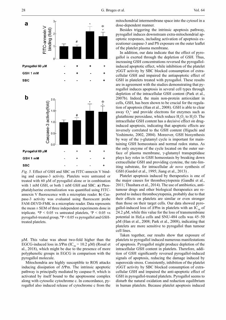
in platelets. We found that GSH completely reverted the cytochrome c release in pyrogallol-treated platelets to the level of the untreated group, while SBC significantly reverted the antiapoptotic effects of GSH on the cy-tochrome c release (Fig. 4). Likely, GSH also prevented the FITC-annexin V binding (Fig. 5a) and activation of caspase-3 (Fig. 5b) in pyrogallol-treated platelets, while SBC completely blocked the anti-apoptotic effects of GSH on both caspase-3 activation and PS exposure.
Thus, the levels of caspase-3 activity and FITC-annexin V binding in platelets treated simultaneously with pyro-gallol, GSH and SBC were similar to those observed in pyrogallol-treated platelets (Fig. 5a,b).
DiscussionPlatelet apoptosis has been reported in response to
various stimuli such as pathological shear stress, platelet storage, hyperthermia, physiological agonists and some chemical compounds (Seghatchian and Krailadsiri, 2001; Leytin et al., 2006, 2009; Towhid et al., 2011; Leytin, 2012; Lien et al., 2013). Recently, we demon-strated that EGCG induces apoptosis in human platelets (Rosal et al., 2018). EGCG is the most abundant flavo-noid present in extracts derived from green tea leaves, which has various beneficial effects related to its anti-oxidant properties (Kinjo et al., 2002). Due to the spe-cial structure of EGCG, which contains a pyrogallol ring, we investigated whether pyrogallol alone can in-fluence the platelet apoptosis. To our knowledge, this is the first report about the apoptotic effect of pyrogallol on human platelets.
Our data showed that exposure of platelets to pyro-gallol induced numerous manifestations of apoptosis including depolarization of the mitochondrial inner membrane. In particular, the effect of pyrogallol on the platelet ΔΨm was dose-dependent, with an IC50 of 24.2
Fig. 3. Effect of GSH and SBC on pyrogallol-induced de-crease of ΔΨm in human platelets. a) Dose-response curves in the absence (open circles) or presence of different GSH concentrations; 0.3 mM (closed circles); 1 mM (closed squares); and 10 mM (open squares). The IC50 calculated by non-linear regression software are indicated in the in-serted table. b) Platelets were incubated with increasing concentrations of pyrogallol alone (open circles) or in combination with 1 mM GSH (closed circles), or both 1 mM GSH and SBC (closed squares). Data represents the mean ± SEM of three independent experiments done in triplicate. *P < 0.05 vs pyrogallol-treated group, #P < 0.05 vs pyrogallol and GSH-treated group.
Fig. 4. Effect of GSH and SBC on pyrogallol-induced cy-tochrome c release in human platelets. Platelets were un-treated or treated with 60 μM of pyrogallol alone or in combination with 1 mM GSH, or both 1 mM GSH and SBC. Cytochrome c release was determined using an anti-cytochrome monoclonal antibody by western blot. The graph shows densitometric quantification (N = 3/group) of western blot bands using the GelAnalyser software. *P < 0.05 vs untreated platelets, #P < 0.05 vs pyrogallol-treated group, FP < 0.05 vs pyrogallol and GSH-treated platelets.
Pyrogallol Induces Platelet Apoptosis
28 Vol. 64
μM. This value was about two-fold higher than the EGCG-induced loss in ΔΨm (IC50 = 10.2 μM) (Rosal et al., 2018), which might be due to the presence of more polyphenolic groups in EGCG in comparison with the pyrogallol molecule.
Mitochondria are highly susceptible to ROS attacks inducing dissipation of ΔΨm. The intrinsic apoptotic pathway is principally mediated by caspase-9, which is activated by itself bound to the apoptosome complex along with cytosolic cytochrome c. In concordance, py-rogallol also induced release of cytochrome c from the
mitochondrial intermembrane space into the cytosol in a dose-dependent manner.
Besides triggering the intrinsic apoptosis pathway, pyrogallol induces downstream extra-mitochondrial ap-optotic responses, including activation of apoptosis ex-ecutioner caspase-3 and PS exposure on the outer leaflet of the platelet plasma membrane.
In addition, our data indicate that the effect of pyro-gallol is exerted through the depletion of GSH. Thus, increasing GSH concentrations reversed the pyrogallol-induced apoptotic effect, while inhibition of the platelet γGGT activity by SBC blocked consumption of extra-cellular GSH and impaired the antiapoptotic effect of GSH in platelets treated with pyrogallol. These results are in agreement with the studies demonstrating that py-rogallol induces apoptosis in several cell types through depletion of the intracellular GSH content (Park et al., 2007b). Indeed, the main non-protein antioxidant in cells, GSH, has been shown to be crucial for the regula-tion of apoptosis (Han et al., 2008). GSH is able to clear away O2
•- and provide electrons for enzymes such as glutathione peroxidase, which reduce H2O2 to H2O. The intracellular GSH content has a decisive effect on drug-induced apoptosis, indicating that apoptotic effects are inversely correlated to the GSH content (Higuchi and Yoshimoto, 2002, 2004). Moreover, GSH biosynthesis by way of the γ-glutamyl cycle is important for main-taining GSH homeostasis and normal redox status. As the only enzyme of the cycle located on the outer sur-face of plasma membrane, γ-glutamyl transpeptidase plays key roles in GSH homeostasis by breaking down extracellular GSH and providing cysteine, the rate-lim-iting substrate, for intracellular de novo synthesis of GSH (Gurdol et al., 1995; Jiang et al., 2013) .
Platelet apoptosis induced by therapeutics is one of the major causes for thrombocytopenia (Gandhi et al., 2011; Thushara et al., 2014). The use of antibiotics, anti-tumour drugs and other biological therapeutics are re-ported to induce thrombocytopenia, probably due to that their effects on platelets are similar or even stronger than those on their target cells. Our data showed pyro-gallol-induced loss of ΔΨm in platelets with an IC50 of 24.2 μM, while this value for the loss of transmembrane potential in HeLa cells and SNU-484 cells was 45–50 μM (Han et al., 2008; Park et al., 2008), indicating that platelets are more sensitive to pyrogallol than tumour cell lines.
Taken together, our results show that exposure of platelets to pyrogallol induced numerous manifestations of apoptosis. Pyrogallol might produce depletion of the intracellular GSH content in platelets. Therefore, addi-tion of GSH significantly reversed pyrogallol-induced signals of apoptosis, reducing the damage induced by superoxide stress. Consistently, inhibition of the platelet γGGT activity by SBC blocked consumption of extra-cellular GSH and impaired the anti-apoptotic effect of GSH in pyrogallol-treated platelets. Pyrogallol seems to disturb the natural oxidation and reduction equilibrium in human platelets. Because platelet apoptosis induced
Fig. 5. Effect of GSH and SBC on FITC-annexin V bind-ing and caspase-3 activity. Platelets were untreated or treated with 60 μM of pyrogallol alone or in combination with 1 mM GSH, or both 1 mM GSH and SBC. a) Phos-phatidylserine externalization was quantified using FITC-annexin V fluorescence with a microplate reader. b) Cas-pase-3 activity was evaluated using fluorescent probe FAM-DEVD-FMK in a microplate reader. Data represents the mean ± SEM of three independent experiments done in triplicate. *P < 0.05 vs untreated platelets, #P < 0.05 vs pyrogallol-treated group, FP < 0.05 vs pyrogallol and GSH-treated platelets.
G. Bruges et al.
Vol. 64 29
by therapeutics is one of the major causes for thrombo-cytopenia, it is worth highlighting the importance of in-vestigating the possible adverse effects of new potential therapeutic drugs by inducing platelet apoptosis and in consequence, thrombocytopenia.
Disclosure of conflicts of interestThe authors declare no conflicts of interest in the re-
search.
AcknowledgmentWe are grateful to Prof. Dr. Klaus T. Preissner (JLU
University, Giessen, Germany) for his critical reading of this manuscript.
References Amstad, P., Johnson, G., Lee, B., Dhawan, S. (2000) An in situ
marker for the detection of activated caspases. Biotechnol. Lab. 18, 52-56.
Ashkenazi, A., Dixit, V. M. (1998) Death receptors: signaling and modulation. Science 281, 1305-1308.
Bedner, E., Smolewski, P., Amstad, P., Darzynkiewicz, Z. (2000) Activation of caspases measured in situ by binding of fluo-rochrome-labeled inhibitors of caspases (FLICA): correla-tion with DNA fragmentation. Exp. Cell Res. 259, 308-313.
Budihardjo, I., Oliver, H., Lutter, M., Luo, X., Wang, X. (1999) Biochemical pathways of caspase activation during apoptosis. Annu. Rev. Cell Dev. Biol. 15, 269-290.
Chung, E. Y., Liu, J., Homma, Y., Zhang, Y., Brendolan, A., Saggese, M., Han, J., Silverstein, R., Selleri, L., Ma, X. (2007) Interleukin-10 expression in macrophages during phagocytosis of apoptotic cells is mediated by homeodo-main proteins Pbx1 and Prep-1. Immunity 27, 952-964.
Dale, G. L., Friese, P. (2006) Bax activators potentiate coated-platelet formation. J. Thromb. Haemost. 4, 2664-2669.
Gandhi, L., Camidge, D. R., Ribeiro de Oliveira, M., Bonomi, P., Gandara, D., Khaira, D., Hann, C. L., McKeegan, E. M., Litvinovich, E., Hemken, P. M., Dive, C., Enschede, S. H., Nolan, C., Chiu, Y. L., Busman, T., Xiong, H., Krivoshik, A. P., Humerickhouse, R., Shapiro, G. I., Rudin, C. M. (2011) Phase I study of Navitoclax (ABT-263), a novel Bcl-2 family inhibitor, in patients with small-cell lung can-cer and other solid tumors. J. Clin. Oncol. 29, 909-916.
Gurdol, F., Nwose, O. M., Mikhailidis, D. P. (1995) Gamma-glutamyl transferase activity in human platelets: quantifi-cation of activity, isoenzyme characterization and potential clinical relevance. Platelets 6, 200-203.
Han, Y. H., Kim, S. Z., Kim, S. H., Park, W. H. (2008). Pyro-gallol as a glutathione depletor induces apoptosis in HeLa cells. Int. J. Mol. Med. 21, 721-730.
Han, Y. H., Kim, S. Z., Kim, S. H., Park, W. H. (2009a) Pyro-gallol inhibits the growth of lung cancer Calu-6 cells via caspase-dependent apoptosis. Chem. Biol. Interact. 177, 107-114.
Han, Y. H., Moon, H. J., You, B. R., Kim, S. Z., Kim, S. H., Park, W. H. (2009b) JNK and p38 inhibitors increase and decrease apoptosis, respectively, in pyrogallol-treated calf pulmonary arterial endothelial cells. Int. J. Mol. Med. 24, 717-722.
Han, Y. H., Park, W. H. (2010) Pyrogallol-induced calf pulmo-nary arterial endothelial cell death via caspase-dependent apoptosis and GSH depletion. Food Chem. Toxicol. 48, 558-563.
Hengartner, M. O. (2000) The biochemistry of apoptosis. Na-ture 407, 770-776.
Higuchi, Y., Yoshimoto, T. (2002) Arachidonic acid converts the glutathione depletion-induced apoptosis to necrosis by promoting lipid peroxidation and reducing caspase-3 activ-ity in rat glioma cells. Arch. Biochem. Biophys. 400, 133-140.
Higuchi, Y., Yoshimoto, T. (2004) Promoting effects of poly-unsaturated fatty acids on chromosomal giant DNA frag-mentation associated with cell death induced by glutathione depletion. Free Radic. Res. 38, 649-658.
Hottz, E. D., Oliveira, M. F., Nunes, P. C., Nogueira, R.M ., Valls-de-Souza, R., Da Poian, A. T., Weyrich, A. S., Zim-merman, G. A., Bozza, P. T., Bozza, F. A. (2013) Dengue induces platelet activation, mitochondrial dysfunction and cell death through mechanisms that involve DC-SIGN and caspases. J. Thromb. Haemost. 11, 951-962.
Hottz, E. D., Medeiros-de-Moraes, I. M., Vieira-de-Abreu, A., de Assis, E. F., Vals-de-Souza, R., Castro-Faria-Neto, H. C., Weyrich, A. S., Zimmerman, G. A., Bozza, F. A., Boz-za, P. T. (2014) Platelet activation and apoptosis modulate monocyte inflammatory responses in dengue. J. Immunol. 193, 1864-1872.
Hvas, A. M. (2016) Platelet function in thrombosis and hemo-stasis. Semin. Thromb. Hemost. 42, 183-184.
Jiang, S., Jiang, D., Tao, Y. (2013) Role of γ-glutamyltransferase in cardiovascular diseases. Exp. Clin. Cardiol. 18, 53-56.
Kile, B. T. (2009). The role of the intrinsic apoptosis pathway in platelet life and death. J. Thromb. Haemost. 7(Suppl 1), 214-217.
Kim, S. W., Han, Y. W., Lee, S. T., Jeong, H. J., Kim, S. H., Kim, I. H., Lee, S. O., Kim, D. G., Kim, S. H., Kim, S. Z., Park, W. H. (2008) A superoxide anion generator, pyro-gallol, inhibits the growth of HeLa cells via cell cycle ar-rest and apoptosis. Mol. Carcinog. 47, 114-125.
Kinjo, J., Nagao, T., Tanaka, T., Nonaka, G., Okawa, M., No-hara, T., Okabe, H. (2002) Activity-guided fractionation of green tea extract with antiproliferative activity against hu-man stomach cancer cells. Biol. Pharm. Bull. 25, 1238-1240.
Leytin, V., Allen, D. J., Mykhaylov, S., Lyubimov, E., Freed-man, J. (2006) Thrombin-triggered platelet apoptosis. J. Thromb. Haemost. 4, 2656-2663.
Leytin, V., Allen, D. J., Mutlu, A., Gyulkhandanyan, A. V., Mykhaylov, S., Freedman, J. (2009) Mitochondrial control of platelet apoptosis: effect of cyclosporin A, an inhibitor of the mitochondrial permeability transition pore. Lab. In-vest. 89, 374-384.
Leytin, V. (2012) Apoptosis in the anucleate platelet. Blood Rev. 26, 51-63.
Li, Z., Zhang, G., Feil, R., Han, J., Xiaoping, D. (2006) Se-quential activation of p38 and ERK pathways by cGMP-dependent protein kinase leading to activation of the plate-let integrin αIIbβ3. Blood 107, 965-972.
Lien, L. M., Su, C. C., Hsu, W. H., Lu, W. J., Chung, C. L., Yen, T. L., Chiu, H. C., Sheu, J. R., Lin, K. H. (2013)
Pyrogallol Induces Platelet Apoptosis
30 Vol. 64
Mechanisms of andrographolide-induced platelet apopto-sis in human platelets: regulatory roles of the extrinsic ap-optotic pathway. Phytother. Res. 27, 1671-1677.
Liu, X., Yue, P., Zhou, Z., Khuri, F.R., Sun, S. Y. (2004) Death receptor regulation and celecoxib-induced apoptosis in hu-man lung cancer cells. J. Natl. Cancer Inst. 96, 1769-1780.
Lou, P. H., Hansen, B. S., Olsen, P. H., Tullin, S., Murphy, M. P., and Brand, M. D. (2007) Mitochondrial uncouplers with an extraordinary dynamic range. Biochem. J. 407, 129-140.
Mehmet, H. (2000) Caspases find a new place to hide. Nature 403, 29-30.
Mitsuhashi, S., Saito, A., Nakajima, N., Shima, H., Ubukata, M. (2008) Pyrogallol structure in polyphenols is involved in apoptosis-induction on HEK293T and K562 cells. Mol-ecules 13, 2998-3006.
Nurden, A. T., Nurden, P., Sanchez, M., Andia, I., Anitua, E. (2008) Platelets and wound healing. Front. Biosci. 13, 3532-3548.
Park, W. H., Han, Y. H., Kim, S. H., Kim, S. Z. (2007a) Pyro-gallol, ROS generator inhibits As4.1 juxtaglomerular cells via cell cycle arrest of G2 phase and apoptosis. Toxicology 235, 130-139.
Park, W. H., Han, Y. W., Kim, S. H., Kim, S.Z. (2007b) A su-peroxide anion generator, pyrogallol induces apoptosis in As4.1 cells through the depletion of intracellular GSH con-tent. Mutat. Res. 619, 81-92.
Park, W. H., Park, M. N., Han, Y. H., Kim, S.W. (2008) Pyro-gallol inhibits the growth of gastric cancer SNU-484 cells via induction of apoptosis. Int. J. Mol. Med. 22, 263-268.
Rakesh, K. S., Jagadish, S., Vinayaka, A. C., Hemshekhar, M., Paul, M., Thushara, R. M., Sundaram, M. S., Swaroop, T. R., Mohan, C. D., Basappa, Sadashiva, M. P., Kemparaju, K., Girish, K. S., Rangappa, K. S. (2014) A new ibuprofen derivative inhibits platelet aggregation and ROS mediated platelet apoptosis. PloS One 9, e107182.
Rosal, K., Useche, A., Moran, L., López, M., Bruges, G. (2018) Epigallocatechin-3-gallate induces apoptosis in platelets. Invest. Clín. (in press). (in Spanish)
Saeki, K., Hayakawa, S., Isemura, M., Miyase, T. (2000) Im-portance of a pyrogallol-type structure in catechin com-pounds for apoptosis-inducing activity. Phytochemistry 53, 391-394.
Seghatchian, J., Krailadsiri, P. (2001) Platelet storage lesion and apoptosis: are they related? Transfus. Apher. Sci. 24, 103-105.
Shi, Y. (2002) Mechanisms of caspase activation and inhibi-tion during apoptosis. Mol. Cell 9, 459-470.
Singh, M., Sharma, H., Singh, N. (2007) Hydrogen peroxide induces apoptosis in HeLa cells through mitochondrial pathway. Mitochondrion 7, 367-373.
Thachil, J. (2015) Platelets in inflammatory disorders: a pathophysiological and clinical perspective. Semin. Thromb. Hemost. 41, 572-581.
Thushara, R. M., Hemshekhar, M., Santhosh, M. S., Jnanesh-wari, S., Nayaka, S. C., Naveen, S., Kemparaju, K., Girish, K. S. (2013) Crocin, a dietary additive protects platelets from oxidative stress-induced apoptosis and inhibits plate-let aggregation. Mol. Cell. Biochem. 373, 73-83.
Thushara, R. M., Hemshekhar, M., Kemparaju, K., Rangappa, K. S., Devaraja, S., Girish, K. S. (2014) Therapeutic drug-induced platelet apoptosis: an overlooked issue in pharma-cotoxicology. Arch. Toxicol. 88, 185-198.
Towhid, S. T., Schmidt, E. M., Schmid, E., Munzer, P., Qadri, S. M., Borst, O., Lang, F. (2011) Thymoquinone-induced platelet apoptosis. J. Cell. Biochem. 112, 3112-3121.
Tsao, R. (2010) Chemistry and biochemistry of dietary poly-phenols. Nutrients 2, 1231-1246.
Vieira-de-Abreu, A., Campbell, R. A., Weyrich, A. S., Zim-merman, G. A. (2012) Platelets: versatile effector cells in hemostasis, inflammation, and the immune continuum. Semin. Immunopathol. 34, 5-30.
Weyrich, A. S., Lindemann, S., Zimmerman, G. A. (2003) The evolving role of platelets in inflammation. J. Thromb. Hae-most. 1, 1897-1905.
Wu, L., Xi, Z., Guo, R., Liu, S., Yang, S., Liu, D., Dong, S., Guo, D. (2013) Exogenous ARC down-regulates caspase-3 expression and inhibits apoptosis of broiler chicken cardio-myocytes exposed to hydrogen peroxide. Avian Pathol. 42, 32-37.
Yamada, J., Yoshimura, S., Yamakawa, H., Sawada, M., Naka-gawa, M., Hara, S., Kaku, Y., Iwama, T., Naganawa, T., Banno, Y., Nakashima, S., Sakai, N. (2003) Cell permeable ROS scavengers, Tiron and Tempol, rescue PC12 cell death caused by pyrogallol or hypoxia/reoxygenation. Neurosci. Res. 45, 1-8.
Zhang, H., Nimmer, P. M., Tahir, S. K., Chen, J., Fryer, R. M., Hahn, K. R., Iciek, L. A., Morgan, S. J., Nasarre, M. C., Nelson, R., Preusser, L. C., Reinhart, G. A., Smith, M. L., Rosenberg, S. H., Elmore, S. W., Tse, C. (2007) Bcl-2 fam-ily proteins are essential for platelet survival. Cell Death Differ. 14, 943-951.
G. Bruges et al.