original article pentapeptide plnpk ameliorates adjuvant ... · original article pentapeptide plnpk...
TRANSCRIPT
Int J Clin Exp Pathol 201710(5)5252-5262wwwijcepcom ISSN1936-2625IJCEP0034627
Original ArticlePentapeptide PLNPK ameliorates adjuvant arthritis and inhibits T cell activation by suppressing Lck and PI3K activities
Jia Sun1 Song Wang2 Yong-Ci Zhang1 Zheng Fu12 Zhi-Feng Zhu2 Jing Jia2 Qiong Xu2 Li Wang12 Jin-Ping Li2 Rong Lu2 Zhi Yao1
1Department of Immunology Tianjin Medical University 22 Qixiangtai Road Tianjin 300070 China 2Tianjin Kangzhe Pharmaceutical Technology Development Co Ltd 7F Building B Guangyin Tower Youyibei Road Tian-jin 300074 China Equal contributors
Received June 28 2016 Accepted July 15 2016 Epub May 1 2017 Published May 15 2017
Abstract The pentapeptide PLNPK could inhibit the transformation and proliferation of mouse T lymphocytes in mice Rheumatoid arthritis (RA) is essentially a kind of T cell-mediated arthritis Therefore we assessed the thera-peutic ability of PLNPK on adjuvant arthritis (AA) rats and examined its effects on signals of CD4+ T cell activation Isolated CD4+ T cells were stained with CFSE and stimulated by anti-CD3 or anti-CD3 and anti-CD28 monoclonal antibodies The suppression effects of PLNPK on T cell proliferation and CD25 expression were detected by flow cytometry AA rats received PLNPK or saline intraperitoneal injection once a day for 20 days Effects on disease progression were assessed by measurement of paw swelling Inflammation and joint destruction were examined by histology T cell infiltration and activation in joint were detected by immunohistochemistry stain The suppres-sion effects of PLNPK on Lck and PI3K ability in CD4+ T cells and in AA rats were assessed by immunoprecipitation and kinase assay kits PLNPK inhibited the CD4+ T cell proliferation and altered their CD25 expression when they received CD3 mAb or CD3CD28 mAbs stimulation In AA rats PLNPK inhibited T cell infiltration and activation in joint In addition PLNPK inhibited Lck activity in CD3 mAb stimulated CD4+ T cells as well as PI3K activity in CD3CD28 mAbs stimulated CD4+ T cells In vivo PLNPK can also reduce the Lck and PI3K activities in AA rat splenocytesPLNPK exerts its immunosuppressive effects and ameliorates adjuvant arthritis by inhibiting the Lck and PI3K ac-tivities in T cells
Keywords Rheumatoid arthritis immunosuppressive agents flow cytometry immunohistochemistry enzymes
Introduction
As the largest immune system organ in the body the spleen contains many immunocytes and cytokines they play important roles in immune regulation A large number of peptides with immunological activities have been ex- tracted from spleen and some of them are potential to be medicine The pentapeptide PLNPK (Pro-Leu-Asn-Pro-Lys) is one of them Our previous studies showed that PLNPK could inhibit ConA-induced T proliferation and could inhibit immunoglobulin production by B cells in mice suggesting that PLNPK is an immunosup-pressive oligopeptide [1 2] The latest studies discovered the therapeutic effects of PLNPK on autoimmune diseases in animal models such
as anti-glomerular basement membrane (GBM) nephritis [1] and systemic lupus erythematosus (SLE) [3] and PLNPK also prolonged the sur-vival time of skin allograft and cardiac allograft [2] In addition we found the common mecha-nism by which PLNPK plays its therapeutic roles is to inhibit T cell activation [2 3]
CD4+ T cells play a central role in the immune response The mechanisms governing CD4+ T cell activation require fine-tuning because hy- peractivation can lead to immunoproliferative disorders and autoimmunity We firstly studied the impacts of PLNPK on CD4+ T cell activation Peripheral T cells are activated following TCRCD3 triggering in the presence of costimulatory signals The earliest recognizable event after
PLNPK ameliorates arthritis and inhibits T cell
5253 Int J Clin Exp Pathol 201710(5)5252-5262
T cell receptor (TCR) engagement by antigen is the activation of protein tyrosine kinases (PTK) of Src family [4 5] The Src kinase family member p56lck (Lck) is the major PTK dur- ing TCR triggering Lck is mainly responsible for the activation of phospholipase Cγ (PLCγ) PLCγ initiates the subsequent Ca2+calmodu-lincecalcineurinNFAT pathway and PKCNF- κB pathway [5] In addition Lck is involved in ERKMAPK signal activation [6-8] Therefore Lck is critical for TCR triggered T-cell activa- tion In this study we observed the inhibitory effect of PLNPK on Lck in T cells
In addition to TCRCD3 signal complete acti- vation of T cells also needs the second sig- nals provided by pairs of costimulatory mole-cules the most important one of which is B7CD28 CD28 binding to its ligand B7 results in tyrosine phosphorylation of Y173MNM motif The phosphorylated YMNM motif subsequent- ly interacts with the SH2 domain within pho- sphoinositide 3-kinases (PI3K) regulatory sub-unit p85 activating the p110 catalytic sub- unit [9] The p110 generate PIP3 that recruit a variety of proteins to plasma membrane among which is phosphatidylinositol-depen-dent kinase-1 (PDK-1) PDK-1 activates AKT and AKT can significantly promote the T cell activation and proliferation cytokine secretion inhibits Fas-mediated T cell apoptosis [10- 12] We also studied the effect of PLNPK on PI3K activity in T cell
Rheumatoid arthritis (RA) is essentially a kind of T cell-mediated arthritis RA disease origi-nated in T cell activation [13] A lot of self-reac-tive CD4+ T cells were detected in joints of RA patients these abnormal T cells interacts with other pathogenic cells through direct contact or secretion of causative agents [14-16] We established rat adjuvant arthritis (AA) model observed the effect of PLNPK treatment on autoimmune diseases AA and studied the effect of PLNPK on T cell activation in vivo
Materials and methods
Drug and reagents
PLNPK was obtained from Shenzhen Kangzhe Pharmaceutical Co Ltd Anti-human CD3 mAb anti-human CD28 mAb anti-human CD4-PE antibody and anti-human CD25-APC antibody were purchased from BD-PharMingen compa-
ny Carboxyfluorescein diacetate succinimidyl ester (CFSE) and Freundrsquos complete adjuvant were purchased from Sigma-Aldrich Corpor- ation Anti-Lck mAb and anti-P85 mAb was pur-chased from Upstate Company PTK kinase assay kit was purchased from Sigma-Aldrich Corporation PI3K kinase assay kit was pur-chased from Echelon Biosiences company anti-rat CD3 mAb and anti-rat CD25 mAb were purchased from Serotec Company
Animals
Male Wistar rats (SPF grade 180 plusmn 20 g) were provided by Experimental Animal Center of Military Medical Sciences They were housed in an environment with a controlled 1212 lightdark cycle and constant temperature of 21-25degC with a humidity of 55 plusmn 5 All pro- cedures carried out according to NIHrsquos SOP Practice
Ethics statement
Venous blood samples were collected from health volunteers with informed consent and with the approval of local ethics committee and Health Bureau Animal experiment in this study was approved by Ethics Committee of Tianjin Medical University
Isolation of primary peripheral blood CD4+ T cells
The peripheral blood mononuclear cells (PBMC) were isolated by Ficoll-Hypaque density gra- dient (1077 gml) centrifugation at 2000 rpm for 20 minutes After washing 3 times CD4+ T cells were negatively selected by an antibody cocktail according to the manufacturersquos in- structions (Miltenyi Company) A Cell purity of above 90 was achieved by flow cytometry detection
CD4+ T lymphocyte proliferation
CD4+ T cells were adjusted to 1times106ml incu-bating with carboxyfluorescein succinimidyl ester (CFSE 5 mmolml) in 37degC for 15 min-utes for CFSE labeling Finally the reaction was stopped by ice bath for 5 minutes After wash-ing for 3 times CD4+ T cells were resuspended in RPMI-164010 FBS and plated in 96-well plate at 4times105well along with titrating doses of PLNPK at a final concentration of 16 mgml
PLNPK ameliorates arthritis and inhibits T cell
5254 Int J Clin Exp Pathol 201710(5)5252-5262
08 mgml 04 mgml 02 mgml or media control Except for unstimulated control group cells were stimulated with 25 μgml CD3 mAb alone or CD3 mAb (25 μgml) and CD28 mAb (1 μgml) both The plate was placed in 37degC 5 CO2 incubator for 48 hours Data acquisition was processed by CELLQuest soft-ware with flow cytometry (BD-PharMingen Cor- poration) and the proliferation index was ob- tained by Modfit software
CD25 expression in T lymphocytes
In this study 2times106 purified human CD4+ T cells were plated in 24-well plate with the same final concentrations of PLNPK as above Cells also received the same stimulation as above and the unstimulated control wells received no stimulation The plate was placed in 37degC 5 CO2 incubator for 48 hours After incubation the cells were harvested and stained with anti-human CD4-PE and CD25-APC antibodies for evaluation of T cell activation Data acquisition was processed by CELLQuest software with flow cytometry (BD-PharMingen Corporation)
AA model and drug treatment
AA model was established by intradermal injec-tion of 01 ml FCA in the right hind paw of each rat After 7 days the rats were randomly divid-ed into four groups different doses of PLNPK 200 μgkgd 100 μgkgd) model control group and healthy control group there ten ani-mals in each group The 2 PLNPK groups received 1 ml ip injection of 200 μgkgd and 100 μgkgd PLNPK respectively once a day for 20 days at the same time the two control group were treated with 1 ml saline
Histological examination
After the last administration all of the AA rats were sacrificed and each right ankle was removed washed with saline and fixed in 10 neutral formalin After 15 EDTA (2Na+) decal-cification conventional dehydration and paraf-fin embedding sliced the joints were stained by haematoxylin-eosin (HE) for light microscope examination In addition some paraffin sec-tions were microwave repaired and then were incubated with anti-CD3 mAb or anti-CD25 mAb working solution at 4degC overnight On the next day HRP labeled goat anti-mouse IgG and DAB were used to reveal the reaction Positive cells were with brown-yellow or yellow granular material deposits to the cytoplasm
Lck activity detection
For in vitro study 2times106 purified human CD4+ T cells were added in 24-well plate with the same PLNPK treatment as above Cells were stimu-lated with 25 μgml CD3 mAb except for that in unstimulated control wells The plate was placed in 37degC 5 CO2 incubator for 48 hours Total protein was extracted by RIPA lysis buffer and was quantified to a final concentration of 5 μgμl Lck samples were prepared by immuno-precipitation as follows 1 mg total protein was incubated with 3 μl Lck antibody for one hour at 4degC and then incubated with 60 μl 50 Protein A bead at 4degC for one hour The solution was centrifuged at 14000 g for 5 seconds and the supernatant was discarded After washing the samples could be used for the evaluation of Lck kinase activity PTK kinase assay kit was used to detect the Lck activity
In order to observe the effect of PLNPK on Lck activity in vivo total proteins were extracted from spleen tissue of AA rats in each group Lck samples were prepared by immunoprecipita-tion and PTK kinase assay kit was used to detect the Lck kinase activity
PI3K activity detection
2times106 purified human CD4+ T cells were plated in 24 well plates with the same PLNPK treat-ment as above Cells were stimulated with 25 μgml CD3 mAb and 1 μgml CD28 mAb in stimulation wells and unstimulated control received no stimulation The plate was placed in 37degC 5 CO2 incubator for 48 hours After that the samples of p85 subunit were prepared by immunoprecipitation as above and then the activity was examined with a PI3K kinase assay kit The PI3K activity in splenocytes of AA rats was also detected
Statistical analyses
Data were expressed as group means plusmn stan-dard deviation and analyzed with one-way anal-ysis of variance (ANOVA) Plt005 was consid-ered to be statistically significant
Results
PLNPK inhibited the proliferation of CD4+ T cells
In order to observe the effect of PLNPK on CD4+ T cells proliferation we used CD3 mAb and
PLNPK ameliorates arthritis and inhibits T cell
5255 Int J Clin Exp Pathol 201710(5)5252-5262
Figure 1 Effect of PLNPK on McAb induced CD4+ T cells proliferation Human CD4+ T cells were labeled with CFSE and plated in 96-well plate with titrating doses of PLNPK and were simulated with CD3 McAb (A B) or CD3CD28 McAb (A C) After cultured for 48 hrs cell proliferation was detected by flow cytometry Data are expressed as mean plusmn SD versus stimulated control group Plt005 N=3
PLNPK ameliorates arthritis and inhibits T cell
5256 Int J Clin Exp Pathol 201710(5)5252-5262
Figure 2 Effect of PLNPK on CD4+ T cells CD25 expression induced by antibodies Human CD4+ T cells were plated in 96-well plate with titrating doses of PLNPK and were simulated with CD3 McAb (A B) or CD3CD28 McAb (A C) After cultured for 48 hrs cells were stained with anti-human CD4-PE and CD25-APC antibodies and were detected by flow cytometry method Data are expressed as mean plusmn SD versus stimulated control group Plt005 N=5
PLNPK ameliorates arthritis and inhibits T cell
5257 Int J Clin Exp Pathol 201710(5)5252-5262
CD3CD28 mAbs to stimulate CD4+ T cells PLNPK concentrations were 16 mgml 08 mgml 04 mgml 02 mgml The results showed that the proliferation kinetics as shown by CFSE dilution profiles was strikingly differ-ent between stimulated control and unstimu-lated control suggesting that CD3 mAb (25 μgml) and CD3CD28 mAbs (25 μgml1 μgml) can induce CD4+ T cells proliferation As expected anti-CD3 plus anti-CD28 costimula-tion activated more T cells All the doses of PLNPK inhibited both CD3 mAb-induced and CD3CD28 mAb-induced CD4+ T cell prolifera-tion in a dose dependent manner there was significant difference versus the stimulated control group Plt005 (Figure 1)
PLNPK reduced the activation of CD4 + T cells
The CD25 expression is a critical step in T cell activation When the T cells are activated they produce the α chain of IL-2 receptor (CD25) which contributes to IL-2 binding CD25 is also an important marker of activated T cells So we detected CD25 expression to evaluate the effect of PLNPK on CD4 + T cell activation The positive rates of CD25 in stimulated cont- rol groups is much higher (471 plusmn 66 for anti-CD3 stimulated control and 519 plusmn 71 for anti-CD3CD28 stimulated control) than that of unstimulated group (less than 10) PLNPK (16 08 04 and 02 mgml) signifi-cantly inhibited the expression of CD25 both in
Figure 3 Effect of PLNPK on the ankle jointsrsquo pathology and T cell infiltration of AA rats AA model was induced by intradermal injection of FCA in right hind footpads of rats PLNPK were administrated continuously for 20 days from the senventh day after immunization The histological change of ankle joint were examined under light micro-scope after HE stain and immunological histochemistry stain A Saline control (times100) synoviocytes accrementition on cartilage and pannus Mononuclear cells infiltrate severely B PLNPK (200 μgkgd) (times100) group cartilage destruction and synoviocytes accrementition were relieved Mononuclear cells infiltration was lessen C PLNPK (100 μgkgd) (times100) group cartilage destruction and synoviocytes accrementition were relieved Mononuclear cells infiltration was lessen D Saline control (times100) CD3+ T cells infiltrated to synovial tissue severely E PLNPK (200 μgkgd) (times100) group CD3+ T cells infiltration was lessen F PLNPK (100 μgkgd) (times100) group CD3+ T cells infiltration was lessen G Saline control (times100) group CD25+ T cells infiltrated to synovial tissue severely H PLNPK (200 μgkgd) (times100) group CD25+ T cells infiltration was lessen I PLNPK (100 μgkgd) (times100) group CD25+ T cells infiltration was lessen
PLNPK ameliorates arthritis and inhibits T cell
5258 Int J Clin Exp Pathol 201710(5)5252-5262
CD3 stimulated groups and in CD3CD28 stim-ulated groups in a dose dependent manner Plt005 versus stimulated control (Figure 2)
PLNPK reduced AA pathological damage and inhibited joint T lymphocytes infiltration
AA and RA have similar pathological chang- es including synovial hyperplasia mononu- clear cell infiltration cartilage and bone de- struction There are excessive growth of vari-ous cells in the synovial tissue including T cells and polyclonal B cell macrophage-like synovial cells monocytes and so on which produce monokines and chemokines to sti- mulate synovial cell proliferation and polymor-phonuclear leukocytes driven into the joints resulting in proliferation of synovial tissue pannus formation cartilage and bone destru- ction [17 18] In our study the right ankle joints of model control AA rats showed syno- vial cell proliferation synovial thickening pan-nus formation and a large number of inflam- matory cell infiltration cartilage damage was serious AA rats in PLNPK treatment groups exhibited ameliorated joint pathological ch- anges of different degrees for instance mild synovial hyperplasia cartilage erosion were worm-like changes infiltration of inflammatory cells was decreased (Figure 3A-C) T cells are the major AA synovial infiltrating cell subsets it even can induce germinal centers formation in synovial structure inflammatory cytokines excretion synovial tissue proliferation B cell activation and Ig secretion eventually leading to synovial membrane injury [19] CD25 is a
surface marker of T cell activation and CD25+ synovial T cells significantly increased in RA patients Our results revealed that a large num-ber of CD3+ T cells CD25+ T cells infiltrated in synovial tissue in the model control group as well as in PLNPK treatment groups there is no significantly deferent among these groups
PLNPK inhibited Lck activity
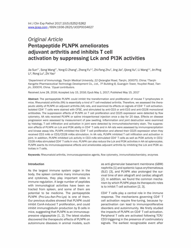
The Src PTK family Lck is essential for T cell development and T cell receptor (TCR) sig- nalling After activated by anti-CD3 antibody Lck activity is much higher than that of unsti- mulated cells PLNPK (16 08 04 and 02 mgml) reduced the Lck activity of CD3 mAb stimulated human CD4+ T cells and there was significant difference versus control group Plt005 (Figure 4A)
During AA pathogenesis Hsp65 protein can activate specific T cell clones causing immune response to the synovial membrane It is report-ed that Lck inhibitors can inhibit the abnormal activation of T cells in AA animals and reduce the early symptoms [20] Our results suggested that Lck activity in AA rat splenocytes was high-er that that in normal rats PLNPK in 200 μgkgd and 100 μgkgd dose was able to sig-nificantly inhibit Lck activity compared with control group Plt005 (Figure 4B)
PLNPK inhibited PI3K activity
P13K aggregation and activation play a key role in CD28 signal pathway When CD28 combined
Figure 4 Effect of PLNPK on Lck activity of CD4+ T lymphocytes and AA ratsrsquo plenocytes A Human CD4+ T cells were added in 24-well plate with titrating doses of PLNPK Cells were stimulated with 25 μgml CD3 mAb except for that in unstimulated control wells After 48 hours Lck activity was detected by ELISA B AA model were established by FCA immunization and drugs were adminsistrated continuously for 20 days from the senventh day after immuniza-tion Spleen Lck protein was purified by immunoprecipitation ELISA method were used to assay Lck activity Data are expressed as mean plusmn SD versus stimulated control group Plt005 N=5
PLNPK ameliorates arthritis and inhibits T cell
5259 Int J Clin Exp Pathol 201710(5)5252-5262
with and its ligand Tyr173 in cytoplasmic domain of CD28 is phosphorylated ensue on P13K activation and ultimately induce AP-1 and NF-AT transcription [21] In order to observe the effect of PLNPK on P13K CD3CD28 mAb were used to stimulate CD4+ T cell activation PI3K activity was detected by ELISA The results discovered that CD3CD28 mAb (25 μgml1 μgml) increased CD4+ T cells PI3K activity and all the dosages of PLNPK (16 08 04 and 02 mgml) inhibited CD3CD28 mAbs induced human CD4+ T cells PI3K activity there was sig-nificant difference versus control group Plt005 (Figure 5A)
In AA animals PI3K pathway is involved in the pathologic processes studies have shown that application of specific PI3K inhibitor wortman-nin or LY294002 can inhibit T cell secretion of TNF and IL-10 in AA and inhibit the activation of macrophage induced by T cells directly [22 23] Our results show that PLNPK in 200 μgkgd and 100 μgkgd dose was able to signifi-cantly inhibit AA rat splenocytes PI3K activity compared with control group Plt005 Inhibition rates were 6174 and 5944 respectively (Figure 5B)
Discussion
Activation of T cells is a central process in immune response so inhibiting T cell activation is an important target for the development of immunosuppressive agents There has been found that certain oligopeptides and polypep-
tides can simulate or block specific protein-pro-tein interactions of T cells The mechanisms of those inhibitory peptides include two aspects inhibition of the first signal and inhibition of the costimulatory signal of T cell activation The HLA-DQA1 peptide is derived from human MHC II molecules α chain it can interfere with the interaction between TCR and MHC and inhibit cytotoxic T cells generation [24-26] Another example is CD80-CAP1 (MQPPGC) it can block combination of CD8086 and CD28152 and is effective to prevent and suppress autoim-mune disease symptoms of collagen-induced arthritis in mice (CIA) inflammatory bowel dis-ease (IBD) experimental allergic encephalomy-elitis (EAE) and trinitrobenzene sulfonic acid (TNBS) colitis [26-30]
The spleen contain a variety of biologically active substances PLNPK is a kind of immuno-suppressive peptide screened from spleen This study showed that PLNPK could signifi-cantly inhibit CD3 mAb and CD3CD28 mAbs induced CD4+ T cells proliferation and activa-tion CD4+ T cells play an important role in assisting B cells to produce immunoglobulin supporting NK cell and CD8+ Tc cells activation and secretion The effect of PLNPK on CD4+ T cells suggests that it is a potential immunosup-pressive agent
In vitro CD3 mAb triggered the first signal transduction pathway of T cell activation At first TCRCD3CD45CD4 rapidly aggregate then CD45 exerts protein tyrosine phospha-
Figure 5 Effect of PLNPK on PI3K activity of CD4+ T lymphocytes and AA ratsrsquo plenocytes A Human CD4+ T cells were added in 24-well plate with titrating doses of PLNPK Cells were stimulated with 25 μgml CD3 mAb except for that in unstimulated control wells After 48 hours PI3K activity of CD4+ T cells was detected by ELISA B AA model was established by FCA immunization and drugs were adminsistrated continuously for 20 days from the seventh day after immunization Spleen PI3K protein was purified by immunoprecipitation ELISA method was used for Lck activity assay Data are expressed as mean plusmn SD versus stimulated control group Plt005 N=5
PLNPK ameliorates arthritis and inhibits T cell
5260 Int J Clin Exp Pathol 201710(5)5252-5262
tase activity resulting Src PTK phosphorylation followed by activation of ZAP-70 LAT [4 31] The ability of PLNPK to inhibit Lck protein tyro-sine kinase activity is likely to lead to inhibit the first signal transduction pathway of T cell activation The expression of CD25 can be used as a sign of T cell activation Inhibition of Lck activity may be the mechanism of PLNPK treatment reduced CD3 mAb stimulated CD25 expression
CD28 mediates a costimulatory signal that cooperates with TCRCD3 activation to pro-mote T cell viability clonal Expansion and cyto-kine production Ligation of CD28 by Abs or its natural ligands B7-1 and B7-2 results in tyrosine phosphorylation at Y173MNM motif within its cytoplasmic tail The phosphorylated YMNM motif subsequently interacts with the Src homology 2 domain within the p85 regu- latory subunit of PI3K activating the p110 catalytic subunit Activated PI3K generates in- tracellular PI [3 4] P2 and PI [3-5] P3 thereby recruiting numerous proteins to plasma mem-brane by pleckstrin homology (PH) domains in- cluding AKT and PDK-1 Akt upon subsequent phosphorylation at Thr-308 and Ser-473 by PDK-1 becomes activated and plays a critical role in cell survival and cell cycle regulation through a wide variety of downstream mole-cules [9 10 32 33] PLNPK significantly inhi- bit CD3CD28 mAb induced PI3K activity indi-cating another mechanism of PLNPK function
Rheumatoid arthritis is essentially a T cell-mediated arthritis Abnormal T-cell repository is the internal factor of RA pathogenesis There are a lot of self-reactive CD4+ T cells control the occurrence and development of RA th- rough direct contact or secretion [34] Experi- ments confirmed that direct specific T cell clones injection into normal animals can lead to destructive arthritis [35] In this study rat AA model were established to observe the immunosuppressive activity of PLNPK in vivo During AA pathogenesis Rheumatoid-associ- ated antigens were presented through HLA and combined with the TCRCD3 of T cells In the presence of costimulatory signals the T cells were abnormally activated Those T cells were recruited into the joints resulting in the occurrence of joints inflammation [36 37] PLNPK can inhibit Lck and PI3K activity of T cells in AA rat naturally inhibits the T cell
downstream signal transduction Thereby PL- NPK can reduce the T cell-mediated inflam- matory response improve the ankle patholo- gical damage
Acknowledgements
This work was supported by grants from the National Major Scientific and Technological Special Project for ldquoSignificant New Drugs De- velopmentrdquo (grant number 2014ZX0910100- 5004) Tianjin application basic and cutting-edge technology research programs Youth Fund (grant number 12JCQNJC08400)
Disclosure of conflict of interest
None
Address correspondence to Dr Zhi Yao Depart- ment of Immunology Tianjin Medical University 22 Qixiangtai Road Tianjin 300070 China Tel +86 22 83336667 Fax +86 22 60368186 E-mail yaozhitijmueducn Rong Lu Tianjin Kangzhe Pharmaceutical Technology Development Co Ltd 7F Building B Guangyin Tower Youyibei Road Tianjin 300074 China Tel +86 22 603- 68186 Fax +86 22 60368186 E-mail lu_rongvipsinacom
References
[1] Zhou CL Lv JQ Lu R Chen LJ Li HQ Cao HL Li QL Wang S Fu Z Yao Z A new pentapeptide compound PLNPK ameliorates anti-glomeru-lar basement membrane nephritis in Wistar rats Peptides 2008 29 1789-97
[2] Wang L Wang S Lu R Lv J Zhou C Fu Z Xu Q Che X Jia J Zhao H Li X Lin G Yao Z The new immunosuppressant PLNPK prolongs allograft survival in mice Transpl Immunol 2010 24 64-8
[3] Lv JQ Zhang W Wang S Zhao L Ma R Hu JW Wang LJ Meng J Zhou CL Lin G Lu R Yao Z The pentapeptide PLNPK inhibits systemic lu-pus erythematosus-associated renal damage Inflamm Res 2010 591081-9
[4] Nel AE T-cell activation through the antigen receptor Part 1 signaling components signal-ing pathways and signal integration at the T-cell antigen receptor synapse J Allergy Clin Immunol 2002 109 758-70
[5] Merino E Avila-Flores A Shirai Y Moraga I Saito N Meacuterida I Lck-dependent tyrosine phosphorylation of diacylglycerol kinase alpha regulates its membrane association in T cells J Immunol 2008 180 5805-15
[6] Lovatt M Filby A Parravicini V Werlen G Palmer E Zamoyska R Lck regulates the
PLNPK ameliorates arthritis and inhibits T cell
5261 Int J Clin Exp Pathol 201710(5)5252-5262
threshold of activation in primary T cells while both Lck and Fyn contribute to the magnitude of the extracellular signal-related kinase re-sponse Mol Cell Biol 2006 26 8655-65
[7] Sugie K Jeon MS Grey HM Activation of naiumlve CD4 T cells by anti-CD3 reveals an important role for Fyn in Lck-mediated signaling Proc Natl Acad Sci U S A 2004 101 14859-64
[8] Jiang Y Cheng H Evidence of LAT as a dual substrate for Lck and Syk in T lymphocytes Leuk Res 2007 31 541-5
[9] Appleman LJ van Puijenbroek AA Shu KM Nadler LM Boussiotis VA CD28 costimulation mediates down-regulation of p27kip1 and cell cycle progression by activation of the PI3KPKB signaling pathway in primary human T cells J Immunol 2002 168 2729-36
[10] Arimura Y Shiroki F Kuwahara S Kato H Dianzani U Uchiyama T Yagi J Akt is a neutral amplifier for Th cell differentiation J Biol Chem 2004 279 11408-16
[11] Lee WW Yang ZZ Li G Weyand CM Goronzy JJ Unchecked CD70 expression on T cells lowers threshold for T cell activation in rheumatoid ar-thritis J Immunol 2007 179 2609-15
[12] Vincenti F Costimulation blockade in auto- immunity and transplantation J Allergy Clin Immunol 2008 12 299-306
[13] Panayi GS Targeting of cells involved in the pathogenesis of rheumatoid arthritis Rheu- matology (Oxford) 1999 38 Suppl 2 8-10
[14] Zhang X Nakajima T Goronzy JJ Weyand CM Tissue trafficking patterns of effector memory CD4+ T cells in rheumatoid arthritis Arthritis Rheum 2005 52 3839-49
[15] Bryl E Vallejo AN Matteson EL Witkowski JM Weyand CM Goronzy JJ Modulation of CD28 expression with anti-tumor necrosis factor al-pha therapy in rheumatoid arthritis Arthritis Rheum 2005 52 2996-3003
[16] Rice JW Veal JM Fadden RP Barabasz AF Partridge JM Barta TE Dubois LG Huang KH Mabbett SR Silinski MA Steed PM Hall SE Small molecule inhibitors of Hsp90 potently af-fect inflammatory disease pathways and ex-hibit activity in models of rheumatoid arthritis Arthritis Rheum 2008 58 3765-75
[17] Holmdahl R Lorentzen JC Lu S Olofsson P Wester L Holmberg J Pettersson U Arthritis induced in rats with nonimmunogenic adju-vants as models for rheumatoid arthritis Immunol Rev 2001 184 184-202
[18] Haas CS Amin MA Ruth JH Allen BL Ahmed S Pakozdi A Woods JM Shahrara S Koch AE In vivo inhibition of angiogenesis by interleu-kin-13 gene therapy in a rat model of rheuma-toid arthritis Arthritis Rheum 2007 56 2535-48
[19] Laragione T Gulko PS mTOR regulates the in-vasive properties of synovial fibroblasts in rheumatoid arthritis Mol Med 2010 16 352-8
[20] Satpute SR Rajaiah R Polumuri SK Moudgil KD Tolerization with Hsp65 induces protect- ion against adjuvant-induced arthritis by mod-ulating the antigen-directed interferon-gam-ma interleukin-17 and antibody responses Arthritis Rheum 2009 60 103-13
[21] Watanabe R Harada Y Takeda K Takahashi J Ohnuki K Ogawa S Ohgai D Kaibara N Koiwai O Tanabe K Toma H Sugamura K Abe R Grb2 and Gads exhibit different interactions with CD28 and play distinct roles in CD28-mediated costimulation J Immunol 2006 177 1085-91
[22] Shripad S Bhagwat Kinase inhibitors for the treatment of inflammatory and autoimmune disorders Purinergic Signal 2009 5 107-15
[23] Camps M Ruumlckle T Ji H Ardissone V Rintelen F Shaw J Ferrandi C Chabert C Gillieron C Franccedilon B Martin T Gretener D Perrin D Leroy D Vitte PA Hirsch E Wymann MP Cirillo R Schwarz MK Rommel C Blockade of PI3Kgamma suppresses joint inflammation and damage in mouse models of rheumatoid arthritis Nat Med 2005 11 936-43
[24] Zang W Lin M Kalache S Zhang N Kruumlger B Waaga-Gasser AM Grimm M Hancock W Heeger P Schroumlppel B Murphy B Inhibition of the alloimmune response through the genera-tion of regulatory T cells by a MHC class II-derived peptide Immunol 2008 181 7499-506
[25] Zang W Murphy B Peptide-mediated immuno-suppression Am J Ther 2005 12 592-9
[26] Zhou CL Lu R Lin G Yao Z The latest develop-ments in synthetic peptides with immunoregu-latory activities Peptides 2011 32 408-14
[27] Srinivasan M Lu D Eri R Brand DD Haque A Blum JS CD80 binding polyproline helical peptide inhibits T cell activation J Biol Chem 2005 280 10149-55
[28] Srinivasan M Eri R Zunt SL Summerlin DJ Brand DD Blum JS Suppression of immune responses in collagen-induced arthritis by a rationally designed CD80-binding peptide agent Arthritis Rheum 2007 56 498-508
[29] Eri R Kodumudi KN Summerlin DJ Srinivasan M Suppression of colon inflammation by CD80 blockade evaluation in two murine models of inflammatory bowel disease Inflamm Bowel Dis 2008 14 458-70
[30] Dudhgaonkar SP Janardhanam SB Kodumudi KN Srinivasan M CD80 blockade enhance glucocorticoid-induced leucine zipper expres-sion and suppress experimental autoimmune encephalomyelitis J Immunol 2009 183 7505-13
PLNPK ameliorates arthritis and inhibits T cell
5262 Int J Clin Exp Pathol 201710(5)5252-5262
[31] Smith-Garvin JE Koretzky GA Jordan MS T cell activation Annu Rev Immunol 2009 27 591-619
[32] Gigoux M Shang J Pak Y Xu M Choe J Mak TW Suh WK Inducible costimulator promotes helper T-cell differentiation through phos-phoinositide 3-kinase Proc Natl Acad Sci U S A 2009 106 20371-6
[33] Alcaacutezar I Corteacutes I Zaballos A Hernandez C Fruman DA Barber DF Carrera AC p85beta phosphoinositide 3-kinase regulates CD28 co-receptor function Blood 2009 113 3198-208
[34] Sanchez-Lockhart M Graf B Miller J Signals and sequences that control CD28 localization to the central region of the immunological syn-apse J Immunol 2008 181 7639-48
[35] Romagnoli P Strahan D Pelosi M Cantagrel A van Meerwijk JP A potential role for protein ty-rosine kinase p56Lck in rheumatoid arthritis synovial fluid T lymphocyte hyporesponsive-ness Int Immunol 2001 13 305-12
[36] Beech JT Andreakos E Ciesielski CJ Green P Foxwell BM Brennan FM T-cell contact-depen-dent regulation of CC and CXC chemokine pro-duction in monocytes through differential in-volvement of NFkappaB implications for rheu-matoid arthritis Arthritis Res Ther 2006 8 R168
[37] Sobek V Birkner N Falk I Wuumlrch A Kirschning CJ Wagner H Wallich R Lamers MC Simon MM Direct Toll-like receptor 2 mediated co-stimulation of T cells in the mouse system as a basis for chronic inflammatory joint disease Arthritis Res Ther 2004 6 R433-46

PLNPK ameliorates arthritis and inhibits T cell
5253 Int J Clin Exp Pathol 201710(5)5252-5262
T cell receptor (TCR) engagement by antigen is the activation of protein tyrosine kinases (PTK) of Src family [4 5] The Src kinase family member p56lck (Lck) is the major PTK dur- ing TCR triggering Lck is mainly responsible for the activation of phospholipase Cγ (PLCγ) PLCγ initiates the subsequent Ca2+calmodu-lincecalcineurinNFAT pathway and PKCNF- κB pathway [5] In addition Lck is involved in ERKMAPK signal activation [6-8] Therefore Lck is critical for TCR triggered T-cell activa- tion In this study we observed the inhibitory effect of PLNPK on Lck in T cells
In addition to TCRCD3 signal complete acti- vation of T cells also needs the second sig- nals provided by pairs of costimulatory mole-cules the most important one of which is B7CD28 CD28 binding to its ligand B7 results in tyrosine phosphorylation of Y173MNM motif The phosphorylated YMNM motif subsequent- ly interacts with the SH2 domain within pho- sphoinositide 3-kinases (PI3K) regulatory sub-unit p85 activating the p110 catalytic sub- unit [9] The p110 generate PIP3 that recruit a variety of proteins to plasma membrane among which is phosphatidylinositol-depen-dent kinase-1 (PDK-1) PDK-1 activates AKT and AKT can significantly promote the T cell activation and proliferation cytokine secretion inhibits Fas-mediated T cell apoptosis [10- 12] We also studied the effect of PLNPK on PI3K activity in T cell
Rheumatoid arthritis (RA) is essentially a kind of T cell-mediated arthritis RA disease origi-nated in T cell activation [13] A lot of self-reac-tive CD4+ T cells were detected in joints of RA patients these abnormal T cells interacts with other pathogenic cells through direct contact or secretion of causative agents [14-16] We established rat adjuvant arthritis (AA) model observed the effect of PLNPK treatment on autoimmune diseases AA and studied the effect of PLNPK on T cell activation in vivo
Materials and methods
Drug and reagents
PLNPK was obtained from Shenzhen Kangzhe Pharmaceutical Co Ltd Anti-human CD3 mAb anti-human CD28 mAb anti-human CD4-PE antibody and anti-human CD25-APC antibody were purchased from BD-PharMingen compa-
ny Carboxyfluorescein diacetate succinimidyl ester (CFSE) and Freundrsquos complete adjuvant were purchased from Sigma-Aldrich Corpor- ation Anti-Lck mAb and anti-P85 mAb was pur-chased from Upstate Company PTK kinase assay kit was purchased from Sigma-Aldrich Corporation PI3K kinase assay kit was pur-chased from Echelon Biosiences company anti-rat CD3 mAb and anti-rat CD25 mAb were purchased from Serotec Company
Animals
Male Wistar rats (SPF grade 180 plusmn 20 g) were provided by Experimental Animal Center of Military Medical Sciences They were housed in an environment with a controlled 1212 lightdark cycle and constant temperature of 21-25degC with a humidity of 55 plusmn 5 All pro- cedures carried out according to NIHrsquos SOP Practice
Ethics statement
Venous blood samples were collected from health volunteers with informed consent and with the approval of local ethics committee and Health Bureau Animal experiment in this study was approved by Ethics Committee of Tianjin Medical University
Isolation of primary peripheral blood CD4+ T cells
The peripheral blood mononuclear cells (PBMC) were isolated by Ficoll-Hypaque density gra- dient (1077 gml) centrifugation at 2000 rpm for 20 minutes After washing 3 times CD4+ T cells were negatively selected by an antibody cocktail according to the manufacturersquos in- structions (Miltenyi Company) A Cell purity of above 90 was achieved by flow cytometry detection
CD4+ T lymphocyte proliferation
CD4+ T cells were adjusted to 1times106ml incu-bating with carboxyfluorescein succinimidyl ester (CFSE 5 mmolml) in 37degC for 15 min-utes for CFSE labeling Finally the reaction was stopped by ice bath for 5 minutes After wash-ing for 3 times CD4+ T cells were resuspended in RPMI-164010 FBS and plated in 96-well plate at 4times105well along with titrating doses of PLNPK at a final concentration of 16 mgml
PLNPK ameliorates arthritis and inhibits T cell
5254 Int J Clin Exp Pathol 201710(5)5252-5262
08 mgml 04 mgml 02 mgml or media control Except for unstimulated control group cells were stimulated with 25 μgml CD3 mAb alone or CD3 mAb (25 μgml) and CD28 mAb (1 μgml) both The plate was placed in 37degC 5 CO2 incubator for 48 hours Data acquisition was processed by CELLQuest soft-ware with flow cytometry (BD-PharMingen Cor- poration) and the proliferation index was ob- tained by Modfit software
CD25 expression in T lymphocytes
In this study 2times106 purified human CD4+ T cells were plated in 24-well plate with the same final concentrations of PLNPK as above Cells also received the same stimulation as above and the unstimulated control wells received no stimulation The plate was placed in 37degC 5 CO2 incubator for 48 hours After incubation the cells were harvested and stained with anti-human CD4-PE and CD25-APC antibodies for evaluation of T cell activation Data acquisition was processed by CELLQuest software with flow cytometry (BD-PharMingen Corporation)
AA model and drug treatment
AA model was established by intradermal injec-tion of 01 ml FCA in the right hind paw of each rat After 7 days the rats were randomly divid-ed into four groups different doses of PLNPK 200 μgkgd 100 μgkgd) model control group and healthy control group there ten ani-mals in each group The 2 PLNPK groups received 1 ml ip injection of 200 μgkgd and 100 μgkgd PLNPK respectively once a day for 20 days at the same time the two control group were treated with 1 ml saline
Histological examination
After the last administration all of the AA rats were sacrificed and each right ankle was removed washed with saline and fixed in 10 neutral formalin After 15 EDTA (2Na+) decal-cification conventional dehydration and paraf-fin embedding sliced the joints were stained by haematoxylin-eosin (HE) for light microscope examination In addition some paraffin sec-tions were microwave repaired and then were incubated with anti-CD3 mAb or anti-CD25 mAb working solution at 4degC overnight On the next day HRP labeled goat anti-mouse IgG and DAB were used to reveal the reaction Positive cells were with brown-yellow or yellow granular material deposits to the cytoplasm
Lck activity detection
For in vitro study 2times106 purified human CD4+ T cells were added in 24-well plate with the same PLNPK treatment as above Cells were stimu-lated with 25 μgml CD3 mAb except for that in unstimulated control wells The plate was placed in 37degC 5 CO2 incubator for 48 hours Total protein was extracted by RIPA lysis buffer and was quantified to a final concentration of 5 μgμl Lck samples were prepared by immuno-precipitation as follows 1 mg total protein was incubated with 3 μl Lck antibody for one hour at 4degC and then incubated with 60 μl 50 Protein A bead at 4degC for one hour The solution was centrifuged at 14000 g for 5 seconds and the supernatant was discarded After washing the samples could be used for the evaluation of Lck kinase activity PTK kinase assay kit was used to detect the Lck activity
In order to observe the effect of PLNPK on Lck activity in vivo total proteins were extracted from spleen tissue of AA rats in each group Lck samples were prepared by immunoprecipita-tion and PTK kinase assay kit was used to detect the Lck kinase activity
PI3K activity detection
2times106 purified human CD4+ T cells were plated in 24 well plates with the same PLNPK treat-ment as above Cells were stimulated with 25 μgml CD3 mAb and 1 μgml CD28 mAb in stimulation wells and unstimulated control received no stimulation The plate was placed in 37degC 5 CO2 incubator for 48 hours After that the samples of p85 subunit were prepared by immunoprecipitation as above and then the activity was examined with a PI3K kinase assay kit The PI3K activity in splenocytes of AA rats was also detected
Statistical analyses
Data were expressed as group means plusmn stan-dard deviation and analyzed with one-way anal-ysis of variance (ANOVA) Plt005 was consid-ered to be statistically significant
Results
PLNPK inhibited the proliferation of CD4+ T cells
In order to observe the effect of PLNPK on CD4+ T cells proliferation we used CD3 mAb and
PLNPK ameliorates arthritis and inhibits T cell
5255 Int J Clin Exp Pathol 201710(5)5252-5262
Figure 1 Effect of PLNPK on McAb induced CD4+ T cells proliferation Human CD4+ T cells were labeled with CFSE and plated in 96-well plate with titrating doses of PLNPK and were simulated with CD3 McAb (A B) or CD3CD28 McAb (A C) After cultured for 48 hrs cell proliferation was detected by flow cytometry Data are expressed as mean plusmn SD versus stimulated control group Plt005 N=3
PLNPK ameliorates arthritis and inhibits T cell
5256 Int J Clin Exp Pathol 201710(5)5252-5262
Figure 2 Effect of PLNPK on CD4+ T cells CD25 expression induced by antibodies Human CD4+ T cells were plated in 96-well plate with titrating doses of PLNPK and were simulated with CD3 McAb (A B) or CD3CD28 McAb (A C) After cultured for 48 hrs cells were stained with anti-human CD4-PE and CD25-APC antibodies and were detected by flow cytometry method Data are expressed as mean plusmn SD versus stimulated control group Plt005 N=5
PLNPK ameliorates arthritis and inhibits T cell
5257 Int J Clin Exp Pathol 201710(5)5252-5262
CD3CD28 mAbs to stimulate CD4+ T cells PLNPK concentrations were 16 mgml 08 mgml 04 mgml 02 mgml The results showed that the proliferation kinetics as shown by CFSE dilution profiles was strikingly differ-ent between stimulated control and unstimu-lated control suggesting that CD3 mAb (25 μgml) and CD3CD28 mAbs (25 μgml1 μgml) can induce CD4+ T cells proliferation As expected anti-CD3 plus anti-CD28 costimula-tion activated more T cells All the doses of PLNPK inhibited both CD3 mAb-induced and CD3CD28 mAb-induced CD4+ T cell prolifera-tion in a dose dependent manner there was significant difference versus the stimulated control group Plt005 (Figure 1)
PLNPK reduced the activation of CD4 + T cells
The CD25 expression is a critical step in T cell activation When the T cells are activated they produce the α chain of IL-2 receptor (CD25) which contributes to IL-2 binding CD25 is also an important marker of activated T cells So we detected CD25 expression to evaluate the effect of PLNPK on CD4 + T cell activation The positive rates of CD25 in stimulated cont- rol groups is much higher (471 plusmn 66 for anti-CD3 stimulated control and 519 plusmn 71 for anti-CD3CD28 stimulated control) than that of unstimulated group (less than 10) PLNPK (16 08 04 and 02 mgml) signifi-cantly inhibited the expression of CD25 both in
Figure 3 Effect of PLNPK on the ankle jointsrsquo pathology and T cell infiltration of AA rats AA model was induced by intradermal injection of FCA in right hind footpads of rats PLNPK were administrated continuously for 20 days from the senventh day after immunization The histological change of ankle joint were examined under light micro-scope after HE stain and immunological histochemistry stain A Saline control (times100) synoviocytes accrementition on cartilage and pannus Mononuclear cells infiltrate severely B PLNPK (200 μgkgd) (times100) group cartilage destruction and synoviocytes accrementition were relieved Mononuclear cells infiltration was lessen C PLNPK (100 μgkgd) (times100) group cartilage destruction and synoviocytes accrementition were relieved Mononuclear cells infiltration was lessen D Saline control (times100) CD3+ T cells infiltrated to synovial tissue severely E PLNPK (200 μgkgd) (times100) group CD3+ T cells infiltration was lessen F PLNPK (100 μgkgd) (times100) group CD3+ T cells infiltration was lessen G Saline control (times100) group CD25+ T cells infiltrated to synovial tissue severely H PLNPK (200 μgkgd) (times100) group CD25+ T cells infiltration was lessen I PLNPK (100 μgkgd) (times100) group CD25+ T cells infiltration was lessen
PLNPK ameliorates arthritis and inhibits T cell
5258 Int J Clin Exp Pathol 201710(5)5252-5262
CD3 stimulated groups and in CD3CD28 stim-ulated groups in a dose dependent manner Plt005 versus stimulated control (Figure 2)
PLNPK reduced AA pathological damage and inhibited joint T lymphocytes infiltration
AA and RA have similar pathological chang- es including synovial hyperplasia mononu- clear cell infiltration cartilage and bone de- struction There are excessive growth of vari-ous cells in the synovial tissue including T cells and polyclonal B cell macrophage-like synovial cells monocytes and so on which produce monokines and chemokines to sti- mulate synovial cell proliferation and polymor-phonuclear leukocytes driven into the joints resulting in proliferation of synovial tissue pannus formation cartilage and bone destru- ction [17 18] In our study the right ankle joints of model control AA rats showed syno- vial cell proliferation synovial thickening pan-nus formation and a large number of inflam- matory cell infiltration cartilage damage was serious AA rats in PLNPK treatment groups exhibited ameliorated joint pathological ch- anges of different degrees for instance mild synovial hyperplasia cartilage erosion were worm-like changes infiltration of inflammatory cells was decreased (Figure 3A-C) T cells are the major AA synovial infiltrating cell subsets it even can induce germinal centers formation in synovial structure inflammatory cytokines excretion synovial tissue proliferation B cell activation and Ig secretion eventually leading to synovial membrane injury [19] CD25 is a
surface marker of T cell activation and CD25+ synovial T cells significantly increased in RA patients Our results revealed that a large num-ber of CD3+ T cells CD25+ T cells infiltrated in synovial tissue in the model control group as well as in PLNPK treatment groups there is no significantly deferent among these groups
PLNPK inhibited Lck activity
The Src PTK family Lck is essential for T cell development and T cell receptor (TCR) sig- nalling After activated by anti-CD3 antibody Lck activity is much higher than that of unsti- mulated cells PLNPK (16 08 04 and 02 mgml) reduced the Lck activity of CD3 mAb stimulated human CD4+ T cells and there was significant difference versus control group Plt005 (Figure 4A)
During AA pathogenesis Hsp65 protein can activate specific T cell clones causing immune response to the synovial membrane It is report-ed that Lck inhibitors can inhibit the abnormal activation of T cells in AA animals and reduce the early symptoms [20] Our results suggested that Lck activity in AA rat splenocytes was high-er that that in normal rats PLNPK in 200 μgkgd and 100 μgkgd dose was able to sig-nificantly inhibit Lck activity compared with control group Plt005 (Figure 4B)
PLNPK inhibited PI3K activity
P13K aggregation and activation play a key role in CD28 signal pathway When CD28 combined
Figure 4 Effect of PLNPK on Lck activity of CD4+ T lymphocytes and AA ratsrsquo plenocytes A Human CD4+ T cells were added in 24-well plate with titrating doses of PLNPK Cells were stimulated with 25 μgml CD3 mAb except for that in unstimulated control wells After 48 hours Lck activity was detected by ELISA B AA model were established by FCA immunization and drugs were adminsistrated continuously for 20 days from the senventh day after immuniza-tion Spleen Lck protein was purified by immunoprecipitation ELISA method were used to assay Lck activity Data are expressed as mean plusmn SD versus stimulated control group Plt005 N=5
PLNPK ameliorates arthritis and inhibits T cell
5259 Int J Clin Exp Pathol 201710(5)5252-5262
with and its ligand Tyr173 in cytoplasmic domain of CD28 is phosphorylated ensue on P13K activation and ultimately induce AP-1 and NF-AT transcription [21] In order to observe the effect of PLNPK on P13K CD3CD28 mAb were used to stimulate CD4+ T cell activation PI3K activity was detected by ELISA The results discovered that CD3CD28 mAb (25 μgml1 μgml) increased CD4+ T cells PI3K activity and all the dosages of PLNPK (16 08 04 and 02 mgml) inhibited CD3CD28 mAbs induced human CD4+ T cells PI3K activity there was sig-nificant difference versus control group Plt005 (Figure 5A)
In AA animals PI3K pathway is involved in the pathologic processes studies have shown that application of specific PI3K inhibitor wortman-nin or LY294002 can inhibit T cell secretion of TNF and IL-10 in AA and inhibit the activation of macrophage induced by T cells directly [22 23] Our results show that PLNPK in 200 μgkgd and 100 μgkgd dose was able to signifi-cantly inhibit AA rat splenocytes PI3K activity compared with control group Plt005 Inhibition rates were 6174 and 5944 respectively (Figure 5B)
Discussion
Activation of T cells is a central process in immune response so inhibiting T cell activation is an important target for the development of immunosuppressive agents There has been found that certain oligopeptides and polypep-
tides can simulate or block specific protein-pro-tein interactions of T cells The mechanisms of those inhibitory peptides include two aspects inhibition of the first signal and inhibition of the costimulatory signal of T cell activation The HLA-DQA1 peptide is derived from human MHC II molecules α chain it can interfere with the interaction between TCR and MHC and inhibit cytotoxic T cells generation [24-26] Another example is CD80-CAP1 (MQPPGC) it can block combination of CD8086 and CD28152 and is effective to prevent and suppress autoim-mune disease symptoms of collagen-induced arthritis in mice (CIA) inflammatory bowel dis-ease (IBD) experimental allergic encephalomy-elitis (EAE) and trinitrobenzene sulfonic acid (TNBS) colitis [26-30]
The spleen contain a variety of biologically active substances PLNPK is a kind of immuno-suppressive peptide screened from spleen This study showed that PLNPK could signifi-cantly inhibit CD3 mAb and CD3CD28 mAbs induced CD4+ T cells proliferation and activa-tion CD4+ T cells play an important role in assisting B cells to produce immunoglobulin supporting NK cell and CD8+ Tc cells activation and secretion The effect of PLNPK on CD4+ T cells suggests that it is a potential immunosup-pressive agent
In vitro CD3 mAb triggered the first signal transduction pathway of T cell activation At first TCRCD3CD45CD4 rapidly aggregate then CD45 exerts protein tyrosine phospha-
Figure 5 Effect of PLNPK on PI3K activity of CD4+ T lymphocytes and AA ratsrsquo plenocytes A Human CD4+ T cells were added in 24-well plate with titrating doses of PLNPK Cells were stimulated with 25 μgml CD3 mAb except for that in unstimulated control wells After 48 hours PI3K activity of CD4+ T cells was detected by ELISA B AA model was established by FCA immunization and drugs were adminsistrated continuously for 20 days from the seventh day after immunization Spleen PI3K protein was purified by immunoprecipitation ELISA method was used for Lck activity assay Data are expressed as mean plusmn SD versus stimulated control group Plt005 N=5
PLNPK ameliorates arthritis and inhibits T cell
5260 Int J Clin Exp Pathol 201710(5)5252-5262
tase activity resulting Src PTK phosphorylation followed by activation of ZAP-70 LAT [4 31] The ability of PLNPK to inhibit Lck protein tyro-sine kinase activity is likely to lead to inhibit the first signal transduction pathway of T cell activation The expression of CD25 can be used as a sign of T cell activation Inhibition of Lck activity may be the mechanism of PLNPK treatment reduced CD3 mAb stimulated CD25 expression
CD28 mediates a costimulatory signal that cooperates with TCRCD3 activation to pro-mote T cell viability clonal Expansion and cyto-kine production Ligation of CD28 by Abs or its natural ligands B7-1 and B7-2 results in tyrosine phosphorylation at Y173MNM motif within its cytoplasmic tail The phosphorylated YMNM motif subsequently interacts with the Src homology 2 domain within the p85 regu- latory subunit of PI3K activating the p110 catalytic subunit Activated PI3K generates in- tracellular PI [3 4] P2 and PI [3-5] P3 thereby recruiting numerous proteins to plasma mem-brane by pleckstrin homology (PH) domains in- cluding AKT and PDK-1 Akt upon subsequent phosphorylation at Thr-308 and Ser-473 by PDK-1 becomes activated and plays a critical role in cell survival and cell cycle regulation through a wide variety of downstream mole-cules [9 10 32 33] PLNPK significantly inhi- bit CD3CD28 mAb induced PI3K activity indi-cating another mechanism of PLNPK function
Rheumatoid arthritis is essentially a T cell-mediated arthritis Abnormal T-cell repository is the internal factor of RA pathogenesis There are a lot of self-reactive CD4+ T cells control the occurrence and development of RA th- rough direct contact or secretion [34] Experi- ments confirmed that direct specific T cell clones injection into normal animals can lead to destructive arthritis [35] In this study rat AA model were established to observe the immunosuppressive activity of PLNPK in vivo During AA pathogenesis Rheumatoid-associ- ated antigens were presented through HLA and combined with the TCRCD3 of T cells In the presence of costimulatory signals the T cells were abnormally activated Those T cells were recruited into the joints resulting in the occurrence of joints inflammation [36 37] PLNPK can inhibit Lck and PI3K activity of T cells in AA rat naturally inhibits the T cell
downstream signal transduction Thereby PL- NPK can reduce the T cell-mediated inflam- matory response improve the ankle patholo- gical damage
Acknowledgements
This work was supported by grants from the National Major Scientific and Technological Special Project for ldquoSignificant New Drugs De- velopmentrdquo (grant number 2014ZX0910100- 5004) Tianjin application basic and cutting-edge technology research programs Youth Fund (grant number 12JCQNJC08400)
Disclosure of conflict of interest
None
Address correspondence to Dr Zhi Yao Depart- ment of Immunology Tianjin Medical University 22 Qixiangtai Road Tianjin 300070 China Tel +86 22 83336667 Fax +86 22 60368186 E-mail yaozhitijmueducn Rong Lu Tianjin Kangzhe Pharmaceutical Technology Development Co Ltd 7F Building B Guangyin Tower Youyibei Road Tianjin 300074 China Tel +86 22 603- 68186 Fax +86 22 60368186 E-mail lu_rongvipsinacom
References
[1] Zhou CL Lv JQ Lu R Chen LJ Li HQ Cao HL Li QL Wang S Fu Z Yao Z A new pentapeptide compound PLNPK ameliorates anti-glomeru-lar basement membrane nephritis in Wistar rats Peptides 2008 29 1789-97
[2] Wang L Wang S Lu R Lv J Zhou C Fu Z Xu Q Che X Jia J Zhao H Li X Lin G Yao Z The new immunosuppressant PLNPK prolongs allograft survival in mice Transpl Immunol 2010 24 64-8
[3] Lv JQ Zhang W Wang S Zhao L Ma R Hu JW Wang LJ Meng J Zhou CL Lin G Lu R Yao Z The pentapeptide PLNPK inhibits systemic lu-pus erythematosus-associated renal damage Inflamm Res 2010 591081-9
[4] Nel AE T-cell activation through the antigen receptor Part 1 signaling components signal-ing pathways and signal integration at the T-cell antigen receptor synapse J Allergy Clin Immunol 2002 109 758-70
[5] Merino E Avila-Flores A Shirai Y Moraga I Saito N Meacuterida I Lck-dependent tyrosine phosphorylation of diacylglycerol kinase alpha regulates its membrane association in T cells J Immunol 2008 180 5805-15
[6] Lovatt M Filby A Parravicini V Werlen G Palmer E Zamoyska R Lck regulates the
PLNPK ameliorates arthritis and inhibits T cell
5261 Int J Clin Exp Pathol 201710(5)5252-5262
threshold of activation in primary T cells while both Lck and Fyn contribute to the magnitude of the extracellular signal-related kinase re-sponse Mol Cell Biol 2006 26 8655-65
[7] Sugie K Jeon MS Grey HM Activation of naiumlve CD4 T cells by anti-CD3 reveals an important role for Fyn in Lck-mediated signaling Proc Natl Acad Sci U S A 2004 101 14859-64
[8] Jiang Y Cheng H Evidence of LAT as a dual substrate for Lck and Syk in T lymphocytes Leuk Res 2007 31 541-5
[9] Appleman LJ van Puijenbroek AA Shu KM Nadler LM Boussiotis VA CD28 costimulation mediates down-regulation of p27kip1 and cell cycle progression by activation of the PI3KPKB signaling pathway in primary human T cells J Immunol 2002 168 2729-36
[10] Arimura Y Shiroki F Kuwahara S Kato H Dianzani U Uchiyama T Yagi J Akt is a neutral amplifier for Th cell differentiation J Biol Chem 2004 279 11408-16
[11] Lee WW Yang ZZ Li G Weyand CM Goronzy JJ Unchecked CD70 expression on T cells lowers threshold for T cell activation in rheumatoid ar-thritis J Immunol 2007 179 2609-15
[12] Vincenti F Costimulation blockade in auto- immunity and transplantation J Allergy Clin Immunol 2008 12 299-306
[13] Panayi GS Targeting of cells involved in the pathogenesis of rheumatoid arthritis Rheu- matology (Oxford) 1999 38 Suppl 2 8-10
[14] Zhang X Nakajima T Goronzy JJ Weyand CM Tissue trafficking patterns of effector memory CD4+ T cells in rheumatoid arthritis Arthritis Rheum 2005 52 3839-49
[15] Bryl E Vallejo AN Matteson EL Witkowski JM Weyand CM Goronzy JJ Modulation of CD28 expression with anti-tumor necrosis factor al-pha therapy in rheumatoid arthritis Arthritis Rheum 2005 52 2996-3003
[16] Rice JW Veal JM Fadden RP Barabasz AF Partridge JM Barta TE Dubois LG Huang KH Mabbett SR Silinski MA Steed PM Hall SE Small molecule inhibitors of Hsp90 potently af-fect inflammatory disease pathways and ex-hibit activity in models of rheumatoid arthritis Arthritis Rheum 2008 58 3765-75
[17] Holmdahl R Lorentzen JC Lu S Olofsson P Wester L Holmberg J Pettersson U Arthritis induced in rats with nonimmunogenic adju-vants as models for rheumatoid arthritis Immunol Rev 2001 184 184-202
[18] Haas CS Amin MA Ruth JH Allen BL Ahmed S Pakozdi A Woods JM Shahrara S Koch AE In vivo inhibition of angiogenesis by interleu-kin-13 gene therapy in a rat model of rheuma-toid arthritis Arthritis Rheum 2007 56 2535-48
[19] Laragione T Gulko PS mTOR regulates the in-vasive properties of synovial fibroblasts in rheumatoid arthritis Mol Med 2010 16 352-8
[20] Satpute SR Rajaiah R Polumuri SK Moudgil KD Tolerization with Hsp65 induces protect- ion against adjuvant-induced arthritis by mod-ulating the antigen-directed interferon-gam-ma interleukin-17 and antibody responses Arthritis Rheum 2009 60 103-13
[21] Watanabe R Harada Y Takeda K Takahashi J Ohnuki K Ogawa S Ohgai D Kaibara N Koiwai O Tanabe K Toma H Sugamura K Abe R Grb2 and Gads exhibit different interactions with CD28 and play distinct roles in CD28-mediated costimulation J Immunol 2006 177 1085-91
[22] Shripad S Bhagwat Kinase inhibitors for the treatment of inflammatory and autoimmune disorders Purinergic Signal 2009 5 107-15
[23] Camps M Ruumlckle T Ji H Ardissone V Rintelen F Shaw J Ferrandi C Chabert C Gillieron C Franccedilon B Martin T Gretener D Perrin D Leroy D Vitte PA Hirsch E Wymann MP Cirillo R Schwarz MK Rommel C Blockade of PI3Kgamma suppresses joint inflammation and damage in mouse models of rheumatoid arthritis Nat Med 2005 11 936-43
[24] Zang W Lin M Kalache S Zhang N Kruumlger B Waaga-Gasser AM Grimm M Hancock W Heeger P Schroumlppel B Murphy B Inhibition of the alloimmune response through the genera-tion of regulatory T cells by a MHC class II-derived peptide Immunol 2008 181 7499-506
[25] Zang W Murphy B Peptide-mediated immuno-suppression Am J Ther 2005 12 592-9
[26] Zhou CL Lu R Lin G Yao Z The latest develop-ments in synthetic peptides with immunoregu-latory activities Peptides 2011 32 408-14
[27] Srinivasan M Lu D Eri R Brand DD Haque A Blum JS CD80 binding polyproline helical peptide inhibits T cell activation J Biol Chem 2005 280 10149-55
[28] Srinivasan M Eri R Zunt SL Summerlin DJ Brand DD Blum JS Suppression of immune responses in collagen-induced arthritis by a rationally designed CD80-binding peptide agent Arthritis Rheum 2007 56 498-508
[29] Eri R Kodumudi KN Summerlin DJ Srinivasan M Suppression of colon inflammation by CD80 blockade evaluation in two murine models of inflammatory bowel disease Inflamm Bowel Dis 2008 14 458-70
[30] Dudhgaonkar SP Janardhanam SB Kodumudi KN Srinivasan M CD80 blockade enhance glucocorticoid-induced leucine zipper expres-sion and suppress experimental autoimmune encephalomyelitis J Immunol 2009 183 7505-13
PLNPK ameliorates arthritis and inhibits T cell
5262 Int J Clin Exp Pathol 201710(5)5252-5262
[31] Smith-Garvin JE Koretzky GA Jordan MS T cell activation Annu Rev Immunol 2009 27 591-619
[32] Gigoux M Shang J Pak Y Xu M Choe J Mak TW Suh WK Inducible costimulator promotes helper T-cell differentiation through phos-phoinositide 3-kinase Proc Natl Acad Sci U S A 2009 106 20371-6
[33] Alcaacutezar I Corteacutes I Zaballos A Hernandez C Fruman DA Barber DF Carrera AC p85beta phosphoinositide 3-kinase regulates CD28 co-receptor function Blood 2009 113 3198-208
[34] Sanchez-Lockhart M Graf B Miller J Signals and sequences that control CD28 localization to the central region of the immunological syn-apse J Immunol 2008 181 7639-48
[35] Romagnoli P Strahan D Pelosi M Cantagrel A van Meerwijk JP A potential role for protein ty-rosine kinase p56Lck in rheumatoid arthritis synovial fluid T lymphocyte hyporesponsive-ness Int Immunol 2001 13 305-12
[36] Beech JT Andreakos E Ciesielski CJ Green P Foxwell BM Brennan FM T-cell contact-depen-dent regulation of CC and CXC chemokine pro-duction in monocytes through differential in-volvement of NFkappaB implications for rheu-matoid arthritis Arthritis Res Ther 2006 8 R168
[37] Sobek V Birkner N Falk I Wuumlrch A Kirschning CJ Wagner H Wallich R Lamers MC Simon MM Direct Toll-like receptor 2 mediated co-stimulation of T cells in the mouse system as a basis for chronic inflammatory joint disease Arthritis Res Ther 2004 6 R433-46
PLNPK ameliorates arthritis and inhibits T cell
5254 Int J Clin Exp Pathol 201710(5)5252-5262
08 mgml 04 mgml 02 mgml or media control Except for unstimulated control group cells were stimulated with 25 μgml CD3 mAb alone or CD3 mAb (25 μgml) and CD28 mAb (1 μgml) both The plate was placed in 37degC 5 CO2 incubator for 48 hours Data acquisition was processed by CELLQuest soft-ware with flow cytometry (BD-PharMingen Cor- poration) and the proliferation index was ob- tained by Modfit software
CD25 expression in T lymphocytes
In this study 2times106 purified human CD4+ T cells were plated in 24-well plate with the same final concentrations of PLNPK as above Cells also received the same stimulation as above and the unstimulated control wells received no stimulation The plate was placed in 37degC 5 CO2 incubator for 48 hours After incubation the cells were harvested and stained with anti-human CD4-PE and CD25-APC antibodies for evaluation of T cell activation Data acquisition was processed by CELLQuest software with flow cytometry (BD-PharMingen Corporation)
AA model and drug treatment
AA model was established by intradermal injec-tion of 01 ml FCA in the right hind paw of each rat After 7 days the rats were randomly divid-ed into four groups different doses of PLNPK 200 μgkgd 100 μgkgd) model control group and healthy control group there ten ani-mals in each group The 2 PLNPK groups received 1 ml ip injection of 200 μgkgd and 100 μgkgd PLNPK respectively once a day for 20 days at the same time the two control group were treated with 1 ml saline
Histological examination
After the last administration all of the AA rats were sacrificed and each right ankle was removed washed with saline and fixed in 10 neutral formalin After 15 EDTA (2Na+) decal-cification conventional dehydration and paraf-fin embedding sliced the joints were stained by haematoxylin-eosin (HE) for light microscope examination In addition some paraffin sec-tions were microwave repaired and then were incubated with anti-CD3 mAb or anti-CD25 mAb working solution at 4degC overnight On the next day HRP labeled goat anti-mouse IgG and DAB were used to reveal the reaction Positive cells were with brown-yellow or yellow granular material deposits to the cytoplasm
Lck activity detection
For in vitro study 2times106 purified human CD4+ T cells were added in 24-well plate with the same PLNPK treatment as above Cells were stimu-lated with 25 μgml CD3 mAb except for that in unstimulated control wells The plate was placed in 37degC 5 CO2 incubator for 48 hours Total protein was extracted by RIPA lysis buffer and was quantified to a final concentration of 5 μgμl Lck samples were prepared by immuno-precipitation as follows 1 mg total protein was incubated with 3 μl Lck antibody for one hour at 4degC and then incubated with 60 μl 50 Protein A bead at 4degC for one hour The solution was centrifuged at 14000 g for 5 seconds and the supernatant was discarded After washing the samples could be used for the evaluation of Lck kinase activity PTK kinase assay kit was used to detect the Lck activity
In order to observe the effect of PLNPK on Lck activity in vivo total proteins were extracted from spleen tissue of AA rats in each group Lck samples were prepared by immunoprecipita-tion and PTK kinase assay kit was used to detect the Lck kinase activity
PI3K activity detection
2times106 purified human CD4+ T cells were plated in 24 well plates with the same PLNPK treat-ment as above Cells were stimulated with 25 μgml CD3 mAb and 1 μgml CD28 mAb in stimulation wells and unstimulated control received no stimulation The plate was placed in 37degC 5 CO2 incubator for 48 hours After that the samples of p85 subunit were prepared by immunoprecipitation as above and then the activity was examined with a PI3K kinase assay kit The PI3K activity in splenocytes of AA rats was also detected
Statistical analyses
Data were expressed as group means plusmn stan-dard deviation and analyzed with one-way anal-ysis of variance (ANOVA) Plt005 was consid-ered to be statistically significant
Results
PLNPK inhibited the proliferation of CD4+ T cells
In order to observe the effect of PLNPK on CD4+ T cells proliferation we used CD3 mAb and
PLNPK ameliorates arthritis and inhibits T cell
5255 Int J Clin Exp Pathol 201710(5)5252-5262
Figure 1 Effect of PLNPK on McAb induced CD4+ T cells proliferation Human CD4+ T cells were labeled with CFSE and plated in 96-well plate with titrating doses of PLNPK and were simulated with CD3 McAb (A B) or CD3CD28 McAb (A C) After cultured for 48 hrs cell proliferation was detected by flow cytometry Data are expressed as mean plusmn SD versus stimulated control group Plt005 N=3
PLNPK ameliorates arthritis and inhibits T cell
5256 Int J Clin Exp Pathol 201710(5)5252-5262
Figure 2 Effect of PLNPK on CD4+ T cells CD25 expression induced by antibodies Human CD4+ T cells were plated in 96-well plate with titrating doses of PLNPK and were simulated with CD3 McAb (A B) or CD3CD28 McAb (A C) After cultured for 48 hrs cells were stained with anti-human CD4-PE and CD25-APC antibodies and were detected by flow cytometry method Data are expressed as mean plusmn SD versus stimulated control group Plt005 N=5
PLNPK ameliorates arthritis and inhibits T cell
5257 Int J Clin Exp Pathol 201710(5)5252-5262
CD3CD28 mAbs to stimulate CD4+ T cells PLNPK concentrations were 16 mgml 08 mgml 04 mgml 02 mgml The results showed that the proliferation kinetics as shown by CFSE dilution profiles was strikingly differ-ent between stimulated control and unstimu-lated control suggesting that CD3 mAb (25 μgml) and CD3CD28 mAbs (25 μgml1 μgml) can induce CD4+ T cells proliferation As expected anti-CD3 plus anti-CD28 costimula-tion activated more T cells All the doses of PLNPK inhibited both CD3 mAb-induced and CD3CD28 mAb-induced CD4+ T cell prolifera-tion in a dose dependent manner there was significant difference versus the stimulated control group Plt005 (Figure 1)
PLNPK reduced the activation of CD4 + T cells
The CD25 expression is a critical step in T cell activation When the T cells are activated they produce the α chain of IL-2 receptor (CD25) which contributes to IL-2 binding CD25 is also an important marker of activated T cells So we detected CD25 expression to evaluate the effect of PLNPK on CD4 + T cell activation The positive rates of CD25 in stimulated cont- rol groups is much higher (471 plusmn 66 for anti-CD3 stimulated control and 519 plusmn 71 for anti-CD3CD28 stimulated control) than that of unstimulated group (less than 10) PLNPK (16 08 04 and 02 mgml) signifi-cantly inhibited the expression of CD25 both in
Figure 3 Effect of PLNPK on the ankle jointsrsquo pathology and T cell infiltration of AA rats AA model was induced by intradermal injection of FCA in right hind footpads of rats PLNPK were administrated continuously for 20 days from the senventh day after immunization The histological change of ankle joint were examined under light micro-scope after HE stain and immunological histochemistry stain A Saline control (times100) synoviocytes accrementition on cartilage and pannus Mononuclear cells infiltrate severely B PLNPK (200 μgkgd) (times100) group cartilage destruction and synoviocytes accrementition were relieved Mononuclear cells infiltration was lessen C PLNPK (100 μgkgd) (times100) group cartilage destruction and synoviocytes accrementition were relieved Mononuclear cells infiltration was lessen D Saline control (times100) CD3+ T cells infiltrated to synovial tissue severely E PLNPK (200 μgkgd) (times100) group CD3+ T cells infiltration was lessen F PLNPK (100 μgkgd) (times100) group CD3+ T cells infiltration was lessen G Saline control (times100) group CD25+ T cells infiltrated to synovial tissue severely H PLNPK (200 μgkgd) (times100) group CD25+ T cells infiltration was lessen I PLNPK (100 μgkgd) (times100) group CD25+ T cells infiltration was lessen
PLNPK ameliorates arthritis and inhibits T cell
5258 Int J Clin Exp Pathol 201710(5)5252-5262
CD3 stimulated groups and in CD3CD28 stim-ulated groups in a dose dependent manner Plt005 versus stimulated control (Figure 2)
PLNPK reduced AA pathological damage and inhibited joint T lymphocytes infiltration
AA and RA have similar pathological chang- es including synovial hyperplasia mononu- clear cell infiltration cartilage and bone de- struction There are excessive growth of vari-ous cells in the synovial tissue including T cells and polyclonal B cell macrophage-like synovial cells monocytes and so on which produce monokines and chemokines to sti- mulate synovial cell proliferation and polymor-phonuclear leukocytes driven into the joints resulting in proliferation of synovial tissue pannus formation cartilage and bone destru- ction [17 18] In our study the right ankle joints of model control AA rats showed syno- vial cell proliferation synovial thickening pan-nus formation and a large number of inflam- matory cell infiltration cartilage damage was serious AA rats in PLNPK treatment groups exhibited ameliorated joint pathological ch- anges of different degrees for instance mild synovial hyperplasia cartilage erosion were worm-like changes infiltration of inflammatory cells was decreased (Figure 3A-C) T cells are the major AA synovial infiltrating cell subsets it even can induce germinal centers formation in synovial structure inflammatory cytokines excretion synovial tissue proliferation B cell activation and Ig secretion eventually leading to synovial membrane injury [19] CD25 is a
surface marker of T cell activation and CD25+ synovial T cells significantly increased in RA patients Our results revealed that a large num-ber of CD3+ T cells CD25+ T cells infiltrated in synovial tissue in the model control group as well as in PLNPK treatment groups there is no significantly deferent among these groups
PLNPK inhibited Lck activity
The Src PTK family Lck is essential for T cell development and T cell receptor (TCR) sig- nalling After activated by anti-CD3 antibody Lck activity is much higher than that of unsti- mulated cells PLNPK (16 08 04 and 02 mgml) reduced the Lck activity of CD3 mAb stimulated human CD4+ T cells and there was significant difference versus control group Plt005 (Figure 4A)
During AA pathogenesis Hsp65 protein can activate specific T cell clones causing immune response to the synovial membrane It is report-ed that Lck inhibitors can inhibit the abnormal activation of T cells in AA animals and reduce the early symptoms [20] Our results suggested that Lck activity in AA rat splenocytes was high-er that that in normal rats PLNPK in 200 μgkgd and 100 μgkgd dose was able to sig-nificantly inhibit Lck activity compared with control group Plt005 (Figure 4B)
PLNPK inhibited PI3K activity
P13K aggregation and activation play a key role in CD28 signal pathway When CD28 combined
Figure 4 Effect of PLNPK on Lck activity of CD4+ T lymphocytes and AA ratsrsquo plenocytes A Human CD4+ T cells were added in 24-well plate with titrating doses of PLNPK Cells were stimulated with 25 μgml CD3 mAb except for that in unstimulated control wells After 48 hours Lck activity was detected by ELISA B AA model were established by FCA immunization and drugs were adminsistrated continuously for 20 days from the senventh day after immuniza-tion Spleen Lck protein was purified by immunoprecipitation ELISA method were used to assay Lck activity Data are expressed as mean plusmn SD versus stimulated control group Plt005 N=5
PLNPK ameliorates arthritis and inhibits T cell
5259 Int J Clin Exp Pathol 201710(5)5252-5262
with and its ligand Tyr173 in cytoplasmic domain of CD28 is phosphorylated ensue on P13K activation and ultimately induce AP-1 and NF-AT transcription [21] In order to observe the effect of PLNPK on P13K CD3CD28 mAb were used to stimulate CD4+ T cell activation PI3K activity was detected by ELISA The results discovered that CD3CD28 mAb (25 μgml1 μgml) increased CD4+ T cells PI3K activity and all the dosages of PLNPK (16 08 04 and 02 mgml) inhibited CD3CD28 mAbs induced human CD4+ T cells PI3K activity there was sig-nificant difference versus control group Plt005 (Figure 5A)
In AA animals PI3K pathway is involved in the pathologic processes studies have shown that application of specific PI3K inhibitor wortman-nin or LY294002 can inhibit T cell secretion of TNF and IL-10 in AA and inhibit the activation of macrophage induced by T cells directly [22 23] Our results show that PLNPK in 200 μgkgd and 100 μgkgd dose was able to signifi-cantly inhibit AA rat splenocytes PI3K activity compared with control group Plt005 Inhibition rates were 6174 and 5944 respectively (Figure 5B)
Discussion
Activation of T cells is a central process in immune response so inhibiting T cell activation is an important target for the development of immunosuppressive agents There has been found that certain oligopeptides and polypep-
tides can simulate or block specific protein-pro-tein interactions of T cells The mechanisms of those inhibitory peptides include two aspects inhibition of the first signal and inhibition of the costimulatory signal of T cell activation The HLA-DQA1 peptide is derived from human MHC II molecules α chain it can interfere with the interaction between TCR and MHC and inhibit cytotoxic T cells generation [24-26] Another example is CD80-CAP1 (MQPPGC) it can block combination of CD8086 and CD28152 and is effective to prevent and suppress autoim-mune disease symptoms of collagen-induced arthritis in mice (CIA) inflammatory bowel dis-ease (IBD) experimental allergic encephalomy-elitis (EAE) and trinitrobenzene sulfonic acid (TNBS) colitis [26-30]
The spleen contain a variety of biologically active substances PLNPK is a kind of immuno-suppressive peptide screened from spleen This study showed that PLNPK could signifi-cantly inhibit CD3 mAb and CD3CD28 mAbs induced CD4+ T cells proliferation and activa-tion CD4+ T cells play an important role in assisting B cells to produce immunoglobulin supporting NK cell and CD8+ Tc cells activation and secretion The effect of PLNPK on CD4+ T cells suggests that it is a potential immunosup-pressive agent
In vitro CD3 mAb triggered the first signal transduction pathway of T cell activation At first TCRCD3CD45CD4 rapidly aggregate then CD45 exerts protein tyrosine phospha-
Figure 5 Effect of PLNPK on PI3K activity of CD4+ T lymphocytes and AA ratsrsquo plenocytes A Human CD4+ T cells were added in 24-well plate with titrating doses of PLNPK Cells were stimulated with 25 μgml CD3 mAb except for that in unstimulated control wells After 48 hours PI3K activity of CD4+ T cells was detected by ELISA B AA model was established by FCA immunization and drugs were adminsistrated continuously for 20 days from the seventh day after immunization Spleen PI3K protein was purified by immunoprecipitation ELISA method was used for Lck activity assay Data are expressed as mean plusmn SD versus stimulated control group Plt005 N=5
PLNPK ameliorates arthritis and inhibits T cell
5260 Int J Clin Exp Pathol 201710(5)5252-5262
tase activity resulting Src PTK phosphorylation followed by activation of ZAP-70 LAT [4 31] The ability of PLNPK to inhibit Lck protein tyro-sine kinase activity is likely to lead to inhibit the first signal transduction pathway of T cell activation The expression of CD25 can be used as a sign of T cell activation Inhibition of Lck activity may be the mechanism of PLNPK treatment reduced CD3 mAb stimulated CD25 expression
CD28 mediates a costimulatory signal that cooperates with TCRCD3 activation to pro-mote T cell viability clonal Expansion and cyto-kine production Ligation of CD28 by Abs or its natural ligands B7-1 and B7-2 results in tyrosine phosphorylation at Y173MNM motif within its cytoplasmic tail The phosphorylated YMNM motif subsequently interacts with the Src homology 2 domain within the p85 regu- latory subunit of PI3K activating the p110 catalytic subunit Activated PI3K generates in- tracellular PI [3 4] P2 and PI [3-5] P3 thereby recruiting numerous proteins to plasma mem-brane by pleckstrin homology (PH) domains in- cluding AKT and PDK-1 Akt upon subsequent phosphorylation at Thr-308 and Ser-473 by PDK-1 becomes activated and plays a critical role in cell survival and cell cycle regulation through a wide variety of downstream mole-cules [9 10 32 33] PLNPK significantly inhi- bit CD3CD28 mAb induced PI3K activity indi-cating another mechanism of PLNPK function
Rheumatoid arthritis is essentially a T cell-mediated arthritis Abnormal T-cell repository is the internal factor of RA pathogenesis There are a lot of self-reactive CD4+ T cells control the occurrence and development of RA th- rough direct contact or secretion [34] Experi- ments confirmed that direct specific T cell clones injection into normal animals can lead to destructive arthritis [35] In this study rat AA model were established to observe the immunosuppressive activity of PLNPK in vivo During AA pathogenesis Rheumatoid-associ- ated antigens were presented through HLA and combined with the TCRCD3 of T cells In the presence of costimulatory signals the T cells were abnormally activated Those T cells were recruited into the joints resulting in the occurrence of joints inflammation [36 37] PLNPK can inhibit Lck and PI3K activity of T cells in AA rat naturally inhibits the T cell
downstream signal transduction Thereby PL- NPK can reduce the T cell-mediated inflam- matory response improve the ankle patholo- gical damage
Acknowledgements
This work was supported by grants from the National Major Scientific and Technological Special Project for ldquoSignificant New Drugs De- velopmentrdquo (grant number 2014ZX0910100- 5004) Tianjin application basic and cutting-edge technology research programs Youth Fund (grant number 12JCQNJC08400)
Disclosure of conflict of interest
None
Address correspondence to Dr Zhi Yao Depart- ment of Immunology Tianjin Medical University 22 Qixiangtai Road Tianjin 300070 China Tel +86 22 83336667 Fax +86 22 60368186 E-mail yaozhitijmueducn Rong Lu Tianjin Kangzhe Pharmaceutical Technology Development Co Ltd 7F Building B Guangyin Tower Youyibei Road Tianjin 300074 China Tel +86 22 603- 68186 Fax +86 22 60368186 E-mail lu_rongvipsinacom
References
[1] Zhou CL Lv JQ Lu R Chen LJ Li HQ Cao HL Li QL Wang S Fu Z Yao Z A new pentapeptide compound PLNPK ameliorates anti-glomeru-lar basement membrane nephritis in Wistar rats Peptides 2008 29 1789-97
[2] Wang L Wang S Lu R Lv J Zhou C Fu Z Xu Q Che X Jia J Zhao H Li X Lin G Yao Z The new immunosuppressant PLNPK prolongs allograft survival in mice Transpl Immunol 2010 24 64-8
[3] Lv JQ Zhang W Wang S Zhao L Ma R Hu JW Wang LJ Meng J Zhou CL Lin G Lu R Yao Z The pentapeptide PLNPK inhibits systemic lu-pus erythematosus-associated renal damage Inflamm Res 2010 591081-9
[4] Nel AE T-cell activation through the antigen receptor Part 1 signaling components signal-ing pathways and signal integration at the T-cell antigen receptor synapse J Allergy Clin Immunol 2002 109 758-70
[5] Merino E Avila-Flores A Shirai Y Moraga I Saito N Meacuterida I Lck-dependent tyrosine phosphorylation of diacylglycerol kinase alpha regulates its membrane association in T cells J Immunol 2008 180 5805-15
[6] Lovatt M Filby A Parravicini V Werlen G Palmer E Zamoyska R Lck regulates the
PLNPK ameliorates arthritis and inhibits T cell
5261 Int J Clin Exp Pathol 201710(5)5252-5262
threshold of activation in primary T cells while both Lck and Fyn contribute to the magnitude of the extracellular signal-related kinase re-sponse Mol Cell Biol 2006 26 8655-65
[7] Sugie K Jeon MS Grey HM Activation of naiumlve CD4 T cells by anti-CD3 reveals an important role for Fyn in Lck-mediated signaling Proc Natl Acad Sci U S A 2004 101 14859-64
[8] Jiang Y Cheng H Evidence of LAT as a dual substrate for Lck and Syk in T lymphocytes Leuk Res 2007 31 541-5
[9] Appleman LJ van Puijenbroek AA Shu KM Nadler LM Boussiotis VA CD28 costimulation mediates down-regulation of p27kip1 and cell cycle progression by activation of the PI3KPKB signaling pathway in primary human T cells J Immunol 2002 168 2729-36
[10] Arimura Y Shiroki F Kuwahara S Kato H Dianzani U Uchiyama T Yagi J Akt is a neutral amplifier for Th cell differentiation J Biol Chem 2004 279 11408-16
[11] Lee WW Yang ZZ Li G Weyand CM Goronzy JJ Unchecked CD70 expression on T cells lowers threshold for T cell activation in rheumatoid ar-thritis J Immunol 2007 179 2609-15
[12] Vincenti F Costimulation blockade in auto- immunity and transplantation J Allergy Clin Immunol 2008 12 299-306
[13] Panayi GS Targeting of cells involved in the pathogenesis of rheumatoid arthritis Rheu- matology (Oxford) 1999 38 Suppl 2 8-10
[14] Zhang X Nakajima T Goronzy JJ Weyand CM Tissue trafficking patterns of effector memory CD4+ T cells in rheumatoid arthritis Arthritis Rheum 2005 52 3839-49
[15] Bryl E Vallejo AN Matteson EL Witkowski JM Weyand CM Goronzy JJ Modulation of CD28 expression with anti-tumor necrosis factor al-pha therapy in rheumatoid arthritis Arthritis Rheum 2005 52 2996-3003
[16] Rice JW Veal JM Fadden RP Barabasz AF Partridge JM Barta TE Dubois LG Huang KH Mabbett SR Silinski MA Steed PM Hall SE Small molecule inhibitors of Hsp90 potently af-fect inflammatory disease pathways and ex-hibit activity in models of rheumatoid arthritis Arthritis Rheum 2008 58 3765-75
[17] Holmdahl R Lorentzen JC Lu S Olofsson P Wester L Holmberg J Pettersson U Arthritis induced in rats with nonimmunogenic adju-vants as models for rheumatoid arthritis Immunol Rev 2001 184 184-202
[18] Haas CS Amin MA Ruth JH Allen BL Ahmed S Pakozdi A Woods JM Shahrara S Koch AE In vivo inhibition of angiogenesis by interleu-kin-13 gene therapy in a rat model of rheuma-toid arthritis Arthritis Rheum 2007 56 2535-48
[19] Laragione T Gulko PS mTOR regulates the in-vasive properties of synovial fibroblasts in rheumatoid arthritis Mol Med 2010 16 352-8
[20] Satpute SR Rajaiah R Polumuri SK Moudgil KD Tolerization with Hsp65 induces protect- ion against adjuvant-induced arthritis by mod-ulating the antigen-directed interferon-gam-ma interleukin-17 and antibody responses Arthritis Rheum 2009 60 103-13
[21] Watanabe R Harada Y Takeda K Takahashi J Ohnuki K Ogawa S Ohgai D Kaibara N Koiwai O Tanabe K Toma H Sugamura K Abe R Grb2 and Gads exhibit different interactions with CD28 and play distinct roles in CD28-mediated costimulation J Immunol 2006 177 1085-91
[22] Shripad S Bhagwat Kinase inhibitors for the treatment of inflammatory and autoimmune disorders Purinergic Signal 2009 5 107-15
[23] Camps M Ruumlckle T Ji H Ardissone V Rintelen F Shaw J Ferrandi C Chabert C Gillieron C Franccedilon B Martin T Gretener D Perrin D Leroy D Vitte PA Hirsch E Wymann MP Cirillo R Schwarz MK Rommel C Blockade of PI3Kgamma suppresses joint inflammation and damage in mouse models of rheumatoid arthritis Nat Med 2005 11 936-43
[24] Zang W Lin M Kalache S Zhang N Kruumlger B Waaga-Gasser AM Grimm M Hancock W Heeger P Schroumlppel B Murphy B Inhibition of the alloimmune response through the genera-tion of regulatory T cells by a MHC class II-derived peptide Immunol 2008 181 7499-506
[25] Zang W Murphy B Peptide-mediated immuno-suppression Am J Ther 2005 12 592-9
[26] Zhou CL Lu R Lin G Yao Z The latest develop-ments in synthetic peptides with immunoregu-latory activities Peptides 2011 32 408-14
[27] Srinivasan M Lu D Eri R Brand DD Haque A Blum JS CD80 binding polyproline helical peptide inhibits T cell activation J Biol Chem 2005 280 10149-55
[28] Srinivasan M Eri R Zunt SL Summerlin DJ Brand DD Blum JS Suppression of immune responses in collagen-induced arthritis by a rationally designed CD80-binding peptide agent Arthritis Rheum 2007 56 498-508
[29] Eri R Kodumudi KN Summerlin DJ Srinivasan M Suppression of colon inflammation by CD80 blockade evaluation in two murine models of inflammatory bowel disease Inflamm Bowel Dis 2008 14 458-70
[30] Dudhgaonkar SP Janardhanam SB Kodumudi KN Srinivasan M CD80 blockade enhance glucocorticoid-induced leucine zipper expres-sion and suppress experimental autoimmune encephalomyelitis J Immunol 2009 183 7505-13
PLNPK ameliorates arthritis and inhibits T cell
5262 Int J Clin Exp Pathol 201710(5)5252-5262
[31] Smith-Garvin JE Koretzky GA Jordan MS T cell activation Annu Rev Immunol 2009 27 591-619
[32] Gigoux M Shang J Pak Y Xu M Choe J Mak TW Suh WK Inducible costimulator promotes helper T-cell differentiation through phos-phoinositide 3-kinase Proc Natl Acad Sci U S A 2009 106 20371-6
[33] Alcaacutezar I Corteacutes I Zaballos A Hernandez C Fruman DA Barber DF Carrera AC p85beta phosphoinositide 3-kinase regulates CD28 co-receptor function Blood 2009 113 3198-208
[34] Sanchez-Lockhart M Graf B Miller J Signals and sequences that control CD28 localization to the central region of the immunological syn-apse J Immunol 2008 181 7639-48
[35] Romagnoli P Strahan D Pelosi M Cantagrel A van Meerwijk JP A potential role for protein ty-rosine kinase p56Lck in rheumatoid arthritis synovial fluid T lymphocyte hyporesponsive-ness Int Immunol 2001 13 305-12
[36] Beech JT Andreakos E Ciesielski CJ Green P Foxwell BM Brennan FM T-cell contact-depen-dent regulation of CC and CXC chemokine pro-duction in monocytes through differential in-volvement of NFkappaB implications for rheu-matoid arthritis Arthritis Res Ther 2006 8 R168
[37] Sobek V Birkner N Falk I Wuumlrch A Kirschning CJ Wagner H Wallich R Lamers MC Simon MM Direct Toll-like receptor 2 mediated co-stimulation of T cells in the mouse system as a basis for chronic inflammatory joint disease Arthritis Res Ther 2004 6 R433-46

PLNPK ameliorates arthritis and inhibits T cell
5257 Int J Clin Exp Pathol 201710(5)5252-5262
CD3CD28 mAbs to stimulate CD4+ T cells PLNPK concentrations were 16 mgml 08 mgml 04 mgml 02 mgml The results showed that the proliferation kinetics as shown by CFSE dilution profiles was strikingly differ-ent between stimulated control and unstimu-lated control suggesting that CD3 mAb (25 μgml) and CD3CD28 mAbs (25 μgml1 μgml) can induce CD4+ T cells proliferation As expected anti-CD3 plus anti-CD28 costimula-tion activated more T cells All the doses of PLNPK inhibited both CD3 mAb-induced and CD3CD28 mAb-induced CD4+ T cell prolifera-tion in a dose dependent manner there was significant difference versus the stimulated control group Plt005 (Figure 1)
PLNPK reduced the activation of CD4 + T cells
The CD25 expression is a critical step in T cell activation When the T cells are activated they produce the α chain of IL-2 receptor (CD25) which contributes to IL-2 binding CD25 is also an important marker of activated T cells So we detected CD25 expression to evaluate the effect of PLNPK on CD4 + T cell activation The positive rates of CD25 in stimulated cont- rol groups is much higher (471 plusmn 66 for anti-CD3 stimulated control and 519 plusmn 71 for anti-CD3CD28 stimulated control) than that of unstimulated group (less than 10) PLNPK (16 08 04 and 02 mgml) signifi-cantly inhibited the expression of CD25 both in
Figure 3 Effect of PLNPK on the ankle jointsrsquo pathology and T cell infiltration of AA rats AA model was induced by intradermal injection of FCA in right hind footpads of rats PLNPK were administrated continuously for 20 days from the senventh day after immunization The histological change of ankle joint were examined under light micro-scope after HE stain and immunological histochemistry stain A Saline control (times100) synoviocytes accrementition on cartilage and pannus Mononuclear cells infiltrate severely B PLNPK (200 μgkgd) (times100) group cartilage destruction and synoviocytes accrementition were relieved Mononuclear cells infiltration was lessen C PLNPK (100 μgkgd) (times100) group cartilage destruction and synoviocytes accrementition were relieved Mononuclear cells infiltration was lessen D Saline control (times100) CD3+ T cells infiltrated to synovial tissue severely E PLNPK (200 μgkgd) (times100) group CD3+ T cells infiltration was lessen F PLNPK (100 μgkgd) (times100) group CD3+ T cells infiltration was lessen G Saline control (times100) group CD25+ T cells infiltrated to synovial tissue severely H PLNPK (200 μgkgd) (times100) group CD25+ T cells infiltration was lessen I PLNPK (100 μgkgd) (times100) group CD25+ T cells infiltration was lessen
PLNPK ameliorates arthritis and inhibits T cell
5258 Int J Clin Exp Pathol 201710(5)5252-5262
CD3 stimulated groups and in CD3CD28 stim-ulated groups in a dose dependent manner Plt005 versus stimulated control (Figure 2)
PLNPK reduced AA pathological damage and inhibited joint T lymphocytes infiltration
AA and RA have similar pathological chang- es including synovial hyperplasia mononu- clear cell infiltration cartilage and bone de- struction There are excessive growth of vari-ous cells in the synovial tissue including T cells and polyclonal B cell macrophage-like synovial cells monocytes and so on which produce monokines and chemokines to sti- mulate synovial cell proliferation and polymor-phonuclear leukocytes driven into the joints resulting in proliferation of synovial tissue pannus formation cartilage and bone destru- ction [17 18] In our study the right ankle joints of model control AA rats showed syno- vial cell proliferation synovial thickening pan-nus formation and a large number of inflam- matory cell infiltration cartilage damage was serious AA rats in PLNPK treatment groups exhibited ameliorated joint pathological ch- anges of different degrees for instance mild synovial hyperplasia cartilage erosion were worm-like changes infiltration of inflammatory cells was decreased (Figure 3A-C) T cells are the major AA synovial infiltrating cell subsets it even can induce germinal centers formation in synovial structure inflammatory cytokines excretion synovial tissue proliferation B cell activation and Ig secretion eventually leading to synovial membrane injury [19] CD25 is a
surface marker of T cell activation and CD25+ synovial T cells significantly increased in RA patients Our results revealed that a large num-ber of CD3+ T cells CD25+ T cells infiltrated in synovial tissue in the model control group as well as in PLNPK treatment groups there is no significantly deferent among these groups
PLNPK inhibited Lck activity
The Src PTK family Lck is essential for T cell development and T cell receptor (TCR) sig- nalling After activated by anti-CD3 antibody Lck activity is much higher than that of unsti- mulated cells PLNPK (16 08 04 and 02 mgml) reduced the Lck activity of CD3 mAb stimulated human CD4+ T cells and there was significant difference versus control group Plt005 (Figure 4A)
During AA pathogenesis Hsp65 protein can activate specific T cell clones causing immune response to the synovial membrane It is report-ed that Lck inhibitors can inhibit the abnormal activation of T cells in AA animals and reduce the early symptoms [20] Our results suggested that Lck activity in AA rat splenocytes was high-er that that in normal rats PLNPK in 200 μgkgd and 100 μgkgd dose was able to sig-nificantly inhibit Lck activity compared with control group Plt005 (Figure 4B)
PLNPK inhibited PI3K activity
P13K aggregation and activation play a key role in CD28 signal pathway When CD28 combined
Figure 4 Effect of PLNPK on Lck activity of CD4+ T lymphocytes and AA ratsrsquo plenocytes A Human CD4+ T cells were added in 24-well plate with titrating doses of PLNPK Cells were stimulated with 25 μgml CD3 mAb except for that in unstimulated control wells After 48 hours Lck activity was detected by ELISA B AA model were established by FCA immunization and drugs were adminsistrated continuously for 20 days from the senventh day after immuniza-tion Spleen Lck protein was purified by immunoprecipitation ELISA method were used to assay Lck activity Data are expressed as mean plusmn SD versus stimulated control group Plt005 N=5
PLNPK ameliorates arthritis and inhibits T cell
5259 Int J Clin Exp Pathol 201710(5)5252-5262
with and its ligand Tyr173 in cytoplasmic domain of CD28 is phosphorylated ensue on P13K activation and ultimately induce AP-1 and NF-AT transcription [21] In order to observe the effect of PLNPK on P13K CD3CD28 mAb were used to stimulate CD4+ T cell activation PI3K activity was detected by ELISA The results discovered that CD3CD28 mAb (25 μgml1 μgml) increased CD4+ T cells PI3K activity and all the dosages of PLNPK (16 08 04 and 02 mgml) inhibited CD3CD28 mAbs induced human CD4+ T cells PI3K activity there was sig-nificant difference versus control group Plt005 (Figure 5A)
In AA animals PI3K pathway is involved in the pathologic processes studies have shown that application of specific PI3K inhibitor wortman-nin or LY294002 can inhibit T cell secretion of TNF and IL-10 in AA and inhibit the activation of macrophage induced by T cells directly [22 23] Our results show that PLNPK in 200 μgkgd and 100 μgkgd dose was able to signifi-cantly inhibit AA rat splenocytes PI3K activity compared with control group Plt005 Inhibition rates were 6174 and 5944 respectively (Figure 5B)
Discussion
Activation of T cells is a central process in immune response so inhibiting T cell activation is an important target for the development of immunosuppressive agents There has been found that certain oligopeptides and polypep-
tides can simulate or block specific protein-pro-tein interactions of T cells The mechanisms of those inhibitory peptides include two aspects inhibition of the first signal and inhibition of the costimulatory signal of T cell activation The HLA-DQA1 peptide is derived from human MHC II molecules α chain it can interfere with the interaction between TCR and MHC and inhibit cytotoxic T cells generation [24-26] Another example is CD80-CAP1 (MQPPGC) it can block combination of CD8086 and CD28152 and is effective to prevent and suppress autoim-mune disease symptoms of collagen-induced arthritis in mice (CIA) inflammatory bowel dis-ease (IBD) experimental allergic encephalomy-elitis (EAE) and trinitrobenzene sulfonic acid (TNBS) colitis [26-30]
The spleen contain a variety of biologically active substances PLNPK is a kind of immuno-suppressive peptide screened from spleen This study showed that PLNPK could signifi-cantly inhibit CD3 mAb and CD3CD28 mAbs induced CD4+ T cells proliferation and activa-tion CD4+ T cells play an important role in assisting B cells to produce immunoglobulin supporting NK cell and CD8+ Tc cells activation and secretion The effect of PLNPK on CD4+ T cells suggests that it is a potential immunosup-pressive agent
In vitro CD3 mAb triggered the first signal transduction pathway of T cell activation At first TCRCD3CD45CD4 rapidly aggregate then CD45 exerts protein tyrosine phospha-
Figure 5 Effect of PLNPK on PI3K activity of CD4+ T lymphocytes and AA ratsrsquo plenocytes A Human CD4+ T cells were added in 24-well plate with titrating doses of PLNPK Cells were stimulated with 25 μgml CD3 mAb except for that in unstimulated control wells After 48 hours PI3K activity of CD4+ T cells was detected by ELISA B AA model was established by FCA immunization and drugs were adminsistrated continuously for 20 days from the seventh day after immunization Spleen PI3K protein was purified by immunoprecipitation ELISA method was used for Lck activity assay Data are expressed as mean plusmn SD versus stimulated control group Plt005 N=5
PLNPK ameliorates arthritis and inhibits T cell
5260 Int J Clin Exp Pathol 201710(5)5252-5262
tase activity resulting Src PTK phosphorylation followed by activation of ZAP-70 LAT [4 31] The ability of PLNPK to inhibit Lck protein tyro-sine kinase activity is likely to lead to inhibit the first signal transduction pathway of T cell activation The expression of CD25 can be used as a sign of T cell activation Inhibition of Lck activity may be the mechanism of PLNPK treatment reduced CD3 mAb stimulated CD25 expression
CD28 mediates a costimulatory signal that cooperates with TCRCD3 activation to pro-mote T cell viability clonal Expansion and cyto-kine production Ligation of CD28 by Abs or its natural ligands B7-1 and B7-2 results in tyrosine phosphorylation at Y173MNM motif within its cytoplasmic tail The phosphorylated YMNM motif subsequently interacts with the Src homology 2 domain within the p85 regu- latory subunit of PI3K activating the p110 catalytic subunit Activated PI3K generates in- tracellular PI [3 4] P2 and PI [3-5] P3 thereby recruiting numerous proteins to plasma mem-brane by pleckstrin homology (PH) domains in- cluding AKT and PDK-1 Akt upon subsequent phosphorylation at Thr-308 and Ser-473 by PDK-1 becomes activated and plays a critical role in cell survival and cell cycle regulation through a wide variety of downstream mole-cules [9 10 32 33] PLNPK significantly inhi- bit CD3CD28 mAb induced PI3K activity indi-cating another mechanism of PLNPK function
Rheumatoid arthritis is essentially a T cell-mediated arthritis Abnormal T-cell repository is the internal factor of RA pathogenesis There are a lot of self-reactive CD4+ T cells control the occurrence and development of RA th- rough direct contact or secretion [34] Experi- ments confirmed that direct specific T cell clones injection into normal animals can lead to destructive arthritis [35] In this study rat AA model were established to observe the immunosuppressive activity of PLNPK in vivo During AA pathogenesis Rheumatoid-associ- ated antigens were presented through HLA and combined with the TCRCD3 of T cells In the presence of costimulatory signals the T cells were abnormally activated Those T cells were recruited into the joints resulting in the occurrence of joints inflammation [36 37] PLNPK can inhibit Lck and PI3K activity of T cells in AA rat naturally inhibits the T cell
downstream signal transduction Thereby PL- NPK can reduce the T cell-mediated inflam- matory response improve the ankle patholo- gical damage
Acknowledgements
This work was supported by grants from the National Major Scientific and Technological Special Project for ldquoSignificant New Drugs De- velopmentrdquo (grant number 2014ZX0910100- 5004) Tianjin application basic and cutting-edge technology research programs Youth Fund (grant number 12JCQNJC08400)
Disclosure of conflict of interest
None
Address correspondence to Dr Zhi Yao Depart- ment of Immunology Tianjin Medical University 22 Qixiangtai Road Tianjin 300070 China Tel +86 22 83336667 Fax +86 22 60368186 E-mail yaozhitijmueducn Rong Lu Tianjin Kangzhe Pharmaceutical Technology Development Co Ltd 7F Building B Guangyin Tower Youyibei Road Tianjin 300074 China Tel +86 22 603- 68186 Fax +86 22 60368186 E-mail lu_rongvipsinacom
References
[1] Zhou CL Lv JQ Lu R Chen LJ Li HQ Cao HL Li QL Wang S Fu Z Yao Z A new pentapeptide compound PLNPK ameliorates anti-glomeru-lar basement membrane nephritis in Wistar rats Peptides 2008 29 1789-97
[2] Wang L Wang S Lu R Lv J Zhou C Fu Z Xu Q Che X Jia J Zhao H Li X Lin G Yao Z The new immunosuppressant PLNPK prolongs allograft survival in mice Transpl Immunol 2010 24 64-8
[3] Lv JQ Zhang W Wang S Zhao L Ma R Hu JW Wang LJ Meng J Zhou CL Lin G Lu R Yao Z The pentapeptide PLNPK inhibits systemic lu-pus erythematosus-associated renal damage Inflamm Res 2010 591081-9
[4] Nel AE T-cell activation through the antigen receptor Part 1 signaling components signal-ing pathways and signal integration at the T-cell antigen receptor synapse J Allergy Clin Immunol 2002 109 758-70
[5] Merino E Avila-Flores A Shirai Y Moraga I Saito N Meacuterida I Lck-dependent tyrosine phosphorylation of diacylglycerol kinase alpha regulates its membrane association in T cells J Immunol 2008 180 5805-15
[6] Lovatt M Filby A Parravicini V Werlen G Palmer E Zamoyska R Lck regulates the
PLNPK ameliorates arthritis and inhibits T cell
5261 Int J Clin Exp Pathol 201710(5)5252-5262
threshold of activation in primary T cells while both Lck and Fyn contribute to the magnitude of the extracellular signal-related kinase re-sponse Mol Cell Biol 2006 26 8655-65
[7] Sugie K Jeon MS Grey HM Activation of naiumlve CD4 T cells by anti-CD3 reveals an important role for Fyn in Lck-mediated signaling Proc Natl Acad Sci U S A 2004 101 14859-64
[8] Jiang Y Cheng H Evidence of LAT as a dual substrate for Lck and Syk in T lymphocytes Leuk Res 2007 31 541-5
[9] Appleman LJ van Puijenbroek AA Shu KM Nadler LM Boussiotis VA CD28 costimulation mediates down-regulation of p27kip1 and cell cycle progression by activation of the PI3KPKB signaling pathway in primary human T cells J Immunol 2002 168 2729-36
[10] Arimura Y Shiroki F Kuwahara S Kato H Dianzani U Uchiyama T Yagi J Akt is a neutral amplifier for Th cell differentiation J Biol Chem 2004 279 11408-16
[11] Lee WW Yang ZZ Li G Weyand CM Goronzy JJ Unchecked CD70 expression on T cells lowers threshold for T cell activation in rheumatoid ar-thritis J Immunol 2007 179 2609-15
[12] Vincenti F Costimulation blockade in auto- immunity and transplantation J Allergy Clin Immunol 2008 12 299-306
[13] Panayi GS Targeting of cells involved in the pathogenesis of rheumatoid arthritis Rheu- matology (Oxford) 1999 38 Suppl 2 8-10
[14] Zhang X Nakajima T Goronzy JJ Weyand CM Tissue trafficking patterns of effector memory CD4+ T cells in rheumatoid arthritis Arthritis Rheum 2005 52 3839-49
[15] Bryl E Vallejo AN Matteson EL Witkowski JM Weyand CM Goronzy JJ Modulation of CD28 expression with anti-tumor necrosis factor al-pha therapy in rheumatoid arthritis Arthritis Rheum 2005 52 2996-3003
[16] Rice JW Veal JM Fadden RP Barabasz AF Partridge JM Barta TE Dubois LG Huang KH Mabbett SR Silinski MA Steed PM Hall SE Small molecule inhibitors of Hsp90 potently af-fect inflammatory disease pathways and ex-hibit activity in models of rheumatoid arthritis Arthritis Rheum 2008 58 3765-75
[17] Holmdahl R Lorentzen JC Lu S Olofsson P Wester L Holmberg J Pettersson U Arthritis induced in rats with nonimmunogenic adju-vants as models for rheumatoid arthritis Immunol Rev 2001 184 184-202
[18] Haas CS Amin MA Ruth JH Allen BL Ahmed S Pakozdi A Woods JM Shahrara S Koch AE In vivo inhibition of angiogenesis by interleu-kin-13 gene therapy in a rat model of rheuma-toid arthritis Arthritis Rheum 2007 56 2535-48
[19] Laragione T Gulko PS mTOR regulates the in-vasive properties of synovial fibroblasts in rheumatoid arthritis Mol Med 2010 16 352-8
[20] Satpute SR Rajaiah R Polumuri SK Moudgil KD Tolerization with Hsp65 induces protect- ion against adjuvant-induced arthritis by mod-ulating the antigen-directed interferon-gam-ma interleukin-17 and antibody responses Arthritis Rheum 2009 60 103-13
[21] Watanabe R Harada Y Takeda K Takahashi J Ohnuki K Ogawa S Ohgai D Kaibara N Koiwai O Tanabe K Toma H Sugamura K Abe R Grb2 and Gads exhibit different interactions with CD28 and play distinct roles in CD28-mediated costimulation J Immunol 2006 177 1085-91
[22] Shripad S Bhagwat Kinase inhibitors for the treatment of inflammatory and autoimmune disorders Purinergic Signal 2009 5 107-15
[23] Camps M Ruumlckle T Ji H Ardissone V Rintelen F Shaw J Ferrandi C Chabert C Gillieron C Franccedilon B Martin T Gretener D Perrin D Leroy D Vitte PA Hirsch E Wymann MP Cirillo R Schwarz MK Rommel C Blockade of PI3Kgamma suppresses joint inflammation and damage in mouse models of rheumatoid arthritis Nat Med 2005 11 936-43
[24] Zang W Lin M Kalache S Zhang N Kruumlger B Waaga-Gasser AM Grimm M Hancock W Heeger P Schroumlppel B Murphy B Inhibition of the alloimmune response through the genera-tion of regulatory T cells by a MHC class II-derived peptide Immunol 2008 181 7499-506
[25] Zang W Murphy B Peptide-mediated immuno-suppression Am J Ther 2005 12 592-9
[26] Zhou CL Lu R Lin G Yao Z The latest develop-ments in synthetic peptides with immunoregu-latory activities Peptides 2011 32 408-14
[27] Srinivasan M Lu D Eri R Brand DD Haque A Blum JS CD80 binding polyproline helical peptide inhibits T cell activation J Biol Chem 2005 280 10149-55
[28] Srinivasan M Eri R Zunt SL Summerlin DJ Brand DD Blum JS Suppression of immune responses in collagen-induced arthritis by a rationally designed CD80-binding peptide agent Arthritis Rheum 2007 56 498-508
[29] Eri R Kodumudi KN Summerlin DJ Srinivasan M Suppression of colon inflammation by CD80 blockade evaluation in two murine models of inflammatory bowel disease Inflamm Bowel Dis 2008 14 458-70
[30] Dudhgaonkar SP Janardhanam SB Kodumudi KN Srinivasan M CD80 blockade enhance glucocorticoid-induced leucine zipper expres-sion and suppress experimental autoimmune encephalomyelitis J Immunol 2009 183 7505-13
PLNPK ameliorates arthritis and inhibits T cell
5262 Int J Clin Exp Pathol 201710(5)5252-5262
[31] Smith-Garvin JE Koretzky GA Jordan MS T cell activation Annu Rev Immunol 2009 27 591-619
[32] Gigoux M Shang J Pak Y Xu M Choe J Mak TW Suh WK Inducible costimulator promotes helper T-cell differentiation through phos-phoinositide 3-kinase Proc Natl Acad Sci U S A 2009 106 20371-6
[33] Alcaacutezar I Corteacutes I Zaballos A Hernandez C Fruman DA Barber DF Carrera AC p85beta phosphoinositide 3-kinase regulates CD28 co-receptor function Blood 2009 113 3198-208
[34] Sanchez-Lockhart M Graf B Miller J Signals and sequences that control CD28 localization to the central region of the immunological syn-apse J Immunol 2008 181 7639-48
[35] Romagnoli P Strahan D Pelosi M Cantagrel A van Meerwijk JP A potential role for protein ty-rosine kinase p56Lck in rheumatoid arthritis synovial fluid T lymphocyte hyporesponsive-ness Int Immunol 2001 13 305-12
[36] Beech JT Andreakos E Ciesielski CJ Green P Foxwell BM Brennan FM T-cell contact-depen-dent regulation of CC and CXC chemokine pro-duction in monocytes through differential in-volvement of NFkappaB implications for rheu-matoid arthritis Arthritis Res Ther 2006 8 R168
[37] Sobek V Birkner N Falk I Wuumlrch A Kirschning CJ Wagner H Wallich R Lamers MC Simon MM Direct Toll-like receptor 2 mediated co-stimulation of T cells in the mouse system as a basis for chronic inflammatory joint disease Arthritis Res Ther 2004 6 R433-46
PLNPK ameliorates arthritis and inhibits T cell
5258 Int J Clin Exp Pathol 201710(5)5252-5262
CD3 stimulated groups and in CD3CD28 stim-ulated groups in a dose dependent manner Plt005 versus stimulated control (Figure 2)
PLNPK reduced AA pathological damage and inhibited joint T lymphocytes infiltration
AA and RA have similar pathological chang- es including synovial hyperplasia mononu- clear cell infiltration cartilage and bone de- struction There are excessive growth of vari-ous cells in the synovial tissue including T cells and polyclonal B cell macrophage-like synovial cells monocytes and so on which produce monokines and chemokines to sti- mulate synovial cell proliferation and polymor-phonuclear leukocytes driven into the joints resulting in proliferation of synovial tissue pannus formation cartilage and bone destru- ction [17 18] In our study the right ankle joints of model control AA rats showed syno- vial cell proliferation synovial thickening pan-nus formation and a large number of inflam- matory cell infiltration cartilage damage was serious AA rats in PLNPK treatment groups exhibited ameliorated joint pathological ch- anges of different degrees for instance mild synovial hyperplasia cartilage erosion were worm-like changes infiltration of inflammatory cells was decreased (Figure 3A-C) T cells are the major AA synovial infiltrating cell subsets it even can induce germinal centers formation in synovial structure inflammatory cytokines excretion synovial tissue proliferation B cell activation and Ig secretion eventually leading to synovial membrane injury [19] CD25 is a
surface marker of T cell activation and CD25+ synovial T cells significantly increased in RA patients Our results revealed that a large num-ber of CD3+ T cells CD25+ T cells infiltrated in synovial tissue in the model control group as well as in PLNPK treatment groups there is no significantly deferent among these groups
PLNPK inhibited Lck activity
The Src PTK family Lck is essential for T cell development and T cell receptor (TCR) sig- nalling After activated by anti-CD3 antibody Lck activity is much higher than that of unsti- mulated cells PLNPK (16 08 04 and 02 mgml) reduced the Lck activity of CD3 mAb stimulated human CD4+ T cells and there was significant difference versus control group Plt005 (Figure 4A)
During AA pathogenesis Hsp65 protein can activate specific T cell clones causing immune response to the synovial membrane It is report-ed that Lck inhibitors can inhibit the abnormal activation of T cells in AA animals and reduce the early symptoms [20] Our results suggested that Lck activity in AA rat splenocytes was high-er that that in normal rats PLNPK in 200 μgkgd and 100 μgkgd dose was able to sig-nificantly inhibit Lck activity compared with control group Plt005 (Figure 4B)
PLNPK inhibited PI3K activity
P13K aggregation and activation play a key role in CD28 signal pathway When CD28 combined
Figure 4 Effect of PLNPK on Lck activity of CD4+ T lymphocytes and AA ratsrsquo plenocytes A Human CD4+ T cells were added in 24-well plate with titrating doses of PLNPK Cells were stimulated with 25 μgml CD3 mAb except for that in unstimulated control wells After 48 hours Lck activity was detected by ELISA B AA model were established by FCA immunization and drugs were adminsistrated continuously for 20 days from the senventh day after immuniza-tion Spleen Lck protein was purified by immunoprecipitation ELISA method were used to assay Lck activity Data are expressed as mean plusmn SD versus stimulated control group Plt005 N=5
PLNPK ameliorates arthritis and inhibits T cell
5259 Int J Clin Exp Pathol 201710(5)5252-5262
with and its ligand Tyr173 in cytoplasmic domain of CD28 is phosphorylated ensue on P13K activation and ultimately induce AP-1 and NF-AT transcription [21] In order to observe the effect of PLNPK on P13K CD3CD28 mAb were used to stimulate CD4+ T cell activation PI3K activity was detected by ELISA The results discovered that CD3CD28 mAb (25 μgml1 μgml) increased CD4+ T cells PI3K activity and all the dosages of PLNPK (16 08 04 and 02 mgml) inhibited CD3CD28 mAbs induced human CD4+ T cells PI3K activity there was sig-nificant difference versus control group Plt005 (Figure 5A)
In AA animals PI3K pathway is involved in the pathologic processes studies have shown that application of specific PI3K inhibitor wortman-nin or LY294002 can inhibit T cell secretion of TNF and IL-10 in AA and inhibit the activation of macrophage induced by T cells directly [22 23] Our results show that PLNPK in 200 μgkgd and 100 μgkgd dose was able to signifi-cantly inhibit AA rat splenocytes PI3K activity compared with control group Plt005 Inhibition rates were 6174 and 5944 respectively (Figure 5B)
Discussion
Activation of T cells is a central process in immune response so inhibiting T cell activation is an important target for the development of immunosuppressive agents There has been found that certain oligopeptides and polypep-
tides can simulate or block specific protein-pro-tein interactions of T cells The mechanisms of those inhibitory peptides include two aspects inhibition of the first signal and inhibition of the costimulatory signal of T cell activation The HLA-DQA1 peptide is derived from human MHC II molecules α chain it can interfere with the interaction between TCR and MHC and inhibit cytotoxic T cells generation [24-26] Another example is CD80-CAP1 (MQPPGC) it can block combination of CD8086 and CD28152 and is effective to prevent and suppress autoim-mune disease symptoms of collagen-induced arthritis in mice (CIA) inflammatory bowel dis-ease (IBD) experimental allergic encephalomy-elitis (EAE) and trinitrobenzene sulfonic acid (TNBS) colitis [26-30]
The spleen contain a variety of biologically active substances PLNPK is a kind of immuno-suppressive peptide screened from spleen This study showed that PLNPK could signifi-cantly inhibit CD3 mAb and CD3CD28 mAbs induced CD4+ T cells proliferation and activa-tion CD4+ T cells play an important role in assisting B cells to produce immunoglobulin supporting NK cell and CD8+ Tc cells activation and secretion The effect of PLNPK on CD4+ T cells suggests that it is a potential immunosup-pressive agent
In vitro CD3 mAb triggered the first signal transduction pathway of T cell activation At first TCRCD3CD45CD4 rapidly aggregate then CD45 exerts protein tyrosine phospha-
Figure 5 Effect of PLNPK on PI3K activity of CD4+ T lymphocytes and AA ratsrsquo plenocytes A Human CD4+ T cells were added in 24-well plate with titrating doses of PLNPK Cells were stimulated with 25 μgml CD3 mAb except for that in unstimulated control wells After 48 hours PI3K activity of CD4+ T cells was detected by ELISA B AA model was established by FCA immunization and drugs were adminsistrated continuously for 20 days from the seventh day after immunization Spleen PI3K protein was purified by immunoprecipitation ELISA method was used for Lck activity assay Data are expressed as mean plusmn SD versus stimulated control group Plt005 N=5
PLNPK ameliorates arthritis and inhibits T cell
5260 Int J Clin Exp Pathol 201710(5)5252-5262
tase activity resulting Src PTK phosphorylation followed by activation of ZAP-70 LAT [4 31] The ability of PLNPK to inhibit Lck protein tyro-sine kinase activity is likely to lead to inhibit the first signal transduction pathway of T cell activation The expression of CD25 can be used as a sign of T cell activation Inhibition of Lck activity may be the mechanism of PLNPK treatment reduced CD3 mAb stimulated CD25 expression
CD28 mediates a costimulatory signal that cooperates with TCRCD3 activation to pro-mote T cell viability clonal Expansion and cyto-kine production Ligation of CD28 by Abs or its natural ligands B7-1 and B7-2 results in tyrosine phosphorylation at Y173MNM motif within its cytoplasmic tail The phosphorylated YMNM motif subsequently interacts with the Src homology 2 domain within the p85 regu- latory subunit of PI3K activating the p110 catalytic subunit Activated PI3K generates in- tracellular PI [3 4] P2 and PI [3-5] P3 thereby recruiting numerous proteins to plasma mem-brane by pleckstrin homology (PH) domains in- cluding AKT and PDK-1 Akt upon subsequent phosphorylation at Thr-308 and Ser-473 by PDK-1 becomes activated and plays a critical role in cell survival and cell cycle regulation through a wide variety of downstream mole-cules [9 10 32 33] PLNPK significantly inhi- bit CD3CD28 mAb induced PI3K activity indi-cating another mechanism of PLNPK function
Rheumatoid arthritis is essentially a T cell-mediated arthritis Abnormal T-cell repository is the internal factor of RA pathogenesis There are a lot of self-reactive CD4+ T cells control the occurrence and development of RA th- rough direct contact or secretion [34] Experi- ments confirmed that direct specific T cell clones injection into normal animals can lead to destructive arthritis [35] In this study rat AA model were established to observe the immunosuppressive activity of PLNPK in vivo During AA pathogenesis Rheumatoid-associ- ated antigens were presented through HLA and combined with the TCRCD3 of T cells In the presence of costimulatory signals the T cells were abnormally activated Those T cells were recruited into the joints resulting in the occurrence of joints inflammation [36 37] PLNPK can inhibit Lck and PI3K activity of T cells in AA rat naturally inhibits the T cell
downstream signal transduction Thereby PL- NPK can reduce the T cell-mediated inflam- matory response improve the ankle patholo- gical damage
Acknowledgements
This work was supported by grants from the National Major Scientific and Technological Special Project for ldquoSignificant New Drugs De- velopmentrdquo (grant number 2014ZX0910100- 5004) Tianjin application basic and cutting-edge technology research programs Youth Fund (grant number 12JCQNJC08400)
Disclosure of conflict of interest
None
Address correspondence to Dr Zhi Yao Depart- ment of Immunology Tianjin Medical University 22 Qixiangtai Road Tianjin 300070 China Tel +86 22 83336667 Fax +86 22 60368186 E-mail yaozhitijmueducn Rong Lu Tianjin Kangzhe Pharmaceutical Technology Development Co Ltd 7F Building B Guangyin Tower Youyibei Road Tianjin 300074 China Tel +86 22 603- 68186 Fax +86 22 60368186 E-mail lu_rongvipsinacom
References
[1] Zhou CL Lv JQ Lu R Chen LJ Li HQ Cao HL Li QL Wang S Fu Z Yao Z A new pentapeptide compound PLNPK ameliorates anti-glomeru-lar basement membrane nephritis in Wistar rats Peptides 2008 29 1789-97
[2] Wang L Wang S Lu R Lv J Zhou C Fu Z Xu Q Che X Jia J Zhao H Li X Lin G Yao Z The new immunosuppressant PLNPK prolongs allograft survival in mice Transpl Immunol 2010 24 64-8
[3] Lv JQ Zhang W Wang S Zhao L Ma R Hu JW Wang LJ Meng J Zhou CL Lin G Lu R Yao Z The pentapeptide PLNPK inhibits systemic lu-pus erythematosus-associated renal damage Inflamm Res 2010 591081-9
[4] Nel AE T-cell activation through the antigen receptor Part 1 signaling components signal-ing pathways and signal integration at the T-cell antigen receptor synapse J Allergy Clin Immunol 2002 109 758-70
[5] Merino E Avila-Flores A Shirai Y Moraga I Saito N Meacuterida I Lck-dependent tyrosine phosphorylation of diacylglycerol kinase alpha regulates its membrane association in T cells J Immunol 2008 180 5805-15
[6] Lovatt M Filby A Parravicini V Werlen G Palmer E Zamoyska R Lck regulates the
PLNPK ameliorates arthritis and inhibits T cell
5261 Int J Clin Exp Pathol 201710(5)5252-5262
threshold of activation in primary T cells while both Lck and Fyn contribute to the magnitude of the extracellular signal-related kinase re-sponse Mol Cell Biol 2006 26 8655-65
[7] Sugie K Jeon MS Grey HM Activation of naiumlve CD4 T cells by anti-CD3 reveals an important role for Fyn in Lck-mediated signaling Proc Natl Acad Sci U S A 2004 101 14859-64
[8] Jiang Y Cheng H Evidence of LAT as a dual substrate for Lck and Syk in T lymphocytes Leuk Res 2007 31 541-5
[9] Appleman LJ van Puijenbroek AA Shu KM Nadler LM Boussiotis VA CD28 costimulation mediates down-regulation of p27kip1 and cell cycle progression by activation of the PI3KPKB signaling pathway in primary human T cells J Immunol 2002 168 2729-36
[10] Arimura Y Shiroki F Kuwahara S Kato H Dianzani U Uchiyama T Yagi J Akt is a neutral amplifier for Th cell differentiation J Biol Chem 2004 279 11408-16
[11] Lee WW Yang ZZ Li G Weyand CM Goronzy JJ Unchecked CD70 expression on T cells lowers threshold for T cell activation in rheumatoid ar-thritis J Immunol 2007 179 2609-15
[12] Vincenti F Costimulation blockade in auto- immunity and transplantation J Allergy Clin Immunol 2008 12 299-306
[13] Panayi GS Targeting of cells involved in the pathogenesis of rheumatoid arthritis Rheu- matology (Oxford) 1999 38 Suppl 2 8-10
[14] Zhang X Nakajima T Goronzy JJ Weyand CM Tissue trafficking patterns of effector memory CD4+ T cells in rheumatoid arthritis Arthritis Rheum 2005 52 3839-49
[15] Bryl E Vallejo AN Matteson EL Witkowski JM Weyand CM Goronzy JJ Modulation of CD28 expression with anti-tumor necrosis factor al-pha therapy in rheumatoid arthritis Arthritis Rheum 2005 52 2996-3003
[16] Rice JW Veal JM Fadden RP Barabasz AF Partridge JM Barta TE Dubois LG Huang KH Mabbett SR Silinski MA Steed PM Hall SE Small molecule inhibitors of Hsp90 potently af-fect inflammatory disease pathways and ex-hibit activity in models of rheumatoid arthritis Arthritis Rheum 2008 58 3765-75
[17] Holmdahl R Lorentzen JC Lu S Olofsson P Wester L Holmberg J Pettersson U Arthritis induced in rats with nonimmunogenic adju-vants as models for rheumatoid arthritis Immunol Rev 2001 184 184-202
[18] Haas CS Amin MA Ruth JH Allen BL Ahmed S Pakozdi A Woods JM Shahrara S Koch AE In vivo inhibition of angiogenesis by interleu-kin-13 gene therapy in a rat model of rheuma-toid arthritis Arthritis Rheum 2007 56 2535-48
[19] Laragione T Gulko PS mTOR regulates the in-vasive properties of synovial fibroblasts in rheumatoid arthritis Mol Med 2010 16 352-8
[20] Satpute SR Rajaiah R Polumuri SK Moudgil KD Tolerization with Hsp65 induces protect- ion against adjuvant-induced arthritis by mod-ulating the antigen-directed interferon-gam-ma interleukin-17 and antibody responses Arthritis Rheum 2009 60 103-13
[21] Watanabe R Harada Y Takeda K Takahashi J Ohnuki K Ogawa S Ohgai D Kaibara N Koiwai O Tanabe K Toma H Sugamura K Abe R Grb2 and Gads exhibit different interactions with CD28 and play distinct roles in CD28-mediated costimulation J Immunol 2006 177 1085-91
[22] Shripad S Bhagwat Kinase inhibitors for the treatment of inflammatory and autoimmune disorders Purinergic Signal 2009 5 107-15
[23] Camps M Ruumlckle T Ji H Ardissone V Rintelen F Shaw J Ferrandi C Chabert C Gillieron C Franccedilon B Martin T Gretener D Perrin D Leroy D Vitte PA Hirsch E Wymann MP Cirillo R Schwarz MK Rommel C Blockade of PI3Kgamma suppresses joint inflammation and damage in mouse models of rheumatoid arthritis Nat Med 2005 11 936-43
[24] Zang W Lin M Kalache S Zhang N Kruumlger B Waaga-Gasser AM Grimm M Hancock W Heeger P Schroumlppel B Murphy B Inhibition of the alloimmune response through the genera-tion of regulatory T cells by a MHC class II-derived peptide Immunol 2008 181 7499-506
[25] Zang W Murphy B Peptide-mediated immuno-suppression Am J Ther 2005 12 592-9
[26] Zhou CL Lu R Lin G Yao Z The latest develop-ments in synthetic peptides with immunoregu-latory activities Peptides 2011 32 408-14
[27] Srinivasan M Lu D Eri R Brand DD Haque A Blum JS CD80 binding polyproline helical peptide inhibits T cell activation J Biol Chem 2005 280 10149-55
[28] Srinivasan M Eri R Zunt SL Summerlin DJ Brand DD Blum JS Suppression of immune responses in collagen-induced arthritis by a rationally designed CD80-binding peptide agent Arthritis Rheum 2007 56 498-508
[29] Eri R Kodumudi KN Summerlin DJ Srinivasan M Suppression of colon inflammation by CD80 blockade evaluation in two murine models of inflammatory bowel disease Inflamm Bowel Dis 2008 14 458-70
[30] Dudhgaonkar SP Janardhanam SB Kodumudi KN Srinivasan M CD80 blockade enhance glucocorticoid-induced leucine zipper expres-sion and suppress experimental autoimmune encephalomyelitis J Immunol 2009 183 7505-13
PLNPK ameliorates arthritis and inhibits T cell
5262 Int J Clin Exp Pathol 201710(5)5252-5262
[31] Smith-Garvin JE Koretzky GA Jordan MS T cell activation Annu Rev Immunol 2009 27 591-619
[32] Gigoux M Shang J Pak Y Xu M Choe J Mak TW Suh WK Inducible costimulator promotes helper T-cell differentiation through phos-phoinositide 3-kinase Proc Natl Acad Sci U S A 2009 106 20371-6
[33] Alcaacutezar I Corteacutes I Zaballos A Hernandez C Fruman DA Barber DF Carrera AC p85beta phosphoinositide 3-kinase regulates CD28 co-receptor function Blood 2009 113 3198-208
[34] Sanchez-Lockhart M Graf B Miller J Signals and sequences that control CD28 localization to the central region of the immunological syn-apse J Immunol 2008 181 7639-48
[35] Romagnoli P Strahan D Pelosi M Cantagrel A van Meerwijk JP A potential role for protein ty-rosine kinase p56Lck in rheumatoid arthritis synovial fluid T lymphocyte hyporesponsive-ness Int Immunol 2001 13 305-12
[36] Beech JT Andreakos E Ciesielski CJ Green P Foxwell BM Brennan FM T-cell contact-depen-dent regulation of CC and CXC chemokine pro-duction in monocytes through differential in-volvement of NFkappaB implications for rheu-matoid arthritis Arthritis Res Ther 2006 8 R168
[37] Sobek V Birkner N Falk I Wuumlrch A Kirschning CJ Wagner H Wallich R Lamers MC Simon MM Direct Toll-like receptor 2 mediated co-stimulation of T cells in the mouse system as a basis for chronic inflammatory joint disease Arthritis Res Ther 2004 6 R433-46

PLNPK ameliorates arthritis and inhibits T cell
5259 Int J Clin Exp Pathol 201710(5)5252-5262
with and its ligand Tyr173 in cytoplasmic domain of CD28 is phosphorylated ensue on P13K activation and ultimately induce AP-1 and NF-AT transcription [21] In order to observe the effect of PLNPK on P13K CD3CD28 mAb were used to stimulate CD4+ T cell activation PI3K activity was detected by ELISA The results discovered that CD3CD28 mAb (25 μgml1 μgml) increased CD4+ T cells PI3K activity and all the dosages of PLNPK (16 08 04 and 02 mgml) inhibited CD3CD28 mAbs induced human CD4+ T cells PI3K activity there was sig-nificant difference versus control group Plt005 (Figure 5A)
In AA animals PI3K pathway is involved in the pathologic processes studies have shown that application of specific PI3K inhibitor wortman-nin or LY294002 can inhibit T cell secretion of TNF and IL-10 in AA and inhibit the activation of macrophage induced by T cells directly [22 23] Our results show that PLNPK in 200 μgkgd and 100 μgkgd dose was able to signifi-cantly inhibit AA rat splenocytes PI3K activity compared with control group Plt005 Inhibition rates were 6174 and 5944 respectively (Figure 5B)
Discussion
Activation of T cells is a central process in immune response so inhibiting T cell activation is an important target for the development of immunosuppressive agents There has been found that certain oligopeptides and polypep-
tides can simulate or block specific protein-pro-tein interactions of T cells The mechanisms of those inhibitory peptides include two aspects inhibition of the first signal and inhibition of the costimulatory signal of T cell activation The HLA-DQA1 peptide is derived from human MHC II molecules α chain it can interfere with the interaction between TCR and MHC and inhibit cytotoxic T cells generation [24-26] Another example is CD80-CAP1 (MQPPGC) it can block combination of CD8086 and CD28152 and is effective to prevent and suppress autoim-mune disease symptoms of collagen-induced arthritis in mice (CIA) inflammatory bowel dis-ease (IBD) experimental allergic encephalomy-elitis (EAE) and trinitrobenzene sulfonic acid (TNBS) colitis [26-30]
The spleen contain a variety of biologically active substances PLNPK is a kind of immuno-suppressive peptide screened from spleen This study showed that PLNPK could signifi-cantly inhibit CD3 mAb and CD3CD28 mAbs induced CD4+ T cells proliferation and activa-tion CD4+ T cells play an important role in assisting B cells to produce immunoglobulin supporting NK cell and CD8+ Tc cells activation and secretion The effect of PLNPK on CD4+ T cells suggests that it is a potential immunosup-pressive agent
In vitro CD3 mAb triggered the first signal transduction pathway of T cell activation At first TCRCD3CD45CD4 rapidly aggregate then CD45 exerts protein tyrosine phospha-
Figure 5 Effect of PLNPK on PI3K activity of CD4+ T lymphocytes and AA ratsrsquo plenocytes A Human CD4+ T cells were added in 24-well plate with titrating doses of PLNPK Cells were stimulated with 25 μgml CD3 mAb except for that in unstimulated control wells After 48 hours PI3K activity of CD4+ T cells was detected by ELISA B AA model was established by FCA immunization and drugs were adminsistrated continuously for 20 days from the seventh day after immunization Spleen PI3K protein was purified by immunoprecipitation ELISA method was used for Lck activity assay Data are expressed as mean plusmn SD versus stimulated control group Plt005 N=5
PLNPK ameliorates arthritis and inhibits T cell
5260 Int J Clin Exp Pathol 201710(5)5252-5262
tase activity resulting Src PTK phosphorylation followed by activation of ZAP-70 LAT [4 31] The ability of PLNPK to inhibit Lck protein tyro-sine kinase activity is likely to lead to inhibit the first signal transduction pathway of T cell activation The expression of CD25 can be used as a sign of T cell activation Inhibition of Lck activity may be the mechanism of PLNPK treatment reduced CD3 mAb stimulated CD25 expression
CD28 mediates a costimulatory signal that cooperates with TCRCD3 activation to pro-mote T cell viability clonal Expansion and cyto-kine production Ligation of CD28 by Abs or its natural ligands B7-1 and B7-2 results in tyrosine phosphorylation at Y173MNM motif within its cytoplasmic tail The phosphorylated YMNM motif subsequently interacts with the Src homology 2 domain within the p85 regu- latory subunit of PI3K activating the p110 catalytic subunit Activated PI3K generates in- tracellular PI [3 4] P2 and PI [3-5] P3 thereby recruiting numerous proteins to plasma mem-brane by pleckstrin homology (PH) domains in- cluding AKT and PDK-1 Akt upon subsequent phosphorylation at Thr-308 and Ser-473 by PDK-1 becomes activated and plays a critical role in cell survival and cell cycle regulation through a wide variety of downstream mole-cules [9 10 32 33] PLNPK significantly inhi- bit CD3CD28 mAb induced PI3K activity indi-cating another mechanism of PLNPK function
Rheumatoid arthritis is essentially a T cell-mediated arthritis Abnormal T-cell repository is the internal factor of RA pathogenesis There are a lot of self-reactive CD4+ T cells control the occurrence and development of RA th- rough direct contact or secretion [34] Experi- ments confirmed that direct specific T cell clones injection into normal animals can lead to destructive arthritis [35] In this study rat AA model were established to observe the immunosuppressive activity of PLNPK in vivo During AA pathogenesis Rheumatoid-associ- ated antigens were presented through HLA and combined with the TCRCD3 of T cells In the presence of costimulatory signals the T cells were abnormally activated Those T cells were recruited into the joints resulting in the occurrence of joints inflammation [36 37] PLNPK can inhibit Lck and PI3K activity of T cells in AA rat naturally inhibits the T cell
downstream signal transduction Thereby PL- NPK can reduce the T cell-mediated inflam- matory response improve the ankle patholo- gical damage
Acknowledgements
This work was supported by grants from the National Major Scientific and Technological Special Project for ldquoSignificant New Drugs De- velopmentrdquo (grant number 2014ZX0910100- 5004) Tianjin application basic and cutting-edge technology research programs Youth Fund (grant number 12JCQNJC08400)
Disclosure of conflict of interest
None
Address correspondence to Dr Zhi Yao Depart- ment of Immunology Tianjin Medical University 22 Qixiangtai Road Tianjin 300070 China Tel +86 22 83336667 Fax +86 22 60368186 E-mail yaozhitijmueducn Rong Lu Tianjin Kangzhe Pharmaceutical Technology Development Co Ltd 7F Building B Guangyin Tower Youyibei Road Tianjin 300074 China Tel +86 22 603- 68186 Fax +86 22 60368186 E-mail lu_rongvipsinacom
References
[1] Zhou CL Lv JQ Lu R Chen LJ Li HQ Cao HL Li QL Wang S Fu Z Yao Z A new pentapeptide compound PLNPK ameliorates anti-glomeru-lar basement membrane nephritis in Wistar rats Peptides 2008 29 1789-97
[2] Wang L Wang S Lu R Lv J Zhou C Fu Z Xu Q Che X Jia J Zhao H Li X Lin G Yao Z The new immunosuppressant PLNPK prolongs allograft survival in mice Transpl Immunol 2010 24 64-8
[3] Lv JQ Zhang W Wang S Zhao L Ma R Hu JW Wang LJ Meng J Zhou CL Lin G Lu R Yao Z The pentapeptide PLNPK inhibits systemic lu-pus erythematosus-associated renal damage Inflamm Res 2010 591081-9
[4] Nel AE T-cell activation through the antigen receptor Part 1 signaling components signal-ing pathways and signal integration at the T-cell antigen receptor synapse J Allergy Clin Immunol 2002 109 758-70
[5] Merino E Avila-Flores A Shirai Y Moraga I Saito N Meacuterida I Lck-dependent tyrosine phosphorylation of diacylglycerol kinase alpha regulates its membrane association in T cells J Immunol 2008 180 5805-15
[6] Lovatt M Filby A Parravicini V Werlen G Palmer E Zamoyska R Lck regulates the
PLNPK ameliorates arthritis and inhibits T cell
5261 Int J Clin Exp Pathol 201710(5)5252-5262
threshold of activation in primary T cells while both Lck and Fyn contribute to the magnitude of the extracellular signal-related kinase re-sponse Mol Cell Biol 2006 26 8655-65
[7] Sugie K Jeon MS Grey HM Activation of naiumlve CD4 T cells by anti-CD3 reveals an important role for Fyn in Lck-mediated signaling Proc Natl Acad Sci U S A 2004 101 14859-64
[8] Jiang Y Cheng H Evidence of LAT as a dual substrate for Lck and Syk in T lymphocytes Leuk Res 2007 31 541-5
[9] Appleman LJ van Puijenbroek AA Shu KM Nadler LM Boussiotis VA CD28 costimulation mediates down-regulation of p27kip1 and cell cycle progression by activation of the PI3KPKB signaling pathway in primary human T cells J Immunol 2002 168 2729-36
[10] Arimura Y Shiroki F Kuwahara S Kato H Dianzani U Uchiyama T Yagi J Akt is a neutral amplifier for Th cell differentiation J Biol Chem 2004 279 11408-16
[11] Lee WW Yang ZZ Li G Weyand CM Goronzy JJ Unchecked CD70 expression on T cells lowers threshold for T cell activation in rheumatoid ar-thritis J Immunol 2007 179 2609-15
[12] Vincenti F Costimulation blockade in auto- immunity and transplantation J Allergy Clin Immunol 2008 12 299-306
[13] Panayi GS Targeting of cells involved in the pathogenesis of rheumatoid arthritis Rheu- matology (Oxford) 1999 38 Suppl 2 8-10
[14] Zhang X Nakajima T Goronzy JJ Weyand CM Tissue trafficking patterns of effector memory CD4+ T cells in rheumatoid arthritis Arthritis Rheum 2005 52 3839-49
[15] Bryl E Vallejo AN Matteson EL Witkowski JM Weyand CM Goronzy JJ Modulation of CD28 expression with anti-tumor necrosis factor al-pha therapy in rheumatoid arthritis Arthritis Rheum 2005 52 2996-3003
[16] Rice JW Veal JM Fadden RP Barabasz AF Partridge JM Barta TE Dubois LG Huang KH Mabbett SR Silinski MA Steed PM Hall SE Small molecule inhibitors of Hsp90 potently af-fect inflammatory disease pathways and ex-hibit activity in models of rheumatoid arthritis Arthritis Rheum 2008 58 3765-75
[17] Holmdahl R Lorentzen JC Lu S Olofsson P Wester L Holmberg J Pettersson U Arthritis induced in rats with nonimmunogenic adju-vants as models for rheumatoid arthritis Immunol Rev 2001 184 184-202
[18] Haas CS Amin MA Ruth JH Allen BL Ahmed S Pakozdi A Woods JM Shahrara S Koch AE In vivo inhibition of angiogenesis by interleu-kin-13 gene therapy in a rat model of rheuma-toid arthritis Arthritis Rheum 2007 56 2535-48
[19] Laragione T Gulko PS mTOR regulates the in-vasive properties of synovial fibroblasts in rheumatoid arthritis Mol Med 2010 16 352-8
[20] Satpute SR Rajaiah R Polumuri SK Moudgil KD Tolerization with Hsp65 induces protect- ion against adjuvant-induced arthritis by mod-ulating the antigen-directed interferon-gam-ma interleukin-17 and antibody responses Arthritis Rheum 2009 60 103-13
[21] Watanabe R Harada Y Takeda K Takahashi J Ohnuki K Ogawa S Ohgai D Kaibara N Koiwai O Tanabe K Toma H Sugamura K Abe R Grb2 and Gads exhibit different interactions with CD28 and play distinct roles in CD28-mediated costimulation J Immunol 2006 177 1085-91
[22] Shripad S Bhagwat Kinase inhibitors for the treatment of inflammatory and autoimmune disorders Purinergic Signal 2009 5 107-15
[23] Camps M Ruumlckle T Ji H Ardissone V Rintelen F Shaw J Ferrandi C Chabert C Gillieron C Franccedilon B Martin T Gretener D Perrin D Leroy D Vitte PA Hirsch E Wymann MP Cirillo R Schwarz MK Rommel C Blockade of PI3Kgamma suppresses joint inflammation and damage in mouse models of rheumatoid arthritis Nat Med 2005 11 936-43
[24] Zang W Lin M Kalache S Zhang N Kruumlger B Waaga-Gasser AM Grimm M Hancock W Heeger P Schroumlppel B Murphy B Inhibition of the alloimmune response through the genera-tion of regulatory T cells by a MHC class II-derived peptide Immunol 2008 181 7499-506
[25] Zang W Murphy B Peptide-mediated immuno-suppression Am J Ther 2005 12 592-9
[26] Zhou CL Lu R Lin G Yao Z The latest develop-ments in synthetic peptides with immunoregu-latory activities Peptides 2011 32 408-14
[27] Srinivasan M Lu D Eri R Brand DD Haque A Blum JS CD80 binding polyproline helical peptide inhibits T cell activation J Biol Chem 2005 280 10149-55
[28] Srinivasan M Eri R Zunt SL Summerlin DJ Brand DD Blum JS Suppression of immune responses in collagen-induced arthritis by a rationally designed CD80-binding peptide agent Arthritis Rheum 2007 56 498-508
[29] Eri R Kodumudi KN Summerlin DJ Srinivasan M Suppression of colon inflammation by CD80 blockade evaluation in two murine models of inflammatory bowel disease Inflamm Bowel Dis 2008 14 458-70
[30] Dudhgaonkar SP Janardhanam SB Kodumudi KN Srinivasan M CD80 blockade enhance glucocorticoid-induced leucine zipper expres-sion and suppress experimental autoimmune encephalomyelitis J Immunol 2009 183 7505-13
PLNPK ameliorates arthritis and inhibits T cell
5262 Int J Clin Exp Pathol 201710(5)5252-5262
[31] Smith-Garvin JE Koretzky GA Jordan MS T cell activation Annu Rev Immunol 2009 27 591-619
[32] Gigoux M Shang J Pak Y Xu M Choe J Mak TW Suh WK Inducible costimulator promotes helper T-cell differentiation through phos-phoinositide 3-kinase Proc Natl Acad Sci U S A 2009 106 20371-6
[33] Alcaacutezar I Corteacutes I Zaballos A Hernandez C Fruman DA Barber DF Carrera AC p85beta phosphoinositide 3-kinase regulates CD28 co-receptor function Blood 2009 113 3198-208
[34] Sanchez-Lockhart M Graf B Miller J Signals and sequences that control CD28 localization to the central region of the immunological syn-apse J Immunol 2008 181 7639-48
[35] Romagnoli P Strahan D Pelosi M Cantagrel A van Meerwijk JP A potential role for protein ty-rosine kinase p56Lck in rheumatoid arthritis synovial fluid T lymphocyte hyporesponsive-ness Int Immunol 2001 13 305-12
[36] Beech JT Andreakos E Ciesielski CJ Green P Foxwell BM Brennan FM T-cell contact-depen-dent regulation of CC and CXC chemokine pro-duction in monocytes through differential in-volvement of NFkappaB implications for rheu-matoid arthritis Arthritis Res Ther 2006 8 R168
[37] Sobek V Birkner N Falk I Wuumlrch A Kirschning CJ Wagner H Wallich R Lamers MC Simon MM Direct Toll-like receptor 2 mediated co-stimulation of T cells in the mouse system as a basis for chronic inflammatory joint disease Arthritis Res Ther 2004 6 R433-46
PLNPK ameliorates arthritis and inhibits T cell
5261 Int J Clin Exp Pathol 201710(5)5252-5262
threshold of activation in primary T cells while both Lck and Fyn contribute to the magnitude of the extracellular signal-related kinase re-sponse Mol Cell Biol 2006 26 8655-65
[7] Sugie K Jeon MS Grey HM Activation of naiumlve CD4 T cells by anti-CD3 reveals an important role for Fyn in Lck-mediated signaling Proc Natl Acad Sci U S A 2004 101 14859-64
[8] Jiang Y Cheng H Evidence of LAT as a dual substrate for Lck and Syk in T lymphocytes Leuk Res 2007 31 541-5
[9] Appleman LJ van Puijenbroek AA Shu KM Nadler LM Boussiotis VA CD28 costimulation mediates down-regulation of p27kip1 and cell cycle progression by activation of the PI3KPKB signaling pathway in primary human T cells J Immunol 2002 168 2729-36
[10] Arimura Y Shiroki F Kuwahara S Kato H Dianzani U Uchiyama T Yagi J Akt is a neutral amplifier for Th cell differentiation J Biol Chem 2004 279 11408-16
[11] Lee WW Yang ZZ Li G Weyand CM Goronzy JJ Unchecked CD70 expression on T cells lowers threshold for T cell activation in rheumatoid ar-thritis J Immunol 2007 179 2609-15
[12] Vincenti F Costimulation blockade in auto- immunity and transplantation J Allergy Clin Immunol 2008 12 299-306
[13] Panayi GS Targeting of cells involved in the pathogenesis of rheumatoid arthritis Rheu- matology (Oxford) 1999 38 Suppl 2 8-10
[14] Zhang X Nakajima T Goronzy JJ Weyand CM Tissue trafficking patterns of effector memory CD4+ T cells in rheumatoid arthritis Arthritis Rheum 2005 52 3839-49
[15] Bryl E Vallejo AN Matteson EL Witkowski JM Weyand CM Goronzy JJ Modulation of CD28 expression with anti-tumor necrosis factor al-pha therapy in rheumatoid arthritis Arthritis Rheum 2005 52 2996-3003
[16] Rice JW Veal JM Fadden RP Barabasz AF Partridge JM Barta TE Dubois LG Huang KH Mabbett SR Silinski MA Steed PM Hall SE Small molecule inhibitors of Hsp90 potently af-fect inflammatory disease pathways and ex-hibit activity in models of rheumatoid arthritis Arthritis Rheum 2008 58 3765-75
[17] Holmdahl R Lorentzen JC Lu S Olofsson P Wester L Holmberg J Pettersson U Arthritis induced in rats with nonimmunogenic adju-vants as models for rheumatoid arthritis Immunol Rev 2001 184 184-202
[18] Haas CS Amin MA Ruth JH Allen BL Ahmed S Pakozdi A Woods JM Shahrara S Koch AE In vivo inhibition of angiogenesis by interleu-kin-13 gene therapy in a rat model of rheuma-toid arthritis Arthritis Rheum 2007 56 2535-48
[19] Laragione T Gulko PS mTOR regulates the in-vasive properties of synovial fibroblasts in rheumatoid arthritis Mol Med 2010 16 352-8
[20] Satpute SR Rajaiah R Polumuri SK Moudgil KD Tolerization with Hsp65 induces protect- ion against adjuvant-induced arthritis by mod-ulating the antigen-directed interferon-gam-ma interleukin-17 and antibody responses Arthritis Rheum 2009 60 103-13
[21] Watanabe R Harada Y Takeda K Takahashi J Ohnuki K Ogawa S Ohgai D Kaibara N Koiwai O Tanabe K Toma H Sugamura K Abe R Grb2 and Gads exhibit different interactions with CD28 and play distinct roles in CD28-mediated costimulation J Immunol 2006 177 1085-91
[22] Shripad S Bhagwat Kinase inhibitors for the treatment of inflammatory and autoimmune disorders Purinergic Signal 2009 5 107-15
[23] Camps M Ruumlckle T Ji H Ardissone V Rintelen F Shaw J Ferrandi C Chabert C Gillieron C Franccedilon B Martin T Gretener D Perrin D Leroy D Vitte PA Hirsch E Wymann MP Cirillo R Schwarz MK Rommel C Blockade of PI3Kgamma suppresses joint inflammation and damage in mouse models of rheumatoid arthritis Nat Med 2005 11 936-43
[24] Zang W Lin M Kalache S Zhang N Kruumlger B Waaga-Gasser AM Grimm M Hancock W Heeger P Schroumlppel B Murphy B Inhibition of the alloimmune response through the genera-tion of regulatory T cells by a MHC class II-derived peptide Immunol 2008 181 7499-506
[25] Zang W Murphy B Peptide-mediated immuno-suppression Am J Ther 2005 12 592-9
[26] Zhou CL Lu R Lin G Yao Z The latest develop-ments in synthetic peptides with immunoregu-latory activities Peptides 2011 32 408-14
[27] Srinivasan M Lu D Eri R Brand DD Haque A Blum JS CD80 binding polyproline helical peptide inhibits T cell activation J Biol Chem 2005 280 10149-55
[28] Srinivasan M Eri R Zunt SL Summerlin DJ Brand DD Blum JS Suppression of immune responses in collagen-induced arthritis by a rationally designed CD80-binding peptide agent Arthritis Rheum 2007 56 498-508
[29] Eri R Kodumudi KN Summerlin DJ Srinivasan M Suppression of colon inflammation by CD80 blockade evaluation in two murine models of inflammatory bowel disease Inflamm Bowel Dis 2008 14 458-70
[30] Dudhgaonkar SP Janardhanam SB Kodumudi KN Srinivasan M CD80 blockade enhance glucocorticoid-induced leucine zipper expres-sion and suppress experimental autoimmune encephalomyelitis J Immunol 2009 183 7505-13
PLNPK ameliorates arthritis and inhibits T cell
5262 Int J Clin Exp Pathol 201710(5)5252-5262
[31] Smith-Garvin JE Koretzky GA Jordan MS T cell activation Annu Rev Immunol 2009 27 591-619
[32] Gigoux M Shang J Pak Y Xu M Choe J Mak TW Suh WK Inducible costimulator promotes helper T-cell differentiation through phos-phoinositide 3-kinase Proc Natl Acad Sci U S A 2009 106 20371-6
[33] Alcaacutezar I Corteacutes I Zaballos A Hernandez C Fruman DA Barber DF Carrera AC p85beta phosphoinositide 3-kinase regulates CD28 co-receptor function Blood 2009 113 3198-208
[34] Sanchez-Lockhart M Graf B Miller J Signals and sequences that control CD28 localization to the central region of the immunological syn-apse J Immunol 2008 181 7639-48
[35] Romagnoli P Strahan D Pelosi M Cantagrel A van Meerwijk JP A potential role for protein ty-rosine kinase p56Lck in rheumatoid arthritis synovial fluid T lymphocyte hyporesponsive-ness Int Immunol 2001 13 305-12
[36] Beech JT Andreakos E Ciesielski CJ Green P Foxwell BM Brennan FM T-cell contact-depen-dent regulation of CC and CXC chemokine pro-duction in monocytes through differential in-volvement of NFkappaB implications for rheu-matoid arthritis Arthritis Res Ther 2006 8 R168
[37] Sobek V Birkner N Falk I Wuumlrch A Kirschning CJ Wagner H Wallich R Lamers MC Simon MM Direct Toll-like receptor 2 mediated co-stimulation of T cells in the mouse system as a basis for chronic inflammatory joint disease Arthritis Res Ther 2004 6 R433-46